Embed Size (px)
Citation preview
Journal ofMaterials Chemistry B
FEATURE ARTICLE
Design strategies f
DfgTshopda
aDepartment of Bioengineering, University
76010, USAbJoint Biomedical Engineering Program, T
Medical Center and The University of Texas
Cite this: J. Mater. Chem. B, 2013, 1,132
Received 4th September 2012Accepted 5th October 2012
DOI: 10.1039/c2tb00071g
www.rsc.org/MaterialsB
132 | J. Mater. Chem. B, 2013, 1, 132
or fluorescent biodegradablepolymeric biomaterials
Yi Zhangab and Jian Yang*c
The combination of biodegradable polymer and fluorescent imaging has resulted in an important area of
polymeric biomaterials: biodegradable fluorescent polymers. Researchers have made significant efforts in
developing versatile fluorescent biomaterials due to their promising applications in biological/biomedical
labeling, tracking, monitoring, imaging, and diagnostics, especially in drug delivery, tissue engineering, and
cancer imaging. Biodegradable fluorescent polymers can function not only as implant biomaterials but also
as imaging probes. Currently, there are two major classes of biodegradable polymers, which are used as
fluorescent materials. The first class is the combination of non-fluorescent biodegradable polymers and
fluorescent agents such as organic dyes and quantum dots. Another class of polymers shows intrinsic
photoluminescence as polymers by themselves carrying integral fluorescent chemical structures in or
pendent to their polymer backbone, such as Green Fluorescent protein (GFP), and the recently
developed biodegradable photoluminescent polymer (BPLP). Thus there is no need to conjugate or
encapsulate additional fluorescent materials for the latter. In the present review, we will review the
fluorescent biodegradable polymers with emphases on material fluorescence mechanism, design criteria
for fluorescence, and their cutting-edge applications in biomedical engineering. We expect that this
review will provide an insightful discussion on the fluorescent biomaterial design and lead to
innovations for the next generation of fluorescent biomaterials and fluorescence-based biomedical
technology.
r Yi Zhang received his PhDrom the Department of Bioen-ineering at the University ofexas at Arlington under theupervision of Dr Jian Yang. Heas been working on the devel-pment of novel biodegradableolymers for tissue engineering,rug delivery and bioimagingpplications.
Dr Jian Yang is an AssociateProfessor in the Department ofBioengineering at the Pennsyl-vania State University. Dr Yangdirects a TransformativeBiomaterials and BiotechnologyLaboratory (TBBL) focusing oncreating novel biomaterialsbased on the evolving under-standing on the interactions ofbiomaterials and living organ-isms and developing trans-formative biotechnologies for
applications in tissue engineering, drug delivery, medical device,and bioimaging. He has received an Early CAREER Award from theNational Science Foundation (2010), and an Outstanding YoungFaculty Member Award (2011) at the University of Texas, Arling-ton. He has published over 55 journal articles, and 12 issued orpending patents.
of Texas at Arlington, Arlington, TX
he University of Texas Southwestern
at Arlington, Dallas, TX 75390, USA
cDepartment of Bioengineering, Materials Research Institute, The Huck Institutes of
The Life Sciences, The Pennsylvania State University, W340 Millennium Science
Complex, University Park, PA 16802, USA. E-mail: [email protected]
–148 This journal is ª The Royal Society of Chemistry 2013

Feature Article Journal of Materials Chemistry B
1 Introduction
Biomaterials are critical components of biomedical devices andproducts.1–5 A novel biomaterial may create new elds of studiesand opportunities to tackle unmet clinical problems. During thepast few decades, biodegradable polymers have been central inbiomaterial science for a wide variety of biomedical applicationssuch as drug delivery, tissue engineering, andmedical devices.3,6
There are many types of biodegradable polymers such as poly-esters, polyanhydrides, polyurethanes, poly(ester amides), andpolyphosphazenes. Thereof, biodegradable polyesters are themost studied polymers, e.g., polylactides, polyglycolides, andtheir copolymers, which are currently used in many food anddrug administration (FDA) approved medical devices. Usingbiodegradable polymers as implant materials is benecial as theimplants may be degraded, absorbed, and cleared by the bodyonce their missions are completed, leaving no foreign materialsin the body. Among the above-mentioned biodegradable poly-mers, polyesters are the most attractive for many biomedicalapplications as these polymers mostly undergo degradation byhydrolysis in the body where water is ubiquitous, although thepolymers may also possibly be degraded enzymatically.7
With careful molecular design, the physical properties ofbiodegradable polymers can be tailored into hard and stiff, soand elastic, water soluble or insoluble, photo-crosslinkable orthermo-crosslinkable in order to meet the versatile needs ofprocessing and use conditions in various applications.8–10 Onthe other hand, orescent labeling and imaging have fueled thesignicant growth of life science and medical research due tothe increasing demands on analyzing biomolecules, trackingbiological processes, and visualizing diseases and therapeuticefficacy.11,12 There has been an increasing interest in designinguorescent biodegradable polymers to address some criticalchallenges in major biomedical applications such as those intissue engineering and (cancer) drug delivery and imaging thatare delineated below.
For tissue engineering, it has been somewhat disappointingthat the expected success of tissue engineering still seems out ofreach at this stage although the eld of tissue engineering isevolving. Some fundamental understanding of the key elementsof tissue engineering is still missing. For example, scaffolddegradation in vivo is oen predicted by the outcome of in vitrodegradation studies.13 However, the degradation rate of abiomaterial in vitro might not reect its actual degradation ratein vivo. Quantitative determination of polymeric scaffolddegradation in vivo has been problematic due to the difficulty ofseparating the inltrated/regenerated tissues from the porousscaffolds. Although it is recognized that the scaffold degrada-tion rate should match the rate of new tissue formation,biomaterial designs to control the in vivo scaffold degradationrate remain empirical due to the lack of in vivo quantitativevalidation. It is imperative to nd an in situ real-time method tofacilitate tracking or monitoring tissue regeneration and scaf-fold degradation processes without sacricing animals. Thisissue has been rarely addressed previously. Thus, the eld oftissue engineering remains a trial and error process, to some
This journal is ª The Royal Society of Chemistry 2013
degree. New measurement tools, engineering methods, designprinciples, non-invasive, and real-time assays are urgentlyneeded to move the eld of tissue engineering forward. Toobtain in situ and real-time information on scaffold degradationand tissue inltration/regeneration in vivo without traumati-cally explanting samples or sacricing animals, it is essentialthat the biodegradable polymers can be used as non-invasivein vivo bioimaging probes, in addition to providing a suitablethree-dimensional (3D) cell growth environment.
For drug delivery, it is well established that polymeric drugdelivery systems can enhance efficacy and safety for cancertherapy by transporting chemotherapy agents directly to thetumor cells/tissues.14,15 One of the major areas identied by theNational Cancer Institute (NCI), in which nanotechnology mayhave a major impact on drug delivery, is “multifunctional ther-apeutics for combined diagnostic and therapeutic applications.”The National Institute of Health (NIH) also seeks proposals viaThe American Recovery and Reinvestment Act of 2009 Programto develop theranostic smart biomaterials for combined deliveryof diagnostic and therapeutic agents for cancermanagement. Allthese national efforts in searching for novel cancer technologiespoint out a potential future breakthrough in cancer research:theranostic nano-biomaterials for cancer imaging and treat-ment. Theranostic nanomedicine intertwines drug delivery anddiagnostic bioimaging, especially emphasizing on the use ofnon-invasive high-throughput imaging tools.16 For cancer treat-ment, multifunctional polymeric nanoparticle systems aredesigned to simultaneously deliver therapeutic, targeting moie-ties, and imaging agents in a single setting.17,18 For uorescenceimaging, polymeric nanoparticles are usually conjugated withadditional organic dyes or quantum dots (QDs).19 However, thepoor photobleaching-resistance and low dye-to-particle conju-gation ratios of organic dyes and the toxicity of QDs prevent theirpractical use in vivo. Since imaging agents themselves cannot beused as implants such as drug delivery carriers, the conjugation/encapsulation of imaging agents with drug delivery carriers isrequired to produce theranostic nanomaterials.20,21 Unfortu-nately, encapsulating/conjugating imaging agents in/on nano-particles may result in increased particle sizes, addedcomplexity, and higher risk of adverse biological reactions. Suchchallenges might be resolved using biodegradable polymers,which themselves exhibit dual-functionality as drug deliverycarriers and imaging probes.
Given the growing body of research in uorescent biomate-rial design and the critical needs in addressing some of thechallenges in but not limited to tissue engineering and drugdelivery, this review is aimed to emphasize on the designstrategies for biodegradable uorescent polymeric biomate-rials. Key issues in the design criteria will be reviewed anddiscussed including material uorescence mechanism, impor-tant uorescence parameters such as excitation, emission,quantum yields, and extinction coefficient, and strategies toconfer uorescence to biodegradable polymers. The applica-tions of biodegradable uorescent polymers will also bereviewed. It is our hope that this review will serve as a guidelinefor anyone who is interested in innovating or using biode-gradable uorescent polymers for biomedical applications.
J. Mater. Chem. B, 2013, 1, 132–148 | 133
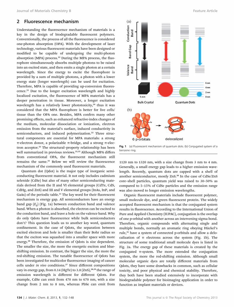
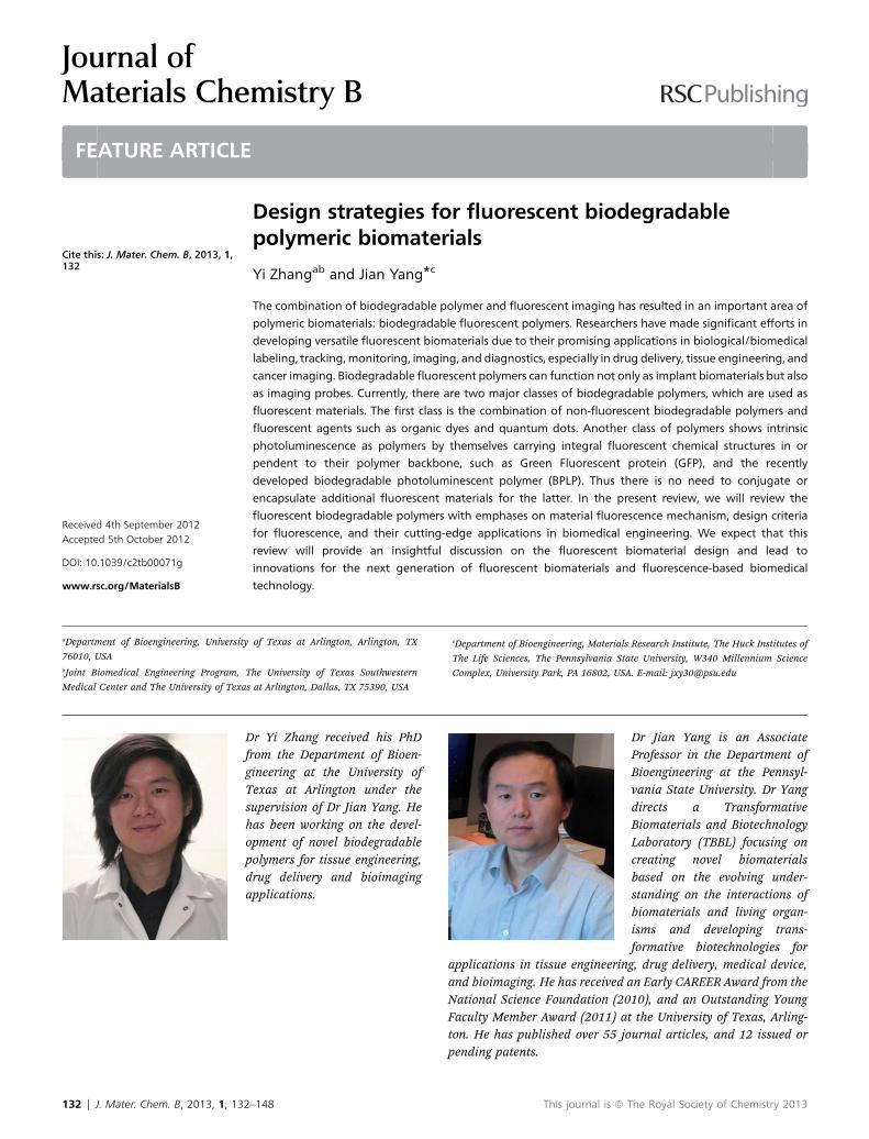
Fig. 1 (a) Fluorescent mechanism of quantum dots. (b) Conjugated system of abenzene ring.
Journal of Materials Chemistry B Feature Article
2 Fluorescence mechanism
Understanding the uorescence mechanism of materials is akey in the design of biodegradable uorescent polymers.Conventionally, the process of all the uorescence is consideredone-photon absorption (OPA). With the development of lasertechnology, various uorescent materials have been designed ormodied to be capable of undergoing the multi-photonabsorption (MPA) process.22 During the MPA process, the uo-rophore simultaneously absorbs multiple photons to be raisedinto an excited state, and then emits a single photon at a certainwavelength. Since the energy to excite the uorophore isprovided by a sum of multiple photons, a photon with a lowerenergy state (longer wavelength) can be used for excitation.Therefore, MPA is capable of providing up-conversion uores-cence.23 Due to the longer excitation wavelength and highlylocalized excitation, the uorescence of MPA materials has adeeper penetration in tissue. Moreover, a longer excitationwavelength has a relatively lower phototoxicity,24 thus it wasconsidered that the MPA uorophore is better for live cells/tissue than the OPA one. Besides, MPA confers many otherpromising effects, such as enhanced refractive-index changes ofthe medium, molecular dissociation or ionization, electronemission from the material’s surface, induced conductivity insemiconductors, and induced polymerization.22 Three struc-tural components are essential for MPA materials: a strongp-electron donor, a polarizable p-bridge, and a strong p-elec-tron acceptor.22 The structural–property relationship has beenwell summarized in previous reviews.22,25 Although MPA differsfrom conventional OPA, the uorescent mechanism stillremains the same.25 Below we will review the uorescencemechanism of the commonly used uorescent materials.
Quantum dot (Qdot) is the major type of inorganic semi-conducting uorescent material. It not only includes cadmiumselenide (CdSe) but also of many other semiconducting mate-rials derived from the II and VI elemental groups (CdTe, CdS,CdHg, and ZnS) and III and V elemental groups (InAs, InP, andGaAs) of the periodic table.11 The key word for their uorescentmechanism is energy gap. All semiconductors have an energyband gap (Eg) (Fig. 1a) between conduction band and valenceband. When a photon is absorbed, the electron can be excited tothe conduction band, and leave a hole on the valence band.Whydo only Qdots have uorescence while bulk semiconductorsdon’t? This question leads us to another key word, quantumconnement. In the case of Qdots, the separation betweenexcited electron and hole is smaller than their Bohr radius sothat the exciton was squashed into a smaller space with moreenergy.26 Therefore, the emission of Qdots is size dependent.The smaller the size, the more the energetic exciton and blue-shiing emission. In contrast, the bigger the size, the more thered-shiing emission. The tunable uorescence of Qdots hasbeen investigated for multicolor uorescence imaging of cancercells under in vivo conditions.27 Since different compositionsvary in energy gap, from 0.14 (HgTe) to 3.8 (ZnS),28,29 the range ofemission wavelength is different for different Qdots. Forexample, CdSe can emit from 470 nm to 670 nm, with a sizechange from 2 nm to 8 nm, whereas PbSe can emit from
134 | J. Mater. Chem. B, 2013, 1, 132–148
1120 nm to 1320 nm, with a size change from 3 nm to 4 nm.Generally, a small energy gap leads to a higher emission wave-length. Recently, quantum dots are capped with a shell ofanother semiconductor, mostly ZnS.30 In the case of CdSe/ZnScore–shell particles, quantum yield was raised to 30–50% ascompared to 5–15% of CdSe particles and the emission rangewas also moved to longer emission wavelengths.
Organic uorescent materials include uorescent polymer,small molecule dye, and green uorescent protein. The widelyaccepted uorescent mechanism is that the conjugated systemresults in uorescence. According to the International Union ofPure and Applied Chemistry (IUPAC), conjugation is the overlapof one p-orbital with another across an intervening sigma bond.Therefore, organic compounds with alternating single andmultiple bonds, normally an aromatic ring obeying Huckel’srule,31 have a system of connected p-orbitals and allow a delo-calization of p electrons across the system (Fig. 1b). Thestructure of some traditional small molecule dyes is listed inFig. 2a. The energy gap of these materials is created by theconjugated p-system. The more extended the conjugationsystem, the more the red-shiing emission. Although smallmolecular organic dyes are totally different materials fromQdots, they have some drawbacks in common, such as cellulartoxicity, and poor physical and chemical stability. Therefore,they both have been studied extensively to incorporate withbiodegradable polymer for bioimaging application in order tofunction as implant materials or devices.
This journal is ª The Royal Society of Chemistry 2013

Fig. 2 (a) Chemical structure of traditional small molecular dye (red represents the conjugated system). (b) Chemical structure of fluorescent polymers, poly(2-vinylnaphthalene), poly(9-anthracenylmethyl acrylate), and poly(p-phenylene vinylene) (from left to right) (red represents the conjugated system).
Fig. 3 (a) The formation and final chemical structure of the fluorophore of GFP.(b) Different colors from various fluorescent proteins.111
Feature Article Journal of Materials Chemistry B
Fluorescent polymers can be divided into two classes. One ishaving a conjugated system pendent to the backbone (Fig. 2b).The other one is with the conjugated system along the back-bone, like poly(p-phenylene vinylene) (Fig. 2b). The uorescentmechanism is the same as for a small molecular dye. Recently,there is a new family of uorescent dendrimers with the tetra-mine group, including poly(amido amine) (PAMAM), poly-(propyleneimine) (PPI), and poly(ethyleneimine) (PEI). The rstproposed uorescent mechanism was the oxidation of hydroxylend groups of PAMAM.32 This hypothesis was later disproved byImae’s group that PPI and PEI with various end groups can emitblue uorescence.33Withmore detailed studies, Imae and Chu34
found that a more rigid, crowded structure of tertiary aminesexhibits a higher uorescence yield. Although the uorescentmechanism still remains unclear, the tertiary amino on thedendritic backbone is speculated to be the key of uorescence,34
which is still under the rules of conjugation.With the tremendously increased researches on Green Fluo-
rescent Protein (GFP), the uorescent mechanism of GFP hasbeen revealed in decent detail. Although there are still somearguments, researchers have agreed on a cyclic ring uorophore,p-hydroxybenzylideneimidazolinone.35 With thorough studies onthe primary, secondary, tertiary, and quaternary structure of GFP,it was found that the cyclic ring in GFP is composed of residuesSer or Thr65, Try66, and Gly67. Fig. 3a illustrates the currentlyaccepted mechanism of the uorophore formation. Aer a seriesof folding, cyclization, dehydration, and aerial oxidation,36 theconjugated system is formed. As a natural organic compound, theuorescent mechanism of GFP still obeys the conjugatedsystem. Therefore, emission of GFP can also be tuned byextending the conjugation system (Fig. 3b). The task can beachieved by oligomerization and conjugating a more aromaticstructure onto the uorophore by a series of folding mutation.Gross et al.37have reported a red uorescent protein “DsRed.” Thered uorophore results from the autonomous multi-step post-translational modication of residues Gln66, Tyr67, and Gly68into an imidazolidinone heterocycle with p-hydroxybenzylideneand acylimine substituents. Shaner et al.38,39 have concludedmonomeric uorescent proteins that emit from yellow to red.
This journal is ª The Royal Society of Chemistry 2013
By obeying the conventional rule of a conjugation system,uorescence seems to be an irreconcilable conict with biode-gradability as water and re. Therefore, there has not been anyreport of conventional uorescent polymer being biodegrad-able. Recently, the authors’ lab has developed a new type ofbiodegradable photoluminescent polymers (BPLPs) whichpossess an intriguing uorescent mechanism although it is not
J. Mater. Chem. B, 2013, 1, 132–148 | 135

Fig. 4 (a) Synthesis schematic of BPLPs, (b) chemical structure of six-memberring of BPLP-Cys and (c) test stripe turning black shows the release of hydrogensulfide.
Journal of Materials Chemistry B Feature Article
fully understood yet.40 BPLPs were synthesized by reacting citricacid, aliphatic diol such as 1,8-octanediol, and a-amino acidsthrough a convenient condensation reaction (Fig. 4a). Poly-(octamethylene citrate) (POC) synthesized from only 1,8-octa-nediol and citric acid has very weak autouorescence. Citricacid has been replaced with succinic acid and tricarboxylic acidfor the synthesis, which turned out that neither of those poly-mers is uorescent. A plausible hypothesis can be drawn fromthose simple reactions in that the side carboxylic and thegerminal hydroxyl group from citric acid, together with anamino acid, results in uorescence. All 20 essential a-aminoacids have distinct uorescence, including glycine which has noR groups. This further excludes the R group of the amino acidfrom the list of indispensables, as amidation and estericationare two possible reactions among amine, hydroxyl, and carboxylgroups. Considering the reacting rate and energy, the fastestreaction is between the amine and the side carboxyl group fromcitric acid. Based on the product of this amidation, wehypothesize a six-member ring structure (Fig. 4b) to be theuorophore. The 13C-NMR spectra of BPLP-cysteine providedevidence for the six-member ring structure.40 For organiccompounds, conjugation is the only known law of uorescence.The basic requirement of a structure being conjugated isplanarity. The hypothesized six-member ring has a similarstructure as morpholine-2,5-dione. In the present case (Fig. 4b),hydrogen on C2 is substituted with an R-group, while hydrogenson C1 are substituted by polymer chains. Studies from othergroups have proven the planarity of the ring, when hydrogenson C1 and C2 have been substituted.41 In order to explain theconjugation of the whole ring structure, the theory of hyper-conjugation needed to be introduced. This is a well-studiedphenomenon, which was rst dened by R. S. Mulliken in thelate 1930s. It refers to the interaction of d with an adjacentp-orbital. There is evidence showing that hyperconjugation notonly extend the conjugation but also lead to uorescence on its
136 | J. Mater. Chem. B, 2013, 1, 132–148
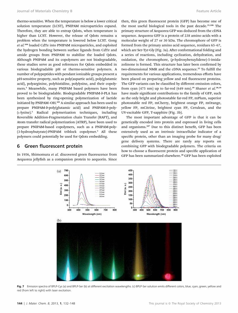
own.42 In the current six-member ring, the carbonyl group on C3and the electronic pair from O1 both interact with a C–C d bondon C1 to form hyperconjugation to explain the conjugation of asix-member ring. It is noteworthy that there is an outstandingred-edge effect (REE) on BPLP-serine (BPLP-Ser).40,43 Previousstudies have shown that uorescence spectra can depend onexcitation wavelength, when polar uorophores are embeddedinto different rigid and highly viscous media.43 The six-memberring structure of BPLPs is pendent on polymer chain, which canbe considered as a rigid media. The excitation dependentemission of BPLP-Ser is a remarkable property, not only becauseit makes the uorescence more controllable but also pushes itinto the window of near infrared. This 6-member ring uo-rophore hypothesis was further supported by the result that theuse of b-amino acids can switch off the uorescence due to thepossible formation of a seven-member ring structure, whichdoes not suffice the conjugation system as stated above.
3 Design criteria of fluorescent materialsfor biomedical application
To develop an ideal biodegradable uorescent material forvarious bioimaging, the physiological, physicochemical, andphotophysical properties should all be taken into consider-ation. For different bioimaging applications, a uorophoreshould be chosen with careful consideration of its photo-physical, physiological, and physicochemical property.
To better describe the photophysical property of a uores-cent material, there are several parameters to be understood.Firstly, excitation and emission, they determine the color of theuorescence. As we discussed above, the emission of quantumdots is size dependent, while the one of organic compounds canbe manipulated by chemical modication.27,38,44 Consideringthe biomedical application, penetration depth in the biologicaltissue is important as well. Light with a longer wavelength has alonger penetration depth in tissues.44 The brightness of uo-rescence is the second consideration aer uorescence color.Precisely speaking, the brightness is determined by twoparameters, extinction coefficient and quantum yield. Theextinction coefficient stands for how many photons a substancecan absorb under a given wavelength, which is excitationwavelength. Quantum yield shows the efficiency of a substancethat emits light, specied at a given emission wavelength. In acommon word, it represents how many protons can be emitted,when 100 of them are absorbed. In the comparison of thebrightness of different uorescent proteins, the product ofmolar extinction coefficient and quantum yield has been used.39
Although each uorophore has a xed value of extinctioncoefficient and quantum yield at a given wavelength, thosevalues can be varied with different factors such as solvent,temperature, and pH. The endurance of uorescent materials tophotobleaching is also a very important optical property. It isdetermined by the time to bleach from an initial emission rateof 1000 photons per s down to 500 photons per s.38,39 In mostcases, it stands for the photostability. Aggregation causedquenching (ACQ) of uorescent materials should be taken intoconsideration when uorophores are required at a high
This journal is ª The Royal Society of Chemistry 2013
Table 1 Optical properties of different fluorescent materials (for quantum yield and extinction coefficient, samples were tested using water as solvent, unlessspecified)
Category Fluorescent material Emission (nm) Quantum yield Extinction coefficient (M�1 cm�1) t1/2 for bleach (s)
Quantum dots CdS 370–500 <0.60 100 000–950 000CdSe 470–660 0.65–0.85 100 000–700 000CdTe 520–750 0.30–0.75 130 000–600 000
Organic dyes FITC 541 0.97 (ethanol) 92 000 (ethanol)Rhodamine B 610 0.49 (ethanol) 106 000 (ethanol)Texas red 615 0.93 (ethanol) 140 000 (ethanol)
Fluorescent proteins Cerulean 475 0.62 43 000 36T-sapphire 511 0.60 44 000 25mOrange 562 0.69 71 000 9.0mPlum 649 0.10 41 000 53
BPLPs BPLP-Cys 437 0.62 133 (1,4-dioxane)BPLP-Ser 441 0.32 (1,4-dioxane) 117 (1,4-dioxane)
535 0.12 (1,4-dioxane) 51 (1,4-dioxane)540 0.02 (1,4-dioxane) 10 (1,4-dioxane)
Feature Article Journal of Materials Chemistry B
concentration or to be loaded in a condensed or solid phase. Toalleviate ACQ effects, steric hindered groups, such as bulkycyclics, branched chains, and dendritic wedges have beenconjugated onto uorophores to impede aggregation forma-tion.45 With a specic molecule design, various aggregationenhanced emission (AEE) uorescent materials have beenobtained to address this issue.46,47 The optical properties ofsome typical uorescent materials were listed in (Tables 1 and 2and Fig. 5). Autouorescence from the examined objective isanother concern. For in vivo imaging application, biological
Table 2 Comparison of properties among fluorescent materials
Property Organic dyes Quantum dots
Emission range All range from UV to IR All range from UV to(size dependent)
Molar extinctioncoefficient
2.5 � 104–2.5 � 105 M�1
cm�1105–106 M�1 cm�1
Quantum yield 0.5–1.0 (visible), 0.05–0.25(NIR)
0.1–0.8 (visible), 0.2–0(NIR)
Size/molecular weight Up to 0.5 nm 6–60 nmFluorescent lifetime 1–10 ns 10–100 nsSolubility Depending on the
chemical structureDepending on surfacechemistry
Bioconjugation Using conjugationchemistry based on thefunctional group, usuallymulti-dyes on a singlebiomolecule
Well-established protof ligand chemistry
Processability Small molecule, usuallyloaded with a polymericcarrier
Normally used as auorescent label
Body clearance Retention and clearancedepending on dyes
<5.5 nm, rapid and erenal clearance; >15 nprevented renal excre
Toxicity Potential cellular toxicitydue to the aromaticstructure
Main issue for the usQDots due to the heametal, and potentialnanotoxicity
This journal is ª The Royal Society of Chemistry 2013
tissue has strong light scattering and autouorescence.48
Plasma also has massive absorption at 400–670 nm (hemo-globin).49 Therefore, a near infrared (NIR) uorophore is usuallypreferred for tissue imaging to avoid an overlap with tissue-autouorescence and light scattering/absorption.
From a physiological point of view, biocompatibility of theuorescent materials has the rst priority. Generally, traditionaluorescent probes, including organic dyes, quantum dots, andGFPs, have different degrees of cytotoxicity.50–53 Conjugationwith or encapsulation in polymers are the most common and
Fluorescent proteins BPLPs
IR 440–649 nm 434–725 nm
103–1.5 � 105 M�1 cm�1
(per chain)100–200 M�1 cm�1
.7 0.10–0.79 0.02–0.62
�27 kD 1000–1500 g mol�1
1–10 ns 1–5 nsCan be water-soluble via aseries of site-directedmutations
Different solubility based onvarious monomers
ocol Can be conjugated easily viaconjugation chemistry, due tothe ubiquitous presence ofcysteine and lysine residues
Rich of –COOH, and –OHgroup, which can be used forbioconjugation
Working individually as abiosensor, but not as anydevices, like nanoparticles,scaffolds
Used for scaffolds,nanoparticles, and biosensors
fficientm,tion
Clearance from body viarenal proximal tubules
Completely degradationinto non-toxic monomers
e ofvy
Generally nontoxic to cell,but still have issues due to theover-expression, and proteinaggregation
Have the comparable toxicitywith PLA
J. Mater. Chem. B, 2013, 1, 132–148 | 137

Fig. 5 Extinction coefficient, optimal emission wavelength, and correspondingquantum yield of different fluorescent materials.
Journal of Materials Chemistry B Feature Article
effective way to considerably reduce the cytotoxicity. It has beenfound that polymer coating can effectively block the release ofheavy metal ion from the core, which is considered the maincourse of its alarming cytotoxicity.54 Polymers can also providethe potential for targeted delivery, and protection against aphysiological environment. The dosage of uorophore can beconsiderably reduced, in other words, lowering cytotoxicity.Although there is no standard protocol to evaluate biocompat-ibility of custom-made uorescent materials, comparison withFDA approved ones in the same category is able to providestrong evidence. For example, biodegradable polymer should becompared with PLA, PGA, or PLGA. Indocyanine green (ICG) canbe a standard to all organic dyes. To our knowledge, there is noFDA approved quantum dots as of now. However, the siliconbased quantum dots, Cdots, have been recently approved by theFDA for a clinical trial.55 Therefore, Cdots can be set as a controlfor all quantum dots. The body clearance should also be givengreat concern. Research has shown that a nanosphere with adynamic diameter smaller than 5.5 nm can be rapidly cleared byrenal. However, like a double-edge sword, rapid body clearancegreatly reduces the toxicity, but it also increases the neededdosage of uorophore for a sustained window. Biodegradablepolymer seems like a perfect solution. An increased size aerconjugation or encapsulation with polymers helps to avoidrenal clearance. Some surface modication, such as PEGylation,confer propensity to evade scavenging by the Reticuloendothe-lial system (RES).56 Thus, a prolonged window period can beobtained while everything can still be renal clearable aer thefull degradation of polymers.
Meanwhile, the physicochemical property can also beimproved by incorporation of biodegradable polymers. Due tothe aromatic nature of organic dyes and hydrophobic surface ofQdots from synthesis, conjugation with water-soluble polymeror encapsulated with polymeric colloids will considerablyincrease their aqueous solubility. Aggregation stability of theuorophore should also be concerned. For examples, Qdotswith smaller size and damaged surface shield exhibit lowstability against aggregation.54,57 Moreover, a negative surfacecharge also causes unexpected ionic interactions with a bio-logical environment.54 Sufficient free functional groups are alsocrucial due to the need for further conjugation of multiple
138 | J. Mater. Chem. B, 2013, 1, 132–148
moieties, such as PEG, drug, and targeting moiety. Althoughnumerous derivates of traditional organic dyes have beensynthesized with functional groups present, the limited numberof functional groups hinders multiple conjugations for variouspurposes such as targeting and longer circulation time. Incor-poration of a uorophore into biodegradable polymers withsufficient functional groups, such as PLGA, poly(L-glutamicacid), and PCL, has been a common solution for the aboveconcern.
4 Organic dye-enabled biodegradablefluorescent polymers
The rst synthetic organic dye, Mauveine, was discovered byWilliam Henry Perkin in 1856. It has been proven to be effectivein dyeing silk and textiles. Since its inception, thousands oforganic dyes have been prepared for a wide range of applica-tions. For bioimaging purposes, a large number of organic dyeshave been developed to examine the fundamental processes atthe organ, tissue, cellular, and molecular levels.12,58 Based onchemical structure, they can be divided into several classes,cyanine, porphyrin, squaraine, BODIPY, and xanthenes. All thecommonly used dyes are derivates from these classes, such asindocyanine green from cyanine, uorescein and rhodamineBfrom xanthenes. In order to meet the requirement for differentbiomedical applications, various chemical modications havebeen made upon traditional dyes to tune emission wavelength,increase uorescence intensity, and introduce functionalgroups. A series of commercial organic dyes have been devel-oped, such as Cy� and Alex Flour� by Molecular Probes, andDyLight� by Thermo Fisher Scientic.
Of all the currently available organic dyes, only indocyaninegreen (ICG) has been approved by the FDA for clinical use asdiagnostic agents. Bare organic dyes tend to be non-specic totarget tissue, unstable, toxic, and rapidly cleared from the body.Major limitations of bare organic dyes include the potentialcarcinogenesis, which comes from aromatic structure, lowthreshold of photobleaching, and the lack of functional groupsfor further conjugation. Incorporation with biodegradablepolymers shows great potential to solve these obstacles. In thepresent section, two approaches will be discussed thoroughly.One is single water-soluble macromolecule conjugation, andthe other is encapsulation with polymeric colloids. Organicdye-conjugated polymeric scaffold will also be discussed.
4.1 Conjugation chemistry
Before reviewing the conjugates of polymer and organic dye, anoverview of conjugation chemistry should rst be summarized.Various conjugation strategies have been developed coveringall the functional groups that exist in organic dyes andbiodegradable polymer. The most commonly used conjugationchemistry is listed in Table 3. The carboxyl group is presentin many biodegradable polymers, most of which aresynthesized via ring-opening polymerization, such as PGA,poly(a-malic acid), and their copolymers. The carboxyl groupcan be easily activated by carbodiimide for conjugation.
This journal is ª The Royal Society of Chemistry 2013
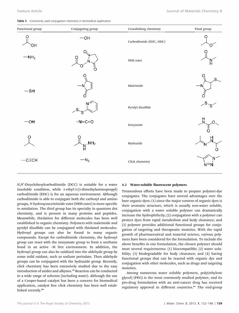
Table 3 Commonly used conjugation chemistry in biomedical application
Functional group Conjugating group Crosslinking chemistry Final group
Carbodiimide (EDC, DDC)
NHS ester
Maleimide
Pyridyl disulde
Isocyanate
Hydrazide
Click chemistry
Feature Article Journal of Materials Chemistry B
N,N0-Dicyclohexylcarbodiimide (DCC) is suitable for a waterinsoluble condition, while 1-ethyl-3-(3-dimethylaminopropyl)carbodiimide (EDC) is for an aqueous environment. Althoughcarbodiimide is able to conjugate both the carboxyl and aminogroups,N-hydroxysuccinimide ester (NHS-ester) is more specicto amidation. The thiol group has its specialty in quantum dotchemistry, and is present in many proteins and peptides.Meanwhile, thiolation for different molecules has been wellestablished in organic chemistry. Polymers with maleimide andpyridyl disulde can be conjugated with thiolated molecules.Hydroxyl groups can also be found in many organiccompounds. Except for carbodiimide chemistry, the hydroxylgroup can react with the isocyanate group to form a urethanebond in an active –H free environment. In addition, thehydroxyl group can also be oxidized into the aldehyde group bysome mild oxidant, such as sodium periodate. Then aldehydegroups can be conjugated with the hydrazide group. Recently,click chemistry has been extensively studied due to the easyintroduction of azides and alkynes.59 Reaction can be conductedin a wide range of solvents (including water). Although the useof a Cooper-based catalyst has been a concern for biomedicalapplication, catalyst free click chemistry has been well estab-lished recently.60
This journal is ª The Royal Society of Chemistry 2013
4.2 Water-soluble uorescent polymers
Tremendous efforts have been made to prepare polymer-dyeconjugates. The conjugates have several advantages over thebare organic dyes: (1) since the major content of organic dyes istheir aromatic structure, which is usually non-water soluble,conjugation with a water soluble polymer can dramaticallyincrease the hydrophilicity; (2) conjugation with a polymer canprotect dyes from rapid metabolism and body clearance; and(3) polymer provides additional functional groups for conju-gation of targeting and therapeutic moieties. With the rapidgrowth of pharmaceutical and material science, various poly-mers have been considered for the formulation. To include theabove benets in one formulation, the chosen polymer shouldmeet several requirements: (1) biocompatible; (2) water solu-bility; (3) biodegradable for body clearance; and (4) havingfunctional groups that can be reacted with organic dye andconjugation with other molecules, such as drugs and targetingmoieties.
Among numerous water soluble polymers, poly(ethyleneglycol) (PEG) is the most commonly studied polymer, and itspro-drug formulation with an anti-cancer drug has receivedregulatory approval in different countries.61 The end-group
J. Mater. Chem. B, 2013, 1, 132–148 | 139

Journal of Materials Chemistry B Feature Article
chemistry of PEG has been intensively studied. PEG withdifferent molecular weight and with different end groups arecommercially available, or can be synthesized with reportedprotocol. Multi-armed PEG provides the multiple conjugationsites for different functional molecules. Although PEG is notbiodegradable, it can be served as a standard for PEG-basedpolymers and many other water soluble polymers. Meanwhile,many efforts have been put on the strategies to producebiodegradable derivatives of PEG. Zhao et al.62 described thesynthesis of biodegradable multiarm PEGs, and it has enteredclinical trials. Poly(L-glutamic acid) (PG) is a biodegradablepolymer and its breakdown product, L-(glutamic acid), canenter normal cellular metabolism. Every repeating unit hascarboxyl groups, so PG has sufficient sites for conjugation. Thepro-drug of PG and Paclitaxel is now under phase III clinicaldevelopment in the U.S.6 Melancon et al.63 conjugated PG witha cyanine derivate, near infrared dyes (NIRF). Aer conjuga-tion of NIRF, there are still numerous free carboxyl groups forconjugation of targeting moieties and drugs. Although thisPG-NIRF was used to study in vivo degradation of PG basedpolymer–drug conjugates, it showed great potential for wholebody imaging or tumor imaging. A number of biodegradableN-(2-hydroxypropyl)methacrylamide (HPMA) based copoly-mers (co-pHPMA) caught much attention over the last30 years.64 Co-pHPMA can be conjugated with differentimaging moieties through copolymerization and chemicalconjugation. Jensen et al.65 conjugated co-pHPMA with uo-rescein-cadaverine, and used it to study the intracellularmetabolism of the copolymer. Dextran is another polymerthat has been conjugated with organic dyes for imaging.Helmchen and Denk66 used dextran-FITC/rhodamine conju-gates for high resolution brain imaging. The vicinal diolstructure could be oxidized by periodate and further conju-gated with various functional molecules.67
In addition to linear polymers, dendrimer is a class ofbranched macromolecules forming a star-like structure. Thephysiochemical properties can be easily tuned due to its step-wise fashion synthesis. Theoretically, generation 5 (G5)dendrimers will have 128 free functional end-groups on thesurface. Thus, a high density of functional groups on the outerlayer of dendrimer confers sufficient sites for the conjugation oforganic dyes, targeting moieties, and drugs. A major advantageof dendrimers over a linear polymer is that hydrophobicmolecules can be encapsulated in the cavity formed by anadjacent branch.68 Some dendrimers with positive surfacecharges, such as poly(ethyleneimine), have also been investi-gated as carriers for negatively charged DNA.69 Polyamidoamine(PAMAM) is the most commonly used dendrimer. Biodegrad-able PAMAMs were prepared by many labs via different strate-gies, and have been used for drug delivery and gene therapy.70
Majoros et al.71 conjugated uorescein isothiocyanate (FITC)onto PAMAM, and investigated it for both in vivo and in vitroimaging. Interestingly, some of PAMAMs have been found tohave uorescent properties.33,34,72 The uorescent mechanismhas been discussed above. This unique property makes PAMAMa dye-free imaging probe. Therefore, complexity of the designcan be greatly reduced.
140 | J. Mater. Chem. B, 2013, 1, 132–148
4.3 Methods of dye incorporation into biodegradablepolymers
With the rapid development of biodegradable polymericcolloids, immense studies have been focused on incorporatingorganic dyes with those colloids for various bioimaging appli-cations, especially polymeric nanoparticles for cancer imaging.Generally, organic dyes can be combined with polymer colloidsvia two different ways, chemical conjugation and physicalencapsulation.
4.3.1 DYE ENCAPSULATION. Biodegradable polymers can befabricated into nanoparticles of different structures, such asnanocapsules, nanospheres, and micelles depending on physi-cochemical properties of polymers and fabrication techniques.Nanocapsule is a core–shell structure with a polymer membraneand cavity inside, which can be a reservoir for organic dyes,whereas nanosphere is a polymeric matrix in which the dyemolecule can be evenly dispersed. Polymeric micelles can beself-assembled in an aqueous solution by amphiphilic poly-mers. Diblock, triblock, and random copolymers of bothhydrophobic and hydrophilic blocks can self-assemble intomicelles. They have a generally small size (<100 nm), dependingon the critical micelle concentration (CMC), and a propensity toevade scavenging by the reticuloendothelial system (RES).Those structures confer protection to an organic dye andconsiderably improve the stability. Targeted delivery can also beachieved by surface conjugation of targeting moieties. Organicdye encapsulated nanoparticles have been generally exploitedfor the study of pure imaging purposes, such as cellular uptake,intracellular fate, metabolism, and biodistribution.73–75
4.3.2 CHEMICAL CONJUGATION. Biodegradable polymericcolloids have been intensively investigated as a delivery vehiclefor drugs, genes, proteins, and cells. To save more loading spacefor those components and avoid interference with the aromaticuorophore, organic dyes are chemically conjugated withpolymeric colloids either during the synthesis of polymers or viasurface conjugation of polymeric colloids. Poly(alkyl cyanoac-rylate) (PACA) and its copolymers are a family of biodegradablepolymers. Droumaguet et al.76 synthesized PACA copolymersby three monomers, hexadecyl cyanoacetate, methoxypoly-(ethylene glycol) cyanoacetate, and rhodamine B conjugatedcyanoacetate. The uorescent intensity can be tuned by varyingthe feeding ratios of dye conjugated monomers. The resultedamphiphilic polymers were self-assembled into nanoparticles.Those nanoparticles have been demonstrated suitable forin vitro imaging of human brain endothelial cells. Numerousbiodegradable polymeric colloids have functional groups on thesurface, such as colloids made from PLGA, Poly(L-glutamicacid), poly(3-caprolactone) (PCL), and their amphiphilic copol-ymers with PEG as a hydrophilic block, as well as nature poly-mers, such as chitosan, and gelatin. PLGA nanoparticles havebeen surface labeled with FITC, Cy�, Alex Fluor�, and Rhoda-mine for various imaging studies.77–79 The label of Near-Infraredcyanine dye (NIR-797) helped the real-time biodistributionstudy of PCL based micelles.80 For natural polymer-basednanoparticles, Nam et al.81 conjugated Cy5.5 was labeled on thesurface of chitosan nanoparticles. This uorescent nanoparticle
This journal is ª The Royal Society of Chemistry 2013

Feature Article Journal of Materials Chemistry B
has been successfully used for the study of nanoparticlebiodistribution and tumor accumulation.
4.4 In situ imaging of biodegradable uorescent polymericscaffold
With the rapid development of biodegradable polymers for insitu tissue engineering, non-invasively or minimal-invasivelymonitoring the behavior of polymeric implants becomescrucial. Although the properties of biodegradable polymershave been carefully evaluated in vitro, such as degradationspeed andmechanical properties, understanding thesematerialproperties in vivo remains elusive as the physiological envi-ronment provides a more complicated degradation or erosionthan in vitro. There is an urgent need to assess material prop-erties in situ and in real time.
Recently, Edelman et al.82 presented a decent study ontracking polymeric scaffold using uorescence imaging. In thisstudy, uorescein was conjugated onto PEG, and then mixedwith dextran to form uorescent hydrolyzable hydrogel. Enzy-matically degradable collagen labeled with Texas-red was alsoused to assess its enzymatic degradation. In vitro and in vivodegradation were both performed for comparison. It was foundthat the in vivo hydrolytical degradation rates of the PEG-basedscaffolds could well correlate with the in vitro degradation of thesamples, while the enzymatic degradation of collagen sampleshas a more complicated behavior in different sites of the body.This study represents an advance in tissue engineering wherethere has been a dearth of understanding on the scaffolddegradation and tissue replacement in situ and in real time.Fluorescent scaffolds enable a real-time quantitative evaluationof the scaffold evolution over time via a uorescence imagingmethod. This study also implies the added uorescent proper-ties can be benecial in the design of the next wave of tissueengineering scaffolds to function as both implants and imagingprobes.
5 Inorganic dyes enabled biodegradablefluorescent polymers
Generally, naked Qdots are not ready for bioimaging due to theeasy surface oxidation, insufficient functional groups, and mostimportantly the water-insolubility.54 To address these problems,polymers have been introduced to make Qdots more suitablefor bioimaging. Various polymers can be incorporated withQdots focusing on the different facets of benets, such asgreatly increasing the stability and hydrophilicity of Qdots,lowering the toxicity, offering sufficient sites for further modi-cation, and providing a reservoir for drug loading.83 As illus-trated in Fig. 6a, polymer coating converts the hydrophobicsurface of Qdots to hydrophilic. Fig. 6b showed that the tunableuorescence of Qdots has been investigated for multicoloruorescence imaging of cancer cells under an in vivo environ-ment. The polymer layer also improves the colloidal stabilityand lowers the chance of aggregation.84 In addition, a simplepolymer coating has proved to act as a signicant barrier forheavy metal ion (Cd2+) diffusion, which was considered a major
This journal is ª The Royal Society of Chemistry 2013
cause of toxicity of Qdots.54 Polymers with various functionalgroups, such as amine, carboxyl group, and maleimide, can beexploited to further conjugate with antibodies, peptides,hydrophilic therapeutics, or aptamers. From a therapeutic pointof view, the polymer network provides sufficient room for thehydrophobic drug compared to the solid semiconductorcomponent of Qdots, which is simply an imaging probe. A largepool of polymers has been studied to encapsulate or conjugatewith Qdots via various techniques.30 All those techniques can beclassied into two ways, surface coating and bulky embedding.There are also numerous studies on incorporating Qdots withinorganic substances, such as silica or titania, non-biodegrad-able polymers, such as poly(maleic anhydride alt-1-tetrade-cene), and dendrimers, such as poly(amido amine).85,86
However, we will only focus on the strategies of incorporatinginorganic dyes with biodegradable polymers below.
5.1 Biodegradable polymer coating on Qdots
Qdots are usually characterized for their photophysical prop-erties, such as emission wavelength, quantum yield, photo-stability, and physicochemical properties, such as size, surfacecharge, and aggregation stability. Both of them may changeaer surface coating with biodegradable polymers.54 There aretwo distinct strategies usually adopted for Qdot coating: ligandexchange and ligand capping.
5.1.1 LIGAND EXCHANGE. The ligand exchange strategyinvolves completely replacing the surface bound ligandsremaining during the decomposition of metal–organic ororganometallic precursors at elevated temperatures.87 Thoseligands include trioctylphosphine oxide (TOPO) and lipophilictrioctylphosphine for most cases. Physicochemically, theadvantage of ligand exchange is maintaining the small naldiameter. Choi et al.88 pointed out that Qdots with a hydrody-namic diameter smaller than 5.5 nm can be cleared rapidlyfrom the body by renal ltration and urinary excretion. There-fore, keeping a small nal diameter aer surface coating can bebenecial in reducing toxicity. However, Qdots with a smalldiameter aer ligand exchange suffer from low stability againstaggregation.54 From a photophysical point of view, replacing theoriginal ligand of Qdots may result in several disadvantages.The exchange process raises the risk of surface damage ofQdots, leading to a decrease of quantum yield. It also increasesthe likelihood for surface oxidation, which will lead to poorphotostability and a blue shi of emission wavelength.
Since there is a specic interaction between the thiol groupand heavy metal, such as gold, silver, cadmium and so forth,thiolated polymers are the most common ligands involved inthe ligand exchange strategy.89 Thiolated PEG has been exten-sively exploited due to its ease of synthesis, ease of handling,and versatile applications. Hou et al.90 reported a disuldebond-bearing and symmetric PLLA–SS–PLLA synthesized byring-opening polymerization, and its reduced product PLLA–SH. The thiolated PLLA has been successfully coated onto CdSewith the ligand exchange strategy. A quantum yield of 53% wasreported by tuning the molecular weight of PLLA–SH, and thefeeding ratios between ligand and Qdots. The hydroxyl groups
J. Mater. Chem. B, 2013, 1, 132–148 | 141

Fig. 6 (a) Schematic illustration of multifunctional Qdots coated with biodegradable polymers. (b) Cellular and animal imaging showing Qdots with different colorsunder the same light source.27
Journal of Materials Chemistry B Feature Article
on the other end of PLLA–SH also provide sites for furtherconjugation. The synthesis of thiolated PLLA sets as a protocolfor thiolation of various biodegradable polymers that can besynthesized through ring-opening polymerization, such asfamily of cyclic lactone, and morpholine-2,5-dione.3 Except forthe thiol group, amine bond and phosphine bond have alsobeen exploited for ligand exchange.54 However, rarely has anybiodegradable polymer been reported involving the latter twochemical groups.
5.1.2 LIGAND CAPPING. Different from ligand exchange,ligand capping only caps the original ligands on Qdots withsuitable amphiphilic polymers. Without damaging the
142 | J. Mater. Chem. B, 2013, 1, 132–148
protecting ligands of Qdots, the photophysical property will bebetter retained. The thicker layer of coating provides not only abetter protection against surface oxidation but also a goodchemical stability and a reliable protection against aggregation.Typically, the coating will also increase the particle size by asmuch as 5–10 nm.91 This number depends on the molecularweight of both hydrophilic and hydrophobic blocks. A largersize of Qdots aer ligand capping may have a low renal clear-ance. However, the bare Qdots still can undergo renal clearanceaer polymer coating is fully degraded. On the other hand, alonger circulation time can extend the targeting and potentialdrug delivery window once injected in the blood circulation.
This journal is ª The Royal Society of Chemistry 2013

Feature Article Journal of Materials Chemistry B
The surfaces of naked Qdots are occupied by hydrophobicligands from the organometallic compounds during thesyntheses of Qdots, such as the stabilizing ligand, tri-octylphosphine oxide (TOPO), or hexadecylamine.92 A ligandcapping strategy exploits the physical interaction between ahydrophobic ligand from Qdots and a hydrophobic part of anamphiphilic polymer. Aer the rst successful case of coatingamphiphilic polymers on Qdots by Dubertret et al.,93 manyothers have explored this approach to coat Qdots with smallorganic molecules and non-degradable polymers. Althoughthere have been extensive research on biodegradable amphi-philic block copolymers,94 very few studies have been reportedon coating Qdots with these polymers via physical interactionsdue to their weak binding. Stabilization of the coating layer hasbeen proved by crosslinking the coating polymers. Variouscrosslinking methods have been introduced to stabilize thepolymer layer, such as lysine or diamine for polymers with anabundant carboxylic group, and free radical crosslinking forpolymers with an abundant double bond.54
Currently, most studies have focused on non-degradableamphiphilic polymers. Nevertheless, those studies providedgeneral protocols for coating biodegradable polymers on Qdotsvia ligand capping. Pellegrino et al.95 have reported a generalroute to coat various Qdots with poly(maleic anhydride alt-1-tetradecene) via ligand capping, and further crosslinked thelayer with bis(6-aminohexyl) amine. Gao et al.27 reported thatCdSe/ZnS was coated with a triblock copolymer consisting of apolybutylacrylate segment, a polyethylacrylate segment, and apolymethacrylic acid segment. The polymer layer was furthercrosslinked by peptide. These polymer coated Qdots showedgreat potential in cancer imaging. These reports can serve asstandard approaches for any –COOH containing biodegradableamphiphilic polymers, such as PEG/PLGA and PEG/poly-(aspartic acid).
5.2 Qdots-embedded biodegradable polymers
A number of studies have attempted to embed Qdots intopolymers such as polymeric nano/microparticles and micelles.Similar to organic dye incorporation, techniques of incorpo-rating Qdots in polymers can be divided into two methods:chemical bonding and physical encapsulation.
To chemically bond Qdots with biodegradable polymers,functional groups should be introduced on Qdots rst, whichcan be achieved with either ligand exchange or ligand cappingstrategies. Different from organic dyes, Qdots can be chemicallybonded to polymer colloids during polymerization. Variouspolymers can be synthesized and form particles via emulsionand dispersion polymerization. Many studies successfullydemonstrated the incorporation of Qdots into polymercolloids.96 During the process, Qdots are required to be dis-solved in oil droplets. In other words, there is no need forsurface modication of Qdots to make them hydrophilic.However, polymers that are applicable for emulsion polymeri-zation are normally non-degradable. Interestingly, a biode-gradable polyurethane nanoparticle was fabricated viaminiemulsion techniques by Cramail et al.97 Although no
This journal is ª The Royal Society of Chemistry 2013
further research has been conducted on these polyurethanenanoparticles, it represents a new way of synthesizing biode-gradable polymer colloids in the presence of Qdots.
Although conjugating Qdots onto the surface of polymerspheres has been rarely reported, Yeh et al.98 reported Qdotsconjugated PLGA nanoparticles as a potential candidate forgene delivery. By conjugating the nuclear localization signal(NLS) on the surface of nanoparticles, HeLa cells exhibit uo-rescence at the nuclei region aer incubating with the NLS-nanoparticles. However, Qdots are normally encapsulatedinside the polymer sphere due to their relatively poor stabilityand potential cellular toxicity. Like organic dyes, Qdots can beencapsulated into biodegradable polymeric colloids viadifferent fabrication techniques. Desai et al.99 encapsulatedCdSe/Zns into PLGA nanoparticles via nanoprecipitation.Kim et al.100 encapsulated CdTe/CdSe into PLGA nanoparticlesvia a double emulsion technique. The Qdots loaded nano-particles have a similar value of quantum yields to the bareQdots (52%), and the emission wavelength remained the same(760 nm). Another group also reported the encapsulation ofprotein-conjugated Qdots into PLGA via a double emulsiontechnique.101 The carboxylic groups on the polymers wereconjugated with Herceptin, a monoclonal antibody that targetsErbB2 cell membrane receptors. Specic targeting to ErbB2-positive SKBR3 breast tumor cells and intracellular controlledrelease of quantum dots was achieved. As we discussed above,amphiphilic copolymers have been intensively studied for thesurface modication of Qdots. Due to the ability to self-assemble into micelles, those copolymers have been used forencapsulation of Qdots. Due to the hydrophobic core/hydro-philic shell structure of micelles, Qdots can be distributedwithin the hydrophobic core. Cao et al.102 reported a PbS loadedN-succinyl-N0-octyl chitosan micelles for targeted imaging ofliver cancer. The emission wavelength of PbS loaded micellespresents a red shi (800–870 nm) due to the stabilization andenergy transfer of Qdots. The unnoticeable toxicity both in vitroand in vivo implied that low leakage of QDs from the micelle.However, the long term toxicity of the micelle needs to befurther studied. Aer 12 h of intravenous injection, micelle hasbeen found to accumulate at a tumor site via an enhancedpermeability and retention (EPR) effect. The strong uorescencecan be observed up to 96 h. Liu et al. loaded hydrophilic CdSe/ZnS into PLA-b-poly(2-methacryloyloxyethylphosphorylcholine)(PLA-b-PMPC). TEM imaging indicated that most of the Qdotswere located in the core of nanoparticles.
Stimulus-responsive polymers have also been used to loadQdots. The external stimulus can trigger the conformationchange of those polymers. The most notable stimulus-responsive polymers are pH or temperature sensitive ones. Themechanism of loading Qdots in such polymers is an expansion-uptake/shrink-entrap process. Xu et al.103 reported a pH-sensitivecopolymer of N-isopropylacrylamide and 4-vinylpyridine(PNIPVP), and the loading of CdTe into the PNIPVP nano-particles. Qdots were loaded during expansion of the PNIPVPnanoparticle under pH 3, and entrapped within the particles atpH 3–10. Eventually, the Qdots can be released when pH > 11.Poly(N-isopropylacrylamide) (PNIPAM) also proved to be
J. Mater. Chem. B, 2013, 1, 132–148 | 143
Journal of Materials Chemistry B Feature Article
thermo-sensitive. When the temperature is below a lower criticalsolution temperature (LCST), PNIPAM microparticles expand.Therefore, they are able to entrap Qdots, when temperature ishigher than LCST. However, the release of Qdots remains aproblem when the temperature is lowered below LCST. Gonget al.104 loaded CdTe into PNIPAM microparticles, and exploitedthe hydrogen bonding between surface ligands from CdTe andamide groups from PNIPAM to stabilize the loaded Qdots.Although PNIPAM and its copolymers are not biodegradable,these studies serve as good references for Qdots embedded invarious biodegradable pH or thermo-sensitive polymers. Anumber of polypeptides with pendant ionizable groups present apH-sensitive property, such as poly(aspartic acid), poly(glutamicacid), polyarginine, polyhistidine, polylysine, and their copoly-mers.3 Meanwhile, many PNIPAM based polymers have beenproved to be biodegradable. Biodegradable PNIPAM-b-PLA hasbeen synthesized by ring-opening polymerization of lactideinitiated by PNIPAM–OH.105 A similar approach has been used toprepare PNIPAM-b-poly(glutamic acid) and PNIPAM-b-poly-(L-lysine).3 Radical polymerization techniques, includingReversible Addition-Fragmentation chain Transfer (RAFT), andAtom transfer radical polymerization (ATRP), have been used toprepare PNIPAM-based copolymers, such as a PNIPAM-poly-(3-hydroxybutyrate)-PNIPAM triblock copolymer.3 All thesepolymers could potentially be used for Qdots embedding.
6 Green fluorescent protein
In 1956, Shimomura et al. discovered green uorescence fromAequorea jellysh as a companion protein to aequorin. Since
Fig. 7 Emission spectra of BPLP-Cys (a) and BPLP-Ser (b) at different excitation wavred (from left to right) with laser excitation.
144 | J. Mater. Chem. B, 2013, 1, 132–148
then, this green uorescent protein (GFP) has become one ofthe most useful biological tools in the past decade.35,106 Theprimary structure of Aequorea GFP was deduced from the cDNAsequence. Aequorea GFP is a protein of 238 amino acids with amolecular weight of 27 or 30 kDa. The chromophore of GFP isformed from the primary amino acid sequence, residues 65–67,which are Ser-Tyr-Gly (Fig. 3a). Aer conformational folding anda series of reactions, including cyclization, dehydration, andoxidation, the chromophore, (p-hydroxybenzylidene)-5-imida-zolinone is formed. This structure has later been conrmed bytwo-dimensional NMR and the cDNA sequence.35 To fulll therequirements for various applications, tremendous efforts havebeen placed on preparing yellow and red uorescent proteins.The GFP variants can be classied by different emission colors,from cyan (475 nm) up to far-red (649 nm).39 Shaner et al.38,39
have made signicant contributions to the family of GFP, suchas the only bright and photostable far-red FP, mPlum, superiorphotostable red FP, mCherry, brightest orange FP, mOrange,yellow FP, mCitrine, brightest cyan FP, Cerulean, and theUV-excitable GFP, T-sapphire (Fig. 3b).
The most important advantage of GFP is that it can begenetically encoded into protein and expressed in living cellsand organisms.107 Due to this distinct benet, GFP has beenextensively used as an intrinsic intracellular indicator of aspecic protein, other than an imaging probe for many drug/gene delivery systems. There are rarely any reports oncombining GFP with biodegradable polymers. The criteria onhow to choose a uorescent protein and specic application ofGFP has been summarized elsewhere.38 GFP has been exploited
elengths. (c) BPLP-Ser solution emits different colors, blue, cyan, green, yellow and
This journal is ª The Royal Society of Chemistry 2013

Feature Article Journal of Materials Chemistry B
as pH sensitive and redox sensitive indicator for dynamicintracellular activity. Based on the variety of uorescentproteins, there are many chances of crosstalk in excitation andemission channels from two uorescent proteins, whichconfers a great opportunity for uorescence resonance energytransfer (FRET) on a pair of uorescent proteins.108 The FRETeffect of uorescent proteins has been exploited for studyingprotease action and Ca2+ sensitivity.109
7 Biodegradable photoluminescentpolymers
All previous studies have to utilize either organic dyes or otheruorescent agents to confer uorescent properties to biode-gradable polymers. Recently, the authors’ laboratory hasmade abreakthrough on synthesizing a new family of biodegradablephotoluminescent polymers based via a convenient poly-condensation reaction.40 Briey, a diol such as 1,8-octanediol,citric acid, and a-amino acid were reacted to form a pre-polymer(pre-BPLP). All 20 essential a-amino acids and their derivativeshave been used for the syntheses of BPLPs all of which showedremarkable uorescence. All three monomers used forsynthesis are non-toxic. The multifunctional monomer citricacid is a non-toxic metabolic product of the human body (Krebsor citric acid cycle).110 There is no reported toxicity for 1,8-octanediol. Amino acids are critical to life, and play an impor-tant role in metabolism. As the most basic units for protein,amino acids are water-soluble and biocompatible.
With a polyester backbone, pre-BPLP can be degraded inPBS within 16 d. The uorescence intensity decayed with thedegradation, and completely died out aer the polymers werefully degraded. The complete degradation also eliminates theconcern of the toxicity raised by accumulation in tissue.Compared with a traditional organic dye, rhodamine-B, BPLP-Cys only lost less than 2% of its original uorescent intensityaer 3 h continuous UV excitation, which is signicantly lowercompared to a 10% loss of rhodamine-B. Based on oursystematic studies on BPLPs with different amino acids, BPLPshave a high quantum yield, up to 62.3%, which is BPLP-Cys.The higher quantum yield of BPLP-Cys over other amino acidsis because of the formation of a double bond which extends theconjugation system. The release of hydrogen sulde wasconrmed by H2S test strips (Fig. 4c). This high quantum yield
Fig. 8 (a) Fluorescence image of BPLP-Ser nanoparticles injected subcutaneouslyin a nude mouse. (b) Fluorescence image of BPLP-Ser porous scaffold implantedsubcutaneously in a nude mouse.
This journal is ª The Royal Society of Chemistry 2013
is comparable to the CdTe/ZnS quantum dots, which isregarded as a bright uorescent agent,11,58 and is much higherthan that of GFP.35 The study of BPLP-Ser showed that it has atunable emission by changing excitation wavelength, up to700 nm, which is different from BPLP-Cys (Fig. 7a and b). TheBPLP-Ser solution can emit different colors, from blue to red,under laser excitation (Fig. 7c). This unique property promisesa great potential of BPLP-Ser as not only a NIR imagingmaterial but also a candidate for multiplex imaging. Thecytotoxicity study of BPLP showed that it has a comparablecyto-compatibility to PLGA 75/25. The in vivo evaluation alsoproved that there is no noticeable edema and tissue necrosis. Athin brous capsule and a weak chronic inammatoryresponse have been observed aer 5 months of implantationfor the crosslinked BPLP.
Different from the dye/polymer system reviewed above, BPLPhas both a high molecular weight and uorescence as onematerial. Therefore, BPLP is free of aromatic structure andheavy metal, in other words, reduced cytotoxicity. Due to itsgood processability, BPLP can be fabricated into micro/nano-particles, lms, and porous scaffold. Fluorescence is sustainedaer fabrication. Inherited from polymer, BPLP nanoparticlesexhibit a strong, tunable, stable uorescence for in vivo imaging(Fig. 8a). BPLP nanoparticles fabricated by nanoprecipitationhave sizes around 100 nm. The degradation speed of BPLPs canbe adjusted to control the drug-releasing rate. The abundantfunctional group, such as amine, carboxylic, and hydroxylgroups, provide sufficient sites for further conjugation. On theother hand, BPLPs can be fabricated into lm, porous scaffold,and other devices by thermal crosslinking to meet the versatileneeds of biomedical application. Fig. 8b showed that uores-cence of a BPLP-Ser porous scaffold can be clearly observed invivo. Comparing to the dye conjugated polymers for real-timetracking of in vivo degradation,82 the uorophore of BPLP ismore evenly distributed, and degradation can potentially bemore accurately correlated with the decrease of uorescence. Inother words, unlike other uorescent agents such as organicdyes and Qdots which simply only act as imaging agents, BPLPsalso function as implants or devices, thus more potent in manybiomedical applications.
Understanding the uorescence mechanism will enable usto expand the biodegradable photoluminescent polymer intodifferent classes of functional polymers. It is well known that–OH may initiate ring-opening polymerization to synthesizebiodegradable polymers, such as PLA, PGA, and their copoly-mers. By controlling the molar ratio of citric acid/diol, we will beable to make a BPLP oligomer with the –OH group capped onboth ends of the polymer chains, and exploit it as a starter forring-opening polymerization. Moreover, without interruptinguorophores, various monomers with functionality can beintroduced to BPLPs. Hydrophilicity can be increased bypartially or fully replacing diol with PEG. Hydrophilic BPLPs canbe further reacted with regular hydrophobic BPLPs to formamphiphilic copolymer for self-assembling micelles. A doublebond can be incorporated by maleic acid to make BPLPs pho-tocrosslinkable (injectable). Urethane bond can be introducedto extend BPLPs to dramatically increase the mechanical
J. Mater. Chem. B, 2013, 1, 132–148 | 145

Journal of Materials Chemistry B Feature Article
property. A novel methodology for synthesizing different classesof citric acid-based biodegradable photoluminescent polymerscan be developed based on our understanding of the uores-cence mechanism.
8 Conclusion and perspectives
In the present reviews, we have summarized the designand applications of both uorescent dye enabled biodegradablematerials and biodegradable materials with intrinsic uores-cence. Biodegradable polymers were endued with importantmissions to improve uorescent dyes, such as protecting,stabilizing, reserving, functionalizing, and enabling the thera-nostic ability. The biodegradability leads to complete bodyabsorbance or clearance of those polymers aer their missionswere accomplished. Biodegradable polymers with intrinsicuorescence (BPLPs) make one stride of improvement. It cutsoff the design complexity and reduces the potential cytotoxicityfrom both organic and inorganic dyes. It is a ready to usematerial with low cost-efficiency and processing verities(injectable hydrogel, scaffold, lm, and micro/nanoparticles).Those promising properties make BPLPs become the nextgeneration uorescent materials.
With the advances in synthetic organic chemistry, biode-gradable polymers can be manipulated to have more and morepromising properties. Recently, a smart design has beenmassively introduced to building polymers and enabling uo-rescent switch. According to the difference between normaltissue and targeted tissue, redox, pH, and thermo sensitivitieshave been incorporated into biodegradable polymers to makethem stimulus responsive. Therefore, on site activities of tar-geted tissue can be achieved, such as drug release and gener-ating a uorescent signal. Fluorescence resonance energytransfer (FRET) has begun to attract attention for its uniquebehavior that can switch on/off uorescence. FRET of a pair ofuorescent proteins has been widely used for tracking physio-logical activities. In the same manner, FRET can also be used toelucidate drug release or polymer degradation. Thus, a real-time, on site, and quantitative monitoring can be achieved. Inconclusion, the success of biodegradable uorescent materialslies in our ability to custom design biodegradable polymers withtunable uorescent properties to achieve appropriate physico-chemical and photophysical properties to elicit favorable bio-logical responses and meet the versatile needs in biological andbiomedical applications.
Acknowledgements
This work was supported in part by a R01 award (EB012575)from the National Institute of Biomedical Imaging and Bioen-gineering (NIBIB), and a National Science Foundation (NSF)CAREER award 0954109.
References
1 E. S. Gil and S. M. Hudson, Prog. Polym. Sci., 2004, 29, 1173–1222.
146 | J. Mater. Chem. B, 2013, 1, 132–148
2 M. Okada, Prog. Polym. Sci., 2002, 27, 87–133.3 H. Y. Tian, Z. H. Tang, X. L. Zhuang, X. S. Chen andX. B. Jing, Prog. Polym. Sci., 2012, 37, 237–280.
4 L. S. Nair and C. T. Laurencin, Prog. Polym. Sci., 2007, 32,762–798.
5 W. Amass, A. Amass and B. Tighe, Polym. Int., 1998, 47, 89–144.
6 N. Larson and H. Ghandehari, Chem. Mater., 2012, 24, 840–853.
7 Y. Ikada and H. Tsuji, Macromol. Rapid Commun., 2000, 21,117–132.
8 J. Yang, A. R. Webb and G. A. Ameer, Adv. Mater., 2004, 16,511–516.
9 Y. D. Wang, G. A. Ameer, B. J. Sheppard and R. Langer, Nat.Biotechnol., 2002, 20, 602–606.
10 S. L. He, M. J. Yaszemski, A. W. Yasko, P. S. Engel andA. G. Mikos, Biomaterials, 2000, 21, 2389–2394.
11 X. Michalet, F. F. Pinaud, L. A. Bentolila, J. M. Tsay,S. Doose, J. J. Li, G. Sundaresan, A. M. Wu, S. S. Gambhirand S. Weiss, Science, 2005, 307, 538–544.
12 J. O. Escobedo, O. Rusin, S. Lim and R. M. Strongin, Curr.Opin. Chem. Biol., 2010, 14, 64–70.
13 Y. H. Gong, Q. L. Zhou, C. Y. Gao and J. C. Shen, ActaBiomater., 2007, 3, 531–540.
14 E. M. Pridgen, R. Langer and O. C. Farokhzad,Nanomedicine, 2007, 2, 669–680.
15 H. Ghandehari, Adv. Drug Delivery Rev., 2008, 60, 956.16 B. Sumer and J. M. Gao, Nanomedicine, 2008, 3, 137–140.17 O. C. Farokhzad, A. Khademhosseini, S. Y. Yon,
A. Hermann, J. J. Cheng, C. Chin, A. Kiselyuk, B. Teply,G. Eng and R. Langer, Anal. Chem., 2005, 77, 5453–5459.
18 V. Bagalkot, L. Zhang, E. Levy-Nissenbaum, S. Jon,P. W. Kantoff, R. Langer and O. C. Farokhzad, Nano Lett.,2007, 7, 3065–3070.
19 F. Wang, W. B. Tan, Y. Zhang, X. P. Fan and M. Q. Wang,Nanotechnology, 2006, 17, R1–R13.
20 J. H. Kim, K. Park, H. Y. Nam, S. Lee, K. Kim and I. C. Kwon,Prog. Polym. Sci., 2007, 32, 1031–1053.
21 S. M. Janib, A. S. Moses and J. A. MacKay, Adv. Drug DeliveryRev., 2010, 62, 1052–1063.
22 G. S. He, L. S. Tan, Q. Zheng and P. N. Prasad, Chem. Rev.,2008, 108, 1245–1330.
23 A. Brenier and A. M. Jurdyc, J. Lumin., 1996, 69, 131–140.24 R. K. P. Benninger, M. Hao and D. W. Piston, Rev. Physiol.,
Biochem., Pharmacol., 2008, 160, 71–92.25 T. Kogej, D. Beljonne, F. Meyers, J. W. Perry, S. R. Marder
and J. L. Bredas, Chem. Phys. Lett., 1998, 298, 1–6.26 J. Smyder and T. Krauss, Mater. Today, 2011, 14, 16.27 L. W. K. Chung, X. H. Gao, Y. Y. Cui, R. M. Levenson and
S. M. Nie, Nat. Biotechnol., 2004, 22, 969–976.28 K. Rajeshwar, N. R. de Tacconi and
C. R. Chenthamarakshan, Chem. Mater., 2001, 13, 2765–2782.
29 T. Trindade, P. O’Brien and N. L. Pickett, Chem. Mater.,2001, 13, 3843–3858.
30 N. Tomczak, D. Janczewski, M. Y. Han and G. J. Vancso,Prog. Polym. Sci., 2009, 34, 393–430.
This journal is ª The Royal Society of Chemistry 2013

Feature Article Journal of Materials Chemistry B
31 A. Hirsch, Z. F. Chen and H. J. Jiao, Angew. Chem., Int. Ed.,2000, 39, 3915.
32 W. I. Lee, Y. J. Bae and A. J. Bard, J. Am. Chem. Soc., 2004,126, 8358–8359.
33 D. Wang, T. Imae and M. Miki, J. Colloid Interface Sci., 2007,306, 222–227.
34 C. C. Chu and T. Imae,Macromol. Rapid Commun., 2009, 30,89–93.
35 R. Y. Tsien, Annu. Rev. Biochem., 1998, 67, 509–544.36 A. B. Cubitt, R. Heim, S. R. Adams, A. E. Boyd, L. A. Gross
and R. Y. Tsien, Trends Biochem. Sci., 1995, 20, 448–455.37 L. A. Gross, G. S. Baird, R. C. Hoffman, K. K. Baldridge and
R. Y. Tsien, Proc. Natl. Acad. Sci. U. S. A., 2000, 97, 11990–11995.
38 N. C. Shaner, P. A. Steinbach and R. Y. Tsien, Nat. Methods,2005, 2, 905–909.
39 N. C. Shaner, R. E. Campbell, P. A. Steinbach,B. N. G. Giepmans, A. E. Palmer and R. Y. Tsien, Nat.Biotechnol., 2004, 22, 1567–1572.
40 J. Yang, Y. Zhang, S. Gautam, L. Liu, J. Dey, W. Chen,R. P. Mason, C. A. Serrano, K. A. Schug and L. Tang, Proc.Natl. Acad. Sci. U. S. A., 2009, 106, 10086–10091.
41 M. Martinez-Palau, L. Urpi, X. Solans and J. Puiggali, ActaCrystallogr., Sect. C: Cryst. Struct. Commun., 2006, 62,O262–O264.
42 k. Knyazhanskii, P. Karmazin, P. Olekhnovich andN. Dorofeenko, J. Appl. Spectrosc., 1975, 23, 3.
43 A. P. Demchenko, Luminescence, 2002, 17, 19–42.44 S. Luo, E. Zhang, Y. Su, T. Cheng and C. Shi, Biomaterials,
2011, 32, 7127–7138.45 A. Qin, J. W. Y. Lam and B. Z. Tang, Prog. Polym. Sci., 2012,
37, 182–209.46 Y. N. Hong, J. W. Y. Lam and B. Z. Tang, Chem. Commun.,
2009, 4332–4353.47 B. S. Gaylord, S. J. Wang, A. J. Heeger and G. C. Bazan, J. Am.
Chem. Soc., 2001, 123, 6417–6418.48 K. Chang and F. Jaffer, J. Nucl. Cardiol., 2008, 15, 417–428.49 A. Paganin-Gioanni, E. Bellard, L. Paquereau, V. Ecochard,
M. Golzio and J. Teissie, Radiol. Oncol., 2010, 44, 142–148.50 A. M. Derfus, W. C. W. Chan and S. N. Bhatia, Nano Lett.,
2004, 4, 11–18.51 C. Kirchner, T. Liedl, S. Kudera, T. Pellegrino, A. M. Javier,
H. E. Gaub, S. Stolzle, N. Fertig and W. J. Parak, Nano Lett.,2005, 5, 331–338.
52 D. W. Han, K. Matsumura, B. Kim and S. H. Hyon, Bioorg.Med. Chem., 2008, 16, 9652–9659.
53 H. S. Liu, M. S. Jan, C. K. Chou, P. H. Chen and N. J. Ke,Biochem. Biophys. Res. Commun., 1999, 260, 712–717.
54 A. F. E. Hezinger, J. Tessmar and A. Gopferich, Eur. J. Pharm.Biopharm., 2008, 68, 138–152.
55 L. Donaldson, Mater. Today, 2011, 14, 131.56 M. L. Schipper, G. Iyer, A. L. Koh, Z. Cheng, Y. Ebenstein,
A. Aharoni, S. Keren, L. A. Bentolila, J. Q. Li, J. H. Rao,X. Y. Chen, U. Banin, A. M. Wu, R. Sinclair, S. Weiss andS. S. Gambhir, Small, 2009, 5, 126–134.
57 W. A. Hild, M. Breunig and A. Goepferich, Eur. J. Pharm.Biopharm., 2008, 68, 153–168.
This journal is ª The Royal Society of Chemistry 2013
58 U. Resch-Genger, M. Grabolle, S. Cavaliere-Jaricot,R. Nitschke and T. Nann, Nat. Methods, 2008, 5, 763–775.
59 M. G. Finn and V. V. Fokin, Chem. Soc. Rev., 2010, 39, 1231–1232.
60 J. Hong, Q. Luo, X. M. Wan, Z. S. Petrovic and B. K. Shah,Biomacromolecules, 2012, 13, 261–266.
61 J. Rautio, H. Kumpulainen, T. Heimbach, R. Oliyai, D. Oh,T. Jarvinen and J. Savolainen, Nat. Rev. Drug Discovery,2008, 7, 255–270.
62 H. Zhao, B. Rubio, P. Sapra, D. C. Wu, P. Reddy, P. Sai,A. Martinez, Y. Gao, Y. Lozanguiez, C. Longley,L. M. Greenberger and I. D. Horak, Bioconjugate Chem.,2008, 19, 849–859.
63 M. P. Melancon, W. Wang, Y. Wang, R. Shao, X. Ji,J. G. Gelovani and C. Li, Pharm. Res., 2007, 24, 1217–1224.
64 Z. R. Lu, Adv. Drug Delivery Rev., 2010, 62, 246–257.65 K. D. Jensen, P. Kopeckova, J. H. Bridge and J. Kopecek,
AAPS PharmSci, 2001, 3, E32.66 F. Helmchen and W. Denk, Nat. Methods, 2005, 2, 932–940.67 X. Hong, W. Guo, H. Yuang, J. Li, Y. M. Liu, L. Ma, Y. B. Bai
and T. J. Li, J. Magn. Magn. Mater., 2004, 269, 95–100.68 D. Bhadra, S. Bhadra, S. Jain and N. K. Jain, Int. J. Pharm.,
2003, 257, 111–124.69 V. Sokolova, S. Neumann, A. Kovtun, S. Chernousova,
R. Heumann and M. Epple, J. Mater. Sci., 2010, 45, 4952–4957.
70 H. Y. Nam, K. Nam, H. J. Hahn, B. H. Kim, H. J. Lim,H. J. Kim, J. S. Choi and J. S. Park, Biomaterials, 2009, 30,665–673.
71 I. J. Majoros, A. Myc, T. Thomas, C. B. Mehta andJ. R. Baker, Jr, Biomacromolecules, 2006, 7, 572–579.
72 T. Imae and D. J. Wang, J. Am. Chem. Soc., 2004, 126, 13204–13205.
73 S. Santra and A. Malhotra, Wiley Interdiscip. Rev.: Nanomed.Nanobiotechnol., 2011, DOI: 10.1002/wnan.134.
74 I. Brigger, C. Dubernet and P. Couvreur, Adv. Drug DeliveryRev., 2002, 54, 631–651.
75 Y. R. Lee, Y. H. Lee, S. A. Im, K. Kim and C. K. Lee, ImmuneNetw., 2011, 11, 163–168.
76 B. Le Droumaguet, H. Souguir, D. Brambilla, R. Verpillot,J. Nicolas, M. Taverna, P. Couvreur and K. Andrieux, Int.J. Pharm., 2011, 416, 453–460.
77 S. H. Kim, J. H. Jeong, K. W. Chun and T. G. Park, Langmuir,2005, 21, 8852–8857.
78 Y. I. Chung, J. C. Kim, Y. H. Kim, G. Tae, S. Y. Lee, K. Kimand I. C. Kwon, J. Controlled Release, 2010, 143, 374–382.
79 P. Kocbek, N. Obermajer, M. Cegnar, J. Kos and J. Kristl,J. Controlled Release, 2007, 120, 18–26.
80 Q. Liu, R. Li, Z. Zhu, X. Qian, W. Guan, L. Yu, M. Yang,X. Jiang and B. Liu, Int J Pharm., 2012, 430, 350–358.
81 T. Nam, S. Park, S. Y. Lee, K. Park, K. Choi, I. C. Song,M. H. Han, J. J. Leary, S. A. Yuk, I. C. Kwon, K. Kim andS. Y. Jeong, Bioconjugate Chem., 2010, 21, 578–582.
82 N. Artzi, N. Oliva, C. Puron, S. Shitreet, S. Artzi, A. bonRamos, A. Groothuis, G. Sahagian and E. R. Edelman,Nat. Mater., 2011, 10, 704–709.
J. Mater. Chem. B, 2013, 1, 132–148 | 147

Journal of Materials Chemistry B Feature Article
83 L. F. Qi and X. H. Gao, Expert Opin. Drug Delivery, 2008, 5,263–267.
84 M. A. Walling, J. A. Novak and J. R. Shepard, Int. J. Mol. Sci.,2009, 10, 441–491.
85 Y. Y. Cheng, L. B. Zhao, Y. W. Li and T. W. Xu, Chem. Soc.Rev., 2011, 40, 2673–2703.
86 D. J. Bharali, M. Khalil, M. Gurbuz, T. M. Simone andS. A. Mousa, Int. J. Nanomed., 2009, 4, 1–7.
87 D. Crouch, S. Norager, P. O’Brien, J. H. Park and N. Pickett,Philos. Trans. R. Soc., A, 2003, 361, 297–310.
88 H. S. Choi, W. Liu, P. Misra, E. Tanaka, J. P. Zimmer,B. I. Ipe, M. G. Bawendi and J. V. Frangioni, Nat.Biotechnol., 2007, 25, 1165–1170.
89 Y. J. Zhang and A. Clapp, Sensors, 2011, 11, 11036–11055.90 X. D. Hou, Q. B. Li, L. Jia, Y. Li, Y. D. Zhu and A. M. Cao,
Macromol. Biosci., 2009, 9, 551–562.91 H. S. Choi, W. H. Liu, F. B. Liu, K. Nasr, P. Misra,
M. G. Bawendi and J. V. Frangioni, Nat. Nanotechnol.,2010, 5, 42–47.
92 M. J. Murcia, D. L. Shaw, E. C. Long and C. A. Naumann,Opt. Commun., 2008, 281, 1771–1780.
93 B. Dubertret, P. Skourides, D. J. Norris, V. Noireaux,A. H. Brivanlou and A. Libchaber, Science, 2002, 298,1759–1762.
94 N. Kumar, M. N. V. Ravikumar and A. J. Domb, Adv. DrugDelivery Rev., 2001, 53, 23–44.
95 R. Di Corato, A. Quarta, P. Piacenza, A. Ragusa, A. Figuerola,R. Buonsanti, R. Cingolani, L. Manna and T. Pellegrino, J.Mater. Chem., 2008, 18, 1991–1996.
96 H. Mattoussi, G. Palui and H. B. Na, Adv. Drug Delivery Rev.,2012, 64, 138–166.
97 B. G. Zanetti-Ramos, E. Lemos-Senna, V. Soldi, R. Borsali,E. Cloutet and H. Cramail, Polymer, 2006, 47, 8080–8087.
148 | J. Mater. Chem. B, 2013, 1, 132–148
98 F. Y. Cheng, S. P. H. Wang, C. H. Su, T. L. Tsai, P. C. Wu,D. B. Shieh, J. H. Chen, P. C. H. Hsieh and C. S. Yeh,Biomaterials, 2008, 29, 2104–2112.
99 B. J. Nehilla, P. G. Allen and T. A. Desai, ACS Nano, 2008, 2,538–544.
100 J. S. Kim, K. J. Cho, T. H. Tran, M. Nurunnabi, T. H. Moon,S. M. Hong and Y. K. Lee, J. Colloid Interface Sci., 2011, 353,363–371.
101 B. Y. S. Kim, W. Jiang, J. Oreopoulos, C. M. Yip,J. T. Rutka and W. C. W. Chan, Nano Lett., 2008, 8,3887–3892.
102 J. Cao, H. Y. Zhu, D. W. Deng, B. Xue, L. P. Tang,D. Mahounga, Z. Y. Qian and Y. Q. Gu, J. Biomed. Mater.Res., Part A, 2012, 100, 958–968.
103 Y. L. Xu, L. Q. Shi, R. J. Ma, W. Q. Zhang, Y. L. An andX. X. Zhu, Polymer, 2007, 48, 1711–1717.
104 Y. J. Gong, M. Y. Gao, D. Y. Wang and H. Mohwald, Chem.Mater., 2005, 17, 2648–2653.
105 C. L. Wu, R. J. Ma, H. He, L. Z. Zhao, H. J. Gao, Y. L. Anand L. Q. Shi, Macromol. Biosci., 2009, 9, 1185–1193.
106 J. M. Tavare, L. M. Fletcher and G. I. Welsh, J. Endocrinol.,2001, 170, 297–306.
107 D. M. Chudakov, M. V. Matz, S. Lukyanov andK. A. Lukyanov, Physiol. Rev., 2010, 90, 1103–1163.
108 D. W. Piston and G. J. Kremers, Trends Biochem. Sci., 2007,32, 407–414.
109 A. Miyawaki, J. Llopis, R. Heim, J. M. McCaffery,J. A. Adams, M. Ikura and R. Y. Tsien, Nature, 1997, 388,882–887.
110 Y. Kang, J. Yang, S. Khan, L. Anissian and G. A. Ameer,J. Biomed. Mater. Res., Part A, 2006, 77, 331–339.
111 R. Y. Tsien, Angew. Chem., Int. Ed., 2009, 48, 5612–5626.
This journal is ª The Royal Society of Chemistry 2013