Embed Size (px)
Citation preview

Publication ofInfectious Disease Association of Thailand
In Co-operation withWestern Pacific Society of Chemotherapy
International Advisory Board
Jacques Acar, France Dieter Adam, GermanyT Arai, Japan Somchai Bovornkitti, Thailand Giuliana Gialdroni Grassi, Italy Geoffrey M Kellerman, Australia Chev Kidson, Thailand Victor KE Lim, Malaysia Somsak Lolekha, Thailand Faridah Moosdeen, UK Barbara E Murray, USA Ronald HH Nelwan, Indonesia Tyrone L Pitt, UK John MB Smith, New Zealand Thelma E Tupasi, Phillippines John Turnidge, Australia Richard P Wenzel, USA John David William, UK John B Zabriskie, USA
Editor-in-Chief Chitsanu Pancharoen
Associate EditorsTerapong Tantawichien Anuwat Keerasuntonpong
Editorial Advisory BoardThanomsak Anekthananon Nalinee AswapokeePantip Chayakul Ploenchan ChetchotisakdKulkanya Chokephaibulkit Anan ChongthaleongManoon Leechawengwong Amorn LeelarasameeSombat Leelasupasri Somsak LolekhaSornchai Looareesuwan Kumthorn MalathumPraphan Phanuphak Sompone PunyaguptaBoonmee Sathapatayavongs Thira SirisanthanaMondej Sookpranee Yupin SuputtamongkolSurapol Suwanagool Panpit SuwangoolUsa Thisyakorn Sataporn ThitivichianlertSurapee Tiengrim Malai VorachitChantapong Wasi
Editorial Office:For all correspondence membership, editorial matters and inquiry Chitsanu Pancharoen, M.D. Infectious Disease Unit, Department of Pediatrics, Chulalongkorn Hospital, Rama IV Road, Bangkok 10330, THAILAND. Tel/Fax 662-02-256-4930, 662-02-252-8181-9 ext. 3349
Journal of INFECTIOUS DISEASES and ANTIMICROBIAL AGENTS

The Journal of Infectious Diseases and Antimicro-bial Agents publishes original research articles, case reports and article reviews on various aspects of infectious diseases and antimicrobial agents. The contributions submitted to this journal should not be published else where in whole or in part, without the Editor’s permission. Accepted contributions become the copyright of Journal.
Manuscripts should be accompanied by a covering letter from the author who is responsible for correspon-dence regarding the manuscript. The covering letter should contain a statement that the manuscript has been approved by all authors. A manuscript, tables and illustrations should be sent in duplicate together with a 3.5-inch diskette to the Editor.
Preparation of ManuscriptAll papers must be written in English language. All
sections of the manuscript should be typed double-spaced on one side of good quality A4-sized paper with margins of at least 2.5 cm. Each of the following sections should be on separate pages: title page, abstract, text, acknow-ledgements, references, individual tables, and legends for illustrations. All pages should be numbered consecutively, beginning with the title page.
Title pageThe title page should contain (1) the title of the
article; (2) a short running head; (3) first name, middle initial, and last name of each author; (4) name of departments(s) and institution(s), (5) keywords.
AbstractThe abstract should not contain more than 150
words. The authors should list 5-10 index keywords for subject classification following the abstract.
TextOriginal article should be divided into sections
with the following headings: Introduction, Materials and Methods, Results and Discussion.
For Case reports, the Materials, Methods and Results sections should be replaced by the Case report(s). This section should include patient history, diagnosis, treatment, outcome and any other information pertinent to the case(s). All other sections should follow the format for original articles.
AcknowledgementsAll acknowledgements including financial support
should be mentioned under the heading “Acknowledge-ments” and not as a footnote on the first page or in the text
ReferencesReferences should be cited above the line of text,
without brackets or parentheses.Number references consecutively in the order
in which they are first mentioned in the text. Identify references in text, tables, and legends by arabic numerals.
Follow the reference format and journal abbrevia-tions as given in Index Medicus. Titles of journals not listed in the current Index Medicus should be spelled out
A GUIDE FOR CONTRIBUTORS
in full.Only works that have been published or accepted
for publication should be listed as references. Examples of correct forms of references are given
below.1. Standard Journal Article (List all authors when six or
less, for seven or more authors, list only the first three and add et al.)
Rongrungruang T, Kaewmanee S, Jannhu V, Sripienjan J. The efficacy of different regimens of albendazole against Strongyloides stercoralist. J Infect Dis Anti-microb Agents 1994;11:113-6.
2. Corporate Author in Journal Committee for Computer Application in Clinical
Microbiology. Bacterial antimicrobial susceptibility pattern 1988. J Infect Dis Antimicrob Agents 1991; 8:25-39.
3. Corporate Author in Book World Health Organization. On being in charge:
a guide to management in primary health care, 2nd ed. England: World Health Organization, 1992.
4. Chapter in Book Wenzel RP. Organization for infection control. In:
Mandell GL, Douglas RG, Bennett JE, eds. Principles and Practice of Infectious Diseases, 3rd ed. USA: Churchill Livingstone Inc., 1990:2176-80.
IllustrationsSubmit 2 complete sets of figures. Figures should
be professionally drawn and photographed. Send sharp, glossy black-and-white photographic prints, usually 12 by 17 cm but not larger than 20 by 25 cm. Titles and detailed explanations belong in the legends for illustrations, not on the illustrations themselves.
Each figure should have a label pasted on its back indicating the number of the figure, the names of the authors, and the top of the figure. For figures, do not write on the back, mount on cardboard, or scratch or mark with paper clips.
Photomicrographs should include internal scale markers. Symbols, arrows, or letters used in photomicro-graphs should contrast with the background.
Cite each figure in the text in consecutive order. If a figure has been published, acknowledge the original source and submit written permission from the copyright holder to reproduce the material.
ProofsGalley proofs will be sent to the corresponding
author for minor corrections and should be returned to the Editor within one month. Major alterations cannot be accepted.
PublicationsThe journal reserves the right to edit for clarity,
conciseness, precision of expression, grammar and to alter a manuscript to conform to the format of Journal of Infectious Diseases and Antimicrobial Agents
Annual Subscription RatesAnnual subscription rates via surface mail are as
follows: $40 US for country member of Western Pacific Society of Chemotherapy, $50 US dollars for non-member. An extra $15 US dollars per year is added for airmail delivery. Cheque payable to Infectious Disease

CONTENTS
ORIGINAL ARTICLEOutcome of lamivudine plus stavudine combination therapy after treatment failure of zidovudine plus didanosinePunpanich W, Chotpitayasunondh T, Kanjanapattanakul W ................................................................ 83-87
In vitro antimicrobial susceptibility and exoenzymes production of two biotypes of Burkholderia pseudomalleiLulitanond A, Worawat S, Harnsri A, Masook T, Chunpum A, Puapermpoonsiri S, Homchampa P, Na-Ngam N .................................................................................................................. 88-92
Study of high serum alkaline phosphatase in HIV-infected patientsWiwanitkit V ...........� 93-95
Bacteriologic profile of acute and chronic maxillary sinusitisJareoncharsri P, Bunnag C, Tunsuriyawong P, Voraprayoon S, Srifuengfung S, Dhiraputra C, Bedavanija A ................................................................................................................. 96-102
Urinary tract infection in Thai childrenJungthirapanich J, Tungsathapornpong A, Chaumrattanakul U, Chotipanich C ............................... 103-107
In vitro susceptibility of Streptococcus pneumoniae to penicillin and seven other antimicrobial agents: a study from Southern ThailandPruekprasert P, Tunyapanit W, Kaewjungwad L, Kaewpaiboon S ..................................................... 108-111
CASE REPORTCryptococcus laurentii fungemia: a case reportKiertiburanakul S, Sungkanuparph S, Pracharktam R ....................................................................... 112-114
REVIEW ARTICLEDengue infectionPancharoen C, Thisyakorn U, Thisyakorn C ...................................................................................... 115-121
LETTER TO EDITOR ........................................................................................................................... 122-123
Author Index ....................� 124
Subject Index ...................� 125
and ANTIMICROBIAL AGENTSVol. 18 No. 3 Sep-Dec 2001Journal of INFECTIOUS DISEASES

Western Pacific Society of Chemotherapy
President Joichi Kumazawa (Japan)
Past President Thelma E Tupasi (Philippines)
Secretary-General Victor KE Lim (Malaysia)
Treasurer Jingoro Shimada (Japan)
Executive Councillors Somsak Lolekha (Thailand)
Ronal HH Nelwan (Indonesia)
Councillors Keryn Christiansen (Australia)
Hiroyuki Kobayashi (Japan)
Julius Lecciones (Philippines)
Amorn Leelarasamee (Thailand)
Cheng-Yi Liu (Taiwan)
Yasmin Malik (Malaysia)
Seung-Chull Park (Korea)
Melecia Velmonte (Philippines)
Wing-Hong Seto (Hong Kong)
Jae-Hoon Song (South Korea)
Dominic Tsang (Hong Kong)
John Turnidge (Australia)
Sin Yew Wong (Singapore)


Vol.18 No.3 Outcome of 3TC+d4T after treatment failure of ZDV+ddI:- Punpanich W, et al.
Abstract
Original Article
83
Outcome of Lamivudine plus Stavudine Combina-tion Therapy after Treatment Failure of Zidovudine plus Didanosine
Warunee Punpanich, M.D.*Tawee Chotpitayasunondh, M.D.*Wiboon Kanjanapattanakul, M.D.**
Background: In infants and children with maternally acquired human immunodeficiency virus type 1 (HIV-1) infection, treatment with zidovudine (ZDV) and didanosine (ddI) combination has limited efficacy. Objective: To evaluate the one year clinical and immunological outcome of lamivudine (3TC) and stavudine (d4T) combination treatment in HIV-infected children who failed from ZDV and ddI combination.Methods: Fifteen HIV-infected children who had been treated with antiretroviral agents (ZDV+ddI) and continued to show disease progression (clinical or immunological), were enrolled for treatment with 3TC+d4T. The study was a nonrandomized and single arm study. Tolerance safety and efficacy of antiretroviral treatment were evaluated by serial clinical monitoring, disease progression and immunologic responses. Results: 3TC and d4T combination was well tolerated, without clinically important adverse events. The one year survival rate is 60.0 percent and 26.6 percent of children have sustained clinical benefit. The study shows that there are no clinical parameters (age at first symptomatic HIV infection, clinical or immunologic stage (CD4, CD8 lymphocyte) when changing treatment or prior clinical benefit of ZDV+ddI combination therapy) found to determine the one year clinical benefit of sequential treatment with 3TC+d4T.Conclusion: Switching to 3TC+d4T therapy in ZDV+ddI treated HIV-infected children yields an unfavorable outcome in this study. Further studies using NRTI with a large sample size and more useful monitoring of laboratory measurements (viral load and genotypic resistance) should be performed. (J Infect Dis Antimicrob Agents 2001;18:83-7.)
* Division of Pediatric Infectious Disease,** Division of Neonatology, Queen Sirikit National Institute of Child Health, Rajvithi Rd., Bangkok 10400, Thailand.Received for publication: April 23, 2001.Reprint request: Warunee Punpanich, M.D., Division of Pediatric Infectious Disease, Queen Sirikit National Institute of Child Health,
Rajvithi Rd., Bangkok 10400, Thailand.
Keywords: lamivudine, stavudine, treatment, failure, zidovudine, didanosine
INTRODUCTIONOver the past decade, the number of infants
infected by the transmission of human immuno-deficiency virus type 1 (HIV-1) from their mothers has dramatically increased. In infants and children, the depletion of CD4 cells and the progression of HIV-1-related disease are more rapid than in adults infected with HIV-1. Antiretroviral therapy for HIV-infected children results in virological, immunological, and
clinical benefits. In Queen Sirikit National Institute of Child Health, combination therapy with zidovudine (ZDV) and didanosine (ddI) has been the first line treatment for HIV-infected children since 1995. Unfortunately, this regimen is limited by intolerance, toxicity, and HIV disease progression. Some patients have disease progression during this combination treatment. The limitations of ZDV+ddI as well as the

J INFECT DIS ANTIMICROB AGENTS Sep-Dec 2001
assessed as well as a complete physical examination. Height and weight were measured on entry into the study and monthly thereafter. Studies of lymphocyte surface markers (CD4+ and CD8+ number and percent) and laboratory tests (complete blood counts and routine blood chemistries (creatinine, liver enzymes and amylase) were made at the enrollment, 3, 6 and 12 months thereafter.
Disease-progression endpoints included death, weight and growth failure, neuropsychologic, neurolo-gical deterioration, occurrence of > 2 opportunistic illnesses, and occurrence of > 2 new clinical conditions such as nephropathy, cardiomyopathy, or development or worsening of lymphoid interstitial pneumonia/pulmonary lymphoid hyperplasia.
Statistical analysis Data analysis is descriptive. The clinical
para-meters related to the one year durability of clini-cal benefit of this sequential therapy were analyzed by Cox Proportional Hazard, Stata version 4.0, Stata corpora-tion, Stata statistic software: released 4. Stata Corporation, Texas.
RESULTS A total of 15 children were enrolled in the study
between January 1999 and January 2000. All of them were vertically infected from their mothers. Most of them were diagnosed by positive anti-HIV antibody testing at the time of symptomatic HIV infection except for 2 cases diagnosed by HIV-PCR before 18 months of age. The most common manifestations were pneumonia and diarrhea. All of them were initially treated with ZDV+ddI with the median duration of 16 months (range 3-62 months) before switching to 3TC+d4T therapy. The median age at the time of changing therapy was 63 months (range 22-105 months). Selected characteristics of study subjects are shown in Table 1.
The regimen of antiretroviral drugs used was well tolerated by all study patients. No clinically significant adverse events related to the study drugs were reported. After a 12-month follow-up period, 3 cases (20.0%) died, 4 cases (26.6%) were lost to follow-up, 5 cases (33.3%) had clinically progressed and 4 cases (26.6%) clinically improved. One year survival rate is 60.0 percent and median durability of clinical benefit is 6 months (range 0->12 months) (Table 2).
At entry into the study, the percentage of CD4 + lymphocyte in the peripheral blood was relatively low i.e. almost all of them were classified into immunologic category 3 (severe immunosuppression). The per-centage of CD4 + lymphocyte in the peripheral blood of most patients decreased over time. However, this trend cannot be accurately evaluated because of the
poor prognosis of HIV infection in children, have led to studies on the use of more effective antiretroviral agents. The ideal combination should be highly potent, use different metabolic pathways, different target cells (activated versus resting cells), no overlaping toxicity and resistance profiles, and for adherence, they should be easy to take with infrequent daily dosing.1
Stavudine (d4T) has shown potent in vitro anti-HIV activity when used as monotherapy in patients with asymptomatic or advanced HIV-1 infection2-5, as well as clinical efficacy in ZDV-experienced patients.6 Lamivudine (3TC) has been studied primarily in combination therapy with ZDV in both treatment-naive and treatment-experienced patients and has been shown to have clinical benefit due to its potent antiviral activity.7-10
The purpose of the study was to evaluate the clinical and immunological outcome of 3TC+d4T combination treatment in HIV-infected children who failed from ZDV+ddI combination.
MATERIALS AND METHODSPatients and study design
This open-label, nonrandomized study was conducted at Queen Sirikit National Institute of Child Health. The study population included antiretroviral experienced (ZDV+ddI) HIV-infected children aged from 22 months to 9 years, who had evidence of disease progression (clinical or laboratory), were enrolled to receive 3TC+d4T. The change in antiretroviral therapy for HIV-infected children was based on the US CDC guideline for the use of antiretroviral agent in pediatric HIV infection.11 The study was approved by the Human Subject Research Board of the institution. Informed consent was obtained from the child’s legal guardians.
Study medicationOne milligram of d4T per kg and 2 mg of 3TC
per kg were given every 12 hours. All medications were administered as oral suspensions or syrup (3TC 10 mg/mL and d4T 1 mg/mL). Medications were dispensed monthly, and the doses were adjusted for the child’s weight.
All patients were receiving prophylaxis for Pneu-mocystis carinii pneumonia and additional antibiotic therapy as needed. The use of immuno-modulators (including corticosteroids and immuno-globulin) or antiretroviral agents other than the study drugs was prohibited.
Clinical and laboratory monitoring At each study visit, clinical well being, number
of hospitalizations, outpatient visits, medications for HIV-related illness and their potential toxic effects were

Vol.18 No.3 Outcome of 3TC+d4T after treatment failure of ZDV+ddI:- Punpanich W, et al.
large proportion of deaths and lost-to-follow-up cases (Table 3).
There were no clinical features, such as age at first symptomatic HIV infection, clinical or immuno-logic or clinical benefit of prior ZDV+ddI therapy, that could determine the one year clinical benefit of 3TC+d4T (Table 4).
Table 1. Baseline characteristics of study patients.
Variables Median age at diagnosis (months) 22 (range 1-89)Median age at switching therapy (months) 63 (range 22-105)Median duration of first regimen (ZDV+ddI) (months) 16 (range 3-62)Male (%) 5 (33.3%)Median CD4+ lymphocyte count (cell × 106/l) 34 (range 9-926)
Table 2. Clinical outcomes of the study patients after changing treatment.
Improved No improvement Lost to follow-up Death (accumulated number) (accumulated number) (accumulated number) (accumulated number) 3-month 11 3 0 1 6-month 9 3 3* 112-month 4 5 4* 3
Table 3. Median CD4+ cell count in study patients after changing treatment.
Baseline 6-month 12-month (n=13) (n=11) (n=6) Percent (range) 8 (1-28) 3 (2-18) 2 (1-8)
Duration
Median CD4+
DISCUSSIONAccording to our data, treatment with this
sequential NRTI combination resulted in 60.0 percent one year survival rate, and only 26.6 percent of patients sustained clinical benefit. At one year the clinical benefit was relatively low, probably due to the fact
Table 4. Predicted indicators for 1-year durability of clinical benefit.
Variables Haz. ratio P value 95% CI
Age at first symptomatic 1.00 0.883 0.97 -1.03Clinical classification before changing treatment 1.01 0.678 0.97- 1.05Immunologic classification before changing treatment 1.53 0.411
J INFECT DIS ANTIMICROB AGENTS Sep-Dec 2001
that most of the patients began their therapy after symptomatic infection with suboptimal regimens (dual NRTI only) and without quantitative plasma HIV-1 RNA measurements for treatment monitoring which occurs routinely in many countries.
This study finds no clinical parameter, such as age at first symptomatic HIV infection, clinical or immunologic classification at the time of changing treatment or clinical benefit of ZDV+ddI combination therapy, that can determine the one year durability of clinical benefit. The relatively poor clinical response rate of this study resulted from not only the advanced stage of the disease but also the possibility of cross resistance of ZDV and d4T by 18C RT gene12 or thymidine analogue (TAM) and multidrug resistance (MDR) mutation.13 The 4th International Workshop on HIV-1 Drug Resistance and Treatment Strategies14 strongly suggested that ZDV and d4T select a share set of drug resistant mutation that could be developed during prior ZDV therapy. In addition, the NARVAL investigators15 demonstrated that the problem of NRTI cross-resistance extends beyond ZDV and d4T. Their analysis showed that so-called thymidine analogue mutations (TAMs; mutations at codons 41, 67, 70, 210, 215, or 219) had a negative impact on virological response to all NRTIs, including the nonthymidine analogues abacavir, lamivudine, and didanosine. From our study we have not been able to determine the potential candidates who would benefit from this NRTI sequential combination. The limitations of the study are small sample size, the unavailability of viral load measurement and genotypic resistance testing of the virus. Whether this combination treatment can either slow the progression of HIV-infection to AIDS and death or have the potential to facilitate healthcare savings, which partly offset the drug acquisition costs, remained to be determined.
SUMMARY Switching to 3TC+d4T therapy in ZDV+ddI
experienced HIV-infected children yields an un-favorable outcome. These children may need more aggressive regimens such as triple therapy (with or without protease inhibitor). Increase sample size and other laboratory measurements including plasma HIV-1 RNA and genotypic resistance should be required for better determination of the treatment outcome.
References 1. Katlama C, Valantin MA, Matheron S, et al. Efficacy
and tolerability of stavudine plus limudine in treatment-naive and treatment-experienced patients with HIV-1 infection. Ann Intern Med 1998;127:525-31.
2. Eron JJ, Benoit SL, Jemsek J, et al. Treatment with
lamivudine, zidovudine, or both in HIV-positive pa-tients with 200 to 500 CD4+ cells per cubic millimeter. North American HIV Working Party. N Engl J Med 1995;333:1662-9.
3. Katlama C, Ingrand D, Loveday C, et al. Safety an efficacy of lamivudine-zidovudine combination therapy in antiretroviral-naive patients. A randomized controlled comparison with zidovudine monotherapy, Lamivudine European HIV Wording Group. JAMA 1996;276:118-25.
4. Staszewki S, Loveday C, Picazo JJ, et al. Safety and efficacy of lamivudine-zidovudine combination therapy in zidovudine-experienced patients. A randomized controlled comparison with zidovudine monotherpy. Lamivudine European HIV Wording Group. JAMA 1996;276:111-7.
5. Bartlett JA, Benoit SL, Johnson VA, et al. Lamivudine plus zidovudine compared with zalcitabine plus zido-vudine in patients with HIV infection. A randomized, double-blind, placebo-controlled trial. North American HIV Working Party. Ann Intern Med 1996;125:161-72.
6. CDC. Guidelines for the Use of Antiretroviral Agents in Pediatric HIV Infection. MMWR 1998;47(RR-4):1-31.
7. Duan CY, Poticha D, Stoeckli TC, et al. Biochemi-cal evidence of cross-resistance to stavudine (d4T) triphosphate in purified HIV-1 reverse transcriptase (RT) derived from a zidovudine (AZT)-resistant iso-late. Program and abstracts of the 8th Conference on Retroviruses and Opportunistic Infections; February 4-8, 2001; Chicago, Illinois. Abstract 442. (Available at: http://www.retroconference.org/2001/abstracts/ab-stracts/abstracts/442.htm)
8. Cohen C, Graham N, St. Clair M, Hirani A, Rinehart A. Virologic suppression from different thymidine analogue (TA)-containing HAART regimen sequenc-ing strategies: VIRA3001. Program and abstracts of the 8th Conference on Retroviruses and Opportunistic Infections; February 4-8, 2001; Chicago, Illinois. Ab-stract 444. (Available at: http://www.retroconference. org/2001/abstracts/abstracts/abstracts/444.htm)
9. Kuritzkes D. Conference report: 4th International Workshop on HIV Drug Resistance and Treatment Strategies; June 12-17, 2000; Sitges, Spain. (Avail-able at: (http://hiv.medscape.com/Medscape/HIV/journal/2000/v06.n03/mha0630.kuri/mha0630.kuri-01.html) Accessed July 14, 2000.
10. Costagliola D, Descamps D, Calvez V. Presence of thymidine-associated mutations and response to d4T, abacavir and ddI in the control arm of the NARVAL ANRS 088 trial. Program and abstracts of the 8th Conference on Retroviruses and Opportunistic Infec- tions; February 4-8, 2001; Chicago, Illinois. Abstract

Vol.18 No.3 Outcome of 3TC+d4T after treatment failure of ZDV+ddI:- Punpanich W, et al.
450. (Available at: http://www.retroconference.org/2001/abstracts/abstracts/abstracts/450.htm)
11. Griffith BP, Brett-Smith H, Kim G, et al. Effect of sta-vudine on human immunodeficiency virus type 1 virus load as measured by quantitative mononuclear cello culture, plasma RNA, and immune complex- dis-sociated antigenemia. J Infect Dis 1996;173:1252-5.
12. Murray HW, Squires KE, Weiss WS, et al. Stavudine in patients with AIDS and AIDS-relanted complex: AIDS Clinical Trials Group 089. J Infect Dis 1995;171 (Suppl 2):S123-30.
13. Petersen EA, Ramirez-Ronda CH, Hardy WD, et al. Dose-related activity of stavudine patients infected with human immunodeficiency virus. J Infect Dis 1995;171 (Suppl 2):S131-9.
14. Katlama C, Molina JM, Rozenbaum W, et al. Stavudine
(d4T) in HIV infected patients with CD4 less than 350/mm3: results of a double-blind randomized placebo controlled study. [Abstract]. Program and Abstracts of the 3rd Conference on Retroviruses and Opportunistic Infection, Was hington, DC, 28 January-1 February 1996:196.
15. Spruance SL, Pavia AT, Mellors JW, et al. Clinical ef-ficacy of monotherapy with stavudine compared with zidovudine in HIV-infected zidovudine-experienced patients. A randomized, double-blind, controlled trial. Bristol-Myers Squibb Stavudine/019 Study Group.
Ann Intern Med 1997;126:355-63.

J INFECT DIS ANTIMICROB AGENTS Sep-Dec 2001
Abstract
Original Article
88
In Vitro Antimicrobial Susceptibility and Exoen-zymes Production of Two Biotypes of Burkholderia pseudomallei
Aroonlug Lulitanond, M.Sc.*Songsri Worawat, B.Sc.*Ankana Harnsri, B.Sc.*Teerarat Masook, B.Sc.*As-chareeya Chunpum, B.Sc.*Supaporn Puapermpoonsiri, Ph.D.*Preecha Homchampa, Ph.D.**Narisorn Na-Ngam, D.V.M., M.P.H.***
Ninety-eight isolates of Burkholderia pseudomallei (B. pseudomallei) were studied for in vitro susceptibility to five antimicrobial agents (tetracycline, trimethoprim/sulfamethoxazole, cefotaxime, ceftazidime, ceftriaxone) and for production of collagenase, lipase and heparinase. Two biotypes of B. pseudomallei were tested. Samples included 24 Ara+ isolates from soil, 24 Ara- isolates from soil and 50 Ara- isolates from patients. Most isolates were resistant to tetracycline and susceptible to the other four antimicrobial agents above. Less than half (46.6%) of the isolates were able to produce collagenase. All Ara+ isolates from soil were able to produce lipase while 40 percent of the other two groups were not. Only 8 percent of the Ara- and 20 percent of the Ara+ isolates from soil produced heparinase, whereas 42 percent of the Ara- from patients produced heparinase. (J Infect Dis Antimicrob Agents 2001;18:88-92.)
* Faculty of Associated Medical Sciences, Department of Clinical Microbiology, ** Faculty of Associated Medical Sciences, Department of Clinical Immunology, *** Faculty of Veterinary Medicine, Department of Veterinary Public Health, Khon Kaen University, Khon Kaen 40002, Thailand.Received for publication: June 21, 2000.Reprint request: Lulitanond A, M.Sc., Faculty of Associated Medical Sciences, Department of Clinical Microbiology, Khon Kaen University, Khon Kaen 40002, Thailand. Keywords: Susceptibility, exoenzymes, B. pseudomallei
INTRODUCTION Burkholderia pseudomallei (B. pseudomallei)
is an opportunistic pathogen, inhabiting soil, stagnant water and rice paddies, and in humans it causes infectious melioidosis. The disease is endemic in the Northeast Thailand, where rice culture is practised by the majority of the 20 million inhabitants. Two biotypes of B. pseudomallei have been differentiated on the basis of their ability to assimilate L-arabinose.1 In the Northeast Thailand, 75 percent of the soil isolated
B. pseudomallei cannot assimilate L-arabinose (Ara-) whereas 25 percent can assimilate L-arabinose (Ara+).1 In a murine model, Ara- strains were more virulent than the Ara+ strains.2 Studies on the genetic structure of the two biotypes showed that they differ in their 16S ribosomal RNA encoding gene3 and in their genomic macrorestriction pattern.4 The objective of this study is to examine the phenotypic differences between the Ara+ and Ara- on antimicrobial susceptibility and on exoenzymes production.

Vol.18 No.3 Susceptibility and exoenzymes production of B. pseudomallei:- Lulitanond A, et al.
MATERIALS AND METHODS
Bacterial isolates Ninety-eight strains of B. pseudomallei were studied. Fifty Ara- isolates were obtained from patients admitted to Srinagarind Hospital, Faculty of Medicine, Khon Kaen University, and 24 Ara- and 24 Ara+ isolates came from soil in Northeast Thailand. All isolates were collected between 1997 and 1998, and stored in skimmed milk with 10 percent glycerol at -70o C.
Antimicrobial agents Five antimicrobial agents (tetracycline, tri-methoprim/sulfamethoxazole, cefotaxime, ceftazidime and ceftriaxone) were tested. The standard powder for these antimicrobial agents was purchased from Sigma Chemical Co. All antibiotics were prepared and stored in accordance with the National Committee for Clinical Laboratory Standards (NCCLS).5
Susceptibility testing In this study, the minimum inhibitory con-centration (MIC) was determined by the agar dilution method described by the NCCLS.5 Escherichia coli ATCC 25922 and Pseudomonas aeruginosa ATCC 27853 were used as controls.
Arabinose assimilation test The arabinose assimilation was conducted by culturing the organisms on minimal agar medium with 0.2 percent L-arabinose.1
Detection of exoenzymes Lipase production was achieved by growing isolates of B. pseudomallei on egg yolk agar plate incubated at 37o C for 7 days. A positive lipase reaction was indicated by an iridescent sheen on the colony surface.6 Collagenase and heparinase were detected by incubating the isolates in peptone yeast broth containing collagen and heparin at 37o C for 7 days. A positive collagenase test was indicated by disappearance of the insoluble collagen. A positive heparinase reaction was indicated by blue color after adding toluidine blue solution.7
RESULTS Most strains of B. pseudomallei were found to be susceptible to the four antimicrobial agents used; these were trimethoprim/sulfamethoxazole, cefotaxime, ceftriaxone and ceftazidime. The Ara+ isolates from
soil were 100 percent susceptible to trimethoprim/ sulfamethoxazole, cefotaxime and ceftazidime and were 87.5 percent susceptible to ceftriaxone. The suscep-tibility of Ara- isolates from soil to trimethoprim/sulfamethoxazole, cefotaxime, ceftazidime and ceftriaxone was 95.8, 100, 100 and 100 percent, respectively. The susceptibility of B. pseudomallei from patients’ isolates to trimethoprim/sulfamethoxazole, cefotaxime, ceftazidime and ceftriaxone were 96, 96, 96 and 86 percent, respectively. Only 8.3 percent of Ara+ isolated from soil were susceptible to tetracycline, whereas all isolates of Ara- from both soil and patients were tetracycline resistant. The MICs for B. pseu-domallei are shown in Table 1. The MIC range, MIC50 (MIC when 50% of isolates are inhibited), MIC90
and
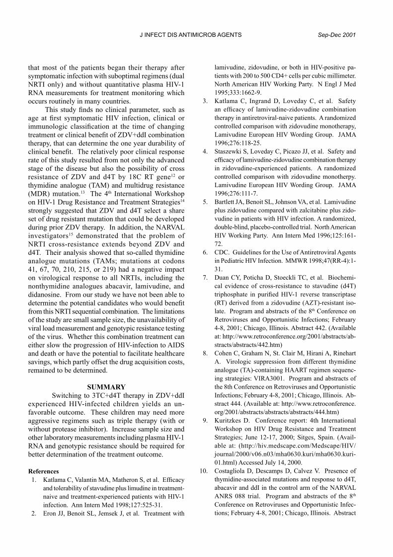
the susceptibility percentage of the isolates are given in Table 2. Only one clinical isolate of B. pseudomallei out of 98 was resistant to all five antimicrobial agents. In this case, the MICs were: tetracycline (32 mcg/ml), trimethoprim/sulfamethoxazole (8/152 mcg/ml), cefotaxime (64 mcg/ml), ceftazidime (32 mcg/ml) and ceftriaxone (128 mcg/ml). Plasmid preparation from this isolate was performed by alkaline lysis technique.8 There was no plasmid band as shown by agarose gel electrophoresis. The results of exoenzyme production are shown in Table 3. Our study revealed that 54.2 percent of Ara+ soil isolates, 45.8 percent of Ara- soil isolates and 40 percent of Ara- patients’ isolates, produced colla-genase. All the Ara+ isolates produced lipase while 62.5 and 58 percent of Ara- isolates from soil and patients, respectively, produced lipase. Only 8.3 percent of Ara- and 20.8 percent of Ara+ from soil produced heparinase while 42 percent of the Ara- isolates from patients produced heparinase.
DISCUSSION The arabinose assimilation is a simple test
to distinguish nonvirulent from virulent isolates of B. pseudomallei.1 Ara- strains are highly virulent whereas Ara+ strains are essentially nonvirulent.2 In our study, we assessed the differences of phenotypic properties of these two biotypes by testing antimicrobial susceptibility and exoenzymes production.
Most isolates of B. pseudomallei are susceptible to conventional drugs such as trimethoprim/sulfa-methoxazole and to newer drugs such as the third generation cephalosporins (cefotaxime, ceftazidime and ceftriaxone). Though trimethoprim/ sulfamethoxazole show in vitro effect against B. pseudomallei, it does

J INFECT DIS ANTIMICROB AGENTS Sep-Dec 2001
Table 2. Antimicrobial susceptibility of B. pseudomallei from different sources.
MIC (mcg/ml) Range MIC50
MIC90
Tetracycline S+ 0.25-128 16 64 8.3 S- 8-64 16 32 0 P- 8-64 32 32 0Trimethoprim/ S+ <0.125/2.38-2/38 1/19 1/19 100Sulfamethoxazole S- 0.25/4.75-16/304 1/19 1/19 95.8 P- <0.125/2.38-128/2,432 1/19 1/19 96Cefotaxime S+ <0.125-8 2 8 100 S- 0.5-8 4 8 100 P- 0.5-64 4 8 96Ceftazidime S+ 0.5-8 1 8 100 S- 0.5-2 2 2 100 P- 1-32 1 4 96Ceftriaxone S+ <0.125-16 4 8 87.5 S- 1-8 4 8 100 P- 2-128 8 16 86
Note: S+ = Ara+ isolates from soil (24 isolates), S- = Ara- isolates from soil (24 isolates), P- = Ara- isolates from patients (50 isolates); break points of tetracycline, trimethoprim/sulfamethoxazole, cefotaxime, ceftazidime and ceftriaxone
Antimicrobial agent
Source oforganisms
% susceptibility
Table 1. Minimum inhibitory concentrations of antimicrobial agents for B. pseudomallei.
No. of strains with indicated MIC (mcg/ml)
< 0.125 0.25 0.5 1 2 4 8 16 32 64 128
Tetracycline S+ 1 1 6 6 4 5 1 S- 1 14 8 1 P- 16 4 25 5
Trimethoprim/ S+ 1 2 19 2 Sulfamethoxazole S- 3 4 15 1 1 P- 5 18 25 1 1
Cefotaxime S+ 1 3 1 9 5 5 S- 3 1 17 3 P- 4 1 14 8 21 1 1
Ceftazidime S+ 10 9 1 4 S- 1 16 7 P- 27 15 6 2
Ceftriaxone S+ 1 4 9 7 3 S- 1 1 10 12 P- 1 3 39 4 2 1
Note: S+ = Ara+ isolates from soil (24 isolates), S- = Ara- isolates from soil (24 isolates), P- = Ara- isolates from patients (50 isolates).
Antimicrobial agent
Source oforganisms
Vol.18 No.3 Susceptibility and exoenzymes production of B. pseudomallei:- Lulitanond A, et al.
Table 3. Production of exoenzymes from B. pseudomallei.
No. of positive isolates (%)
S+ S- P-
Collagenase 13 (54.2) 11 (45.8) 20 (40)Lipase 24 (100) 15 (62.5) 29 (58)Heparinase 5 (20.8) 2 (8.3) 21 (42)
Note: S+ = Ara+ isolates from soil (24 isolates), S- = Ara- isolates from soil (24 isolates), P- = Ara- isolates from patients (50 isolates).
Enzymes
not have a complete bactericidal effect.9 Studies from Australia have shown that trimethoprim/sulfa-methoxazole was effective10, but had a high failure rate in severe melioidosis.11 Effective clinical treatment requirs a combination of trimethoprim/sulfametho-xazole and the other drugs.12
Our study shows that third generation cephalo-sporins have high activity against B. pseudomallei. Ceftazidime had MIC50
and MIC90 values similar to
those previously reported.13,14 Similarly, the organisms were 96-100 percent susceptible to cefotaxime with MIC50
and MIC90 values close to those reported by
Aswapokee15 and Tharavichitkul.16 Although suscepti-bility to cefotaxime was similar, our MIC50
and MIC90
values for this drug were four times lower than those reported by Vorachit.14 This might be associated with the different use of antimicrobial agents. Ceftriaxone was slightly less effective than ceftazidime and cefotaxime. The MIC50 and MIC90 of ceftriaxone were similar to those previously reported.14-16 Amongst the five antimicrobial agents tested in this study, tetra-cycline was least effective against B. pseudomallei with the MIC50
and the MIC90 at 32 and 64 mcg/ml,
respectively. Clinical trials recommend that the drug of choice for treatment of acute and severe melioidosis is ceftazidime or a combination of ceftazidime with the other drugs.12,17 Resistant strains of ceftazidime have been reported.18
Ara+ isolates tend to have lower MIC than the Ara- isolates (Table 1). This may be due to their different origin or individual differences in anti-microbial exposure. Further studies with additional samples should be done to discern the reason.
Lipase production of Ara+ and Ara- isolates, was statistically significant (p < 0.05, Chi-square test). Heparinase production of the isolates from soil and from the patients was different.
In summary, the two biotypes (Ara+ and Ara-)
of B. pseudomallei have no significant difference in susceptibility to the antimicrobial agents tested. Lipase and heparinase, exoenzymes which indicate virulence, are significantly different with Ara+ and Ara-.
ACKNOWLEDGEMENT This work was supported by Faculty of
Associ-ated Medical Sciences, Khon Kaen University. We thank Mr. Bryan Hamman for assistance with English language editing.
References 1. Wuthiekanun V, Smith MD, Dance DAB, Walsh AL,
Pitt TL, White NJ. Biochemical characteristics of clinical and environmental strains of Burkholderia pseudomallei. J Med Microbiol 1996;45:408-12.
2. Smith MD, Angus BJ, White NJ. Arabinose assimi- lation defines a nonvirulent biotype of Burkholderia pseudomallei. Infect Immun 1997;65:4319-21.
3. Brett PJ, Deshazer D, Woods DE. Characterization of Burkholderia pseudomallei and Burkholderia pseu-domallei like strains. Epidemiol Infect 1997;118:137-48.
4. Chaiyaroj SC, Kotrnen K, Koonpaew S, Anantagool N, White NJ, Sirisinha S. Differences in genomic macroestriction patterns of arabinose-positive (Burkholderia thailandensis) and arabinose-negative Burkhold-eria pseudomallei. Microbiol Immunol 1999;43:625-30.
5. National committee for Clinical Laboratory Standards. Methods for dilution antimicrobial susceptibility tests for bacteria that grow aerobically. 7th ed. Approved Standard NCCLS Document M7-A4. Villanova Pa: NCCLS, 1997.
6. Phillips E, Nash P. Culture media. In: Lennette EH, Balows A, Hausler JR WJ, Shadomy HJ, eds. Manual of Clinical Microbiology, 4th ed. Washington DC:

J INFECT DIS ANTIMICROB AGENTS Sep-Dec 2001
American Society for Microbiology, 1985:1051-92. 7. Steffen EK, Hentges DJ. Hydrolytic enzymes of
anaerobic bacteria isolated from human infection. J Clin Microbiol 1981;14:153-6.
8. Takahashi S, Nagano Y. Rapid procedure for isolation of plasmid DNA and application to epidemiological analysis. J Clin Microbiol 1984;20:608-13.
9. Vorachit M, Chongtrakool P, Arkomsean S, Boonsong S. Antimicrobial resistance in Burkholderia pseu-domallei. Acta Trop 2000;7:139-44.
10. Currie B, Howard D, Nguyen VT, Withnall K, Merianos A. The 1990-1991 outbreak of melioidosis in the northern territory of Australia: clinical aspects. Southeast Asian J Trop Med Public Health 1993; 24:436-43.
11. White NJ, Dance DAB. Clinical and laboratory studies of malaria and melioidosis. Trans R Soc Trop Med Hyg 1988;82:15-20.
12. Chaowagul W. Recent advances in the treatment of severe melioidosis. Acta Trop 2000;74:133-37.
13. Ashdown LR. In vitro activities of the newer β-lactam and quinolone agents against Pseudomonas pseu-domallei. Antimicrob Agents Chemother 1988;2:1435-6.
14. Vorachit M, Jayanetra P, Boonsung S. Pseudomonas pseudomallei II. Antimicrobial Susceptibility Patterns. In: Punyagupta S, Sirisanthana T, Stapatayavong B,
eds. Melioidosis, Proceedings of National Workshop on Melioidosis. Bangkok: Bangkok Medical Publisher, 1989:161-5.
15. Aswapokee N, Pruksachatvudhi S, Aswapokee P. In vitro susceptibility of Pseudomonas pseudomallei to Conventional and Newer antimicrobial Agents. In: Punyagupta S, Sirisanthana T, Stapatayavong B, eds. Melioidosis, Proceedings of National Workshop on Melioidosis. Bangkok: Bangkok Medical Publisher, 1989:177-80.
16. Tharavichitkul P, Khanthawa B, Preuksakorn S, Sirisanthana T. Comparative activity of cefoperazone, cefotaxime, ceftriaxone and piperacillin against Pseudomonas pseudomallei isolated from Maharaj Hospital, Chiang Mai. In: Punyagupta S, Sirisanthana T, Stapatayavong B, eds. Melioidosis, Proceedings of National Workshop on Melioidosis. Bangkok: Bangkok Medical Publisher, 1989:170-3.
17. White NJ, Dance DAB, Chaowagul W, Wattanagoon Y, Wuthiekanun V, Pitakwatchara N. Halving of mortality of severe melioidosis by ceftazidime. Lancet 1989;2:697-701.
18. Dance DAB, Wuthiekanun V, Chaowagul W, Su-puttamongkol Y, White NJ. Development of resistance to ceftazidime and Co-amoxyclav in Pseudomonas pseudomallei. J Antimicrob Chemother 1991;24:295-

Vol.18 No.3 High ALP in HIV patients: Wiwanitkit V.
Abstract
Original Article
93
Study of High Serum Alkaline Phosphatase in HIV-infected Patients
Viroj Wiwanitkit, M.D. *
In 1998, this study was done to investigate the cause of hyperalkaline-phos-phatasemia among patients with human immunodeficiency virus (HIV) in different clinical stages at King Chulalongkorn Memorial Hospital, Bangkok, Thailand. Data was collected from 187 HIV-infected patients whose serum alkaline phosphatase (ALP) level was deter-mined. Twenty-four patients with known CD4 count and raised serum ALP levels alkaline were evaluated. Opportunistic infections appeared to be the most common possible cause of hyperalkalinephosphatasemia (79.2%), followed by non-opportunistic infections (16.6%). Mycobacterium tuberculosis infection is associated with hyperalkalinephosphatasemia. In conclusion, hyperalkalinephosphatasemia in HIV-infected patients may indicate opportunistic infection. (J Infect Dis Antimicrob Agents 2001;18:93-5)
* Department of Laboratory Medicine, Faculty of Medicine, Chulalongkorn University, Bangkok 10330, Thailand.Received for publication: January 4, 2001.Reprint request: Viroj Wiwanitkit, M.D., Department of Laboratory Medicine, Faculty of Medicine, Chulalongkorn University, Bangkok
10330, Thailand.Keywords: Alkaline phosphatase, ALP, HIV
INTRODUCTIONHuman immunodeficiency virus (HIV) infection,
is a worldwide epidemic with high prevalence in many areas of the world including Thailand. In Thailand, approximately one million people are estimated to be HIV infected1.
Hepatobiliary disease is one of the common pro-blems among patients with HIV. A number of liver function abnormalities including alkaline phos-phatase (ALP) among HIV-infected patients have been mentioned.2 However, the etiology of high ALP level in HIV-infected patients has not been systematically evaluated. This study was performed to determine the clinical association of high ALP level in Thai HIV-in-fected patients with different immune status.
MATERIALS AND METHODSThis cross-sectional descriptive study was re-tro-
spectively performed in the clinical chemistry unit service of King Chulalongkorn Memorial Hospital from January to December 1998. All HIV-infected patients with known CD4 cell count and raised serum ALP lev-els were recruited in the study. Determination of ALP was measured by the optimized method of Boehringer
Manheim, with a normal range of 98-279 IU/L. ALP of > 1,000 IU/L are considered as high levels and > 2,000 IU/L are considered as extremely high levels. According to the 1993 revised classification system for HIV infection3, CD4 cell count is used to categorize the immunological status of the study subjects. Medi-cal records including history, physical examinations, investigations, diagnostic procedures and outcome were reviewed. Data was analyzed using descriptive statistical analysis.
RESULTS Of 187 HIV-infected patients whose serum
ALP levels were determined, 26 (13.9%) had hyper-alkalinephosphatasemia. The study patients consisted of one patient (4.2%) with a CD4 count > 500 cells/mm3 (Class A), 3 patients (12.5%) with CD4 200-499 cells/mm3 (Class B), and 20 patients (83.3%) with CD4 < 200 cells/mm3. The mean age of the subjects was 35.5 + 10.9 years. Mean serum ALP and total bilirubin levels were 1,596.6 + 711.5 IU/L and 2.4 + 2.8 mg/dl, respectively. Five patients (20.8%) had extremely high serum ALP levels, and all of them were classified in class A.

J INFECT DIS ANTIMICROB AGENTS Sep-Dec 2001
Table 1. Study subjects, classified by the number of CD4 cell count.
Number of cases (%)
Class A Class B Class C Total CD4 > 500 cells/mm3 CD4 200-499 cells/mm3 CD4 < 200 cells/mm3
(n = 24) (n = 1) (n = 3) (n = 20) 1. Subjects with OIs Tuberculosis - 2 (8.3) 9 (37.5) 11(45.8) Penicilliosis - - 2 (8.3) 2 (8.3) Histoplasmosis - - 2 (8.3) 2 (8.3) Norcardiosis - - 1 (4.2) 1 (4.2) Pneumocystis carinii - - 1 (4.2) 1 (4.2) Cryptococcosis - - 1 (4.2) 1 (4.2) Cytomegalovirus - - 1 (4.2) 1 (4.2)2. Subjects with non OIs
Salmonellosis - 1 (4.2) - 3 (12.4)Urinary tract infection 1 (4.2) - - 1 (4.2)
3. Subjects with malignanciesNon-Hodgkin lymphoma - - - 1 (4.2)
Table 2. Study subjects, classified by serum ALP levels.
Number (%)
ALP = 1,000 - 1,999 IU/L ALP = 2,000 - 3,000 IU/L (n = 19) (n = 5)
1. Subjects with OI Tuberculosis 8 (33.4) 3 (12.4) Penicilliosis 2 (8.3) - Histoplasmosis 2 (8.3) - Norcardiosis 1 (4.2) - Pneumocystis carinii 1 (4.2) - Cryptococcosis 1 (4.2) - Cytomegalovirus - 1 (4.2)2. Subjects with non-OI infections Salmonellosis 3 (12.4) - Urinary tract infection 1 (4.2) -3. Subjects with malignancies Non-Hodgkin lymphoma - 1 (4.2)
Category of subjects
Category of subjects
The patients were calegorized into three major groups i.e. subjects with opportunistic infections (OIs) (79.2%), subjects with non-OI infections (16.6%) and subjects with malignancies. The most common OIs found in our subjects was disseminated tuber-culosis. It was found that 17 of 20 patients (85.0%) in class C developed OIs (Table 1). After classifying study patients according to serum ALP levels, it was found that 15 of 19 patients (78.9%) with high ALP (> 1,000 IU/L) and 4 of 5 patients (80.0%) with extremely
high ALP (> 2,000 IU/L) developed OIs. One subject in the extremely high ALP group had lymphoma (Table 2) and three patients (12.5%) in this group died within 6 months.
DISCUSSION HIV infection is associated with a variety of
hepatobiliary manifestations. A marked increase in serum ALP level (> 1,000 IU/L) can be seen in many conditions, including hepatobiliary disorders, systemic
Vol.18 No.3 High ALP in HIV patients: Wiwanitkit V.
infections and malignancies.4 It can also be a pres-entation of disorders among HIV-infected patients. Previous literature reviews have not demonstrated the factors associated with raised ALP among HIV-infected patients.
Approximately ten percent of our patients were observed to have hyperalkalinephosphatasemia. OIs was determined as factors associated with HIV among patients with hyperalkalinephosphatasemia. It is found that there is a significantly high number of patients with hyperalkalinephosphatasemia were found in class C.
A previous study revealed that extremely high ALP level in AIDS patients were associated with super-imposed infections.5 However, our data show a similar prevalence of OIs in those with high and extremely high ALP.
There were two findings features learned from our study: (1) high prevalence of OIs among HIV patients may be associated with hyperalkalinephos-phatasemia, and (2) the high prevalence of Mycobacterium tubercu-losis associated with hyperalkaline-phosphatasemia is found in subjects with advanced disease. Information about the level of ALP in Thai patients with HIV could be an indicator of OIs and assist with investigation and care.
However, this study reviewed a specific group of hospital-based patients. Therefore, the results may not be applied to the general population. Further studies on large number of cases in multicenter is suggested.
References1. Surasiengsunk S, Kiranandana S, Wongboonsin
K, Garnett GP, Anderson RM, van Griensven GJ. Demographic impact of the HIV epidemic in Thailand. AIDS 1998;12:775-84.
2. Roulot D. Hepatobiliary and pancreatic diseases in hu-man immunodeficiency virus infection. Gastroenterol Clin Biol 1995;19:B150-6.
3. Centers for Disease Control and Prevention. 1993 revised classification system for HIV infection and expanded surveillance on definition of AIDS among adolescents and adults. Morb Weekly Rep 1993;41:1-19.
4. Neuschwander-Tetri BA. Common blood tests for liver disease. Which ones are most useful? Postgrad Med 1995;98:49-56.
5. Dworkin BM, Stahl RE, Glardina MA, et al. The liver in acquired immune deficiency syndrome: emphasis on patients with intravenous drug abuse. Am J Gastro-enterol 1987;82:231-6.

J INFECT DIS ANTIMICROB AGENTS Sep-Dec 2001
This recent case occurred in November 1999 and had reduced susceptibility to penicillin (minimum inhi-bitory concentration/MIC=0.125 mcg/ml), and was susceptible to cefotaxime and ceftriaxone. After appropriate antimicrobial treatment, improvement occurred within the first three days of hospitalization in two-thirds of patients (65.2%).
The most common central nervous system com-plications were subdural effusion (13%) and hearing impairment (13%), followed by hydrocephalus (8.6%) and ventriculitis (4.3%). Ultrasonographic studies of the head were performed in 4 cases and hearing tests were performed in 6 cases. Final outcomes were excellent in all except one child who had fulminant meningococcemia and died within the first day of hospitalization. The overall fatality rate was 4.3 percent.
DISCUSSIONMeningococcal infections are commonly found in
developing countries such as African countries, and are sometimes found in developed countries like the United States. In Thailand between 1979 and 1994, the national incidence of meningococcal disease ranged from 0.03-0.20/100,000,3 and approximately 50 percent of cases occurred in children. Compared with the average incidence of 1.1/100,000 in 15 African countries4, meningococcal infection is relatively uncommon in Thailand. Usa Thisyakorn et al5 reported 13 children with culture-proven invasive menin-gococcal infections from the Childen’s Hospital (now known as Queen Sirikit National Institute of Child Health) in Bangkok during the 10-year study from 1975 to 1984. From our study, 25 children with menin-gococcal infections were reported in the same hospital in 12-year period from 1988 to 1999.
As in other studies6-8, the most susceptible age group was between 3 months to 2 years (47.8%). Clinical manifestations of invasive meningococcal infection noted in this study ranged from fever with nonspecific symptoms such as irritability, vomiting and tachypnea to seizure and shock with purpura fulminans. Fever associated with a petechial rash or purpura has classically been noted in patients with meningococ-cemia. However from our study, the occurrence of fever, particularly with either petechiae or purpura, allowed prompt diagnosis in only 21.7 percent of invasive menincoccal infection. This figure is low, compared to reports from equatorial African, Spain and other western countries.9-10
The presence of meningitis in patients with
meningococcemia has previously been correlated with favorable outcome.11-12 This is true in our study, in which all cases had CSF abnormalities in addition to positive culture results from either blood or CSF. However, there was one case who presented with fulminant meningococcemia and died within the first day of hospitalization.
Overall fatality rate of invasive meningococcal infection is 5-16 percent, although these rates are difficult to assess as some studies only take into account meningococcal meningitis, while others reflect overall fatality from meningococcal disease.6,13,14 Reported mortality from meningococcemia ranges from 18-35 percent.5,15-26 For overall invasive meningococcal infection, the fatality rate in our study was low (4.3%). For meningococcemia, the fatality rate is 12.5 percent which is related to the presence of the poor prognostic signs such as shock on arrival, coma and absence of meningism.
Morbidity has been much less frequently reported but varies from 11-19 percent and consists of neurologic disability, sensorineural hearing loss, limb loss and/or need for skin grafting.20,24,26 In general, the most important long-term sequelae is sensorineural hearing impairment.27 From our study subdural effusion and hearing impairment occurred in 13 percent of the patients. However, due to the limited number of study patients, we cannot make conclusion concerning the long-term sequelae produced by meningococcal meningitis.
The discovery of sulfa in the 1930s and its success in treating meningococcal disease has represented a major advance and replaced serum therapy as the treatment of choice. With the development of sulfa resistance in the 1950s, penicillin became the antibiotic of choice for invasive meningococcal disease. Penicillin resistant strains of meningococci (MIC=0.1-1.28 mcg/ml) have been reported from Spain28, England29, South Africa30, Canada31, the United States32,33, and Thailand by Pancharoen C in 1998.2
In our study, disc susceptibility were performed in all 23 strains, and all were sensitive to penicillin except for the last case, in November 1999, which was found to have reduced susceptibility to penicillin (MIC=0.125 mcg/ml). Fortunately, it remained susceptible to ceftriaxone and cefotaxime by disc susceptibility and the patient recovered without immediate sequelae.
In the past, sporadic cases or outbreaks emerged in the military services in boot camp, where young and susceptible recruits are overworked, submitted to 32.

Vol.18 No.3 High ALP in HIV patients: Wiwanitkit V.

J INFECT DIS ANTIMICROB AGENTS Sep-Dec 2001

Vol.18 No.3 High ALP in HIV patients: Wiwanitkit V.

J INFECT DIS ANTIMICROB AGENTS Sep-Dec 2001Original Article
96
Abstract
* Rhinology Division, Department of Otolaryngology,** Department of Microbiology, Faculty of Medicine Siriraj Hospital, Mahidol University, Bangkok 10700, Thailand.Received for publication: June 6, 2001.Reprint request: Perapun Jareoncharsri, M.D., Rhinology Division, Department of Otolaryngology, Faculty of Medicine Siriraj Hospital,
Mahidol University, Bangkok 10700, Thailand.Keywords: Bacteriologic profile, acute maxillary sinusitis, chronic maxillary sinusitis, acute exacerbation on chronic sinusitis, antral
aspiration culture
Bacteriologic Profile of Acute and Chronic Maxillary Sinusitis
Perapun Jareoncharsri, M.D.*Chaweewan Bunnag, M.D.*Prayuth Tunsuriyawong, M.D.*Siriporn Voraprayoon, B.Ed.*Somporn Srifuengfung, Ph.D. (Microbiol)** Chertsak Dhiraputra, M.D., M.Sc. (Microbiol)**Anan Bedavanija, M.D.*
Sinusitis is a very common disease. Antimicrobial therapy can be used for the treatment of sinusitis due to bacterial infection. This study of the bacterial etiology of maxillary antral aspiration is done in patients with maxillary sinusitis treated at Siriraj Hospital, Bangkok, Thailand between January 1999 and June 2000. There were 64 patients with acute maxillary sinus infection, 48 with acute maxillary sinusitis (AMS) and 16 with acute exacerbation of chronic maxillary sinusitis (AECS), and 27 patients with chronic maxillary sinusitis (CMS). There were positive cultures in 64.6 percent of AMS, 93.8 percent of AECS and 92.6 percent of CMS. Anaerobes were found in 14.6 percent of AMS, 37.5 percent of AECS and 66.6 percent of CMS. Gram-negative bacteria were more common than gram-positive bacteria and occurred in 52.8 percent of AMS, 70.6 percent of AECS, and 68.0 percent of CMS. Common aerobes in AMS were Haemophilus influenzae (H. influenzae) (34.3%, with 50.0% being beta-lactamase producing strains). Streptococcus pneumoniae (17.1%), and other Streptococcus species (17.1%). Common aerobes in AECS were Pseudomonas aeruginosa (23.5%), H. influenzae (17.6%, with 66.7% being beta-lactamase producing strains), and Klebsiella pneumoniae (K. pneumoniae) (11.8%). Aerobes in CMS were H. influenzae (28.0%), non-fermentative gram-negative rods (20.0%), and K. pneumoniae (12.0%). The most common anaerobes were Fusobacterium spp., Peptostreptococcus spp., Bacteroides spp., non-sporing gram-positive rods, and Prevotella spp., which were found in AMS, AECS and CMS. The microbiology of sinusitis is important in prescribing appropiate antimicrobial therapy. (J Infect Dis Antimicrob Agents 2001;18:96-102)
INTRODUCTION Sinusitis is a common disease and it is increas-
ingly being reported in clinical practice. In the United States, the incidence of sinusitis is 14.7 percent in the general population.1 In 1992, there were more than 13 million antibiotic prescriptions for sinusitis,
costing about $ 375 million2, and the annual costs of sinusitis rose to $ 6.1 billion in 1997.3 Sinusitis is an expensive disease and has a major impact on individual patients and the health care system. Appropriate antibiotic selection is important for the cure of bacterial sinusitis. Inappropriate therapy wastes money, results

Vol. 18 No. 3 Bacteriologic profile of maxillary sinusitis:- Jareoncharsri P, et al.
in resistance of the organisms and leads to chronic sinusitis. The selection of the antimicrobial therapy for community-acquired sinusitis is usually based on the information from the previous studies. Studies on sinusitis usually involve the maxillary sinuses since it is more accessible than the other sinuses for antral aspiration and specimens can be collected without nasal flora contamination.
During the last ten years, bacterial culture rates for acute maxillary sinusitis (AMS) have varied from 60 to 80 percent.4-10 Cultures for AMS show that Streptococcus pneumoniae (S. pneunoniae) and Haemophilus influenzae (H. influenzae) are the two most common organisms, followed by other Strep-tococcus species, Morexella catarrhalis (M. catar-rhalis) and Staphylococcus aureus (S. aureus). The incidence of anaerobic infection in AMS varies from 0 to 19.1 percent.4-10 The most common anaerobes are Peptostreptococcus spp., Prevotella spp., and Propioni-bacterium acnes.
In chronic maxillary sinusitis (CMS), most studies report positive bacterial cultures in 70 to 100 percent.7-11 The most common bacteria are Strep-tococcus, Staphylococcus coagulase negative, S. aureus, H. infuenzae, and other gram-negative rods such as Klebsiella pneumoniae (K. pneumoniae) and Pseu-domonas aeruginosa (P. aeruginosa). The incidence of anaerobic infection in CMS varies from 8.1 to 100 percent.11-15 The most common anaerobes were Prevotella species, Fusobacterium species, Pepto-streptococcus species, Propionibacterium species, anaerobic gram-negative rod, and Veillonella species.
There is a wide range of variation in the percentage of positive cultures and the culture profile varies with the type of sinusitis (AMS or CMS). It is also important whether the data are retrospectively or prospectively collected, because in prospective study antibiotics are withheld and the specimens are better handled to the laboratory, which may result in a higher percentage of positive cultures. It is important to include the anaerobic culture, and that there is satisfactory collection and transportation of the specimens to the laboratory. The objective of this study was to study the bacteriological profile of acute and chronic maxillary sinusitis in patients in Thailand.
PATIENTS AND METHODS This is a prospective study of maxillary antral
aspiration culture in patients with maxillary sinusitis and was done by the Rhinology Division of the Department of Otolaryngology and by the Department of Microbiology at Siriraj Hospital between January 1999 and June 2000.
Patients with AMS Inclusion criteria:1. Outpatients with symptoms and signs of AMS,
i.e. persistent upper respiratory tract infection more than 7 to 10 days, or mucopurulent nasal or posterior pharyngeal discharge, cough, fever, headache or facial pain, and persisting for 2 to 8 weeks.2
2. Positive radiological findings of maxillary sinus, defined as one or more of the following findings, i.e. opacification, air fluid levels and mucosal thickening in one or both maxillary sinuses.
3. The presence of mucopurulent discharge in the middle meatus or maxillary ostia seen by nasal endoscopic examination.
4. Antibiotics stopped for at least 48 to 72 hours prior to the study.
A total of 64 patients (22 males and 42 females) were included in this study. The mean age was 38 + 13.2 years (17-70 years). The acute group was classified into 2 groups: 1) 48 patients with AMS and 2) 16 patients with acute exacerbation on chronic sinusitis (AECS). The mean duration of symptoms in AMS group was 17.4 + 13.6 days, and in AECS group was 31.6 + 20.6 days.
Patients with CMS
Twenty-seven consecutive patients with CMS who were scheduled to undergo elective endoscopic sinus surgery were included. There were 12 males and 15 females, with the mean age of 39.9 + 13.3 years (15-70 years). The mean duration of symptoms was 15.6 + 18.9 months (3-84 months). Antibiotics were withheld one week before admission to hospital.
Written informed consent was obtained from each subject prior to study entry. Patients with AMS and AECS were part of two ongoing clinical trials.16,17 The protocol was approved by the Committee on Human Rights Research in Human Subjects at the Faculty of Medicine Siriraj Hospital.
Culture procedure Specimens for bacteriological study were
collected by maxillary antral aspiration using sterile technique. If there was no secretion on aspiration, the maxillary sinus was irrigated with 2 ml of normal saline and the irrigation fluid was sent for both aerobic and anaerobic culture.18 All specimens were sent to the microbiology laboratory immediately. For the CMS group, specimens for anaerobic culture were incubated immediately on blood agar plate, when specimens were obtained and put into Gaspak anaerobic jars. All specimens were sent to the laboratory within 2 hours. Microorganisms were isolated and identified

J INFECT DIS ANTIMICROB AGENTS Sep-Dec 2001
accor- ding to standard bacteriological methods. The antimicrobial susceptibility was performed by the stan-dard disk diffusion method as recommended by the National Committee for Clinical Laboratory Standards (NCCLS).19 Mueller-Hinton agar (MH) was used for testing nonfastidous bacteria. MH supplemented with 5 percent sheep blood was used for testing fastidious bacteria such as S. pneumoniae and other Streptococcus species. Haemophilus test medium was used for testing H. influenzae.
Each bacterial inoculum was prepared by suspending 4-5 isolated colonies from pure culture into peptone broth and the suspension was adjusted equal to the turbidity of McFarland standard solution No. 0.5. The diameter of the inhibition zone around each antimicrobial disk after incubation on agar plates at 35° C for 18-24 hours was measured and read as sensitive or resistant by referring to the zone diameter interpretive standards as recommended by the NCCLS. Beta-lactamase enzyme was detected by a rapid test for hydrolysis of nitrocefin solution (10 mg/ml).20 A positive reaction occurs when the color of the solution changes from yellow to red within 5 minutes of the procedure carried out at room temperature.
RESULTS In the AMS group, positive cultures were found
in 64.6 percent of cases (31/48) i.e. aerobes (50%), anaerobes (4.2%), and mixed aerobes and anaerobes (10.4%). Sixty microorganisms were isolated from 48 specimens (1.3 isolates/specimen) (Table 1). The most common aerobes were H. influenzae (34.3% of aerobes), S. pneumoniae (17.1%), other Streptococcus species (17.1%), P. aeruginosa (11.4%), and S. aureus (5.7%) (Table 2). Fifty percent of H. influenzae were betalactamase-producing (BLP). The most common anaerobes were Peptostreptococcus species (28% of anaerobes), Fusobacterium species (28%), Bacteroides species (20%), Prevotella species (8%), and Tissierella praeacuta (8%) (Table 3).
In AECS group, positive cultures were found in 93.8 percent of cases (15/16), which were aerobes (56.3%), anaerobes (31.3%), and mixed aerobes and anaerobes (6.2%). Forty microorganisms were isolated (2.5 isolates/specimen) (Table 1). The most common aerobes were P. aeruginosa (23.5%), H. influenzae (17.6%), K. pneumoniae (11.8%), non-fermentative gram-negative rods (11.8%), and other Streptococcus species (11.8%) (Table 2). Two-thirds of H. influenzae were BLP. The most common anaerobes were Fusobacterium species (34.8%), Peptostreptococcus species (21.7%), Prevotella species (13.0%), Bacte-roides species (8.7%), and Tissierella praeacuta (4.3%)
(Table 3). In the CMS group, positive cultures were
found in 92.5 percent of cases (25/27) which were aerobes (25.9%), anaerobes (22.2%), mixed aerobes and anaerobes (44.4%). The number of microorganisms isolated was 65 (2.4 isolates/specimen) (Table 1). The most common aerobes were H. influenzae (28.0%), non-fermentative gram-negative rod (20.0%), K. pneumo-niae (12.0%), β-hemolytic streptococci (8.0%), and S. aureus (8.0%) (Table 2). No isolates of BLP H. influ-enzae were found. The most common anaerobes were non-sporing gram-positive rods (30.0%), Peptostrep-tococcus spp. (27.5%), Fusobacterium spp. (22.5%), Bacteroides spp. (10.0%), and Porphyromonas (5.0%) (Table 3).
The antimicrobial susceptibility of pathogenic microorganisms in AMS and AECS are shown in Table 4.
DISCUSSIONPositive culture (Table 1) In this study, the percentage of positive cultures (64.6%) in AMS are similar to the findings from most previous studies.4-12 The study by Brook et al15 showed 100 percent positive culture rates but there were only ten patients, which was too small to represent the true positive rates. In contrast with AECS and CMS, the positive culture rates are as high as 93.8 percent and 92.5 percent, respectively. This study finds more than one microorganisms/specimen in AECS (2.5 isolates/specimen) and in CMS (2.4 isolates/specimen) compared with only 1.3 isolates/specimen in AMS. This shows the polymicrobial nature and mixed type (aerobes + anaerobes) infection in AECS and CMS. This study finds entirely anaerobes in 4.2 percent of AMS. Vogan et al5 and Brook et al15 found no pure anaerobic infection in AMS, similar to another study from Thailand,6 where pure anaerobic infection was isolated in 14.9 percent of AMS. The percentage of anaerobic infections (pure anaerobes + mixed aerobes and anaerobes) in AMS from our study was 14.6 percent, which is higher than other studies reported by Vogan et al5 (12.5%), Brook et al15 (5.9%), Gwaltney et al8 (7%) and Jousimies-Somer et al9 (1.5%). Anaerobes are more likely to be present in CMS. The finding of anaerobes in AMS infection suggests chronic disease or dental infection. In this study, associated dental infection was found in 1 patient (2.1%) with AMS and 3 patients (18.8%) with AECS. The percentage of anaerobic infection was 37.5 percent in AECS and 66.6 percent in CMS, suggesting that prolonged obstruction of maxillary sinus ostium is an important predisposing factor. In this situation, antimicrobial therapy for anaerobic infection may not

Vol. 18 No. 3 Bacteriologic profile of maxillary sinusitis:- Jareoncharsri P, et al.
Table 1. Percentage of microorganisms in AMS, AECS and CMS.
AMS AECS CMS (n=48) (n=16) (n=21)
Positive culture (%) 64.6 93.8 92.6Total No. of bacterial isolates 60 40 65No. of bacterial isolates/specimen 1.3 2.5 2.4Aerobes (%) 50 56.3 25.9Anaerobes (%) 4.2 31.3 22.2Mixed (%) 10.4 6.2 44.4Gram-positive bacteria (%) 47.2 29.4 32.0Gram-negative bacteria (%) 52.8 70.6 68.0
Note: AMS = acute maxillary sinusitis, AECS = acute exacerbation on chronic sinusitis,
CMS = chronic maxillary sinusitis.
Table 2. Percentage of aerobes in AMS, AECS and CMS.
AMS (35 isolates) AECS (17 isolates) CMS (25 isolates)
H. influenzae 34.3% P. aeruginosa 23.5% H. influenzae 28.0%S. pneumoniae 17.1% H. influenzae 17.6% NF. GNR 20.0%Other Streptococcus spp. 17.1% K. pneumoniae 11.8% K .pneumoniae 12.0%P. aeruginosa 11.4% NF. GNR 11.8% β-hemo. Strep. 8.0%S. aureus 5.7% Other Streptococcus spp. 11.8% S. aureus 8.0%
Note: NF-GNR = Non-fermentative gram-negative rod.
Table 3. Percentage of anaerobes in AMS, AECS and CMS.
AMS (25 isolates) AECS (23 isolates) CMS (40 isolates)
Peptostreptococcus spp. 28.0% Fusobact. spp. 34.8% Non-spore forming GPR 30.0%Fusobacterium spp. 28.0% Peptostrep spp. 21.7% Peptostreptococcus spp. 27.5%Bacteroides spp. 20.0% Prevotella spp. 13.0% Fusobacterium. spp. 22.5%Prevotella spp. 8.0% Bacteroides spp. 8.7% Bacteroides spp. 10.0%Tissierella spp. 8.0% Tissierella spp. 4.3% Porphyromonas spp. 5.0%
Note: GPR=Non-sporing gram-positive rod.
be sufficient. Surgical drainage and ventilation of the sinuses has to be included in the treatment process.
Common microorganisms: Aerobes (Table 2) In this study, as with other previous studies,
H. influenzae and S. pneumoniae are the two most common aerobic organisms in AMS. Our study shows that H. influenzae (34.3%) was twice as common as S. pneumoniae (17.1%) (Table 2), with differs from other studies, which report S. pneumoniae predominates.
M. catarrhalis was not reported in AMS and the AECS groups and only 1 culture was isolated from the CMS group. Our study results differ from those of European countries7 and of the United States5,15,21
where M. catarrhalis is cultured in 10 to 14 percent of adult studies, and even higher percentages are reported in pediatric patients.22 This could be due to our study including only adults (M. catarrhalis is an important pathogen in children) or the organism could be rare in community-acquired sinusitis in Thailand.
Another surprising finding was the presence of P. aeruginosa (11.4%) in AMS. Other studies have found P. aeruginosa to be very rare in AMS. The difference in the bacteriology of AMS in Thailand could be due to geographical variations in preva- lence of P. aeruginosa (Thailand vs Europe and America). The percentages of aerobes found in AECS

J INFECT DIS ANTIMICROB AGENTS Sep-Dec 2001
Tabl
e 4.
Ant
imic
robi
al s
usce
ptib
ility
test
of o
rgan
ism
s fr
om p
atie
nt w
ith A
MS
and
AE
CS.
%
sen
sitiv
ity
A
MP
ER
Y
SX
T
A
UG
CX
M
C
AZ
CR
O
C
TX
CH
O
FL
CIP
LV
X
GA
F
S. p
neum
onia
e 3/
8 (3
7.5)
2/
8 (2
5)
2/8
(25)
9
/9 (1
00)
2/2
(100
)
8/8
(100
) 8/
8 (1
00)
4/4
(100
) 0/
2 (0
)
5/6
(83.
3)
5/7
(71.
4)
5/5
(100
) 2/
2 (1
00)
H. i
nflue
nzae
(BL-
ve)
9/10
(90)
3/
10 (3
0)
6/10
(60)
10
/10
(100
) 6/
6 (1
00)
7/
8 (8
7.5)
7/
8 (8
7.5)
6/
6 (1
00)
3/3
(100
)
3/3
(100
) 6/
6 (1
00)
4/4
(100
) 3/
3 (1
00)
H. i
nflue
nzae
(BL+
ve)
1/6
(16.
7)
3/7
(42.
9)
4/8
(50)
8
/8 (1
00)
2/2
(100
)
7/7
(100
) 7/
7 (1
00)
7/7
(100
) 2/
2 (1
00)
2/
2 (1
00)
7/7
(100
) 5/
5 (1
00)
2/2
(100
)
Oth
er S
trep
toco
ccus
spp
. 7/
9 (7
7.8)
7/
9 (7
7.8)
7/
9 (7
7.8)
9
/9 (1
00)
6/6
(100
)
7/7
(100
) 9/
9 (1
00)
7/7
(100
) 0/
1 (0
)
7/9
(77.
8)
6/8
(75)
6/
6 (1
00)
1/1
(100
)
S. a
ureu
s 0/
5 (0
) 4/
5 (8
0)
5/5
(100
) 5
/5 (1
00)
3/3
(100
)
4/5
(100
) 5/
5 (1
00)
5/5
(100
) 2/
2 (1
00)
5/
5 (1
00)
4/4
(100
) 2/
2 (1
00)
2/2
(100
)
NF
GN
R
0/5
(0)
-
1/
4 (2
5)
1/4
(25)
0/
1 (0
)
4/4
(100
) 0/
1 (0
) 6/
6 (1
00)
0/1
(0)
3/
4 (7
5)
3/4
(75)
3/
3 (1
00)
0/1
(0)
K. p
neum
onia
e 0/
3 (0
)
-
2/3
(66.
7)
3/3
(100
) 3/
3 (1
00)
3/
3 (1
00)
3/3
(100
) 3/
3 (1
00)
0/2
(0)
3/3
(100
) 3/
3 (1
00)
- 2/
2 (1
00)
P. a
erug
inos
a 0/
2 (0
)
-
0/1
(0)
1/9
(11.
1)
0/3
(0)
11/1
1 (1
00)
2/11
(18.
2)
2/11
(18.
2)
0/1
(0)
10/1
0 (1
00)
8/8
(100
) 7/
7 (1
00)
1/1
(100
)
Not
e:
AM
P =
Am
pici
llin,
ERY
= E
ryth
rom
ycin
, SX
T =
Co-
trim
oxaz
ole,
AU
G =
Am
oxyc
illin
/cla
vula
nic
acid
, CX
M =
Cef
urox
ime,
CA
Z =
Cef
tazi
dine
, CR
O =
Cef
riaxo
ne,
CTX
=C
efot
axim
e,
C
H =
Cla
rithr
omyc
in, O
FL =
Oflo
xaci
n, C
IP =
Cip
roflo
xaci
n, L
VX
= L
evofl
oxac
in, G
AF
= G
atifl
oxac
in, B
L =
Bet
alac
tam
ase
prod
ucin
g, N
F G
NR
= N
onfe
rmen
tativ
e gr
am-n
egat
ive
rod
Bac
teri
a

Vol. 18 No. 3 Bacteriologic profile of maxillary sinusitis:- Jareoncharsri P, et al.
and CMS are similar to other studies (Table 2). The percentage of gram-negative bacteria in AMS
and CMS has increased over the past twenty years (Table 5).11,23,24 The percentage of gram-negative bacteria in CMS has increased from 25 percent in 1979 11 to 68 percent in 2000 (our study), and in AMS it has increased from 33.3 percent in 199524 to 52.8 percent in 2000 (our study).
Common microorganisms: anaerobes (Table 3) Common anaerobes in AMS, AECS and CMS
are similar. Peptostreptococcus species, Fusobacterium species and Bacteroides species account for 60 to 70 percent of anaerobic isolates. In CMS, the non-spore forming gram-positive rod (species not identified) was found in 30 percent of anaerobic isolates. Non-spore forming rods have also been found in chronic ethmoiditis.25
Tissierella praeacuta and Prevotella species were isolated in the AMS and the AECS groups and not found in the CMS study group. Other anaerobes found in the CMS group were Porphyromonas species, Veillonella species, and anaerobic gram-negative rods. These findings are similar to other studies.5,12,15
Antimicrobial resistance Antibiotic treatment is changing because of
the changing trend in sinusitis of the antimicrobial susceptibility to organisms (Table 4). This is a major problem in the treatment of sinusitis. Our study found the incidence of BLP H. influenzae in AMS was 50 percent, and in AECS was 66.7 percent. This finding is similar to other studies.6,15 In CMS, there were no BLP H. influenzae detected, but only 7 specimens had H. influenzae in CMS group, which would be too small to represent the true prevalence of BLP H. influenzae. Overall the high prevalence of BLP H. influenzae favors the use of beta-lactamase inhibitory drugs in the treatment of bacterial sinusitis. However, it is
economically cheaper to use antibiotics without beta-lactamase inhibitor such as amoxycillin in the treatment of acute uncomplicated sinusitis in Thailand and then use a stepwise approach; if the symptoms do not improve within 5-7 days, antibiotics should be changed to amoxicillin/clavulanic acid or oral cephalosporins.
The prevalence of penicillin-resistance S. pneumoniae varies from 4 to 48 percent.26 In this study, the prevalence of penicillin-resistant S. pneumoniae was 62.5 percent. Penicillin-resistant strains are more likely to exhibit multidrug resistance. In this study, 75 percent of S. pneumoniae were resistant to erythromycin and co-trimoxazole which means that the choice of antimicrobial therapy for sinusitis is more limited. The recommended treatment would be amoxicillin/clavulanic acid, newer macrolides or oral cephalo-sporins.
SUMMARY The bacteriological profiles of AMS, AECS,
and CMS have been studied in 91 cases seen at Siriraj Hospital in Thailand. Bacterial infection, as shown by positive antral aspiration cultures, was found in 64.6 percent in AMS, 93.8 percent in AECS, and 92.6 percent in CMS. Anaerobes were isolated in 14.6 percent of the AMS group, 37.5 percent of the AECS group, and 66.6 percent of the CMS. There were three important observations in this study: 1) gram-negative bacteria were found more frequently than gram-positive bacteria. 2) BLP H. influenzae was identified in 50 percent of the AMS group, 66.7 percent of the AECS group and was not found in the CMS group. 3) S. pneumoniae showed multidrug resistance to as shown by penicillin (62.5%), erythromycin (75%) and co-trimoxazole (75%). Physicians need to keep alert to the changing bacteriology in sinusitis, so they can prescribe appropiate antimicrobial therapy for patients with sinusitis.References 1. Kaliner MA. Rhinosinusitis - the role of the allergist
Table 5. Percentage of gram-positive and gram-negative bacteria in maxillary sinusitis in Siriraj Hospital during different periods.
Years 1974-197811 197911 1990-199423 199524 2000 (This study) Type of study Retrospective Prospective Retrospective Prospective Prospective No. of specimens (n = 40) (n = 20) (n = 887) (n = 25) Type of sinusitis CMS CMS CMS AMS (n = 48) (n = 16) (n = 27) (middle meatus swab) AMS AECS CMS Gram-positive bacteria 55.6% 75% 36.7% 66.7% 47.2% 29.4% 32.0%

J INFECT DIS ANTIMICROB AGENTS Sep-Dec 2001
in diagnosis and treatment. ACI International 1998; 10:141-8.
2. Kaliner MA, Osguthorpe JD, Frieman P, et al. Sinusitis: Bench to bedside. J Allergy Clin Immunol 1997; 99:S829-48.
3. Ray N, Rinehart C, Thamer M, et al. The cost of sinusitis: Estimation by the Delphi consensus me- thod. J Allergy Clin Immunol 1998;107:S177.
4. Prakunhungsit S, Boonchert C, Suetrong S. Bac-teriology of acute maxillary sinusitis at Ramathibodi Hospital. Rama Med J 1993;16:323-9.
5. Vogan JC, Bolger WE, Keyes AS. Endoscopically guided sinonasal cultures: a direct comparison with maxillary sinus aspirate cultures. Otolaryngol Head Neck Surg 2000;122:370-3.
6. Kongkaew T, Prakunhungsit S, Kulpraditharom B. Bacteriologic study of acute and subacute maxillary sinusitis at Ramathibodi Hospital. Thai J Otolaryngol H&N Surg 2000;1:21-6.
7. Penttila M, Savolainen S, Kiukaanniemi H, et al. Bacterial findings in acute maxillary sinusitis-European Study. Acta Otolaryngol (Stockh) 1997;529:165-8.
8. Gwaltney JM Jr, Scheld WM, Sande MA, Sydnor A. The microbial etiology and antimicrobial therapy of adult with acute community-acquired sinusitis: a fifteen-year experience at the University of Virginia and review of other selected studies. J Allergy Clin Immunol 1992;90:457-62.
9. Jousimies-Somer HR, Savolainen S, Ylikoshi JS. Bacteriological findings of acute maxillary sinusitis in adults. J Clin Microbiol 1988;26:1919-25.
10. Wald ER. Microbiology of acute and chronic sinusitis in children and adults. Am J Med Sci 1998;316:13-20.
11. Bunnag C, Dhiraputra C, Santivijai C, et al. Bacterio-logical findings in chronic maxillary sinusitis. Otolaryngol H&N Surg 1986;1:21-9.
12. Hartog B, Degener JE, Van Benthem PPG, Hordijk GJ. Microbiology of chronic maxillary sinusitis in adults: isolated aerobic and their susceptibility to twenty antibiotics. Acta Otolaryngol (Stockh) 1995;115:672-7.
13. Sener B, Hascelik G, Onerci M, Tunckanat F. Evaluation of the microbiology of chronic sinusitis. J Laryngol Otol 1996;110:547-50.
14. Klossek JM, Dubreuil L, Richet H, et al. Bacteriology of chronic purulent secretions in chronic rhinosinusitis. J Laryngol Otol 1998;112:1162-6.
15. Brook I, Yocum P, Frazier EH. Bacteriology and β-lactamase activity in acute and chronic maxillary sinusitis. Arch Otolaryngol Head Neck Surg 1996;
122:418-23.16. Jareoncharsri P, Bunnag C, Tunsuriyawong P, et al. The
efficacy and tolerability of levofloxacin in comparison with amoxycillin/clavulanic acid in the treatment of maxillary sinusitis. The 7th Western Pacific Congress of Chemotherapy & Infectious Diseases. Hong Kong SAR, China, December 11-14, 2000. (Poster presen-tation).
17. Bunnag C, Jareoncharsri P, Tunsuriyawong P. An open-label, multicenter, non-comparative, phase III b study of oral gatifloxacin in the treatment of acute, uncomplicated bacterial sinusitis. (submitted 2001).
18. Carenfelt C. Antral aspiration. Acta Otolaryngol (Stockh) 1982;93:237-41.
19. NCCLS. Performance standards for antimicrobial susceptibility testing; Ninth informational supplement. NCCLS document 1999; M100-S9. The National Committee for Clinical Laboratory Standards, USA.
20. O’Callaghan CH, Morris A, Kirby SM, Shingler AH. Novel method for detection of beta-lactamase by using chromogenic cephalosporin substrate. Antimicrob Agents Chemother 1972;1:283-8.
21. Johnson PA, Rodriguez HP, Wazen JJ, et al. Cipro-floxacin versus cefuroxime axetil in the treatment of acute bacterial sinusitis. Sinusitis Infection Study Group. J Otolaryngol 1999;28:3-12.
22. Wald ER. Antimicrobial therapy of pediatric patients with sinusitis. J Allergy Clin Immunol 1992;90:469-73.
23. Nuntavudtipunt P. Bacteriology of chronic maxillary sinusitis and chronic ethmoidal sinusitis (Dissertation). Bangkok: Faculty of Medicine Siriraj Hospital, 1995:1-20.
24. Jareoncharsri P, Bunnag C, Fooanant S, et al. The efficacy, safety, and toleration of a three-day regimen of azithromycin in the treatment of adult upper respiratory tract infection: a Thailand multicentre study. The “Forum for the evaluation of anti-infective therapy (FEAT)”. Athens, Greece, November 9-12, 1995. (Poster presentation).
25. Jareoncharsri P, Nuntavudhipunt P, Lekprasert V, Bunnag C, Vitavasiri A. Pathogenic organisms of chronic ethmoiditis. The 6th ASEAN ORL Congress. Chiang Rai, Thailand. November 14-18, 1994. (Poster presentation).
26. Ahuja GS, Thompson J. What role for antibiotics in otitis media and sinusitis? Poster Grad Med 1998; 104:93-9.

Vol.18 No.3 Childhood urinary tract infection:-Jungthirapanich J, et al.
Abstract
Original Article
103
Urinary Tract Infection in Thai ChildrenJaakchai Jungthirapanich, M.D.*Auchara Tungsathapornpong, M.D.*Utairat Chaumrattanakul, M.D.**Chanisa Chotipanich, M.D.**
A study of urinary tract infection (UTI) in children aged < 15 years was conducted at Thammasat Chalermprakiat Hospital from June 1997 to May 2001. The study objective was to review history, symptoms, causative organisms, antimicrobial susceptibility test, abnormalities of urinary tract, treatment, and the follow-up of children with UTI. One hundred and twenty cases of childhood UTI, based on urine culture, were retrospectively analyzed. There were 50 boys and 70 girls with a male to female ratio of 1:1.4. Fifty-two children (43.3%) were less than 1 year old. Fever was the most common clinical presentation (80.0%). Pyuria was detected in 97.5 percent of cases and the urine gram stain revealed microorganisms in 58 of 78 cases (74.4%). The most common causative organism was Eschericia coli (E. coli) (80.8%). The percentage of susceptibility of E. coli to ceftriaxone and gentamicin was greater than 90 percent, and less susceptibility to cotrimoxazole and ampicillin (18.9% and 7.1%, respectively). Vesicoureteral reflux (VUR) was detected in 16 of 72 children (22.2%). Most patients became afebrile within 3 days (79.1%) after starting antimicrobial therapy. Recurrent UTI occurred in 8 of 60 cases (13.3 %) during the mean duration of follow-up of 189.8 days. (J Infect Dis Antimicrob Agents 2001;18:103-7)
* Department of Pediatrics, ** Department of Radiology, Faculty of Medicine, Thammasat University, Klong Luang, Pathumthani 12120, Thailand.Received for publication: September 3, 2001.Reprint request: Jaakchai Jungthirapanich, M.D., Department of Pediatrics, Faculty of Medicine, Thammasat University, Klong Luang,
Pathumthani 12120, Thailand.Keywords: Urinary tract infection, vesicoureteral reflux, UTI, VUR, children
INTRODUCTION Urinary tract infection (UTI) is one of the most
common bacterial infections in children. In Sweden, approximately 3-5 percent of girls and 1 percent of boys have experienced UTI. The prevalence of UTI varies with age. During the first year of life, the male to female ratio varies from 2.8-5.8:1. After the first year, there is a striking female preponderance, with a male to female ratio of 1:10.1 UTI is mainly caused by colonic bacteria. Eschericia coli (E. coli) accounts for 80-90 percent of UTI in children and less common organisms are Klebsiella spp., Proteus spp., and Staphylococcus saprophyticus. Viral infections, particularly adeno-virus, may also account for some cases of UTI and can be a cause of hemorrhagic cystitis.2 UTI can occur as an isolated event or may signal the presence of an underlying urinary tract abnormality. Prompt and appropriate treatment leads not only to resolution of symptoms among children with pyelonephritis but
also reduces long term sequelae such as renal scarring, hypertension, chronic renal failure, and toxemia of pregnancy.3 Accurate diagnosis of UTI in children is essential for proper evaluation and management.4-8
The present study was conducted to review history, clinical manifestation, laboratory tests, imaging studies, treatment and follow-up of children with UTI.
MATERIALS AND METHODS This study was conducted at Thammasat
Chalermprakiat Hospital from June 1997 to May 2001. Children aged ≤ 15 years with the first documented UTI, based on urine culture, were analyzed retrospec-tively. A positive urine culture was defined as growth of >105 colony-forming units (CFU)/ml of a single organism in a urine bag or mid stream clean catch specimen, or >103 CFU/ml in one obtained by urine catheterization, or any colony count in a urine aspirated

J INFECT DIS ANTIMICROB AGENTS Sep-Dec 2001
from the bladder. Urine samples were routinely tested for blood, protein, leukocyte esterase and nitrite, using commercially available dipsticks. Urinary sediment was examined immediately after collection of a freshly voided sample in all children. Pyuria was defined as the presence of > 5 white blood cells (WBC) per high power field (hpf) and hematuria was defined as > 5 red blood cells per hpf, respectively. Parenteral adminis-tration of antimicrobial was recommended in infants or children who were toxic-appearing, dehydrated, unable to retain oral fluid or medications. After the clinical improvement, antimicrobial was changed to oral route to complete a 10-14 days course of therapy. Subsequently, prophylactic antibiotic was initiated until a voiding cystourethrography (VCUG) was performed, and continued when vesicoureteral reflux (VUR) was present. The imaging of the urinary tract was indicated in children ≤ 5 years which consisted of an ultra-sonography (US) of urinary tract during the acute phase and VCUG within several weeks after the infection. Any child who was detected to have VUR by VCUG or US suggested renal parenchymal damage also underwent dimercaptosuccinic acid (DMSA) renal scan as soon as possible. Children were excluded from the study if they had bladder dysfunction such as neuro-
Table 1. Age and sex distribution of study patients.
Age Male Female Total (%) Male : Female
< 1 month - 4 4 (3.3) 0 : 41 month – 1 year 30 18 48 (40.0) 1.7 : 1> 1 – 2 years 6 15 21 (17.5) 1 : 2.5> 2 – 5 years 5 16 21 (17.5) 1 : 3.2> 5 – 10 years 6 12 18 (15.0) 1 : 2> 10 – 15 years 3 5 8 (6.7) 1 : 1.7 Total 50 70 120 (100) 1 : 1.4
genic bladder or an immunocompromised state. Data were reviewed and standard descriptive
statistics were utilized.
RESULTSPatient characteristics
One hundred and twenty patients met study criteria. Forty-eight patients (40%) were aged 1 month to 1 year. There were 30 males and 18 females, giving a male to female ratio of 1.7:1. Above 1 year of age, the sex ratio was reversed and female to male ratio varied from 1.7-3.2:1(Table 1). A history of chronic constipation was detected in 22 patients (18.3%). Of these, 6 were males and 16 were females. Family history of renal disease or UTI was not found in this study population.
Symptoms at presentation Fever (≥ 38° C) was the major symptom of
UTI occuring in 96 patients (80.0%). Eleven cases with fever developed febrile seizures. The second most common symptom was dysuria (26.7%). Twenty-seven patients (22.5%) presented with abdominal pain, vomiting and diarrhea. Gross hematuria was the initial presentation in 5 patients (4.2%) (Table 2). The mean
Table 2. Symptoms at presentation of study patients.
Symptoms N Percent of total patient ( N = 120)
Fever 96 80.0Dysuria 32 26.7Vomiting 12 10.0Diarrhea 11 9.2Convulsion 11 9.2Cloudy urine 8 6.7Gross hematuria 5 4.2Abdominal pain 4 3.3Foul smell urine 2 1.7Enuresis 2 1.7Flank pain 1 0.8
Note: some patients had more than one symptoms

Vol.18 No.3 Childhood urinary tract infection:-Jungthirapanich J, et al.
afebrile on day 9. Recurrent UTI occurred in 8 out of 60 patients (13.3%) during the mean duration of follow-up of 189.8 days (range 3-1,395 days). Five cases developed recurrent infections within 2 months of the first episode of UTI.
DISCUSSIONSimilar to the previous studies of UTI among
Thai children9,10, our study shows that more than half (60.8%) of our patients were younger than 2 years of age. Male predominance was observed in the group of children 1 month - 1 year of age and above 1 year, female predominance was observed. These are iden-tical to most previous reports.6,8
Fever was the most common symptom in this study. Nonspecific symptoms, such as abdominal pain, vomiting, and diarrhea were commonly observed in young children. The presence of UTI should be consi-dered in infants and young children with unexplained fever because others specific clinical signs and symp-toms of UTI do not usually appear.11
Constipation is commonly associated factor with bladder dysfunction and recurrent UTI in children.12,13 Neumann et al12 found that 34 percent of children with UTI had abnormal bowel pattern. Improvement of bowel habits will generally decrease the incidence of recurrent UTI.14,15 Functional constipation was found in less percentage of our children, comparing to other report.12 Further studies should be performed to identify the correlation between constipation and UTI in Thai children.
Urinalysis and urine gram stain are the initial laboratory screening tests which are commonly used to guide decisions about the need for urine culture or prompt initiation of treatment. Hoberman A et al16 found that the presence of either pyuria or bacteriuria and the presence of both pyuria and bacteriuria have the highest sensitivity (95%) with a positive predictive value of 85 percent, for predicting positive urine cultures. The American Academy of Pediatrics (AAP) recommends that the urinalysis cannot be substitute for a urine culture to document the presence of UTI in infants and toddlers.4 Any of the following is suggestive of UTI i.e. positive leukocyte esterase or nitrite test, > 5 WBC/hpf of a properly spun specimen, or presence of bacteria in an unspun gram-stained specimen. In this study, if pyuria alone had been used to determine the need for urine culture, 2.5 percent of cases would not have initially been identified. Based on this information, urine culture should be obtained in all patients when the clinical index of suspicion of UTI is high.
E. coli is the most frequently isolated organism, being responsible for approximately 80 percent of cases of UTI in our study, which is consistent with other
duration of preceding symptoms were 3.4 days (range 1-30 days).
Laboratory findingsPyuria was the major urinary abnormality (n=117,
97.5%) and 86 patients (71.7%) had a positive leu-kocyte esterase test. Forty-one patients had hema- turia (34.2%, 5 with gross hematuria and 36 with mi-croscopic hematuria). Proteinuria (albustix >1+) was detected in 26 patients (21.7%) and this resolved in all cases during the course of treatment. Urine gram stain was performed on 78 patients (65.0%). Of these, 58 (74.4%) revealed positive results.
Microbiologic findings E. coli was isolated from the urine of 97 children
(80.8%). Other organisms included Staphylococcus coagulase negative (n = 11, 9.2%), Klebsiella spp.(n = 5, 4.2%), Proteus spp.(n = 3, 2.5%) , Enterobacter spp. (n = 2, 1.7%), Streptococcus pneumoniae (n = 1, 0.8%) and Citrobacter spp.(n = 1, 0.8%).
Antimicrobial susceptibility testMost E. coli were susceptible to ceftriaxone
(100%) and gentamicin (90.2%). Only 18.9 and 7.1 percent were susceptible to cotrimoxazole and ampi-cillin, respectively.
TherapyAll patients were initially treated with antibiotics.
Ninety-two patients (76.7%) received gentamicin. Amoxicillin/clavulanic acid was used in 60 cases (50%), and the other antibiotics included cotrimoxazole, quinolones and ampicillin.
Imaging studies US, performed in 107 children (89.2%), showed
abnormalities in 38 cases (35.5%). Dilatation of renal pelvis was the major abnormal finding (n = 26,24.3%). The second most common was cystitis (n = 14,13.1%). Three cases (2.8%) had evidence of renal parenchymal damage. Hydronephrosis and a double calyceal system were observed in 2 children (1.9%).
VCUG was performed in 72 children (60.0%), and found to be abnormal in 27 (37.5%). VUR was observed in 16 children (22.2%) cystitis in 11 children (15.3%), increased residual urine in 3 children (4.2%) and bladder diverticulum in 2 children (2.7%).
DMSA renal scan, performed in 30 children, was abnormal in 26 (86.7%). Of these, 20 (66.6%) had acute pyelonephritis and 6 (20.1%) had renal scarring.
OutcomeFollowing treatment, 76 febrile patients (79.1%)
became afebrile within 3 days. One child became

J INFECT DIS ANTIMICROB AGENTS Sep-Dec 2001
for UTI should continue antimicrobial prophylaxis until the imaging studies are completed and assessed.4 Our study has shown lower percentage of recurrent UTI, comparing to other studies.7,14 An appropriate follow-up schedule should be established and patients should be encouraged to return whenever symptoms occur that can be due to UTI.
In summary, UTI is common among young children with unexplained fever. The infection may occur as an isolated event or may signal the presence of an underlying urinary tract abnormality. E. coli is the most common causative pathogen. Empirical therapy should be initiated with gentamicin or a third generation cephalosporin, and a clinical response should be observed within 3 days. We suggest that US and VCUG should be performed in all infants or young children with UTI, and DMSA renal scan should be reserved for children suggesting renal parenchymal damage from VUR or US. An appropriate follow-up schedule should be established.
ACKNOWLEDGEMENT This study was supported by the Academic
Research Committee, Thammasat University, Thailand.
References 1. Elder JS. Urinary tract infections. In: Behrman RE,
Kliegman RM, Jenson HB, eds. Nelson Textbook of Pediatrics. 16thed. Philadelphia: W.B. Saunders Company, 2000:1621-4.
2. Sobel JD, Kaye D. Urinary tract infections. In: Mandell GL, Bennett JE, Dolin R, eds. Principles and Practice of Infectious Diseases. 5thed. Philadelphia: Churchill Livingstone Inc., 2000:773-805.
3. Hansson S, Jodal U. Urinary tract infection. In: Barratt TM, Avner ED, Harmon WE, eds. Pediatric Nephrology. 4th ed. Baltimore: Williams & Wilkins, 1998:835-50.
4. American Academy of Pediatrics. Committee on Quality Improvement. Subcommittee on Urinary Tract Infection. Practice Parameter: The Diagnosis, Treatment, and Evaluation of the Initial Urinary Tract Infection in Febrile Infants and Young Children. Pediatrics 1999;103:843-52.
5. Johnson CE. New advances in childhood urinary tract infection. Pediatr Rev 1999;20:335-42.
6. Shaw KN, Gorelick MH. Urinary tract infection in the pediatric patient. Pediatr Clin North Am 1999;46:1111-24.
7. Shapiro E, Elder JS. The office management of recurrent urinary tract infection and vesicoureteral reflux in children. Urol Clin North Am 1998;25:725-34.
8. Rushton HG. Urinary tract infections in children. Epidemiology, evaluation and management. Pediatr
reviews.5,6,8 The high rate of UTI due to coagulase negative Staphylococcus seen in our report differs from other studies.6,8 Coagulase negative Staphylococcus could be a causative pathogen of UTI in adolescent children and may be related to sexual activity.17 Honkinen O et al18 has shown that hospitalized infants and children with UTI caused by Enterococcus spp., Klebsiella spp., and coagulase negative Staphylococcus have significantly more urinary tract abnormalities than those caused by E. coli. Further studies are needed to clarify the relevance of coagulase negative Staphy-lococcus in pediatric patients with UTI.
Prompt treatment of children with UTI leads not only to earlier resolution of symptoms and in children with pyelonephritis, reduces the risk for renal scarring and associated sequelae. Based on in vitro suscep-tibilities of E. coli in our study, the appropriate empirical antibiotics should be initiated with a third generation cephalosporin or gentamicin. Most of our children with UTI showed a clinical response within 3 days of antimicrobial therapy. Hoberman A et al19 has reported the mean duration of defervescence of 23.3 hours in febrile UTI children. The AAP recommends that infants and young children 2 months to 2 years of age with UTI who have not had the expected clinical response with 2 days of antimicrobial therapy should be re-evaluated and another urine specimen should be cultured.4
UTI in young children is an important indicator of abnormalities of the urinary tract. Imaging of the urinary tract is recommended in every febrile infant or young child with the first episode of UTI to identify abnormalities that predispose to renal damage. Imaging should consist of US and VCUG. Goldman M et al20 has found that urinary tract abnormalities were observed in 22 of 45 male neonates with UTI and VUR was the most common finding (31%). The incidence of VUR in our study is similar to that observed by Jirawa-tanaworakul T et al10 but higher than Tapaneya-olarn C et al.21 The DMSA renal scan is a very sensitive mean of identifying acute change from pyelonephritis or renal scarring. Rosenberg AR et al22 reported that renal parenchymal lesions were detected by DMSA renal scan in 52 percent of children with UTI. The incidence of abnormal DMSA renal scan in our patients was higher than that reported by previous studies.19,22 The reason for this discrepancy could be because we performed the DMSA renal scan only in patients who had VUR or the US suggested renal parenchymal damage.
The recurrence rate of symptomatic UTI in children aged less than 1 year old is 18 percent and 26 percent for boys and girls, respectively.7,14 The risk for recurrence is greatest in the first year after the initial UTI and declines in subsequent years.23 Children treated

Vol.18 No.3 Childhood urinary tract infection:-Jungthirapanich J, et al.
Clin North Am 1997;44:1133-69. 9. Tapaneya-olarn C, Tapaneya-olarn W, Tunlayade-
chamanont S. Primary vesicoureteric reflux in Thai children with urinary tract infection. J Med Assoc Thai 1993;76:187-93.
10. Jirawatanaworakul T, Vachvanichsanong P, Dissa-neewate P. Childhood community acquired urinary tract infections in Songklanagarind Hospital. Thai J Pediatr 1998;37:259-68.
11. Honkinen O, Jahnukainen T, Mertsala J, Eskola J, Ruuskanen O. Bacteremic urinary tract infection in children. Pediatr Infect Dis J 2000;19:630-4.
12. Neumann PZ, de Domenico IJ, Nogardy MB. Con-stipation and urinary tract infection. Pediatrics 1973; 52:241.
13. O’ Regan S, Yazbeck S, Schick E. Constipation, bladder instability, urinary tract infection syndrome. Clin Nephrol 1985;23:152-4.
14. Hodson E, Pokorny CS. Investigation and managing the child with recurrent UTIs. J Pediatr Obstet Gynecol 2001;27:18-24.
15. Loening-Baucke V. Urinary incontinence and urinary tract infection and their resolution with treatment of chronic constipation of childhood. Pediatrics 1997; 100:226-32.
16. Hoberman A, Wald ER, Reynolds EA, Penchansky L, Charron M. Is urine culture necessary to rule out urinary
tract infection in young febrile children? Pediatr Infect Dis J 1996;5:304-9.
17. Khan AJ, Evans HE, Bombach E, Ratner H. Coagulase negative Staphylococcal bacteriuria-a rarity in infants and children. J Pediatr 1975;86:309.
18. Honkinen O, Lehtonen OP, Russkanen O, Huavinen P, Mertsola J. Cohort study of bacterial species causing urinary tract infection and urinary tract abnormalities in children. Br Med J 1995;318:770-1.
19. Hoberman A, Wald ER, Hickey RW, et al. Oral versus initial intravenous therapy for urinary tract infections in young febrile children. Pediatrics 1999;104:79-86.
20. Goldman M, Lahat E, Strauss S, et al. Imaging after urinary tract infection in male neonates. Pediatrics 2000;105:1232-5.
21. Tapaneya-olarn C, Tapaneya-olarn W, Assadamongkol K. Genitourinary tract anomalies in Thai children with urinary tract infection. J Med Assoc Thai 1989;72 (Suppl 1):47-51.
22. Rosenberg AR, Rossleigh MA, Brydon MP, et al. Evaluation of acute urinary tract infection in children by dimercaptosuccinic acid scintigraphy: a prospective study. J Urol 1992;148:1746-9.
23. Hellerstein S. Recurrent urinary tract infections in children. Pediatr Infect Dis 1982;1:271-81.

J INFECT DIS ANTIMICROB AGENTS Sep-Dec 2001Original Article
108
Abstract
In vitro Susceptibility of Streptococcus pneumoniae to Penicillin and Seven Other Antimicrobial Agents: A Study from Southern Thailand
Pornpimol Pruekprasert, M.D.*Wanutsanun Tunyapanit, B.Sc.*Lamy Kaewjungwad, Cert. (Lab Technician)*Sukon Kaewpaiboon, Cert. (Lab Technician)**
Objective: To determine the susceptibility of Streptococcus pneumoniae (S. pneumoniae) to penicillin and seven other antimicrobial agents.Material and Methods: S. pneumoniae from clinical specimens were tested for susceptibility to eight antimicrobial agents i.e. penicillin by standard broth macrodilution method; cefpro-zil and cefuroxime by the Ellips test (E test) method; oxacillin, clindamycin, erythromycin, chloramphenicol, tetracycline, and trimethoprim-sulfamethoxazole (TMP-SMX) by the disk diffusion method.Results: Fifty isolates of S. pneumoniae were identified from 27 males and 23 females, aged 2-84 years old. Only 58 percent of the isolates were susceptible to penicillin, and 42 percent of the isolates were resistant to penicillin, of which 28 percent were highly resistant to penicillin. The MIC50 and MIC90 to penicillin were 0.06 and 16 mg/L respectively. The sensitivity of an oxacillin disk to determine penicillin resistant S. pneumoniae (PRSP) was 93 percent. The susceptibility of all isolates to cefprozil, clindamycin, cefuroxime, erythromycin, chloram-phenicol, tetracycline and TMP-SMX were 74, 72, 60, 52, 42, 36 and 30 percent respec- tively. The susceptibility of PRSP to the above antimicrobial agents were 48, 62, 38, 29, 24, 33 and 19 percent respectively.Conclusions: Twenty-one isolates (42%) of S. pneumoniae were resistant to penicillin, of which 14 isolates (28%) were highly resistant. PRSP isolates had a higher rate of resistance to other antimicrobial agents. (J Infect Dis Antimicrob Agents 2001;18:108-11)
* Department of Pediatrics, Faculty of Medicine, Songklanagarind Hospital, Prince of Songkla University,
** Microbiology Laboratory, Department of Clinical Pathology, Hat Yai Hospital, Hat Yai, Songkhla 90110, Thailand.
Received for publication: July 12, 2001.
Re�
Songkla University, Hat Yai, Songkhla 90110, Thailand.
Keywords: Streptococcus pneumoniae, Penicillin resistant Streptococcus pneumoniae (PRSP)
INTRODUCTION Streptococcus pneumoniae (S. pneumoniae)
is the most common bacterial etiology of pneumonia, meningitis and otitis media. The highest incidence is found in young children and the elderly. The organism was previously found to be susceptible to penicillin, but during the past two decades, the incidence of resistance
to penicillin and other antimicrobial agents has been increasing in many parts of the world.1-2 In Thailand, previous studies have reported that the incidence of penicillin resistant S. pneumoniae (PRSP) varies be-tween 15-69 percent.3-6 Data on the antimicro-bial susceptibility pattern of S. pneumoniae in the southern Thailand is limited, especially that of the second gen-

Vol. 18 No. 3 Susceptibility of S. pneumoniae to antimicrobial agents:- Pruekprasert P, et al.
eration oral cephalosporins which are recommended as alternative agents in treating otitis media and respira-tory tract infections.7 In this study, we examined the in vitro activity of penicillin, cefprozil, cefuroxime and five antimicrobial agents against S. pneumoniae.
MATERIAL AND METHODSMicroorganisms
Fifty isolates of S. pneumoniae from clinical isolates were identified from patients hospitalized at Songklanagarind Hospital and Hat Yai Hospital, Songkhla, Thailand from July 1997 to February 1999. The isolates were frozen at -70° C in the glycerol brainheart infusion broth prior to use. The control strain was S. pneumoniae ATCC 49619.
Antimicrobial agents Antimicrobial disks included 1-mcg oxacillin,
15-mcg erythromycin, 30-mcg chloramphenicol, 30-mcg tetracyclin, 1.25/23.75-mcg trimethoprim/sulfa-methoxazole (TMP/SMX) (Oxoid, Detroit, USA) and 2-mcg clindamycin (Upjohn, Kalamazoo, USA).
Standard powder used was penicilln (Sigma, Steinheim Germany).
Gradient diffusion test strips or E tests used were cefuroxime and cefprozil (AB Biodisk, Solna, Sweden), with a gradient range of 0.016 to 256 mg/L.
Susceptibility studiesInocula of S. pneumoniae were prepared by the
direct colony method and used for disk diffusion, broth macrodilution and E test. The tests were performed as described by the National Committee for Clinical Laboratory Standards (NCCLS).8-9
The minimal inhibitory concentration (MIC) is the lowest concentration of antimicrobial agents that will inhibit the growth of the organism in vitro.
The susceptibility of S. pneumoniae to penicil-lin, cefuroxime and cefprozil were classified into three levels i.e. susceptible, low level resistance and high level resistance, depending on the MIC breakpoints according to the criteria of NCCLS.9
RESULTSFifty isolates of S. pneumoniae were isolated
from 27 males and 23 females, with an age range from 2-84 years old. The clinical specimens were sputum (30), discharge from ear (10), blood (5), cerebrospinal fluid (3), and peritoneal fluid (2).
The distribution of the MICs of penicillin, ce-furoxime and cefprozil against S. pneumoniae are shown in Table 1. Only 58 percent of the strains were susceptible to penicillin, while 14 percent were interme-diately resistant and 28 percent were highly resistant to penicillin. Sixty percent of S. pneumoniae strains were susceptible to cefuroxime and 74 percent were suscep-tible to cefprozil. The MIC50 and MIC90 of penicillin, cefuroxime and cefprozil against S. pneumoniae and PRSP are shown in Table 2.
The susceptibility to antimicrobial agents is shown in Fig. 1.
Two isolates of penicillin susceptible S. pneu-moniae had an oxacillin zone size of less than 19 mm. The sensitivity of an oxacillin disk for determining penicillin resistant strains was 93 percent.
DISCUSSION Resistance to S. pneumoniae has been a grow-
ing concern since it was first described in 1967 in Papua, New Guinea.10 This organism may cause serious problems in children, the elderly, the immuno-compro-mised and previously hospitalized patients.11-12 Several day-care center outbreaks of this pathogen have been described.13-15 In this study, the prevalence of PRSP was similar to reports from other parts of Thailand.3-6
Table 1. Minimal inhibitory concentration (MIC) of antibiotics against 50 isolates of S. pneumoniae.
Break point9
Cumulative percent susceptibility MIC
(mg/L)
Antimicrobial MIC (mg/L)
agents
Resistance
Susceptible
0.06 0.12 0.25 0.5 1 2 4 8 16 32 64 128 256 Intermediate High

J INFECT DIS ANTIMICROB AGENTS Sep-Dec 2001
Table 2. The MIC50 and MIC90 of penicillin, cefuroxime and cefprozil against S. pneumoniae and PRSP.
Antimicrobial S. pneumoniae PRSP agents (50 isolates) (21 iso-lates)
MIC50 MIC90 MIC50 MIC90
Penicillin 0.06 16 2 128Cefuroxime 1 4 2 4
yielding oxacillin zone sizes less than 20 mm have been previously reported.18-19 Because of the limitation of the oxacillin diffusion test, MIC tests should be performed directly in cases of meningitis and septicemia. The rapidity and accuracy of the MIC tests are extremely important in antimicrobial selection and in determining the outcome of these diseases.
The susceptibility to other alternative agents recommended for treating invasive infections including cefotaxime, ceftriaxone, imipenem, meropenem and vancomycin20 should be studied. This information is crucial for informed selection of empirical antimicro-bial agents.
In conclusion, this study showed that 42 percent of S. pneumoniae were resistant to penicillin, of which
Fig. 1 Susceptibility of S. pneumoniae (50 isolates) and PRSP (21 isolates) to antimicrobial agents.
However, the prevalence in Thailand is higher than reports from the United States.16 The organisms were also resistant to other antimicrobial agents as observed in other studies.5-6,16
The diffusion method with a 1-mcg oxacillin disk is recommended by NCCLS8-9 as an effective screen-ing method for detection of PRSP with a sensitivity of 99 percent17 and is commonly used by most clinical laboratories. However, we found two fully penicillin-susceptible strains with an oxacillin zone size less than 20 mm. In this study, the sensitivity of an oxacillin disk for determining penicillin-resistant strains is 93 percent. Eleven and 8.2 percent of penicillin-susceptible strains
% suscep-

Vol. 18 No. 3 Susceptibility of S. pneumoniae to antimicrobial agents:- Pruekprasert P, et al.
28 percent were highly resistant. PRSP had a higher rate of resistant to other antimicrobial agents. The relatively high percentage of susceptibility of S. pneumoniae and PRSP to clindamycin and cefprozil renders these two antimicrobial agents as alternative agents for treating nonmeningeal infections of S. pneumoniae such as otitis media and sinusitis.
References 1. Schreiber J, Jacobs MR. Antimicrobial resistant pneu-
mococci. Pediatr Clin North Am 1995;42:519-37. 2. Caputo GM, Appelbaum PC, Liu HH. Infections due
to penicillin-resistant pneumococcci: clinical, epide-miologic and microbiologic features. Arch Intern Med 1993;153:1301-10.
3. Sunakorn P, Bamrungtrakul T, Kusum M, Dejsirilert S, Sareebutara W. Antimicrobial resistance pattern of Streptococcus pneumoniae isolated from ARI patients. Thai J Epidemiol 1996;4:61-7.
4. Sirisanthana V, Baosoung V. Invasive penicillin-resis-tant Streptococcus pneumoniae infection in children at Chiang Mai University Hospital. Thai J Pediatr 1997;36:107-18.
5. Aswapokee N, Tiengrim S, Charoensook B, Dhiraputra C. Resistant pneumococci in a University Hospital. J Infect Dis Antimicrob Agents 1998;15:111-4.
6. Sirisantana V. Infections caused by drug-resistant S. pneumoniae. In: Sathapatayawong B, ed. An update on Infectious Disease V. Bangkok: Sawichan Pub-lishing, 1998:52-65.
7. Nelson JD, Bradley JS. Antimicrobial therapy accord-ing to clinical syndromes. In: 2000-2001 Nelson’s Pocket Book of Pediatric Antimicrobial Therapy. 14th ed. Philadelphia: Lippincott Williams & Wilkins, 2000:18-43.
8. National Committee for Clinical Laboratory Standards. Performance standards for antimicrobial disk sus-ceptibility testing; eight informational supplements. National Committee for Clinical Laboratory Standards, Villanova (Pa), 1998;18:28-9.
9. National Committee for Clinical Laboratory Standards. Methods for dilution antimicrobial susceptibility tests for bacteria that grow aerobically; approved standard-5th ed. National Committee for Clinical Laboratory Standards, Wayne, Pennsylvania, 2000;20:34-6.
10. Hansman D, Glasgow H, Sturt J, Devitt L, Douglas
R. Increased resistance to penicillin of pneumococci isolated from man. N Engl J Med 1971;284:175-7.
11. Pallares R, Gudiol F, Linares J, et al. Risk factors and response to antibiotic therapy in adults with bacteremic pneumonia caused by penicillin-resistant pneumococci. N Engl J Med 1987:18-22.
12. Nava JM, Bella F, Garau J, et al. Predictive factors for invasive disease due to penicillin-resistant Strep-tococ-cus pneumoniae : a population-based study. Clin Infect Dis 1994;19:884-90.
13. Radetsky MS, Istre GR, Johansen TL, et al. Multiply resistant pneumococcus causing meningitis; its epide-miology within a day-care centre. Lancet 1981;2:771-3.
14. Reichler MR, Allphin AA, Breiman RF, et al. The spread of multiply resistant Streptococcus pneumoniae at a day-care centre in Ohio. J Infect Dis 1992;166: 1346-53.
15. Rauch AM, O’Ryan M, Van R, Pickering LK. Invasive disease due to multiply resistant Streptococcus pneu-moniae in a Houston, Tex, day-care center. Am J Dis Child 1990;144:923-7.
16. Doern GV, Brueggemann A, Holley HP, Rauch AM. Antimicrobial resistance of S. pneumoniae recovered from outpatients in the United States during the winter months of 1994 to 1995: results of a 30 center national surveillance study. Antimicrob Agents Chemother 1996;40:1208-13.
17. Jacobs MR, Gasper MN, Robins-Brown RM, Koornhof HJ. Antimicrobial susceptibility testing of pneumo-cocci, 2: determination of optimal disc diffusion test for the detection of penicillin G resistance. Antimicrob Chemother 1980;6:53-64.
18. Doern GV, Brueggemann AB, Pierce G. Assessment of the oxacillin disk screening test for determining penicil-lin resistance in Streptococcus pneumoniae. Eur J Clin Microbiol Infect Dis 1997;16:311-3.
19. Jette LP, Sinave C. Use of an oxacillin disk screening test for detection of penicillin and ceftriaxone resistant pneumococci. J Clin Microbiol 1999;37:1178-81.
20. Committee on Infectious Diseases: American Academy of Pediatrics. Pneumococcal infection. 2000 Red Book: report of the committee on infectious diseases. 25th ed. Illinois: American Academy of Pediatrics, 2000:452-60.

J INFECT DIS ANTIMICROB AGENTS Sep-Dec 2001
Abstract
Case Report
112
Cryptococcus laurentii Fungemia: A Case Report
Sasisopin Kiertiburanakul, M.D.*Somnuek Sungkanuparph, M.D.*Roongnapa Pracharktam, M.Sc.**
Cryptococcus spp. other than Cryptococcus neoformans are generally considered non-pathogenic in man. Cryptococcus laurentii is a non-neoformans cryptococcus that has rarely been associated with human infection. The spectrum of clinical infection can range from skin lesions to fungemia. We reported the first case of C. laurentii fungemia in Thailand in a young female non HIV-infected patient. The patient had prolonged hospitalization due to intracerebral hemorrhage and repeated nosocomial infections. She made good recovery and was treated with amphotericin B followed by fluconazole. We expect there will be in an increase in the occurrence of unusual fungal infections due to advance in medicine. (J Infect Dis Antimicrob Agents 2001;18:112-4)
* Department of Medicine,** Department of Pathology, Faculty of Medicine Ramathibodi Hospital, Mahidol University, Bangkok 10400, Thailand.Received for publication: September 18, 2001.Reprint request: Sasisopin Kiertiburanakul, M.D., Department of Medicine, Faculty of Medicine Ramathibodi Hospital, Mahidol
University, Bangkok 10400, Thailand.Keywords: Cryptococcus laurentii, non-neoformans cryptococci, fungemia
INTRODUCTIONCryptococcus spp. other than Cryptococcus
neoformans (C. neoformans) are considered as saprophytes and considered to be nonpathogenic. The numbers of reported cases of infection in humans has been rare.1-4 However, infection caused by species other than C. neoformans has increased recently. Crypto-coccus laurentii (C. laurentii) is one of several non-neoformans cryptococci that are rarely associated with human infection. This is the first case report of fungemia caused by C. laurentii in a non HIV-infected patient. The case is a young woman who had prolonged hospitalization and repeated nosocomial infection. Archibald et al reviewed the etiology of bloodstream infections in febrile hospitalized patients in Thailand and reported one HIV-infected patient with C. laurentii fungemia,6 however there were limited details about that patient.
CASE REPORT An 18-year-old woman without underlying
disease was admitted to Ramathibodi Hospital with acute severe headache, right hemiparesis and
alternation of consciousness for 5 days. Computerized tomography revealed an intracerebral hemorrhage at left temporo-pariatal area with brain edema. Craniotomy with clot removal and partial temporal lobe resection was performed. The pathology of the brain tissue showed acute hemorrhagic infarct secondary to venous thrombosis. The cause of venous thrombosis may probably due to a hypercoagulable state from oral pills that she had taken for the last 4 months.
On the 5th day of admission, she developed nosocomial pneumonia caused by Pseudomonas aeruginosa. Ceftazidime and amikacin were given. On the 15th day, she developed urinary tract infection caused by Klebsiella pneumoniae and the antibiotics were switched to meropenem for 2 weeks. She had stayed in intensive care unit for 30 days and was then transfered to the ward after coming off the respirator. On the 47th day, methicillin resistant Staphylococcus aureus (MRSA) was found on the surgical wound. She was treated with vancomycin for 2 weeks. On the 58th day, the patients had developed leukopenia (white blood cell count < 1,200/mm3) which lasted for 5 days. On the 63rd day, the patient’s temperature was 40º C,

Vol.18 No.3 Cryptococcus laurentii fungemia:- Kiertiburanakul S, et al.
and pulse rate, respiratory rate and blood pressure were normal. The patient was alert and slightly pale. There were no jaundice, cervical lymphadenopathy and skin lesions. The cardiovascular and pulmonary systems were normal. The abdominal examination was normal. Neurological examination showed a right hemiparesis which had improved since admission.
Laboratory investigation showed the white blood cell count of 21,800/mm3, 94 percent of neutrophil, haemoglobin of 9.5 g/dl and normal platelets count. Alkaline phosphatase and gammaglutamyl transferase were 171 (40-105) and 363 (7-55) U/L respectively. Other laboratory investigations were normal. Chest roentgenogram and ultrasonography of abdomen were normal. Anti-HIV antibody was negative.
The patient had a fever with no identifiable source of infection. Four days later (67th day) the microbiology laboratory reported the growth of yeast in the blood culture from peripheral veins. Treatment with amphotericin B was started. The patient was afebrile on the 7th day after treatment. Repeated blood cultures on the 11th day of amphotericin B therapy were negative. The culture identified C. laurentii. A positive growth was detected in 24 hours after incubation. The gram stain from hemoculture bottles revealed budding yeasts. Subculture on Sabouraud’s dextrose agar and brain heart blood agar were incubated in both 25° C and 37° C. Two days after incubation, the colonies showed smooth, round and dome shape with creamy white appearance. Identification of C. laurentii was based on the round budding yeast without hyphae or pseudohyphae, lack of the ability to ferment carbo-hydrate and assimilate inorganic nitrate, the production of urease, and the definitive assimilation of inositol and lactose.
Lumbar puncture was carried out on the 14th day of amphotericin B treatment and 4 mononuclear cells/mm3
were seen. After 12 days of intravenous amphotericin B, the patients was switched to oral fluconazole 400 mg/d for 7 days and then discharged on the 85th day after admission. Regular out-patient follow-up for 6 months has shown no evidence of complication of fungemia or clinical relapse. Complete blood count and liver function tests returned to be normal within 2 weeks after discharge.
DISCUSSION Cryptococcosis is a worldwide infectious
disease, caused by an encapsulated, basidiomycetous, yeast like fungus. Cryptococcus spp. is transmitted to humans primarily through inhalation of fomites
or directly from the digestive tract. The organism is found in soil contaminated by pigeons feces as well as certain vegetable skins and milk from infected cattle.2,7,8 Cryptococcus spp. other than C. neoformans are generally considered nonpathogenic to humans. The spectrum of clinical infection due to non-neoformans species ranges from skin lesion to fungemia.9 Cryptococcus laurentii is one of several non-neoformans cryptococci that has rarely been associated with human infection. The incidence of C. laurentii isolated from sterile and non-sterile body sites is less than 0.1 percent.10
The natural habitat and prevalence of C. laurentii in the environment has not been established. No data exists on the isolation from normal respiratory or conjunctival flora, and it would appear to be extra-ordinarily rare.2 Cheng et al reviewed that there have been only 15 reported cases of human disease caused by C. laurentii, seven of which were due to fungemia.5 There were four more reported cases in other published journals.10,17 Less common clinical features are cutaneous,1 lung abscess,2 peritonitis,3 keratitis,7 endophthalmitis8 and meningitis.11 Potential sources of bloodstream infection include dissemination from the pulmonary tract or by transmission along intra-venous catheters.9 The source of infection in our case are not both from pulmonary infection or from catheter use. C. laurentii is genetically heterogenous species, and this must be taken into consideration when identifying C. laurentii clinical isolates.12 Underlying diseases and predisposing risk factors seem to play an important role in these cases. Major risk factors for fungemia included serious underlying disease, intravascular catheter and parenteral hyperalimentation. Possible risk factors in our patient were a short period of leukopenia and the use of broad spectrum anti-biotics.13,14 The fungemia in this patient was nosocomial infection because she had clinical symptoms with positive blood cultures that were obtained more than 48 hours after hospitalization.15
Other reports show that the signs and symptoms of C. laurentii infection vary considerably according to site of infection. Two common presentations are abnormal temperature and hypotension. The measure-ment of cryptococcal antigen in serum or cerebrospinal fluid has not been reported in the majority of these cases, including the present case. However, if reported, the results would be consistently negative.5 Ikeda et al investigated the antigenic patterns of 34 Cryptococcus spp. and found that C. laurentii did not cross-react with C. neoformans serotypes.16
Because of a limited information available on

J INFECT DIS ANTIMICROB AGENTS Sep-Dec 2001
non-neoformans cryptococci infection and the thera-peutic approaches to these patients had varied. The management of our patient was based on data derived from other reports. Treatment with amphotericin B and followed by fluconazole had a good outcome.
Undoubtedly, there will be an increasing number fungal infections concomitant with further advances in medicine, such as the availability of immuno-suppressive therapy, use of corticosteroids, broad spectrum antibiotics, and the wide spread use of central venous catheters. A high degree of suspicion and relevant tests should be carried out in these groups of patients. There is likely to be an increased recognition of cases.
In summary, cryptococcosis caused by C. laurentii is rare. Clinical suspicion and laboratory identification are needed for case detection. The occurrence of this unsual fungal infection may be more common in the future due to the increased number of immunocompromised hosts and invasive medical devices.
References 1. Kamalam A, Yesudian P, Thambiah AS. Cutaneous
infection by Cryptococcus laurentii. Br J Dermatol 1977;97:221-3.
2. Lynch JP 3rd, Schaberg DR, Kissner DG, Kauffman CA. Cryptococcus laurentii lung abscess. Am Rev Respir Dis 1981;123:135-8.
3. Sinnott JT, Rodnite J, Emmanuel PJ, Campos A. Cryptococcus laurentii infection complicating peritoneal dialysis. Pediatr Infect Dis J 1989;8:803-5.
4. Waren NG, Hazen KC. Candida, Cryptococcus and other yeasts of medical importance. In: Murry PR, Baron EJ, Pfaller MA, Tenover FC, Yolken RH, eds. Manual of Clinical Microbiology, 7th ed. Washington DC: ASM Press, 1999:1184-99.
5. Cheng MF, Chiou CC, Liu YC, Wang HZ, Hsieh KS. Cryptococcus laurentii fungemia in a premature neonate. J Clin Microbiol 2001;39:1608-11.
6. Archibald LK, Mc Donald LC, Rheanpumikankit S, et al. Fever and human immunodeficiency virus infection as sentinals for emerging mycobacterial and fungal bloodstream infection in hospitalized patients > 15 years old, Bangkok. J Infect Dis 1999;180:87-92.
7. Ritterband DC, Seedor JA, Shah MK, Waheed S, Schorr I. A unique case of Cryptococcus laurentii keratitis spread by a rigid gas permeable contact lens in a patient with onychomycosis. Cornea 1998;17:115-8.
8. Custis PH, Haller JA, de Juan E Jr. An unusual case of cryptococcal endophthalmitis. Retina 1995;15:300-4
9. Johnson LB, Bradley SF, Kauffman CA. Fungaemia due to Cryptococcus laurentii and a review of non-neoformans cryptococcaemia. Mycoses 1998;41:277-80.
10. Kunova A, Krcmery V. Fungaemia due to thermophilic cryptococci: 3 cases of Cryptococcus laurentii bloodstream infections in cancer patients receiving antifungals. Scand J Infect Dis 1999;31:328.
11. Kordossis T, Avlami A, Velegraki A, et al. First report of Cryptococcus laurentii meningitis and a fatal case of Cryptococcus albidus cryptococcaemia in AIDS patients. Med Mycol 1998;36:335-9.
12. Sugita T, Takashima M, Ikeda R, Nakase T, Shinoda T. Intraspecies diversity of Cryptococcus laurentii as revealed by sequences of internal transcribed spacer regions and 28S rRNA gene and taxonomic position of C. laurentii clinical isolates. J Clin Microbiol 2000;38:1468-71.
13. Young RC, Bennett JE, Geelhoed GW, Levine AS. Fungemia with compromised host resistance. Ann Intern Med 1974;80:605-12.
14. Weinstein MP, Towns ML, Quartey SM, et al. The clinical significance of positive blood cultures in the 1990s: a prospective comprehensive evaluation of the microbiology, epidemiology, and outcome of bac-teremia and fungemia in adults. Clin Infect Dis 1997;24:584-602.
15. Piltet D. Nosocomial bloodstream infection. In: Wenzel RP, ed. Prevention and Control of Nosocomial Infections, 2nd ed. Maryland: William & Wilkins, 1993:512-55.
16. Ikeda R, Sugita T, Shinoda T. Serological relationships of Cryptococcus spp.: distribution of antigenic factors in Cryptococcus and intraspecies diversity. J Clin Microbiol 2000;38:4021-5.
17. Krcmery V Jr, Kunova A, Mardiak J. Nosocomial Cryptococcus laurentii fungemia in a bone marrow transplant patient after prophylaxis with ketoconazole successfully treated with oral fluconazole. Infection 1997;25:30.
∨ ‘

J INFECT DIS ANTIMICROB AGENTS Sep-Dec 2001Letter to Editor
122
To The Editor, Journal of Infectious Diseases and Antimicrobial Agents,
Now that Dr. Manoon Leechawengwong has announced, through the mail, the establishment of The Research Fund for Drug Resistance Tuberculosis, Siriraj Foundation1, with the offering to perform the drug susceptibility testing for every patient with sputum positive smear detected from all governmental hospitals in the kingdom, free of charge, so that the treating doctor would be able to adjust the drug regimen according to the result of the susceptibility test, we welcome the generosity and good intention of Dr. Manoon in trying to control the drug resistance of TB problem, particularly resistance to rifampicin, and to isoniazid and rifampicin (MDR-TB).
However, although we have learned that Dr. Manoon has already made arrangement with many hospitals, both in Bangkok and upcountry, for the proposed activities, we would like to express our reservations on his recommendations as circulated with the announcement as follows:
First, since the recommendations for the adjustment of drug treatment regimens are individualized on the direct susceptibility test and involving mostly second-line drugs, at present, there is not enough data nor any report on a systematic application of the treatment of drug-resistant tuberculosis in a TB control programme condition. Hence there is a necessity to conduct a “DOTS-Plus” pilot project2, using directly observed treatment (DOT), which requires rigorous quality assurance, monitoring and evaluation of treatment outcome, by international standards of criteria and indicators, in order to develop an evidence-based approach for expansion, in conjunction with the treatment of drug-susceptible TB patients.2
Second, according to the monitoring data from the TB Division, Ministry of Public Health (MOPH), in spite of the progress in the expansion of Directly Observed Treatment, Short course Strategy (DOTS) coverage of up to more than 77 percent of district health services (each with a district community hospital as the focal point) in the whole country, a large number of the big general and regional hospitals have still not been able to start DOTS. The cohort analysis (accountable for every case enrolled for treatment) on the outcome of treatment of new TB cases with the National Tuberculosis Programme (NTP)’s standard 6-month short course regimen in two hospitals of the Bangkok Metropolitan Administration, one MOPH hospital in Bangkok and a regional one upcountry, revealed a success rate (cure + completed treatment with no sputum exam. result) of only 50-60 percent, with a high defaulter rate of 20-40 percent (unpublished data).
As the “second-line” reserve drugs, besides being much more expensive, are less effective, have many more side effects and require much longer period of up to two years of treatment than the standard drugs, thus it should preferrably be given by specialized units.3 However, as low-compliance treatment programme or ineffective DOT increases the prevalence of MDR-TB, to use second-line drugs in this situation would not only give poor result but also increase resistance to second-line drugs.2
What should be of the utmost concern for us, is to regard strategies and practices to manage MDR-TB, which are more complex and require much more sustained resources, logistically and economically, than the usual management of TB, as “easy.”
Looking back to Dr. Manoon’s earlier recommendation in 1998 to do sputum culture and drug susceptibility test (DST) in every TB patient4, because it is a necessity that costs so little, probably at about three USD each, from the NTP point of view as explained at that time5,6, actually the cost for the rapid DST will have to be matched by a hugh amount of fund many times more than the current available budget, to provide for the much more expensive second-line drugs, (together with additional expenses for increased frequency for sputum smear and culture and the indirect DST). This was only confirmed by Dr. Manoon in his talk early this year on the subject of “MDR-TB in Thailand7: … that Thailand has to allocate more resources in rapid susceptibility test, uninterrupted supply of second line drugs…”
Hence in the situation of implementing and expanding DOTS, the top priority is the prevention of MDR tuberculosis3; DOT is not only essential in ensuring full compliance and high cure rate but also to stop creating more MDR-TB, in the treatment for the drug-sensitive, and the drug-resistant TB with second-line drugs.
Meanwhile, the NTP is awaiting for the almost completed result of the DOTS-Plus pilot comparative project of treatment of MDR-TB in Peru2, which has had an effective DOTS programme for more than 10 years, between the individualized treatment regimen relying upon DST, for first- and second-line drugs, and the standardized treatment regimen with no DST for second-line drugs on a systematic basis. The result of the pilot DOTS-Plus should be a very useful guide for Thailand NTP to determine for the most effective and cost-benefit use of both

Vol.18 No.3 Letter to Editor
the indirect and direct DST, possibly with the cooperation of the laboratory facilities set up by the Research Fund for Drug Resistance Tuberculosis Siriraj Foundation, on a countrywide programme. Finally, on behalf of the Anti-Tuberculosis Association of Thailand, we would like to make an urgent plea to hospitals and their responsible clinicians, which have been cooperating with Dr. Manoon in sending sputum specimens for rapid DST, that unless the monitoring and follow-up service for TB treatment of the hospital is strengthened to start DOT to ensure compliance for those patients concerned, the effort and good intention of Dr. Manoon may not be fulfilled.
Nadda Sriyabhaya, M.D. Anti-Tuberculosis Association of Thailand September 29, 2001
References 1. Á¹Ù ÅÕàªÇ§Ç§Èì. â¤Ã§¡ÒÃ�´Êͺ¤ÇÒÁäǢͧàª×éÍÇѳâäµèÍÂÒ ¨Ò¡ÊÔè§�ÕèÊ觵ÃǨ�ÕèÂéÍÁ¾ºàªÕéÍ â´ÂäÁè¤Ô´¤èÒãªé¨èÒÂÊÓËÃѺʶҹ
¾ÂÒºÒŢͧÃÑ°�Ø¡áËè§ Ë¹Ñ§Ê×Í�Õè 018/2544 �عÇÔ¨ÑÂÇѳâä´×éÍÂÒ ÈÔÃÔÃÒªÁÙÅ¹Ô¸Ô Å§Çѹ�Õè 1 ÁԶعÒ¹ 2544. 2. WHO: Guidelines for Establishing DOTS-PLUS Pilot Projects for the Management of MDR-TB, WHO/CDS/TB/2000.279,
WHO, 2000. 3. WHO: Guidelines for the Management of Drug-Resistant Tuberculosis, WHO/TB/96.210 (Rev 1), WHO 1997. 4. Leechawengwong M, Punyagupta S. Letter to Editor, in response to Drs. Payanandana and Sawert. J Infect Dis
Antimicrob Agents 1998;15:155-61. 5. Payanandana V, Sawert H. Letter to Editor. J Infect Dis Anitmicrob Agents 1998;15:97-8. 6. Sriyabhaya N. Letter to Editor. J Infect Dis Anitmicrob Agents 1998;15:97-8. 7. Leechawengwong M. MDR-TB in Thailand, American College of Chest Physicians Thai Chapter/ Thoracic Society of
Thailand Scientific Meeting, Bangkok. March 13, 2001.
Dear Dr. Nadda Sriyabhaya,
Your letter was forwarded to Dr. Manoon Leechawengwong. I hope that our readers will take your comments into their considerations.
Chitsanu Pancharoen, M.D. Editor-in-Chief Journal of Infectious Diseases and Antimicrobial
Agents February 22, 2002
Vol.18 No.3 Dengue infection:- Pancharoen C, et al.
Dengue Infection
Chitsanu Pancharoen, M.D.* Usa Thisyakorn, M.D.* Chule Thisyakorn, M.D.*
* Department of Pediatrics, Faculty of Medicine, Chulalongkorn University, Bangkok 10330, Thailand.Received for publication: October 25, 2001.Reprint request: Usa Thisyakorn, M.D., Department of Pediatrics, Faculty of Medicine, Chulalongkorn University, Bangkok 10330,
Thailand.Keywords: Dengue virus, dengue fever, dengue hemorrhagic fever, dengue shock syndrome, DF, DHF, DSS
INTRODUCTION Dengue hemorrhagic fever (DHF) is an acute
febrile illness of mainly children. It is characterized by fever, bleeding diathesis and a tendency to develop a potentially fatal shock syndrome. The disease is a major public health problem in South and Southeast Asia, the Western Pacific regions, Central and South America and is now being reported in other tropical countries. It is among the ten leading causes of hospitalization and death in children in countries where it is prevalent.1
Epidemiology Dengue is the most important human viral
di-sease transmitted by arthropod vectors. Annually there are an estimated 50-100 million cases of dengue fever (DF) and 250,000 to 500,000 cases of DHF in the world. Over half of the world’s population live in areas at risk of infection. The factors responsible for this resurgence of DF and the emergence of DHF
include unpre-cedented population growth, unplanned and uncontrolled urbanisation, increased air travel, the lack of effective mosquito control and the deterioration of public health infrastructure.2
The etiologic agents include all four dengue serotypes which belong to the genus flavivirus in the family Flaviviridae. Infection with a particular dengue serotype confers long lasting immunity for that serotype (homotypic immunity). The protection or immunity to other dengue serotypes (heterotypic immunity) lasts for a few months, after which patients are susceptible to infection with other serotypes. The principal vector is the mosquito, Aedes aegypti, which breeds largely indoors in clean water mainly artificial water containers, and feeds on humans in the daytime.
Extensive epidemiological studies in South-east Asia have shown that DHF occurs when two or more dengue serotypes are simultaneously endemic or sequentially epidemic and where ecological condi-
Abstract Dengue infection, one of the most important mosquito-borne viral diseases of humans, is now a significant problem in several tropical countries. The disease, caused by the four dengue virus serotypes, ranges from asymptomatic infection, undifferentiated fever, dengue fever (DF) to severe dengue hemorrhagic fever (DHF) with or without shock. DHF is characterized by fever, bleeding diathesis and a tendency to develop a potentially fatal shock syndrome. Hematological findings include vasculopathy, coagulopathy and thrombo-cytopenia as the most constant findings. During the last twenty-five years, there have been increa-sing reports of dengue infection with unusual manifestations, mainly cerebral and hepatic symptoms. Laboratory diagnosis includes virus isolation, serology and detection of dengue ribonucleic acid. Successful treatment, which is mainly supportive, depends on early recognition of the disease and careful monitoring for shock. Prevention depends on control of the mosquito vector. More efforts must be made to understand the pathogenesis of DHF in order to develop a safe and effective dengue vaccine. (J Infect Dis Antimicrob Agents 2001;18:115-21)
115
Review Article

J INFECT DIS ANTIMICROB AGENTS Sep-Dec 2001
tions favor efficient virus transmission by the mosquito vector. Serological studies demonstrate that there is an association between DHF and a secondary type antibody response in most cases. These epidemiologi-cal and serological observations clearly link DHF to individuals who have had previous dengue infection, or alternatively have acquired maternal dengue antibody. Nisalak A et al reviewed dengue virus incidence from 1973 to 1999 in Bangkok, and demonstrated that all four dengue serotypes can be found circulating in any one year with one predominant serotype emerging and re-emerging as the cause of the epidemic over time. The author concludes that the pathogenesis of DHF is complex and a product of host determinants, dengue serotype and environmental factors, as explained under the pathogenesis section.3
Dengue virus infection in humans causes a spectrum of illness ranging from inapparent or mild febrile illness to severe and fatal hemorrhagic disease. Vertical transmission of dengue virus from a mother to her newborn baby has been reported.4 The manifesta-tions of DF are largely age dependent; the disease is mild in children and more severe in adults. Infants and children with DF have symptoms ranging from an undifferentiated fever to mild febrile illness, some-times associated with a rash. Older children and adults frequently suffer a more severe form with the triad of high fever, pain in various parts of the body, and a maculopapular rash. The infection is rarely fatal.
On the other hand, DHF is considered a distinct disease characterized by increased vascular permeabi-lity leading to hemoconcentration and dengue shock syndrome (DSS). DHF mostly affects children less than 14 years of age and causes significant mortality.1,5,6 There is a strong association between good nutritional status and an increased risk of developing DSS. DHF and DSS are rarely seen in children with severe mal-nutrition.7
Pathophysiology and pathogenesis The pathophysiological abnormality of DHF
is an acute increase in vascular permeability without an inflammatory response, ultimately resulting in hypovolemic shock. Supporting evidence of plasma leakage includes serous effusions found at autopsy, pleural effusions and ascites on chest and abdominal roentgenograms (Fig. 1), hemoconcentration and hy-poproteinemia. The immunological response plays a central role in disease pathogenesis since there is little or no viable virus in the circulation during the occur-rence of increased vascular permeability giving further credibility that these events are mediated by pro-cesses
not directly related to infection, but rather to media-tors such as cytokines. Elevated levels of cytokines and other markers of activated T cells support the role of cytokines in increased capillary permeability.8-10 Increased capillary permeability may be due to gaps in the endothelium and a recent study suggests that endothelial cells are a target for dengue virus infection and dengue virus-induced IL-6 and IL-8 production by endothelial cells, which may contribute to the increased capillary permeability.11,12
Activation of the complement system with pro-
Fig. 1 Chest roentgenogram of a patient with dengue hemorrhagic fever showing right
found depression of C3 and C5 levels in serum and the formation of immune complexes are found in all cases. The peak in complement activation and the presence of C3a and C5a anaphylatoxins coincide with the onset of shock and plasma leakage. Levels of C3a correlate closely with disease severity.13
“Immune enhancement” of virus infection has been proposed in the pathogenesis of DHF. Dengue virus shows enhanced replication in human and simian peripheral blood leukocytes (most likely monocytes) in the presence of subneutralising concentrations of specific antibody.14,15 Halstead proposed that an

Vol.18 No.3 Dengue infection:- Pancharoen C, et al.
immune elimination response, probably mediated by T-lymphocytes, activates these dengue-infected mono-cytes to release a variety of factors which pro-duce hemorrhage and shock. These include vascular permeability factor, complement activating factors and thromboplastin.16,18
Other hypotheses suggest that DHF results from infection by a more virulent serotype or strains within serotypes of the virus. DHF has been diagnosed in patients with primary dengue infection without hav-ing pre-existing dengue virus antibodies. Molecular cha-racterization of dengue virus has suggested that genetic variation between strains may be correlated with clinical disease expression and epidemiological characte-ristics.19 Murgue et al showed that the dura-tion and magnitude of dengue viremia, which were not significantly different between primary and secondary dengue infection, determine the disease severity. The results did not support the immune enhancement hypo-thesis.20 On the contrary, Sudiro et al did not find a significant difference in maximal plasma viral RNA levels between children with DHF and those with DF.21 Vaughn et al determined the duration and magnitude of dengue viremia in serial plasma samples by viral culture and showed that viremia during primary infection was prolonged compared to secondary infection. The study also showed that the rate of virus clearance was faster in patients experiencing secondary infection compared to primary infection, and was faster in those with DHF than those with DF.22
For the last thirty years, the mechanism of DHF has been debated by two hypotheses, secondary infection or viral virulence. A combination of both hypo-theses is most plausible.
Certain ethnic groups may be more suscepti-ble or resistant to the dengue virus since DHF is more common in Southeast Asia compared with Africa and America. It was observed that black individuals are relatively resistant to DHF/DSS during the 1981 Cuba outbreak and a “resistance gene” present in the African population has been speculated.23 Further epidemiologi-cal studies are needed to evaluate the effect of immune enhancement with risk factors such as viral virulence, other environmental or infectious agents, genetic sus-ceptibility or unknown host factors.
Diagnosis The incubation period of DHF is 4-6 days (range 3-10 days). Clinical and laboratory criteria for the diagnosis of DHF as established by the World Health organization are as follows:24
Clinical criteria • Fever - acute in onset, high, continuous, lasting for
2-7 days. • Hemorrhagic manifestations - a positive tourniquet
test, petechiae, purpura, ecchymosis, epistaxis, bleeding gums, hematemesis, melena.
• Hepatomegaly - observed in 90-96 and 60 percent of Thai children and adults respectively
• Shock - a rapid, weak pulse with a narrow pulse pressure; hypotension with cold, clammy skin and restlessness.
In patients with DHF grade I, a positive tour-niquet test is the only hemorrhagic manifestation, whereas in DHF grade II, spontaneous bleeding occurs. Patients with circulatory failure (narrowing of the pulse pressure and a rapid and weak pulse) have DHF grade III. Patients in profound shock (no detectable blood pressure and pulse) have DHF grade IV. Grades III and IV DHF are also referred to as DSS. In the initial febrile period, flushing of the skin is common and a centrifugal maculopapular rash is less common. In the convalescent stage, a confluent petechial rash with round pale areas of normal skin is commonly seen.
Clinical manifestations of dengue infection vary with age as DSS is more common in children as compared with adults. Infants with dengue infection present more frequently with convulsions, diarrhea, rash, cyanosis and splenomegaly.25-27
Laboratory criteria • Thrombocytopenia (platelet count < 100,000/
mm3) • Hemoconcentration (hematocrit increased by >
20%)
Diagnosis is conclusive on these criteria above in 90 percent of patients. The presence of the first two or three clinical criteria with thrombocytopenia and he-moconcentration is sufficient to establish the diagnosis of DHF. The diagnosis is highly likely when shock occurs with high hematocrit levels except in patients with severe bleeding.24
Other common laboratory findings are hypo-proteinemia, hyponatremia, elevation of hepatic enzymes and blood urea nitrogen levels. Metabolic acidosis can be found in patients with prolonged shock. White blood cell count is variable, ranging from leukopenia to mild leukocytosis with increase in the percentage of lymphocytes and the presence of atypical forms.28-30
Hematological findings include vasculopathy,

J INFECT DIS ANTIMICROB AGENTS Sep-Dec 2001
reduction of several coagulation factors, reducted plate-let count and platelet dysfunction.31 Thrombocytopenia could be caused by the virus reducing hematopoietic progenitor cell growth and subsequent decrease in thrombopoiesis.32 Interaction of the virus with the platelets through IgM anti-platelet autoantibody has been demonstrated in dengue patients.33 Disseminated intravascular clotting can occur in all grades of dengue infection. However, only in severe cases and those with prolonded shock, disseminated intravascular coagulo-pathy is a cause of uncontrolled bleeding and death.31 The bleeding tendency should be monitored in any dengue patients since it may cause severe uncontrol-lable hemorrhage.4,34
The etiological diagnosis of dengue infection can be confirmed by serological tests or by isolation of the virus from blood specimens. Virus isolation is easier during the early febrile phase.3,35 Enzyme-linked immunosorbent assay (ELISA) for dengue antibodies is an improvement over the previous hemagglutination inhibition assay for serological confirmation.36 Com-mercial kits based on serological approach for dengue diagnosis are available and need further testing on sensitivity and specificity before they can be recom-mended for routine use. Detection of viral RNA by reverse transcription polymerase chain reaction is a highly sensitive technique for the early diagnosis of dengue infection.37
Unusual Manifestations During the past 25 years, there have been
increa-sing reports of dengue infection with unusual manifestations including encephalopathy, encephalitis and fulminant hepatitis. Patients with these manifesta-tions tend to be in the younger age group and have a significantly higher mortality rate than those with the more common form of the infection.38,39 Complications of severe infection such as cerebral edema, acidosis, fulminant hepatic failure and bleeding may lead to en-cephalopathy.40 Occasionally, dengue viruses can cross the blood brain barrier and lead to encephalitis.38,39,41,42
Neurological manifestations of dengue include altera-tion of consciousness, seizures, pyramidal tract signs, meningeal signs and headache. Cerebrospinal fluid (CSF) examination shows lymphocytic pleocytosis in 20 percent while presence of anti-dengue IgM anti- bodies in CSF is detected in few patients. Dengue antigens have been demonstrated in the brain of fatal cases, however, pathological evidence of encephalitis is rarely seen.41,42 In endemic areas dengue should be considered in patients who present with clinical features of encephalitis, whether or not classical manifestations
of dengue are present.41,43 Dengue encephalopathy and encephalitis should be defined by WHO and this would clarify the importance worldwide.
Hepatocellular injury manifested by hepatome-galy, elevations of alanine aminotransferase and mild coagulopathy are common in DHF and even in DF, though hepatomegaly is absent.44 Acute liver failure is an important cause of death. Virus culture, immu-nocytochemistry and electron microscopy confirm that dengue virus replicates in liver. Whether the mode of liver injury is a direct effect of virus replication or a consequence of host response to infection cannot be inferred.45
In all cases of unusual findings or unusual manifestations of dengue infection, one should search for coinfection which can modify the clinical presenta-tion. This could result in missed or delayed diagnosis and treatment of dengue infections, and possible mis-interpretation as unusual manifestations.46
Treatment Treatment of dengue infection is symptomatic and
supportive. In most cases early and effective replace-ment of lost plasma with fluid and electrolyte solutions, plasma, and/or plasma expander results in a favourable outcome. A single high dose of methylprednisolone does not reduce mortality in severe DSS is not required for conventional critical care.47 The outcome depends on early recognition of infection and careful monitoring. Serial determinations of platelet and hematocrit levels are essential for the early recognition and prevention of shock. In rare cases, blood products are required. Blood transfusion is indicated for patients with signifi-cant clinical bleeding mainly from the gastrointestinal tract. Fresh frozen plasma and/or platelet concentrate are required when consumptive coagulopathy causes massive bleeding. Persistent shock, despite adequate fluid administration, and a decline in the hematocrit level suggests significant clinical bleeding requiring prompt treatment. Disseminated intravascular coagula-tion occurs in cases with severe shock and may play an important role in the development of massive bleeding and irreversible shock. Coagulation tests should be monitored in all cases of shock to document the onset and severity of disseminated intravascular coagulation Blood grouping and matching should be carried out as a routine precaution for every patient in shock.
The rate of fluid infusion needs to be care-fully tailored according to the vital signs, hematocrit, and urine output. In general, there is no need for fluid therapy beyond 48 hours after the cessation of shock. Reabsorption of extravasated plasma takes place, mani-

Vol.18 No.3 Dengue infection:- Pancharoen C, et al.
fested by a further drop in the hematocrit level after intravenous administration of fluid has been withdrawn. This may cause hypervolemia, pulmonary edema or heart failure if more fluid is given. It is extremely im-portant that a drop in the hematocrit level at this stage is not taken as a sign of internal hemorrhage. A strong pulse and blood pressure, with a wide pulse pressure and diuresis, are good vital signs. They rule out the likelihood of gastrointestinal hemorrhage, mostly found during the stage of shock.48
Post-mortem findings In DHF, the most frequent gross anatomical
fin-dings at post-mortem are petechial hemorrhages especially of the mucosa of the gastrointestinal tract, atrophy of the thymus and increase in extravascular fluid with effusions in serous cavities, increased weight of organs and edema most commonly in the retroperi-toneum. Microscopically, there is no vasculitis. There is widespread evidence of diapedesis of red blood cells around blood vessels, and interstitial edema in all tissues of the body. In capillaries and precapillary arterioles, swelling of occasional endothelial cells sug-gested that functional alterations are accompanied by structural derangements. Evidences of intravascular thrombosis are seen in some cases.49 There are degrees of coagulative necrosis of hepatocytes, varying from scattered cells within the liver lobule to submassive and massive involvement are seen. Necrotic areas contain cells identical to Councilman bodies seen in yellow fever which are accompanied by activation of Kupffer cells.45,49
Prevention Prevention of DHF depends on control of the
mosquito vector by limiting the breeding places, treat-ment of stored water with larvicide. These measures against dengue are effective only with a high level of government commitment, education and community participation.2
An effective, safe, affordable vaccine against dengue virus is not an immediate prospect since pre-existing heterotypic antibodies within the host increase the risk for DHF and DSS, an effective vaccine will have to offer protection against all 4 serotypes of the virus. Dengue vaccines under development include the first generation live attenuated tetravalent vaccine developed at Mahidol University in Thailand, and a se- cond generation attenuated vaccine using genetic engineering and the vaccine using new molecular ap-proach.50-52
There has been geographical expansion of
DHF in Thailand and other tropical countries and it is crucial to maintain well-documented clinical, epidemio-logical and virological descriptions of the syndrome. Biological and social research are essential to develop effective mosquito control, treatment such as medica-tions to reduce capillary leakage and a safe vaccine.
References 1. Thisyakorn U, Thisyakorn C. Dengue hemorrhagic
fever. In: Dupont HL, Steffen R, eds. Textbook of Travel Medicine and Health. 2nd ed. Hamilton: B.C. Decker Inc., 2001:312-4.
2. Rigau-Perez JG, Clark GG, Reiter P, Sanders EJ, Vorndam AV. Dengue and dengue haemorrhagic fever. Lancet 1998;352:971-7.
3. Nisalak A, Endy TP, Nimmannitya S. Serotype-speci-fic dengue virus circulation and dengue diseases in Bangkok, Thailand from 1973 to 1999. (Submitted 2001).
4. Thaithumyanon P, Thisyakorn U, Deerojanawong J, Innis BL. Dengue infection during parturition compli-cated by severe hemorrhage and vertical transmission. Clin Infect Dis 1994;18:248-9.
5. Thisyakorn U, Thisyakorn C. Studies on flaviviruses in Thailand. In: Miyai K, Kanno T, Ishikawa E, eds. Progress in Clinical Biochemistry: Proceedings of the 5th Asian-Pacific Congress of Clinical Biochemistry. September 29-October 4, 1991. Kobe, Japan. Am-sterdam: Excerpta Medica, 1992:985-7.
6. Thisyakorn U, Thisyakorn C. Diseases caused by arboviruses-dengue haemorrhagic fever and Japanese B encephalitis. Med J Aust 1994;160:22-6.
7. Thisyakorn U, Nimmannitya S. Nutritional status of children with dengue hemorrhagic fever. Clin Infect Dis 1993;16:295-7.
8. Rothman AL, Ennis FA. Immunopathogenesis of dengue haemorrhagic fever. Virology 1999;257:1-6.
9. Bethell DB, Flobbe K, Cao XT, et al. Pathophysiologic and prognostic role of cytokines in dengue hemorrhagic fever. J Infect Dis 1998;177:778-82.
10. Hober D, Nguyen TL, Shen L, et al. Tumor necrosis factor alpha levels in plasma and whole-blood culture in dengue-infected patients: relationship between virus detection and pre-existing specific antibodies. J Med Virol 1998;54:210-8.
11. Chaturvedi UC, Dhawan R, Mukerjee R. Immunosup-pression and cytotoxicity of dengue infection in the mouse model. In: Gubler DJ, Kuno G, eds. Dengue and dengue hemorrhagic fever. Wallingford, UK: CAB International, 1997:291-311.
12. Huang YH, Lei HY, Liu HS, Lin YS, Liu CC, Yeh TM. Dengue virus infects human endothelial cells and in-

J INFECT DIS ANTIMICROB AGENTS Sep-Dec 2001
duces IL-6 and IL-8 production. Am J Trop Med Hyg 2000;63:71-5.
13. Malasit P. Complement and dengue hemorrhagic fever/shock syndrome. Southeast Asian J Trop Med Public Health 1987;18:316-20.
14. Halstead SB, O’Rourke EJ. Dengue virus and mono-nuclear phagocytes. I. Infection enhancement by non-neutralizing antibody. J Exp Med 1977;146:201-17.
15. Halstead SB, O’Rourke EJ, Allison AC. Dengue viruses and mononuclear phagocytes. II. Identify of blood and tissue leukocytes supporting in vitro infec-tion. J Exp Med 1977;146:218-29.
16. Halstead SB. Immunological parameters of togavirus disease syndrome. In: Schlesinger RW, ed. The toga-viruses. New York: Academic Press, 1980:107-73.
17. Halstead SB. The pathogenesis of dengue: molecular epidemiology in infectious disease. Am J Epidemiol 1981;114:632-48.
18. Green S, Vaughn DW, Kalayanarooj S, et al. Early immune activation in acute dengue illness is related to development of plasma leakage and disease severity. J Infect Dis 1999;179:755-62.
19. Leitmeyer K, Vaughn DW, Watts DM, et al. Dengue virus structural differences that correlate with patho-genesis. J Virol 1999;73:4738-47.
20. Murgue B, Roche C, Chungue E, Deparis X. Prospec-tive study of the duration and magnitude of viraemia in children hospitalised during the 1996-1997 dengue-2 outbreak in French Polynesia. J Med Virol 2000; 60:432-8.
21. Sudiro TM, Zivny J, Ishiko H, et al. Analysis of plasma viral RNA levels during acute dengue virus infection using quantitative competitor reverse transcription-polymerase chain reaction. J Med Virol 2001;63:29-34.
22. Vaughn DW, Green S, Kalayanarooj S, et al. Dengue viremia titer, antibody response pattern, and virus serotype correlate with disease severity. J Infect Dis 2000;181:2-9.
23. Guzman MG. Dengue haemorrhagic fever in Cuba, 1981: a retrospective seroepidemiologic study. Am J Trop Med Hygiene 1981;42:179-84.
24. World Health Organization. Dengue haemorrhagic fever: diagnosis, treatment and control. 2nd ed. Geneva: WHO, 1997.
25. Pancharoen C, Thisyakorn U. Dengue virus infection during infancy. Trans R Soc Trop Med Hyg 2001; 95:307-8.
26. Pancharoen C, Thisyakorn U. Dengue infection in teenage children. J Infect Dis Antimicrob Agents 2000;17:93-6.
27. Tantawichien T, Thisyakorn U, Pisarnpong A, Israsena
S, Suwangool P. Dengue fever and dengue hemorrhagic fever in adults. The First International Conference on dengue and dengue haemorrhagic fever, Chiang Mai, Thailand. November 20-24, 2000. Page 16-7. (Abstract).
28. Wells RA, Scott RM, Pavanand K, Sathitsathein V, Chiamudom U, Macdermott RP. Kinetics of peri- ph-eral blood leukocyte alteration in Thai children with dengue hemorrhagic fever. Infect Immun 1980;28:428-33.
29. Thisyakorn U, Nimmannitya S, Ningsanond V, Sooga-run S. Atypical lymphocyte in dengue hemor-rhagic fever: its value in diagnosis. Southeast Asian J Trop Med Public Health 1984;15:32-6.
30. Wongsawat J, Pancharoen C, Ningsanond V, This-yakorn U. Kinetics of peripheral blood leukocyte alterations in Thai children with dengue infection. The First International Conference on dengue and dengue haemorrhagic fever, Chiang Mai, Thailand. November 20-24, 2000. Page 37. (Abstract).
31. Mitrakul C, Thisyakorn U. Hemostatic studies in dengue hemorrhagic fever. In: Suvatte V, Tuchinda M, eds. Proceedings of the 1st International Congress of Tropical Pediatrics, Bangkok, Thailand. November 8-12, 1989. Bangkok: Ruen Kaew Press, 1989:215-7.
32. Murgue B, Cassar O, Guigon M, Chungue E. Dengue virus inhibits human hematopoietic progenitor growth in vitro. J Infect Dis 1997;175:1497-1501.
33. Lin CF, lei HY, Liu CC, et al. Generation of IgM anti-platelet autoantibody in dengue patients. J Med Virol 2001;63:143-9.
34. Bunyavejchevin S, Tanawattanacharoen S, Taecha-krai-chana N, Thisyakorn U, Tannirandorn Y, Limpa-phay-om K. Dengue hemorrhagic fever during pregnancy: antepartum, intrapartum and postpartum ma-nagement. J Obstet Gynaecol Res 1997;23:445-8.
35. Scott RM, Nisalak A, Chiamudom U, Seridhoranakul S, Nimmannitya S. Isolation of dengue viruses from peripheral blood leukocytes of patients with hemor-rhagic fever. J Infect Dis 1980;141:1-6.
36. Innis BL, Nisalak A, Nimmannitya S, et al. An enzyme-linked immunosorbent assay to characterize dengue infections where dengue and Japanese encephalitis co-circulate. Am J Trop Med Hyg 1989;40:418-27.
37. Deubel V, Laille M, Hugnot J, et al. Identification of dengue sequences by genomic amplification: rapid di-agnosis of dengue virus serotypes in peripheral blood. J Virol Methods 1990;30:41-54.
38. Thisyakorn U, Thisyakorn C. Dengue hemorrhagic fever: unusual manifestation and problems in manage-ment. JAMA SEA 1994;10:102-3.
39. Thisyakorn U, Thisyakorn C. Dengue infection with

Vol.18 No.3 Dengue infection:- Pancharoen C, et al.
unusual manifestations. J Med Assoc Thai 1994; 10:102-3.
40. Nimmannitya S, Thisyakorn U, Hemsrichart V. Den-gue hemorrhagic fever with unusual manifestations. Southeast Asian J Trop Med Public Health 1978; 18:398-406.
41. Thisyakorn U, Thisyakorn C, Limpitikul W, Nisalak A. Dengue infection with central nervous system mani-festations. Southeast Asian J Trop Med Public Health 1999;30:504-6.
42. Pancharoen C, Thisyakorn U. Neurological manifesta-tions in dengue patients. Southeast Asian J Trop Med Public Health 2001;32:341-5.
43. Solomon T, Dung NM, Vaughn DW, et al. Neuro-logical manifestations of dengue infection. Lancet 2000;355:1053-9.
44. Pancharoen C, Rungsarannont A, Thisyakorn U. Hepat-ic function in dengue patients. The First International Conference on dengue and dengue haemorrhagic fever, Chiang Mai, Thailand. November 20-24, 2000. Page 22. (Abstract).
45. Innis BL, Myint KSA, Nisalak A. Acute liver failure is one important cause of fatal dengue infection. South-east Asian J Trop Med Public Health 1990;21:695-6.
46. Pancharoen C, Thisyakorn U. Coinfections in dengue patients. Pediatr Infect Dis J 1998;17:81-2.
47. Tassniyom S, Vasanawathana S, Chirawatkul A, Roja-nasuphot S. Failure of high-dose methylprednisolone in established dengue shock syndrome: a placebo-control-led, double-blind study. Pediatrics 1993;92:111-5.
48. Thisyakorn U. Current status of the knowledge on dengue/dengue hemorrhagic fever/dengue shock syndrome in Thailand. IV. Clinical aspects. Mosquito Borne Diseases Bulletin 1994;11:61-3.
49. Bhamarapravati N, Tuchinda P, Boonyapaknavik V. Pathology of Thailand hemorrhagic fever: a study of 100 autopsy cases. Ann Trop Med Parasitol 1967; 61:500-10.
50. Bhamarapravati N, Yoksan S. Live attenuated tetrava-lent dengue vaccine. Vaccine 2000;18:S44-7.
51. Kanesa-thasan N, Sun W, Kim-Ahn G, et al. Safety and immunogenicity of attenuated dengue virus vac-cines (Aventis Pasteur) in human volunteers. Vaccine 2001;19:3179-88.
52. Trent DW, Kinney RM, Huang CYH. Recombinant dengue virus vaccines. In: Gubler DJ, Kuno G, eds. Dengue and dengue hemorrhagic fever. Oxon: CAB International, 1997:379-403.