Embed Size (px)
Citation preview

INTRODUCTION
Breast ultrasonography (US) is appropriate for the
initial evaluation of women younger than 30 yr with a
palpable lump and as an adjunctive method for the eval-
uation of mammographically detected masses, persistent
focal asymmetric densities and palpable abnormalities
not seen on mammography.(1) Recently, US has been
widely used for screening in women with a dense breast
parenchyma.
The analysis of US features of solid masses continues
to improve, though observer variability remains to be
problematic to avoid a biopsy.(2) The American College
of Radiology illustrated Breast Imaging Reporting and
Data System (BI-RADS) US lexicon is helpful to improve
observer performance.(3)
BI-RADS defines ductal changes as an abnormal cal-
iber and/or arborization and describes as a change of the
surrounding tissue associated with a solid breast mass.(3)
However, ductal changes, especially duct ectasia itself,
is a frequently encountered finding during a US exam-
ination. Although several reports have described the
galactographic findings of malignant duct ectasia,(4,5)
US findings of duct ectasia have not yet been investi-
gated and there is no morphological criteria suggesting
malignant duct ectasia using the BI-RADS US lexicon.
Thus, confusion remains in the description and manage-
ment for this abnormality and radiologists often interpret
Purpose: This study was designed to investigate differencesin ultrasonographic findings between malignant and benignmammary duct ectasia. Methods: From January 2003 toJune 2005, 54 surgically proven mammary duct ectasialesions depicted on sonograms were included in this study.We evaluated the ultrasonographic (US) findings in terms ofinvolved ductal location, size, margin, intraductal echogenic-ity, presence of an intraductal nodule, calcification, ductalwall thickening and echo changes of the surrounding breastparenchyma. The US findings were correlated with the patho-logical features. Results: Of the 54 lesions, 46 lesions werebenign and eight lesions were malignant. Benign lesionsincluded an inflammatory change (n=7), ductal epithelialhyperplasia (n=7), fibrocystic change (n=18), intraductal
papilloma (n=11), atypical ductal hyperplasia (n=2) and scle-rosing adenosis (n=1). Malignant lesions included ductalcarcinoma in situ (DCIS) (n=6), infiltrating ductal carcinoma(n=1) and mucinous carcinoma (n=1). On US images, theperipheral ductal location, an ill-defined margin, ductal wallthickening and a hypoechoic change of the surroundingparenchyma were features significantly associated withmalignant duct ectasia. Conclusion: For ill-defined periph-eral duct ectasia with ductal wall thickening and surroundinghypoechogenicity as depicted on US, the possibility of malig-nancy should be considered and radiologists should nothesitate to recommend a prompt biopsy.
Key Words: Breast, Breast neoplasms, Diagnosis, Mammary ultrasonography
Journal ofBreast
CancerORIGINAL ARTICLE
Sonographic Findings of Mammary Duct Ectasia: Can Malignancy beDifferentiated from Benign Disease?Keum Won Kim, Kyu Ran Cho1, Bo Kyoung Seo1, Kyu Won Whang1, Ok Hee Woo1, Yu Whan Oh1, Yun Hwan Kim1,Jeoung Won Bae2, Yong Sung Park, Cheol Mog Hwang, Moo Sik Lee3, Kwang Ill Kim4
Department of Radiology, Konyang University College of Medicine, Daejeon; Departments of 1Radiology and 2Surgery, Korea UniversityCollege of Medicine, Seoul; 3Department of Statistics, Konyang University College of Medicine, Daejeon; 4Department of Pathology, CHAUniversity College of Medicine, Seoul, Korea
Correspondence: Kyu Ran Cho
Department of Radiology, Korea University Anam Hospital, Korea
University School of Medicine, 126-1 Anam-dong 5-ga,
Seongbuk-gu, Seoul 136-705, Korea
Tel: 02-920-5579, Fax: 02-929-3796
E-mail: [email protected]
Received:September 1, 2009 Accepted:November 23, 2009
19
J Breast Cancer 2010 March; 13(1): 19-26 DOI: 10.4048/jbc.2010.13.1.19
20 Keum Won Kim, et al.
findings in an arbitrary manner. In this study, we inves-
tigated differences of US features between malignant
and benign mammary duct ectasia.
METHODS
Patient population
The institutional review board of Korea University
Anam Hospital approved this study. This study was con-
ducted for 54 sonographically detected mammary duct
ectasia lesions in 51 female patients that were diagnos-
ed pathologically from January 2003 to June 2005. We
excluded cases that showed a definite solid mass on ultra-
sound. All were female and the mean patient age was
46 yr (age range, 32-63 yr). All 54 lesions were path-
ologically confirmed after a sonography-guided core biopsy
using a 14-gauge needle (Bard, Covington, USA) and 11
lesions were surgically excised after a core biopsy.
Imaging analysis
A radiologist reviewed and recorded the clinical symp-
toms from the medical records. Breast US was performed
using a broadband width (14-5 MHz) linear-array trans-
ducer with a LOGIQ 9 (GE Healthcare, Milwaukee, USA)
or HDI 5000 unit (Philips Medical Systems, Bothell, USA).
In our practices, entire breasts are scanned as a diag-
nostic workup or screening for women with dense breasts
by one experienced breast radiologist and once an abnormal
finding is detected, US is performed in the radial and
antiradial planes as well as in the longitudinal and trans-
verse planes.
Two experienced breast radiologists reviewed the US
findings in consensus. The US findings of duct ectasia
were evaluated in terms of lesion location, size, margin,
intraductal echogenicity, presence of an intraductal nodule
and calcification, ductal wall thickening and change of
the surrounding parenchymal echo. The location of duct
ectasia was divided into central (defined as less than 2 cm
from the nipple) and peripheral locations (more than 2 cm
from the nipple). The size of a duct ectasia lesion was
measured by the longest length and divided into less than
or greater than 1 cm. The margin of a lesion was described
as well defined or ill defined and the intraductal echo
pattern was described as homogeneous or heterogeneous.
In addition, we investigated the presence of an intra-
ductal nodule, intraductal calcification and ductal wall
thickening. We also examined the echo change of the
surrounding parenchyma. After that, based on our results,
we gave one point to each suspicious sonographic findings
and investigated the difference of malignancy rate accord-
ing to the score. An experienced pathologist correlated
the US findings with the histopathological features.
Statistical analysis
To compare the US findings of mammary duct ectasia
between benign and malignant lesions, Fisher’s exact test
was used. Statistical analyses were performed by the
statistician by the use of SPSS software (version 11.0;
SPSS Inc., Chicago, USA). A p-value less than 0.05 was
considered as statistically significant.
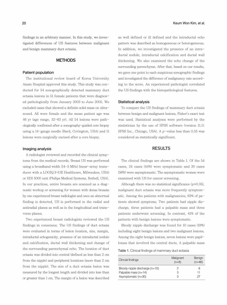
RESULTS
The clinical findings are shown in Table 1. Of the 54
cases, 24 cases (44%) were symptomatic and 30 cases
(56%) were asymptomatic. The asymptomatic women were
examined with US for cancer screening.
Although there was no statistical significance (p>0.05),
malignant duct ectasia was more frequently symptom-
atic. Among the patients with malignancies, 63% of pa-
tients showed symptoms. Two patients had nipple dis-
charge, three patients had a palpable mass and three
patients underwent screening. In contrast, 41% of the
patients with benign lesions were symptomatic.
Bloody nipple discharge was found for 10 cases (19%)
including eight benign lesions and two malignant lesions.
Among the eight benign lesions, seven lesions were papil-
lomas that involved the central ducts. A palpable mass
Table 1. Clinical findings of mammary duct ectasia
Malignant(n=8)
Benign (n=46)
Clincial findings
Bloody nipple discharge (n=10) 2 8Palpable mass (n=14) 3 11Asymptomatic (n=30) 3 27

was seen for 14 cases (26%) that consisted of 11 benign
lesions and three malignant lesions.
Of the 54 lesions, 46 lesions (85%) were benign and
eight lesions (15%) were malignant based on the histology.
The pathological diagnoses of 46 benign lesions included
an inflammatory change (n=7), ductal epithelial hyper-
plasia (n=7), fibrocystic change (n=18), intraductal papil-
loma (n=11), atypical ductal hyperplasia (n=2) and scle-
rosing adenosis (n=1). The malignant lesions included
ductal carcinoma in situ (DCIS) (n=6), infiltrating ductal
carcinoma (n=1) and mucinous carcinoma (n=1) (Table 2).
US findings of mammary duct ectasia are summarized
in Table 3. Of the 46 benign lesions, 74% (34/46) of the
lesions developed from the central ducts (Figures 1, 2).
In contrast, 88% (7/8) of the malignant lesions involved
the peripheral ducts (p<0.05) (Figures 3, 5).
The mean sizes of the benign and malignant lesions
were 1.5 cm and 1.8 cm, respectively. Of 46 benign lesions,
26 lesions (57%) were greater than 1 cm in length. Of
eight malignant lesions, seven lesions (88%) were greater
than 1 cm in length. This difference was not significant
Sonographic Findings of Mammary Duct Ectasia: Differentiation Malignancy from Benign Disease 21
Table 2. Pathologic diagnosis of mammary duct ectasia
Frequency (%)Pathology
Benign (n=46)Duct ectasia with inflammatory change 7 (12.9)Ductal epithelial hyperplasia 7 (12.9)Fibrocystic change 18 (33.3)Papilloma 11 (20.4)ADH 2 (3.7)Sclerosing adenosis 1 (1.85)
Malignancy (n=8)DCIS 6 (11.1)Infiltrating ductal carcinoma 1 (1.85)Mucinous carcinoma 1 (1.85)
ADH=atypical ductal hyperplasia; DCIS=ductal carcinoma in situ.
Figure 1. A 44-yr-old-woman without symptom and moderate to florid ductal epithelial hyperplasia. (A) Transverse (left) and longitudinal(right) scans of US reveal a well defined duct ectasia with intraductal homogeneous echogenecity (arrows) in the subareolar area ofthe left breast. An echogenic intraductal nodule (arrowhead) is shown with no evidence of intraductal calcification. (B) A photomicrographdemonstrates protruding ductal epithelial hyperplasia (arrow) and the hyperplastic epithelial gland, with moderate to florid ductal epithelialhyperplasia (arrowhead) (H&E stain, ×200).
A B
Figure 2. A 49-yr-old woman with left bloody nipple dischargeand intraductal papilloma and ductal epithelial hyperplasia. (A)Radial (left) and antiradial scans (right) of the left central ductdemonstrate an isoechoic intraductal nodule (arrows) within welldefined duct ectasia. The intraductal echogenicity is homoge-neous with no abnormal ductal wall thickening. (B) A papillaryprojected papilloma (arrow) is well depicted on a histologicalexamination (H&E stain, ×200).
A
B

statistically (p>0.05).
Heterogeneous intraductal echo was more frequently
22 Keum Won Kim, et al.
Figure 3. A 34-yr-old woman with a palpable lump in the right breast and comedo type DCIS. (A) Radial (left) and antiradial scans (right)of US show ill defined duct ectasia filled with intraductal heterogeneous echogenecities and intraductal nodules in the peripheral portion(5 cm apart from the nipple) of the right breast. There is an associated ductal wall thickening (arrows). (B) A photomicrograph demonstratestumor cells that invade the epithelium and basement membrane (arrows) as well as central necrosis (H&E stain, ×200).
A B
Figure 4. A 41-yr-old-woman with bloody nipple discharge fromthe left breast and cribriform DCIS. (A) Radial (left) and transverse(right) scans of US depict ill defined central duct ectasia withintraductal heterogeneous echogenicities. Associated intraductalnodules and calcifications (arrows) are well seen and irregularductal wall thickening is prominent (arrowheads). (B) On a his-tological examination, tumor cells invade the ductal epitheliumand basement membrane (arrows) (H&E stain, ×200).
Figure 5. A 52-yr-old-woman with a palpable lump in the rightbreast and a mucinous carcinoma with infiltrating ductal com-ponents. (A) On US, several aggregated cysts with ill-definedduct ectasia are noted in the peripheral ducts (5 cm apart fromthe nipple) of the right breast. Intraductal heterogeneous hypo-echogenicities with ductal wall thickenings (arrows) and a sur-rounding hypoechoic parenchymal change (arrowheads) arealso demonstrated. (B) A photomicrograph demonstrates floatingtumor cells (arrow) within the mucin pool (arrowheads) and asso-ciated infiltrating tumor cells (double arrows) (H&E stain, ×100).
A
A
BB

seen for malignant lesions (88%) (Figures 3-5) as com-
pared to benign lesions (63%). The presence of an intra-
ductal nodule and calcification were found in 12% and
50% of malignant lesions, respectively (Figures 3, 4), and
the presence of an intraductal nodule and calcification
were found in 52% and 28% of benign lesions, respec-
tively (Figures 1, 2). However, there was no significant
difference in the lesion size, the pattern of intraductal
echo and the presence of an intraductal nodule or cal-
cification between benign and malignant duct ectasia
lesions (p>0.05) (Table 3).
In terms of lesion margin, 88% (7/8) of malignant lesions
demonstrated an ill-defined margin (Figures 3-5), but
85% (39/46) of benign lesions had a well-defined margin
(Figures 1, 2) (p<0.05). Ductal wall thickening was seen
for 75% (6/8) of malignant lesions (Figures 3-5), and for
13% (6/46) of benign lesions (p<0.05). Two of six benign
lesions that showed ductal wall thickening were atypical
ductal hyperplasia. Hypoechoic changes at the surround-
ing breast tissue were seen for 38% (3/8) of malignant
lesions (Figure 5) and 4% (2/46) of benign lesions (p<0.05).
Table 4 presents the difference of malignancy rate by
score. Based on our results, suspicious sonographic find-
ings were peripheral location, ill defined margin, presence
of ductal wall thickening and surrounding hypoechoic
parenchymal changes. From this results, the malignancy
rate of score 0 and 1 was 0%, of score 2 was 25%, and of
score 3 and 4 was 100% (p<0.05).
DISCUSSION
Mammary duct ectasia is defined as a dilated duct larger
than 2 mm in diameter or a dilated ampullary portion
larger than 3 mm in diameter. Duct ectasia affects the
major ducts in the subareolar region, but sometimes the
smaller segmental ducts can be involved. Thick, unre-
sorbed secretions and cellular debris may fill the dis-
tended ducts. Periductal fibrosis is found in association
with an inflammatory infiltrates.(6) Based on our results,
in contrast to benign lesions, most malignant duct ectasia
lesions involved the peripheral ducts and the margin of
duct ectasia lesions was ill-defined (p<0.05).
The cause of duct ectasia is not clear. Duct dilatation
may be secondary to periductal inflammation where the
ducts become patulous and the ducts are filled with unre-
sorbed material and cellular debris.(6)
Sonographic Findings of Mammary Duct Ectasia: Differentiation Malignancy from Benign Disease 23
Table 4. Malignancy rate by score
Malignant(n=8)
Benign(n=46)
Malignantrate
p-value*Score
0 0 28 0% <0.051 0 9 0%2 3 9 25%3 3 0 100%4 2 0 100%
Table 3. Comparison of US findings in duct ectasia betweenmalignant and benign disease
US finding
Location 0.017 0.05 (0.00-0.49)Central 1 34Peripheral 7 12
Size 0.13 0.19 (0.01-1.75)Less than 1 cm 1 20Larger than 1 cm 7 26
Margin 0.0001 0.03 (0.00-0.27)Well defined 1 39Ill defined 7 7
Intraductal echo 0.2446 0.24 (0.01-2.32)Homogeneous 1 17Heterogeneous 7 29
Intraductal nodule 0.056 0.13 (0.01-1.23)Present 1 24Absent 7 22
Calcification 0.243 2.54 (0.44-14.81)Present 4 13Absent 4 33
Ductal wall thickening 0.00079 20.0 (2.63-192.69)Present 6 6Absent 2 40
Surrounding hypoe- 0.01936 13.2 (1.31-159.79)chogenecityPresent 3 2Absent 5 44
Malig-nant(n=8)
Benign(n=46)
p-value Odds ratio(95% CI*)
US=ultrasonographic; CI=confidence interval.*Fisher exact test.
Location (central=0, periphery=1); Margin (well defined=0, ill defined=1); ductal wall thickening (absent=0, present=1); Surrounding hypoe-chogenecity (absent=0, present=1).Score=sum of the points showing suspicious sonographic findings.*Chi-square test.

Reported diagnoses of pathological nipple discharge
include intraductal papilloma (33-48%), fibrocystic dis-
ease (14-33%), papillomatosis (14-28%), mammary duct
ectasia (2-11%) and a breast malignancy (8-15%).(7-10)
In our study, 15% of mammary duct ectasia lesions were
was malignant and three-quarters of the lesions were
low grade DCIS. Cho et al.(11) reported that 17% of non-
calcified DCIS lesions detected on US were manifested
as pure ductal changes without a solid mass and all lesions
were designated as group 1 based on the Van Nuys clas-
sification. The recent use of high resolution US can help
to detect subtle findings of non-calcified DCIS, especially
lesions that manifest as a pure ductal change.
The radiological findings of mammary duct ectasia
based on mammography and galactography have been
well described in many reports(4-6) but US features have
not been investigated to the best of our knowledge.
On mammography,(6)duct ectasia appears as a tubular,
serpinginous structure converging on the nipple at the
subareolar region in the fatty breast. However, duct ecta-
sia is not visible since the lesions are obscured by the
dense breast parenchyma.
When a serous or bloody nipple discharge is present,
ductography can be easily performed for the evaluation
of the intraductal pathology. Several reports have des-
cribed the ductographic findings according to the differ-
ent pathological diagnoses.(12) Diner(12) showed that the
ducts of fibrocystic disease may be irregular in diameter
and may be displaced by cysts. lntraductal papilloma is
depicted as a localized rounded or lobulated filling defect
within the dilated major ducts in the subareolar region.
For secretory disease, the ducts are markedly dilated,
somewhat tortuous and may contain irregular filling
defects produced by secretions. If the ducts are irregu-
lar in outline, encased and straightened, distorted or
obstructed, the presence of an invasive carcinoma should
be considered.(12)
A study by Hou et al.(4) demonstrated the galacto-
graphic findings of malignant tumors. Malignant duct
ectasia is typically depicted as an irregular intraductal
filling defect and ductal stenosis that affects the periph-
eral ducts. Cardenosa et al.(5) showed that a malignant
intraductal nodule, pathologically proven DCIS or papil-
loid hyperplasia appear as irregular filling defects on
galactography and benign lesions such as an intraductal
papilloma are seen as a round well circumscribed filling
defect involving the central ducts. Benign intraductal
nodules are seen as a round well circumscribed intra-
ductal filling defect, and a malignant intraductal nodule
is more common in a peripheral duct and shows an irreg-
ular filling defect with the use of galactography.
When nipple discharge is vague or originates from
multiple openings, successful ductography is impossible
and the evaluation of intraductal pathology is difficult.
In such cases, with US it is possible to visualize mam-
mary duct ectasia distinctively regardless of nipple dis-
charge and to evaluate the location, size and intraductal
features such as the presence of a nodule or calcification
and the echo-pattern. US is also able to evaluate a sur-
rounding parenchymal echo change. Furthermore, US
has an advantage of being a non-invasive procedure as
compared to ductography. US should be performed in
the radial and antiradial planes as well as in the longi-
tudinal and transverse planes. Radial US is particularly
useful to depict the intraductal pathology and to eval-
uate the extent of ductal disease, whereas antiradial US
is more helpful to evaluate the surface characteristics of
a mass.(2,13)However, US features suggestive of malig-
nant duct ectasia have not yet been investigated.
Several histopathological conditions affect the intra-
ductal echotexture. Calcified DCIS lesions usually show
a mild hypoechoic, heterogeneous echotexture, probably
due to the presence of punctuate echogenic calcifica-
tions.(13) Similar US findings may also be seen for some
benign lesions such as sclerosing adenosis, atypical duc-
tal hyperplasia and a radial scar.(14) Our study demon-
strated heterogeneous intraductal hypoechogenicity in
88% (7/8) of malignant lesions and 63% (29/46) of benign
lesions. Intraductal echogenicity was due to intraductal
secretion, inflammatory cellular debris and intraductal
calcification based on histological findings. In one case,
intraductal heterogeneous hypoechogenicity was due to
deposition of fibrin and lymphocytes within dilated ducts.
However, in our study, the heterogeneity of intraductal
24 Keum Won Kim, et al.

Sonographic Findings of Mammary Duct Ectasia: Differentiation Malignancy from Benign Disease 25
echogenicity did not provide useful information to differ-
entiate malignant from benign duct ectasia.
In our study, malignant duct ectasia was not associ-
ated with a discrete intraductal nodule except for one
case. An intraductal nodule was commonly associated
with benign duct ectasia, with intraductal papilloma as
the most common finding.
Several studies have examined the clinical significance
of microcalcifications detected on US.(15-17) Soo et al.(17)
mentioned that suspicious microcalcifications were seen
infrequently on sonography (23%), but when detected
could be successfully biopsied with sonographic guidance
and more frequently were malignant lesions and repre-
sented invasive cancer as compared to lesions seen on
mammography alone. Likewise, in our study, 72% of
benign lesions showed no intraductal calcifications but
half of the malignant lesions contained intraductal cal-
cification, even though this difference was not signifi-
cant statistically. For cases that show suspicious sono-
graphic features of duct ectasia, the presence of intra-
ductal calcifications can be a helpful finding that can
increase the confidence of a radiologist for a diagnosis
of a malignancy.
From our results, the most striking feature of malig-
nant duct ectasia was an ill-defined margin with ductal
wall thickening. An ill-defined margin was seen for 88%
of malignant lesions and 15% of benign lesions. Ductal
wall thickening was seen only in 13% of benign lesions
but in 75% of malignant lesions. Histological examina-
tions revealed accumulation of tumor cells within ducts
without evidence of invasion through the basement mem-
brane for malignant lesions.
Pure clusters of microcysts without discrete solid com-
ponents can be considered probably benign and should
be subjected to follow-up. Such lesions are often due to
apocrine metaplasia, though a fibrocystic change without
apocrine metaplasia can have a similar appearance.(18)
Berg(19) reported 66 lesions prospectively characterized
as clustered microcysts with no malignancies. Our study
showed a case that was depicted as several aggregated
cysts with ill-defined duct ectasia and a surrounding
hypoechoic parenchymal change. We considered the lesion
as a probably benign lesion, but the lesion was diagnosed
as a mucinous carcinoma with infiltrating ductal carci-
noma component based on the histopathology. From our
results, 38% (3/8) of malignant duct ectasia lesions and
only 4% (2/46) of benign lesions demonstrated a hypo-
echoic change of the surrounding breast tissue. One malig-
nant lesion was due to direct tumor cell infiltration but
two cases were proliferative lesions without atypia and
fibrocystic changes based on the histology.
CONCLUSION
In conclusion, mammary duct ectasia without a solid
mass frequently depicted on breast US is a commonly
benign condition, especially a fibrocystic change. In con-
trast, if there is peripheral, ill-defined duct ectasia with
ductal wall thickening and associated hypoechogencity
of the surrounding breast tissue, the possibility of a
malignancy such as carcinoma in situ should be con-
sidered and radiologists should not hesitate to recommend
a prompt biopsy.
REFERENCES
1. Bassett LW. Imaging of breast masses. Radiol Clin North Am 2000;38:669-91.
2. Stavros AT, Thickman D, Rapp CL, Dennis MA, Parker SH, SisneyGA. Solid breast nodules: use of sonography to distinguish betweenbenign and malignant lesions. Radiology 1995;196:123-34.
3. Mendelson EB, Berg WA, Merritt CR. Toward a standardized breastultrasound lexicon, BI-RADS: ultrasound. Semin Roentgenol 2001;36:217-25.
4. Hou MF, Huang TJ, Liu GC. The diagnostic value of galactographyin patients with nipple discharge. Clin Imaging 2001;25:75-81.
5. Cardenosa G, Eklund GW. Benign papillary neoplasms of the breast:mammographic findings. Radiology 1991;181:751-5.
6. Kopans DB. Histologic, pathologic, and image correlation. In: KopansDB, editor. Breast Imaging. 3rd ed. Philadelphia: Lippincott Williams& Wilkins; 2007. p.783-888.
7. Leis HP Jr. Management of nipple discharge. World J Surg 1989;13:736-42.
8. Oh KK, Park YH, Ji H, Park CY. Values of contrast galactographyin nonlactating nipple discharge in Korean women. J Korean RadiolSoc 1988;24:782-94.
9. Taba@r L, Dean PB, Péntek Z. Galactography: the diagnostic procedureof choice for nipple discharge. Radiology 1983;149:31-8.
10. Cho N, Oh KK, Cho HY. Galactographic differentiation between

malignant and benign disease in patients with pathologic nipple dis-charge. J Korean Radiol Soc 2003;48:511-6.
11. Cho KR, Seo BK, Kim CH, Whang KW, Kim YH, Kim BH, et al.Non-calcified ductal carcinoma in situ: ultrasound and mammographicfindings correlated with histological findings. Yonsei Med J 2008;49:103-10.
12. Diner WC. Galactography: mammary duct contrast examination.Am J Roentgenol 1981;137:853-6.
13. Moon WK, Myung JS, Lee YJ, Park IA, Noh DY, Im JG. US ofductal carcinoma in situ. Radiographics 2002;22:269-80.
14. Stavros AT. Ultrasound of ductal carcinoma in situ. In: SilversteinM, editor. Ductal Carcinoma In Situ of the Breast. Baltimore: Williams& Wilkins; 1997. p.135-58.
15. Gufler H, Buitrago-Te@llez CH, Madjar H, Allmann KH, Uhl M, Rohr-
Reyes A. Ultrasound demonstration of mammographically detectedmicrocalcifications. Acta Radiol 2000;41:217-21.
16. Cheung YC, Wan YL, Chen SC, Lui KW, Ng SH, Yeow KM, et al.Sonographic evaluation of mammographically detected microcal-cifications without a mass prior to stereotactic core needle biopsy. JClin Ultrasound 2002;30:323-31.
17. Soo MS, Baker JA, Rosen EL. Sonographic detection and sonogra-phically guided biopsy of breast microcalcifications. AJR Am J Roent-genol 2003;180:941-8.
18. Warner JK, Kumar D, Berg WA. Apocrine metaplasia: mammo-graphic and sonographic appearances. AJR Am J Roentgenol 1998;170:1375-9.
19. Berg WA. Can clusters of microcysts appropriately be followed?Radiological Society of North America. 2002. abstract # E14-510.
26 Keum Won Kim, et al.