Embed Size (px)
Citation preview

BioMed Central
Journal of Brachial Plexus and Peripheral Nerve Injury
ss
Open AcceShort reportHyperglycemia alters enzyme activity and cell number in spinal sensory gangliaRichard A Zaruba1, Paul N Epstein2 and Patrick A Carr*1Address: 1Department of Anatomy and Cell Biology, School of Medicine and Health Sciences, University of North Dakota, Grand Forks, ND 58202, USA and 2Department of Pediatrics, University of Louisville, Louisville, KY 40202, USA
Email: Richard A Zaruba - [email protected]; Paul N Epstein - [email protected]; Patrick A Carr* - [email protected]
* Corresponding author
AbstractPeripheral sensory diabetic neuropathy is characterized by morphological, electrophysiological andneurochemical changes to a subpopulation of primary afferent neurons. Here, we utilized atransgenic mouse model of diabetes (OVE26) and age-matched controls to histologically examinethe effect of chronic hyperglycemia on the activity or abundance of the enzymes acid phosphatase,cytochrome oxidase and NADPH-diaphorase in primary sensory neuron perikarya and the dorsalhorn of the spinal cord. Quantitative densitometric characterization of enzyme reaction productrevealed significant differences between diabetic, compared to control, animals for all threeenzymes. Levels of acid phosphatase reaction product were found to be significantly reduced inboth small diameter primary sensory somata and the dorsal horn. Cytochrome oxidase activity wasfound to be significantly lower in small primary sensory somata while NADPH-diaphorase labelingwas found to be significantly higher in small primary sensory somata and significantly lower in thedorsal horn. In addition to these observed biochemical changes, ratiometric analysis of the numberof small versus large diameter primary sensory perikarya in diabetic and control animalsdemonstrated a quantifiable decrease in the number of small diameter cells in the spinal ganglia ofdiabetic mice. These results suggest that the OVE26 model of diabetes mellitus produces anidentifiable disturbance in specific metabolic pathways of select cells in the sensory nervous systemand that this dysfunction may reflect the progression of a demonstrated cell loss.
BackgroundDiabetic sensory neuropathies are a common, clinicallyobserved sequelae of hyperglycemia and are characterizedby a progressive degradation of primary afferent function[1,2]. Functional and structural evidence suggest an earlyand frequent involvement of small diameter primary sen-sory neurons leading to nociceptive abnormalities [2-4].In order to examine basic mechanisms underlying thisdisorder, we utilized the OVE26 transgenic mouse model
of diabetes mellitus [5,6] to examine the effects of long-standing hyperglycemia on enzyme histochemical indica-tors of sensory neuron metabolism and evaluate thepotential utility of this model for future studies of diabeticneuropathy. The OVE26 mouse line uses cell-specificoverexpression of calmodulin to destroy pancreatic β-cellsand the result is a viable diabetic mouse (>1 year survival)that displays both early-onset (<1 week after birth) andchronic elevated blood glucose (>500 mg/dl) and
Published: 25 April 2007
Journal of Brachial Plexus and Peripheral Nerve Injury 2007, 2:11 doi:10.1186/1749-7221-2-11
Received: 22 March 2007Accepted: 25 April 2007
This article is available from: http://www.JBPPNI.com/content/2/1/11
© 2007 Zaruba et al; licensee BioMed Central Ltd. This is an Open Access article distributed under the terms of the Creative Commons Attribution License (http://creativecommons.org/licenses/by/2.0), which permits unrestricted use, distribution, and reproduction in any medium, provided the original work is properly cited.
Page 1 of 6(page number not for citation purposes)

Journal of Brachial Plexus and Peripheral Nerve Injury 2007, 2:11 http://www.JBPPNI.com/content/2/1/11
decreased serum and pancreatic insulin (<50% of normal)[5,6]. Enzyme histochemical techniques demonstrated tobe sensitive to neuronal perturbation [7] were used toexamine the impact of long-standing hyperglycemia andhypoinsulinemia on the distribution and activity of lyso-somal acid β-glycerophosphatase (AP), cytochrome oxi-dase (CO), and NADPH-diaphorase (NADPH-d; acorrelate of nitric oxide synthase in aldehyde fixed tissue[8]) in both sensory ganglia and the spinal cord. It hasbeen previously demonstrated [7,11] that the metabolicstatus of sensory neurons, as reflected by the endogenousactivity of specific homeostatic enzymes, is sensitive toinjury and perturbation. Therefore, these enzymes wereselected as putatively reflective of mitochondrial function(CO), lysosomal or degradative activity (AP) and primarysensory neuron injury or repair (NADPH-diaphorase).
The OVE26 transgenic mouse line (characterized by theinsulin promoter-linked overexpression of calmodulin inpancreatic β-cells) used in this study displays a well-char-acterized chronic hyperglycemia and hypoinsulinemiawithin days after birth. [5,6]. Ten (five OVE26 transgenicand five age-matched, control FVB animals) aged (>365days old) mice were anesthetized with pentobarbital, per-fused with 4% paraformaldehyde and the lumbar spinalcord and sensory ganglia removed, sectioned and proc-essed for AP, CO or NADPH-d enzyme histochemistry aspreviously described [7,9-12]. Counts of primary sensorysomata were conducted on toluidine blue counterstainedsections of L5 spinal ganglia and quantified using previ-ously published methodologies [7,12].
Quantitative analysis of CO, AP and NADPH-d stainingwas undertaken on both the dorsal horn of the L5 seg-ment of the spinal cord and the large and small cells of L5sensory ganglion using previously described densitomet-ric analysis [7,12]. The entire mediolateral extent of lam-ina I to III was selected for staining intensitymeasurement. Statistical analyses (t-test, Mann-WhitneyRank Sum test, one way analysis of variance, Kruskal-Wal-lis analysis of variance on ranks, z-test of proportions)were conducted using SigmaStat (Jandel). Controls fordensitometric analysis consisted of: 1) simultaneous sec-tioning and mounting of diabetic and control tissue onthe same slide to ensure identical histological processing;2) statistical analysis to verify consistency of stainingbetween animals within control and experimental groups;3) correction for small fluctuations in tissue opacity/thick-ness by subtractive illumination whereby the densityvalue of white matter was subtracted from the immedi-ately adjacent ventral horn; and 4) manual adjustmentand calibration of the video camera parameters andmicroscope illumination and acquisition of all imagesusing identical settings. All experiments were conductedin accordance with the guidelines of our institutions and
the National Institutes of Health regarding the care anduse of animals for experimental procedures.
Prior to fixative perfusion, the phenotypic status ofOVE26 diabetic mice were confirmed by their characteris-tic small eyes caused by the GR19 gene in their transgenicconstruct [5]. All adult OVE26 mice maintained fed bloodglucose levels of at least 400 mg/dl. At the histologicallevel, a survey [13] of the ratio of small (50 and 500 μm2
area) to large (500 and 1950 μm2 area) primary sensorysomata in the fifth lumbar spinal ganglia revealed a signif-icant decrease in the proportion of small to large cells indiabetic (1.29:1 small:large perikarya) compared to con-trol (1.94:1 small:large perikarya) mice (P < 0.05 by z-testof proportions; 417 cells measured). Quantitative densit-ometric analysis of the abundance and distribution ofenzyme histochemical reaction product in dorsal rootganglia (DRG) revealed substantive differences betweendiabetic and control mice (720 cells were quantified forboth densitometry and cell size; 240 cells for eachenzyme). Small somata from the ganglia of diabetic miceexhibited lower levels of AP (13.4% decrease; P < 0.001)and CO (Fig 1A,B; 9% decrease; P < 0.001) reaction prod-uct and an increase in the density of the reaction productfor NADPH-d (Fig. 1C,D; 13.2% increase; P < 0.001) incomparison to control animals. No differences wereobserved in large diameter neurons from diabetic as com-pared to control animals.
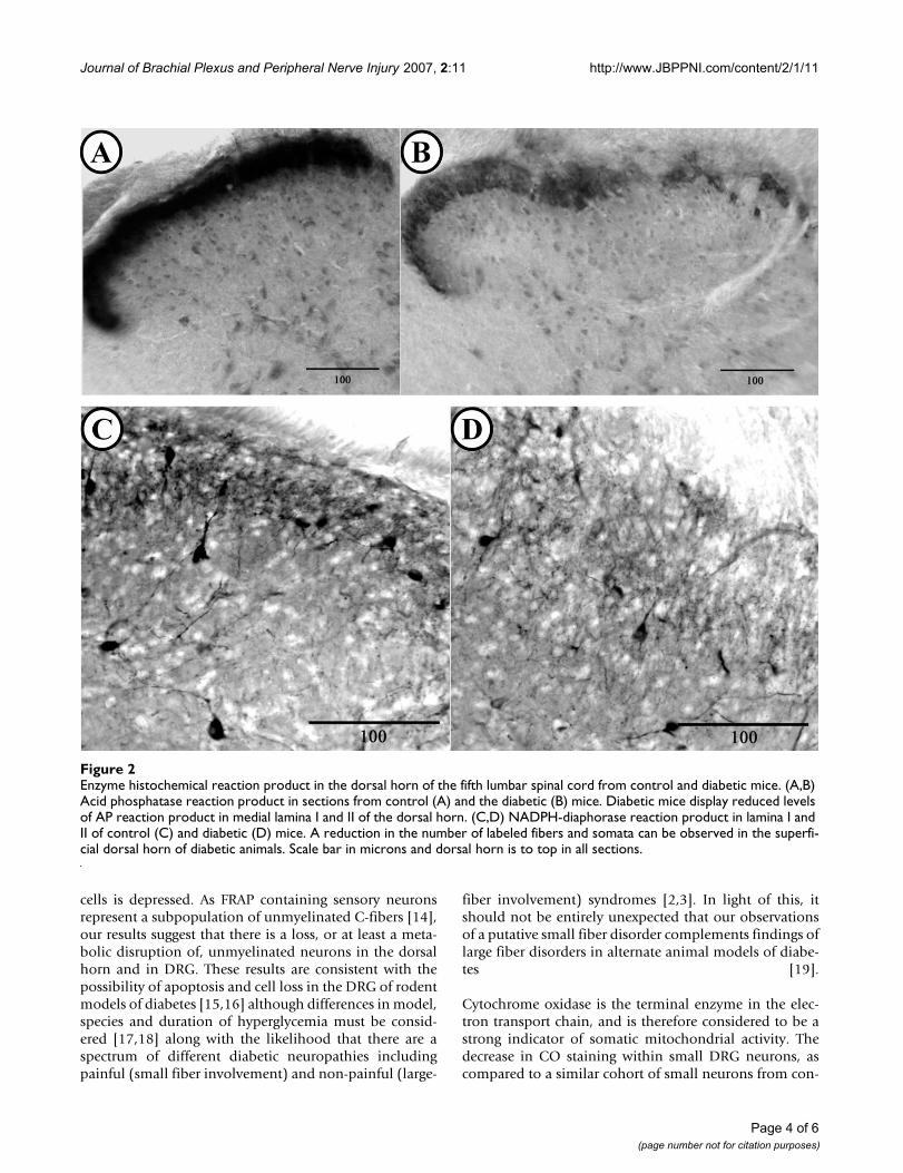
In the spinal cord, all observed differences were confinedto lamina I to III. Motoneuron somata in the ventral hornappeared both qualitatively and quantitatively similar indiabetic and control animals. In diabetic animals, therewas an observable loss of AP reaction product in lamina Iand II of the dorsal horn (Fig. 2A) as compared to controlmice (Fig. 2B). Similarly, these laminae appeared to havequalitatively fewer NADPH-d labeled fibers and neuronalsomata in diabetic (Fig. 2C) as compared to control ani-mals. (Fig. 2D). The decrease of both AP and NADPH-dlabeling was most profound in the medial portion of lam-ina I and II. Quantitative densitometric analysis sup-ported the qualitative observations and revealedsignificantly reduced levels of AP (P = 0.026; 27 sectionsquantified) and NADPH-d (P < 0.001; 27 sections quanti-fied) reaction product in lamina I and II of the dorsal hornof control and diabetic mice (Table 1). No significant dif-ferences were observed in qualitative staining appearanceor intensity of CO reaction product labeling in the dorsalhorn of diabetic, compared to non-diabetic animals (31sections quantified).
Here we have demonstrated that chronic hyperglycemiahas an impact on both the survival and metabolic profileof primary sensory neurons. The observed decrease in theratio of small to large diameter primary sensory somata in
Page 2 of 6(page number not for citation purposes)

Journal of Brachial Plexus and Peripheral Nerve Injury 2007, 2:11 http://www.JBPPNI.com/content/2/1/11
diabetic animals most likely represents a loss of unmyeli-nated or small myelinated primary sensory neuronsalthough a relative increase in the number of large myeli-nated neurons, however, unlikely, cannot be discounted.Nonetheless, the former interpretation is supported by the
observed decrease in AP labeling in the dorsal horn of thespinal cord. The observed decrease in AP intensity in thesurviving small neuronal somata from the DRG of theOVE26 animals, as compared to small neurons from con-trol DRG, suggests that acid phosphatase activity in those
Table 1: Quantitative histochemical reaction product in the dorsal horn of control and diabetic mice.
Enzyme Control Mean ± S.D. Diabetic Mean ± S.D t-test significance level
AP 6.058 ± 0.254 5.949 ± 0.210 P = 0.026*CO 3.997 ± 0.172 3.989 ± 0.137 P = 0.800NADPH-d 1.320 ± 0.354 0.733 ± 0.228 P < 0.001*
* indicates significant difference. Standard deviation (S.D.). Following densitometric measurements, the numbers were transformed to a 0 to 10 scale for clarity of presentation and comparison. Optical density values fall along a range of 0 representing minimal staining intensity (no staining) and 10 representing extremely dense staining (black).
Enzyme histochemical reaction product in the fifth lumbar dorsal root ganglion from control and diabetic miceFigure 1Enzyme histochemical reaction product in the fifth lumbar dorsal root ganglion from control and diabetic mice. (A,B) Cyto-chrome oxidase reaction product in sections from control (A) and the diabetic (B) mice. Diabetic mice display reduced levels of CO reaction product compared to control. Quantitative analysis revealed a decrease in CO reaction product density in small neuronal somata. (C,D) NADPH-diaphorase reaction product in sections from control (C) and diabetic (D) mice. Dia-betic mice display an increase in NADPH-diaphorase reaction product density compared to control. Scale bar in microns.
Page 3 of 6(page number not for citation purposes)
Journal of Brachial Plexus and Peripheral Nerve Injury 2007, 2:11 http://www.JBPPNI.com/content/2/1/11
cells is depressed. As FRAP containing sensory neuronsrepresent a subpopulation of unmyelinated C-fibers [14],our results suggest that there is a loss, or at least a meta-bolic disruption of, unmyelinated neurons in the dorsalhorn and in DRG. These results are consistent with thepossibility of apoptosis and cell loss in the DRG of rodentmodels of diabetes [15,16] although differences in model,species and duration of hyperglycemia must be consid-ered [17,18] along with the likelihood that there are aspectrum of different diabetic neuropathies includingpainful (small fiber involvement) and non-painful (large-
fiber involvement) syndromes [2,3]. In light of this, itshould not be entirely unexpected that our observationsof a putative small fiber disorder complements findings oflarge fiber disorders in alternate animal models of diabe-tes [19].
Cytochrome oxidase is the terminal enzyme in the elec-tron transport chain, and is therefore considered to be astrong indicator of somatic mitochondrial activity. Thedecrease in CO staining within small DRG neurons, ascompared to a similar cohort of small neurons from con-
Enzyme histochemical reaction product in the dorsal horn of the fifth lumbar spinal cord from control and diabetic miceFigure 2Enzyme histochemical reaction product in the dorsal horn of the fifth lumbar spinal cord from control and diabetic mice. (A,B) Acid phosphatase reaction product in sections from control (A) and the diabetic (B) mice. Diabetic mice display reduced levels of AP reaction product in medial lamina I and II of the dorsal horn. (C,D) NADPH-diaphorase reaction product in lamina I and II of control (C) and diabetic (D) mice. A reduction in the number of labeled fibers and somata can be observed in the superfi-cial dorsal horn of diabetic animals. Scale bar in microns and dorsal horn is to top in all sections.
Page 4 of 6(page number not for citation purposes)

Journal of Brachial Plexus and Peripheral Nerve Injury 2007, 2:11 http://www.JBPPNI.com/content/2/1/11
trol animal ganglia, suggests a disruption of oxidativemetabolism which corresponds well with results fromother animal models of diabetes that demonstrate dimin-ished CO activity or disruptions in mitochondrial mor-phology or function [16,20-22]. Alternatively, ourobserved decrease in CO staining may reflect a simpledecrease in mitochondrial number, as DRG neuronsexposed to high glucose in vitro exposure contain fewermitochondria [23]. The lack of an observed change in COactivity in the dorsal horn is not unexpected as both phys-ical (axotomy) and functional (tetrodotoxin) disconnec-tion have previously been shown to leave CO activity inthe dorsal horn unaltered [7].
In DRG and the dorsal horn of the spinal cord, NADPH-dactivity levels have been previously shown to be respon-sive to peripheral neuronal injury or attenuation of elec-trical activity [7]. Our results suggest that in addition tothe pathological state that led to our observed loss of sen-sory neurons (and diminished NADPH-d labeling in thedorsal horn), there is a ongoing perturbation resulting inincreased NADPH-d labeling in small DRG neurons fromhyperglycemic animals as compared to the primary sen-sory somata from normoglycemic animals. As utilizedhere, NADPH-d enzyme histochemical reaction productrepresents nitric oxide synthase activity [8]. The elevatedganglionic NADPH-diaphorase and diminished CO labe-ling in the DRG of hyperglycemic mice is consistent withpreviously proposed inhibition of CO activity [24] by theproduct of nitric oxide synthase, nitric oxide. Althoughthe reported statistically significant changes may appear tobe quantitatively modest, these percent changes representgroup averages. Qualitatively and quantitatively, thechanges are more pronounced in some animals and tis-sues sections, and less obvious in others. This is not unex-pected as chronic diseases processes impact individualswith profound variability in both severity and temporalprogress.
Our results suggest that the OVE26 model of chronichyperglycemia does alter the overall neurochemical pro-file of the sensory nervous system through cell loss and/oraltered enzyme activity and that this pathology seems tospecifically impact unmyelinated and/or small myeli-nated primary sensory neurons.
Declaration of competing interestsThe author(s) declare that they have no competing inter-ests.
Authors' contributionsRZ completed this work as part of his doctoral dissertationand was involved in the writing of this manuscript andcontributed both intellectually and practically to the con-tent. PE created, characterized and supplied the transgenic
mice and was also involved in the writing of this manu-script and contributed both intellectually and practicallyto the content. PC provided the lab, supervision, and sup-port for this work, exclusive of that associated with gener-ation and characterization of the mouse model. PC wasalso involved in the design and coordination of this studyand participated in the writing of this manuscript andcontributed both intellectually and practically to the con-tent. All authors read and approved the final manuscript.
AcknowledgementsThis work was supported by ND EPSCoR and UNDSOMH.
References1. Vinik AI, Park TS, Stansberry KB, Pittenger GL: Diabetic neuropa-
thies. Diabetologia 2000, 43:957-973.2. Sinnreich M, Taylor B, Dyck PJB: Diabetic neuropathies. The Neu-
rologist 2005, 11:63-79.3. Berti-Mattera LN, Gariepy CE, Burke RM, Hall AK: Reduced
expression of endothelin B receptors and mechanical hyper-algesia in experimental chronic diabetes. Exp Neurol 2006,201:399-406.
4. Walwyn WM, Matsuka Y, Arai D, Bloom DC, Lam H, Tran C, Spigel-man I, Maidment NT: HSV-1-mediated NGF delivery delaysnociceptive deficits in a genetic model of diabetic neuropa-thy. Exp Neurol 2006, 198:260-270.
5. Epstein PN, Overbeek PA, Means AR: Calmodulin-induced early-onset diabetes in transgenic mice. Cell 1989, 58:1067-1073.
6. Epstein PN, Ribar TJ, Decker GL, Yaney G, Means AR: Elevatedbeta-cell calmodulin produces a unique insulin secretorydefect in transgenic mice. Endocrinology 1992, 130:1387-1393.
7. Carr PA, Haftel V, Alvarez FJ, Cope TC, Fyffe RE: Effect of sciaticnerve transection or TTX application on enzyme activity inrat spinal cord. Neuroreport 1998, 9:357-361.
8. Dawson TM, Snyder SH: Gases as biological messengers: nitricoxide and carbon monoxide in the brain. J Neurosci 1994,14:5147-5159.
9. Carr PA, Huang A, Noga BR, Jordan LM: Cytochemical character-istics of cat spinal neurons activated during fictive locomo-tion. Brain Res Bull 1995, 37:213-218.
10. Carr PA, Liu M, Zaruba RA: Enzyme histochemical profile ofimmunohistochemically identified Renshaw cells in rat lum-bar spinal cord. Brain Res Bull 2001, 54:669-674.
11. Carr PA, Yamamoto T, Nagy JI: Calcitonin gene-related peptidein primary afferent neurons of rat: co-existence with fluo-ride-resistant acid phosphatase and depletion by neonatalcapsaicin. Neuroscience 1990, 36:751-760.
12. Carr PA, Yamamoto T, Staines WA, Whittaker ME, Nagy JI: Quanti-tative histochemical analysis of cytochrome oxidase in ratdorsal root ganglia and its co-localization with carbonicanhydrase. Neuroscience 1989, 33:351-362.
13. Lawson SN: The postnatal development of large light andsmall dark neurons in mouse dorsal root ganglia: a statisticalanalysis of cell numbers and size. J Neurocytol 1979, 8:275-294.
14. Ribeiro-Da-Silva A, Castro-Lopes JM, Coimbra A: Distribution ofglomeruli with fluoride-resistant acid phosphatase (FRAP)-containing terminals in the substantia gelatinosa of the rat.Brain Res 1986, 377:323-329.
15. Tolkovsky A: Apoptosis in diabetic neuropathy. Int Rev Neurobiol2002, 50:145-159.
16. Schmeichel AM, Schmelzer JD, Low PA: Oxidative injury andapoptosis of dorsal root ganglion neurons in chronic experi-mental diabetic neuropathy. Diabetes 2003, 52:165-171.
17. Sasaki K, Chancellor MB, Phelan MW, Yokoyama T, Fraser MO, SekiS, Kubo K, Kumon H, DeGroat WC, Yoshimura N: Diabeticcystopathy correlates with a long-term decrease in nervegrowth factor levels in the bladder and lumbosacral dorsalroot ganglia. J Urol 168:1259-1264.
18. Sango K, Horie H, Saito H, Ajiki K, Tokashiki A, Takeshita K, Ishigat-subo Y, Kawano H, Ishikawa Y: Diabetes is not a potent inducerof neuronal cell death in mouse sensory ganglia, but it
Page 5 of 6(page number not for citation purposes)

Journal of Brachial Plexus and Peripheral Nerve Injury 2007, 2:11 http://www.JBPPNI.com/content/2/1/11
Publish with BioMed Central and every scientist can read your work free of charge
"BioMed Central will be the most significant development for disseminating the results of biomedical research in our lifetime."
Sir Paul Nurse, Cancer Research UK
Your research papers will be:
available free of charge to the entire biomedical community
peer reviewed and published immediately upon acceptance
cited in PubMed and archived on PubMed Central
yours — you keep the copyright
Submit your manuscript here:http://www.biomedcentral.com/info/publishing_adv.asp
BioMedcentral
enhances neurite regeneration in vitro. Life Sci 2002,71:2351-2368.
19. Kishi M, Tanabe J, Schmelzer JD, Low PA: Morphometry of dorsalroot ganglion in chronic experimental diabetic neuropathy.Diabetes 51:819-824.
20. Russell JW, Sullivan KA, Windebank AJ, Herrmann DN, Feldman EL:Neurons undergo apoptosis in animal and cell culture mod-els of diabetes. Neurobiol Dis 1999, 6:347-363.
21. Al-Abdulla NA, Martin LJ: Apoptosis of retrogradely degenerat-ing neurons occurs in association with the accumulation ofperikaryal mitochondria and oxidative damage to thenucleus. Am J Pathol 1998, 153:447-456.
22. Vincent AM, Brownlee M, Russell JW: Oxidative stress and pro-grammed cell death in diabetic neuropathy. Ann N Y Acad Sci2002, 959:368-383.
23. Leinninger GM, Backus C, Sastry AM, Yi Y-B, Wang C-W, Feldman EL:Mitochondria in DRG neurons undergo hyperglycemic medi-ated injury through Bim, Bax and the fission protein Drp1.NeurobiolDis 2006, 23:11-22.
24. Sharpe MA, Cooper CE: Interaction of peroxynitrite with mito-chondrial cytochrome oxidase. Catalytic production of nitricoxide and irreversible inhibition of enzyme activity. J BiolChem 1998, 273:30961-30972.
Page 6 of 6(page number not for citation purposes)