Embed Size (px)
Citation preview

Journal of Blood Group Serology and Molecular Genetics
Vo lu m e 30, Nu m b e r 1, 2014


39 Inst ruct I o ns fo r Au t h o rs
34 Adv ert Isem en ts
ImmunohematologyJournal of Blood Group Serology and Molecular Genetics
Volume 30, Number 1, 2014
CONTENTS
14 cAse rep o rt
Evans syndrome in a pediatric liver transplant recipient with an autoantibody with apparent specificity for the KEL4 (Kpb) antigenS.A. Koepsell, K. Burright-Hittner, and J.D. Landmark
24 rep o rt
Demonstration of IgG subclass (IgG1 and IgG3) in patients with positive direct antiglobulin testsA. Singh, A. Solanki, and R. Chaudhary
1 rep o rt
Indirect antiglobulin test-crossmatch using low-ionic-strength saline–albumin enhancement medium and reduced incubation time: effectiveness in the detection of most clinically significant antibodies and impact on blood utilizationC.L. Dinardo, S.L. Bonifácio, and A. Mendrone, Jr.
6 re v I e w
Raph blood group systemM. Hayes
11 rep o rt
I-int phenotype among three individuals of a Parsi community from Mumbai, IndiaS.R. Joshi
18 re v I e w
JMH blood group system: a reviewS.T. Johnson
30 An n ou n cem en ts
28 In mem o rI A m
George Garratty, 1935–2014Patricia A. Arndt and Regina M. Leger

Immunohematology is published quarterly (March, June, September, and December) by the American Red Cross, National Headquarters, Washington, DC 20006.
Immunohematology is indexed and included in Index Medicus and MEDLINE on the MEDLARS system. The contents are also cited in the EBASE/Excerpta Medica and Elsevier
BIOBASE/Current Awareness in Biological Sciences (CABS) databases.
The subscription price is $50 for individual, $100 for institution (U.S.) and $60 for individual, $100 for institution (foreign) per year.
Subscriptions, Change of Address, and Extra Copies:
Immunohematology, P.O. Box 40325 Philadelphia, PA 19106
Or call (215) 451-4902
Web site: www.redcross.org/about-us/publications/immunohematology
Copyright 2014 by The American National Red Cross ISSN 0894-203X
ed I to r- I n-ch I ef
Sandra Nance, MS, MT(ASCP)SBBPhiladelphia, Pennsylvania
mA n Ag I n g ed I to r
Cynthia Flickinger, MT(ASCP)SBBPhiladelphia, Pennsylvania
tec h n I cA l ed I to rs
Christine Lomas-Francis, MScNew York City, New York
Joyce Poole, FIBMSBristol, United Kingdom
Dawn M. Rumsey, ART(CSMLT)Norcross, Georgia
sen I o r med I cA l ed I to r
Ralph R. Vassallo, MDPhiladelphia, Pennsylvania
As s o c I At e med I cA l ed I to rs
P. Dayand Borge, MDBaltimore, Maryland
David Moolten, MDPhiladelphia, Pennsylvania
mo lec u l A r ed I to r
Margaret A. KellerPhiladelphia, Pennsylvania
ed I to r I A l As s IstA n t
Sheetal Patel
pro d u ct I o n As s IstA n t
Marge Manigly
co p y ed I to r
Mary L. Tod
pro o f r e A d er
Lucy OppenheimWendy Martin-Shuma
elect ro n I c pu b l Is h er
Paul Duquette
ed I to r I A l boA r d
Patricia Arndt, MT(ASCP)SBBPomona, California
James P. AuBuchon, MDSeattle, Washington
Barbara J. Bryant, MDMilwaukee, Wisconsin
Lilian Castilho, PhDCampinas, Brazil
Martha R. Combs, MT(ASCP)SBBDurham, North Carolina
Geoffrey Daniels, PhDBristol, United Kingdom
Anne F. Eder, MDWashington, District of Columbia
Brenda J. Grossman, MDSt. Louis, Missouri
Christine Lomas-Francis, MScNew York City, New York
Geralyn M. Meny, MDSan Antonio, Texas
Paul M. Ness, MDBaltimore, Maryland
Thierry Peyrard, PharmD, PhDParis, France
Mark Popovsky, MDBraintree, Massachusetts
S. Gerald Sandler, MDWashington, District of Columbia
Jill R. Storry, PhD Lund, Sweden
David F. Stroncek, MDBethesda, Maryland
Nicole ThorntonBristol, United Kingdom
em er I t us ed I to rs
Delores Mallory, MT(ASCP) SBBSupply, North Carolina
Marion E. Reid, PhD, FIBMSNew York City, New York
on ou r cov er
Henri Rousseau, or Le Douanier (the customs officer), as he was nicknamed for his primary occupation as a toll collector, failed to impress either the art world or the public in his lifetime (1844–1910). Self-taught, he adhered to a simple, childlike style at odds with his contemporaries. Although he received little recognition from and was even ridiculed by critics, he became the darling of artists and writers such as Picasso, Brancusi, and Apollinaire and is now regarded as a genius. The Tabby, or Le Chat Tigre, exemplifies both the primitive and yet subtly dreamlike character of his work. The JMH blood group discussed by S.T. Johnson in this issue includes “The Cat” among its nicknames.
David Moolten, MD

IMMUNOHEMATOLOGY, Volume 30, Number 1, 2014 1
Indirect antiglobulin test-crossmatch using low-ionic-strength saline–albumin enhancement medium and reduced incubation time: effectiveness in the detection of most clinically significant antibodies and impact on blood utilizationC.L. Dinardo, S.L. Bonifácio, and A. Mendrone, Jr.
Indirect antiglobulin test-crossmatch (IAT-XM) using enhancement media such as low-ionic-strength saline (LISS) and polyethylene glycol (PEG) usually requires 15 minutes of incubation. These methods are necessary when testing samples from blood recipients who have a higher risk of alloimmunization. In emergency situations, IAT-XM can be time-consuming and can influence presurgery routine, resulting in more red blood cell (RBC) units being tested and stored to avoid the transfusion of uncrossmatched ones. The objective of this study was to evaluate the performance of a LISS-albumin enhancer to intensify antigen-antibody reaction after 5 minutes of 37°C incubation and compare this performance with that of other enhancers, gel, and conventional tube testing. Second, the study evaluated the impact of this method’s implementation in the C:T ratio (crossmatched to transfused RBC units) of a transfusion laboratory. Ninety serum samples containing alloantibodies of potential clinical significance were tested against phenotyped RBCs using four different methods: (1) tube with LISS-albumin enhancer (5 minutes of incubation), (2) tube with LISS-albumin and PEG (15 minutes of incubation), (3) gel, and (4) conventional tube method (60 minutes of incubation). In parallel, the study compared the C:T ratio of a tertiary-hospital transfusion laboratory in two different periods: 3 months before and 3 months after the implementation of the 5-minute IAT-XM protocol. The use of LISS-albumin with 5 minutes of incubation exhibited the same performance as LISS-albumin, PEG, and gel with 15 minutes of incubation. Conventional tube method results were equally comparable, but reactions were significantly less intense, except for anti-c (p = 0.406). Accuracy was 100 percent for all selected methods. After the implementation of the 5-minute IAT-XM protocol, the C:T ratio fell from 2.74 to 1.29 (p < 0.001). IAT-XM can have its incubation time reduced to 5 minutes with the use of LISS-albumin enhancement. We suggest this strategy should be used to quickly prepare RBC units for surgical patients, keeping transfusion safety without compromising blood supplies. Immunohematology 2014;30:1–5.
Key Words: crossmatch, alloantibody, antibody screening, LISS, PEG, gel testing
Crossmatching of donor red blood cells (RBCs) and recipient’s serum is an important step required to complete pretransfusion tests. Crossmatch can be performed electronically or by an immediate-spin method, in which no incubation or anti-human globulin (AHG) steps are performed. Those modalities of crossmatch are applied to nonalloimmunized recipients, with the detection of ABO mismatches their main goal. Whenever there is a positive antibody screen (current or past) or a history of pregnancy, crossmatch demands 37°C incubation and AHG steps (indirect antiglobulin test-crossmatch; IAT-XM), increasing the time needed for completion of pretransfusion tests, which can become critical in emergency situations.
In Brazil, legislation requires IAT-XM for all recipients who have ever been transfused, pregnant, or alloimmunized, and as a consequence, the routine of performing a type and screen on samples from surgical patients is frequently not applied, as most transfusion laboratories perform IAT-XM before the surgery and segregate all the units that may be used during the surgical procedure until its end. This obviously increases the number of blood units in store and the costs of the pretransfusion tests. Also, the implementation of any automation may be compromised.
Classic saline indirect antibody test (SIAT) demands 60 minutes of incubation (37°C) and three wash steps before the addition of AHG and interpretation of the results. This method allows 99 percent of antibody uptake onto RBCs for the detection of an immunoglobulin G (IgG) antibody present in the recipient’s serum.1 This length of incubation can be shortened with the help of enhancement media such as low-ionic-strength saline (LISS), albumin, polyethylene glycol (PEG), and hexadimethrine bromide (Polybrene), whose
RepoRt

2 IMMUNOHEMATOLOGY, Volume 30, Number 1, 2014
C.L. Dinardo et al.
major functions are to speed up the rate of antigen-antibody association, promoting a higher rate of antibody uptake by RBCs in a shorter time.
In emergency situations, even when data referring to recipients’ previous alloimmunization is not known, saline immediate-spin crossmatch (IS-XM) is frequently chosen by laboratory analysts, as it allows fast availability of RBC units. For recipients who present a higher risk of alloimmunization, such as those with sickle cell anemia, myelodysplastic syndrome, or multiparity, this approach may be risky. Enhancement medium containing a mixture of LISS and albumin typically requires 15 minutes of incubation before the AHG phase. Our hypothesis is that this time can be shortened to 5 minutes without losing sensitivity, making this method suitable for RBC and serum crossmatch in emergency situations.
The main objective of this study is to evaluate the performance of a LISS-albumin enhancer to intensify the antigen-antibody reaction after 5 minutes of 37°C incubation, allowing the detection of clinically significant alloantibodies present in recipients’ serum. Second, the impact of this method’s implementation in the routine of a transfusion laboratory will also be evaluated. To meet this second objective, we chose to evaluate the C:T ratio (ratio of units crossmatched to units transfused) before and after the implementation of the type and screen associated with the 5-minute IAT-XM protocol in our presurgery routine. In most countries, a C:T ratio less than 2:1 is an indicator of efficient preparation of blood units for elective surgery.
Materials and Methods
Ninety serum samples containing alloantibodies of potential clinical significance (anti-D, -c, -K, -Jka, -Jkb, -Fya) from our inventory were tested against phenotyped RBCs using two different methods: tube and column agglutination gel (DiaMed Latino América, Lagoa Santa, Brazil). Tube testing was performed conventionally (without enhancers) and with two different enhancement media: LISS-albumin (Dialiss, DiaMed, enhancer 1, and LISS ADD, Lorne Laboratories, Lower Earley, UK, enhancer 2) and PEG (BioPeg, Fresenius HemoCare, São Paulo, Brazil, enhancer 3). One of the LISS-albumin enhancers (enhancer 1) was chosen to be tested with the proposed protocol of 5 minutes of incubation. All tests were performed in parallel by the same analyst, according to the specific method described in a later section. We chose RBCs from donors who were heterozygous for the allele encoding the corresponding antigen against the alloantibody being tested.
Gel Microcolumn Assay MethodRBCs were washed once in 0.9 percent saline and
suspended in LISS (ID-Diluent 2, DiaMed) to achieve a final 0.8 to 1 percent concentration. Then 50 µL of 0.8 to 1 percent RBC suspension and 25 µL of sera were added to LISS-Coombs gel cards (IgG/C3d, DiaMed), incubated for 15 minutes at 37°C, and centrifuged (85g) for 10 minutes according to manufacturer’s instructions.
Tube TestingRBCs were washed three times in 0.9 percent saline
and then adjusted to a final concentration of 3 percent. After this step, 50 µL of each RBC suspension and 100 µL of each specific serum were added to each tube. For the conventional method, tubes were incubated for 1 hour at 37°C. For the methods involving enhancers 2 and 3, tubes were incubated for 15 minutes at 37°C. After addition of enhancer 1, the tubes were incubated for 5 minutes at 37°C. After the incubation step, all tubes were washed three times with 0.9 percent saline before AHG was added and final reading of the results was performed. All reactions were read macroscopically. Positive reactions were graded from 1+ to 4+ and expressed in scores.2
Statistical AnalysisWe compared the scores obtained from the five studied
methods (median value) for each specific antibody using the Friedman statistical test (nonparametric statistical test suitable for multiple comparisons of different methods or treatments, SPSS version 17, SPSS Inc., Chicago, IL). We considered as statistically significant a probability value less than 0.05. Scores were calculated based on AABB standards.
Analysis of Crossmatched-to-Transfused RatioFrom June 2011 to September 2011, after the implemen-
tation of our new legislation, only an ABO/Rh type and antibody screen was performed for all preoperative transfusion requests (type and screen strategy), except in cases of alloimmunization, in which phenotyped units were prepared. If during the surgery any RBC unit was requested, our laboratory analysts performed the IAT-XM using the LISS-albumin enhancer and 5 minutes of 37°C incubation. We calculated the C:T ratio at the end of this 3-month period and compared it with our retrospective C:T ratio (March 2011 to May 2011).
Results
Enhancer 1 (LISS-albumin, 5 minutes of incubation), enhancer 2 (LISS-albumin, 15 minutes of incubation),

IMMUNOHEMATOLOGY, Volume 30, Number 1, 2014 3
Rapid technique for IAT-crossmatch
enhancer 3 (PEG, 15 minutes of incubation), and gel microcolumn agglutination methods exhibited similar performance, expressed in terms of score, for all selected antibodies. Conventional tube method results were equally comparable, but reactions were significantly less intense than those presented by the other methods, except in the case of anti-c (p = 0.406). Sensitivity and specificity were 100 percent for all selected methods. Enhancer 1 presented the highest scores in the presence of anti-Jkb, -c, -K, and -Fya. In the case of anti-D and -Jka, gel microcolumn agglutination method exhibited higher scores, but they were not statistically different from those presented by all other enhancers. Table 1 shows mean scores for the different antibodies using each selected method. Figures 1 and 2 summarize the data.
After the implementation of the type and screen routine associated with the 5-minute IAT-XM protocol using a LISS-albumin enhancer, we had 2080 elective surgeries: 360 RBC units were crossmatched and only 81 were not used during the surgery. Three months before the implementation of this protocol, we had a C:T ratio of 2.74 (2040 elective surgeries, 795 units crossmatched and 290 units transfused). Our C:T ratio fell from 2.74 to 1.29 (p < 0.001) in 3 months.
After the implementation of the 5-minute IAT-XM protocol, no transfusion reactions suggestive of hemolysis were reported to the blood bank. In one case, antibody screening was negative (gel microcolumn agglutination method) and IAT-XM was positive. The antibody identified after the performance of 16°C antibody screening (tube method) was an anti-M without activity at 37°C. In all other cases, IAT-XM was negative.
Discussion
Our results demonstrate that the required 37°C incubation time for detection of significant RBC alloantibodies can be decreased to 5 minutes when using a LISS-albumin
enhancement medium, without loss of sensitivity or specificity. This strategy improved our C:T ratio, an important quality indicator of blood utilization, as unexpected needs for urgent transfusions directed to surgical patients could be met in less than 10 minutes.
Reducing time without losing sensitivity in RBC antigen-antibody reactions was always a subject of discussion in the literature before the emergence of the gel microcolumn agglutination method. PEG and LISS, either isolated or combined with albumin, are the most widely used enhancement media and intensify RBC sensitization while decreasing the required incubation time of 60 to 90 minutes (conventional tube method) to 15 to 20 minutes.3 Even though extending the incubation time in the presence of enhancers may increase the detection of antibodies directed against antigens of clinical significance,4 extending this time to more than 40 minutes may result in a paradoxical loss of sensitivity.3
Performance of RBC crossmatch between donor and recipient before transfusion is suggested either when the antibody screen is positive or when the risk of previous alloimmunization is higher than usual (multiply transfused patients and multiparous). There are a considerable number of reports of hemolytic transfusion reactions caused by antibodies that were not detected by antibody screen or by IS-XM, but that would have been detected by IAT-XM.5–8 Even though those antibodies mainly are cold-reactive or are against low-incidence antigens, the odds of encountering them increase in patients who are immunologic responders and consequently prone to RBC alloimmunization.9 Indeed, the decision to eliminate IAT-XM is associated with a risk of hemolytic transfusion reactions of 1:2000 transfused units,10 which is a significant value, especially in large transfusion laboratories.
The IS-XM approach is suitable for detecting donor-patient ABO incompatibilities and may be used in situations when antibody screening is negative. However, there is the
Table 1. Performance of different IAT methods in detecting clinically significant antibodies*
Antibody (number of samples)
Enhancer 1 (LISS-albumin): 5-min incubation
Enhancer 2 (LISS-albumin): 15-min incubation
Enhancer 3 (PEG): 15-min incubation Gel Tube: 60-min incubation
Anti-D (50) 9.32 ± 0.79 9.3 ± 1.18 9.18 ± 0.92 9.52 ± 0.93 5.64 ± 1.27
Anti-Jka (12) 3.34 ± 2.23 4 ± 2.89 3.92 ± 1.88 4.25 ± 3.17 1.67 ± 1.37
Anti-Fya (11) 6.82 ± 2.75 6 ± 2.24 6 ± 2.28 6 ± 2.24 2.91 ± 1.87
Anti-Jkb (4) 8.5 ± 1 8.5 ± 1 6.5 ± 1.73 8 ± 0 4 ± 0
Anti-c (11) 12 ± 0 12 ± 0 11.81 ± 0.6 12 ± 0 11.81 ± 0.6
Anti-K (48) 9.38 ± 1.61 9.15 ± 1.51 8.75 ± 1.64 7.48 ± 1.89 5.27 ± 1.97
IAT = indirect antiglobulin test; LISS = low-ionic-strength saline; PEG = polyethylene glycol.*Mean ± standard deviation.

4 IMMUNOHEMATOLOGY, Volume 30, Number 1, 2014
substantial risk of not detecting an A2B (donor)-B (recipient) mismatch or the presence of an alloantibody against a low-incidence antigen.11 In countries where IAT-XM is required for all patients with a previous history of pregnancy or transfusion, as in ours (Brazil), or in areas where the incidence of some low-incidence antigens is higher owing to ethnic characteristics, a 5-minute IAT-XM may be a less time-consuming and more effective strategy than a 15-minute IAT-XM.
Our present results indicate that the LISS-albumin enhancement medium detects clinically significant antibodies after 5 minutes of incubation and that the intensity of reactions did not differ from that presented by this same enhancer or by PEG after 15 minutes of incubation. After implementation of routine type and screen testing on samples from surgical patients and of preparing RBC units only after intraoperation
requests, we were able to reduce significantly the number of units that were prepared and not transfused. In our country this strategy is of great importance, as our legislation requires IAT-XM for all recipients who have ever been transfused, pregnant, or alloimmunized. As a referral hospital, we have plenty of previously transfused or pregnant patients, and preparing and reserving RBC units for all of them compromised our blood supplies.
In conclusion, crossmatch incubation time can be reduced to 5 minutes with the use of LISS-albumin enhancement medium. We suggest this strategy be used to quickly prepare RBC units for surgical patients, maintaining transfusion safety without compromising blood supplies.
C.L. Dinardo et al.
Fig. 1 Mean scores ± 95 percent confidence interval (CI) of antigen-antibody reactions using different indirect antiglobulin test methods for the detection of anti-D, -Jka, and -Fya. Enhancers 1, 2, and 3 had a performance similar to that of gel testing. Conventional tube testing expressed significantly lower reaction intensity. *p < 0.05; **p < 0.01, ***p < 0.001.
Mea
n Sc
ore
10
8
6
4
2
0Enhancer 1 Enhancer 1 Enhancer 1Enhancer 2 Enhancer 2 Enhancer 2Enhancer 3 Enhancer 3 Enhancer 3
anti-D anti-Jka anti-Fya
GEL GEL GELConventional Conventional Conventional
xxx
xxx
x
Error bars: 95% Cl
anti-Jkb anti-c anti-K
Fig. 2 Mean scores ± 95 percent confidence interval (CI) of antigen-antibody reactions using different indirect antiglobulin test methods for the detection of anti-Jkb, -c, and -K. Enhancers 1, 2, and 3 had a performance similar to that of gel testing. Conventional tube testing expressed significantly lower reaction intensity, except for anti-c. *p < 0.05; **p < 0.01, ***p < 0.001.
Mea
n Sc
ore
10
8
6
4
2
0Enhancer 1 Enhancer 1 Enhancer 1Enhancer 2 Enhancer 2 Enhancer 2Enhancer 3 Enhancer 3 Enhancer 3GEL GEL GELConventional Conventional Conventional
xx
xxx
Error bars: 95% Cl
12

IMMUNOHEMATOLOGY, Volume 30, Number 1, 2014 5
References
1. Rumsey DH, Ciesielski DJ. New protocols in serologic testing: a review of techniques to meet today’s challenges. Immunohematology 2000;16:131–7.
2. Roback JD, Grossman BJ, Harris T, Hillyer CD. Technical manual. 17th ed. Bethesda, MD: American Association of Blood Banks, 2011.
3. Jørgensen J, Nielsen M, Nielsen CB, Nørmark J. The influence of ionic strength, albumin and incubation time on the sensitivity of the indirect Coombs’ test. Vox Sang 1979;36:186–91.
4. Skaik YA. Detection of Rh antibodies using two low ionic diluents: extension of the incubation time and the number of Rh antibodies detected. J Postgrad Med 2008;54:4–6.
5. Reznicek MJ, Cordle DG, Strauss RG. A hemolytic reaction implicating Sda antibody missed by immediate spin crossmatch. Vox Sang 1992;62:173–5.
6. Judd WJ, Steiner EA, Abruzzo LV, et al. Anti-i causing acute hemolysis following a negative immediate-spin crossmatch. Transfusion 1992;32:572–5.
7. Wise SC, Larson PJ, Cook LO. Antibodies to low-incidence antigens and elimination of the antihuman globulin phase of the crossmatch-case report: anti-Wra. Immunohematology 1997;13:20–2.
8. Koshy R, Patel B, Harrison JS. Anti-Kpa-induced severe delayed hemolytic transfusion reaction. Immunohematology 2009;25:44–7.
9. Higgins JM, Sloan SR. Stochastic modeling of human RBC alloimmunization: evidence for a distinct population of immunologic responders. Blood 2008;112:2546–53.
10. Judd WJ, Fullen DR, Steiner EA, Davenport RD, Knafl PC. Revisiting the issue: can the reading for serologic reactivity following 37 degrees C incubation be omitted? Transfusion 1999;39:295–9.
11. Chapman JF, Milkins C, Voak D. The computer crossmatch: a safe alternative to the serological crossmatch. Transfus Med 2000;10:251–6.
Carla Luana Dinardo, MD, PhD, student (corresponding author), Chief of Immunohematology Division, Sílvia Leão Bonifácio, MSc student, Chief of Immunohematology Quality Control Department, and Alfredo Mendrone Júnior, MD, PhD, Director of Fundação Pró-Sangue/Hemocentro de São Paulo, Avenida Dr. Enéas de Carvalho Aguiar, 155—1st floor, Cerqueira César, São Paulo—SP, Brazil 05403-000.
Rapid technique for IAT-crossmatch

6 IMMUNOHEMATOLOGY, Volume 30, Number 1, 2014
Raph blood group systemM. Hayes
This review describes the current state of knowledge of the Raph blood group system, which consists of a single antigen, MER2. MER2 was initially classified as a high-incidence antigen in the 901 series of blood groups, formerly known as 901011, but was reclassified as an antigen in the Raph blood group system in 2004. There have been six reports of human alloantibodies to MER2. Three of the subjects were found to have a stop codon in the CD151 gene, which encodes a member of the tetraspanin family of proteins. These three individuals had nephropathy and deafness, and two of the three, who are siblings, also had skin lesions and β-thalassemia minor. The fourth subject had missense mutation c.533G>A (p.Arg178His). Subjects 5 and 6 shared missense mutation c.511C>T (p.Arg171Cys) as well as a synonymous single-nucleotide mutation (c.579A>G) and had no clinical features. Although the CD151 protein is critical to cell-to-cell interactions and cell signaling and is implicated in cancer progression, the significance in transfusion medicine is limited to one report of a hemolytic transfusion reaction in Subject 5. Immunohematology 2014;30:6–10.
Key Words: MER2, CD 151, MER2 red blood cell polymorphism
History
MER2 , the single antigen in the Raph blood group system, was first described in 1985.1 The authors described a red blood cell (RBC) polymorphism called DEN that subsequently was named MER2. MER2 was the first RBC surface antigen to be defined by monoclonal antibodies, reacting with monoclonal antibodies 2F7 and 1D12.2 The incidence of MER2 in a random white population was estimated at 92 percent, with approximately 8 percent of individuals showing the Raph-null phenotype.2
The first report of human alloantibodies with MER2 specificity was in 1988 and involved three Jews originating in India and living in Israel (Table 1).3 Two were siblings (Subjects 1 and 2) and the third was unrelated (Subject 3); all possessed the Raph-null phenotype.3,4 A fourth example of an antibody with MER2 specificity was described in 2002 in a healthy Turkish blood donor (Subject 4).4 In 2008, two more examples of anti-MER2 were reported in pregnant women, one of Pakistani and the other of Turkish origin (Subjects 5 and 6, respectively).5
Biochemistry
In 2004, Crew et al.4 demonstrated that MER2 is carried on CD151, a member of the cluster of differentiation (CD) family. CD proteins are found mostly on leukocytes and are often used as laboratory tools to determine developmental and functional characteristics of T and B cells.7 Tetraspanins (TSPs) span the cell membrane, with two extracellular and two intracellular loops and short intracellular, cytoplasmic N- and C-termini. The first extracellular loop (EC1) is small, whereas the second (EC2) is large. EC2, sometimes characterized as a “mushroom head,”8 contains cysteines that form at least two conserved disulfide bonds (Fig. 1). CD151 is a TSP with six cysteines in EC2 including those in CCG, PXXCC, and GC cysteine patterns found in other TSP EC2 domains (Fig. 1).8 Tetraspanins interact with each other as well as a variety of other transmembrane proteins, including integrins. They are thought to be involved in cell membrane stability, cell-to-cell communication, cell migration, and maintenance of cell-to-cell contacts.4
Revie w
Table 1. Molecular studies of CD151
Subject number Ethnicity Antigen/ antibody status Nucleotide changes Amino acid changes Allele name Reference
1, 2, 3 Israeli MER2–Anti-MER2 present c.383insG p.Lys127fs + Glu140X RAPH*01N.01 4
4 Turkish MER2–Anti-MER2 present c.533G>A p.Arg178His RAPH*-01.02 4
5 Pakistani MER2–Anti-MER2 present c.511C>Tc.579A>G
p.Arg171Cys RAPH*-01.01 5
6 Turkish MER2–Anti-MER2 present c.511C>Tc.579A>G
p.Arg171Cys RAPH*-01.01 5
7 White MER2–Anti-MER2 present c.494G>A p.Arg165Gln N/A 6
N/A = not available.

IMMUNOHEMATOLOGY, Volume 30, Number 1, 2014 7
CD151 is the first member of the TSPs to be found on RBCs, being present on immature RBCs in the bone marrow. As the RBC matures, the amount of CD151 on the cell surface decreases.4 MER2 is present on cord cells.7 CD151 also has been found on epithelium, endothelium, muscle, renal glomeruli and proximal and distal tubules, Schwann cells, and dendritic cells.9 High levels of CD151 have been found on both platelets and megakaryocytes4,10 as well as on other cell types.11 CD151 is co-located with integrins α3β1 and α6β4 in hemi-desmosomes at the basolateral surface of basal keratinocytes12 and with α6β4 on endothelial cells.13 Given the impaired bone marrow responsiveness of CD151-deficient individuals, this molecule may play an important role in erythroid progenitor membrane assembly such that the cells are responsive to signals including those via the erythropoietin receptor.4
CD151 plays a role in signal transduction. Overexpression of CD151 leads to activation of phosphoinositide 3-kinase (PI3K), hepatocyte growth factor (HGF), and the c-Met signaling pathway. In turn, PI3K upregulates matrix metallopeptidase 9 (MMP-9).14 CD151 also regulates RhoA and cell-to-cell contacts, and, in doing so, maintains vascular stability.15–17 Regulation of cell-to-cell contact through CD151 involves palmitoylation.18
Genetics and Inheritance
The CD151 gene is 4.3 kilobases in length and located on chromosome 11p15.5.2,19,20 The CD151 gene is made up of 8 exons (Fig. 2) and encodes a 253–amino acid protein product.21 MER2 is inherited as a Mendelian dominant trait. As a result, individuals who express MER2 via one CD151 allele will type positive for MER2.7 The reference allele is called RAPH*01 and encodes a RAPH:1 or MER2+ phenotype.
After the CD151 gene was found to encode the blood group antigen MER2, it was reclassified a blood group system by the International Society of Blood Transfusion Committee on Terminology for Red Cell Surface Antigens19 as described in Table 2.22
The CD151 genes of Subjects 1, 2, and 3, all with the Raph-null phenotype and anti-MER2 , were subjected to DNA sequence analysis.4 Subjects 1 and 2 were homozygous for a single-nucleotide insertion in exon 5 (c.383insG). The insertion causes a frameshift mutation that leads to a premature stop codon at amino acid 140. The resulting truncated protein would be predicted to lack a significant part of the second large extracellular loop (EC2) and would be unlikely to fold properly or reach the plasma membrane.4 Subject 4 was found to be homozygous for a nonsynonymous single-nucleotide polymorphism (SNP) at nucleotide 533 in exon 6. The c.533G>A nucleotide change results in an arginine to histidine at amino acid 178 (p.Arg178His) in the EC2 domain. Protein modeling did not predict that the amino acid change would have a functional effect on the protein.5 Subjects 5 and 6 were homozygous for a nonsynonymous SNP in exon 6 at c.511.5 The nucleotide 511C>T change results in an amino acid change of arginine to cysteine at amino acid 171 (p.Arg171Cys). In addition, both individuals were homozygous for a synonymous
Fig. 1 CD151 protein structure shows the protein with its four transmembrane domains spanning the cell membrane, including the small first extracellular domain (EC1) and the large second extracellular domain (EC2). EC2 contains six cysteines (C, black circles) in patterns of amino acids found in other TSP family members. The gray circles represent noncysteine residues including proline (P) and glycine (G). X represents any amino acid.
N-terminus C-terminus
Fig. 2 CD151 gene structure shows the 4.3 kilobases of the gene with its 8 exons (boxes). Exonic regions in gray depict the untranslated regions whereas those in black depict the coding regions.
Table 2. Raph blood group system terminology21
SystemSystem name
System symbol
Gene name Antigen
Phenotype: numerical
terminology
Phenotype: alternative terminology
025 Raph RAPH CD151 RAPH1 MER2 RAPH:1 MER2+
Raph blood group system review

8 IMMUNOHEMATOLOGY, Volume 30, Number 1, 2014
SNP (c.579A>G) in exon 6. Protein modeling of this variant suggested that the p.Arg171Cys variant protein would retain its integrin-binding capacity but would not have the MER2 epitope.5 Subject 7 is a white woman negative for MER2 with anti-MER2 reported to have a CD151 c.494G>A change resulting in an amino acid change of arginine to glutamine at amino acid 165 (p.Arg165Gln).6
Antibodies
Anti-MER2 is reactive in the antiglobulin phase of testing. MER2 is resistant to treatment with sialidase, papain, and neuraminidase.7,23 Dithiothreitol (DTT), trypsin, chymotrypsin, pronase, and 2-aminoethylisothiouronium bromide (AET) denature MER2 by breaking the disulfide bonds within EC2.7,24 Blocking studies found that human alloanti-MER2 blocked binding of both murine anti-CD151 and murine anti-MER2 with MER2+ RBCs.4
Alloanti-MER2 has been reported in individuals who do not have the antigen expressed on any cell types.4 Most people whose RBCs type as MER2– express MER2 on other cell types. The presence of CD151 on multiple cell types supports early speculation that although 8 percent of the population phenotypically types as MER2– on RBCs, they are not at risk of alloimmunization to MER2 because of the presence of the antigen on other cells.
Clinical Significance
Subjects 1 and 2, who were siblings, as well as Subject 3 had kidney disease.5 All three subjects with the null mutation had nephritic syndrome, which progressed to end-stage renal failure requiring dialysis. Subjects 1 and 2 also showed pretibial bullous skin lesions, neurosensory deafness, bilateral lacrimal duct stenosis, nail dystrophy, and β-thalassemia minor.25 The male sibling had a single right kidney and defective teeth, and the female sibling had agenesis of the distal vagina and bilateral cervical ribs.25
The fourth subject with anti-MER2 was a Turkish blood donor (Subject 4).4 This individual had no apparent clinical symptoms or abnormalities.
Subjects 5 and 6 were pregnant women; neither was reported to have symptoms associated with renal failure.5 Subject 5 experienced a hemolytic transfusion reaction after transfusion of three units of RBCs. A monocyte monolayer assay suggested the antibody could be clinically significant, and the patient’s serum contained no other alloantibodies.5
MER2 may be associated with other disease states. CD151 has been linked to modulation of platelet function.26 CD151-null mice show increased bleeding time and decreased clotting ability.27 Intravital microscopy in mice with and without CD151 expression demonstrated that the protein is required for regulating thrombus formation in vivo.9 In addition, there is a growing body of evidence suggesting that increased expression of CD151 is associated with poor prognosis in a variety of cancers.28–32
Weakened or reduced expression of MER2 on RBCs has been associated with the inheritance of In(Lu).7 The In(Lu) phenotype is caused by a mutation in the promoter of EKLF (KLF1),33 a transcription factor that is required for expression of Lutheran and other blood group antigens. Although regulation of CD151 is not well understood,34 if EKLF plays a role in its expression, this is consistent with the finding of weakened MER2 expression on RBCs of the In(Lu) phenotype.
Mice lacking CD151 show abnormal basement membrane biosynthesis and maturation, maintenance, and function of the kidney filter.35 Thus, animal modeling and the clinical presentation of Subjects 1, 2, and 3 suggest a link between Raph and the basement membrane of the kidney, skin, inner ear, and other tissues. Mice lacking CD151 also demonstrate abnormal wound healing.36
Conclusions
The Raph blood group system is currently made up of a sole antigen, MER2, encoded by CD151. MER2 is expressed on RBCs as well as other cell types. CD151 has been associated with kidney function, cell-to-cell interactions, platelet function, and cancer progression. Evidence suggests that MER2– individuals express MER2 on other cell types.4 This would be similar to Fyb in the Duffy system, in which the FYB allele can be silenced in RBCs based on the presence of an SNP in the promoter region, while being expressed in other tissues.37
The clinical significance of anti-MER2 in RBC transfusion is not clear. Additional examples of the antibody will need to be studied to determine the impact on RBC survival. Given that approximately 8 percent of whites are MER2–, it should be possible to crossmatch such individuals.
M. Hayes

IMMUNOHEMATOLOGY, Volume 30, Number 1, 2014 9
Raph blood group system review
References
1. Daniels GL, Tippett P, Palmer DK, Miller YE, Geyer D, Jones C; MRC Blood Group Unit. DEN, a new human red cell polymorphism defined by monoclonal antibodies and controlled by a locus on chromosome 11. (Abstract) Transfusion 1985;25:482.
2. Daniels GL, Tippett P, Palmer DK, Miller YE, Geyer D, Jones C. MER2: a red cell polymorphism defined by monoclonal antibodies. Vox Sang 1987;52:107–10.
3. Daniels GL, Levene C, Berrebi A, et al. Human alloantibodies detecting a red cell antigen apparently identical to MER2. Vox Sang 1988;55:161–4.
4. Karamatic Crew V, Burton N, Kagan A, et al. CD151, the first member of the tetraspanin (TM4) superfamily detected on erythrocytes, is essential for the correct assembly of human basement membranes in kidney and skin. Blood 2004;104:2217–23.
5. Karamatic Crew V, Poole J, Long S, et al. Two MER2-negative individuals with the same novel CD151 mutation and evidence for clinical significance of anti-MER2. Transfusion 2008;48:1912–16.
6. Karamatic Crew V, Poole J, Bullock T, Burton N, Muniz-Diaz E, Daniels G. A new case and a novel molecular background in a MER2-negative (RAPH:−1) individual with anti-MER2 (abstract). Vox Sang 2012;103(Suppl 1):210–11.
7. Issitt PD, Anstee DJ. Applied blood group serology. 4th ed. Durham, NC: Montgomery Scientific Publications, 1998.
8. Huang S, Yuan S, Dong M, et al. The phylogenetic analysis of tetraspanins projects the evolution of cell-cell interactions from unicellular to multicellular organisms. Genomics 2005; 86:674–84.
9. Sincock PM, Mayrhofer G, Ashman LK. Localization of the transmembrane 4 superfamily (TM4SF) member PETA-3 (CD151) in normal human tissues: comparison with CD9, CD63, and α5β1 integrin. J Histochem Cytochem 1997; 45:515–25.
10. Orlowski E, Chand R, Yip J, et al. A platelet tetraspanin superfamily member, CD151, is required for regulation of thrombus growth and stability in vivo. J Thromb Haemost 2009;7:2074–84.
11. Rojewski MT, Schrezenmeier H, Flegel WA. Tissue distribution of blood group membrane proteins beyond red cells: evidence from cDNA libraries. Transfus Apher Sci 2006;35:71–82.
12. Sterk LMT, Geuijen CA, Oomen LC, Calafat J, Janssen H, Sonnenberg A. The tetraspanin molecular CD151, a novel constituent of hemidesmosomes, associates with the integrin α6β4 and may regulate the spatial organization of hemidesmosomes. J Cell Biol 2000;149:969–82.
13. Sincock PM, Fitter S, Parton RG, Berndt MC, Gamble JR, Ashman LK. PETA-3/CD151, a member of the transmembrane 4 superfamily, is localised to the plasma membrane and endocytic system of endothelial cells, associates with multiple integrins and modulates cell function. J Cell Sci 1999;112: 833–44.
14. Yañez-Mó N, Barreiro O, Gonzalo P, et al. MT1-MMP collagenolytic activity is regulated through association with tetraspanin CD151 in primary endothelial cells. Blood 2008;112:3217–26.
15. Zhang F, Michaelson JE, Moshiach S, et al. Tetraspanin CD151 maintains vascular stability by balancing the forces of cell adhesion and cytoskeletal tension. Blood 2011;118:4274–84.
16. Johnson JL, Winterwood N, DeMali KA, Stipp CS. Tetraspanin CD151 regulates RhoA activation and the dynamic stability of carcinoma cell-cell contacts. J Cell Sci 2009;122:2263–73.
17. Yamada M, Sumida Y, Fujibayashi A, et al. The tetraspanin CD151 regulates cell morphology and intracellular signaling on laminin-511. FEBS J 2008;275:3335–51.
18. Sharma C, Yang XH, Helmer ME. DHHC2 affects palmitoylation, stability, and functions of tetraspanins CD9 and CD151. Mol Biol Cell 2008;19:3415–25.
19. Daniels GL, Fletcher A, Garratty G, et al; International Society of Blood Transfusion. Blood group terminology 2004: from the International Society of Blood Transfusion committee on terminology for red cell surface antigens. Vox Sang 2004;87:304–16.
20. Lögdberg L, Reid ME, Lamont RE, Zelinski T. Human blood group genes 2004: chromosomal locations and cloning strategies. Transfus Med Rev 2005;19:45–57.
21. Whittock NV, McLean WHI. Genomic organization, amplification, fine mapping, and intragenic polymorphisms of the human hemidesmosomal tetraspanin CD151 gene. Biochem Biophys Res Comm 2001;281:425–30.
22. International Society of Blood Transfusion. Blood Group Allele Terminology. Available at: http://www.isbtweb.org/working-parties/red-cell-immunogenetics-and-blood-group-terminology/blood-group-terminology/blood-group-allele-terminology/. Accessed January 21, 2014.
23. Daniels G. Human blood groups. 3rd ed. Oxford: Wiley-Blackwell, 2013.
24. Roback JD, Combs MR, Grossman BJ, Hillyer CD. Technical manual. 16th ed. Bethesda, MD: AABB, 2008:432.
25. Kagan A, Feld S, Chemke J, Bar-Khayim Y. Occurrence of hereditary nephritis, pretibial epidermolysis bullosa and beta-thalassemia minor in two siblings with end-stage renal disease. Nephron 1988;49:331–2.
26. Lau L, Wee JL, Wright MD, et al. The tetraspanin superfamily member CD151 regulates outside-in integrin αIIbβ3 signaling and platelet function. Blood 2004;104:2368–75.
27. Wright MD, Geary SM, Fitter S, et al. Characterization of mice lacking the tetraspanin superfamily member CD151. Mol Cell Biol 2004;24:5978–88.
28. Ang J, Lijovic M, Ashman LK, Kan K, Frauman AG. CD151 protein expression predicts the clinical outcome of low-grade primary prostate cancer better than histologic grading: a new prognostic indicator? [Erratum in Cancer Epidemiol Biomarkers Prev 2005;14:553]. Cancer Epidemiol Biomarkers Prev 2004;13:1717–21.
29. Hashida H, Takabayashi A, Tokuhara T, et al. Clinical significance of transmembrane 4 superfamily in colon cancer. Br J Cancer 2003;89:158–67.
30. Ke AW, Shi GM, Zhou J, et al. Role of overexpression of CD151 and/or c-Met in predicting prognosis of hepatocellular carcinoma. Hepatology 2009;49:491–503.
31. Tokuhara T, Hasegawa H, Hattori N, et al. Clinical significance of CD151 gene expression in non-small cell lung cancer. Clin Cancer Res 2001;7:4109–14.

10 IMMUNOHEMATOLOGY, Volume 30, Number 1, 2014
M. Hayes
32. Sadej R, Romanska H, Baldwin G, et al. CD151 regulates tumorigenesis by modulating the communication between tumor cells and endothelium. Mol Cancer Res 2009;7:787–98.
33. Singleton BK, Burton NM, Green C, Brady RL, Anstee DJ. Mutations in EKLF/KLF1 form the molecular basis of the rare blood group In(Lu) phenotype. Blood 2008;112:2081–8.
34. Wang J, Liu X, Ni P, Gu Z, Fan Q. SP1 is required for basal activation and chromatin accessibility of CD151 promoter in liver cancer cells. Biochem Biophys Res Commun 2010;393:291–6.
35. Baleato RM, Guthrie PL, Gubler M, Ashman LK, Roselli S. Deletion of Cd151 results in a strain-dependent glomerular disease due to severe alterations of the glomerular basement membrane. Am J Pathol 2008;173:927–37.
36. Cowin AJ, Adams D, Geary SM, Wright MD, Jones JC, Ashman LK. Wound healing is defective in mice lacking tetraspanin CD151. J Invest Dermatol 2006;126:680–9.
37. Chaudhuri A, Polyakova J, Zbrzezna V, Pogo AO. The coding sequence of Duffy blood group gene in humans and simians: restriction fragment length polymorphism, antibody and malarial parasite specificities, and expression in nonerythroid tissues in Duffy-negative individuals. Blood 1995;85:615–21.
Michele Hayes, MT(ASCP)SBB, MS HHSA, Director, IRL, Greater Alleghenies American Red Cross, 250 Jari Drive, Johnstown, PA 15904.
Immunohematology is on the Web!
www.redcross.org/about-us/publications/immunohematology
For more information, send an e-mail to [email protected]
For information concerning the National Reference
Laboratory for Blood Group Serology, including the American
Rare Donor Program, contact Sandra Nance, by phone at
(215) 451-4362, by fax at (215) 451-2538, or by e-mail at
Attention: State Blood Bank Meeting Organizers
If you are planning a state meeting and would like copies of Immunohematology for distribution, please send request, 4 months in advance, to [email protected]
Notice to Readers
All articles published, including communications and book reviews, reflect the opinions of the authors and do not necessarily reflect the official policy of the American Red Cross.
IMMUNOHEMATOLOGY, Volume 30, Number 1, 2014 11
I-int phenotype among three individuals of a Parsi community from Mumbai, IndiaS.R. Joshi
RepoRt
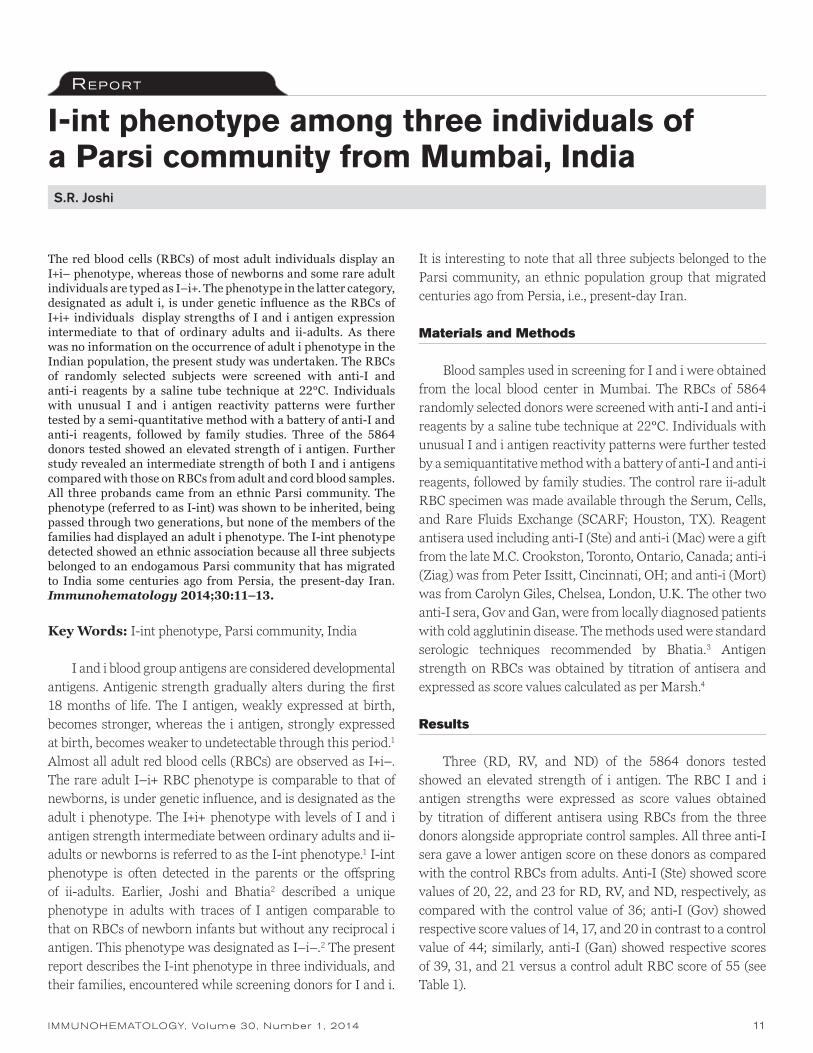
The red blood cells (RBCs) of most adult individuals display an I+i– phenotype, whereas those of newborns and some rare adult individuals are typed as I–i+. The phenotype in the latter category, designated as adult i, is under genetic influence as the RBCs of I+i+ individuals display strengths of I and i antigen expression intermediate to that of ordinary adults and ii-adults. As there was no information on the occurrence of adult i phenotype in the Indian population, the present study was undertaken. The RBCs of randomly selected subjects were screened with anti-I and anti-i reagents by a saline tube technique at 22°C. Individuals with unusual I and i antigen reactivity patterns were further tested by a semi-quantitative method with a battery of anti-I and anti-i reagents, followed by family studies. Three of the 5864 donors tested showed an elevated strength of i antigen. Further study revealed an intermediate strength of both I and i antigens compared with those on RBCs from adult and cord blood samples. All three probands came from an ethnic Parsi community. The phenotype (referred to as I-int) was shown to be inherited, being passed through two generations, but none of the members of the families had displayed an adult i phenotype. The I-int phenotype detected showed an ethnic association because all three subjects belonged to an endogamous Parsi community that has migrated to India some centuries ago from Persia, the present-day Iran. Immunohematology 2014;30:11–13.
Key Words: I-int phenotype, Parsi community, India
I and i blood group antigens are considered developmental antigens. Antigenic strength gradually alters during the first 18 months of life. The I antigen, weakly expressed at birth, becomes stronger, whereas the i antigen, strongly expressed at birth, becomes weaker to undetectable through this period.1 Almost all adult red blood cells (RBCs) are observed as I+i–. The rare adult I–i+ RBC phenotype is comparable to that of newborns, is under genetic influence, and is designated as the adult i phenotype. The I+i+ phenotype with levels of I and i antigen strength intermediate between ordinary adults and ii-adults or newborns is referred to as the I-int phenotype.1 I-int phenotype is often detected in the parents or the offspring of ii-adults. Earlier, Joshi and Bhatia2 described a unique phenotype in adults with traces of I antigen comparable to that on RBCs of newborn infants but without any reciprocal i antigen. This phenotype was designated as I–i–.2 The present report describes the I-int phenotype in three individuals, and their families, encountered while screening donors for I and i.
It is interesting to note that all three subjects belonged to the Parsi community, an ethnic population group that migrated centuries ago from Persia, i.e., present-day Iran.
Materials and Methods
Blood samples used in screening for I and i were obtained from the local blood center in Mumbai. The RBCs of 5864 randomly selected donors were screened with anti-I and anti-i reagents by a saline tube technique at 22°C. Individuals with unusual I and i antigen reactivity patterns were further tested by a semiquantitative method with a battery of anti-I and anti-i reagents, followed by family studies. The control rare ii-adult RBC specimen was made available through the Serum, Cells, and Rare Fluids Exchange (SCARF; Houston, TX). Reagent antisera used including anti-I (Ste) and anti-i (Mac) were a gift from the late M.C. Crookston, Toronto, Ontario, Canada; anti-i (Ziag) was from Peter Issitt, Cincinnati, OH; and anti-i (Mort) was from Carolyn Giles, Chelsea, London, U.K. The other two anti-I sera, Gov and Gan, were from locally diagnosed patients with cold agglutinin disease. The methods used were standard serologic techniques recommended by Bhatia.3 Antigen strength on RBCs was obtained by titration of antisera and expressed as score values calculated as per Marsh.4
Results
Three (RD, RV, and ND) of the 5864 donors tested showed an elevated strength of i antigen. The RBC I and i antigen strengths were expressed as score values obtained by titration of different antisera using RBCs from the three donors alongside appropriate control samples. All three anti-I sera gave a lower antigen score on these donors as compared with the control RBCs from adults. Anti-I (Ste) showed score values of 20, 22, and 23 for RD, RV, and ND, respectively, as compared with the control value of 36; anti-I (Gov) showed respective score values of 14, 17, and 20 in contrast to a control value of 44; similarly, anti-I (Gan) showed respective scores of 39, 31, and 21 versus a control adult RBC score of 55 (see Table 1).

12 IMMUNOHEMATOLOGY, Volume 30, Number 1, 2014
S.R. Joshi
On the other hand, the three anti-i reagents showed higher antigen scores on RBCs from these donors as compared with the control RBCs from adults. Anti-i (Ziag) showed score values of 22, 23, and 26 for RD, RV, and ND, respectively, whereas control RBCs from adults showed a score value of 0; anti-i (McD) gave respective score values of 56, 50, and 61 as compared with control RBCs from adults showing score values of 26; and anti-i (Mort) showed respective scores of 21, 10, and 12 as compared with control adult RBCs showing a score value of 2. The i antigen strengths on these donors’ RBCs were lower than those found on control RBCs of the newborns or ii-adults. The results are displayed in Table 1.
In the family of donor ND, the I-int phenotype was found in three generations in the father (II-1), sister (III-4), and niece (IV-2) of the proband (III-1; Fig. 1), whereas in the family of donor RD, the phenotype was detected through two generations in the father (I-1) and the proband (II-1; Fig. 2).
Although the phenotype showed vertical inheritance, none of the members in these families have the ii-adult phenotype.
Discussion
I and i antigens are considered to be developmental antigens as they show a remarkable reciprocal relationship of expression on RBCs of individuals in early childhood and subsequent adult life.1 Most adult RBCs are characterized by a strong expression of I antigen and a weak expression of i antigen, whereas the
RBCs of newborns have weak I and strong i antigens. The rare individuals defined as ii-adult have an I/i antigen profile similar to that found in newborn infants. Additionally, these individuals have naturally occurring alloanti-I.
Joshi and Bhatia2 earlier found weak expression of I antigen without concurrent increase in i antigen on RBCs of certain individuals and defined the phenotype as I–i–. Family study showed that this I–i– phenotype demonstrated vertical inheritance with some members showing an apparently partial expression of the I–i– character. However, the phenotype appeared to be passing through generations without showing any pattern of Mendelian inheritance.5 Joshi and Bhatia5 reported an association with group A1 or A1B phenotype that was characterized by a remarkable increase in A1 antigen expression on RBCs, thereby suggesting an influence of A1 blood group on expression of I in this phenotype. In the present cases, the I-int phenotype showed no bearing on the
Table 1. Comparison of the I and i antigen scores of the proband and their family members having I-int phenotype with control RBCs
Anti-I Anti-i
Red blood cells ABO groups Ste Gov Gan Ziag McD Mort
RD FamilyRD (Proband) O 20 14 39 22 56 21
Father O 23 18 34 26 56 25
ND Family
ND (Proband) B 23 20 21 26 61 12
Father B 21 25 25 26 64 19
Sister B 22 16 19 23 53 19
Niece B 23 25 25 18 59 21
RV Family RV (Proband) A1B 22 17 31 23 50 10
Controls
Adults B; O 36 44 55 0 26 2
Cord B; O 4 0 3 34 64 36
ii-Adult O 2 0 1 38 70 NT
NT = not tested.
Fig. 1 I-int phenotype in members of the ND family.
1
I
II
III
IV
1
1
2
2
2
3
3
1
4
2
Fig. 2 I-int phenotype in members of the RD family.
1
I
II
1
2
2
Deceased Normal I-I Phenotype I-int phenotype Proband
LEGEND

IMMUNOHEMATOLOGY, Volume 30, Number 1, 2014 13
A1 blood group, as two of the three probands were group B and O. The reduced I antigen in the present I-int phenotype appeared with an increase in i as is seen among the obligate heterozygotes (e.g., parents or offspring) in the families of the ii-adult probands.6 Although there was no ii-adult phenotype found in the present families studied, such a rare phenotype was investigated by Joshi et al.7 in a blood donor from Iran with depressed RBC ABH antigen expression. However, the three donors in the present study had normal features of ABH antigens. The Parsi community has its ancestral origin in Iran, so it is conceivable that the I-int phenotype in the present cases bears some ethnic relation to the ii-adult phenotype detected in the Irani donor. However, the level of I and i antigen strength found among those donors with an I-int phenotype as well as those reported among the family members of the ii-adult probands may potentially reflect a dosage effect shown by the I and i antibodies, a concept that, as per the author’s knowledge, has not been proposed to date.
References
1. Jenkins WJ, Marsh WL, Noades J, Tippett P, Sanger R, Race RR. The I antigen and antibody. Vox Sang 1960;5:97–106.
2. Joshi SR, Bhatia HM. A new red cell phenotype I–i–: red cells lacking both l and i antigens. Vox Sang 1979;36:34–8.
3. Bhatia HM, ed. Procedures in blood banking and immunohaematology. Bombay: Blood Group Reference Centre (ICMR), 1977.
4. Marsh WL. Scoring of hemagglutination reactions. Transfusion 1972;12:352–3.
5. Joshi SR, Bhatia HM. I–i– phenotype in a large kindred Indian family. Vox Sang 1984;46:157–60.
6. Daniels G. I and I antigens, and cold agglutination. In: Human blood groups. 3rd ed. Chichester, UK: John Wiley & Sons, 2013:469–84.
7. Joshi SR, Pourazar A, Clarke VA, Ala FA. Para-Bombay phenotype with altered I-i blood group antigens in an Iranian donor. Transfusion Today 2000:3–6.
Sanmukh R. Joshi, PhD, Associate Professor, Allianze University College of Medical Sciences, Waziria Medical Square, Jalan Bertam 2, Mukim 6, Kepala Batas 13200, Penang, Malaysia.
I-int phenotype among Parsi community
Notice to ReadersImmunohematology is printed on acid-free paper.
For information concerning Immunohematology or the Immunohematology Methods and Procedures manual, contact us by e-mail at [email protected]
Important Notice About Manuscripts for Immunohematology
Please e-mail all manuscripts to [email protected]
Attention: SBB and BB Students
You are eligible for a free 1-year subscription to
Immunohematology.
Ask your education supervisor to submit the name and
complete address for each student and the inclusive dates
of the training period to [email protected]

14 IMMUNOHEMATOLOGY, Volume 30, Number 1, 2014
Evans syndrome in a pediatric liver transplant recipient with an autoantibody with apparent specificity for the KEL4 (Kpb) antigenS.A. Koepsell, K. Burright-Hittner, and J.D. Landmark
Although most warm red blood cell (RBC) autoantibodies react broadly with panel cells in addition to the patient’s own RBCs, occasionally an autoantibody with specificity for a specific blood group antigen is encountered. Rare cases of warm autoantibodies with specificity for the Kpb antigen of the Kell blood group system have been described. We report a pediatric transplant recipient with anemia, immune-mediated hemolysis, thrombocytopenia, and a warm autoantibody with apparent anti-Kpb specificity. The patient’s autoimmune anemia and thrombocytopenia responded well to discontinuing the immunosuppressant tacrolimus, trans-fusions with Kp(b–) RBCs, and intravenous immunoglobulin therapy, with disappearance of the pathologic antibody. During the autoimmune hemolysis, the patient’s RBCs did not react with antisera specific for Kpb. However, repeat testing of the patient’s RBCs with Kpb-specific antisera 15 months after the resolution of hemolysis showed reactivity, indicating that the RBC autoantibody was associated with a transient disappearance of the Kpb antigen. Immunohematology 2014;30:14–17.
Key Words: autoimmune hemolytic anemia, Kell blood group, Evans syndrome
Warm autoantibodies are a broad category of immuno-globulins that react with a patient’s own antigens optimally at 37°C. Their incidence is 1 in 50,000 to 80,000 patients.1 Depending on clinical background, antibody specificity, antibody titer, immunoglobulin subclass, and other less defined variables, warm autoantibodies may be associated with an autoimmune hemolytic anemia (AIHA), especially if the direct antiglobulin test (DAT) is reactive for complement C3 in addition to immunoglobulin G (IgG).2
Although most warm autoantibodies display broad specificity, many examples have been reported of autoanti-bodies with well-defined specificity, most commonly against antigens of the Rh and LW blood group systems.2,3 Although extremely rare, warm autoantibodies with anti-Kpb specificity have been reported.4–8 The most common of three antithetical antigens, Kpb (KEL4) is an antigen that is formed by the presence of an arginine at amino acid 281 of the Kell protein. Substitution of the arginine with tryptophan generates the Kpa antigen (present in 2.3% of whites), and substitution with glutamine generates the Kpc antigen, which is extremely rare.
In transplant recipients, the presence of an apparent autoantibody also raises the possibility that the pathologic antibody may not be a true autoantibody, but rather an alloantibody arising from lymphocytes of donor origin from the transplanted organ, a term called passenger lymphocyte syndrome (PLS).9 PLS often occurs within weeks of transplant and usually involves an antibody with defined specificity that is mismatched between the donor and the recipient. An additional consideration in transplant recipients who develop red blood cell (RBC) autoantibodies are immunosuppressant drugs, which may have unknown consequences on the ability of the immune system to regulate autoimmunity. Tacrolimus in particular is an immunosuppressant that has been reported to be associated with AIHA by an unknown mechanism in transplant recipients based on the observation that pathologic warm autoantibodies seem to resolve after discontinuation of the drug.10,11
We report here an unusual case of a liver transplant recipient who developed an AIHA and thrombocytopenia (Evans syndrome), with the autoantibody having apparent specificity for the Kpb antigen. The patient responded well to discontinuing her tacrolimus, transfusion of Kp(b–) RBCs, and intravenous immunoglobulin (IVIG) therapy, with the autoantibody becoming undetectable.
Case Report
The patient was a 2-year-old group O, D+ girl who received a cadaveric liver transplant at 7 months of age to treat congenital biliary atresia. Her postoperative course was significant for pneumonia, one episode of liver rejection, and ascites. Eight months after transplantation, the patient presented with difficulty breathing and fatigue. She was found to have a hemoglobin (Hb) concentration of 6.2 g/dL, down from 10.0 g/dL 2 weeks earlier. Her immunosuppression regimen was tacrolimus at 1.5 mg, twice daily. Pretransfusion testing at this time showed a negative antibody screen. The patient subsequently received 15 mL/kg of irradiated,
Case RepoRt

IMMUNOHEMATOLOGY, Volume 30, Number 1, 2014 15
Autoantibody with Kpb specificity
leukocyte-reduced, cytomegalovirus (CMV)-negative group O, D+ RBCs. Her Hb level increased to 9.4 g/dL after transfusion. Infectious disease testing at this time showed positive nucleic acid amplification results for Epstein-Barr virus (28,000 copies/mL), human herpes virus 6, and rhinovirus. The patient recovered and was discharged 1 day after admission.
Five days after being discharged, the patient was referred to a pediatric hematologist after being seen in the clinic with an Hb concentration of 8.4 g/dL. Eleven days after discharge, the patient had an Hb level of 6.8 g/dL, an absolute reticulocyte count of 70,000/µL, and a platelet count of 45,000/µL, and her RBCs were negative in the DAT with polyspecific antiglobulin reagent. Nucleic acid amplification testing for parvovirus B19 was negative. Physical examination was significant for an enlarged spleen. The patient again had a negative antibody screen and received 90 mL of irradiated, leukocyte-reduced, CMV-negative group O, D+ RBCs. A bone marrow biopsy was performed, which showed trilineage hematopoiesis with a decreased number of megakaryocytes.
Ten days after the bone marrow biopsy and transfusion, the patient’s Hb concentration was 8.7 g/dL and her platelet count was 15,000/µL. The patient then received two units of single-donor, leukocyte-reduced, irradiated, CMV-negative platelets, and her platelet count increased to 228,000/µL. Again, the patient’s Hb level continued to decline to 7.8 g/dL, and the patient was given RBCs, which brought her Hb concentration up to 10.8 g/dL. On the following day, the patient received another unit of platelets. Six days after the last unit of RBCs and 5 days after the last unit of platelets, the patient was again anemic with an Hb concentration of 7.4 g/dL. An antibody screen performed at this time was positive, with all three cells in the panel reacting with 1+ reactivity at the anti-human globulin phase. A polyspecific DAT was 2+ positive, with similar reactivity observed with anti-IgG monospecific reagent. A peripheral blood smear showed RBCs with crenated and teardrop morphology.
The patient’s specimen was forwarded to the American Red Cross regional reference laboratory, where the DAT was confirmed to be positive (1+) with anti-IgG reagent, but negative with anti-complement reagent. An antibody screen including an auto-control was reactive with all cells at room temperature, showing 1+ to 2+ reactivity. As part of the protocol for initial testing of a suspected autoantibody, the sample was prewarmed, which did not resolve the reactivity. An eluate was prepared and tested, which subsequently reacted with all cells tested. Its reactivity with the ficin-treated reagent cells increased to 4+ at 37°C, and no reactivity was observed with dithiothreitol-treated reagent cells. The patient’s cells were
treated with an ethylenediaminetetraacetic acid–glycine acid (EGA) solution and were DAT-negative. The patient’s plasma and eluate both reacted with the EGA-treated cells, indicating a true autoantibody.
The patient’s plasma was then tested against a panel of antigen-negative rare RBCs that included cells negative for the Rh, Joa, Hy, PP1Pk, and Kpb antigens in low-ionic-strength saline (LISS) at 37°C. Reactivity was observed with all the cells tested, with the exception of the Kp(b–) cells. The patient’s plasma was nonreactive with four different reagent cells that were Kp(a+b–). Antisera with Kpb specificity did not agglutinate the patient’s RBCs. The American Red Cross reference laboratory also performed molecular genotyping of the patient’s peripheral blood leukocytes using HEA Beadchip™. This technology identified that patient as demonstrating the Kpb genotype in her peripheral blood leukocyte DNA, despite the nonreactivity with the Kpb-specific antisera.
During this time, the patient underwent both a lower and upper endoscopy, which ruled out significant gastrointestinal bleeding or pathology. Her immunosuppressant regimen was changed from tacrolimus to cyclosporin, 50 mg twice per day. A lactate dehydrogenase (LDH) level at this time was elevated at 392 units (normal, 140–304 units). Sixteen days after the first positive DAT, the patient was provided with Kp(b–) and K– RBCs, which were nonreactive with the patient’s serum without enhancement but did have weak reactivity with LISS and anti-IgG reagent. Her Hb concentration increased from 6.0 g/dL to 10.8 g/dL. Four days after her Kp(b–) RBC transfusion, the patient was started on IVIG, 12 g per day for 4 days. During the third day of IVIG treatment, an antibody screen was negative and a subsequent polyspecific DAT was also negative. All antibody screens and DATs performed since this episode have been negative. Fifteen months later, the patient’s RBCs were reactive with Kpb antisera. The patient’s clinical course is summarized in Table 1.
Discussion
We report a liver transplant recipient who developed AIHA. The etiology of the autoantibody is unknown, but may have been associated with her antecedent viral illnesses that occurred near the time of her first bout of anemia and before her first positive antibody screen. Although the possibility exists that the anti-Kpb was actually an alloantibody as a result of PLS, this is unlikely, as the Kpb antigen is highly prevalent in the population. In addition, the timing of the patient’s hemolysis occurred 8 months after transplantation,

16 IMMUNOHEMATOLOGY, Volume 30, Number 1, 2014
S.A. Koepsell et al.
whereas PLS usually occurs within a couple of weeks after transplantation. Further, genetic analysis performed on the formalin-fixed paraffin-embedded liver biopsy obtained from the transplanted liver while the organ was being prepared for transplant was homozygous for the Kpb allele, essentially ruling out the possibility that donor lymphocytes could recognize the Kpb antigen as foreign.12
Serologic workup found a warm autoantibody in her plasma that had specificity for the Kpb antigen. However, antisera specific for the Kpb antigen did not agglutinate the patient’s RBCs at the time of the anemia. To classify the pathologic antibody as an autoantibody or alloantibody, molecular genotyping was performed that identified the patient as being homozygous for the gene encoding Kpb. The patient was successfully treated with discontinuing her tacrolimus, transfusion of Kp(b–) blood, and administration of IVIG, with complete resolution of the pathologic antibody.
Table 1 shows that from mid-November 2009 through mid-January 2010, the child developed repeatedly low hemoglobin levels requiring transfusions. Clinically, there was no evidence of bleeding. During this period, the child’s hemoglobin decreased at an initial rate of 0.24 g/dL/day, which increased to over 1 g/dL/day before starting IVIG therapy. Although the patient did not have elevated bilirubin during
this period, she was clinically diagnosed as having AIHA because of the lack of clinical evidence of bleeding, the increased LDH level, and the increased level of circulating reticulocytes along with the warm autoantibody that was identified. The patient also had persistent thrombocytopenia during this time, which may indicate that this case of AIHA may actually be part of an Evans syndrome. Interestingly, a case of Evans syndrome in a pediatric liver transplant recipient has been reported in which the cytopenias were successfully treated by switching the patient from tacrolimus to cyclosporine.13
Previous case reports have identified that patients with Kpb-specific autoantibodies have a depression in the Kell antigens on their RBCs that rebounds after resolution of the autoantibody.8,14 A similar observation was made in this case, as the patient’s RBCs failed to agglutinate with Kpb-specific antisera during the peak of her AIHA. Appropriate controls showed that the Kpb-specific antisera agglutinated reagent cells. Fifteen months after the resolution of the patient’s anemia, her cells did agglutinate with Kpb-specific antisera. Thus, the apparent negative reaction with anti-Kpb may be a result of a similar mechanism of Kell antigen downregulation in response to an autoantibody as previously reported,15 or that our patient simply had a serologically “blocked” antigen owing to her specific autoantibody. The process of how an
Table 1. Clinical and laboratory data obtained from a pediatric liver transplant recipient who developed autoimmune hemolytic anemia*
Hemoglobin Platelets
Date Before transfusion After transfusion Before transfusion After transfusion DAT Ab Screen Transfusion
10/29/2009 10 288
11/13/2009 6.2 9.4 212 Neg 135 mL PRBC
11/25/2009 6.8 8.7 45 Neg Neg 90 mL PRBC
12/7/2009 15 133 50 mL A+ platelets
12/8/2009 54 228 45 mL A+ platelets
12/15/2009 7.8 64 Neg
12/16/5009 10.8 64 135 mL PRBC
12/17/2009 29 92 51 mL A+ platelets
12/22/2009 7.4 54 Pos Pos
12/23/2009 Tacrolimus discontinued
1/7/2010 6.0 7.8 121 100 mL Kp(b–) PRBC
1/8/2010 7.8 10.8 100 mL Kp(b–) PRBC
1/12/2010 Initiation of IVIG therapy
1/14/2010 9.0 179 Neg
1/22/2010 9.9 231 Neg
3/14/2010 12.6 13.3 343 Neg 100 mL PRBC
3/24/2010 11.9 12.3 278 Neg
4/6/2010 10.9 364 Neg
DAT = direct antiglobulin test; Ab = antibody; Neg = negative; PRBC = packed red blood cells; Pos = positive; IVIG = intravenous immunoglobulin G.* Timeline of the patient’s hemoglobin (g/dL), platelet count (cells/μL), DAT, Ab screen, and quantity and type of transfused product. All cellular blood products were irradiated.

IMMUNOHEMATOLOGY, Volume 30, Number 1, 2014 17
autoantibody to the Kpb antigen results in downregulation of the antigen is not known.
A common theme among the handful of case reports describing warm autoantibodies with Kpb specificity is resolu-tion of the autoantibody with either time or treatment.8,14,16 Our case report affirms this trend, with our patient’s Kpb autoantibody completely disappearing 23 days after first being discovered. The patient’s hemoglobin responded well to Kp(b–) blood transfusions. Discontinuing tacrolimus and administering IVIG may have played a role as well, as the antibody became undetectable shortly after IVIG therapy was administered. In summary, we report a pediatric solid-organ transplant recipient who developed a warm autoantibody with apparent Kpb specificity that responded well to treatment and was associated with transient depression of the Kpb antigen on the patient’s RBC surface.
References
1. Issitt PD, Anstee DJ. Applied blood group serology. 4th ed. Durham, NC: Montgomery Scientific Publications, 1998.
2. Wheeler CA, Calhoun L, Blackall DP. Warm reactive autoantibodies: clinical and serologic correlations. Am J Clin Pathol 2004;122:680–5.
3. Garratty G. Specificity of autoantibodies reacting optimally at 37 degrees C. Immunohematology 1999;15:24–40.
4. Villa MA, Coluccio E, Revelli N, Drago F, Morelati F, Rebulla P. Successful transfusion of Kp (a–b+) red cells incompatible for auto anti-Kpb. Haematologica 2005;90:ECR07.
5. Lee E, Burgess G, Win N. Autoimmune hemolytic anemia and a further example of autoanti-Kpb. Immunohematology 2005;21:119–21.
6. Puig N, Carbonell F, Marty ML. Another example of mimicking anti-Kpb in a Kp(a+b–) patient. Vox Sang 1986;51:57–9.
7. Manny N, Levene C, Sela R, Johnson CL, Mueller KA, Marsh WL. Autoimmunity and the Kell blood groups: auto-anti-Kpb in a Kp(a+b–) patient. Vox Sang 1983;45:252–6.
Autoantibody with Kpb specificity
8. Beck ML, Marsh WL, Pierce SR, DiNapoli J, Oyen R, Nichols ME. Auto anti-Kpb associated with weakened antigenicity in the Kell blood group system: a second example. Transfusion 1979;19:197–202.
9. Audet M, Panaro F, Piardi T, et al. Passenger lymphocyte syndrome and liver transplantation. Clin Dev Immunol 2008; 2008:715769.
10. Valentini RP, Imam A, Warrier I, et al. Sirolimus rescue for tacrolimus-associated post-transplant autoimmune hemolytic anemia. Pediatr Transplant 2006;10:358–61.
11. Botija G, Ybarra M, Ramos E, et al. Autoimmune cytopaenia after paediatric intestinal transplantation: a case series. Transpl Int 2010;23:1033–7.
12. Koepsell SA, Landmark JD. Passenger lymphocyte syndrome: use of archived donor organ biopsy obtained at the time of transplantation for diagnosis. Am J Transplant 2013;13:2227.
13. Domenech C, Mialou V, Galambrun C, et al. Successful treatment of Evans syndrome post liver transplant with splenectomy and switch from tacrolimus to cyclosporine. Transpl Int 2008;21:397–9.
14. Seyfried H, Gorska B, Maj S, Sylwestrowicz T, Giles CM, Goldsmith KL. Apparent depression of antigens of the Kell blood group system associated with autoimmune acquired haemolytic anaemia. Vox Sang 1972;23:528–36.
15. Williamson LM, Poole J, Redman C, et al. Transient loss of proteins carrying Kell and Lutheran red cell antigens during consecutive relapses of autoimmune thrombocytopenia. Br J Haematol 1994;87:805–12.
16. Win N, Kaye T, Mir N, Damain-Willems C, Chatfield C. Autoimmune haemolytic anaemia in infancy with anti-Kpb specificity. Vox Sang 1996;71:187–8.
Scott A. Koepsell, MD, PhD (corresponding author), Assistant Professor of Pathology and Microbiology, University of Nebraska Medical Center; Kerry Burright-Hittner, MT(ASCP)SBB, Director, Immunohematology Reference Laboratory, American Red Cross, Omaha; and James D. Landmark, MD, Associate Professor of Pathology and Microbiology, University of Nebraska Medical Center, 983135 Nebraska Medical Center, Omaha, NE 68198-3135.

18 IMMUNOHEMATOLOGY, Volume 30, Number 1, 2014
JMH blood group system: a reviewS.T. Johnson
Revie w
The JMH blood group system consists of six high-prevalence antigens. These antigens are located on the Sema7A protein. The molecular basis of the JMH1– phenotype is not known; however, single nucleotide changes in the SEMA7A gene on chromosome 15 account for the other JMH antigens. JMH1, commonly known as JMH, is most notable because transient depression of the antigen occurs and anti-JMH may develop. These antibodies are most commonly observed and are not significant in transfusion. Antibodies developed in the rare JMH variant types may cause reduced red cell survival. This review provides a general overview of the JMH blood group system, including the serologic and molecular characteristics as well as proposed functions of the Sema7A protein. Immunohematology 2014;30:18–23.
Key Words: John Milton Hagen group, semaphorin 7A (CDw108 glycoprotein), SEMA7A gene, high-prevalence red cell blood group antigen
Introduction
The JMH blood group system has the distinction of being the blood group with the most “nicknames.” The “over 60 group,” “John Milton Hagen group,” “The Boys,” “The Cat,” and the “Old Boys’ Club” have all been used in reference to the antibody and antibody makers.1 The earliest accounts in the 1970s were of antibody reactivity, followed by description of the protein carrying the antigens (semaphorin 7A or CD108), and finally with identification of the gene SEMA7A, JMH earned its rightful status of blood group system 026 in 2001.
Antibodies to JMH antigens are encountered infrequently but have unique characteristics landing them their nicknames. The “over 60 group,” “The Boys,” and the “Old Boys’ Club” all are derived from early observations that many of the antibodies were found in older gentleman. Older has been defined as >50 or 60 years of age. Like Issitt,2 I concur that old is recognized as mature, wiser, and experienced patients.
Not to leave the ladies out of describing these antibodies, the nickname “The Cat” came from one of the first women who produced anti-JMH. She is said to have claimed the anti-JMH occurred when her cat died (Marilyn Moulds, March 2012, personal communication).
The significance of antibodies to JMH antigens in trans-fusion and pregnancy are minimal and will be discussed.
History
In March 1973, a male patient in his 60s was to have elective orthopedic surgery. He had no history of transfusions. A weakly reactive antibody detected by the indirect antiglobulin test (IAT) was not reactive with papain- or ficin-pretreated cells. No compatible blood was available, and units collected for autologous transfusions were never given. It was reported as an “antibody to high-incidence unknown factor,” and samples were sent to other laboratories for investigation. This result led to a group of antibodies being collected in the 1970s that were compatible with this individual’s red blood cells (RBCs) and had similar reactivity. They were first mentioned in print by Issitt in 1975 as the John Milton Hagen group of antibodies, as he termed them “belonging to a group of high-incidence antigens of which little is known about.” His remarks were based on personal communication with John J. Moulds.3 The first antibody was reported to be recognized in 1970 per Sabo et al.,4 who further characterized 49 sera with similar reactivity and proposed giving this antibody the symbol JMH, naming it after one of the original antibody makers, and adding it to the list of high-titer, low-avidity antibodies. These antibodies were of high titer and weakly reactive in saline IAT with all RBCs tested, except autologous cells and other JMH– RBCs.
DedicationThis review is dedicated to John J. Moulds, MT(ASCP)SBB, as he was instrumental in bringing a group of antibodies with interesting serologic characteristics to the blood group it is today. In the classic “Moulds” way throughout his life and career, he always challenged young SBBs and laboratory scientists to question unusual serologic results. He would say, “They are telling you something.” The JMH blood group system is a testament to his beliefs. He is coauthor on many papers in this review. John called me to provide additional information he found important to the JMH story. I am honored and privileged to have known John, to have been challenged by John, and to write this review as a tribute to his work.

IMMUNOHEMATOLOGY, Volume 30, Number 1, 2014 19
JMH blood group system review
In the 1980s, work was done to further define the serologic characteristics of anti-JMH and its reactivity with chemically modified RBCs, namely those treated with proteases, sulfhydryl-reducing agents, and neuraminidase. In addition, attempts were made to predict the clinical significance of anti-JMH in transfusion using chromium-51 RBC survival studies and subclassing.5 In 1982, a monoclonal antibody named H8 was described with JMH specificity.6,7 This antibody was important to further work in characterizing the JMH protein. In addition, J.J. Moulds reported evidence that there was heterogeneity in reactivity of different anti-JMH.8
Telen et al.9 reported in 1990 that several high-incidence antigens including the JMH antigen were absent on RBCs of individuals with paroxysmal nocturnal hemoglobinuria (PNH). PNHIII RBCs were previously shown to lack glycosylphosphatidylinositol (GPI)-linked proteins, and when these RBCs were tested with human anti-JMH and monoclonal H8, no reactivity was seen. This result suggested that JMH must reside on a GPI-linked membrane protein. Immunoprecipitation and immunoblotting experiments on human anti-JMH and H8 showed JMH resides on a 76-kD phosphatidylinositol-linked protein.10 Finally, the location of JMH was determined to be on the CDw108 glycoprotein, now known as semaphorin 7A.11
Having knowledge of the location of JMH, the cDNA clone containing the CDw108 gene was identified in 1999.12 The gene resides on the middle of the long arm of chromosome 15. This CDw108 gene is now known as SEMA7A and is located on 15q23–24. The genetic information earned JMH its own blood group system in 2001 named John Milton Hagen, 026, and its symbol JMH.13 JMH1 is the antigen detected by most individuals making anti-JMH. Four additional variant JMH genotypes were added in 2007.14 JMHQ was proposed in 2011, reported in four Native Americans whose RBCs were JMH1–, with most examples of anti-JMH, and officially recognized later that year.15
Today, there are six recognized antigens in the system summarized in Table 1.16,17 Each antigen is defined by antibodies nonreactive with JMH1– RBCs.
Nomenclature
Nomenclature resulting from serologic observations and biologic and molecular studies has remained very simple for the blood group with so many nicknames. The antigen name commonly known by serologists remains JMH and is officially recognized by the International Society for Blood Transfusion (ISBT) as JMH1. Confirmed JMH variants are sequentially numbered for example, JMH2 and named with the first letter from the antibody maker’s first name (JMHK) following JMH.
JMH Glycoprotein
Antigens in the JMH blood group system are carried on the protein semaphorin 7A, also known as Sema7A and CD108. Mature Sema7A consists of 525 amino acids. Changes in amino acids 207 and 460/461 were noted by Seltsam in JMH-variant individuals. A three-dimensional model of the crystal structure of Sema7A was proposed (Fig. 1A and B).14 Position 207 is located at the top face of the sema domain, whereas positions 460 and 461 are on the bottom.
As mentioned previously, Sema7A protein binds to the cell membrane by a GPI linkage. Not only is it present on RBCs, it is present on lymphoid and myeloid cells as well as bone cells, neurons of the brain and spinal cord, thymus, spleen, gut, kidney, heart, and placenta. Sema7A is found primarily in activated T cells and thymocytes.18
Table 1. The John Milton Hagen blood group system
Number Name Prevalence Molecular basis of antigen-negative phenotype
JMH1 JMH High Not known
JMH2 JMHK High 619C>T R207W
JMH3 JMHL High 620G>A R207Q
JMH4 JMHG High 1379G>A R460H
JMH5 JMHM High 1381C>T R461C
JMH6 JMHQ High 1040G>T R347L
Fig. 1 Three-dimensional model of Sema7A (reprinted with permission from AABB). Image courtesy of A. Setsam. (A) A cartoon of the sema domain when looking down on the protein. The numbers represent propellers. (B) A side view of the sema domain. The polymorphic amino acid positions 207, 460, and 461 are circled.

20 IMMUNOHEMATOLOGY, Volume 30, Number 1, 2014
S.T. Johnson
Numerous roles for Sema7A have been proposed. It is clear it is involved in cytokine expression, chemotaxis, and axon guidance signaling.19 In effector T cells, it likely promotes macrophage recruitment to sites of inflammation.18,20,21 A recent report shows the Sema7A_R461C variant (JMH5) causes different T-cell responses than the wild-type Sema7A.22 It is able to strongly activate CD4+ T cells and change their phenotype to a more cytotoxic one, and evidence is provided that Sema7A_R461C delivers costimulatory signals to T cells, causing them to increase their release of cytokines such as interleukin (IL)-1β, IL-6, and IL-8.
Sema7A has also been reported to be an important regulator of tissue remodeling. In genetic studies in Korean women with decreased bone mineral density, mutations in SEMA7A were found.23 Sema7A also contributes to regulation of tissue fibrosis and remodeling.24
The exact function of semaphorin 7A on RBCs is not known, but it likely plays a role in cell migration as an adhesion molecule.25 It may also play a role as a receptor for Plasmodium falciparum. It was identified as a receptor for the P. falciparum merozoite-specific thrombospondin-related anonymous pro- tein (TRAP) homolog in laboratory experiments using recombinant protein.26
JMH Antigen Characteristics
JMH1 is the primary antigen in the system and is present in greater than 99 percent of all individuals. The JMH1-negative phenotype occurs as either inherited or more commonly acquired depression of the antigen. The JMH antigen is most commonly depressed in a transient manner. In these individuals, the antigen is often below the level of detection by standard serologic methods and they make anti-JMH. This result likely explains the serologic observation of a positive direct antiglobulin test (DAT) seen in many individuals with anti-JMH. There has been no genetic change identified in the SEMA7A gene of these individuals.14 The transient phenomenon may be triggered by an autoimmune process like that seen in other blood group systems.
There is only one family described with the inherited negative phenotype identified when randomly screening donors. In this family, it was shown that three generations of individuals possessed JMH– RBCs, consistent with autosomal dominant inheritance.27 None had made anti-JMH, and all had a negative DAT.
Rare JMH variants have been described with reduced or variable expression of JMH antigen (Table 1).14 All possess an antibody that is not reactive with JMH1– RBCs. However,
variable reactivity is seen when the specificity of each antibody is cross-tested with other JMH variants (Table 2).28 JMH antigens are destroyed by various chemicals, as shown in Table 3, characteristic of other blood group antigens on GPI-linked proteins. PNHIII cells can be used as a source of JMH– RBCs as well.29
The SEMA7A Gene: Genetics and Inheritance
SEMA7A is located on 15q22.3-q23, the middle of the long arm of chromosome 15 (GenBank accession number BC101647), and is organized in 14 exons. To date, no nucleotide changes have been identified in individuals with JMH– RBCs. JMH variants result from changes in nucleotides in exons 6 and 11 (Table 1).
Rare individuals who lack Sema7A on their RBCs possess Sema7A on other cells. Given the information provided from molecular analysis performed to date, this is likely because of a posttranscriptional mechanism.14
JMH Antibodies
Anti-JMH is found most often as the acquired form and as such can be found in individuals with no prior transfusions
Table 2. Anti-JMH and JMH-related antibodies tested with JMH– and variant JMH red blood cells
Anti-
Phenotype JMH1 JMH2 JMH3 JMH4 JMH5 JMH6
JMH:-1 0 0 0 0 0 0
JMH:-2 + 0 0 + + NA
JMH:-3 + 0 0 + + NA
JMH:-4 + + + 0 + NA
JMH:-5 + + + 0 0 NA
JMH:-6 + NA NA NA NA NA
NA = not available.Adapted from Daniels.27
Table 3. Characteristics of antigens in the John Milton Hagen blood group system
Sensitive to ficin or papain treatment
Sensitive to trypsin and alpha chymotrypsin treatment
Sensitive to 2-aminoethyl-isothiouronium bromide and 200 mmol/L/ 50 mmol/L dithiothreitol treatment
Sensitive to sialidase treatment
Resistant to chloroquine treatment
Absent from paroxysmal nocturnal hemoglobinuria-III red blood cells
Weakly expressed on cord cells
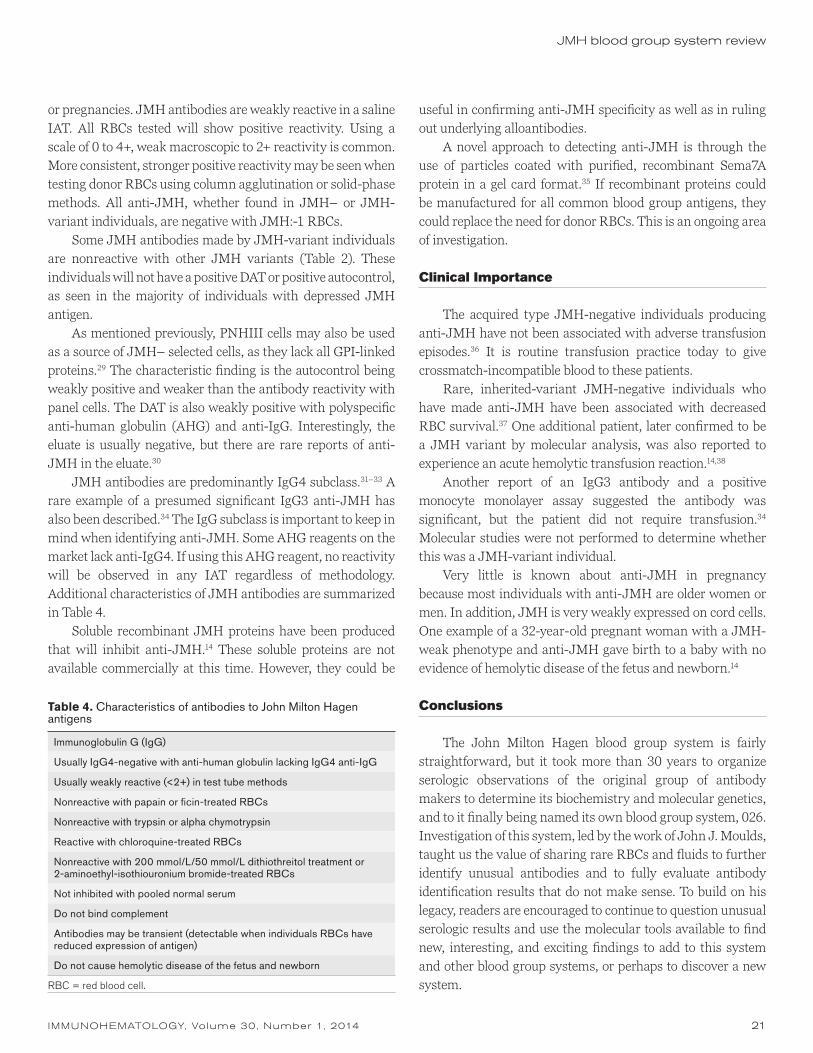
IMMUNOHEMATOLOGY, Volume 30, Number 1, 2014 21
JMH blood group system review
or pregnancies. JMH antibodies are weakly reactive in a saline IAT. All RBCs tested will show positive reactivity. Using a scale of 0 to 4+, weak macroscopic to 2+ reactivity is common. More consistent, stronger positive reactivity may be seen when testing donor RBCs using column agglutination or solid-phase methods. All anti-JMH, whether found in JMH– or JMH-variant individuals, are negative with JMH:-1 RBCs.
Some JMH antibodies made by JMH-variant individuals are nonreactive with other JMH variants (Table 2). These individuals will not have a positive DAT or positive autocontrol, as seen in the majority of individuals with depressed JMH antigen.
As mentioned previously, PNHIII cells may also be used as a source of JMH– selected cells, as they lack all GPI-linked proteins.29 The characteristic finding is the autocontrol being weakly positive and weaker than the antibody reactivity with panel cells. The DAT is also weakly positive with polyspecific anti-human globulin (AHG) and anti-IgG. Interestingly, the eluate is usually negative, but there are rare reports of anti-JMH in the eluate.30
JMH antibodies are predominantly IgG4 subclass.31–33 A rare example of a presumed significant IgG3 anti-JMH has also been described.34 The IgG subclass is important to keep in mind when identifying anti-JMH. Some AHG reagents on the market lack anti-IgG4. If using this AHG reagent, no reactivity will be observed in any IAT regardless of methodology. Additional characteristics of JMH antibodies are summarized in Table 4.
Soluble recombinant JMH proteins have been produced that will inhibit anti-JMH.14 These soluble proteins are not available commercially at this time. However, they could be
useful in confirming anti-JMH specificity as well as in ruling out underlying alloantibodies.
A novel approach to detecting anti-JMH is through the use of particles coated with purified, recombinant Sema7A protein in a gel card format.35 If recombinant proteins could be manufactured for all common blood group antigens, they could replace the need for donor RBCs. This is an ongoing area of investigation.
Clinical Importance
The acquired type JMH-negative individuals producing anti-JMH have not been associated with adverse transfusion episodes.36 It is routine transfusion practice today to give crossmatch-incompatible blood to these patients.
Rare, inherited-variant JMH-negative individuals who have made anti-JMH have been associated with decreased RBC survival.37 One additional patient, later confirmed to be a JMH variant by molecular analysis, was also reported to experience an acute hemolytic transfusion reaction.14,38
Another report of an IgG3 antibody and a positive monocyte monolayer assay suggested the antibody was significant, but the patient did not require transfusion.34 Molecular studies were not performed to determine whether this was a JMH-variant individual.
Very little is known about anti-JMH in pregnancy because most individuals with anti-JMH are older women or men. In addition, JMH is very weakly expressed on cord cells. One example of a 32-year-old pregnant woman with a JMH-weak phenotype and anti-JMH gave birth to a baby with no evidence of hemolytic disease of the fetus and newborn.14
Conclusions
The John Milton Hagen blood group system is fairly straightforward, but it took more than 30 years to organize serologic observations of the original group of antibody makers to determine its biochemistry and molecular genetics, and to it finally being named its own blood group system, 026. Investigation of this system, led by the work of John J. Moulds, taught us the value of sharing rare RBCs and fluids to further identify unusual antibodies and to fully evaluate antibody identification results that do not make sense. To build on his legacy, readers are encouraged to continue to question unusual serologic results and use the molecular tools available to find new, interesting, and exciting findings to add to this system and other blood group systems, or perhaps to discover a new system.
Table 4. Characteristics of antibodies to John Milton Hagen antigens
Immunoglobulin G (IgG)
Usually IgG4-negative with anti-human globulin lacking IgG4 anti-IgG
Usually weakly reactive (<2+) in test tube methods
Nonreactive with papain or ficin-treated RBCs
Nonreactive with trypsin or alpha chymotrypsin
Reactive with chloroquine-treated RBCs
Nonreactive with 200 mmol/L/50 mmol/L dithiothreitol treatment or 2-aminoethyl-isothiouronium bromide-treated RBCs
Not inhibited with pooled normal serum
Do not bind complement
Antibodies may be transient (detectable when individuals RBCs have reduced expression of antigen)
Do not cause hemolytic disease of the fetus and newborn
RBC = red blood cell.

22 IMMUNOHEMATOLOGY, Volume 30, Number 1, 2014
S.T. Johnson
References
1. Rolih SD. High-titer, low-avidity (HTLA) antibodies and antigens: a review. Transfus Med Rev 1989;3:128–39.
2. Issitt PD. Applied blood group serology. 4th ed. Durham, NC: Montgomery Scientific Publications, 1998.
3. Issitt PD. Applied blood group serology. 2nd ed. Oxnard, CA: Spectra Biologicals, 1975.
4. Sabo B, Moulds JJ, McCreary J. Anti-JMH: another high titer-low avidity antibody against a high frequency antigen (abstract). Transfusion 1978;18:387.
5. Baldwin ML, Ness PM, Barrasso C, et al. In vivo studies of the long-term 51Cr red cell survival of serologically incompatible red cell units. Transfusion 1985;25:34–8.
6. Daniels GL, Green C, Lomas C, Tippet P. Monoclonal antibodies recognizing high-frequency RBC antigens, including type 2H and JMH (abstract). Transfusion 1981:21;612.
7. Daniels GL, Knowles RW. A monoclonal antibody to the high frequency red cell antigen JMH. J Immunogenet 1982;9:57–9.
8. Moulds JJ, Levene C, Zimmernam S. Serological e vidence for heterogeneity among antibodies compatible with JMH-negative red cells (abstract). Abstracts of the Joint Meeting of the 19th Congress of the International Society of Haematology and the 17th Congress of the International Society of Blood Transfusion. 1982:287.
9. Telen MJ, Rosse WF, Parker CJ, Moulds MK, Moulds JJ. Evidence that several high-frequency human blood group antigens reside on phosphatidylinositol-linked erythrocyte membrane proteins. Blood 1990;75:1404–7.
10. Bobolis KA, Moulds JJ, Telen MJ. Isolation of the JMH antigen on novel phosphatidylinositol-linked human membrane protein. Blood 1992;79:1574–81.
11. Mudud R, Rao N, Angelisova P, Horejsi V, Telen MJ. Evidence that CDw108 membrane protein bears the JMH blood group antigen. Transfusion 1995;35:566–70.
12. Yamada A, Kubo K, Takeshita T, et al. Molecular cloning of a glycosylphosphatidylinositol-anchored molecule CDw108. J Immunol 1999;162:4094–100.
13. Daniels GL, Anstee DJ, Cartron JP, et al. International Society of Blood Transfusion Working Party on Terminology for Red Cell Surface Antigens. Vox Sang 2001;80:193–7.
14. Seltsam A, Strigens S, Levene C, et al. The molecular diversity of Sema7A, the semaphorin that carries the JMH blood group antigens. Transfusion 2007;47:133–46.
15. Richard M, St-Laurent J, Perreault J, Long A, St-Louis M. A new SEMA7A variant found in Native Americans with alloantibody. Vox Sang 2011;100:322–6.
16. Daniels G, Flegel WA, Fletcher A, et al. International Society of Blood Transfusion Committee on Terminology for Red Cell Surface Antigens: Cape Town report. Vox Sang 2007;92: 250–3.
17. Storry JR, Castilho L, Daniels G, et al. International Society of Blood Transfusion Working Party on red cell immunogenetics
and blood group terminology: Berlin report. Vox Sang 2011; 101:77–82.
18. Suzuki K, Okuno T, Yamamoto M, et al. Semaphorin 7A initiates T-cell-mediated inflammatory responses through alpha1beta1 integrin. Nature 2007;446:680–4.
19. Kikutani H, Kumanogh A. Semaphorins in interactions between T cells and antigen-presenting cells. Nat Rev Immunol 2003;3:159–67.
20. Yazdani U, Terman JR. The semaphorins. Genome Biol 2006; 7:211.
21. Holmes S, Downs AM, Fosberry A, et al. Sema7A is a potent monocyte stimulator. Scand J Immunol 2002;56:270–5.
22. Gras C, Eiz-Vesper B, Seltsam A, Immenschuh S, Blasczyk R, Figueiredo. Semaphorin 7A protein variants differentially regulate T-cell activity. Transfusion 2013;53:270–83.
23. Koh J-M, Oh B, Lee JY, et al. Association study of semaphorin 7a (sema7a) polymorphisms with bone mineral density and fracture risk in postmenopausal Korean women. J Hum Genet 2006;51:112–17.
24. Kang H-R, Lee CG, Homer RJ, Elias JA. Semaphorin 7A plays a critical role in TGF-beta1-induced pulmonary fibrosis. J Exp Med 2007;204:1083–93.
25. Anstee DJ. The functional importance of blood group-active molecules in human red blood cells. Vox Sang 2011;100:140–9.
26. Bartholdson SJ, Bustamente LY, Crosnier C, et al. Semaphorin-7A is an erythrocyte receptor for P. falciparum merozoite- specific TRAP homolog, MTRAP. PLoS Pathog 2012; 8:e1003031.
27. Kollmar M, South SF, Tregellas WM. Evidence of a genetic mechanism for the production of the JMH negative phenotype (abstract). Transfusion 1981;21:612.
28. Daniels G. Human blood groups. 3rd ed. Wiley-Blackwell, 2013:467.
29. Stroka-Lee AH, Halverson GR. Collecting GPI-negative RBCs from patients with paroxysmal nocturnal hemoglobinuria for use in serological investigations (abstract). Transfusion 2010;50(Suppl):167A.
30. Whitsett CF, Moulds M, Pierce JA, Hare V. Anti-JMH identified in serum and eluate from red cells of a JMH-negative man. Transfusion 1983;23:344–5.
31. Tregellas WM, Pierce SR, Harding JT, Beck ML. Anti-JMH: IgG subclass composition and clinical significance (abstract). Transfusion 1980;20:628.
32. Garratty G, Arndt P, Nance S. IgG subclass of blood group alloantibodies to high frequency antigens (abstract). Transfusion 1996;36(Suppl 9S):50S.
33. Pope J, Lubenko A, Lai WYY. A survey of the IgG subclasses of antibodies to high frequency red cell antigens (abstract). Transf Med 1991;1(Suppl 2):58.
34. Geisland J, Corgan M, Hillard B. An example of anti-JMH with characteristics of a clinically significant antibody. Immunohematology 1990;6:9–11.

IMMUNOHEMATOLOGY, Volume 30, Number 1, 2014 23
JMH blood group system review
35. Seltsam A, Agaylan A, Grueger D, Meyer O, Blasczyk R, Salama A. Rapid detection of JMH antibodies with recombinant Sema7A (CD108) protein and the particle gel immunoassay. Transfusion 2008;48:1151–5.
36. Baldwin ML, Ness PM, Barrasso C, et al. In vivo studies of the long-term 51Cr red cell survival of serologically incompatible red cell units. Transfusion 1985;25:34–8.
37. Mudad R, Rao N, Issitt PD, Roy RB, Combs MR, Telen MJ. JMH variants: serologic, clinical, and biochemical analyses in two cases. Transfusion 1995;35:925–30.
38. Hoppe B, Pastucha L, Seltsam A, Greinacher A, Salama A. Acute haemolytic transfusion reactions due to weak antibodies that in vitro did not seem to be clinically significant. Vox Sang 2002;82:207–10.
Susan T. Johnson, MSTM, MT(ASCP) SBB, Director, Transfusion Medicine Program, Marquette University, Milwaukee, WI; Director of the SBB Program, Director, Clinical Education, Blood Center of Wisconsin, 638 N. 18th Street, Milwaukee, WI.

24 IMMUNOHEMATOLOGY, Volume 30, Number 1, 2014
Demonstration of IgG subclass (IgG1 and IgG3) in patients with positive direct antiglobulin testsA. Singh, A. Solanki, and R. Chaudhary
Serologic characterization of autoantibodies helps in the management and monitoring of the course of autoimmune hemolytic anemia (AIHA). The purpose of this study was to evaluate gel centrifugation test (GCT) cards for immunoglobulin G (IgG) titer and determination of IgG subclasses IgG1 and IgG3 and their influence on hemolysis. Eighty direct antiglobulin test (DAT)-positive patients were examined with the help of GCT cards for IgG titer and IgG subclasses. The results were correlated with the presence and absence of hemolysis. A statistically significant (p < 0.005) association of hemolysis with increasing anti-IgG titer was observed. When IgG titer was 30 or less, 28 (50.91%) patients had no hemolysis, whereas 15 (93.75%) patients had features of hemolysis when titer was at least 300. Statistically significant (p < 0.005) association of subclass of IgG (IgG1, IgG3) coating the red blood cells with intravascular hemolysis was also seen. Twenty-nine (80.56%) patients had evidence of hemolysis when IgG1 or IgG1-IgG3 both were present. Gel technology is helpful to demonstrate red blood cell–bound autoantibodies and their characterization with regard to class, subclass, and titer. This information is useful to identify patients with AIHA who are at risk of severe hemolysis with adverse prognosis. Immunohematology 2014;30:24–27.
Key Words: autoimmune hemolytic anemia, gel centrifuga-tion test, direct antiglobulin test, hemolysis
Autoimmune hemolytic anemia (AIHA) is characterized by increased red blood cell (RBC) destruction or decreased RBC survival as a result of autoantibodies directed against self-antigen on RBCs.1 The degree of hemolysis depends on characteristics of the bound antibody as well as the target antigen. Immunoglobulin M (IgM) antibodies readily activate the classical complement pathway and produce cytolysis. IgG antibodies are relatively poor activators of the classical complement pathway but are easily recognized by phagocytic cells.2–4 IgG subclasses have a varying affinity for Fc receptors, with IgG3 having a higher affinity for mononuclear phagocytes. Therefore, monocyte reactions with IgG3 are much more rapid, requiring few molecules for initiation of erythrophagocytosis and thus having greater potential for RBC destruction.
RBC-bound Ig can be detected using conventional tube technique, gel technique, flow cytometry (FC), or capillary
electrophoresis.5 The tube agglutination assay has been the standard antibody detection method for RBC serology tests for decades; however, use of the gel microcolumn antibody detection system is growing and can be used in place of the tube agglutination assay. Gel technology, which was introduced by Lapierre et al. in 1990,6 is used to determine ABO group and D type,6,7 detect unexpected serum antibodies,8–14 and perform the direct antiglobulin test (DAT).15 Compared with the tube agglutination assay, the gel microcolumn assay requires less test sample and reagent, is easier to read, needs no washing, and is more readily automated.15 Although FC is a very sensitive method and can detect very small amounts of Ig molecules coating the RBCs, it has the disadvantage of being costly and not easily available in many of the hospital-based blood transfusion services in developing countries.
Here we discuss our experience of gel technology to demonstrate IgG subclasses (IgG1 and IgG3) in DAT+ cases and their correlation with hemolysis.
Materials and Methods
The study was conducted in the immunohematology laboratory of the Department of Transfusion Medicine, Sanjay Gandhi Postgraduate Institute of Medical Sciences, Lucknow, India, from January 2009 to June 2012. Ethylenediaminetetraacetic acid (EDTA) whole blood samples from 80 DAT+ patients were tested for IgG subclasses using gel technology (Diamed, Cressier, Switzerland). Normal blood donors were not included in this retrospective study, which was a limitation of this study.
Hemolysis in a patient was documented when at least three of the following laboratory parameters were abnormal: (1) hemoglobin (Hb; <9 g/dL), (2) percentage of reticulocytes (>2%), (3) total serum bilirubin (>2 mg/dL), and (4) lactate dehydrogenase (LDH; >500 IU/mL).16
RepoRt

IMMUNOHEMATOLOGY, Volume 30, Number 1, 2014 25
IgG subclass in DAT positive patients
Gel TechnologyFor quantification and IgG subclass determination, three
types of gel centrifugation test (GCT) dilution cards were used. The first type (IgG dilution cards) contained dilutions of rabbit anti-human polyclonal anti–IgG-g-chain serum of 1 in 10, 1 in 30, 1 in 100, 1 in 300, and 1 in 1000 and a negative diluent control. The second type contained monospecific AHG (ID-Card DC Screening I, Diamed). This card consisted of five different monospecific AHG reagents to anti-IgG, anti-IgA, anti-IgM, anti-C3c (all rabbit), and anti-C3d (monoclonal cell line C139-9). If antibody was IgG in nature, then the subclass was determined by a third type of monospecific anti-IgG1 and anti-IgG3 gel card (ID-Card DAT IgG1/IgG3, Diamed). This card consisted of monoclonal anti-IgG1 (cell line M345/795) in two different dilutions (1:1 and 1:100), anti-IgG3 (cell line M346/805) in two different dilutions (1:1 and 1:100), anti-IgG 1:10 (rabbit), and a negative control. The tests were performed following the department’s standard operating procedure manual as described previously.17
ElutionTo confirm that the positive DAT result is caused by an
autoantibody, cold acid elution was performed.18 The eluate was tested with a three-cell screening panel, ID DiaCell I-II-III (Diamed), by indirect antiglobulin test using gel cards.
Statistical AnalysisData were entered in Microsoft Office Excel (Microsoft
Corporation, Redmond, WA) and analyzed with Statistical Package for the Social Sciences (SPSS) version 16.0 (SPSS, Inc., Chicago, IL). Pearson’s χ2 test was used to analyze the relations between IgG titer with hemolysis and IgG subclass with hemolysis. Probability values less than 0.05 were considered significant.
Results
A total of 80 DAT+ samples were evaluated. The median age of patients was 32.5 years, and the female-to-male ratio was 2.33. Of the 80 patients, 49 had features of hemolysis as shown in Fig. 1. The median levels of Hb, reticulocyte count, total serum bilirubin, and LDH were 6.9 g/dL (range, 3–14.3 g/dL), 4.7 percent (range, 0.4–41%), 1.35 mg/dL (range, 0.3–22.7 mg/dL), and 847.5 IU/mL (range, 125–9620 IU/mL), respectively.
A statistically significant (p < 0.005) association of hemolysis with increasing anti-IgG titer was observed (Fig. 1). When anti-IgG titer was 30 or less, 27 (49.09%) patients
had evidence of hemolysis. Similarly, 77.78 percent and 93.75 percent of the patients experienced hemolysis when anti-IgG titer was 100 and at least 300, respectively.
There was a statistically significant (p < 0.005) association of subclass of IgG (IgG1, IgG3) coating the RBCs with intravascular hemolysis. Of 80 DAT+ samples, 25 had IgG1 and 11 had both IgG1 and IgG3, of which 20 (80.0%) and 9 (81.82%) had evidence of hemolysis, respectively. In 44 patients, IgG1 or both IgG1 and IgG3 were not detected; of these, 24 (54.55%) had no hemolysis (Table 1).
Whenever DAT was more than 2+ in strength, cold acid elution was performed. Eluate was tested with reagent RBC panels (Diamed). It was found to be pan-reactive in all the samples, confirming the presence of autoantibodies.
Discussion
Introduction of specialized gel cards, such as monospecific Ig cards, IgG subclass cards, and IgG dilution cards, have made it possible to characterize RBC-bound antibodies in a simplified and rapid way. These detailed investigations help predict the severity of hemolysis and disease outcome and permit the planning of therapy in autoimmune disorders.17
Hemolysis
No hemolysis
≤ 30
p value <0.05
5
10
15
20
25
30
0= 100 ≥ 30
2850.91%27
49.09%
777.78%
1593.75%
16.25%
222.22%
Fig. 1 Correlation of IgG titer with in vivo hemolysis.
Table 1. Correlation of IgG subclass with hemolysis
IgG subclassNumber
of patients Hemolysis No hemolysis
IgG1 25 20 (80.00%) 5 (20.00%)
IgG1 and IgG3 11 9 (81.82%) 2 (18.18%)
IgG3 only or neither IgG1 nor IgG3 detected
44 20 (45.45%) 24 (54.55%)
Total 80 49 (61.25%) 31 (38.75%)
p value <0.005.

26 IMMUNOHEMATOLOGY, Volume 30, Number 1, 2014
A. Singh et al.
In this study, we demonstrated IgG subclasses (IgG1 and IgG3) in DAT+ patients using specialized gel cards (Diamed). IgG2 and IgG4 were not studied, as these are rare antibodies involved in AIHA; moreover, Diamed does not provide gel cards for detection of these subclasses. IgG1 or both IgG1 and IgG3 were observed with hemolysis in 29 (59.18%) patients. This finding was almost the same as that of Das et al.16 They reported IgG subclasses, IgG1 or IgG3 or both, coating the RBC membrane in 46.5 percent of their DAT+ patients with AIHA. We have observed a statistically significant association of IgG1 and IgG3 with intravascular hemolysis (Table 1, Fig. 2). This result is in agreement with previous studies.19,20 Garratty21 found that most of the RBC-bound IgG was of the IgG1 subclass similar to our study, but did not find any correlation with the quantity of RBC-bound IgG and the rate of in vivo RBC destruction.
One factor that possibly contributes to the degree of hemolysis is the IgG subclass. Four human subclasses of the IgG molecule can be differentiated (IgG1, IgG2, IgG3, and IgG4) of which IgG1 is predominant. Although all four subclasses are able to induce hemolysis, types 1 and 3 bind to the Fc receptor of phagocytic cells with greater affinity than do types 2 and 4. Hemolysis is therefore expected to occur to a greater extent in association with the former.22
The extent of RBC destruction depends not only on the density of the cell-bound antibodies and the IgG subclass, but also on the type of effector cells, the Fc receptors of the latter, and complement activation. Immune hemolysis also depends on other factors influencing the activity of the monocyte/macrophage system, on the kind and stage of underlying disease, or on therapeutic actions.23
We found that increasing the titer of IgG was significantly associated with hemolysis. When titer was at least 300, 15 of 16 (93.75%) patients had features of hemolysis. Lai et al.24 found in their study that in 81 percent of AIHA patients, the IgG titer was at least 300; however, they reported a strong association of DAT strength with hemolysis compared with anti-IgG titer. Lynen et al.20 reported that immune hemolysis is unlikely if the titer is 30 or less, whereas immune mechanisms are probably the cause of hemolysis if the titer is at least 300. Although there is a correlation between the amount of immunoglobulin molecules coating the RBCs and in vivo hemolysis, the actual threshold required to induce hemolysis is not exactly known.
There are several methods of quantitative determination of IgG subclasses of antibodies.25 In general, they are very useful, especially for a better understanding of immune RBC destruction, but for routine use, they are not very practical. The simplicity of GCT, as it requires less test sample and
reagent, is easier to read, needs no washing, and achieves a stable agglutination that allows reading the result over a long period, makes it very useful in practice.
We have used gel technology for demonstration of IgG subclasses on DAT+ RBCs. Flow cytometry has been used by some workers20 to demonstrate IgG subclasses on RBC membranes, as it is more precise and reproducible than standard techniques. In addition, FC can actually quantitate the number of IgG subclass molecules on RBC membranes.20 However, FC may not actually be useful in predicting hemolysis.
Although blood samples from donors were not included in this study, the testing performed on referred patient samples illustrated that gel technology is helpful to demonstrate RBC-bound autoantibodies and their characterization with regard to class, subclass, and titer. This information is useful to identify patients of AIHA who are at risk of severe hemolysis with adverse prognosis.
Fig. 2 Correlation of IgG subclass (G1 or G3) with various hemolytic parameters: (A) hemoglobin (Hb), (B) reticulocyte count (Retic), (C) total bilirubin (TB), and (D) lactate dehydrogenase (LDH).
14
12
10
8
6
4
2
Hb
G1 G1, G3, Both
G1 or G3
No G1/G3
A
50
40
30
20
10
0
Retic
G1 G1, G3, Both
G1 or G3
No G1/G3
B

IMMUNOHEMATOLOGY, Volume 30, Number 1, 2014 27
References
1. Gehrs BC, Friedberg RC. Autoimmune hemolytic anemia. Am J Hematol 2002;69:258–71.
2. Abramson N, Gelfand EW, Jandl JH, Rosen FS. The interaction between human monocytes and red cells: specificity for IgG subclasses and IgG fragments. J Exp Med 1970;132:1207–15.
3. Anderson CL, Looney RJ. Human leukocyte IgG Fc receptors. Immunol Today 1986;7:264–6.
4. Ravetch J, Kinet JP. Fc receptors. Annu Rev Immunol 1991; 9:457–92.
5. Katsougraki P, Heliopoulos J, Lamari F, et al. Identification of kappa and lambda chains of the major immunoglobulin G subclasses by capillary zone electrophoresis. Biomed Chromatogr 2002;16:234–6.
6. Lapierre Y, Rigal D, Adam J, et al. The gel test: a new way to detect red cell antigen-antibody reactions. Transfusion 1990; 30:109–13.
7. Langston MM, Procter JL, Cipolone KM, Stroncek DF. Evaluation of the gel system for ABO grouping and D antigen typing. Transfusion 1999;39:300–5.
8. Reis KJ, Chachowski R, Cupido A, Davies D, Jakway J, Setcavage TM. Column agglutination technology: the antiglobulin test. Transfusion 1993;33:639–43.
9. Cate JC 4th, Reilly N. Evaluation and implementation of the gel test for indirect antiglobulin testing in a community hospital
laboratory. Arch Pathol Lab Med 1999;123:693–7. 10. Titlestad K, Georgsen J, Andersen H, Kristensen T. Detection
of irregular red cell antibodies: more than 3 years of experience with a gel technique and pooled screening cells. Vox Sang 1997;73:246–51.
11. Rumsey DH, Ciesielski DJ. New protocols in serologic testing: a review of techniques to meet today’s challenges. Immunohematology 2000;16:131–7.
12. Eichler H, Kretschmer V. The gel test: investigation into the aetiology and the significance of a positive auto-control. Infusionsther Transfusionsmed 1995;22:232–6.
13. Novaretti MCZ, Jens Silveira E, Filho EC, Dorlhiac-Llacer PE, Chamone DA. Comparison of tube and gel techniques for antibody identification. Immunohematology 2000;16:138–41.
14. Weisbach V, Ziener A, Zimmerman R, Glaser A, Zingsen J, Eckstein R. Comparison of the performance of four microtube column agglutination systems in the detection of red cell alloantibodies. Transfusion 1999;39:1045–50.
15. Greco VA, Byrne KM, Procter JL, Stroncek DF. Detection of antibodies in acid eluates with the gel microcolumn assay. Transfusion 2002;42:698–701.
16. Das SS, Nityanand S, Chaudhary R. Clinical and serological characterization of autoimmune hemolytic anemia in a tertiary care hospital in north India. Ann Hematol 2009;88:727–32.
17. Das SS, Chaudhary RK. Application of gel technology in the serologic characterization of autoantibody in DAT-positive autoimmune disease. Immunohematology 2007;23:59–62.
18. Rekvig OP, Hannestad K. Acid elution of blood group antibodies from intact erythrocytes. Vox Sang 1977;33:280–5.
19. Li Z, Shao Z, Xu Y, et al. Subclasses of warm autoantibody IgG in patients with autoimmune hemolytic anemia and their clinical implications. Chin Med J (Engl) 1999;112:805–8.
20. Lynen R, Krone O, Legelar TJ, Köhler M, Mayer WR. A newly developed gel centrifugation test for quantification of RBC-bound IgG antibodies and their subclasses IgG1 and IgG3: comparison with flow cytometry. Transfusion 2002;42: 612–18.
21. Garratty G. Effect of cell-bound proteins on the in vivo survival circulating blood cells. Gerontology 1991;37:68–94.
22. Mackenzie SB. Hemolytic anemia due to extrinsic factors. In: Textbook of hematology. Baltimore, MD: Williams & Wilkins, 1996:245–74.
23. Engelfriet C, Overbeeke M, Dooren M, Ouwehand WH, von dem Borne AE. Bioassays to determine the clinical significance of red cell alloantibodies based on Fc receptor-induced destruction of red cells sensitized by IgG. Transfusion 1994;34:617–26.
24. Lai M, Leone G, Landolf R. Autoimmune hemolytic anemia with gel-based immunohematology tests. Am J Clin Pathol 2013;139:457–63.
25. Fabijańska-Mitek J, Lopieńska H, Zupańska B. Gel test application for IgG subclass detection in auto-immune haemolytic anaemia. Vox Sang 1997;72:233–7.
Ashutosh Singh, MD, Senior Resident, Archana Solanki, MD, Senior Resident, and Rajendra Chaudhary, MD (corresponding author), Professor and Head, Department of Transfusion Medicine, Sanjay Gandhi Postgraduate Institute of Medical Sciences (SGPGIMS), Lucknow 226014, India.
25.0
20.0
15.0
10.0
5.0
0
TB
G1 G1, G3, Both
G1 or G3
No G1/G3
C
10000
8000
6000
4000
2000
0
LDH
G1 G1, G3, Both
G1 or G3
No G1/G3
D
IgG subclass in DAT positive patients

28 IMMUNOHEMATOLOGY, Volume 30, Number 1, 2014
In Memoriam
George Garratty1935–2014
Dr. George Garratty was born in 1935 in England. His original career plan was to attend the Royal Veterinary College in London and become a veterinarian. While waiting to go into the Army Veterinary Corps, he applied for a summer job at the local hospital (knowing that hospitals kept animal houses). The hospital was Hammersmith Hospital in London, and Dr. Dacie (later Professor Sir John Dacie) had some openings in his laboratory in the Hematology Department. Professor Patrick L. Mollison, the Director of Blood Transfusion, worked just down the hall. George became excited about the fields of hematology and transfusion medicine, and his career plans changed. At the time, he did not realize exactly how famous Dr. Dacie and Prof. Mollison were and imposed on them, asking many questions and discussing interesting cases. Hematologists from all over the world came to study with Dr. Dacie at the Royal Postgraduate Medical School of London; one of them was Dr. Lawrence Petz from the United States.
George’s friend Peter Issitt also worked at Hammersmith in Dr. Dacie’s department and with Prof. Mollison before moving to the New York Blood Center in 1964. Peter wrote letters to George, persuading him to come to the United States. In 1968, George joined Dr. Petz in San Francisco, California, to carry out research on complement for 2 years. He and Dr. Petz ended up working together for 10 years, studying autoimmune hemolytic anemia (AIHA) and drug-induced immune hemolytic anemia (DIIHA). The culmination of their work was the first edition of Acquired Immune Hemolytic Anemias published in 1980.
In 1978, the American Red Cross in Los Angeles, California, offered George a job as Scientific Director to start a research program. The Research Department began with two technologists and expanded over the years to eventually include both Cellular Immunology and Transfusion Transmitted Diseases in addition to the Immunohematology Research Laboratory. Much of the latter group’s work was applied research on immune cell destruction. Some major projects included development of assays to help diagnose AIHA, functional assays to predict the clinical significance of antibodies (e.g., monocyte monolayer assay), applications of flow cytometry to the study of blood group antigens and antibodies, research on “stealth” cells (e.g., red blood cells treated with polyethylene glycol), and continuing studies on DIIHA. George received his PhD in immunology in 1985 and his Fellow of Royal College of Pathologists
IMMUNOHEMATOLOGY, Volume 30, Number 1, 2014 29
(FRCPath) in 1990. He published more than 300 scientific papers and was coauthor of three and editor of six textbooks. The second edition of Petz and Garratty’s Immune Hemolytic Anemias was published in 2004.
In addition to his continuing research at the American Red Cross Blood Services, Southern California Region, Dr. Garratty became responsible for the Immunohematology Reference Laboratory (red cell and HLA/platelet immunology) and Community Education department (including a Specialist in Blood Banking program). He was also Clinical Professor of Pathology and Laboratory Medicine at the University of California, Los Angeles. Education was important to Dr. Garratty. He was always eager to share information via face-to-face discussions at meetings, phone and e-mail consultations, and lectures, publications, and textbooks. Dr. Garratty was very supportive of new people in the field and was a personal inspiration to many. He was a highly sought lecturer and traveled the world giving invited lectures. In addition, visitors from around the world came to Southern California to learn from him.
Dr. Garratty was honored during his 50+ years in the field with numerous awards at the state, national, and international levels as well as through lectureships. Two honors most special to him were the James Blundell Award from the British Blood Transfusion Society in 2007 and the AABB’s Bernard Fantus Lifetime Achievement Award with Dr. Petz in 2010. The latter award is given only every 5 or more years. In 2014, he posthumously received the Herb Perkins Lectureship Award at the California Blood Bank Society (CBBS) meeting; this award will now be known as the Perkins-Garratty Memorial Award to honor Dr. Garratty and his achievements.
Dr. Garratty was very involved over the years with committees at the state level (e.g., CBBS President 1985–86), national level (e.g., the AABB’s Standards, Annual Meeting, and Nominating and Awards committees), and the international level (e.g., the International Society of Blood Transfusion’s Working Party on Red Cell Immunogenetics and Blood Group Terminology). He was an Associate Editor of Transfusion for 31 years (1982–2013), and on the boards of other journals, including Immunohematology.
Dr. Garratty will be remembered by many for his great sense of humor and ability to connect to anyone he met. He and his wife Eileen shared their home with two to three Great Danes (12 over the years). His presence among us will be missed, and probably most significantly, his passing marks a defining moment at the end of an era. He lived the history that we must teach to those who follow.
Patricia A. (Pat) ArndtRegina M. (Gina) LegerImmunohematology Research LaboratoryAmerican Red Cross Blood Services, Southern
California Region100 Red Cross CirclePomona, CA 91768(909) 859-7407 phone(909) 859-7718 [email protected]@redcross.org

30 IMMUNOHEMATOLOGY, Volume 30, Number 1, 2014
announCements
Free Classified Ads and Announcements
Immunohematology will publish classified ads and announcements (SBB schools, meetings, symposia, etc.) without charge.
E-mail information to [email protected] or fax to (215) 451-2538
The editorial staff of Immunohematology welcomes manuscripts pertaining to blood group serology and education for consideration for publication. We are especially interested in case reports, papers on platelet and white cell serology, scientific articles covering original investigations, and papers on new methods for use in the blood bank. For instructions for scientific articles, case reports,
and review articles, see Instructions for Authors in every issue of Immunohematology or e-mail a request to [email protected]. Include fax and phone numbers and e-mail address with all manuscripts and correspondence. E-mail all manuscripts to [email protected]
Manuscripts
September 17, 2014Red Cell Genotyping 2014: Clinical Benefits. The department of Transfusion Medicine, Clinical Center, National Institutes of Health and the BloodCenter of Wisconsin are co-hosting the 4th Annual Symposium on Red Cell Genotyping. For information and registration fee, contact Phyllis Kirchner, BloodCenter of Wisconsin, P.O. Box 2178, Milwaukee, WI 53021-2178, e-mail: [email protected] or visit our Web site: www.bcw.edu/rcg2014
Annual Symposium Announcements
September 18, 2014National Institutes of Health, Clinical Center, Department of Transfusion Medicine, 33rd Annual Symposium: Immunohematology and Blood Transfusion. The symposium is co-hosted by the American Red Cross and is free of charge, but advance registration is encouraged. Contact Karen Byrne, NIH/CC/DTM, Bldg. 10/Rm. 1C711, 10 Center Drive MSC 1184, Bethesda, MD 20892-1184, e-mail: [email protected] or visit our Web site: www.cc.nih.gov/dtm/education.html.

IMMUNOHEMATOLOGY, Volume 30, Number 1, 2014 31
The Johns Hopkins Hospital Specialist in Blood Bank Technology Program
The Johns Hopkins Hospital was founded in 1889. It is located in Baltimore, Maryland, on the original founding site, just 45 minutes from Washington, DC. There are approximately 1,000 inpatient beds and another 1,200 outpatient visits daily; nearly 600,000 patients are treated each year.
The Johns Hopkins Hospital Transfusion Medicine Division is one of the busiest in the country and can provide opportunities to perform tasks that represent the entire spectrum of immunohematology and transfusion medicine practice. It provides comprehensive support to all routine and specialized areas of care for surgery, oncology, cardiac, obstetrics, neonatal and pediatric, solid organ and bone marrow transplant, therapeutic apheresis, and patients with hematological disorders to name a few. Our intradepartment immunohematology reference laboratory provides resolution of complex serologic problems, transfusion management, platelet antibody, and molecular genotype testing.
The Johns Hopkins Hospital Specialist in Blood Bank Technology Program is an onsite work-study, graduate-level training program for certified medical technologists, medical laboratory scientists, and technologists in blood banking with at least two years of full-time blood bank experience.
The variety of patients, the size, and the general intellectual environment of the hospital provide excellent opportunities for training in blood banking. The program is a challenging one that will prepare competent and knowledgeable graduates who will be able to effectively apply practical and theoretical skills in a variety of employment settings. The Johns Hopkins Hospital Specialist in Blood Bank Technology Program is accredited by the Commission on Accreditation of Allied Health Education Programs (CAAHEP). Please visit our Web site at http://pathology.jhu.edu/department/divisions/transfusion/sbb.cfm for additional information.
Contact: Lorraine N. Blagg, MA, MLS(ASCP)CMSBB Program Director E-mail: [email protected] Phone: (410) 502-9584
The Johns Hopkins Hospital Department of Pathology Division of Transfusion Medicine Sheikh Zayed Tower, Room 3100 1800 Orleans Street Baltimore, Maryland 21287
Phone (410) 955-6580 Fax (410) 955-0618 Web site: http://pathology.jhu.edu/department/divisions/transfusion/index.cfm
Announcements, cont.
32 IMMUNOHEMATOLOGY, Volume 30, Number 1, 2014
Announcements, cont.

IMMUNOHEMATOLOGY, Volume 30, Number 1, 2014 33
Masters (MSc) in Transfusion and Transplantation Sciences at
The University of Bristol, England
Applications are invited from medical or science graduates for the Master of Science (MSc) degree in Transfusion and Transplantation Sciences at the University of Bristol. The course starts in October 2014 and will last for 1 year. A part-time option lasting 2 or 3 years is also available. There may also be opportunities to continue studies for PhD or MD following the MSc. The syllabus is organized jointly by The Bristol Institute for Transfusion Sciences and the University of Bristol, Department of Pathology and Microbiology. It includes:
• Scientific principles of transfusion and transplantation
• Clinical applications of these principles
• Practical techniques in transfusion and transplantation
• Principles of study design and biostatistics
• An original research project
Application can also be made for Diploma in Transfusion and Transplantation Science or a Certificate in Transfusion and Transplantation Science.
The course is accredited by the Institute of Biomedical Sciences.
Further information can be obtained from the Web site:http://ibgrl.blood.co.uk/MSc/MscHome.htm
For further details and application forms please contact:
Dr. Patricia Denning-Kendall
University of Bristol
Paul O’Gorman Lifeline Centre
Department of Pathology and Microbiology
Southmead Hospital
Westbury-on-Trym, Bristol BS10 5NB, England
Fax +44 1179 595 342, Telephone +44 1779 595 455, e-mail: [email protected].
Announcements, cont.

34 IMMUNOHEMATOLOGY, Volume 30, Number 1, 2014
adveRtisements
Antibody identification and problem resolution
HLA-A, B, C, and DR typing
HLA-disease association typing
Paternity testing/DNA
For inFormation, contact:
Mehdizadeh Kashi
at (503) 280-0210
or write to:
Tissue Typing Laboratory
American Red Cross Biomedical Services
Pacific Northwest Region
3131 North Vancouver
Portland, OR 97227
Reference and Consultation Services
CLIA licensed, ASHI accredited
IgA and anti-IgA testing are available to do the
following:
• Identify IgA-deficient patients
• Investigate anaphylactic reactions
• Confirm IgA-deficient donors
Our ELISA for IgA detects protein to 0.05 mg/dL.
For additional inFormation contact:
Cynthia Flickinger at (215) 451-4909
or e-mail:
or write to:
American Red Cross Biomedical Services
Musser Blood Center
700 Spring Garden Street
Philadelphia, PA 19123-3594
ATTN: Cynthia Flickinger
IgA/Anti-IgA Testing
CLIA licensed
• Effective tool for screening large volumes of donors
• Gel diffusion test that has a 15-year proven track record:
Approximately 90 percent of all donors identified as
IgA deficient by this method are confirmed by the more
sensitive testing methods
For additional inFormation:
Kathy Kaherl
at (860) 678-2764
e-mail:
or write to:
Reference Laboratory
American Red Cross Biomedical Services
Connecticut Region
209 Farmington Ave.
Farmington, CT 06032
Donor IgA Screening
CLIA licensed
Immunohematology Reference LaboratoryAABB, ARC, New York State, and CLIA licensed
24-hour phone number: (215) 451-4901
Fax: (215) 451-2538
American Rare Donor Program24-hour phone number:
(215) 451-4900Fax: (215) [email protected]
ImmunohematologyPhone, business hours:
(215) 451-4902Fax: (215) 451-2538
Quality Control of Cryoprecipitated–AHFPhone, business hours:
(215) 451-4903Fax: (215) 451-2538
National Reference Laboratory for Blood Group Serology

IMMUNOHEMATOLOGY, Volume 30, Number 1, 2014 35
Advertisements, cont.

36 IMMUNOHEMATOLOGY, Volume 30, Number 1, 2014
Advertisements, cont.
Diagnostic testing for:• Neonatal alloimmune thrombocytopenia (NAIT)• Posttransfusion purpura (PTP)• Refractoriness to platelet transfusion• Heparin-induced thrombocytopenia (HIT)• Alloimmune idiopathic thrombocytopenia purpura (AITP)
Medical consultation available
Test methods:• GTI systems tests — detection of glycoprotein-specific platelet antibodies — detection of heparin-induced antibodies (PF4 ELISA)• Platelet suspension immunofluorescence test (PSIFT)• Solid phase red cell adherence (SPRCA) assay• Monoclonal immobilization of platelet antigens (MAIPA)• Molecular analysis for HPA-1a/1b
For Further inFormation, contact:
Platelet Serology Laboratory (215) 451-4205
Cynthia Flickinger (215) 451-4909 [email protected]
Sandra Nance (215) 451-4362 [email protected]
American Red Cross Biomedical ServicesMusser Blood Center
700 Spring Garden StreetPhiladelphia, PA 19123-3594
National Platelet Serology Reference Laboratory
CLIA licensed
Our laboratory specializes in granulocyte antibody detection and granulocyte antigen typing.
Indications for granulocyte serology testing include:• Alloimmune neonatal neutropenia (ANN)• Autoimmune neutropenia (AIN)• Transfusion-related acute lung injury (TRALI)
Methodologies employed:• Granulocyte agglutination (GA)• Granulocyte immunofluorescence by flow cytometry (GIF)• Monoclonal antibody immobilization of neutrophil antigens
(MAINA)
TRALI investigations also include:• HLA (PRA) Class I and Class II antibody detection
For Further inFormation, contact:
Neutrophil Serology Laboratory (651) 291-6797
Randy Schuller (651) 291-6758 [email protected]
American Red Cross Biomedical ServicesNeutrophil Serology Laboratory
100 South Robert StreetSt. Paul, MN 55107
National Neutrophil Serology Reference Laboratory
CLIA licensed

IMMUNOHEMATOLOGY, Volume 30, Number 1, 2014 37
Advertisements, cont.
Program Contact Name Phone Contact E-mail Contact Web Site
Onsite or : Online Program
Blood Systems Laboratories Marie P. Holub 602-996-2396 [email protected] www.bloodsystemslaboratories.org :Walter Reed Army Medical Center William Turcan 301-295-8605 [email protected]
[email protected]/Fellow/default.aspx
American Red Cross, Southern California Region Catherine Hernandez 909-859-7496 [email protected] www.redcrossblood.org/socal/communityeducation
ARC-Central OH Region Joanne Kosanke 614-253-2740 ext. 2270 [email protected] none
Blood Center of Wisconsin Phyllis Kirchner 414-937-6271 [email protected] www.bcw.edu
Community Blood Center/CTS Dayton, Ohio Nancy Lang 937-461-3293 [email protected] www.cbccts.org/education/sbb.htm :Gulf Coast Regional Blood Center Clare Wong 713-791-6201 [email protected] www.giveblood.org/services/education/sbb-distance-program :Hoxworth Blood Center, University of Cincinnati Medical Center
Pamela Inglish 513-558-1275 [email protected] www.grad.uc.edu
Indiana Blood Center Jayanna Slayten 317-916-5186 [email protected] www.indianablood.org :Johns Hopkins Hospital Lorraine N. Blagg 410-502-9584 [email protected] http://pathology.jhu.edu/department/divisions/transfusion/sbb.cfm
Medical Center of Louisiana Karen Kirkley 504-903-3954 [email protected] www.mclno.org/webresources/index.html
NIH Clinical Center Blood Bank Karen Byrne 301-496-8335 [email protected] www.cc.nih.gov/dtm
Rush University Yolanda Sanchez 312-942-2402 [email protected] www.rushu.rush.edu/cls :Transfusion Medicine Center at Florida Blood Services Marjorie Doty 727-568-5433 ext. 1514 [email protected] www.fbsblood.org :Univ. of Texas Health Science Center at San Antonio Linda Myers 210-731-5526 [email protected] www.sbbofsa.org
University of Texas Medical Branch at Galveston Janet Vincent 409-772-3055 [email protected] www.utmb.edu/sbb :University of Texas SW Medical Center Lesley Lee 214-648-1785 [email protected] www.utsouthwestern.edu/education/school-of-health-professions/
programs/certificate-programs/medical-laboratory-sciences/index.html:
Revised May 2012
What is a certified Specialist in Blood Banking (SBB)?• Someone with educational and work experience qualifications who successfully passes the American Society for Clinical Pathology
(ASCP) Board of Certification (BOC) examination for the Specialist in Blood Banking.• This person will have advanced knowledge, skills, and abilities in the field of transfusion medicine and blood banking.
Individuals who have an SBB certification serve in many areas of transfusion medicine: • Serve as regulatory, technical, procedural, and research advisors• Perform and direct administrative functions • Develop, validate, implement, and perform laboratory procedures• Analyze quality issues preparing and implementing corrective actions to prevent and document nonconformances• Design and present educational programs• Provide technical and scientific training in transfusion medicine• Conduct research in transfusion medicine
Who are SBBs?Supervisors of Transfusion Services Executives and Managers of Blood Centers LIS Coordinators EducatorsSupervisors of Reference Laboratories Research Scientists Consumer Safety OfficersQuality Assurance Officers Technical Representatives Reference Lab Specialists
Why become an SBB?Professional growth Job placement Job satisfaction Career advancement
How does one become an SBB?CAAHEP-accredited SBB Technology program or grandfather the exam based on ASCP education and experience criteria.
Fact: In recent years, a greater percentage of individuals who graduate from CAAHEP-accredited programs pass the SBB exam compared to individuals who grandfather the exam. The BEST route for obtaining an SBB certification is to attend a CAAHEP-accredited Specialist in Blood Bank Technology Program.
Which approach are you more compatible with?
Contact the following programs for more information:
Additional information can be found by visiting the following Web sites: www.ascp.org, www.caahep.org, and www.aabb.org
Becoming a Specialist in Blood Banking (SBB)

38 IMMUNOHEMATOLOGY, Volume 30, Number 1, 2014

IMMUNOHEMATOLOGY, Volume 30, Number 1, 2014 39
I. GENERAL INSTRUCTIONSBefore submitting a manuscript, consult current issues of Immunohematology for style. Number the pages consecutively, beginning with the title page.
II. SCIENTIFIC ARTICLE, REVIEW, OR CASE REPORT WITH LITERATURE REVIEW
A. Each component of the manuscript must start on a new page in the following order:1. Title page2. Abstract3. Text4. Acknowledgments5. References6. Author information7. Tables8. Figures
B. Preparation of manuscript 1. Title page
a. Full title of manuscript with only first letter of first word capitalized (bold title)
b. Initials and last name of each author (no degrees; all CAPS), e.g., M.T. JONES, J.H. BROWN, AND S.R. SMITH
c. Running title of ≤40 characters, including spacesd. Three to ten key words
2. Abstracta. One paragraph, no longer than 300 wordsb. Purpose, methods, findings, and conclusion of study
3. Key wordsa. List under abstract
4. Text (serial pages): Most manuscripts can usually, but not necessarily, be divided into sections (as described below). Survey results and review papers may need individualized sectionsa. Introduction — Purpose and rationale for study, including pertinent
background referencesb. Case Report (if indicated by study) — Clinical and/or hematologic data and
background serology/molecularc. Materials and Methods — Selection and number of subjects, samples, items,
etc. studied and description of appropriate controls, procedures, methods, equipment, reagents, etc. Equipment and reagents should be identified in parentheses by model or lot and manufacturer’s name, city, and state. Do not use patient’s names or hospital numbers.
d. Results — Presentation of concise and sequential results, referring to pertinent tables and/or figures, if applicable
e. Discussion — Implication and limitations of the study, links to other studies; if appropriate, link conclusions to purpose of study as stated in introduction
5. Acknowledgments: Acknowledge those who have made substantial contributions to the study, including secretarial assistance; list any grants.
6. Referencesa. In text, use superscript, Arabic numbers.b. Number references consecutively in the order they occur in the text.
7. Tablesa. Head each with a brief title; capitalize the first letter of first word (e.g., Table
1. Results of…) use no punctuation at the end of the title.
b. Use short headings for each column needed and capitalize first letter of first word. Omit vertical lines.
c. Place explanation in footnotes (sequence: *, †, ‡, §, ¶, **, ††).8. Figures
a. Figures can be submitted either by e-mail or as photographs (5 × 7″ glossy).b. Place caption for a figure on a separate page (e.g. Fig. 1 Results of…), ending
with a period. If figure is submitted as a glossy, place first author’s name and figure number on back of each glossy submitted.
c. When plotting points on a figure, use the following symbols if possible: l l s s n n.
9. Author informationa. List first name, middle initial, last name, highest degree, position held,
institution and department, and complete address (including ZIP code) for all authors. List country when applicable. Provide e-mail addresses of all authors.
III. EDUCATIONAL FORUM
A. All submitted manuscripts should be approximately 2000 to 2500 words with pertinent references. Submissions may include:1. An immunohematologic case that illustrates a sound investigative approach with
clinical correlation, reflecting appropriate collaboration to sharpen problem solving skills
2. Annotated conference proceedings
B. Preparation of manuscript1. Title page
a. Capitalize first word of title.b. Initials and last name of each author (no degrees; all CAPs)
2. Texta. Case should be written as progressive disclosure and may include the
following headings, as appropriatei. Clinical Case Presentation: Clinical information and differential diagnosisii. Immunohematologic Evaluation and Results: Serology and molecular
testingiii. Interpretation: Include interpretation of laboratory results, correlating
with clinical findingsiv. Recommended Therapy: Include both transfusion and nontransfusion-
based therapiesv. Discussion: Brief review of literature with unique features of this casevi. Reference: Limited to those directly pertinentvii. Author information (see II.B.9.)viii. Tables (see II.B.7.)
IV. LETTER TO THE EDITOR
A. Preparation1. Heading (To the Editor)2. Title (first word capitalized)3. Text (written in letter [paragraph] format)4. Author(s) (type flush right; for first author: name, degree, institution, address
[including city, state, Zip code and country]; for other authors: name, degree, institution, city and state)
5. References (limited to ten)6. Table or figure (limited to one)
Send all manuscripts by e-mail to [email protected]
ImmunohematologyInstructions for Authors

40 IMMUNOHEMATOLOGY, Volume 30, Number 1, 2014

IMMUNOHEMATOLOGY, Volume 30, Number 1, 2014 41
A. For describing an allele which has not been described in a peer-reviewed publication and for which an allele name or provisional allele name has been assigned by the ISBT Working Party on Blood Group Allele Terminology (http://www.isbtweb.org/working-parties/red-cell-immunogenetics-and-blood-group-terminology/blood-group-terminology/blood-group-allele-terminology/)
B. Preparation
1. Title: Allele Name (Allele Detail)
ex. RHCE*01.01 (RHCE*ce48C)
2. Author Names (initials and last name of each (no degrees, ALL CAPS)
C. Text
1. Case Report
i. Clinical and immunohematologic data
ii. Race/ethnicity and country of origin of proband, if known
2. Materials and Methods
Description of appropriate controls, procedures, methods, equipment, reagents, etc. Equipment and reagents should be identified in parentheses by model or lot and manufacturer’s name, city, and state. Do not use patient names or hospital numbers.
3. Results Complete the Table Below:
Phenotype Allele Name Nucleotide(s) Exon(s) Amino Acid(s) Allele Detail References
e weak RHCE*01.01 48G>C 1 Trp16Cys RHCE*ce48C 1
Column 1: Describe the immunohematologic phenotype (ex. weak or negative for an antigen).
Column 2: List the allele name or provisional allele name.
Column 3: List the nucleotide number and the change, using the reference sequence (see ISBT Blood Group Allele Terminology Pages for reference sequence ID).
Column 4: List the exons where changes in nucleotide sequence were detected.
Column 5: List the amino acids that are predicted to be changed, using the three-letter amino acid code.
Column 6: List the non-consensus nucleotides after the gene name and asterisk.
Column 7: If this allele was described in a meeting abstract, please assign a reference number and list in the Reference section.
4. Additional Information
i. Indicate whether the variant is listed in the dbSNP database (http://www.ncbi.nlm.nih.gov/snp/); if so, provide rs number and any population frequency information, if available.
ii. Indicate whether the authors performed any population screening and if so, what the allele and genotype frequencies were.
iii. Indicate whether the authors developed a genotyping assay to screen for this variant and if so, describe in detail here.
iv. Indicate whether this variant was found associated with other variants already reported (ex. RHCE*ce48C,1025T is often linked to RHD*DIVa-2)
D. Acknowledgments
E. References
F. Author Information
List first name, middle initial, last name, highest degree, position held, institution and department, and complete address (including ZIP code) for all authors. List country when applicable.
ImmunohematologyInstructions for Authors | New Blood Group Allele Reports


NON PROFITU.S. POSTAGE
PAIDAMERICAN
RED CROSSMusser Blood Center700 Spring Garden StreetPhiladelphia, PA 19123-3594
(Place Label Here)