Embed Size (px)
Citation preview

21Lipid insights 2015:8(s1)
Lipid Flippases for Bacterial Peptidoglycan BiosynthesisSupplementary Issue: Cellular Anatomy of Lipid Traffic
natividad RuizAssociate Professor, Department of Microbiology, The Ohio State University, Columbus, OH, USA.
ABSTR ACT: The biosynthesis of cellular polysaccharides and glycoconjugates often involves lipid-linked intermediates that need to be translocated across membranes. Essential pathways such as N-glycosylation in eukaryotes and biogenesis of the peptidoglycan (PG) cell wall in bacteria share a common strategy where nucleotide-sugars are used to build a membrane-bound oligosaccharide precursor that is linked to a phosphorylated isoprenoid lipid. Once made, these lipid-linked intermediates must be translocated across a membrane so that they can serve as substrates in a different cellular compartment. How translocation occurs is poorly understood, although it clearly requires a transporter or flippase. Identification of these transporters is notoriously difficult, and, in particular, the identity of the flippase of lipid II, an intermediate required for PG biogenesis, has been the subject of much debate. Here, I will review the body of work that has recently fueled this controversy, centered on proposed flippase candidates FtsW, MurJ, and AmJ.
KEY WORDS: MOP exporter, MATE transporter, murein, MviN, YdaH
SUPPLEMENT: Cellular Anatomy of Lipid Traffic
CITATION: Ruiz. Lipid Flippases for Bacterial Peptidoglycan Biosynthesis. Lipid Insights 2015:8(s1) 21–31 doi:10.4137/Lpi.s31783.
TYPE: Review
RECEIVED: October 1, 2015. RESUBMITTED: November 10, 2015. ACCEPTED FOR PUBLICATION: November 30, 2015.
ACADEMIC EDITOR: Tim Levine, Editor in Chief
PEER REVIEW: Two peer reviewers contributed to the peer review report. Reviewers’ reports totaled 755 words, excluding any confidential comments to the academic editor.
FUNDING: This work was supported by funds from the National Institute of General Medical Sciences of the National Institutes of Health under award number R01GM100951. The content is solely the responsibility of the author and does not necessarily represent the official views of the National Institutes of Health. The author confirms that the funder had no influence over the study design, content of the article, or selection of this journal.
COMPETING INTERESTS: Author discloses no potential conflicts of interest.
COPYRIGHT: © the authors, publisher and licensee Libertas Academica Limited. This is an open-access article distributed under the terms of the Creative Commons CC-BY-NC 3.0 License.
CORRESPONDENCE: [email protected]
Paper subject to independent expert blind peer review. All editorial decisions made by independent academic editor. Upon submission manuscript was subject to anti-plagiarism scanning. Prior to publication all authors have given signed confirmation of agreement to article publication and compliance with all applicable ethical and legal requirements, including the accuracy of author and contributor information, disclosure of competing interests and funding sources, compliance with ethical requirements relating to human and animal study participants, and compliance with any copyright requirements of third parties. This journal is a member of the Committee on Publication Ethics (COPE).
Published by Libertas Academica. Learn more about this journal.
IntroductionBacteria are unicellular organisms that often live in environments where the external osmolarity is lower than that in their cytoplasm. To protect themselves from the osmotic lysis that this difference in pressure would cause, most bacteria surround their cytoplasmic membrane with a rigid cell wall. This cell wall, composed of peptidoglycan (PG), is a polymeric macromolecule built with glycan chains that are intercon-nected through peptide bridges.1,2 The resulting structure, or sacculus, is incredibly stable and serves as a scaffold for other envelope structures.
Building the PG matrix is a complex, highly controlled process.1,3 When a bacterium grows, it must add new material into the preexisting PG structure in order to accommodate the increase in cell size. Then, when the bacterium enters the division program, it must synthesize a septum containing a PG cell wall that will separate both daughter cells. The final three-dimensional structure of the PG sacculus is genetically programed and provides the characteristic cell shape (eg, rod, sphere, spiral) to each bacterial species.4
Bacteria use a highly conserved pathway to build their PG sacculus by polymerizing a disaccharide-pentapeptide (Figs. 1 and 2) subunit into long glycan chains that are cross-linked by peptide bonds. Although there are variations mostly in the stem peptide and mode of cross-linking, the chemical
composition of the PG cell wall is highly conserved among bacteria.2 Nonetheless, there is a distinct difference between the PG cell wall from the so-called Gram-negative bacteria and that from their Gram-positive counterparts.5 Gram-negative bacteria possess two membranes (i.e. the inner or cytoplasmic and outer membranes (OMs)) that are separated by an aqueous compartment called the periplasm, where a single layer of PG resides. In contrast, Gram-positive bacteria only contain one (cytoplasmic) membrane that is surrounded by a thick, multi-layered PG cell wall. For simplicity, I will refer next to the PG biogenesis of Escherichia coli, the Gram-negative bacterium used in the studies most relevant to this review.
The steps in PG biogenesis proceed in a linear pathway that spans several cellular compartments (Fig. 2). A simi-lar pathway strategy is used across nature for the synthesis of many glycopolymers and glycoconjugates, including the pathway involved in N-glycosylation.6 In PG biogenesis, nucleotide precursors UDP-N-acetylmuramic acid penta-peptide and UDP-N-acetylglucosamine (UDP-GlcNAc) are made in the cytoplasm,7–9 lipid intermediates lipid I and lipid II are synthesized in the inner leaflet of the cytoplasmic membrane,10–13 and the glycan chains are polymerized and cross-linked in the periplasm using GlcNAc-MurNAc-l-Ala-d-Glu-meso-A2pm-d-Ala-d-Ala disaccharide-pentapeptide as the building block (Fig. 2).14–16 Thus, while synthesis of the
Journal name: Lipid Insights
Journal type: Review
Year: 2016
Volume: 9(S1)
Running head verso: Ruiz
Running head recto: Lipid flippases for bacterial peptidoglycan biosynthesis

Ruiz
22 Lipid insights 2015:8(s1)
Figure 1. Structure of lipid II from E. coli. Undecaprenol is linked by a pyrophosphate to the PG building block composed of a GlcNAc-MurNAc disaccharide and an l-Ala-γ-d-Glu-A2pm-d-Ala-d-Ala stem pentapeptide. Abbreviations: GlcNAc, N-acetylglucosamine; MurNAc, N-acetylmuramic acid; A2pm, meso-diaminopimelic acid.
Figure 2. Schematic of the PG biogenesis pathway in E. coli. Synthesis of PG precursors begins in the cytoplasm where nucleotide-linked precursors UDP-N-acetylglucosamine and UDP-N-acetylmuramic acid-l-Ala-γ-d-Glu-A2pm-d-Ala-d-Ala are made. The latter precursor is linked to Und-P by MraY to generate lipid I. Then, MurG utilizes lipid I and UDP-N-acetylglucosamine to synthesize lipid II. A lipid II flippase translocates lipid II across the inner membrane (IM) so that transglycosylases (TG) can polymerize the disaccharide-pentapeptide into glycan chains. In addition, TPs catalyze peptide bonds between stem peptides that are properly oriented in adjacent glycan chains, while CPs remove the terminal d-Ala residue of stem peptides. For more detailed description, refer to relevant reviews.1,2,12
Lipid flippases for bacterial peptidoglycan biosynthesis
23Lipid insights 2015:8(s1)
disaccharide-pentapeptide takes place in the cytoplasm, its polymerization occurs in the periplasm. Consequently, this building block must be flipped across the membrane, and it does so in the form of lipid II, a disaccharide-pentapeptide conjugate of undecaprenyl pyrophosphate (Und-PP), which is a 55-carbon polyisoprenyl lipid (Fig. 1).10,17–21 It is estimated that at steady state, E. coli contains only 1,000–2,000 molecules of lipid II per cell.22 Even in Gram-positive organisms, which build a thicker PG saccule than E. coli, lipid II is only estimated to be 1 mol percent of the amount of phospholipids that con-stitute their cytoplasmic membrane.23 Therefore, lipid II trans-location must be fast and efficient to keep up with the growth rate of the PG matrix, which is coupled to cell growth.
How bacterial cells translocate lipid II has been the cen-ter of great controversy. Given the structure and chemical composition of lipid II (Fig. 1), the PG community agrees that translocation of this large amphipathic peptidyl-glycolipid across the cytoplasmic membrane requires a flippase(s). Fol-lowing the credit card swiping model proposed for polar lipids,6,24 it is thought that this flippase provides a conduit for the hydrophilic moiety of lipid II so that it can traverse the hydrophobic core of the membrane, while the lipid portion of the molecule likely remains in the membrane during trans-port. What has been debated is the identity of the flippase(s) that translocates lipid II.
It is important to note that bacteria synthesize other Und-PP-linked oligo- and polysaccharides that are also flipped across the cytoplasmic membrane. Several transport-ers belonging to the Wzx and ATP-binding cassette (ABC) families have been assigned to perform this function.25–29 Briefly, Wzx proteins transport Und-PP-linked oligosaccha-rides that bacteria utilize to build polysaccharides that even-tually are displayed on the cell surface. The best characterized member of the Wzx family flips Und-PP-linked O-antigen subunits to the periplasmic leaflet of the cytoplasmic mem-brane, where they are polymerized and subsequently ligated to lipopolysaccharides (LPS) before they are transported to the cell surface of the Gram-negative bacteria.25,26 In contrast to those relevant to Wzx-dependent transport, some Und-PP-linked oligosaccharides are polymerized into Und-PP-linked polysaccharides in the cytoplasmic leaflet of the bilayer prior to membrane translocation. To transport this type of sub-strate, ABC transporters use the energy derived from the binding and hydrolysis of ATP in the cytoplasm.27–29
As mentioned above, the identity of the bacterial lipid II flippase has been highly controversial, with the debate mainly focused on two proteins, FtsW and MurJ.30 Recently, a third protein, AmJ, has also been identified as a transporter of lipid II.31 Next, I will present the arguments and counterarguments for each of these cases. I should disclose here my own research-based bias to MurJ,32–36 which I hope I kept neutralized dur-ing the exposition of each case. Ultimately, you, the reader, should decide which of these proteins, if any, flips lipid II dur-ing PG biogenesis.
Case for FtsWIn order to generate new-born cells, rod-shaped E. coli cells double their length by growing their cell envelope, including the PG cell wall, along the long cell axis. Then, in a highly concerted constriction program, they build a PG septum at mid-cell and split it while invaginating the inner and OMs so that the two daughter cells can separate.37,38 The ftsW gene is part of an operon required for PG biosynthesis39,40 and it owes its name to a mutant allele that confers the filamentous temperature-sensitive phenotype characteristic of fts mutations that cause defects in cell division.40 Cells carrying fts alleles can typically grow and divide normally at low temperatures; however, at high temperatures, these mutants continue to grow laterally but are unable to undergo cell division, resulting in long filamentous cells that ultimately die. Their phenotype is caused by defects in the assembly or function of the divisome, the dynamic multiprotein complex that controls and executes cell division.37,38 FtsW is required for cell division41 and local-izes to the septum42 where it recruits the essential PG trans-peptidase (TP) FtsI (or PBP3).43–45
FtsW is a polytopic membrane composed of 10 trans-membrane domains (TMDs).46 Analysis of its amino acid sequence reveals high similarity to two other membrane proteins, RodA and SpoVE.47 RodA is a conserved protein required for maintaining the rod shape of E. coli cells,48 while SpoVE is required for sporulation in the Gram-positive bacte-rium Bacillus subtilis.47,49 Together, FtsW, RodA, and SpoVE are the founding members of the shape, elongation, division, and sporulation (SEDS) family of proteins.50 Notably, E. coli has both RodA and FtsW, and these proteins are thought to perform the same function during elongation and division, respectively, at each cell cycle.
The suggestion that FtsW and RodA might be lipid II flippases was made decades ago. Once it was recognized that lipid II translocation across the cytoplasmic membrane is a step required for PG biogenesis, the hunt for the lipid II flippase(s) began. It was reasoned that the flippase would be a membrane protein required for PG synthesis. The first candidates to be suggested as lipid II flippases were FtsW and RodA based on the fact that they are polytopic membrane proteins required for septal and lateral growth, respectively, and PG synthesis.51,52 At present, there are no data in the literature supporting that RodA is involved in lipid II translocation. In contrast, in recent years, the role of FtsW in lipid II translocation has been tested using in vitro biochemical experiments.53,54 Nevertheless, we still await experimental evidence confirming this function in vivo. In fact, as described below, recent in vivo studies on MurJ dispute FtsW functioning as a lipid II flippase in E. coli.36 On the flip side, pun intended, in vivo experiments support that the opposing candidate, MurJ, is a lipid II flippase, and attempts to reconstitute its activity in vitro have failed.36,54
An important obstacle faced by those studying lipid transport is the lack of a biochemical assay to probe trans-port. The first advance in studying the mechanism of lipid II
Ruiz
24 Lipid insights 2015:8(s1)
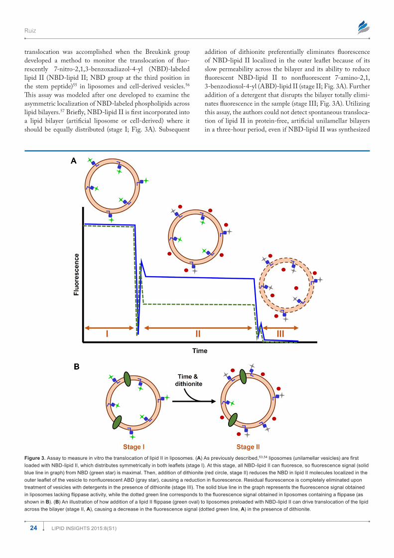
translocation was accomplished when the Breukink group developed a method to monitor the translocation of fluo-rescently 7-nitro-2,1,3-benzoxadiazol-4-yl (NBD)-labeled lipid II (NBD-lipid II; NBD group at the third position in the stem peptide)55 in liposomes and cell-derived vesicles.56 This assay was modeled after one developed to examine the asymmetric localization of NBD-labeled phospholipids across lipid bilayers.57 Briefly, NBD-lipid II is first incorporated into a lipid bilayer (artificial liposome or cell-derived) where it should be equally distributed (stage I; Fig. 3A). Subsequent
addition of dithionite preferentially eliminates fluorescence of NBD-lipid II localized in the outer leaflet because of its slow permeability across the bilayer and its ability to reduce fluorescent NBD-lipid II to nonfluorescent 7-amino-2,1, 3-benzodioxol-4-yl (ABD)-lipid II (stage II; Fig. 3A). Further addition of a detergent that disrupts the bilayer totally elimi-nates fluorescence in the sample (stage III; Fig. 3A). Utilizing this assay, the authors could not detect spontaneous transloca-tion of lipid II in protein-free, artificial unilamellar bilayers in a three-hour period, even if NBD-lipid II was synthesized
Figure 3. Assay to measure in vitro the translocation of lipid II in liposomes. (A) As previously described,53,54 liposomes (unilamellar vesicles) are first loaded with NBD-lipid II, which distributes symmetrically in both leaflets (stage I). At this stage, all NBD-lipid II can fluoresce, so fluorescence signal (solid blue line in graph) from NBD (green star) is maximal. Then, addition of dithionite (red circle, stage II) reduces the NBD in lipid II molecules localized in the outer leaflet of the vesicle to nonfluorescent ABD (gray star), causing a reduction in fluorescence. Residual fluorescence is completely eliminated upon treatment of vesicles with detergents in the presence of dithionite (stage III). The solid blue line in the graph represents the fluorescence signal obtained in liposomes lacking flippase activity, while the dotted green line corresponds to the fluorescence signal obtained in liposomes containing a flippase (as shown in B). (B) An illustration of how addition of a lipid II flippase (green oval) to liposomes preloaded with NBD-lipid II can drive translocation of the lipid across the bilayer (stage II, A), causing a decrease in the fluorescence signal (dotted green line, A) in the presence of dithionite.

Lipid flippases for bacterial peptidoglycan biosynthesis
25Lipid insights 2015:8(s1)
by MurG in situ.56 In contrast, they detected lipid II trans-location in a modified assay using membrane vesicles derived from E. coli cells without the addition of an energy source.56 Together, these data suggested that translocation of lipid II across cell-derived vesicles was mediated by a flippase(s) through an ATP- and pmf-independent mechanism.
This dithionite-based assay was subsequently used to test whether purified FtsW translocates NBD-lipid II across pro-teoliposomes (Fig. 3).54 Specifically, detergent-purified FtsW was incorporated into unilamellar vesicles loaded with NBD-lipid II. While adding dithionite to protein-free liposomes eliminated ~50% of the fluorescence corresponding to the NBD-lipid II molecules randomly incorporated into the outer leaflet of the bilayer, there was a reduction of up to 70% in fluo-rescence in liposomes containing FtsW. This additional FtsW-dependent decrease in fluorescence was not detected when liposomes contained other control proteins, even the lipid II flippase candidate MurJ. In addition, this in vitro reconsti-tution assay was complemented by a novel Förster resonance energy transfer (FRET)-based assay to examine transloca-tion of NBD-lipid II across right-side-out membrane vesicles derived from cells where FtsW was either overproduced or depleted.54 The Breukink group took advantage of the fact that the membrane-impermeable antibiotic vancomycin binds to the d-Ala-d-Ala terminal portion of the stem peptide of lipid II.54,58 In their assay, they monitored the energy trans-fer between NBD-lipid II (donor) and tetramethylrhodamine cadaverine (TMR)-vancomycin (acceptor). If the two fluo-rophores were in close proximity in the same leaflet, energy transfer between NBD and TMR would occur, resulting in a decrease in the NBD signal and a concomitant increase in the TMR fluorescence. This study showed that there was an increase in FRET signal in right-side-out vesicles derived from cells overproducing FtsW and a decrease in those derived from cells depleted of FstW. In contrast, altering the levels of MurJ did not have an effect. From these data, the authors con-cluded that FtsW is a lipid II flippase.54
Mohammadi et al recently addressed two important tests missing in their earlier study: (a) substrate specificity of the reported FtsW-dependent translocation and (b) dependence of transport on FtsW function (ie, testing inactive FtsW proteins).53 With respect to the substrate specificity issue, they first tested whether purified FtsW could flip NBD-phospholipids across artificial unilamellar vesicles using the dithionite-based assay (Fig. 3).53,54 Their results showed that the addition of wild-type FtsW to liposomes promoted the translocation of the three phospholipids tested, phosphatidyl-ethanolamine and phosphatidylglycerol, which are the major phospholipids in E. coli, and phosphatidylcholine, which E. coli does not produce. Furthermore, they showed that FtsW also promoted the translocation of several NBD-lipid II ana-logs larger than NBD-lipid II. From this set of experiments, the authors concluded that FtsW flips both phospholipids and lipid II, and that there is a size and shape limit for substrates.53
The requirement for FtsW function was tested by utilizing a collection of FtsW variants that either lacked some of its 10 TMDs or carried specific residue substitutions.53 Surprisingly, truncated FtsW proteins lacking TMD 10, TMDs 7–10, or TMDs 5–10 were still able to induce translocation of NBD-lipid II across proteoliposomes. Thus, addition of a truncated FtsW variant containing only TMDs 1–4 was sufficient to flip lipid II. This study also showed that full-length FtsW proteins containing single residue substitutions (R145A and K153N) in TMD 4 exhibited dominant-negative effects in E. coli cells and could not promote translocation of NBD-lipid II in their in vitro assay. However, addition of these mutant proteins to liposomes still caused the translocation of NBD-phospholipids. Based on these data, the authors concluded that FtsW transports lipid II and phospholipids through dif-ferent mechanisms of facilitated diffusion and hypothesized that transport occurs through a pore-like structure.53
Case for MurJMurJ (formerly MviN) is a 14-TMD membrane protein34 required for PG biogenesis.32,59–62 I identified MurJ as the only lipid II flippase candidate in E. coli using a reductionist bioinformatics search that took advantage of the small size of the genome of endosymbiotic bacteria that are closely related to the free-living bacterium E. coli.32 Independently, the Kato group also found murJ as a gene required for PG biogenesis when analyzing a set of chromosomal deletion mutants of E. coli.59 Both studies demonstrated that MurJ is required for cell viability and maintenance of cellular shape and integrity. Importantly, they also showed that depletion of MurJ caused a decrease in PG synthesis and an accumulation of PG nucle-otide and lipid intermediates.32,59 Although these data were in agreement with MurJ being an essential lipid II flippase, what led us to propose that MurJ itself was the long sought-after lipid II flippase32,59 was the fact that MurJ belongs to the multidrug/oligo-saccharidyl-lipid/polysaccharide (MOP) exporter superfamily of proteins.63
Members of the MOP exporter superfamily include mul-tidrug transporters64–66 and the aforementioned Wzx pro-teins, which flip Und-PP-oligosaccharides across bacterial cytoplasmic membranes.26,67–70 A notable side note is that a member of the MOP exporter superfamily is the eukaryotic Rft1 protein,63 which has been the center of great controversy regarding its role in flipping polyisoprenoid-oligosaccharide precursors during N-glycosylation.71–75 Briefly, the main arguments are the following. Helenius et al showed that in vivo depletion of the essential yeast protein Rft1 caused a decrease in N-glycosylation and an accumulation of the polyisoprenoid-oligosaccharide precursor that needs to be flipped across the membrane of the endoplasmic reticulum (ER) in this pathway.72 Moreover, increasing production of Rft1 suppressed defects conferred by mutations that lead to the synthesis of an incomplete polyisoprenoid-oligosaccharide precursor of N-glycosylation that is poorly translocated across

Ruiz
26 Lipid insights 2015:8(s1)
the ER membrane. These results demonstrated that Rft1 is necessary for the membrane translocation of N-glycosylation precursors. However, Frank et al concluded that Rft1 is not the flippase itself because liposomes reconstituted with ER proteins extracted from cells depleted of Rft1 still translocated N-glycosylation polyisoprenoid-oligosaccharide precursors in an in vitro assay.71
The best studied members of the MOP exporter super-family are multidrug exporters belonging to the multidrug and toxic extrusion (MATE) family for which there is a large body of genetic, biochemical, and structural data.63,76 Crys-tallography studies have revealed that their 12 TMDs are arranged into two six-helix bundles that form a V-shaped central cavity mainly lined by TMDs 1, 2, 7, and 8, which is essential for transport.77–81 In general, MATE transporters extract amphipathic molecules from the bacterial cytoplasm by an antiport mechanism that takes advantage of the electro-chemical gradient of protons or cations across the cytoplasmic membrane. It has been proposed that through an alternating-access model of transport, their central cavity opens to the cytoplasm in order to load the cargo and then undergoes a conformational change so that it opens to the other side of the membrane to deliver its cargo. During this process, a counter ion (proton or cation) is imported across the membrane. Inter-estingly, these structural studies have also revealed significant differences in how several members of the MATE family bind cations and substrates.78–81
Although there are still many unanswered questions regarding their mechanism of function, it is clear that a key structural and functional feature of the MATE transport-ers is their central cavity.77–81 Also relevant to our discussion are recent studies on MOP exporter member Wzx suggesting that it might transport Und-PP-O-antigen molecules using a mechanism similar to that described for MATE transport-ers. A three-dimensional structural model has predicted that the 12-TMDs82 of the Pseudomonas aeruginosa Wzx protein fold into a V-shaped structure with a central cationic cavity that might transport its anionic Und-PP-oligosaccharide substrate.70 Although transport of the native substrate has not yet been reconstituted, in vitro studies have shown proton-dependent transport of anions in proteoliposomes containing purified Wzx.69 Furthermore, a combination of in vivo and in vitro functional analyses of various Wzx mutant proteins has revealed the importance of charged and aromatic residues within the central cavity, suggesting that they interact with the substrate and/or protons.69,70
Given that structural and functional features might be conserved among members of the MOP exporter superfamily, my laboratory conducted structure–function analyses on MurJ. These studies generated a three-dimensional structural model that predicted that MurJ is structurally similar to other MOP exporters (Fig. 4).34 Further, we probed this model in vivo by determining the accessibility of specific MurJ resi-dues to the periplasm, the cytoplasm, and the hydrophobic
membrane environment. This detailed topological study validated the most salient features of the structural model.34 Namely, it demonstrated that MurJ has 14 TMDs and TMDs 1–12 adopt a V-shaped structure with a solvent-exposed cavity mainly lined by TMDs 1, 2, 7, and 8 (Fig. 4). Moreover, func-tional studies revealed that the native charge of specific residues within this cavity is required for MurJ function in E. coli.34,35
The most logical and simplest explanation of the results on MurJ discussed so far is that MurJ is the lipid II flippase: MurJ is required for PG biogenesis, it belongs to the MOP exporter superfamily, it adopts a structure that resembles that of MATE transporters, it has a solvent-exposed hydrophilic cavity, and depleting MurJ from cells causes the accumulation of lipid-linked PG precursors.32,34,35,59 Nevertheless, several studies have called this proposal into question. Purified FtsW, and not purified MurJ, had been shown to promote lipid II transloca-tion in the in vitro reconstitution system described above.54 In addition, although ytgP, the murJ ortholog in Gram-positive bacteria,33 had been shown to be essential in Staphylococ-cus aureus and Streptococcus pneumoniae,83,84 two independent studies disputed the essentiality of the function of YtgP in B. subtilis, a bacterium encoding multiple YtgP homologs.33,85,86 Because B. subtilis cells lacking four of the ytgP paralogs are viable, it was disputed that these proteins could not perform the essential function of translocating lipid II.85,87 As described below, the reason for this apparent discrepancy is that B. subtilis encodes AmJ, a protein that is redundant with YtgP (MurJ).31
Figure 4. A structural model of MurJ. The front view of the model structure of MurJ of E. coli showing the central cavity opened toward the periplasm.34 The cavity is mainly lined by TMDs 1 (blue), 2 (cyan), 7 (magenta), and 8 (red).

Lipid flippases for bacterial peptidoglycan biosynthesis
27Lipid insights 2015:8(s1)
Ultimately, the question that sparked studies on FtsW and MurJ is the following: which protein(s) flips lipid II in the cell? Focusing on the FtsW–MurJ controversy, a question that needed to be addressed was the following: do any of these two proteins flip or at least participate in the flipping of lipid II in cells? The Bernhardt, Kahne, and Ruiz groups joined forces to address this question in E. coli. First, we developed a method to monitor in vivo the translocation of lipid II across the cyto-plasmic membrane in E. coli (Fig. 5).36 This method takes advantage of the colicin M (ColM) toxin that some strains of E. coli produce to kill other members of its own species.88,89 Specifically, after ColM is secreted into the environment, it
can cross the OM of target cells to enter the periplasm. There, it inhibits PG biogenesis by hydrolyzing periplasmic lipid II into Und and PP-disaccharide-pentapeptide, which can be further cleaved by periplasmic carboxypeptidases (CPs) into a PP-disaccharide-tetrapeptide product.36,88 Because ColM cannot cross the cytoplasmic membrane of target cells, it is specific for periplasmic (or flipped) lipid II. Therefore, if cells are treated with ColM for a given period, the amount of PP-disaccharide-tetrapeptide should reflect how much lipid II was flipped in that period. Second, we developed a method to specifically and rapidly inhibit MurJ function with a small molecule. To do this, we utilized a functional MurJ variant
Figure 5. Assay to measure in vivo the translocation of lipid II in E. coli cells. (A) As previously described,31,36 purified toxin ColM is added to actively growing E. coli cells. ColM crosses the OM to enter the periplasm, where it inhibits PG biogenesis by cleaving lipid II that has been flipped to the periplasm (lipid iiperiplasmic). ColM cleaves lipid II into membrane-bound undecaprenol and soluble PP-disaccharide-pentapeptide, which is further converted by periplasmic CPs into PP-disaccharide-tetrapeptide (marked by blue box). (B) A schematic showing the experimental details of the assay.31,36 PG precursors are specifically labeled with 3H-meso-diaminopimelic acid (3H-DAP) and then treated or not with ColM. Before cell lysis occurs (drop in growth curve), cells are collected and extracted with boiling water. Species in the water-soluble fraction are separated using high-pressure liquid chromatography and radioactivity present in the ColM disaccharide-tetrapeptide product (blue box, A) is then measured. Radiolabeled lipid II that is not cleaved by ColM (lipid IIcytoplasmic) is measured after extracting the water-insoluble fraction with butanol. When flippase activity is not impaired, treatment with ColM leads to the appearance of signal in the fraction corresponding to the ColM product and the disappearance of signal from the lipid IIcytoplasmic fraction. When flippase activity is inhibited, the amount of signal in the lipid IIcytoplasmic fraction increases and the treatment of ColM does not lead to the appearance of signal corresponding to the ColM product.36
Ruiz
28 Lipid insights 2015:8(s1)
(MurJA29C) carrying a single Cys substitution localized in the central cavity of MurJ. Haploid cells producing only Mur-JA29C were viable and could synthesize PG. However, if these mutant cells were treated with MTSES (2-Sulfonatoethyl methanethiosulfonate), a small molecule that reacts with free Cys residues to form adducts, they rapidly lysed because they could not synthesize PG. This lethality was shown to be spe-cific for the presence of the A29C substitution and was caused by the loss of function of the MurJA29C variant. Together, these methods allowed us to specifically inhibit MurJ in cells and rapidly detect the effect that this inhibition had in lipid II translocation using the ColM-based flippase assay. In essence, from these data, we concluded that inhibiting MurJ resulted in the loss of all the measurable translocation and the accu-mulation of lipid II that could only be cleaved by ColM if the cytoplasmic membrane was lysed. Furthermore, we also showed that depletion of FtsW in a strain-lacking RodA did not reduce lipid II translocation.36
From these results36 and the fact that MurJ, a member of the MOP exporter superfamily, is essential32,59,63 and structur-ally similar to MOP exporters, and possesses a solvent-exposed cavity that is essential for function,34,35 we concluded that MurJ is the lipid II flippase in E. coli. Indeed, based on the credit card swipe model proposed for the transport of polar lipids6,24 and the mechanism of function proposed for MATE exporters,78–81 my group has also proposed that MurJ flips lipid II using an alternating-access mechanism: during transport, charged resi-dues in the cavity of MurJ interact with the hydrophilic moiety of lipid II, while its lipid portion stays in the membrane, possibly sliding between TMDs 1 and 8 or 1 and 5.35 Whether the nega-tive charges located in the cavity that we showed to be essential interact with the substrate, promote intramolecular interactions, or interact with a counter ion remains unknown. Moreover, it is interesting that all of the charged residues that are critical for MurJ function are localized to the top half of the central cavity.35 Therefore, it is possible that directional transport is not driven by an antiport mechanism but by an increase in the binding affinity of MurJ for lipid II as transport proceeds from the cyto-plasmic bottom half of the cavity (lower affinity) to the external upper half (higher affinity). It is also possible that MurJ might not use an alternating-access mechanism at all, as has recently been proposed by the Locher group for PglK, the ABC trans-porter of the Und-PP-oligosaccharide used in N-glycosylation in bacteria.90 In their structural study, the authors proposed a novel mechanism where the cavity does not open to the cyto-plasmic side of the membrane. Instead, the polyisoprenoid end of the molecule engages with a helix in the periplasmic face of the membrane, inducing the interaction of the pyrophosphate-oligosaccharide portion of the substrate with positively charged residues within the outward-facing cavity and driving transport.
Case for AmJAs described above, an argument used against MurJ being a lipid II flippase was the fact that a mutant lacking ytgP,
the murJ ortholog in Gram-positive bacteria,33 and three other paralogs was viable.85,87 Recently, the Bernhardt and Rudner laboratories have demonstrated that ytgP is not essential in B. subtilis because this organism encodes a protein, AmJ, that is functionally redundant with YtgP (MurJ).31 After show-ing that a B. subtilis mutant strain lacking all 10 members of the MOP exporter superfamily is viable, the authors used a genetic screen for synthetic lethality with the DytgP allele to identify proteins that might function as lipid II flippases. Spe-cifically, they used a strain lacking the four MOP exporters most closely related to MurJ (including YtgP) and transpo-son mutagenesis in a screen designed to identify genes whose inactivation would cause lethality only in this mutant devoid of MurJ-like proteins. Assuming that YtgP (MurJ) is a lipid II flippase, the rationale of the screen was that if another protein functions as the lipid II flippase in the absence of YtgP, its genetic inactivation would cause lethality, since lipid II trans-location is a process essential for cell viability. With this strategy, the authors identified AmJ (formerly YdaH) as an alternate to MurJ. They further demonstrated that amJ and ytgP are indeed a synthetic lethal pair required for PG biogen-esis in B. subtilis, and that in E. coli, production of B. subtilis AmJ can substitute for native MurJ in lipid II translocation.31
Analysis of the primary sequence of AmJ reveals that this polytopic membrane protein has no similarity to MurJ or any member of either the MOP exporter superfamily or ABC trans-porters, suggesting that AmJ is the founding member of a new type of protein involved in the translocation of Und-PP-linked sugars.31 In addition, AmJ is not widely conserved in bacteria and its transcription is regulated by sM,91,92 a sigma factor that is positively regulated by cell wall stress.91,93 In fact, amJ tran-scription is upregulated in the absence of YtgP in B. subtilis.31 These findings have led to the proposal that having AmJ, which is very different from MurJ, might benefit organisms that somehow might come into contact with environmental condi-tions that inhibit MurJ (YtgP).31 Alternatively, AmJ could be a transporter of a yet-to-be-identified substrate and promiscu-ously transport lipid II since some, but not all, transporters of Und-PP-linked oligosaccharides from both the Wzx and ABC transporter types have relaxed substrate specificity.94–100
Finding AmJ raises the question of whether there could be additional types of transporters capable of translocating Und-PP-oligosaccharides. Indeed, the Raetz laboratory proposed that ArnE and ArnF (formerly, PmrM and PmrL) work, pos-sibly in a complex, to translocate Und-PP-L-aminoarabinose across the cytoplasmic membrane of E. coli and Salmonella.101 Each of these proteins is predicted to have four TMDs, and they are distantly related to the drug/metabolite transporter superfamily.102 However, how these transporters function remains unknown.
CounterargumentsYou have now read the arguments based on positive and nega-tive results that support FtsW and discount MurJ (and AmJ),
Lipid flippases for bacterial peptidoglycan biosynthesis
29Lipid insights 2015:8(s1)
respectively, or vice versa. The FtsW argument is fueled by results obtained in an in vitro reconstitution system but lacks support from in vivo evidence. On the contrary, the MurJ model is fueled by a collection of evidence obtained in vivo but lacks proof from a reconstitution system.
Pro-MurJ (and AmJ) counterargument against FtsW. It should first be noted that the lack of lipid II flippase activ-ity of MurJ in the in vitro assay54 could be the result of trivial explanations, such as the inactivation of the protein during purification. In addition, validity of the in vitro reconstitu-tion system supporting FtsW as a lipid II flippase requires that the addition of purified FtsW does not cause disorder of the lipid membrane, which could result in a nonspecific increase in permeability to either dithionite or lipid II.57,103 Although other proteins tested did not induce lipid II translocation, this issue is protein-specific and it has not been thoroughly ruled out for FtsW. In fact, this artifact could explain why in the dithionite-based assay, FtsW is reported to rapidly flip lipid II and several types of phospholipids.53,54 More importantly, regardless of whether the in vitro flippase assay is flawed or not, ultimately, we want to know if FtsW flips lipid II in cells. At present, there is no in vivo experimental evidence sup-porting FtsW’s function as a lipid II flippase in a cell. On the contrary, data from experiments designed to address this issue for FtsW, MurJ, and AmJ have indicated that MurJ and AmJ are required in lipid II translocation in E. coli, while FtsW and RodA are not.31,36 Therefore, the simplest explanation of these results and those demonstrating that MurJ is essential in E. coli and structurally similar to related transporters of lipid II-like substrates is that MurJ is the lipid II flippase in this bacterium. Furthering this reasoning, given that AmJ can substitute for MurJ in E. coli and that amJ and murJ ortholog are a synthetic lethal pair in B. subtilis,31 it follows that AmJ is a transporter that can flip lipid II in cells; whether AmJ is specific to lipid II or not awaits investigation.
Pro-FtsW counterargument against MurJ. In an in vitro reconstitution system, purified MurJ, unlike FtsW, has failed to induce lipid II translocation across liposomes.54 Further-more, levels of MurJ, unlike those of FtsW, do not correlate with lipid II translocation in an in vitro assay using cell-derived vesicles.54 Therefore, FtsW, and not MurJ, is a lipid II flippase. The in vivo system supporting MurJ as a lipid II flip-pase does not allow one to discern whether the effect on lipid II translocation is direct or indirect. Only an in vitro reconstitu-tion system can, and such a system has shown that FtsW, and not MurJ, can flip lipid II. Moreover, the in vivo studies by Sham et al36 did not report values of radioactivity (3H-DAP; Fig. 5) present in the fraction corresponding to the mature PG sacculus that might reflect undetected flippase activity even when MurJ is inhibited. Thus, it is possible that when MurJ was inhibited, there was still some level of lipid II transloca-tion occurring (that could be mediated by FtsW) that could not be detected in the in vivo assay because of the notable background level (or low sensitivity of detection) of this assay.
Could Both FtsW and MurJ be Lipid II Flippases?Both FtsW and MurJ are essential in E. coli, implying that they perform nonredundant essential functions. Therefore, if both FtsW and MurJ were lipid II flippases, they either would have to be redundant but perform additional essential func-tions or should each serve as a lipid II flippase required for a unique purpose during growth and division. With respect to the first point, although FtsW is known to be essential for the proper assembly of the divisome,43–45 there are no data hinting that MurJ could perform an additional function. With respect to both FtsW and MurJ being lipid II flippases required for a unique purpose, we should remember that FtsW’s paralog RodA is likely to perform the same function during cell elongation that FtsW plays during cell division, calling into question in what other circumstance MurJ would be essential. In addition, the in vivo assay showed that all the detectable flippase activity was abolished upon MurJ inacti-vation.31,36 Therefore, if all three proteins were lipid II flip-pases, these data would argue that MurJ would be the main flippase and that the activity of RodA and FtsW flippases is minimal (below detection) but essential. There are no current data or model in PG biogenesis that would explain this latter situation. Furthermore, the idea that the cell uses different flippases for specific essential functions seems only possible if lipid II is not free (diffusible) in the membrane. Some mem-bers of the PG community believe that lipid II is handed off from MurG to a flippase and then to a transglycosylase in a PG-synthesizing multiprotein complex. To my knowledge, there is no experimental evidence supporting this model; furthermore, a recent study by Grabowicz et al might argue against it.104 In this work, the authors isolated a mutant of E. coli that produces a variant of WaaL (or RfaL), the ligase that normally adds the polysaccharide portion of Und-PP-O-antigen to the core of a glycolipid in order to synthesize LPS.105 This ligation occurs in the periplasmic side of the cytoplasmic membrane. Interestingly, the mutation in waaL causes the altered WaaL protein to have relaxed substrate specificity so that it can utilize the disaccharide-pentapeptide moiety from lipid II as a substrate and ligate it to produce a novel form of LPS.104 Although it is unclear whether the waaL mutant cells produce normal or elevated levels of lipid II, the fact that flipped, periplasmic lipid II is accessible to the mutant WaaL protein suggests that the lipid II flippase does not directly hand off lipid II after transport to the PG transglycosylases.
Based on these arguments, it seems unlikely that both FtsW and MurJ function as lipid II flippases in E. coli. What is clear is that more studies are needed to understand how FtsW (and RodA) and MurJ (and AmJ) function.
Instructions to the JuryThis review was intended to serve as a guide that highlights the recent investigative developments in the essential process of translocation of lipid II across the bacterial cytoplasmic

Ruiz
30 Lipid insights 2015:8(s1)
membrane. In the past few years, studies have focused on the identification of the lipid II flippase and have resulted in two opposing camps in the PG community: those who defend FtsW based on an in vitro reconstitution system and those who defend MurJ and AmJ based on data generated in vivo. Using this review as a guide, I now encourage you to carefully exam-ine the evidence, the primary literature, and decide whether or not the controversy about the identity of the lipid II flippase has been resolved beyond a reasonable doubt or, at least, based on the preponderance of the evidence.
AcknowledgmentI thank Rebecca M. Davis for her critical reading of the manuscript.
Author ContributionsWrote the first draft of the manuscript: NR. Agree with manuscript results and conclusions: NR. Jointly developed the structure and arguments for the paper: NR. Made criti-cal revisions and approved final version: NR. Author reviewed and approved of the final manuscript.
REFERENCES1. Vollmer W, Bertsche U. Murein (peptidoglycan) structure, architecture and bio-
synthesis in Escherichia coli. Biochim Biophys Acta. 2008;1778(9):1714–1734.2. Vollmer W, Blanot D, de Pedro MA. Peptidoglycan structure and architecture.
FEMS Microbiol Rev. 2008;32(2):149–167.3. Typas A, Banzhaf M, Gross CA, et al. From the regulation of peptidoglycan
synthesis to bacterial growth and morphology. Nat Rev Microbiol. 2012;10(2):123–136.
4. Young KD. The selective value of bacterial shape. Microbiol Mol Biol Rev. 2006;70(3):660–703.
5. Silhavy TJ, Kahne D, Walker S. The bacterial cell envelope. Cold Spring HarbPerspect Biol. 2010;2(5):a000414.
6. Sanyal S, Menon AK. Flipping lipids: why an’ what’s the reason for? ACS Chem Biol. 2009;4(11):895–909.
7. Chatterjee AN, Park JT. Biosynthesis of cell wall mucopeptide by a particulatefraction from Staphylococcus aureus. Proc Natl Acad Sci U S A. 1964;51:9–16.
8. Meadow PM, Anderson JS, Strominger JL. Enzymatic polymerization of UDP-acetylmuramyl.L-ala.D-glu.L-lys.D-ala.D-ala and UDP-acetylglucosamine bya particulate enzyme from Staphylococcus aureus and its inhibition by antibiotics. Biochem Biophys Res Commun. 1964;14:382–387.
9. Barreteau H, Kovac A, Boniface A, et al. Cytoplasmic steps of peptidoglycanbiosynthesis. FEMS Microbiol Rev. 2008;32(2):168–207.
10. Mengin-Lecreulx D, Texier L, Rousseau M, et al. The murG gene of Escherichia coli codes for the UDP-N-acetylglucosamine: N-acetylmuramyl-(pentapeptide) pyrophosphoryl-undecaprenol N-acetylglucosamine transferase involvedin the membrane steps of peptidoglycan synthesis. J Bacteriol. 1991;173(15): 4625–4636.
11. Ikeda M, Wachi M, Jung HK, et al. The Escherichia coli mraY gene encodingUDP-N-acetylmuramoyl-pentapeptide: undecaprenyl-phosphate phospho-N-acetylmuramoyl-pentapeptide transferase. J Bacteriol. 1991;173(3):1021–1026.
12. Bouhss A, Trunkfield AE, Bugg TD, et al. The biosynthesis of peptidoglycanlipid-linked intermediates. FEMS Microbiol Rev. 2008;32(2):208–233.
13. van Heijenoort J. Lipid intermediates in the biosynthesis of bacterial peptidogly-can. Microbiol Mol Biol Rev. 2007;71(4):620–635.
14. Van Heijenoort Y, Derrien M, Van Heijenoort J. Polymerization by transglyco-sylation in the biosynthesis of the peptidoglycan of Escherichia coli K 12 and itsinhibition by antibiotics. FEBS Lett. 1978;89(1):141–144.
15. Broome-Smith JK, Edelman A, Yousif S, et al. The nucleotide sequences ofthe ponA and ponB genes encoding penicillin-binding protein 1A and 1B ofEscherichia coli K12. Eur J Biochem. 1985;147(2):437–446.
16. Sauvage E, Kerff F, Terrak M, et al. The penicillin-binding proteins: structure and role in peptidoglycan biosynthesis. FEMS Microbiol Rev. 2008;32(2):234–258.
17. Thorne KJ, Kodicek E. The structure of bactoprenol, a lipid formed by lactoba-cilli from mevalonic acid. Biochem J. 1966;99(1):123–127.
18. Anderson JS, Matsuhashi M, Haskin MA, et al. Biosynthesis of the peptido-glycan of bacterial cell walls. II. Phospholipid carriers in the reaction sequence. J Biol Chem. 1967;242(13):3180–3190.
19. Higashi Y, Strominger JL, Sweeley CC. Structure of a lipid intermediate in cell wall peptidoglycan synthesis: a derivative of a C55 isoprenoid alcohol. Proc Natl Acad Sci U S A. 1967;57(6):1878–1884.
20. Umbreit JN, Strominger JL. Isolation of the lipid intermediate in peptidoglycan biosynthesis from Escherichia coli. J Bacteriol. 1972;112(3):1306–1309.
21. Bupp K, van Heijenoort J. The final step of peptidoglycan subunit assembly inEscherichia coli occurs in the cytoplasm. J Bacteriol. 1993;175(6):1841–1843.
22. van Heijenoort Y, Gomez M, Derrien M, et al. Membrane intermediates in the peptidoglycan metabolism of Escherichia coli: possible roles of PBP 1b and PBP 3. J Bacteriol. 1992;174(11):3549–3557.
23. Kramer NE, Smid EJ, Kok J, et al. Resistance of Gram-positive bacteria tonisin is not determined by lipid II levels. FEMS Microbiol Lett. 2004;239(1):157–161.
24. Pomorski T, Menon AK. Lipid flippases and their biological functions. Cell Mol Life Sci. 2006;63(24):2908–2921.
25. Islam ST, Lam JS. Wzx flippase-mediated membrane translocation of sugarpolymer precursors in bacteria. Environ Microbiol. 2013;15(4):1001–1015.
26. Islam ST, Lam JS. Synthesis of bacterial polysaccharides via the Wzx/Wzy-dependent pathway. Can J Microbiol. 2014;60(11):697–716.
27. Cuthbertson L, Kos V, Whitfield C. ABC transporters involved in export of cell surface glycoconjugates. Microbiol Mol Biol Rev. 2010;74(3):341–362.
28. Greenfield LK, Whitfield C. Synthesis of lipopolysaccharide O-antigens byABC transporter-dependent pathways. Carbohydr Res. 2012;356:12–24.
29. Willis LM, Whitfield C. Structure, biosynthesis, and function of bacterial cap-sular polysaccharides synthesized by ABC transporter-dependent pathways.Carbohydr Res. 2013;378:35–44.
30. Young KD. Microbiology. A flipping cell wall ferry. Science. 2014;345(6193):139–140.
31. Meeske AJ, Sham LT, Kimsey H, et al. MurJ and a novel lipid II flippase arerequired for cell wall biogenesis in Bacillus subtilis. Proc Natl Acad Sci U S A. 2015;112(20):6437–6442.
32. Ruiz N. Bioinformatics identification of MurJ (MviN) as the peptidogly-can lipid II flippase in Escherichia coli. Proc Natl Acad Sci U S A. 2008;105(40): 15553–15557.
33. Ruiz N. Streptococcus pyogenes YtgP (Spy_0390) complements Escherichia colistrains depleted of the putative peptidoglycan flippase MurJ. Antimicrob AgentsChemother. 2009;53(8):3604–3605.
34. Butler EK, Davis RM, Bari V, et al. Structure-function analysis of MurJ reveals a solvent-exposed cavity containing residues essential for peptidoglycan biogenesis in Escherichia coli. J Bacteriol. 2013;195(20):4639–4649.
35. Butler EK, Tan WB, Joseph H, et al. Charge requirements of lipid II flippaseactivity in Escherichia coli. J Bacteriol. 2014;196(23):4111–4119.
36. Sham LT, Butler EK, Lebar MD, et al. Bacterial cell wall. MurJ is the flippaseof lipid-linked precursors for peptidoglycan biogenesis. Science. 2014;345(6193):220–222.
37. de Boer PA. Advances in understanding E. coli cell fission. Curr Opin Microbiol. 2010;13(6):730–737.
38. Egan AJ, Vollmer W. The physiology of bacterial cell division. Ann N Y Acad Sci. 2013;1277:8–28.
39. Miyakawa T, Matsuzawa H, Matsuhashi M, et al. Cell wall peptidoglycanmutants of Escherichia coli K-12: existence of two clusters of genes, mra and mrb, for cell wall peptidoglycan biosynthesis. J Bacteriol. 1972;112(2):950–958.
40. Ishino F, Jung HK, Ikeda M, et al. New mutations fts-36, lts-33, and ftsW clus-tered in the mra region of the Escherichia coli chromosome induce thermosensitive cell growth and division. J Bacteriol. 1989;171(10):5523–5530.
41. Boyle DS, Khattar MM, Addinall SG, et al. ftsW is an essential cell-divisiongene in Escherichia coli. Mol Microbiol. 1997;24(6):1263–1273.
42. Wang L, Khattar MK, Donachie WD, et al. FtsI and FtsW are localized to the septum in Escherichia coli. J Bacteriol. 1998;180(11):2810–2816.
43. Mercer KL, Weiss DS. The Escherichia coli cell division protein FtsW is required to recruit its cognate transpeptidase, FtsI (PBP3), to the division site. J Bacteriol. 2002;184(4):904–912.
44. Fraipont C, Alexeeva S, Wolf B, et al. The integral membrane FtsW protein and peptidoglycan synthase PBP3 form a subcomplex in Escherichia coli. Microbiology. 2011;157(pt 1):251–259.
45. Datta P, Dasgupta A, Singh AK, et al. Interaction between FtsW and penicillin-binding protein 3 (PBP3) directs PBP3 to mid-cell, controls cell septation andmediates the formation of a trimeric complex involving FtsZ, FtsW and PBP3 in mycobacteria. Mol Microbiol. 2006;62(6):1655–1673.
46. Lara B, Ayala JA. Topological characterization of the essential Escherichia colicell division protein FtsW. FEMS Microbiol Lett. 2002;216(1):23–32.
47. Ikeda M, Sato T, Wachi M, et al. Structural similarity among Escherichia coliFtsW and RodA proteins and Bacillus subtilis SpoVE protein, which functionin cell division, cell elongation, and spore formation, respectively. J Bacteriol. 1989;171(11):6375–6378.

Lipid flippases for bacterial peptidoglycan biosynthesis
31Lipid insights 2015:8(s1)
48. Matsuzawa H, Hayakawa K, Sato T, et al. Characterization and genetic analy-sis of a mutant of Escherichia coli K-12 with rounded morphology. J Bacteriol. 1973;115(1):436–442.
49. Henriques AO, de Lencastre H, Piggot PJ. A Bacillus subtilis morphogene clusterthat includes spoVE is homologous to the mra region of Escherichia coli. Biochi-mie. 1992;74(7–8):735–748.
50. Henriques AO, Glaser P, Piggot PJ, et al. Control of cell shape and elongation by the rodA gene in Bacillus subtilis. Mol Microbiol. 1998;28(2):235–247.
51. Holtje JV. Growth of the stress-bearing and shape-maintaining murein sacculus of Escherichia coli. Microbiol Mol Biol Rev. 1998;62(1):181–203.
52. Matsuhashi M. Utilization of lipid-linked precursors and the formation of pepti-doglycan in the process of cell growth and division: membrane enzymes involved in the final steps of peptidoglycan synthesis and the mechanism of their regula-tion. In: Ghuysen J-M, Hakenbeck R, eds. Bacterial Cell Wall. Amsterdam: Else-vier Science B.V.; 1994:55–71.
53. Mohammadi T, Sijbrandi R, Lutters M, et al. Specificity of the transport of lipid II by FtsW in Escherichia coli. J Biol Chem. 2014;289(21):14707–14718.
54. Mohammadi T, van Dam V, Sijbrandi R, et al. Identification of FtsW as atransporter of lipid-linked cell wall precursors across the membrane. EMBO J. 2011;30(8):1425–1432.
55. Breukink E, van Heusden HE, Vollmerhaus PJ, et al. Lipid II is an intrinsiccomponent of the pore induced by nisin in bacterial membranes. J Biol Chem. 2003;278(22):19898–19903.
56. van Dam V, Sijbrandi R, Kol M, et al. Transmembrane transport of peptidogly-can precursors across model and bacterial membranes. Mol Microbiol. 2007;64(4): 1105–1114.
57. McIntyre JC, Sleight RG. Fluorescence assay for phospholipid membrane asym-metry. Biochemistry. 1991;30(51):11819–11827.
58. Perkins HR. Specificity of combination between mucopeptide precursors andvancomycin or ristocetin. Biochem J. 1969;111(2):195–205.
59. Inoue A, Murata Y, Takahashi H, et al. Involvement of an essential gene, mviN, in murein synthesis in Escherichia coli. J Bacteriol. 2008;190(21):7298–7301.
60. Mohamed YF, Valvano MAA. Burkholderia cenocepacia MurJ (MviN) homo-log is essential for cell wall peptidoglycan synthesis and bacterial viability. Glyco-biology. 2014;24(6):564–576.
61. Ling JM, Moore RA, Surette MG, et al. The mviN homolog in Burkholderia pseudo-mallei is essential for viability and virulence. Can J Microbiol. 2006;52(9):831–842.
62. Rudnick PA, Arcondeguy T, Kennedy CK, et al. glnD and mviN are genes of an essential operon in Sinorhizobium meliloti. J Bacteriol. 2001;183(8):2682–2685.
63. Hvorup RN, Winnen B, Chang AB, et al. The multidrug/oligosaccharidyl-lipid/polysaccharide (MOP) exporter superfamily. Eur J Biochem. 2003;270(5):799–813.
64. Long F, Rouquette-Loughlin C, Shafer WM, et al. Functional cloning and char-acterization of the multidrug efflux pumps NorM from Neisseria gonorrhoeae and YdhE from Escherichia coli. Antimicrob Agents Chemother. 2008;52(9):3052–3060.
65. Rouquette-Loughlin C, Dunham SA, Kuhn M, et al. The NorM efflux pump of Neisseria gonorrhoeae and Neisseria meningitidis recognizes antimicrobial cationic compounds. J Bacteriol. 2003;185(3):1101–1106.
66. Huda MN, Morita Y, Kuroda T, et al. Na+-driven multidrug efflux pump VcmA from Vibrio cholerae non-O1, a non-halophilic bacterium. FEMS Microbiol Lett. 2001;203(2):235–239.
67. Stevenson G, Andrianopoulos K, Hobbs M, et al. Organization of the Esch-erichia coli K-12 gene cluster responsible for production of the extracellular poly-saccharide colanic acid. J Bacteriol. 1996;178(16):4885–4893.
68. Liu D, Cole RA, Reeves PR. An O-antigen processing function for Wzx (RfbX): a promising candidate for O-unit flippase. J Bacteriol. 1996;178(7):2102–2107.
69. Islam ST, Eckford PD, Jones ML, et al. Proton-dependent gating and protonuptake by Wzx support O-antigen-subunit antiport across the bacterial innermembrane. MBio. 2013;4(5):e613–e678.
70. Islam ST, Fieldhouse RJ, Anderson EM, et al. A cationic lumen in the Wzx flip-pase mediates anionic O-antigen subunit translocation in Pseudomonas aeruginosa PAO1. Mol Microbiol. 2012;84(6):1165–1176.
71. Frank CG, Sanyal S, Rush JS, et al. Does Rft1 flip an N-glycan lipid precursor? Nature. 2008;454(7204):E3–E4. [discussion E4–E5].
72. Helenius J, Ng DT, Marolda CL, et al. Translocation of lipid-linked oligosaccharidesacross the ER membrane requires Rft1 protein. Nature. 2002;415(6870):447–450.
73. Helenius J, Ng DTW, Marolda CL, et al. Does Rft1 flip an N-glycan lipid pre-cursor? Reply. Nature. 2008;454(7204):E4–E5.
74. Rush JS, Gao N, Lehrman MA, et al. Suppression of Rft1 expression does notimpair the transbilayer movement of Man5GlcNAc2-P-P-dolichol in sealedmicrosomes from yeast. J Biol Chem. 2009;284(30):19835–19842.
75. Jelk J, Gao N, Serricchio M, et al. Glycoprotein biosynthesis in a eukaryote lack-ing the membrane protein Rft1. J Biol Chem. 2013;288(28):20616–20623.
76. Kuroda T, Tsuchiya T. Multidrug efflux transporters in the MATE family. Bio-chim Biophys Acta. 2009;1794(5):763–768.
77. He X, Szewczyk P, Karyakin A, et al. Structure of a cation-bound multidrug and toxic compound extrusion transporter. Nature. 2010;467(7318):991–994.
78. Lu M, Radchenko M, Symersky J, et al. Structural insights into H+-coupled multi-drug extrusion by a MATE transporter. Nat Struct Mol Biol. 2013;20(11):1310–1317.
79. Lu M, Symersky J, Radchenko M, et al. Structures of a Na+-coupled, substrate-bound MATE multidrug transporter. Proc Natl Acad Sci U S A. 2013;110(6):2099–2104.
80. Tanaka Y, Hipolito CJ, Maturana AD, et al. Structural basis for the drug extrusion mechanism by a MATE multidrug transporter. Nature. 2013;496(7444):247–251.
81. Radchenko M, Symersky J, Nie R, et al. Structural basis for the blockade ofMATE multidrug efflux pumps. Nat Commun. 2015;6:7995.
82. Islam ST, Taylor VL, Qi M, et al. Membrane topology mapping of the O-antigen flippase (Wzx), polymerase (Wzy), and ligase (WaaL) from Pseudomonas aerugi-nosa PAO1 reveals novel domain architectures. MBio. 2010;1(3)e00189–10.
83. Thanassi JA, Hartman-Neumann SL, Dougherty TJ, et al. Identification of 113 conserved essential genes using a high-throughput gene disruption system inStreptococcus pneumoniae. Nucleic Acids Res. 2002;30(14):3152–3162.
84. Zalacain M, Biswas S, Ingraham KA, et al. A global approach to identify novel broad-spectrum antibacterial targets among proteins of unknown function. J Mol Microbiol Biotechnol. 2003;6(2):109–126.
85. Vasudevan P, McElligott J, Attkisson C, et al. Homologues of the Bacillus subtilis SpoVB protein are involved in cell wall metabolism. J Bacteriol. 2009; 191(19):6012–6019.
86. Vasudevan P, Weaver A, Reichert ED, et al. Spore cortex formation in Bacillus subtilis is regulated by accumulation of peptidoglycan precursors under the con-trol of sigma K. Mol Microbiol. 2007;65(6):1582–1594.
87. Fay A, Dworkin J. Bacillus subtilis homologs of MviN (MurJ), the putativeEscherichia coli lipid II flippase, are not essential for growth. J Bacteriol. 2009; 191(19):6020–6028.
88. El Ghachi M, Bouhss A, Barreteau H, et al. Colicin M exerts its bacteriolyticeffect via enzymatic degradation of undecaprenyl phosphate-linked peptidogly-can precursors. J Biol Chem. 2006;281(32):22761–22772.
89. Touze T, Barreteau H, El Ghachi M, et al. Colicin M, a peptidoglycan lipid-II-degrading enzyme: potential use for antibacterial means? Biochem Soc Trans. 2012;40(6):1522–1527.
90. Perez C, Gerber S, Boilevin J, et al. Structure and mechanism of an active lipid-linked oligosaccharide flippase. Nature. 2015;524(7566):433–438.
91. Eiamphungporn W, Helmann JD. The Bacillus subtilis sigma(M) regulon and its contribution to cell envelope stress responses. Mol Microbiol. 2008;67(4):830–848.
92. Jervis AJ, Thackray PD, Houston CW, et al. SigM-responsive genes of Bacillus subtilis and their promoters. J Bacteriol. 2007;189(12):4534–4538.
93. Cao M, Kobel PA, Morshedi MM, et al. Defining the Bacillus subtilis sigma(W) reg-ulon: a comparative analysis of promoter consensus search, run-off transcription/macroarray analysis (ROMA), and transcriptional profiling approaches. J Mol Biol. 2002;316(3):443–457.
94. Alaimo C, Catrein I, Morf L, et al. Two distinct but interchangeable mechanisms for flipping of lipid-linked oligosaccharides. EMBO J. 2006;25(5):967–976.
95. Feldman MF, Marolda CL, Monteiro MA, et al. The activity of a puta-tive polyisoprenol-linked sugar translocase (Wzx) involved in Escherichia coli O antigen assembly is independent of the chemical structure of the O repeat. J Biol Chem. 1999;274(49):35129–35138.
96. Hug I, Couturier MR, Rooker MM, et al. Helicobacter pylori lipopolysaccha-ride is synthesized via a novel pathway with an evolutionary connection to pro-tein N-glycosylation. PLoS Pathog. 2010;6(3):e1000819.
97. Marolda CL, Vicarioli J, Valvano MA. Wzx proteins involved in biosynthesis of O antigen function in association with the first sugar of the O-specific lipopoly-saccharide subunit. Microbiology. 2004;150(pt 12):4095–4105.
98. Hong Y, Reeves PR. Diversity of o-antigen repeat unit structures can accountfor the substantial sequence variation of wzx translocases. J Bacteriol. 2014; 196(9):1713–1722.
99. Hong Y, Cunneen MM, Reeves PR. The Wzx translocases for Salmonella enterica O-antigen processing have unexpected serotype specificity. Mol Microbiol. 2012; 84(4):620–630.
100. Liu MA, Stent TL, Hong Y, et al. Inefficient translocation of a truncated O unit by a Salmonella Wzx affects both O-antigen production and cell growth. FEMS Microbiol Lett. 2015;362(9):fnv053.
101. Yan A, Guan Z, Raetz CR. An undecaprenyl phosphate-aminoarabinose flippase required for polymyxin resistance in Escherichia coli. J Biol Chem. 2007;282(49): 36077–36089.
102. Jack DL, Yang NM, Saier MH Jr. The drug/metabolite transporter superfamily. Eur J Biochem. 2001;268(13):3620–3639.
103. Moreno MJ, Estronca LM, Vaz WL. Translocation of phospholipids and dithi-onite permeability in liquid-ordered and liquid-disordered membranes. Biophys J. 2006;91(3):873–881.
104. Grabowicz M, Andres D, Lebar MD, et al. A mutant Escherichia coli that attaches peptidoglycan to lipopolysaccharide and displays cell wall on its surface. Elife. 2014;3:e05334.
105. Raetz CR, Whitfield C. Lipopolysaccharide endotoxins. Annu Rev Biochem. 2002;71:635–700.


![P4-ATPases: lipid flippases in cell membranes · recently treated different aspects of P4-ATPases [ 18, 77, 86, 94]. In this review, we will first provide an overview on the functional](https://img.dokumen.tips/doc/110x75/5ede6227ad6a402d6669b49d/p4-atpases-lipid-flippases-in-cell-membranes-recently-treated-different-aspects.jpg)