Embed Size (px)
Citation preview
8/10/2019 Journal Adenomyosis
http://slidepdf.com/reader/full/journal-adenomyosis 1/6
Journal of Reproductive Immunology 93 (2012) 58–63
Contents lists available at SciVerse ScienceDirect
Journal of Reproductive Immunology
j ourna l homepage: www.e lsev ier .com/locate / j repr imm
The distribution of immune cells and macrophages in the
endometrium of women with recurrent reproductive failure. II:
adenomyosis and macrophages
Kelton P. Tremellen a,b,∗, Peter Russell c,d
a Repromed, Adelaide, Australiab School of Pharmacy andMedical Science, University of South Australia, Australiac GynaePath,DouglassHanly Moir Pathology, Australia
d Department of Obstetrics andGynaecology, TheUniversity of Sydney, Australia
a r t i c l e i n f o
Article history:
Received 22 September 2011
Received in revised form
23 November 2011
Accepted 5 December 2011
Keywords:
Endometrium
IVF
CD163
Macrophages
Adenomyosis
a b s t r a c t
Adenomyosis, a condition usually associated with multiparity, is not generally seen as a
cause of infertility. However, recent studies have reported a reduction in IVF implantation
rates and a link with miscarriage, suggesting that adenomyosis may interfere with success-
ful implantation. To investigate this hypothesis, the clinical records and laboratory results,
which routinely include immunohistochemicalexamination of a late luteal phase endome-
trial biopsy for leukocytes, were retrospectively reviewed for 64 women with implantation
failure and who previously had been screened for the presence of adenomyosis by pelvic
MRI.
The presence of either diffuse or “adenomyoma” type of adenomyosis was associated
with a marked increase ( p= 0.004) in the density of macrophages and natural killer cells
in the endometrial stroma, compared to those women with mild focal adenomyosis or no
disease. These findings point to an immunologicalmechanismby which adenomyosis might
interfere with successful embryo implantation.
Crown Copyright © 2012 Published by Elsevier Ireland Ltd. All rights reserved.
1. Introduction
Adenomyosis, a disorder related to endometriosis (Kunz
et al., 2005; Leyendecker et al., 2006), is characterised
by heterotopic endometrial glands and stroma in the
myometrium, and is an established cause of menorrha-gia, dysmenorrhoea and pelvic pressure symptoms (Peric
and Fraser, 2006). Most commonly it is diagnosed in mul-
tiparous women in their 5th decade of life and therefore is
not traditionally associated with an inability to conceive
(Vercellini et al., 2006). Adenomyosis is primarily diag-
nosed at pathological examination of the uterus following
∗ Corresponding author at: Repromed, 180 Fullarton Road, Dulwich,
South Australia 5065, Australia. Fax: +61 8 8333 8188.
E-mail address: [email protected] (K.P. Tremellen).
hysterectomy, a sterilising procedure unlikely to be under-
taken in an infertile patient. Since as many as one third
of women with pathologically confirmed adenomyosis are
symptom-free (Peric and Fraser, 2006), it is likely that ade-
nomyosis in infertile women is frequently missed.
Recent studies have suggested that adenomyosis maybe associated with impaired reproduction. A study of
46 women undergoing resection of bowel endometriosis
reported that the subsequent natural and IVF assisted preg-
nancy rate in those with co-existing adenomyosis wasvery
significantly reduced (Ferrero et al., 2009). Furthermore,
two small case series have linked the presence of adeno-
myosis withearly miscarriage (Kano etal., 1997; Oliveet al.,
1982). All of these studies suggest that adenomyosis may
interfere with successful implantation.
Pelvic Magnetic Resonance Imaging (MRI) is the radi-
ological investigation of choice for the diagnosis of
0165-0378/$ – see front matter Crown Copyright © 2012 Published by Elsevier Ireland Ltd. All rights reserved.doi:10.1016/j.jri.2011.12.001
8/10/2019 Journal Adenomyosis
http://slidepdf.com/reader/full/journal-adenomyosis 2/6
K.P. Tremellen, P. Russell / Journal of Reproductive Immunology 93 (2012) 58–63 59
adenomyosis, with a sensitivity and specificity approach-
ing 90% (Reinhold et al., 1998; Tamai et al., 2006), now
enabling the non-invasive examination of the relationship
between adenomyosis and infertility. Two prospective MRI
studies (Kissler et al., 2006; Maubonet al., 2010) andamore
recent case-series reported from our group (Tremellen and
Russell, 2011) have confirmed a link between failure of suc-
cessful implantation of good quality embryos during IVF
treatment and adenomyosis. However, other investigators
have not confirmed a significant correlation between MRI
evidence of adenomyosis and implantation failure (Kunz
and Beil, 2010; Turnbull et al., 1994). Therefore we sought
to examine in a large study cohort if adenomyosis may pro-
duce implantation failure, by investigating differences in
endometrial function between women with and without
adenomyosis detected by MRI scan.
Whilst the exact mechanisms by which adenomyosis
may limit fertility are still under debate, alterations in
the endometrial immune environment were suggested as
a possible cause in a small case series of patients with
adenomyosis, who exhibited excessively high endometrial
macrophage density compared to published normal ranges
(Russell et al., 2011; Tremellen and Russell, 2011). Since it
is well recognised that macrophages have the capability
of releasing embryo-toxic cytokines and reactive oxygen
species (Agarwal et al., 2005; Haddad et al., 1995; Lee et al.,
2004), we specifically examined the relationship between
MRI diagnosed adenomyosis and endometrial immune cell
populations in a larger cohort of women experiencing IVF
implantation failure.
2. Materials and methods
2.1. Study population
Investigation of recurrent implantation failure in
Repromed (Adelaide, South Australia) generally consists
of an assessment for thrombophilias (lupus anticoagu-
lant, anticardiolipin antibody, factor V Leiden mutation,
prothrombin mutation, MTHFR genotype, homocysteine),
evidence for autoimmunity (thyroid antibodies, ANA),
coeliac antibody screen and an endometrial biopsy in the
late luteal phase (endometrial morphology and assess-
ment of selected immunocompetent cells). A diagnostic
hysteroscopy and laparoscopy are performed if any abnor-
malities are found on ultrasound or symptoms suggest
pelvic pathology such as endometriosis. Patients with sub-
mucous or cavity-distorting intramural fibroids or pelvic
pathology such as hydrosalpinx were excluded from the
study as these conditions are already known to alter
endometrial immune cell populations (Copperman et al.,
2006; Miura et al., 2006).
Since late 2009, we have routinely performed a pelvic
MRI in patients with recurrent IVF failure, and therefore
we were able to retrospectively correlate the MRI diag-
nosis of adenomyosis with changes in the numbers of
selected endometrial immunocompetent cells. This study
was approved by the local scientific advisory committee
and did not require formal ethics committee review due
to its purely retrospective nature, in line with Australian
National Health and Medical Research Council guidelines.
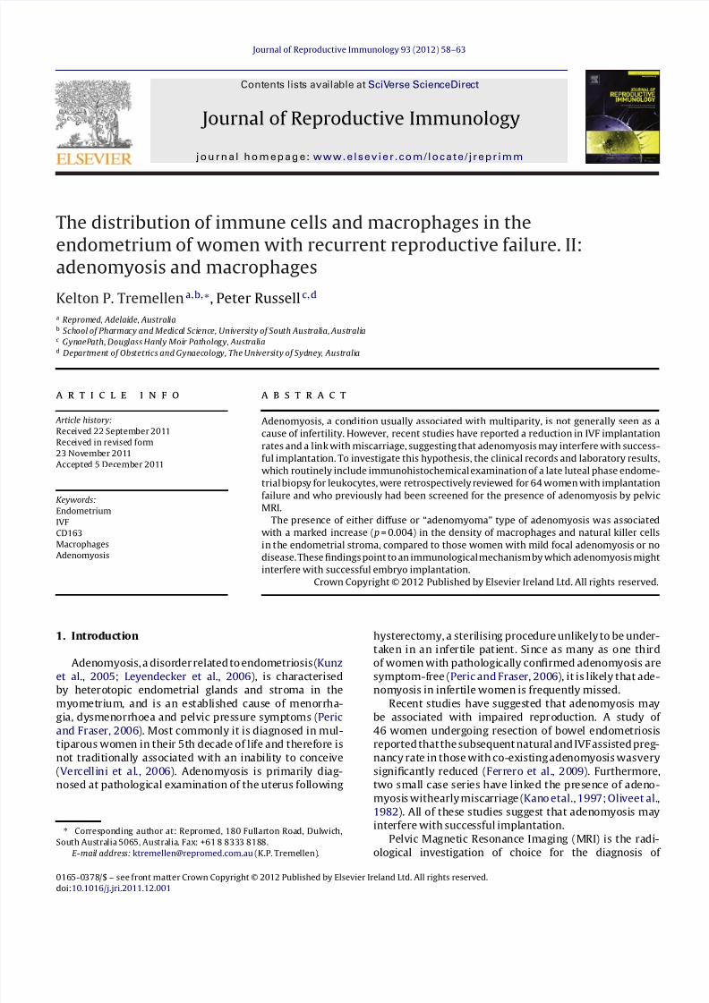
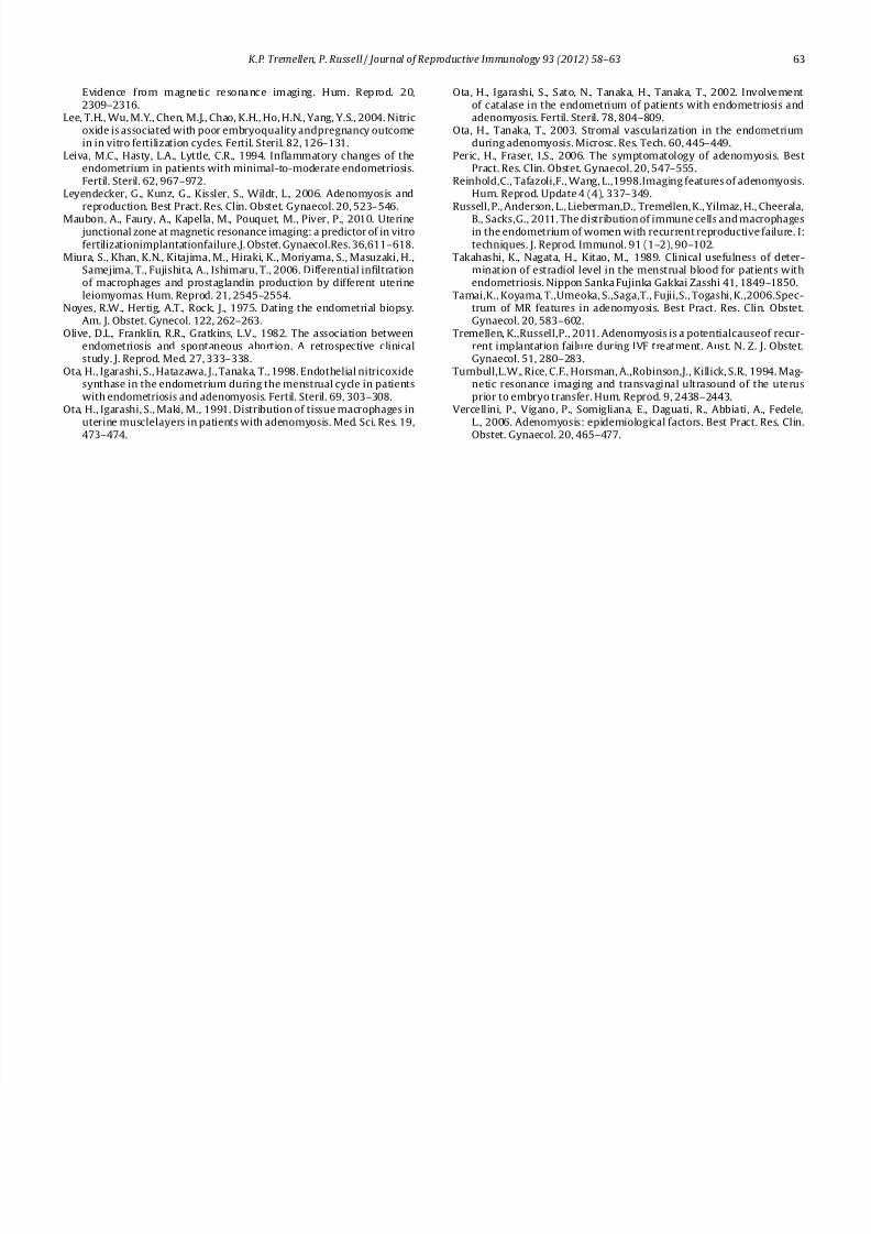
Fig. 1. Immunostain of CD163+ macrophages in the superficial endome-
trial stroma (dark brown) in a background of non-reactive stromal cells
(pale blue nuclei). (For interpretation of the references to colour in this
figure legend, the reader is referred to the web version of the article.)
2.2. Endometrial tissue collection and processing
Endometrial biopsies were conducted between 8 and
12days post ovulation, corresponding to days 22 through
to 26 of a standardised 28-day menstrual cycle. In
patients with irregular cycles, serum hormone profiles
were used to time biopsies. Anovulatory patients were
given clomiphene citrate to produce ovulation or an arti-
ficial hormone replacement cycle (oral oestradiol valerate
for a minimum 2 weeks before commencing vaginal pro-
gesterone). All patients had the endometrial sample dated
according to the time-honoured criteria of Noyes et al.
(1975). An additional six cases where histological dating
of the endometrium, using these traditional morphologi-
cal criteria, was outside the day 22–26 period of interest,
were omitted from the study.
All endometrial samples were fixed in 10% neutral
buffered formalin, processed within 48 h into paraffin, had
sections cut at 4m and stained with haematoxylin and
eosin according to a standard protocol. Serial sections were
also cut at 4m and immunostained in a Ventana Bench-
mark XT using ULTRA view® DAB detection kit (Ventana
Medical Systems, Oro Valley, USA), using commercially
available monoclonal antibodies, for natural killer (NK)
cells (CD56) and macrophages (CD163), as previously pub-
lished by our group (Russell et al., 2011). All histological
studies were performed by one specialist pathologist (PR).
The only information provided to the pathologist before
reporting of the biopsy was the date of the last menstrual
period and a clinical history of recurrent implantation
failure. The pathologist was blinded to the MRI result
so as to remove any chance of bias in the report find-
ings. CD56+ NK cell and CD163+ macrophage numbers
were quantified both as immunopositive cells per mm2 of
endometrial stromain thesuperficial functional layer of the
endometrium, and as a percentage of stromal cells in the
same area (Fig. 1). Stromal macrophage counts were per-
formed on 5 high power fields (40× objective), devoid of
endometrial glands. Percentage counts were performed on
8/10/2019 Journal Adenomyosis
http://slidepdf.com/reader/full/journal-adenomyosis 3/6
8/10/2019 Journal Adenomyosis
http://slidepdf.com/reader/full/journal-adenomyosis 4/6
K.P. Tremellen, P. Russell / Journal of Reproductive Immunology 93 (2012) 58–63 61
Table 2
Immunohistochemistry analysis.
Adenomyosis
positive
Adenomyosis
negative
p-Value
Day of biopsy 24 (23–24) 24 (23–24) 0.321
Stromal CD 163
density (mm2)
280 (190–360) 210 (170–245) 0.013
Superficialglandular CD163
density (0 to 4+)
1 (0–2) 1 (0–2.5) 0.418
Stromal CD 56
density (mm2)
1160±124 913±93 0.112
Macrophages quantified using CD163 and uterine Natural Killer cells
with CD56 immunostains. CD163 data were not normally distributed
and therefore are presented as median (inter-quartile range) and ana-
lysed with Mann Whitney test. CD56 data were normally distributed and
expressed as a mean (±SEM) and analysed by an unpaired t -test.
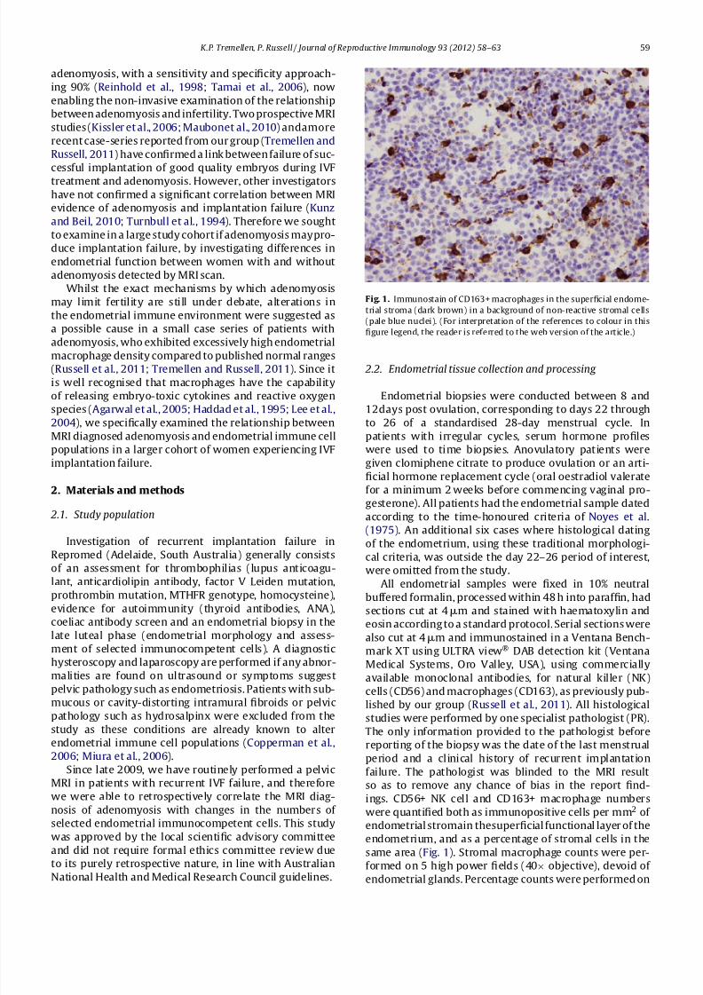
the superficial glands observed between patients with
mild focal adenomyosis, severe diffuse adenomyosis and
those with no MRI evidence of adenomyosis is recorded
in Table 2, and is represented in graphical form in Fig. 3.The median day of sampling was the same in the adeno-
myosis and non-adenomyosis groups, enabling legitimate
comparisons between these two groups.
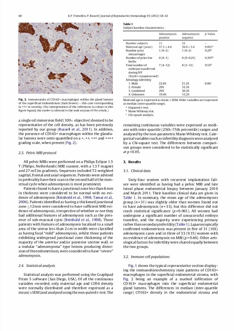
The median density of CD163+ stromal macrophages
(Table 2) was greatly increased in the adenomyosis
group comparedwith thenon-adenomyosis group(median
280/mm2 vs 210/mm2, p= 0.013). However, if the adeno-
myosis population is divided into those with “mild” focal
adenomyosis and those with “severe” diffuse or adenomy-
oma type of adenomyosis, the above difference is almost
entirely due to increased macrophage populations in the
severely affected patients (Fig. 3). The density of stromal
macrophages does not significantly differ between subjectswith no MRI evidence of adenomyosis (median 210/mm2,
IQR 170–245/mm2) compared with those women with
Endometrial macrophage density
C D 1 6 3 p o s i t i v e c e l l s ( m m
2 )
N o a d e
n o
m i l d
a d e n
o
s e v e r e
a d e
n o
0
200
400
600
800 a a b
Fig. 3. Immunohistochemical analysis of macrophage density (CD163)
in the endometrial stromal tissue. Horizontal lines represent the group
medians as data were not normally distributed. Differences between the
groups were analysed by the Mann–WhitneyU test. Groups labelled with
different lettersidentifystatistically significantdifferences between thosegroups.
Endometrial Natural Killer cell density
C D 5 6 p o s i t
i v e c e l l s ( m m
2 )
N o a d e
n o
M i l d
a d e n
o
S e v e
r e a d
e n o
0
1000
2000
3000
4000
a a b
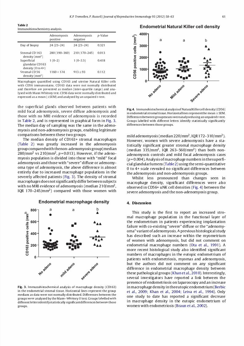
Fig.4. Immunohistochemical analysisof NaturalKillercell density (CD56)
in endometrial stromal tissue. Horizontallines representthe mean±SEM.
Differencesbetween groupmeans wereanalysedusing an unpaired t -test.
Groups labelled with different letters identify statistically significantlydifferences between those groups.
mild adenomyosis (median 220/mm2, IQR 172–310/mm2).
However, women with severe adenomyosis have a sta-
tistically significant greater stromal macrophage density
(median 335/mm2, IQR 263–560/mm2) than both non-
adenomyosis controls and mild focal adenomyosis cases
( p= 0.004). Analysis of macrophage numbers in thesuperfi-
cial glandularlumens (Table 2) using the semi-quantitative
0 to 4+ scale revealed no significant differences between
the adenomyosis and non-adenomyosis groups.
Whilst less pronounced than changes seen in
macrophage density, significant differences were alsoobserved in CD56+ uNK cell densities (Fig. 4) between the
severe adenomyosis and the non-adenomyosis group.
4. Discussion
This study is the first to report an increased stro-
mal macrophage population in the functional layer of
the endometrium in patients experiencing implantation
failure with co-existing “severe” diffuse or the “adenomy-
oma” variant of adenomyosis. A previous histological study
has described such an increase within the myometrium
of women with adenomyosis, but did not comment on
endometrial macrophage numbers (Ota et al., 1991). A
more recent histological study also identified significant
numbers of macrophages in the eutopic endometrium of
patients with endometriosis, myomas and adenomyosis,
but the authors did not comment on any significant
difference in endometrial macrophage density between
these pathological groups (Khan et al., 2010). Interestingly,
several investigators have reported a link between the
presence of endometriosis on laparoscopy and an increase
in macrophage density in theeutopic endometrium (Berbic
et al., 2009; Khan et al., 2004; Leiva et al., 1994). Only
one study to date has reported a significant decrease
in macrophage density in the eutopic endometrium of
women with endometriosis (Braun et al., 2002).
8/10/2019 Journal Adenomyosis
http://slidepdf.com/reader/full/journal-adenomyosis 5/6
62 K.P. Tremellen, P. Russell / Journal of Reproductive Immunology 93 (2012) 58–63
As both endometriosis and adenomyosis have been
linked to impaired implantation potential (Barnhart et al.,
2002; Ferrero et al., 2009; Maubon et al., 2010; Tremellen
and Russell, 2011), it is possible that the observed
increase in macrophage density may represent a com-
mon underlying pathological mechanism by which these
cells contribute to a hostile immune environment for
the implanting embryos. Whilst not all subjects in this
study underwent laparoscopy to identify the presence of
endometriosis,those patientswith symptomssuggestiveof
endometriosis usually did. Furthermore, pelvic MRI isarel-
atively good imaging modality to non-invasively diagnose
severe endometriosis (endometriomas, thick peritoneal
deposits). Taken together, it is unlikely that we have
significantly under-estimated the existence of signifi-
cant endometriosis disease co-existing with adenomyosis.
Finally, the observation that only severe adenomyosis, not
mild focal disease, is linked with an increased endome-
trial macrophage density makes it less likely that mild
undiagnosed endometriosis is primarily responsible for the
observed macrophage infiltration.
It is presently unknown how adenomyosis stimu-
lates an increased endometrial macrophage density or
whether, indeed, these phenomena are related or merely
co-incidental. A recent study has reported that long term
GnRH agonist down-regulation therapy can produce a
very significant fall in endometrial macrophage numbers,
with a co-existent decline in endometrial tissue produc-
tion of MCP-1, a cytokine known to be chemotactic for
macrophages (Khan et al., 2010). Furthermore, this study
reported that down-regulation therapy produced a marked
reduction in endometrial capillary density, a parameter
that is known to be significantly increased in theadenomy-
otic endometrium (Ota and Tanaka, 2003). Taken together,
these studies suggest that under local control of oestro-
gen there is an increase in endometrial capillary density
and production of cytokines chemotactic for macrophages,
both likely to result in a net influx of macrophages into the
endometrium. Adenomyosis is known to be characterised
by a local state of oestrogen dominance (Kitawaki, 2006).
Whilst serum levels of oestrogen do not differ between
women with or without adenomyosis, menstrual blood
oestradiol levelshavebeen reported to be raised in theade-
nomyosis group, reflecting an increased level of aromatase
expression in the adenomyotic endometrium (Kitawaki,
2006; Takahashi et al., 1989). An excess of oestrogen action
in the endometrium may result in an increase in capillary
density and production of pro-inflammatory cytokines,
resulting in an elevated endometrial macrophage density.
When appropriately activated, macrophages are known
to have the capacity to secrete cytokines such as TNF and
IL-1, plus reactive oxygen and nitrogen species, all poten-
tially toxic to embryos (Agarwal et al., 2005; Haddad et al.,
1995). The endometrial environment of patients with ade-
nomyosis has been reported to contain elevated levels of
nitric oxide (Ota et al., 1998), a “free radical” chemical
linked with impaired embryo development and poor preg-
nancy rates (Lee et al., 2004). Furthermore, endometrial
biopsies taken from patients with adenomyosis contain
elevated amounts of anti-oxidant enzymes such as cata-
lase, superoxide dismutase and glutathione peroxidase, a
clear sign of local oxidativestress causedby excessive reac-
tive oxygen species production (Agarwal et al., 2005; Ota
et al., 2002). These reactive oxygen and nitrogen species
will directly damage embryos and have been reported to
increase local production of prostaglandin F2, leading to
an increase in myometrial contractility that is likely to
further impede the implantation process (Agarwal et al.,
2005).
Whilst thecauseand effectrelationship, if any, between
severe adenomyosis and increased macrophage popula-
tions in the endometrium requires elucidation, our novel
observation now provides a pathological mechanism by
which adenomyosis may impede successful implanta-
tion of an embryo. However, as these observations are
only preliminary findings of a small retrospective case
series, they must be verified in a larger prospective study
before firm conclusions can be made. Furthermore, in the
future it would be useful to correlate the immunohis-
tochemistry defined macrophage infiltrations seen with
adenomyosis with changes in endometrial inflammatory
mediators (cytokines and prostaglandins) and the pro-
duction of reactive oxygen species, as these observations
would help strengthen the causative link between adeno-
myosis andfailure of implantation of goodquality embryos.
References
Agarwal,A., Gupta,S.,Sharma,R.K.,2005.Roleof oxidativestressin femalereproduction. Reprod. Biol. Endocrinol. 3, 28.
Barnhart, K., Dunsmoor-Su, R., Coutifaris, C., 2002.Effect of endometriosison in vitro fertilization. Fertil. Steril. 77, 1148–1155.
Berbic, M., Schulke, L., Markham, R., Tokushige, N., Russell, P., Fraser, I.S.,2009. Macrophage expression in endometrium of women with andwithout endometriosis. Hum. Reprod. 24, 325–332.
Braun,D.P.,Ding, J., Shen,J., Rana,N., Fernandez,B.B., Dmowski,W.P., 2002.
Relationship between apoptosis and the number of macrophages ineutopic endometrium from women with and without endometriosis.Fertil. Steril. 78, 830–835.
Copperman, A.B., Wells, V., Luna, M., Kalir, T., Sandler, B., Mukherjee, T.,2006. Presence of hydrosalpinx correlated to endometrial inflamma-tory response in vivo. Fertil. Steril. 86, 972–976.
Ferrero, S., A nserin i, P., A bbamonte, L.H., Ragni, N., Camerini, G.,Remorgida, V., 2009.Fertility afterbowel resection for endometriosis.Fertil. Steril. 92, 41–46.
Haddad, E.K., Duclos, A.J., Baines, M.G., 1995. Early embryo loss is asso-ciated with local production of nitric oxide by decidual mononuclearcells. J. Exp. Med. 182, 1143–1151.
Kano, T., Furudono, M., Nabetani, M., 1997. The incidence of endometrio-sis and adenomyosis in patients with habitual abortion in relation toimmunological abnormalities. Jpn. J. Fertil. Steril. 42, 113–118.
Khan, K.N., Kitajima, M., Hiraki, K., Fujishita, A., Sekine, I., Ishimaru, T.,Masuzaki, H., 2010.Changes in tissue inflammation, angiogenesis and
apoptosis in endometriosis, adenomyosis and uterine myoma afterGnRH agonist therapy. Hum. Reprod. 25, 642–653.
Khan, K.N., Masuzaki , H., Fujishit a, A., Ki tajima, M ., Sekine, I., Ishi-maru, T., 2004. Differential macrophage infiltration in early andadvanced endometriosis and adjacent peritoneum. Fertil. Steril. 81,652–661.
Kissler, S., Hamscho, N., Zangos, S., Wiegratz, I., Schlichter, S., Menzel, C.,Doebert, N., Gruenwald, F., Vogl, T.J., Gaetje, R., Rody, A., Siebzehn-ruebl, E., Kunz, G., Leyendecker, G., Kaufmann, M., 2006. Uterotubaltransport disorder in adenomyosis and endometriosis – a cause forinfertility. BJOG 113, 902–908.
Kitawaki, J., 2006. Adenomyosis: the pathophysiology of an oestrogen-dependent disease. Best Pract . Res. Clin. Obstet. Gynaecol. 20,493–502.
Kunz, G., Beil, D., 2010. Characterization of the uterine junctional zoneprior to IVF/ICSI: an observational study. Arch. Gynecol. Obstet. 281,945–953.
Kunz, G., Beil, D., Huppert, P., Noe, M., Kissler, S., Leyendecker, G., 2005.Adenomyosis in endometriosis – prevalence and impact on fertility.
8/10/2019 Journal Adenomyosis
http://slidepdf.com/reader/full/journal-adenomyosis 6/6
K.P. Tremellen, P. Russell / Journal of Reproductive Immunology 93 (2012) 58–63 63
Evidence from magneti c resonance imagi ng. Hum. Reprod. 20,2309–2316.
Lee, T.H., Wu, M.Y., Chen, M.J., Chao, K.H., Ho, H.N., Yang, Y.S., 2004. Nitricoxide is associated with poor embryoquality andpregnancy outcomein in vitro fertilization cycles. Fertil. Steril. 82, 126–131.
Leiva, M.C., Hasty, L.A., Lyttle, C.R., 1994. Inflammatory changes of theendometrium in patients with minimal-to-moderate endometriosis.Fertil. Steril. 62, 967–972.
Leyendecker, G., Kunz, G., Kissler, S., Wildt, L., 2006. Adenomyosis andreproduction. Best Pract. Res. Clin. Obstet. Gynaecol. 20, 523–546.
Maubon, A., Faury, A., Kapella, M., Pouquet, M., Piver, P., 2010. Uterine junctional zone at magnetic resonance imaging: a predictor of in vitrofertilizationimplantationfailure.J. Obstet. Gynaecol.Res. 36,611–618.
Miura, S., Khan, K.N., Kitajima, M., Hiraki, K., Moriyama, S., Masuzaki, H.,Samejima, T., Fujishita, A., Ishimaru, T., 2006. Differential infiltrationof macrophages and prostaglandin production by different uterineleiomyomas. Hum. Reprod. 21, 2545–2554.
Noyes, R.W., Hertig, A.T., Rock, J., 1975. Dating the endometrial biopsy.Am. J. Obstet. Gynecol. 122, 262–263.
Olive, D.L., Franklin, R.R., Gratkins, L.V., 1982. The association betweenendometriosis and spontaneous abortion. A retrospective clinicalstudy. J. Reprod. Med. 27, 333–338.
Ota, H., Igarashi, S., Hatazawa, J., Tanaka, T., 1998. Endothelial nitricoxidesynthase in the endometrium during the menstrual cycle in patientswith endometriosis and adenomyosis. Fertil. Steril. 69, 303–308.
Ota, H., Igarashi, S., Maki, M., 1991. Distribution of tissue macrophages inuterine musclelayers in patients with adenomyosis. Med. Sci. Res. 19,
473–474.
Ota, H., Igarashi, S., Sato, N., Tanaka, H., Tanaka, T., 2002. Involvementof catalase in the endometrium of patients with endometriosis andadenomyosis. Fertil. Steril. 78, 804–809.
Ota, H., Tanaka, T., 2003. Stromal vascularization in the endometriumduring adenomyosis. Microsc. Res. Tech. 60, 445–449.
Peric, H., Fraser, I.S., 2006. The symptomatology of adenomyosis. BestPract. Res. Clin. Obstet. Gynaecol. 20, 547–555.
Reinhold,C., Tafazoli,F., Wang, L.,1998.Imaging features of adenomyosis.Hum. Reprod. Update 4 (4), 337–349.
Russell, P., Anderson, L., Lieberman,D., Tremellen, K., Yilmaz, H., Cheerala,
B., Sacks,G., 2011. The distribution of immune cells and macrophagesin the endometrium of women with recurrent reproductive failure. I:techniques. J. Reprod. Immunol. 91 (1–2), 90–102.
Takahashi, K., Nagata, H., Kitao, M., 1989. Clinical usefulness of deter-mination of estradiol level in the menstrual blood for patients withendometriosis. Nippon Sanka Fujinka Gakkai Zasshi 41, 1849–1850.
Tamai,K., Koyama, T.,Umeoka, S.,Saga,T., Fujii,S., Togashi, K.,2006.Spec-trum of MR features in adenomyosis. Best Pract. Res. Clin. Obstet.Gynaecol. 20, 583–602.
Tremellen, K.,Russell,P., 2011. Adenomyosis is a potentialcauseof recur-rent implantation failure during IVF treatment. Aust. N. Z. J. Obstet.Gynaecol. 51, 280–283.
Turnbull,L.W., Rice, C.F., Horsman, A.,Robinson,J., Killick, S.R., 1994. Mag-netic resonance imaging and transvaginal ultrasound of the uterusprior to embryo transfer. Hum. Reprod. 9, 2438–2443.
Vercellini, P., Vigano, P., Somigliana, E., Daguati, R., Abbiati, A., Fedele,L., 2006. Adenomyosis: epidemiological factors. Best Pract. Res. Clin.
Obstet. Gynaecol. 20, 465–477.


![Journal für Reproduktionsmedizin und Endokrinologie · 2019-02-26 · Adenomyosis uteri 213 Myometriumdicke). Basierend auf MRT gestützte Erhebungen fanden Kunz et al. [9] eine](https://img.dokumen.tips/doc/110x75/5c9ca8f088c9938d348b62e2/journal-fuer-reproduktionsmedizin-und-endokrinologie-2019-02-26-adenomyosis.jpg)