Embed Size (px)
Citation preview
]74
Tel. 972 8 934 Fax. 972 8 934E-mail:
similar disruption. This movement may provide an explanation for a very slow kinetics of reactivation of the carbamyl enzyme.
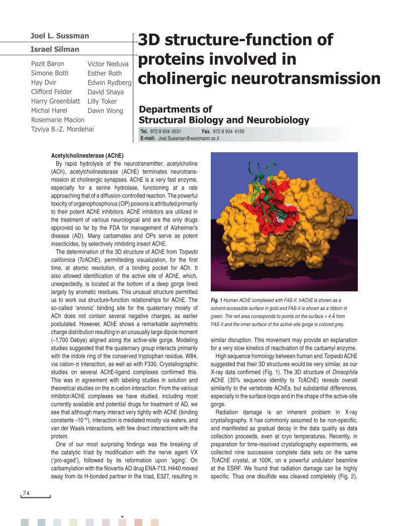
High sequence homology between human and Torpedo AChE suggested that their 3D structures would be very similar, as our X-ray data confirmed (Fig. 1). The 3D structure of Drosophila AChE (35% sequence identity to TcAChE) reveals overall similarity to the vertebrate AChEs, but substantial differences, especially in the surface loops and in the shape of the active-site gorge.
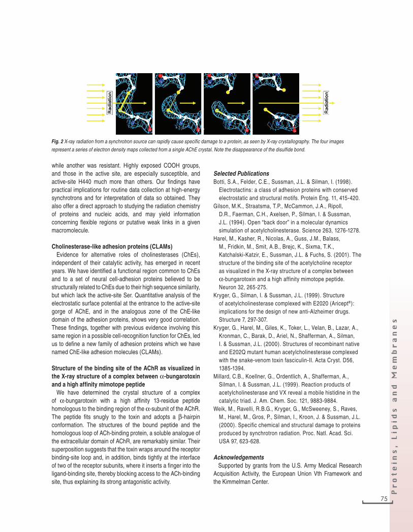
Radiation damage is an inherent problem in X-ray crystallography. It has commonly assumed to be non-specific, and manifested as gradual decay in the data quality as data collection proceeds, even at cryo temperatures. Recently, in preparation for time-resolved crystallography experiments, we collected nine successive complete data sets on the same TcAChE crystal, at 100K, on a powerful undulator beamline at the ESRF. We found that radiation damage can be highly specific. Thus one disulfide was cleaved completely (Fig. 2),
Pazit Baron
Simone Botti
Hay Dvir
Clifford Felder
Harry Greenblatt
Michal Harel
Rosemarie Macion
Tzviya B.-Z. Mordehai
3D structure-function of proteins involved in cholinergic neurotransmission
Departments ofStructural Biology and Neurobiology
Victor Neduva
Esther Roth
Edwin Rydberg
David Shaya
Lilly Toker
Dawn Wong
Joel L. Sussman
4531 [email protected]
Acetylcholinesterase (AChE)By rapid hydrolysis of the neurotransmitter, acetylcholine
(ACh), acetylcholinesterase (AChE) terminates neurotrans- mission at cholinergic synapses. AChE is a very fast enzyme, especially for a serine hydrolase, functioning at a rate approaching that of a diffusion-controlled reaction. The powerful toxicity of organophosphorus (OP) poisons is attributed primarily to their potent AChE inhibitors. AChE inhibitors are utilized in the treatment of various neurological and are the only drugs approved so far by the FDA for management of Alzheimer’s disease (AD). Many carbamates and OPs serve as potent insecticides, by selectively inhibiting insect AChE.
The determination of the 3D structure of AChE from Torpedo californica (TcAChE), permitteding visualization, for the first time, at atomic resolution, of a binding pocket for ACh. It also allowed identification of the active site of AChE, which, unexpectedly, is located at the bottom of a deep gorge lined largely by aromatic residues. This unusual structure permitted us to work out structure-function relationships for AChE. The so-called ‘anionic’ binding site for the quaternary moiety of ACh does not contain several negative charges, as earlier postulated. However, AChE shows a remarkable asymmetric charge distribution resulting in an unusually large dipole moment (~1,700 Debye) aligned along the active-site gorge. Modeling studies suggested that the quaternary group interacts primarily with the indole ring of the conserved tryptophan residue, W84, via cation-π interaction, as well as with F330. Crystallographic studies on several AChE-ligand complexes confirmed this. This was in agreement with labeling studies in solution and theoretical studies on the π-cation interaction. From the various inhibitor/AChE complexes we have studied, including most currently available and potential drugs for treatment of AD, we see that although many interact very tightly with AChE (binding constants ~10-10), interaction is mediated mostly via waters, and van der Waals interactions, with few direct interactions with the protein.
One of our most surprising findings was the breaking of the catalytic triad by modification with the nerve agent VX (‘pro-aged’), followed by its reformation upon ‘aging’. On carbamylation with the Novartis AD drug ENA-713, H440 moved away from its H-bonded partner in the triad, E327, resulting in
Israel Silman
Fig. 1 Human AChE complexed with FAS-II. hAChE is shown as a
solvent-accessible surface in gold and FAS-II is shown as a ribbon in
green. The red area corresponds to points on the surface > 4 Å from
FAS-II and the inner surface of the active-site gorge is colored grey.
joel_sussman 26.12.2001, 15:2674
Pr
ot
ein
s, L
ip
id
s a
nd
M
em
br
an
es
while another was resistant. Highly exposed COOH groups, and those in the active site, are especially susceptible, and active-site H440 much more than others. Our findings have practical implications for routine data collection at high-energy synchrotrons and for interpretation of data so obtained. They also offer a direct approach to studying the radiation chemistry of proteins and nucleic acids, and may yield information concerning flexible regions or putative weak links in a given macromolecule.
Cholinesterase-like adhesion proteins (CLAMs)Evidence for alternative roles of cholinesterases (ChEs),
independent of their catalytic activity, has emerged in recent years. We have identified a functional region common to ChEs and to a set of neural cell-adhesion proteins believed to be structurally related to ChEs due to their high sequence similarity, but which lack the active-site Ser. Quantitative analysis of the electrostatic surface potential at the entrance to the active-site gorge of AChE, and in the analogous zone of the ChE-like domain of the adhesion proteins, shows very good correlation. These findings, together with previous evidence involving this same region in a possible cell-recognition function for ChEs, led us to define a new family of adhesion proteins which we have named ChE-like adhesion molecules (CLAMs).
Structure of the binding site of the AChR as visualized in the X-ray structure of a complex between α-bungarotoxin and a high affinity mimotope peptide
We have determined the crystal structure of a complex of α-bungarotoxin with a high affinity 13-residue peptide homologous to the binding region of the α-subunit of the AChR. The peptide fits snugly to the toxin and adopts a β-hairpin conformation. The structures of the bound peptide and the homologous loop of ACh-binding protein, a soluble analogue of the extracellular domain of AChR, are remarkably similar. Their superposition suggests that the toxin wraps around the receptor binding-site loop and, in addition, binds tightly at the interface of two of the receptor subunits, where it inserts a finger into the ligand-binding site, thereby blocking access to the ACh-binding site, thus explaining its strong antagonistic activity.
Selected PublicationsBotti, S.A., Felder, C.E., Sussman, J.L. & Silman, I. (1998).
Electrotactins: a class of adhesion proteins with conserved electrostatic and structural motifs. Protein Eng. 11, 415-420.
Gilson, M.K., Straatsma, T.P., McCammon, J.A., Ripoll, D.R., Faerman, C.H., Axelsen, P., Silman, I. & Sussman, J.L. (1994). Open “back door” in a molecular dynamics simulation of acetylcholinesterase. Science 263, 1276-1278.
Harel, M., Kasher, R., Nicolas, A., Guss, J.M., Balass, M., Fridkin, M., Smit, A.B., Brejc, K., Sixma, T.K., Katchalski-Katzir, E., Sussman, J.L. & Fuchs, S. (2001). The structure of the binding site of the acetylcholine receptor as visualized in the X-ray structure of a complex between α-bungarotoxin and a high affinity mimotope peptide. Neuron 32, 265-275.
Kryger, G., Silman, I. & Sussman, J.L. (1999). Structure of acetylcholinesterase complexed with E2020 (Aricept®): implications for the design of new anti-Alzheimer drugs. Structure 7, 297-307.
Kryger, G., Harel, M., Giles, K., Toker, L., Velan, B., Lazar, A., Kronman, C., Barak, D., Ariel, N., Shafferman, A., Silman, I. & Sussman, J.L. (2000). Structures of recombinant native and E202Q mutant human acetylcholinesterase complexed with the snake-venom toxin fasciculin-II. Acta Cryst. D56, 1385-1394.
Millard, C.B., Koellner, G., Ordentlich, A., Shafferman, A., Silman, I. & Sussman, J.L. (1999). Reaction products of acetylcholinesterase and VX reveal a mobile histidine in the catalytic triad. J. Am. Chem. Soc. 121, 9883-9884.
Weik, M., Ravelli, R.B.G., Kryger, G., McSweeney, S., Raves, M., Harel, M., Gros, P., Silman, I., Kroon, J. & Sussman, J.L. (2000). Specific chemical and structural damage to proteins produced by synchrotron radiation. Proc. Natl. Acad. Sci. USA 97, 623-628.
AcknowledgementsSupported by grants from the U.S. Army Medical Research
Acquisition Activity, the European Union Vth Framework and the Kimmelman Center.
Fig. 2 X-ray radiation from a synchrotron source can rapidly cause specific damage to a protein, as seen by X-ray crystallography. The four images
represent a series of electron density maps collected from a single AChE crystal. Note the disappearance of the disulfide bond.
] 75
joel_sussman 26.12.2001, 15:2675