Embed Size (px)
Citation preview

Clinical Evaluation of Optic Nerve Head in Glaucoma
Journal of Current Glaucoma Practice, September-December 2010;4(3):115-132 115
JOCGP
Clinical Evaluation of Optic Nerve Head in GlaucomaShibal Bhartiya, Ritu Gadia, Harinder S Sethi, Anita Panda
Glaucoma Services, Dr RP Center for Ophthalmic Sciences, All India Institute of Medical Sciences, New Delhi, India
Correspondence: Shibal Bhartiya, Glaucoma Services, Dr RP Center for Ophthalmic Sciences, All India Institute of MedicalSciences, New Delhi, India, e-mail: [email protected]
CLINICAL EXAMINATION
ABSTRACT
Glaucomatous optic neuropathy is characterized by changes in the intrapapillary and parapapillary region of the optic nerve head,including excavation of the optic nerve head and consequent defects in retinal sensitivity with visual field defects and other psychophysicalalterations. Clinical evaluation of the optic nerve head has been shown to have a high specificity and good precision for glaucomadiagnosis and an experienced observer may in fact be better in distinguishing between normal and glaucomatous disks in comparison withthe HRT or OCT. It is important to differentiate normal physiologic variations in the optic cup, the neural rim, and the peripapillary retina,developmental anomalies and nonglaucomatous optic atrophies from glaucomatous optic nerve head changes.
This review attempts to elucidate the morphology and anatomy of the opic nerve head, and correlate the same to the pathophysiologyof glaucomatous optic neuropathy.
Keywords: Optic nerve head morphology, Glaucomatous optic neuropathy, Optic disk evaluation.
INTRODUCTION
Glaucoma can be defined as syndrome of progressive opticneuropathy characterized by excavation of the optic nerve headand consequent defects in retinal sensitivity with visual fielddefects and other psychophysical alterations. Glaucomatousoptic neuropathy is characterized by changes in theintrapapillary and parapapillary region of the optic nerve head.1,2
One of the most important tests for determining if a patient hasglaucoma is the evaluation of the optic nerve head. Studies haveshown that careful evaluation of the optic nerve head has highspecificity and good precision for glaucoma diagnosis and anexperienced observer may in fact be better in distinguishingbetween normal and glaucomatous disks as compared to anyother technology, like the HRT or OCT.1-3 Normal physiologicvariations in the optic cup, the neural rim, and the peripapillaryretina, developmental anomalies and nonglaucomatous opticatrophies are the common entities that can be confused with aglaucomatous disk.3-5
In the past few years, significant changes have evolved inthe conceptual understanding of the underlying pathogenicmechanisms of primary open-angle glaucoma. Theglaucomatous optic neuropathy is thought of as an optic nervedisorder in which IOP is a major risk factor among severalothers. At one time theories of glaucomatous pathophysiologywere considered either “mechanical” or “vasogenic” butglaucomatous optic neuropathy is caused by a combination ofseveral factors, which include a combination of mechanical andvascular factors which set up a cascade of events finally leadingto biochemical changes and apoptosis of retinal ganglion cells.
Glaucomatous atrophy is thus caused by a progressive deathof retinal ganglion cells, which manifests as characteristicexcavation of the optic nerve head with sequential visual field
deterioration in characteristic patterns. An in-depth under-standing of the anatomy of the optic nerve head (ONH) is crucialfor better understanding of pathophysiology of glaucomatousoptic neuropathy.
Anatomy of the Optic Nerve Head
Each part of the ONH is made up of axons (nerve fibers) ofretinal ganglion cells grouped into bundles, blood vessels andsupporting glial tissue. The optic nerve head (ONH) can bedivided into four anatomic parts:a. Surface layer: The superficial nerve fiber layer (SNFL) of
the ONH has its most anterior limit at the point where thenerve contacts the vitreous. For histopathologic and clinicalpurposes, the peripheral edge of the nerve is defined by theanterior limits of the scleral ring. The posterior limit of theSNFL is recognized histologically as the point at which theaxon bundles have completed their 90° turn from the planeof the retina and have reached the level of the choroids.
b. Prelaminar part: The prelaminar portion of the ONH is theindistinct segment of the axons surrounded by the outerretina, choriocapillaris, and choroid; structurally theastroglial component here is considerably increasedcompared with the SNFL.
c. Laminar part: Laminar part of the nerve is contained withinthe lamina cribrosa; here the glial wrapped axon bundles areconfined in the relatively rigid pores of the specialized laminarscleral plates.
d. Retrolaminar part: Posterior to lamina cribrosa is theretrolaminar portion of the optic nerve, where its thicknessis doubled by the presence of myelinating oligodendrocytes.In the human eye, the distribution of the nerve fibers from
the peripheral retina towards the optic nerve is such that axons
10.5005/jp-journals-10008-1080

Shibal Bhartiya et al
116JAYPEE
from peripheral ganglion cells remain peripheral as they enterthe disk while the central fibers enter centrally, adjacent to thephysiologic cup. This topographic arrangement correlates withthe clinical progression of the glaucomatous visual field;paracentral scotomas appear early in the disease as the cupenlarges, and the peripheral field remains until the peripheralaxons in the nerve are affected.
The arterial blood supply to the ONH may vary amongindividuals, but there is general agreement about its fundamentalcomponents. The central retinal artery (CRA) and the shortposterior ciliary arteries (SPCAs) all contribute directly orindirectly to a capillary plexus that supplies the ONH. Thevenous drainage of the ONH is almost entirely through branchesof the central retinal vein, although important choroidalcollaterals exist; these collaterals may appear as retinociliaryshunts in instances of disturbed retinal circulation. The branchesof the CRA supply the SNFL. This is the network responsiblefor the flame/splinter disk hemorrhages seen clinically, and it isalso the vascular bed that appears in fluorescein angiograms ofthe ONH. The prelaminar ONH is supplied by branches of theSPCAs, which enter the disk substance through the adjacentsclera and posterior to the choroidal bed. Most investigatorsmaintain that vessels derived from the peripapillary choroidmake only a minor contribution to the blood supply of anteriorpart of ONH. The laminar portion is vascularized primarily bycentripetal SPCAs, although an anastomotic capillary bed mayalso contribute to the vascular supply. The anterior portion ofthe retrolaminar nerve has both centripetal vascular supply fromthe piameninges and a significant axial vasculature frombranches of the CRA.
Techniques of Optic Nerve Head Evaluation
There are various techniques of examination of the optic nervehead like the direct ophthalmoscopy, the most commonly used,the indirect ophthalmoscopy for hazy media, the slit lampBinocular indirect ophthalmoscopy using the noncontactmethod (+60, +70, +90 D lenses) or the contact method(Goldmann’s prism).
The identification of structural, contour and color changesof the disk is best done stereoscopically with a dilated pupil.The evaluation of the optic nerve head (ONH) and retinal nervefiber layer (RNFL) may be divided into two parts:
Qualitative Evaluation
a. Contour of the neuroretinal rimb. Optic disk hemorrhagesc. Parapapillary atrophyd. Bared circumlinear vesselse. Appearance of the retinal nerve fiber layer.
Quantitative Evaluation
a. Optic disk size (vertical disk diameter)b. Cup/disk ratio (vertical)
c. Rim disk ratiod. Retinal nerve fiber layer height (RNFLH)
The ONH can be evaluated by following ways:
Direct ophthalmoscopy: This is the simplest and mostcommonly used technique for optic disk evaluation.6 It gives amagnified erect image and is very useful for a quick screening.It lacks stereopsis, which is of great importance in assessingthe topography of the disk. The direct ophthalmoscope cangive three-dimensional information using parallax movements.It is essential to select a spot size with a diameter smaller thanthe diameter of the disk. This is to avoid light spreading fromthe peripapillary retina altering the appearance of the rim. Thesize of the disk can also be estimated by comparison with thecircular small light spot of the ophthalmoscope. The smallest 5°aperture projects to an area of 1.7 mm2.
Indirect Ophthalmoscopy: Though it gives a three-dimensionalpicture, the magnification is not sufficient for detailed evaluationof the disk specially the changes in the blood vessels, neuralrim and disk hemorrhages.7 It also fails to provide an accurateassessment of disk pallor and not being helpful in constrictedpupil. It is useful in an eye with a hazy media.
Slit-lamp biomicroscopy: Slit lamp biomicroscopy is one of themost useful tools to study the ONH. It allows for a time-efficientand detailed stereoscopic examination of the posterior pole byproviding both good magnification and stereopsis. It can bedone by two ways:• Contact method with Goldman three mirror lens• Noncontact method with a Hruby lens or a 78/90 D lens.
A + 78 D lens provides more magnification and a detailwhile a + 90 D give a wider field and is better in cases withsmall pupils.
Although a yellow colored lens may be helpful in increasingpatient comfort, it may mask some early color changes found inthe glaucomatous optic nerve head. For this reason, the highplus lens should be clear. To ensure that the maximum benefit isachieved from the use of the high plus lens, it is crucial that thebiomicroscope is appropriately set for the examiner. Even aslight variation from the correct interpupillary distance settingcan affect the examiner’s stereopsis. Additionally, the anglebetween the illumination system and microscope system shouldbe no more than 10° to ensure stereopsis. It is important toremember that the image seen through a high plus lens is avirtual image, and will be inverted with the right on the left, andthe top on the bottom.
Being familiar with the normal optic nerve head (Fig. 1) isessential in order to critically examine the nerve for the changestypical of glaucoma. Although there is considerable variability,the normal optic nerve contains approximately one million axonswhich leave the eye in multiple bundles through the laminacribrosa. The convergence of the fibers creates a circulardepression in the optic nerve head which is known as the cup.The size of the cup is compared to the size of the disk and is

Clinical Evaluation of Optic Nerve Head in Glaucoma
Journal of Current Glaucoma Practice, September-December 2010;4(3):115-132 117
JOCGP
dependent both on the number of nerve fibers leaving the eye,and the size of the disk. Patients with a decrease in the numberof nerve fibers leaving the eye, as occurs in the generalized lossof axons in moderate to severe glaucoma; or a larger sized opticdisk with all of the axons intact, will both have cup-to-disk (C/D) ratios larger than normal. Equally true, a small disk will havevery little cupping even if a loss of axons has occurred. Becauseof the effect of the nerve size on the cup-to-disk ratio, it is veryimportant to evaluate the optic nerve head size beforecommenting on the C/D ratio.8,9
Normal Optic Disk Morphology
Optic Nerve Head
The disk area varies from 0.80 to 6.00 mm2.5,65,66,67 The opticnerve head is vertically oval with the vertical diameter beingmore than the horizontal diameter by 7 to 10%.5 The averagevertical diameter varies from 1.85 to 1.95 mm (range 0.95-2.9 mm)and the average horizontal diameter varies from 1.70 to 1.80 mm(range 0.9-2.6 mm).10, 11 The ratio between horizontal and verticaldisk diameter varies between 0.70 to 1.37. The ONH size is notconstant. It shows high inter-individual variability withnumerous influencing factors as age, sex, anthropometry,refractive error and race. The disk size becomes constant after 3
to 10 years of age.5, 65, 66, 67 The mean optic disk area is 3.2%larger in males than females67 and the disk size increases by0.02 mm2 for 10 cm increase in body length.68 Recent studieshave shown that increasingly elongated optic disks areassociated with myopia >12 D,63 corneal astigmatism andamblyopia64 suggesting that if an abnormal disk shape is foundin children, a skiascopy should be undertaken to preventamblyopia. Previous studies had quoted that disk size isindependent of refractive error in the range of –5 to +5 D ofametropia5,66 whereas recent investigations have revealed alinear increase in disk area of 1.2% for each 1 D shift towardsmyopia.67 The size of the disk varies considerably in the normalpopulation and among different races. Africans and Asianshave larger disk size as compared to Europeans.
The size of the optic disk can be estimated by using theformula: r/4 × horizontal diameter × vertical diameter; (r is thecorrection factor). The vertical diameter of the optic disk can bemeasured at the slit lamp using a contact or a condensing lens.The slit beam should be coaxial with the observation axis; anarrow beam is used to measure the disk height using the whitescleral ring as a reference landmark. The magnificationcorrections needed vary with the optical dimensions of the eyeand with the lens used for measurement12 (Table 1).
On the basis of Gaussian-like distribution curve of opticdisk area, disks smaller than mean minus twofold standarddeviations are classified microdisks (< 1.29 mm2) and optic diskslarger than mean plus twofold standard deviation are classifiedmacrodisks (> 4.2 mm2). Microdisks are seen in hyperopia,aniridia, optic nerve hypoplasia, nonarteritic anterior ischemicoptic neuropathy, etc. Primary macrodisks are independent ofage after first year of life and slightly influenced by refractiveerror. They may be asymptomatic, e.g. large physiological cupor symptomatic as in optic disk pit (Fig. 21) or Morning Glorydisk (Fig. 22). Secondary macrodisks increase in size after birthas in primary and secondary high myopia. Studies have founddisk size variability to be pathogenically important. The opticdisk size is normal in cases of primary open-angle glaucoma,69,73
Juvenile onset open-angle glaucoma,74 pigmentary glaucoma71
and age related atrophic type glaucoma.72 A study has shownthat size of optic disk is smaller in eyes of pseudoexfoliativeglaucoma than in eyes of POAG.41 The disk size is significantlylarger in eyes with normal pressure glaucoma70 and inglaucomatous eyes with high myopia.
Fig. 1: Normal optic nerve head with small discernible optic cup
Table 1: Magnification correction factors for commonly available high plus lenses
Type of lens Magnification correction factors12 Manufacturer’s specifications
Volk 0.88 0.9260D 1.11 1.1578D 1.33 1.3990D
Nikon 1.03 1.0260D 1.63 1.5490D
Haag-Streit Goldmann — 1.14

Shibal Bhartiya et al
118JAYPEE
Optic Cup
The optic cup is the central excavation in the optic nerve head(Figs 2 to 4). It lies below the level of neural rim and its bottomis formed by lamina cribrosa. It is usually horizontally oval witha horizontal diameter of 0.83 mm (0-2.08 mm) and a verticaldiameter of 0.77 mm (0-2.13 mm) and an area of0.72 mm2 (0-3.41 mm2). The border between the optic cup andthe neuroretinal rim is determined by contour and not by pallor.The combination of the horizontally oval shape of the optic cupand the vertically oval shape of the optic disk explains theconfiguration of the normal neuroretinal rim, which has itsbroadest parts in the inferior and superior disk regions and itssmallest parts in the nasal and temporal region of the opticdisk.10,11
For the evaluation of the optic cup, it is useful to clinicallyexamine the optic nerve head by stereo-optic disk photographyor stereoscopic slit-lamp examination. Especially in eyes withshallow disk cupping, such as in highly myopic eyes withglaucoma, location of the kinking of vessels can be helpful forthe determination of the border of the optic cup (Fig. 5). Innormal eyes, the areas of the optic disk and optic cup arecorrelated with each other, i.e. the larger the optic disk, thelarger the optic cup and vice versa. This feature must beconsidered in the morphologic diagnosis of glaucoma.12,13 Earlyor moderately advanced glaucomatous optic nerve damage mayerroneously be overlooked in small optic disks with relativelylow cup-to-disk ratios, if one does not consider that small opticdisks normally have no optic cup. In contrast, a large optic cupin a large optic disk should not lead to the diagnosis of glaucomaif the other intrapapillary variables are normal, mainly theconfiguration of the neuroretinal rim. One should see if theISNT rule is violated or not. Larger cup size, can be physiologicalif it is bilaterally symmetrical or is associated with large disksize, or high myopia and follows the ISNT rule14 (Fig. 6).
Fig. 2: The vertically oval optic nerve head with central cup Fig. 3A: The optic nerve head with CD ratio of 0.4:1
Fig. 3B: The optic nerve head with CD ratio of 0.7:1, with bayoneting ofvessels, laminar dots, superotemporal thinning of the neuroretinal rim
Fig. 4: The optic nerve head with total glaucomatous optic atrophy

Clinical Evaluation of Optic Nerve Head in Glaucoma
Journal of Current Glaucoma Practice, September-December 2010;4(3):115-132 119
JOCGP
In addition to its area, the optic cup is ophthalmoscopicallydescribed by its depth. In normal eyes, the optic cup depthdepends on the cup area and indirectly on the disk size: Thelarger the optic cup, the deeper it is. In glaucoma, the optic cup
deepens depending on the type of glaucoma and the level ofIOP.3,11
Shape of Optic Cup
Shape of optic cup can be of one of the following types(Elschnig):15
• Type I: Small funnel shaped• Type II: Temporal cylindrical• Type III: Central trough-shaped• Type IV: Temporal or central with steep nasal wall and
sloping temporal margin• Type V: Developmental anomalies.
Cup/Disk Ratio
The cup/disk ratio (CDR) is the decimal value obtained bydividing the cup diameter with the disk diameter. It normallyranges from 0.2 to 0.5. The closer the value is to 1, the worse thedamage. The vertical cup/disk ratio is a better measure ofdeviation from normal than the horizontal ratio, because earlyneuroretinal rim loss occurs preferentially at the upper and lowerpoles of the disk. A difference in cup/disk ratio between eyeswith equal overall optic disk size is suggestive of tissue lossand therefore is highly suspicious of acquired damage.Expressing the size of a cup as a cup/disk ratio (C/D or CDR) isof limited value unless the actual size of the disk is known.
Because of the vertically oval optic disk and the horizontallyoval optic cup, the cup/disk ratios in normal eyes are significantlylarger horizontally than vertically. The quotient of the horizontalto vertical cup/disk ratios is usually higher than 1.0. This isimportant for the diagnosis of glaucoma, in which, in the earlyto medium advanced stages, the vertical cup/disk diameter ratioincreases faster than the horizontal one, leading to an increaseof the quotient of horizontal to vertical cup/disk ratios to valueslower than 1.0. The diagnosis of glaucoma should be stronglyconsidered if the difference in the cup/disk ratio between thetwo eyes is more than 0.2 (seen only in 1% of the normalpopulation)10,11 (Fig. 7) or if the CD ratio is 0.7 or more (seenonly in 10% of population).
Fig. 5: The optic nerve head of high myope with CD ratio of 0. 9:1 withbayoneting of vessels (Note: the shallow disk cup for which the locationof kinking of vessels is helpful for the determination of the border ofthe optic cup)
Fig. 6: The I-S-N-T rule. Note the thickness of the NRR: inferior>superior> nasal> temporal
Fig. 7: The optic nerve head with cup-disk asymmetry of 0.2

Shibal Bhartiya et al
120JAYPEE
When determining the amount of cupping, it is veryimportant to evaluate the contour and not the pallor of the cup.This is because the optic nerve head damaged by glaucomatypically has cupping which is larger than the pallor, whereasthe normal eye has cupping equal to the area of pallor. Pallormore than the cupping should raise suspicion of a non-glaucomatous cause for optic atrophy (Fig. 8).
Neuroretinal Rim (NRR)
Neuroretinal rim is the area of the bending of the axons from thedisk margins to the edge of the optic cup. The evaluation ofNRR width is based on the mark of change in contour than onthe mark of change in color. The average area of NRR is 1.4 to2.0 mm2 and may decline with age. The NRR is notinterindividually constant. The neuroretinal rim size correlateswith the optic disk area; the larger the disk, the larger therim.3,11,16-18 The correlation between rim area and disk areacorresponds with positive correlation between optic disk size,optic nerve fiber count, number and total area of lamina cribrosapores . It points towards a greater anatomic reserve capacity ineyes with large optic disks as compared to eyes with smallerones. Because it is recognized that a large cup/disk size is notdefinitive as a diagnosis of glaucoma, less attention is placedon the size of the cup, and it is more important to focus on theappearance and configuration of the neural rim tissue foundbetween the cup and the edge of the disk. The rim tissue isoften the first area to show changes in glaucoma, and must beexamined very critically during an optic nerve head evaluation.The normal neuroretinal rim tissue is uniformly pink in colorindicating good vascular perfusion (Figs 1 and 2). Becausethere is a round cup located in a vertically elongated oval opticdisk, the width of the neural rim tissue varies by quadrant. Inthe normal eye, the inferior quadrant has the widest rim tissuewith the superior portion second in width. The nasal tissue isslightly thinner than the superior tissue and the tissue in thetemporal quadrant is the thinnest (ISNT rule as termed by
Werner)19,20 (see Fig. 6). This variation in rim sizes causes largephysiologic cups to appear elongated horizontally. The rimtissue will thin as nerve fibers atrophy and this result in pallorin the area of atrophy and a decrease in the size of the rim tissueover time. If the nerve fiber loss is generalized, the atrophy ofnerve fibers will cause an overall decrease in the width of rimtissue and an increase in the size of the cup. This generalizedatrophy is typical in moderate to advanced glaucoma, withcorresponding visual field loss. Because these changes areobvious only in the later stages of the condition, the increase incup size is not very helpful in making a diagnosis of glaucomaearly in the disease process.
In glaucoma, neuroretinal rim is lost in all sectors of theoptic disk with regional preferences, depending on the stage ofthe disease. In early glaucoma, the inferior rim is usually affectedfirst, with the superior rim a close second. The next tissue to bedamaged is typically the temporal rim, with the nasal rim beingthe last to be affected.11,19,20 Thinning in one focal area of thedisk can cause a “notch” to develop in the rim tissue over time.Since the inferior and superior rim tissues are affected first,notching is typically seen in one of these quadrants (Figs 9 to10B). When evaluating the optic nerve, it is helpful to have theresults of a visual field test performed on the same day readilyavailable. This allows the comparison of areas of potential visualfield defects to the nerve fiber responsible for that area of thefield. It is estimated that 20% of the nerve fibers must beatrophied to cause a visual field defect of 5 dB and 40% tocause a 10 dB loss. Because of this, visual field results are bestinterpreted when used in conjunction with the optic nerve headand nerve fiber layer evaluation.19,21
An oblique insertion of optic nerve head as seen in myopia,and occasionally a gray crescent in the optic nerve area, mayobscure the view of NRR thus falsely mimicking NRR thinning.Color of NRR can be misinterpreted due to presence of nuclearsclerosis and use of coated +90 D lens.
Fig. 8: Note that the pallor is out of proportion to the cuppingsuggesting a nonglaucomatous etiology
Fig. 9: The optic nerve head with CD ratio of 0.8:1, inferior notch withthinning of neuroretinal rim inferiorly, bayoneting, baring of thecircumlinear vessels

Clinical Evaluation of Optic Nerve Head in Glaucoma
Journal of Current Glaucoma Practice, September-December 2010;4(3):115-132 121
JOCGP
Rim/Disk Ratio (RDR)
It is the fractional decimal value obtained by dividing the rimthickness by the disk diameter. The closer the value is to 1, thebetter the optic disk appearance. It can be calculated as verticaldiameters as for the cup/disk ratio but obviously with theopposite meaning, as rim area/disk area ratio. This latter canalso be calculated for each degree of the optic disk as a sectorindex of a healthy disk.
Peripapillary Region
Peripapillary region is divided into an outer alpha and an innerbeta zone (Figs 12A and B).22,23
The alpha zone forms the outer crescent characterized byirregular hypo- and hyperpigmentation representing thealteration in the distribution of the melanin pigment in the RPE.It is common finding in normal eyes.
The beta zone lies adjacent to the disk and is characterizedby visible sclera, choroidal vessels and a total loss of pigmentepithelium. This zone is more common in eyes with glaucoma.
In normal eyes, both the alpha zone and beta zone are largestand most frequently located in the temporal horizontal sector,followed by the inferior temporal area and the superior temporalregion. They are smallest and most rarely found in the nasalparapapillary area.
Fig. 10A: The optic nerve head with CD ratio of 0.6:1, superotemporalnotch with thinning of neuroretinal rim in superotemporal quadrant
Fig. 10B: The red free photograph of same patient showing moreretinal nerve fiber loss in superior arcuate zone
Fig. 11: The optic nerve head with near total cupping with laminardot sign, baring of the circumlinear vessel, bayoneting
Figs 12A and B: Peripapillary atrophy (an outer alpha andan inner beta zone)

Shibal Bhartiya et al
122JAYPEE
Both zones are significantly larger and the beta zone occursmore often in eyes with glaucomatous optic nerve atrophy thanin normal eyes. Size of both zones and frequency of the betazone is significantly correlated with variables indicating theseverity of the glaucomatous optic nerve damage, such asneuroretinal rim loss, decrease of retinal vessel diameter,reduced visibility of the retinal nerve fiber bundles, andperimetric defects.21-23 In eyes with small cup-to-disk ratios,the appearance of peripapillary atrophy may be a more sensitiveindicator of glaucomatous optic nerve damage than cup-to-disk ratios. The appearance of peripapillary atrophy shouldraise the suspicion of glaucoma, and be used in conjunctionwith other test results when making clinical decisions on thediagnosis and management of glaucoma.3
Retinal Nerve Fiber Layer Height (RNFLH)
The thickness of the RNFL depends on disk area, age, stage ofthe glaucomatous damage. The vertical polar sectors werethicker than nasal and temporal sectors as the nerve fibersarcuate around the macula and are concentrated at the two poles.
Morphology of the Glaucomatous Optic Disk
Changes in Optic Cup: Glaucoma results in loss of the retinalnerve fibers which manifests as the changes in the opticcup.3,11,21-24
1. Increase in the size of the cup (see Figs 3A to 5)2. Increase in CD ratio (see Figs 3A to 5)3. Vertical enlargement of cup due to localized loss of nerve
fibers at the superior and inferior poles. Focal loss of NRRresults in formation of a polar or a focal notch seen morecommonly at inferior than at superior pole (Fig. 9)
4. Asymmetry between the two eye of more than 0.2 in theCD ratio (see Fig. 7)
5. Increase in depth of the cup6. Diskrepancy in pallor/cupping usually the pallor at the disk
is confined to the area of the physiological cupping, but asthe glaucoma advances the cupping may progress ahead ofthe area pallor. Enlargement of the cup in such cases isevident by the kinking of the vessels at the cup margin.Initial enlargement may lead to a shallow cupping withsloping margins extending (saucerization) up to the diskmargins but the NRR in may retain its normal color (tintedhollow), thus the area of pallor appears smaller than thearea of cupping.
Changes in Lamina Cribrosa
Normally the openings of the lamina are obscured by the nervefibres. However, with advancement of glaucomatous changesthe nerve fibres undergo atrophy and the openings becomevisible as the laminar dot sign (Fig. 11).25
Thinning and backward bowing of the lamina occurs alongwith deepening of the cup. More bowing at the superior andinferior pole results in an hour glass appearance.26
Changes in Neuroretinal Rim
Loss of NRR and decrease in NRR area is seen in glaucoma.3,11,16-
19 The loss of neural rim can be either localized or diffuse. Bothof these precede development of visual field defects. Diffuseloss results in concentric increase in the cup size and is morecommon. Localized loss results in formation of a focal notchmore common at the inferior than at the superior pole (see Fig. 9).Sometimes it may be confused with a congenital pit of the opticdisk. In advanced cases total loss of NRR occurs with extremeposterior bowing of the lamina (bean pot sign) (see Fig. 10).
Vascular Changes
1. Nasalization of vessels: Normally the retinal vessels enterthe eye along the nasal border of the disk and their branchesrun along the margin of disk and cup and emerge somewhattemporally. With enlargement of the optic cup the majorvessels may show a further nasal shift (Fig. 13),27 although,this is not specific for glaucoma.
2. Bayoneting of vessels: With advancement of the cupping,the vessels emerge from the floor of the cup, ascend up thesteep wall of the cup under the overhanging edge of thecup (at which time they are not visible to the observer) andthen emerge again at the disk margin pass making a sharpbend (that resemble bayonet of a rifle), may disappear andthen emerge again at the disk margin (Fig. 14). This is specificfor glaucomatous cupping.3,11,28
3. Over pass cupping: Normally the vessels run over thesurface of the disk and NRR and then come out. A loss ofNRR takes away their posterior support and they appear tohang over the disk, and bridging the cup which is known asover pass cupping.3,11,28
4. Baring of circumlinear vessels: Circumlinear vessels arethe small branches arising from retinal vessels, seen in 50%of the normal eyes. These vessels follow a curvilinear pathand run along the superior and inferior margins of the optic
Fig. 13: Advanced glaucomatous optic atrophy withnasalization of the vessels

Clinical Evaluation of Optic Nerve Head in Glaucoma
Journal of Current Glaucoma Practice, September-December 2010;4(3):115-132 123
JOCGP
cup. Baring of circumlinear vessels occurs with enlargementof the cup as an area of pallor appears between the cupmargin and these vessels (Figs 15A and B). As the rimnarrows the loss of tissue leaves this vessel isolated or‘bared’. It may then remain superficial or come to lie on theinner slope of the rim or on the cup floor. Acquired bearingof circumlinear vessels is an early sign of rim thinning andthus diagnostic of glaucoma.29
Disk Hemorrhages
Disk hemorrhages in glaucoma were reported by Drance andBegg.30,31 The prevalence of small hemorrhages related to theoptic disk has been estimated at 0 to 0.21% in the normalpopulation and 2.2 to 4.1% in glaucomatous patients; they maybe more common in normal-tension glaucoma (up to 40%). Sincethe prevalence of disk hemorrhage is low in the normalpopulation, their presence is very likely to be pathological,especially if recurring. It is a sign of local vascular damage. Thecharacterstic features of these hemorrhages are:1. Hemorrhages typically appear blot-like when located on
the disk, and flame or splinter shaped if they are in closeproximity to the disk in the nerve fiber layer. Splinterhemorrhages are more common than blot ones (Figs 16Aand B).
2. Inferotemporal location is most common.3. It is a sign of progressive disease, may lead to nerve fiber
layer defect, focal notching of NRR and progression ofvisual field defect.
4. Splinter hemorrhages have been shown to precede nervefiber layer and visual field changes in some patients.
5. Although the hemorrhages can resolve in as short as 2 weeksor as long as 35 weeks, the average time to resolution is 10weeks.
Fig. 14: The characteristic bayoneting demonstrated in an opticnerve head with near total cup
Figs 15A and B: Baring of circumlinear vessels
Figs 16A and B: Disk hemorrhages in glaucoma

Shibal Bhartiya et al
124JAYPEE
6. When such a hemorrhage is found in a patient who hasalready been diagnosed, and is being treated for glaucoma,it indicates an unfavorable prognosis, and the need for moreaggressive therapy with resetting of the target IOP to a lowerlevel.
7. More common in patients with large IOP variations.8. More common in diabetics and hypertensives.9. Higher association with normal tension glaucoma
In all cases with disk hemorrhages, other causes leading todisk hemorrhages like systemic anticoagulants, blood disordersand microvascular disease should be ruled out.30-33
Retinal Arterioles/Vessel Changes
Narrowing of the retinal vessels (diffuse or focal) may occursecondary to the loss of nerve fiber layer in cases with glaucoma.This finding is also found in eyes with nonglaucomatous opticdisk damage, such as nonarteritic anterior optic neuropathyand descending optic nerve atrophy indicating that reductionin vessel caliber is typical to optic nerve damage and not toglaucoma. Appearance of collateral vessels on disk may occurin glaucoma but it is not specific for glaucoma.3,11
Patterns of Optic Nerve Head Changesin Glaucoma
A variety of classification schemes have been proposed toclinically distinguish subtypes of glaucoma based on theappearance of the disk but they are not universally accepted.Also there is considerable overlap of features as they appear inclinical practice. The ONH glaucoma patterns has been classifiedinto following five patterns 34-45 (Table 2).
High Myopia Disk Pattern
It is seen in cases with high myopia and open-angle glaucoma(Fig. 5). They have larger and often abnormally shaped opticdisks and their diagnosis represents a special problem in themanagement of glaucoma. Many myopic eyes have lostconsiderable vision from primary or secondary glaucoma beforethe ophthalmologist becomes aware of the diagnosis.
The reasons for this difficulty are:a. The distance between the level of the lamina cribrosa and
the level of the retina is much less than in normal orhyperopic eyes. The average value of this distance in thenormal eye is about 0.7 mm, whereas that of the myopic eyeis between 0.2 and 0.5 mm. Therefore, a completely cuppeddisk in a myopic eye will have only half the depth of theusual glaucomatous cup and therefore such a shallowexcavation is difficult to appreciate clinically.
b. The myopic ONH is masked by the usual myopic conus,tilting of the disk, and peripapillary atrophy. In such cases,disk photographs are superior to drawings for monitoringof the subtle progression of the shallow cup with associatedshifts in vessels or changes in peripappillary area.
c. The ocular rigidity usually is lower than that of normal eyes.Therefore, Schiotz tensions, using the ordinary conversiontables, are lower than the actual lOP. Hence, applanationtonometry should be used in these cases. These patientsmay have associated thin corneas causing falsely lowerapplanation lOP readings.
d. Visual field interpretation is difficult due to presence ofassociated retinal pathology in some cases and the presenceof refractive scotomas.
Table 2: Subtypes of glaucoma by optic nerve head appearance
High myope Focal ischemic Age related Juvenile POAG
Age and Sex < 50 years, M>F > 60 years, F>M > 60 years 10-40 years > 40 yearsOptic disk shape and size Large Normal Normal Normal NormalOptic cupping Concentric, Deep and steep Saucerized, Deep and steep Diffuse, round,
shallow sloping shallow, moth-eaten concentric andsymmetric
Disk hemorrhages Thin superior Frequent disk No No Rare disk hemorrhageor rim notches and inferior rims hemorrhage and and rare rim notch
polar rim notch
Focal RNFL No Yes No No Nodefects
Visual field Dense, focal. Dense, focal, near Relative defects — Diffusechanges Equal superior fixation, superior with diffuse loss
and inferior more than inferiorinvolvement
Peripapillary Marked (may ± Frequent (associated No Nochanges overlap with with tessellated
myopic temporal fundus)crescents)
IOP Normal-high Normal-high Normal-high Normal-high High
Associated systemic — Migraine, Ischemic heart — —abnormalities peripheral disease,
vasospasm hypertension
Clinical Evaluation of Optic Nerve Head in Glaucoma
Journal of Current Glaucoma Practice, September-December 2010;4(3):115-132 125
JOCGP
Focal Normal-Pressure Pattern (Focal Ischemic)
Eyes with the focal type of normal-pressure glaucoma havenormally sized and shaped optic disks, but with characteristiccupping. Often there is a steep and distinct edge to the cup,with the deep cup remaining visible as it vertically progressesto manifest rim notches, disk hemorrhages, and focal RNFLwedge defects. Despite the polar notching, often the remainderof the rim tissue remains relatively intact. The clinical associationsfor this disk appearance include a higher frequency amongwomen, scotomas near fixation in the superior visual field, anda positive history for migraine headaches. These cases havenonspecific circulatory abnormalities in orbital circulation oncolor Doppler analysis. The inclusion of the term ischemic isbased on the clinical impression of frequent disk hemorrhagesin these eyes, which rarely demonstrate highly elevated lOPs.
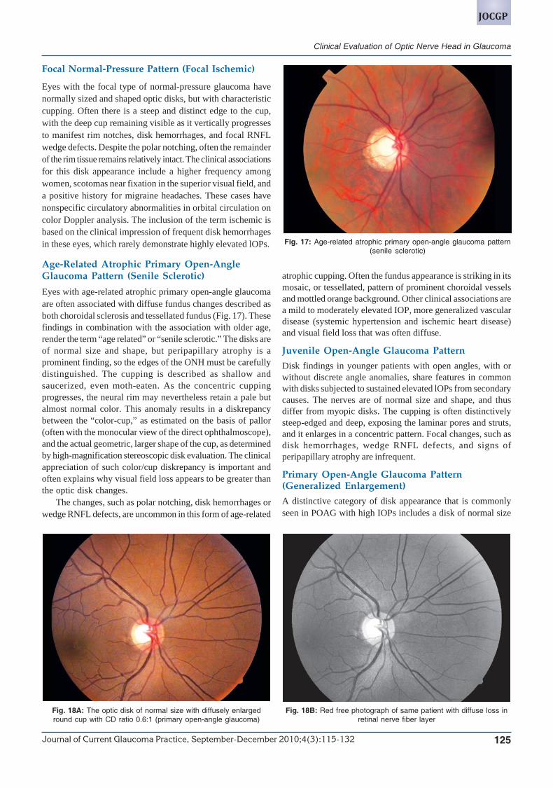
Age-Related Atrophic Primary Open-AngleGlaucoma Pattern (Senile Sclerotic)
Eyes with age-related atrophic primary open-angle glaucomaare often associated with diffuse fundus changes described asboth choroidal sclerosis and tessellated fundus (Fig. 17). Thesefindings in combination with the association with older age,render the term “age related” or “senile sclerotic.” The disks areof normal size and shape, but peripapillary atrophy is aprominent finding, so the edges of the ONH must be carefullydistinguished. The cupping is described as shallow andsaucerized, even moth-eaten. As the concentric cuppingprogresses, the neural rim may nevertheless retain a pale butalmost normal color. This anomaly results in a diskrepancybetween the “color-cup,” as estimated on the basis of pallor(often with the monocular view of the direct ophthalmoscope),and the actual geometric, larger shape of the cup, as determinedby high-magnification stereoscopic disk evaluation. The clinicalappreciation of such color/cup diskrepancy is important andoften explains why visual field loss appears to be greater thanthe optic disk changes.
The changes, such as polar notching, disk hemorrhages orwedge RNFL defects, are uncommon in this form of age-related
Fig. 18A: The optic disk of normal size with diffusely enlargedround cup with CD ratio 0.6:1 (primary open-angle glaucoma)
atrophic cupping. Often the fundus appearance is striking in itsmosaic, or tessellated, pattern of prominent choroidal vesselsand mottled orange background. Other clinical associations area mild to moderately elevated IOP, more generalized vasculardisease (systemic hypertension and ischemic heart disease)and visual field loss that was often diffuse.
Juvenile Open-Angle Glaucoma Pattern
Disk findings in younger patients with open angles, with orwithout discrete angle anomalies, share features in commonwith disks subjected to sustained elevated lOPs from secondarycauses. The nerves are of normal size and shape, and thusdiffer from myopic disks. The cupping is often distinctivelysteep-edged and deep, exposing the laminar pores and struts,and it enlarges in a concentric pattern. Focal changes, such asdisk hemorrhages, wedge RNFL defects, and signs ofperipapillary atrophy are infrequent.
Primary Open-Angle Glaucoma Pattern(Generalized Enlargement)
A distinctive category of disk appearance that is commonlyseen in POAG with high IOPs includes a disk of normal size
Fig. 18B: Red free photograph of same patient with diffuse loss inretinal nerve fiber layer
Fig. 17: Age-related atrophic primary open-angle glaucoma pattern(senile sclerotic)

Shibal Bhartiya et al
126JAYPEE
with diffusely enlarged round cups (Figs 18A and B). Localizedrim defects are uncommon, so abnormality or progression ofcup enlargement necessitates comparison with the fellow eyeor with serial photographs or drawings. The cup increase isoften biased toward the temporal rim, with gradual attenuationof the neural rim. Usually the secondary forms of open-angleglaucoma, such as pseudoexfoliation and pigmentary dispersion,manifest similar disk pattern changes.
Retinal Nerve Fiber Layer
The importance of Retinal nerve fiber layer defects was firstreported in 1973 by Hoyt et al. Evaluation of the nerve fiberlayer is another useful tool to aid in the early diagnosis ofglaucoma. This is because nerve fiber layer defects can occurbefore disk changes and visual field changes are documentedor found.46-49 Retinal nerve fiber layer (RNFL) loss is thus theearliest sign of glaucoma. RNFL thickness is 200 microns nearthe disk, 60 microns in the area of papillomacular bundle and 40microns at rest of the places. The nerve fiber layer (NFL) is bestseen with a 78 D or 90 D lens or a contact lens at the slit lamp.RNFL is best examined in a red free light, because this lightdoes not penetrate beyond the RNFL and is reflected back(Figs 19A and B). In the areas of RFNL loss, the light getabsorbed by the RPE, thus a contrast is created between thenormal and the degenerated area (Fig. 20). Red free light, whichis absorbed by the pigment of the retinal pigment epitheliumand the choroid, is therefore used to provide a dark background.The normal nerve fiber layer reflects light and appears as awhitish haze over the darker underlying retinal structures. Therewill be a striated appearance to the nerve fibers (the fiberbundles are seen as silver striations), with thicker nerve fiberlayers appearing brighter. From about two disks diameters fromthe disk, the NFL thins and feathers out. Slit-like, groove-like orspindle-shaped apparent defects, narrower than the retinalvessels, are seen in the normal fundus. The NFL becomes lessvisible with age, and is more difficult to see in lightly pigmentedfundi. Because the nerve fiber layer is thickest in the superiorand inferior arcades closest to the disk, this area should be thebrightest portion of the view. There will be less brightness inthe thinner papillomacular region and the nasal side of the disk.Symmetry between the reflections in the superior and inferiorarcades and between each of the patient’s eyes is expected.
Patterns of RFNL defect: Experimental studies have shown thatthe defects can be picked if 50% or more of the RNFL is lost.This is due to the sandwich pattern of NFL bundles. The firstglaucomatous axons to be lost are from the temporal raphewhich are in the middle and deep layers of the retina. Followingtypes of NFL defects may be seen:3,11,46-49
1. Slit defects: Dark areas which are slightly larger thanarterioles and reach the disk following the normal course ofthe nerve fiber layer are called slit defects. They representretrograde degeneration of the axons due to focal damage
Fig. 19A: Normal glistening appearance of the retinal nerve fiber layer
Fig. 19B: Normal appearance of the retinal nervefiber layer in red free light
Fig. 20: Retinal nerve fiber layer defect in red free light

Clinical Evaluation of Optic Nerve Head in Glaucoma
Journal of Current Glaucoma Practice, September-December 2010;4(3):115-132 127
JOCGP
Table 3: Summary of the optic nerve head and retinal nervefiber changes in glaucoma
I. Alterations of the cup and neural rimA. Enlargement of the cup
1. Increased cup size {concentric or focal)2. Increased cup-disk ratio3. Alteration in cup shape (vertical-horizontal
disproportion)4. Asymmetry of cup size between the two eyes5. Changes in position and appearance of lamina
cribrosaa. Baring of the laminab. Backward bowing of the laminac. Slit-like laminar openings
B. Loss of neural rim1. Localized2. Diffuse3. Change in normal topographic configuration
(selective narrowing in inferior and superior quadrants)a. Increased central area of pallorb. Pallor of the neural rim
II. Vascular alterationsA. Changes in vessel configuration and caliber
1. Nasalization2. Bayoneting3. Overpass vessel4. Circumlinear and cilioretinal vessel baring5. Narrowing of retinal vessels
B. Disc hemorrhageC. Collateral vessels on disk
III. Peripapillary atrophic changesA. Increased area of peripapillary atrophy
B. Increased frequency of more profound (zone beta) typeof peripapillary atrophy
IV. Loss of the retinal nerve fiber layerA. Localized loss
B. Diffuse loss
of the optic nerve at the lamina. These can occur inapproximately 10% of normal patients. Slit like defect are difficultto identify and may be confused with the normal healthygrooves seen in normal RNFL
2. Wedge defects: Wedge defects are caused by atrophy of manyganglion cells in the same area of the optic nerve. These defectsstart at the disk as narrow lines and expand as they get furtherfrom the disk. Notching of the neural rim tissue, as well as avisual field defect are often associated with wedge defects.Wedge shaped defect are usually seen in superior and inferiorpoles. They are easily detectable as compared to slit defectsand may be preceded by appearance of a splinter hemorrhagein the same site.
3. Diffuse loss: Diffuse loss is the commonest type of RFNL lossseen in glaucoma, but is difficult to pick up. The diffuse atrophytypically occurs in the superior and inferior arcades. The nervefiber layer in these areas loses its consistency and looks like ithas been combed or raked with darker and lighter areas.
4. Nerve fiber layer reversal: In severe cases nerve fiber layerreversal can occur in which the normal pattern of superior andinferior brightness with increasing dimness towards thepapillomacular bundle is lost and the papillomacular areabecomes the brightest structure. Nerve fiber layer reversal isassociated with thinning of the neural rim and a diffusedepression or constriction of the visual field
5. A combination of localized and diffuse loss: RNFL defects arebest seen within two disk diameters of the disk. Wedge andslit defects (wider than retinal vessels) are more apparent inearly disease, when there is little generalized thinning of theNFL, and are seen as dark bands extending from the opticdisk. Generalized thinning of the NFL, with a loss of brightnessand density of striations, is a difficult sign to confirmobjectively. When the NFL is thinned out, the blood vesselwalls are sharp and the vessel appear to stand out in reliefagainst a matt background. The initial abnormality in glaucomamay be either diffuse thinning or localized defects. Since theprevalence of true NFL defects is < 3% in the normal population,their presence is very likely to be pathological.The various changes in optic nerve head in cases of glaucoma
are summarized in Table 3.
Recording of ONH Features
Color disk photos are useful for patient documentation. Colorphotography with a 15° field gives optimal magnification.Stereoscopic photographs are the preferred method. Pseudo-stereoscopic photos are also acceptable. Drawings are better thannothing, if a fundus camera is not available.
Recording of the Nerve FiberLayer (NFL) Features
The photographic methods require specialized processing of film.Patients must have clear media. Photography of lightly colored fundi
are more difficult. The technique is available in some centres,though their use in routine clinical work is limited.
New systems for ONH and NFL assessment, usingalternative technologies, are being evaluated forreproducibility, specificity and sensitivity, although currentlyvery few are available to the general ophthalmologist indeveloping countries due to their prohibitive cost. Theseinclude confocal scanning laser ophthalmoscopy (e.g.Heidelberg Retinal Tomograph), scanning laser polarimetry(e.g. GDx), optical coherence tomography (e.g. OCT) and retinalthickness analyzer (e.g. RTA).
Early or Preperimetric Diagnosis ofGlaucomatous Optic Nerve Damage
For the early detection of glaucomatous optic nerve damagein ocular hypertensive eyes before the development of visualfield loss, the most important variables are shape of theneuroretinal rim, size of the optic cup in relation to the size of

Shibal Bhartiya et al
128JAYPEE
the optic disk, diffusely or segmentally decreased visibility ofthe RNFL, and occurrence of localized RNFL defects and diskhemorrhages.3 If the rim is not markedly broader in the inferiorand superior disk regions as compared with the temporal diskregion, a glaucomatous loss of rim tissue may be suspected inthe inferior and superior disk regions. In other words, if theneuroretinal rim is more or less even in width in all disk sectors,glaucomatous optic nerve damage can be suspected. In theevaluation of the shape of the neuroretinal rim in glaucomatouseyes, one must account for the fact that the rim configurationdepends on the distance to the exit of the central retinal vesseltrunk on the lamina cribrosa surface (in glaucomatous eyeswith the vessel trunk abnormally exiting in the superotemporalquadrant, the neuroretinal rim is often smallest in the inferonasalregion). In eyes with small disks, the neuroretinal rim cannotclearly be delineated from the optic cup, thus, the shape of therim cannot be clearly determined. In these eyes, the variable“cup size in relation to disk size” is the most importantintrapapillary factor to detect glaucomatous optic nerve damagecup.3
Martus et al evaluated whether various types of chronicopen-angle glaucomas differ in predictive factors forprogression of glaucomatous optic nerve damage.50 For patientswith elevated intraocular pressure, significantly predictivefactors for eventual progression were older age, advancedperimetric damage, smaller neuroretinal rim, and larger area ofbeta zone of parapapillary atrophy. In contrast, in the normalintraocular pressure group, a significant predictive factor waspresence of disk hemorrhages at baseline.
The diagnostic power of a novel digital stereoscopic imagingsystem in the diagnosis of glaucomatous optic neuropathy wasstudied by Morgan et al.51A prospective cross-sectional analysisof the diagnostic accuracy of digital stereoscopic optic diskanalysis in the diagnosis of glaucomatous optic neuropathyexhibiting mild to moderate field loss was done by threeobservers. With subjective stereoscopic analysis, sensitivity forglaucoma detection among the three observers was 80.8, 76.9,and 90.4%, with respective specificities of 94.4, 79.6 and 79.6%.Regression analysis of the NRR in 30° segments gavesensitivities between 69.2 and 80.8% and specificities between83.3 and 90.7%. A combination of the subjective and quantitativeanalysis did not significantly improve discrimination. Accordingto them the subjective analysis of digital stereoscopic imagesprovides a useful method for the discrimination of normal andglaucomatous optic nerves. Planimetric analysis does notsignificantly improve the diagnostic precision of this technique.
Jonas reviewed the clinical implications of peripapillaryatrophy in glaucoma.52
Recent studies showed an association of peripapillaryatrophy with glaucoma and the eventual development ofglaucomatous disk hemorrhages independent of a smallneuroretinal rim area, and an association between increasingperipapillary atrophy and progressive glaucoma. A ranking ofoptic disk parameters to detect glaucomatous damage revealed
that the alpha and beta zones of peripapillary atrophy, comparedwith neuroretinal rim parameters, are less useful.Pseudoexfoliation syndrome without glaucoma is not a risk factorfor peripapillary atrophy. In arteritic anterior ischemic opticneuropathy, peripapillary atrophy does not enlarge. Hesummarized that peripapillary chorioretinal atrophy is oneamong several morphologic variables to detect glaucomatousabnormalities. Ranking optic disk variables for the detection ofglaucomatous optic nerve damage, peripapillary atrophy is avariable of second order. It is useful for the differentiation ofvarious types of chronic open-angle glaucomas. In contrast toglaucomatous eyes, eyes with nonglaucomatous optic nerveatrophy, including eyes after arteritic anterior ischemic opticneuropathy, do not show enlarged peripapillary atrophy.
Predictive Value of Nerve HeadEvaluation for Glaucoma
If done properly, evaluation of the optic nerve head and nervefiber layer are very valuable methods to aid clinician in earlydiagnosis of glaucoma. Tielsch reported the specificity andsensitivity of a vertical cup-disk ratio greater than 0.5 as 98%and 29%, respectively.53Airaksinen et al found evaluation ofnerve fiber layer photographs had a specificity of 83% and asensitivity of 94% for glaucoma detection.54 Balazsi and Wernerreported baring of a circumlinear vessel had a specificity of94% and a sensitivity of 65% when the vessel was present.55
More recently investigators have begun to evaluate thevarious features of the optic disk and nerve fiber layer in relationto each other to find which signs or combinations of signs bestdiscriminate between glaucomatous and normal discs.
Jonas et al have studied the qualitative characteristics ofcertain optic disk features in normal and glaucomatous eyes.The features with the highest diagnostic accuracy were thenarrowest neural rim outside the temporal sector, the area ofcupping greater than the area of pallor, detectable retinal nervefiber layer loss, and a large area of peripapillary atrophy. Certainfeatures, such as disk hemorrhage, baring of a cilioretinal vessel,and the overpass vessel sign, which are occasionally seen inglaucoma but almost never seen in normal individuals, had, asexpected, very high specificities but very low sensitivities.56,57
In studies on very large and very small optic disks, wherethe size of the cup and the cup-disk ratio can be very misleading,the following features were most useful in distinguishing normalfrom glaucomatous disks: Vertically oval cup, selective thinningof the inferior neural rim (violation of the ISN’T rule), large areaof peripapillary atrophy with presence of visible inner zonebeta, and nerve fiber layer dropout.
Caprioli et al have reported several studies using optic diskand nerve fiber layer analysis to distinguish normal fromglaucomatous disks. When utilizing computerized quantitativeimage analysis techniques, analysis of features of the retinalnerve fiber layer generally gave the best overall levels of

Clinical Evaluation of Optic Nerve Head in Glaucoma
Journal of Current Glaucoma Practice, September-December 2010;4(3):115-132 129
JOCGP
specificity and sensitivity. Qualitative evaluation of the diskand nerve fiber layer by experienced examiners, however,outperformed the image analysis system.58-60
Montgomery evaluated neural rim area and disk area usingan ophthalmoscopic technique. When corrected for disk area,neural rim measurement achieved a specificity of 95% and asensitivity of 91%.61
Staging and Quantification of Optic NerveHead Damage in GlaucomaStaging and quantification is necessary once optic nerve headchanges have been noticed. Categorizing patients according toseverity is important in giving them prognosis, monitoringprogress deciding management and counseling patients.62
In 1960 Armaly devised the first methodology forquantitatively evaluating disk damage. The method receivedworld wide acceptance and is still commonly used today.75,76
The examiner compared the cup diameter to the entire diskdiameter in any axis and expressed it as a ratio.
Read-Spaeth system: The system was described in 1974 andwas also based upon the cup/disk ratio. The severity of diskdamage was classified into six stages.
Richardson system: The classification system included opticdisk changes and visual fields.
Stage 1a: Low-risk subject; normal visual fields and cup/diskratio (< 0.3 and pink rim of uniform width without asymmetry).
Stage 1b: High-risk subject; 1a with family history of glaucoma,vascular disease, pseudoexfoliation, pigment dispersion or largecups.
Stage 2: Early glaucomatous damage; incomplete Bjerrum defector nasal step with cup disk alterations (cup/disk ratio >0.3 withvertical widening of cup, asymmetry or neuroretinal rim, diskhemorrhage).
Stage 3: Late stage glaucoma; arcuate scotoma with cup/diskratio < 0.8, pale rim of uneven width.
Stage 4: End stage glaucoma; central or temporal visual islandwith narrow pale rim.
The above systems have two major shortcomings as cup/disk ratios are not highly valid indicators of health and diseaseof optic nerve. The systems assume that that cups start centrallyand progress concentrically. Although this occurs in some cases,the nerve damage frequently occurs eccentrically. The secondproblem is that the above systems do not take into considerationthe disk size. It is now well-known that the size of the cup varieswith the size of the disk with lower cup/disk ratios still beingsignificant in small sized disks.
Read and Spaeth for the first time brought attention towardsmeasuring rim width ; they noted that onset of visual field losswas related to remaining rim width. This study formed the basisfor later staging systems like the Nesterov’s system, Jonasmethod and disk damage likelihood scale:
The scale is the latest entry to the list of methodologies forthe the staging of optic nerve damage. It was devised by Spaethet al. The scale divides disk damage into 10 grades of severity.It is better able to monitor disease progression than the otherscales. Disk drawings are made after a slit lamp biomicroscopicexamination and the size of the disk is measured by comparingto the beam length. The DDLS score is derived from the DDLSchart.
DDLS scores of 1 through 3 are rarely associated withglaucomatous visual field loss. Some individuals are born withDDLS three optic disks, whereas others begin with DDLS onedisk. For this reason, noting that a person has a DDLS threeoptic disks indicates that it is reasonably healthy and that thereis no visual field loss. This score is not proof that the disk’shealth has not worsened, however, because it could have beena stage 1 or 2 in the past. The DDLS allows you to quantify theamount of damage that the optic nerve has sustained. Visualfield loss usually will not occur before stage 5. The differen-tiation between very early and no damage is important, becausea neuroretinal rim that has already narrowed is likely to becomenarrower still, whereas an undamaged rim is far more likely toremain stable. Unless glaucomatous progression has stabilized(e.g. in cases of inactive glaucoma secondary to trauma orcorticosteroids), a DDLS score of 6 through 10 strongly supports
Fig. 21: Optic disk pit
Fig. 22: Morning glory disk

Shibal Bhartiya et al
130JAYPEE
aggressive treatment. The DDLS grading performs wellcompared to C/D ratio and HRT-II evaluation.77
DISK DAMAGE LIKELIHOOD SCALE
Narrowest width of rim (rim to disk ratio)
DDLS For small For average For large disk DDLSstage < 1.50 mm 1.50-2.00 mm > 2.00 mm stage
1 0.5 or more 0.4 or more 0.3 or more 0a2 0.4 to 0.49 0.3 to 0.39 0.2 to 0.29 0b3 0.3 to 0.39 0.2 to 0.29 0.1 to 0.19 14 0.2 to 0.29 0.1 to 0.19 Less than 0.1 25 0.1 to 0.19 Less than 0.1 0 for less than 45° 36 Less than 0 for less 0 for 46° 4
0.1 than 45° to 90°7 0 for less 0 for 46° 0 for 91° 5
than 45° to 90° to 180°8 0 for 46° 0 for 91° 0 for 181° 6
to 90° to 180° to 270°9 0 for 91° 0 for 181° 0 for more 7a
to 180° to 270° than 270°10 0 for more 0 for more 7b
than 180° than 270°
Differentiation of Glaucomatous vsNonglaucomatous Optic Neuropathy
Glaucomatous and nonglaucomatous optic neuropathy may bedifficult to distinguish and both can be associated with cuppeddisks along with a decreased diameter of the retinal arterioles,focal arteriole narrowing, and a reduced visibility of the RNFL.3,11
Increasing excavation and enlargement of the optic cup occursmost commonly in glaucoma, but can occur in arteritic anteriorischemic optic neuropathy and compressive lesions on the opticnerve, such as sphenoid wing meningioma. However, in theselast two cases, the neuroretinal rim typically will have pallorwhereas glaucoma will not. Localized RNFL defects can befound in glaucoma and in many types of nonglaucomatous
optic nerve damage, such as in optic disk drusen and long-standing papilledema. Compared with nonglaucomatous opticnerve atrophy, the optic cup enlarges and deepens inglaucomatous optic neuropathy, and, in a complementarymanner, the neuroretinal rim decreases. In addition to glaucoma,an enlargement of the optic cup and a loss of neuroretinal rimmay be found in patients after arteritic anterior ischemic opticneuropathy and in a few patients with intrasellar or suprasellartumors. Because parapapillary atrophy does not usually occurin eyes with nonglaucomatous optic nerve damage, it is helpfulfor the differentiation of glaucomatous versus nonglauco-matous optic neuropathy (Table 4).
In conclusion, a detailed evaluation of the optic disk andretinal fiber layer by stereoscopic slit lamp biomicroscopictechniques provides the clinician with an excellent method forearly detection of glaucoma and in monitoring its progression.Annual disk photographs (both color and red free) should bemade the standard practice pattern for follow up of a glaucomapatient.
REFERENCES
1. O’Connor DJ, Zeyen T, Caprioli J. Comparisons of methods todetect glaucomatous optic nerve damage. Ophthalmology Oct1993;100(10):1498-1503.
2. Caprioli J. Clinical evaluation of the optic nerve in glaucoma.Trans Am Ophthalmol Soc 1994;92:589-641.
3. Jonas JB, Budde WM, Jonas SP. Ophthalmoscopic evaluationof the optic nerve head. Survey of ophthalmology1999;43(4):293-320.
4. Quigley HA, Brown AE, Morrison JD, Drance SM. The sizeand shape of the optic disc in normal human eyes. ArchOphthalmol Jan 1990;108(1):51-57.
5. Jonas JB, Gusek GC, Naumann GOH. Optic disc, cup andneuroretinal rim size, configuration and correlations in normaleyes. Invest Ophthalmol Vis Sci Jul 1988;29(7):1151-58.[published errata appear in Invest Ophthalmol Vis SciMay 1991;32(6):1893 and Feb 1992;32(2):474-5.].
6. Spaeth GL. Direct Ophthalmoscopy. In: Varma R, Spaeth GL,(Eds): The optic nerve in glaucoma, Philadelphia 1993, JBLippincort.
Table 4: Differentiation of glaucomatous from nonglaucomatous optic atrophy in presence of optic disk cupping
Clinical features Glaucomatous optic nerve changes Neurological optic nerve changes
Visual acuity Affected late Affected early
Color vision defect Normal Abnormal
RAPD Absent unless unilateral Present in unilateral casesadvanced involvement
Rim changes Rim defects present Rim pallor
Disk field match Present Disk field mismatch
Visual field changes Respects horizontal meridian Respects vertical meridianEarly cases – arcuate scotomas Early cases – central scotomasMatch with the disk Do not match with the disk
Peripapillary changes Suggestive of glaucomatous Not suggestivedamage be present

Clinical Evaluation of Optic Nerve Head in Glaucoma
Journal of Current Glaucoma Practice, September-December 2010;4(3):115-132 131
JOCGP
7. Shields MB, Tiedman JS. Binocular ophthalmoscopic techniquesfor evaluation of the optic nerve head. In: Varma R, Spaeth GL,(Eds): The optic nerve in glaucoma, Philadelphia 1993, JBLippincort.
8. Varma R, Tielsch JM, Quigley HA, Hilton SC, et al. Race-, age-, gender-, and refractive error-related differences in the normaloptic disc. Arch Ophthalmol Aug 1994;112:1068-76.
9. Jonas JB, Fernandez MC, Naumann, GOH. Glaucomatous opticnerve atrophy in small discs with low cup-to-disc ratios.Ophthalmology Sep 1990;97(9):1211-15.
10. Armaly MF. The optic cup in the normal eye: Cup width, depthvessel displacement, ocular tension and outflow facility. Am Jof Ophthalmol Sept 1969;68(23):401-07.
11. Airakinsen PJ, Tuulonen A, Werner EB. Clinical evaluation of theoptic disc and retianal nerve fibre layer. In: Ritch R, Shields MB,Krupin T: The Glaucomas (Basic Sciences), Vol 1 (2nd ed), StLouis, Missouri: Mosby Inc 1996;617-58.
12. Lim CS, O’Brien C, Bolton NM. A simple clinical method tomeasure the optic disc size in glaucoma. J GlaucomaAug 1996;5(4):241-45.
13. Jonas JB, Fernandez MC, Naumann, GOH. Correlation of theoptic disc size to glaucoma susceptibility. Ophthalmology May1991;98(5):675-80.
14. Jonas JB, Zach FM, Gusek GC, Naumann GO. Pseudoglauco-matous physiologic large cups. American Journal ofOphthalmology Feb 1989;107(2):137-44.
15. Elschnig A. Der normale Sehnerveneintrittt des menschlichenAuges. Denkschrift der kais Akad der Wiss, Wien,Math.naturw.Kl. 1901;70:219-310.
16. Airaksinen PJ, Drance SM. Neuroretinal rim area and RNFL inglaucoma. Arch Ophthalmol 1985;103:203-04.
17. Airaksinen PJ, Drance SM, Schulzer M. Neuroretinal rim area inearly glaucoma. Am J Ophthalmol 1985;99:1-4.
18. Britton RJ, Drance SM, Schulzer MD, et al. The area of theneuroretinal rim of the optic nerve in normal eyes. Am JOphthalmol 1987;103:497-504.
19. Jonas JB, Fernandez MC, Sturmer J. Pattern of glaucomatousneuroretinal rim loss. Ophthalmology Jan 1993;100(1):63-68.
20. Jonas JB, Gusek GC, Naumann GOH: Optic disk, cup andneuroretinal rim size, configuration, and correlations in normaleyes. Invest Ophthalmol Vis Sci 1988;29:1151-58. Correction:Invest Ophthalmol Vis Sci 1991;32:1893.
21. Quigley HA, Dunkelberger BS, Green WR. Retinal ganglion cellatrophy correlated with automated perimetry in human eyeswith glaucoma. American Journal of Ophthalmology1989;107(5):453-64.
22. Jonas JB, Fernández MC, Naumann GOH. Glaucomatousparapapillary atrophy: Occurrence and correlations. ArchOphthalmol 1992;110:214-22.
23. Jonas JB, Nguyne XN, Gusek GC, Naumann GOH: Theparapapillary chorio-retinal atrophy in normal and glaucomaeyes: I. Morphometric data. Invest Ophthalmol Vis Sci 1989;30:908-18.
24. Jonas JB, Berenshtein E, Holbach L. Anatomic relationshipbetween lamina cribrosa, intraocular space, and cerebrospinalfluid space. Invest Ophthalmol Vis Sci 2003;44:5189-95.
25. Read RM, Spaeth GL. The practical clinical appraisal of theoptic disc in glaucoma: The natural history of cup progressionand some specific disc field correlations. Trans Am acadophthalmol Otolaryngol 1974;78:255.
26. Miller KM, Quigley HA. The clinical apperance of the laminacribrosa as a function of the extent of glaucomatous optic nervedamage, Ophthalmology 1988;95:135.
27. Varma R, et al. Positional changes in the vasculature of the opticdisc in glaucoma. Am J Ophthalmol 1987;104:457.
28. Boeglin RJ, Caprioli J. Contemporary clinical evaluation of theoptic nerve in glaucoma. Ophthalmic Clin North 1991;4:711.
29. Balazsi G, Werner EB. Relationship between barring ofcircumlinear vessels of the optic disc and glaucomatous visualfield loss. Can J ophthalmol 1983;18:333.
30. Drance SM, Begg IS: Sector hemorrhage: A probable acute diskchange in chronic simple glaucoma. Can J Ophthalmol1970;5:137-41.
31. Drance SM. Disc hemorrhages in the glaucomas. Survey ofOphthalmol Mar-Apr 1989;33(5):331-37.
32. Hendrickx KH, van den Enden A, Rasker MT, Hoyng PFJ.Cumulative incidence of patients with disc hemorrhages inglaucoma and the effect of therapy. Ophthalmology Jul1994;101(7):1165-72.
33. Diehl DLC, Quigley HA, Miller NR, Sommer A, Burney EN.Prevalence and significance of optic disc hemorrhage in alongitudinal study of glaucoma. Arch Ophthalmol April1990;108:545-50.
34. Caprioli J. Correlation between optic disc appearance and typesof glaucoma. In Varma R, Spaeth GL, Parker KW (Eds): Theoptic nerve in glaucoma, Philadelphia, 1993, Lippincott-Raven.
35. Geijssen HC. Studies on normal pressure glaucoma, Amsterdam,1991, Kugler.
36. Geijssen HC, Greve EL. The spectrum of primary open angleglaucoma. Senile sclerotic glaucoma versus high tension glaucoma,Ophthalmic Surg 1987;18:207.
37. Geijssen HC, Greve EL. Focal ischaemic normal pressureglaucoma versus high pressure glaucoma. Doc OphthalmoI1990;75:291.
38. Nicolela MT, Drance SM. Various glaucomatous optic nerveappearances: Clinical correlations. Ophthalmology 1996;103:640.
39. Spaeth GL. A new classification of glaucoma including focalglaucoma. Surv Ophthalmol 1994;38:S9.
40. Jonas JB, Grundler A. Optic disc morphology in “age-relatedatrophic glaucoma.” Graefes Arch Clin Exp Ophthalmol1996;234:744.
41. Jonas JB, Papastathopoulos KI. Optic disk appearance inpseudoexfoliation syndrome. Am Ophthalmol 1997;123:174.
42. Jonas m, Gusek GC, Naumann GO. Optic disk morphometry inhigh myopia. Graefes Arch Clin Exp Ophthalmol 1988;226:587.
43. Jonas JB, Schiro O. Localised wedge shaped defects of the retinalnerve fibre layer in glaucoma. Br J Ophthalmol 1994;78:285.
44. Jonas JB, Xu L. Parapapillary chorioretinal atrophy in normal-pressure glaucoma. Am J Ophthalmol 1993;115:50.
45. Nicolela MT, et al. Various glaucomatous optic nerveappearances: A color Doppler imaging study of retrobulbarcirculation. Ophthalmology 1996;103:1670.
46. Quigley HA, Katz J, Derick RJ, Gilbert D, Sommer A. Anevaluation of optic disk and nerve fiber layer examinations inmonitoring progression of early glaucoma damage.Ophthalmology 1992;99:19-28.
47. Quigley HA, Miller NR, George T. Clinical evaluation of nervefiber atrophy as indicator of glaucomatous optic nerve damage.Arch Ophthalmol 1980;98:1564-71.
48. Sommer A, Katz J, Quigley HA, et al. Clinically detectablenerve fiber atrophy precedes the onset of glaucomatous fieldloss. Archives of Ophthalmology Jan 1991;109:77-83.
49. Caprioli J, Miller J. Measurement of relative nerve fiber layersurface height in glaucoma. Ophthalmology 1989;96:633-41.

Shibal Bhartiya et al
132JAYPEE
50. Martus P, Stroux A, Budde WM, Mardin CY, Korth M, JonasJB. Predictive factors for progressive optic nerve damage invarious types of chronic open-angle glaucoma. Am J OphthalmolJun 2005;139(6):999-1009.
51. Morgan JE, Sheen NJ, North RV, Goyal R, Morgan S, Ansari E,Wild JM. Discrimination of glaucomatous optic neuropathy bydigital stereoscopic analysis.Ophthalmology. May2005;112(5):855-62.
52. Jonas JB. Clinical implications of peripapillary atrophy inglaucoma. Curr Opin Ophthalmol Apr 2005;16(2):84-88.
53. Tielsch JM. Screening for primary open angle glaucoma:Alternative strategies and future directions. J glaucoma1992;1:214.
54. Airaksinen PJ, et al. Diffuse and localized nerve fibre loss inglaucoma. Am J Ophthalmol 1984;98:566.
55. Balazsi G, Werner EB. Relationship between baring ofcircumlinear vessels of the optic disc and glaucomatous visualfield loss. Can J Ophthalmol Dec 1983;18(7):333-36.
56. Jonas B, Gusek GC, Naumann GOH. Qualitative morphologicalcharacteristics in normal and glaucomatous eyes. Klin MonatsblAugenheilkd 1988;193:481.
57. Jonas B, Nguyen NX, Naumann GOH. Nonquantitativemorphological features in normal and glaucomatous optic discs.Acta Ohthalmol 1989;67:361.
58. Caprioli J, Ortiz-Colberg R, Miller JM, Tressler C.Measurements of peripapillary nerve fiber layer contour inglaucoma. Am J Ophthalmol. Oct 15, 1989;108(4):404-13.
59. Assad A, Caprioli J. Digital image analysis of optic nerve headpallor as a diagnostic test for early glaucoma. Graefes Arch ClinExp Ophthalmol 1992;230(5):432-36.
60. O’Connor DJ, Zeyen T, Caprioli J. Comparisons of methods todetect glaucomatous optic nerve damage. OphthalmologyOct 1993;100(10):1498-503.
61. Montgomery DM. Clinical disc biometry in early glaucoma.Ophthalmology Jan 1993;100(1):52-56.
62. Jonas JB, Papastathapoulos KI: Optic disc shape inglaucoma.Graefes Arch Clin Exp Ophthalmol 1996;234(Suppl1):S167-73.
63. Jonas JB, Dichtl A. Optic disc morphology in myopic primaryopen angle glaucomas. Graefes Arch Clin Exp Ophthalmol1997;235:627-33.
64. Jonas JB, Kling F, Grundler AF. Optic disc shape, cornealastigmatismand amblyopia. Ophthalmology 1997;104:1934-37.
65. Bengtsson B. The variation and covariation of cup and discdiameters. Acta Ophthalmol 1976;54:804-18.
66. Brriton RJ, Drance SM, Schulzer MD, et al. The area of theneuroretinal rim of optic nerve in normal eyes. Am J Ophthalmol1987;103:497-504.
67. Ramrattan RS, Rolfs RCW, Hofman A, de Jong PTVM: Aregender differences in disc and rim area due to differences inrefractive error or height? The Rotterdam Study(abstract). InvestOphthalmol Vis Sci 1997;38(Suppl):S824.
68. Airaksinen PJ, Drance SM. Neuroretinal rim area and RNFL inGlaucoma. Arch Ophthalmol 1985;103:203-04.
69. Caprioli J. Discrimination between normal and glaucomatouseyes. Invest Ophthalmol Vis Sci 1992;33:153-59.
70. Jonas JB. Size of glaucomatous optic discs. Ger J Ophthalmol1992;41-44.
71. Jonas JB, Dichtl A, Budde WM, Lang P. Optic disc morphologyin pigmentary glaucoma. Br J of Ophthalmol 1998;82:875-79.
72. Jonas JB, Grundler AE. Optic disc morphology in “age relatedatrophic glaucoma.” Graefes Arch Clin Exp Ophthalmol1996;234:744-49.
73. Jonas JB, Gusek GC, Naumann GOH. Optic disc morphometryin chronic primary open-angle glaucoma: Morphometricintrapapillary characteristics (I).Graefes Arch Clin ExpOphthalmol 1988;226:522-30.
74. Jonas JB, Grundler AE. Optic disc morphology in juvenileprimary open angle glaucoma. Graefes Arch Clin Exp Ophthalmol1996;234:750-54.
75. Armaly M. genetic determination of cup/disc ratio of the opticnerve. Arch ophthalmol 1967;78:35-43.
76. Armaly M, Sayegi R. The cup/disc ratio. Arch ophthalmol1969;82:191-96.
77. Danesh-Meyer HV, Gaskin BJ, Jayusundera T, Donaldson M,Gamble DD. Comparison of disc damage likelihood scale, cup/disc ratio, and Heidelberg retina tomography in the diagnosis ofglaucoma. Br J Ophthalmol 2006,90:437-41.