Embed Size (px)
Citation preview

J N E R JOURNAL OF NEUROENGINEERING AND REHABILITATION
MCPJ2
IPJ2
IPJ1
Y
Z
X
Y
a) b)
TAB M2
P2 P3P4 P5
D2
D3
D4
D5
M1
P1
D1
SR SU
MCPJ1
M5M3 M4M2
P2
D2
SR
M1
P1
D1
Multi-finger coordination in healthy subjects andstroke patients: a mathematical modellingapproachCarpinella et al.
Carpinella et al. Journal of NeuroEngineering and Rehabilitation 2011, 8:19http://www.jneuroengrehab.com/content/8/1/19 (20 April 2011)

RESEARCH Open Access
Multi-finger coordination in healthy subjects andstroke patients: a mathematical modelling approachIlaria Carpinella1*, Johanna Jonsdottir2 and Maurizio Ferrarin1
Abstract
Background: Approximately 60% of stroke survivors experience hand dysfunction limiting execution of dailyactivities. Several methods have been proposed to objectively quantify fingers’ joints range of motion (ROM), whilefew studies exist about multi-finger coordination during hand movements. The present work analysed this aspect,by providing a complete characterization of spatial and temporal aspects of hand movement, through themathematical modelling of multi-joint finger motion in healthy subjects and stroke patients.
Methods: Hand opening and closing movements were examined in 12 healthy volunteers and 14 hemiplegicstroke survivors by means of optoelectronic kinematic analysis. The flexion/extension angles ofmetacarpophalangeal (MCPJ) and proximal interphalangeal joints (IPJ) of all fingers were computed andmathematically characterized by a four-parameter hyperbolic tangent function. Accuracy of the selected model wasanalysed by means of coefficient of determination (R2) and root mean square error (RMSE). Test-retest reliabilitywas quantified by intraclass correlation coefficient (ICC) and test-retest errors. Comparison between performancesof healthy controls and stroke subjects were performed by analysing possible differences in parameters describingangular and temporal aspects of hand kinematics and inter-joint, inter-digit coordination.
Results: The angular profiles of hand opening and closing were accurately characterized by the selected model,both in healthy controls and in stroke subjects (R2 > 0.973, RMSE < 2.0°). Test-retest reliability was found to beexcellent, with ICC > 0.75 and remarking errors comparable to those obtained with other methods. Comparisonwith healthy controls revealed that hemiparetic hand movement was impaired not only in joints ROM but also inthe temporal aspects of motion: peak velocities were significantly decreased, inter-digit coordination was reducedof more than 50% and inter-joint coordination patterns were highly disrupted. In particular, the stereotypicalproximal-to-distal opening sequence (reversed during hand closing) found in healthy subjects, was altered in strokesubjects who showed abnormally high delay between IPJ and MCPJ movement or reversed moving sequences.
Conclusions: The proposed method has proven to be a promising tool for a complete objective characterization ofspatial and temporal aspects of hand movement in stroke, providing further information for a more targeted planningof the rehabilitation treatment to each specific patient and for a quantitative assessment of therapy’s outcome.
BackgroundIn the last decade, kinematic analysis of upper limbmovements has been increasingly investigated [1,2].Quantitative characterization of upper limb movementsare, indeed, highly required in clinical research and prac-tice, not only to obtain information about pathophysiolo-gical aspects of neural control but also to quantifyimpairment of upper limbs, to plan the appropriate
therapeutic approach and to quantify the effectiveness oftreatment [3]. This is particularly important in the caseof stroke which is the leading cause of disability in theadult worldwide with an estimated incidence of 16 mil-lion new cases per year [4]. Approximately 60% of strokesurvivors experience upper extremity dysfunction limit-ing execution of functional activities and independentparticipation in daily life [5]. Chronic deficits are espe-cially prevalent in the hand, as finger extension is themotor function most likely to be impaired [5].Within recent years, progress in technology has pro-
vided several instruments and methods to objectively
* Correspondence: [email protected] Technology Department, Found. Don C. Gnocchi Onlus, IRCCS,Via Capecelatro 66, 20148, Milan, ItalyFull list of author information is available at the end of the article
Carpinella et al. Journal of NeuroEngineering and Rehabilitation 2011, 8:19http://www.jneuroengrehab.com/content/8/1/19 J N E R JOURNAL OF NEUROENGINEERING
AND REHABILITATION
© 2011 Carpinella et al; licensee BioMed Central Ltd. This is an Open Access article distributed under the terms of the CreativeCommons Attribution License (http://creativecommons.org/licenses/by/2.0), which permits unrestricted use, distribution, andreproduction in any medium, provided the original work is properly cited.

quantify hand kinematics [3]. The most common are elec-trogoniometers [6], instrumented gloves [7], electromag-netic systems [8] and optoelectronic kinematic analysers[9-12]. Some of these methods have been used for the eva-luation of anomalies in hand kinematics due to handinjury [9], focal dystonia [13] and stroke [8,11,14]. Most ofthese studies are mainly focused on the analysis of initialand final position of fingers during a specific movement toevaluate active range of motion, while there is still a lackof studies aimed at analysing temporal aspects of handmotion (i.e. the movement process) and multi-finger coor-dination that is also highly impaired in people with stroke[15].Motion coordination among long fingers (index to little
finger) has been investigated in healthy subjects duringunrestricted flexion/extension movements [16,17] andduring object-grasping [18,19]. Analysis of temporalaspects of these multi-joints movements revealed theexistence of task-specific motion coordination patternsbetween metacarpophalangeal joints (MCPJ) and proxi-mal interphalangeal joints (IPJ) of digits 2-5. In particu-lar, a proximal-to-distal sequence (i.e. MCPJ start movingfirst, followed by IPJ) was noticed during free hand open-ing [16] and hand opening before cylinder-grasping [18],while a reversed sequence (i.e. IPJ-MCPJ sequence) wasfound during unrestricted hand closing [16]. Temporalcoordination of finger motion during the movement tograsp an object was analysed also by Santello et al [19] inunimpaired individuals. Their results demonstrated ahigh degree of covariation among the rotations of theMCPJ and IPJ of long fingers. Specifically, all joint of thesame type (i.e. MCPJ and IPJ) tended to extend and flextogether, simultaneously reaching a maximum excursion.These results gave additional insight into finger
motion control in healthy subjects and provided a usefulstarting point for the analysis of changes in the patternsof joint motion due to neuromuscular disorders, eventhough in these studies the role of the thumb was oftenlacking.Following these considerations, in the present work a
quantitative analysis of unrestricted hand opening andclosing movements, with particular attention to inter-joint,inter-finger coordination was performed on a group ofhealthy subjects and on persons with hemiparesis due tostroke.The selected task (hand opening and closing) was cho-
sen as it represents the most elemental multi-finger move-ment and has previously been demonstrated to be areliable early predictor of recovery of arm function instroke patients [8,20].The analysis was performed by using the method pro-
posed by Braido & Zhang [18], based on the mathematicalcharacterization of fingers joint motion. This specificmethod was chosen since the parametric modelling of
hand kinematics can provide a synthetic representation ofactual movements and facilitate the extraction of spatial,temporal and coordinative features of motion, not imme-diately computable from measured data.With respect to the study of Braido & Zhang [18], which
reported results related to healthy subjects only and didn’tconsider the role of the thumb, the present work hadthree main purposes: i) evaluation of the accuracy of thechosen method in characterizing hand opening/closingmovements, including thumb motion, in healthy subjectsand persons with hemiparesis due to stroke, ii) evaluationof the method’s capacity to discriminate motor perfor-mances of stroke subjects from that of healthy controlsand iii) analysis of the repeatability of the method, andthus, the minimal detectable change in hand performancethat could potentially be used in future work to monitorthe progression of hand function in each stroke subject.
MethodsSubjectsTwelve healthy volunteers (2 women and 10 men, meanage: 36.6 ± 10.8 years), with no history of injury or sur-gery to the hand, and fourteen subjects with hemiparesiscaused by stroke (7 women and 7 men, mean age: 58.4 ±14.8 years) participated in the study. All hemiplegicpatients had sustained a single ischemic (8 subjects) orhemorrhagic (6 subjects) stroke from 3.5 months to 7.5years before the experiments. Three subjects had righthemiparesis and eleven had left hemiparesis. All strokesubjects showed a clinically significant reduction of theparetic upper limb function as indicated by the ActionResearch Arm Test [21] scores ranging from 5 to 46points (maximum score of 57 points indicates a normalupper limb function). Demographic and clinical data arepresented in Table 1. Exclusion criteria were: coexistenceof orthopedic, neurological or other medical conditionsthat limited the affected upper limb, inability to bring theaffected hand to the mouth, inability to extend the pare-tic elbow to at least 120°, spasticity of hand muscles ratedmore than 3 points on the Ashworth scale [22], botuli-num toxin injections in the upper extremity musculaturein the last three months, presence of severe hemispatialneglect, aphasia and/or hemianopsia.All subjects had given written, informed consent to
the experimental protocol, which was conformed to thestandards for human experiments set by the Declarationof Helsinki (last modified in 2004) and approved by thelocal ethical committee.
Experimental protocolSubjects were asked to sit upright in a chair behind atable. The forearm was maintained semi-prone on thetable, the elbow was flexed of about 120° while the wristwas kept in a neutral position (see Figure 1). Healthy
Carpinella et al. Journal of NeuroEngineering and Rehabilitation 2011, 8:19http://www.jneuroengrehab.com/content/8/1/19
Page 2 of 19
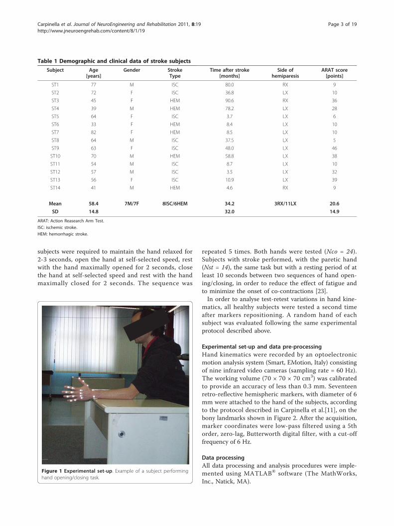
subjects were required to maintain the hand relaxed for2-3 seconds, open the hand at self-selected speed, restwith the hand maximally opened for 2 seconds, closethe hand at self-selected speed and rest with the handmaximally closed for 2 seconds. The sequence was
repeated 5 times. Both hands were tested (Nco = 24).Subjects with stroke performed, with the paretic hand(Nst = 14), the same task but with a resting period of atleast 10 seconds between two sequences of hand open-ing/closing, in order to reduce the effect of fatigue andto minimize the onset of co-contractions [23].In order to analyse test-retest variations in hand kine-
matics, all healthy subjects were tested a second timeafter markers repositioning. A random hand of eachsubject was evaluated following the same experimentalprotocol described above.
Experimental set-up and data pre-processingHand kinematics were recorded by an optoelectronicmotion analysis system (Smart, EMotion, Italy) consistingof nine infrared video cameras (sampling rate = 60 Hz).The working volume (70 × 70 × 70 cm3) was calibratedto provide an accuracy of less than 0.3 mm. Seventeenretro-reflective hemispheric markers, with diameter of 6mm were attached to the hand of the subjects, accordingto the protocol described in Carpinella et al.[11], on thebony landmarks shown in Figure 2. After the acquisition,marker coordinates were low-pass filtered using a 5thorder, zero-lag, Butterworth digital filter, with a cut-offfrequency of 6 Hz.
Data processingAll data processing and analysis procedures were imple-mented using MATLAB® software (The MathWorks,Inc., Natick, MA).
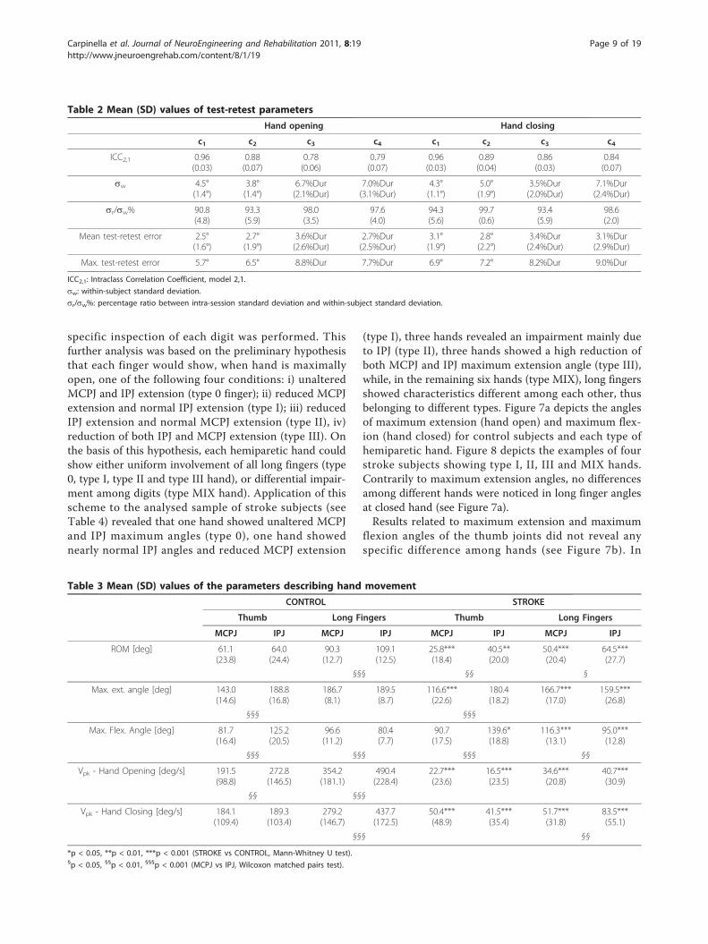
Table 1 Demographic and clinical data of stroke subjects
Subject Age[years]
Gender StrokeType
Time after stroke[months]
Side ofhemiparesis
ARAT score[points]
ST1 77 M ISC 80.0 RX 9
ST2 72 F ISC 36.8 LX 10
ST3 45 F HEM 90.6 RX 36
ST4 39 M HEM 78.2 LX 28
ST5 64 F ISC 3.7 LX 6
ST6 33 F HEM 8.4 LX 10
ST7 82 F HEM 8.5 LX 10
ST8 64 M ISC 37.5 LX 5
ST9 63 F ISC 48.0 LX 46
ST10 70 M HEM 58.8 LX 38
ST11 54 M ISC 8.7 LX 10
ST12 57 M ISC 3.5 LX 32
ST13 56 F ISC 10.9 LX 39
ST14 41 M HEM 4.6 RX 9
Mean 58.4 7M/7F 8ISC/6HEM 34.2 3RX/11LX 20.6
SD 14.8 32.0 14.9
ARAT: Action Reasearch Arm Test.
ISC: ischemic stroke.
HEM: hemorrhagic stroke.
Figure 1 Experimental set-up. Example of a subject performinghand opening/closing task.
Carpinella et al. Journal of NeuroEngineering and Rehabilitation 2011, 8:19http://www.jneuroengrehab.com/content/8/1/19
Page 3 of 19

Joint angle calculation and normalizationA local Cartesian coordinate system XYZ was established,following the procedure described in [11] and the time-courses of the following joint angles computed: metacar-pophalangeal joint (MCPJi) flexion/extension angles,proximal interphalangeal joint (IPJi) flexion/extensionangles of finger i (i = 1-5) and thumb abduction angle(TAB) (see Figure 2 for more details). An automatic algo-rithm was established to identify the initiation and termi-nation of hand opening and closing separately. Theinitiation time of hand opening/closing (Tstart) wasdefined as the instant in which the first joint reached anangular velocity value equal to 10% of its own peak velo-city (Vpk), while movement termination (Tend) wasdefined as the instant in which the angular velocity of thelast joint fell below the 10% of Vpk. Thereafter, angular
profiles were segmented in separated movements of handopening and closing and normalized in time as a percen-tage of the movement duration (%Dur).Joint angle mathematical characterization and accuracyAfter data normalization, each joint angular profile wasmathematically characterized to obtain a synthetic repre-sentation of motion and facilitate the extraction of spa-tial, temporal and coordinative features of multi-fingermovements. The chosen mathematical model was ahyperbolic tangent function with four parameters as sug-gested in [18,24]. The function, graphically representedin Figure 3, is described by Equation 1:
αe (t) = c1 + c2 · tanh(
t − c3�tc4�t
)(1)
MCPJ2
IPJ2
IPJ1
Y
Z
X
Y
a) b)
TABM2
P2 P3P4 P5
D2
D3
D4
D5
M1
P1
D1
SR SU
MCPJ1
M5M3 M4M2
P2
D2
SR
M1
P1
D1
Figure 2 Marker placement, hand local reference system and finger joint angles. Markers position. Mi: head of the metacarpal bone offinger i (i = 1-5); Pi: head of proximal phalanx of finger i (i = 1-5); Di: head of distal phalanx of the thumb (i = 1) and head of middle phalanx oflong fingers (i = 2-5); SU: styloid process of ulna; SR: styloid process of radius. Local reference system XYZ. The origin is in correspondence of themarker M2. Vectors (M2-M5) and (M2 - SR) define the metacarpal plane of the hand (grey triangle). Z-axis is normal to the metacarpal planepointing palmarly, Y-axis has the direction of vector (M2 - SR) pointing distally, while X-axis is calculated as the cross-product of Y and Z-axis,pointing radially. Joint angles in transverse plane YZ (a) and in sagittal plane XY (b) of the hand. MCPJi: metacarpophalangeal joint flexion angleof finger i (i = 1-5); IPJi: proximal interphalangeal joint flexion angle of finger i (i = 1-5); TAB: thumb abduction angle. MCPJi (i = 2-5) is defined asthe angle between Y-axis and the projection of the vector (Pi - Mi) on the YZ plane; IPJi (i = 2-5) is the angle between the projections of vectors(Di-Pi) and (Pi-Mi) on the YZ plane. TAB is the angle between the vector (P1 - M1) and the XY plane. MCPJ1 is the angle between X-axis and theprojection of vector (P1 - M1) on the XY plane. IPJ1 is the angle between vectors (D1 - P1) and (P1 - M1).
Carpinella et al. Journal of NeuroEngineering and Rehabilitation 2011, 8:19http://www.jneuroengrehab.com/content/8/1/19
Page 4 of 19

where ae(t) represents the estimated value of a specificjoint angle ar(t) at instant t (t = 0,..., ΔT), ΔT = Tend-Tstart is the total opening/closing movement duration,c1 = [ae(0)+ ae(ΔT)]/2 is the average of the initial andfinal angles, c2 = [ae(ΔT)- ae(0)]/[tanh((1-c3)/c4) + tanh(c3/c4)] approximates a half of the total angular
displacement (i.e. [ae(ΔT)- ae(0)]/2) when c4 is suffi-ciently small with respect to c3 (e.g. c4 < = 0.5* c3)
1, c3represents the acceleration portion of the total move-ment duration and c4 corresponds to the half of the pri-mary displacement time, where the primarydisplacement is considered the steepest ascending or
0 20 40 60 80 10090
120
150
180
Acc=100*c3
=2*c4*100
0 20 40 60 80 100-50
0
50
100
150
200
250Vpk= c2/100*c4
e(0)
e(100)
2*c2
Primary displacement
c1
Acc= 100*c3
0.42*Vpk
0 20 40 60 80 10060
120
180
240
Acc=100*c3
c1
e(0)
e(100)
2*c2
Primary displacement= 2*c4*100
0 20 40 60 80 100-450
-300
-150
0
Acc=100*c3
a) MCP2 angle [deg]
HAND OPENING
b) MCP2 velocity [deg/s]
c) IPJ2 angle [deg] d) IPJ2 velocity [deg/s]
HAND CLOSING
Measured signal ( r)Modelised signal (
rr)e)
% Duration % Duration
% Duration % Duration
Vpk= c2/100*c4
2*c4*100
0.42*Vpk
0.42*Vpk0.42*Vpk
2*c4*100
Figure 3 Measured and estimated signals. Examples of joint angles and velocity during hand opening (a, b) and hand closing (c, d).Measured signals (grey line) and signals estimated with a four-parameter hyperbolic tangent function (black line) are plotted together. Thekinematic meaning of all four parameters is shown.
Carpinella et al. Journal of NeuroEngineering and Rehabilitation 2011, 8:19http://www.jneuroengrehab.com/content/8/1/19
Page 5 of 19

descending portion of the signal characterized by a velo-city (V) higher than 42% of peak speed (Vpk) [18], asshown by Equations 2 and represented in Figure 3.
Primary displacement = (c3T + c4T) − (c3T − c4T) = 2c4T
V(t) =c2
c4T · cosh2(
t − c3Tc4T
) =Vpk
cosh2(
t − c3Tc4T
)
V(c3T ± c4T) =Vpk
cosh2(±1)= 0.42 · Vpk
(2)
A non-linear least square curve fitting approach wasused to obtain the set of four parameters that best fiteach joint angle profile. The initial estimate of the fourparameters were set according to [24]: c1 = [ar(0)+ ar
(ΔT)]/2, c2 = [ar(ΔT) - ar(0)]/2, c3 = 0.5 and c4 = 0.25.To analyse the accuracy of the model, the coefficient of
determination (R2) and the root mean square error(RMSE) were computed. An angular profile was consid-ered well fitted by the model and included in the subse-quent group analysis if R2 was greater than 0.8. Values ofR2 below this threshold would suggest that the corre-sponding joint motion didn’t show a sygmoidal-shapeprofile and for this reason were treated separately.Test-retest reliabilityTo analyse the test-retest variations on the four para-meters c1, c2, c3, c4, data from the 12 healthy subjectstested two times for reliability purposes were considered.Test-retest reliability was statistically evaluated usingintraclass correlation coefficient, model 2,1 (ICC2,1) cal-culated following the procedure described by McGraw &Wong [25]. ICC2,1 is represented by Equation 3:
ICC2,1 =σ 2
n
σ 2n + σ 2
s + σ 2r
(3)
where sn2 is the inter-subject variance, ss
2 is theinter-session variance and sr
2 is the intra-session var-iance. The following guidelines were used to grade thestrength of reliability: 0.50-0.60 fair, 0.60-0.75 good,0.75-1.00 excellent reliability [12,26]. Within-subjectvariability (sw) was evaluated by the Standard Error ofMeasurement (SEM), computed, from Equation 3, as√(ss
2+sr2). The percentage ratio between intra-session
standard deviation (sr) and within-subject standarddeviation (sw) was also computed. For all angular pro-files and for each parameter, the absolute differencebetween the values obtained from the two sessions wascomputed (absolute test-retest error). Maximum test-retest error and, thus, minimum significant changedetectable by the protocol was calculated as mean
absolute error + 2 standard deviations, following theprinciples of Bland-Altman analysis [27].Extraction of specific parametersFrom data included in the group analysis (R2 > 0.8), thefollowing variables were calculated to analyse three dis-tinct aspects of hand motion:
1) Finger kinematics were analysed through the fol-lowing parameters:
• Dur = Tend -Tstart, movement duration• amin = c1-c2, angle of maximum flexion• amax = c1+c2, angle of maximum extension• ROM = 2*c2, range of motion• Vpk = c2/100*c4, peak velocity
2) Inter-joint coordination was inspected by lookingat the level of synchronization between MCPJ andIPJ, which was defined by the temporal delay (Δi)between IPJ and MCPJ angles of finger i in theinstant of peak velocity (100*c3). The value of Δi wascalculated as 100*[c3(IPJi)-c3(MCPJi)].3) Inter-digit coordination was evaluated consideringthe variability among IPJ-MCPJ delays (Δi) of all fin-gers: a high level of inter-digit coordination is repre-sented by similar values of Δi (low variability), whilepoor coordination is implied by higher differencesamong Δi (high variability). This concept was repre-sented by the coordination index among long fingers(COILF) and among all digits (COIHAND). COILF wasdefined as 100*CVLF(co)/CVLF(j), where CVLF(j) =standard deviation(Δ2, Δ3, Δ4, Δ5)/mean(Δ2, Δ3, Δ4,Δ5) was the coefficient of variation for long fingers ofhand j and CVLF(co) was the mean CVLF value ofhealthy control subjects. COIHAND was calculated inthe same way but considering the coefficient of varia-tions among all 5 fingers. COI values below 100% indi-cated lower coordination with respect to the meanvalue of control subjects, while values above 100%represent a level of coordination higher than the aver-age value of healthy subjects.
Data not well fitted by the selected model (R2 < 0.8)were treated separately and only amin, amax and ROM,as calculated from the measured data, were included inthe analysis.
Statistical analysisConsidering the small sample of data, comparisons weremade using nonparametric tests. Differences between IPJand MCPJ were analysed using Wilcoxon matched pairstest (Wt), variations among fingers were evaluated withFriedman test (Ft) and Bonferroni post-hoc comparisons,while differences between healthy controls and strokesubjects were analysed by means of Mann-Whitney Utest (MWt). Level of significance was set to 0.05.
Carpinella et al. Journal of NeuroEngineering and Rehabilitation 2011, 8:19http://www.jneuroengrehab.com/content/8/1/19
Page 6 of 19

ResultsModel accuracyAnalysis of all hand opening/closing movements per-formed by healthy subjects confirmed that the selectedmathematical model accurately characterized the shapeof angular profiles of MCPJ and IPJ of long fingers andthumb. This was confirmed by R2 and RMSE mean(± SD) values which were, respectively, 0.996 (± 0.009)and 1.6° (± 0.6°) for hand opening and 0.995 (± 0.009)and 1.7°(± 0.7°) for hand closing. With regard tothumb abduction angles (TAB), the mathematicalmodel accurately characterised TAB only in 75% of alltested hands (R2 = 0.964 ± 0.043, RMSE = 0.9° ± 0.5°),as shown in Figure 4a. The remaining thumb abduc-tion angles (25%) showed significantly lower values ofR2 (0.517 ± 0.210) and higher RMSE (2.6° ± 1.3°), asindicated in the example of Figure 4b. For this reason,TAB angles were considered not well fitted by theselected model and, consequently, only the angularvalues reached at maximally closed and open hand, ascalculated from the measured data, were included inthe analysis.Concerning stroke subjects, 5% of all MCPJ and IPJ
angular profiles during hand opening did not show a syg-moidal-shape profile, as indicated by R2 values lower than0.8 (see Figure 4d). The remaining data (95%) were accu-rately characterized by the mathematical model as theyshowed values of R2 and RMSE equal to 0.973 (± 0.045)and 0.9° (± 0.7°), respectively (see Figure 4c). As for handclosing, all angular profiles were well fitted by the hyper-bolic tangent model (R2 = 0.979 ± 0.064, RMSE = 2.0° ±1.3°). The mathematical model accurately characterisedTAB only in 75% of all tested hands (R2 = 0.951 ± 0.050,RMSE = 1.0° ± 0.8°). The remaining thumb abductionangles (25%) showed significantly lower values of R2 (0.549± 0.193) and higher RMSE (2.2° ± 1.0°). Consequently,only the angular values reached at maximally closed andopen hand, as calculated from the measured data, wereincluded in the analysis.
Test-retest reliabilityResults of the test-retest analysis are reported in Table 2.All four parameters showed good to excellent reliability inboth hand opening and closing as indicated by mean ICCvalues greater than 0.75 [12,26]. Mean Standard Error ofMeasurement (sw) was lower than 5.0° for angular para-meters (c1, c2) and lower than 7.1%Dur for temporal para-meters (c3, c4). Angular parameters (c1, c2) showed a meanand a maximum test-retest errors lower than 3.1° and 7.2°,respectively, while mean and maximum test-retest errorsfor temporal parameters (c3, c4) were lower than 3.6%Durand 9.0%Dur. Results on the sr/sw% ratio, revealed thatless than 10% of within-subject variations (sw) was due to
inter-session variability (ss) while more than 90% was dueto intra-session variations (sr).
Hand motion characterization in healthy subjectsFingers kinematicsHealthy controls took 0.9 (± 0.6) seconds to completelyopen and close the hand. Table 3 reports the resultsrelated to the angular variables extracted from MCPJand IPJ motion of long fingers and thumb. IPJ showed asignificantly higher ROM with respect to MCPJ. Thiswas due to a significantly higher maximum flexion angleof IPJ (amin= 80.4° ± 7.7°) with respect to MCPJ (amin =96.6° ± 11.2°), when the hand was completely closed.Contrarily, maximum extension angles, correspondingto the position of maximum hand aperture, were similarfor both types of joints (MCPJ: amax = 186.7° ± 8.1°; IPJ:amax = 189.5° ± 8.7°; p(Wt) = 0.2301, n.s.). As reportedin Table 3, IPJ revealed a higher peak velocity withrespect to MCPJ both in hand opening and closing. IPJpeak speed was similar in the two movements, whileMCPJ speed was significantly lower during extensionthan during flexion.Inter-joint and inter-digit coordinationWithin each long finger, a proximal-to-distal sequencewas evident for hand opening movements (see Figure 5,left panels). In particular, MCPJ started extending first,followed by IPJ after an average delay of 7.4%Dur (seeFigure 6a). Contrarily to long fingers, a distal-to-proxi-mal sequence was noticed in the thumb (see Figure 5,upper-left panel): IPJ started extending first followed byMCPJ after a mean delay of 4% (Figure 6a). Duringhand closing inter-joint sequence was reversed for boththumb and long fingers (see Figure 5, right panels). Inparticular, a proximal-to distal sequence (i.e. MCPJ-IPJ)was noticed in the thumb and a distal-to proximalsequence (i.e. IPJ-MCPJ) was evident in long fingers (seeFigure 6b). In both hand opening and closing MCPJ offinger 2 to 5 moved together, simultaneously reachingpeak velocity at approximately 50% of the movementduration. Synchronous motion was noticed also in IPJ,which reached the maximum speed at nearly 57% of thewhole duration (see Figure 5).These coordination sequences were consistent among
fingers. In fact, analysis of IPJ-MCPJ delay did not revealany significant difference among long fingers in handopening [p(Ft) = 0.2308 n.s.] or closing [p(Ft) = 0.6065n.s.] indicating a high level of inter-digit coordination.
Hand motion characterization in subjects with strokeFingers kinematicsIn both hand opening and closing, stroke patients (ST)took significantly longer time to complete the movementwith respect to healthy control subjects (CO) (Hand
Carpinella et al. Journal of NeuroEngineering and Rehabilitation 2011, 8:19http://www.jneuroengrehab.com/content/8/1/19
Page 7 of 19

opening: ST = 3.9 s ± 1.7 s, CO = 0.9 s ± 0.6 s, p(MWt) <0.001; Hand closing: ST = 5.1 s ± 1.6 s, CO = 1.0 s ± 0.6s, p(MWt) < 0.001). Stroke patients showed a signifi-cantly reduced ROM of thumb and long fingers jointsthat was due to a high reduction of both maximum flex-ion and maximum extension angles (see Table 3). Inthree cases, subject’s attempt to extend IPJ resulted in anundesired flexion of one or two fingers. No significantdifferences between controls and stroke subjects werenoticed in thumb abduction angles neither in hand open-ing (ST: 20.1° ± 18.7°, CO: 18.5° ± 17.3°, p(MWt) =
0.5251, n.s.) nor hand closing (ST: 29.7° ± 15.9°, CO:27.0° ± 11.2°, p(MWt) = 0.5450 n.s.). As reported inTable 3, stroke subjects showed significantly reducedpeak velocities in all joints with respect to controls.Moreover, peak speed during hand opening was signifi-cantly lower than that obtained during hand closing(p(Wt) < 0.01).Considering the high variability of maximum extension
angles of long fingers’ joints, represented by an inter-sub-ject standard deviation 2 to 3 times greater than that ofhealthy controls (see Table 3 and Figure 7a), a more
0 20 40 60 80 10020
25
30
35
40
0 20 40 60 80 10020
25
30
35
40
Hand closed
Hand open
R2=0.9972RMSE=0.2°
R2=0.5164RMSE=3.0°
Measured signal ( r)
Modelised signal ( e)
a) Hand opening – TAB angle [deg] b) Hand opening – TAB angle [deg]
Hand closedHand open
% Duration % Duration
0 20 40 60 80 100145
146
147
148
149
150
R2=0.5639RMSE=1.1°
d) Hand opening – IPJ3 angle [deg]
% Duration
Hand closed
Hand open
0 20 40 60 80 100130
140
150
160
170
c) Hand opening – IPJ2 angle [deg]
R2=0.9967RMSE=0.8°
% Duration
Hand closed
Hand open
Figure 4 Examples of measured and estimated angles during hand opening. Thumb abduction angles (TAB) of two unimpaired individuals(a, b) and proximal interphalangeal joint angles (IPJ2 and IPJ3) of two stroke subjects (c, d) during movements of hand opening. Coefficient ofdetermination (R2) and root mean square error (RMSE) are reported.
Carpinella et al. Journal of NeuroEngineering and Rehabilitation 2011, 8:19http://www.jneuroengrehab.com/content/8/1/19
Page 8 of 19
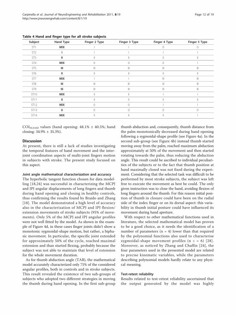
specific inspection of each digit was performed. Thisfurther analysis was based on the preliminary hypothesisthat each finger would show, when hand is maximallyopen, one of the following four conditions: i) unalteredMCPJ and IPJ extension (type 0 finger); ii) reduced MCPJextension and normal IPJ extension (type I); iii) reducedIPJ extension and normal MCPJ extension (type II), iv)reduction of both IPJ and MCPJ extension (type III). Onthe basis of this hypothesis, each hemiparetic hand couldshow either uniform involvement of all long fingers (type0, type I, type II and type III hand), or differential impair-ment among digits (type MIX hand). Application of thisscheme to the analysed sample of stroke subjects (seeTable 4) revealed that one hand showed unaltered MCPJand IPJ maximum angles (type 0), one hand showednearly normal IPJ angles and reduced MCPJ extension
(type I), three hands revealed an impairment mainly dueto IPJ (type II), three hands showed a high reduction ofboth MCPJ and IPJ maximum extension angle (type III),while, in the remaining six hands (type MIX), long fingersshowed characteristics different among each other, thusbelonging to different types. Figure 7a depicts the anglesof maximum extension (hand open) and maximum flex-ion (hand closed) for control subjects and each type ofhemiparetic hand. Figure 8 depicts the examples of fourstroke subjects showing type I, II, III and MIX hands.Contrarily to maximum extension angles, no differencesamong different hands were noticed in long finger anglesat closed hand (see Figure 7a).Results related to maximum extension and maximum
flexion angles of the thumb joints did not reveal anyspecific difference among hands (see Figure 7b). In
Table 2 Mean (SD) values of test-retest parameters
Hand opening Hand closing
c1 c2 c3 c4 c1 c2 c3 c4
ICC2,1 0.96(0.03)
0.88(0.07)
0.78(0.06)
0.79(0.07)
0.96(0.03)
0.89(0.04)
0.86(0.03)
0.84(0.07)
sw 4.5°(1.4°)
3.8°(1.4°)
6.7%Dur(2.1%Dur)
7.0%Dur(3.1%Dur)
4.3°(1.1°)
5.0°(1.9°)
3.5%Dur(2.0%Dur)
7.1%Dur(2.4%Dur)
sr/sw% 90.8(4.8)
93.3(5.9)
98.0(3.5)
97.6(4.0)
94.3(5.6)
99.7(0.6)
93.4(5.9)
98.6(2.0)
Mean test-retest error 2.5°(1.6°)
2.7°(1.9°)
3.6%Dur(2.6%Dur)
2.7%Dur(2.5%Dur)
3.1°(1.9°)
2.8°(2.2°)
3.4%Dur(2.4%Dur)
3.1%Dur(2.9%Dur)
Max. test-retest error 5.7° 6.5° 8.8%Dur 7.7%Dur 6.9° 7.2° 8.2%Dur 9.0%Dur
ICC2,1: Intraclass Correlation Coefficient, model 2,1.
sw: within-subject standard deviation.
sr/sw%: percentage ratio between intra-session standard deviation and within-subject standard deviation.
Table 3 Mean (SD) values of the parameters describing hand movement
CONTROL STROKE
Thumb Long Fingers Thumb Long Fingers
MCPJ IPJ MCPJ IPJ MCPJ IPJ MCPJ IPJ
ROM [deg] 61.1(23.8)
64.0(24.4)
90.3(12.7)
109.1(12.5)
25.8***(18.4)
40.5**(20.0)
50.4***(20.4)
64.5***(27.7)
§§§ §§ §
Max. ext. angle [deg] 143.0(14.6)
188.8(16.8)
186.7(8.1)
189.5(8.7)
116.6***(22.6)
180.4(18.2)
166.7***(17.0)
159.5***(26.8)
§§§ §§§
Max. Flex. Angle [deg] 81.7(16.4)
125.2(20.5)
96.6(11.2)
80.4(7.7)
90.7(17.5)
139.6*(18.8)
116.3***(13.1)
95.0***(12.8)
§§§ §§§ §§§ §§
Vpk - Hand Opening [deg/s] 191.5(98.8)
272.8(146.5)
354.2(181.1)
490.4(228.4)
22.7***(23.6)
16.5***(23.5)
34.6***(20.8)
40.7***(30.9)
§§ §§§
Vpk - Hand Closing [deg/s] 184.1(109.4)
189.3(103.4)
279.2(146.7)
437.7(172.5)
50.4***(48.9)
41.5***(35.4)
51.7***(31.8)
83.5***(55.1)
§§§ §§
*p < 0.05, **p < 0.01, ***p < 0.001 (STROKE vs CONTROL, Mann-Whitney U test).§p < 0.05, §§p < 0.01, §§§p < 0.001 (MCPJ vs IPJ, Wilcoxon matched pairs test).
Carpinella et al. Journal of NeuroEngineering and Rehabilitation 2011, 8:19http://www.jneuroengrehab.com/content/8/1/19
Page 9 of 19

particular, all thumbs showed a significant reduction ofMCPJ maximum extension at hand open and a slightreduction of IPJ maximum flexion at hand closed.Inter-joint and inter-digit coordinationResults related to IPJ-MCPJ delay revealed that the proxi-mal-to-distal sequence typical of controls during hand
opening was highly disrupted in stroke subjects. Figure9a shows the mean (± SD) values of delay parameter forthe different types of long fingers. Delay of type 0 digits(i.e. unaltered extension of both MCPJ and IPJ) was inthe control range (see also Figure 10b). Type I fingers(i.e. impairment of MCPJ extension only) showed a
50
100
150
200Thumb
50
100
150
200Finger 2
50
100
150
200Finger 3
50
100
150
200
Finger 4
0 50 10050
100
150
200Finger 5
Finger 2
Finger 3
Finger 4
0 50 100
Finger 5
Thumb
% Duration % Duration
deg
deg
deg
deg
deg
MCPJ
IPJ
Figure 5 Example from a healthy subject. Joint angles (± SD band) of a representative healthy subject during hand opening (left panels) andhand closing (right panels). Instants of peak velocity are represented as black and white dots, for MCPJ and IPJ respectively.
Carpinella et al. Journal of NeuroEngineering and Rehabilitation 2011, 8:19http://www.jneuroengrehab.com/content/8/1/19
Page 10 of 19

negative average delay (Figure 9a) which indicated areversed opening sequence (i.e. MCPJ followed by IPJ inreaching peak speed). This was caused, in 30% of thedigits, by a delayed motion of MCPJ (Figure 10c), whilein the remaining 70%, by a significantly slowed move-ment of MCPJ (Figure 10d). Type II digits (i.e. impair-ment of IPJ extension only) revealed a significantlyhigher delay with respect to healthy subjects (Figure 9a),which was due, in 30% of the cases, to a segmentedmovement in which IPJ started moving after MCPJ hadalready reached full extension (Figure 10e) and, in 70% ofthe cases, to a significantly slower movement of IPJ withrespect to MCPJ (Figure 10f). In type III digits (i.e.impairment of both MCPJ and IPJ extension), MCPJ andIPJ contemporarily reached peak velocity as indicated bythe delay value approximately equal to 0 (Figure 9a andFigure 10g). Impairment of inter-joint coordination wasnoticed also in the thumb of type II and type III handswhich showed a reversed sequence of movement (MCPJfirst followed by IPJ), as shown in Figure 9b. Inter-jointcoordination was altered also during hand closing.Even though inter-digit variability was extremely high,mean values showed a reduced delay in long fingers,with MCPJ and IPJ which flexed almost synchronously
(Figure 9c). Thumb, instead, revealed an abnormally highdelay with respect to controls (see Figure 9d).Stroke subjects showed a reduction of the inter-digit
coordination indexes greater than 50% with respect tohealthy controls. In particular, COILF mean (± SD) valuewas 32.0% (± 26.8%) during hand opening and 45.2%(± 36.2%) during hand closing. The same trend wasnoticed after inclusion of the thumb, as reported by
a) HAND OPENING – Delay (IPJ-MCPJ) [% duration]
-4.0%
7.4%
-20
-10
0
10
20
30
TH
LF
9.0%
-11.4%
-20
-10
0
10
20
30
TH
LF
b) HAND CLOSING – Delay (IPJ-MCPJ) [% duration]
**
***
Figure 6 Inter-joint coordination in healthy subjects. Resultsrelated to the delay between IPJ and MCPJ of thumb (TH) and longfingers (LF) for healthy subjects, during hand opening (a) and handclosing (b). Columns and whiskers represent mean and standarddeviation, respectively. **p < 0.01, ***p < 0.001 (TH vs LF, Wilcoxonmatched pairs test).
60 80 100 120 140 160 180 20060
80
100
120
140
160
180
200 CO – Open
CO – Closed
ST- Type II Open
ST- Type III Open
ST- Type I Open
ST Closed
ST- Type 0Open
MCP joint angle [deg]
IP jo
int a
ngle
[deg
]
a) Long Fingers
CO Open
ST Open
COClosed
ST Closed
MCP joint angle [deg]
IP jo
int a
ngle
[deg
]
b) Thumb
ST - Open
Figure 7 Maximum flexion and extension angles. Maximumextension angles (OPEN) and maximum flexion angles (CLOSED) ofMCPJ and IPJ of long fingers (a) and thumb (b) for healthy subjects(CO) and stroke patients (ST). Confidence ellipsoids are shown forcontrols (grey) and for type 0, type I, type II and type III fingers ofstroke subjects (black lines).
Carpinella et al. Journal of NeuroEngineering and Rehabilitation 2011, 8:19http://www.jneuroengrehab.com/content/8/1/19
Page 11 of 19
COIHAND values (hand opening: 48.1% ± 40.5%; handclosing: 34.9% ± 35.3%).
DiscussionAt present, there is still a lack of studies investigatingthe temporal features of hand movement and the inter-joint coordination aspects of multi-joint fingers motionin subjects with stroke. The present study focused onthis aspect.
Joint angle mathematical characterization and accuracyThe hyperbolic tangent function chosen for data model-ling [18,24] was successful in characterizing the MCPJand IPJ angular displacements of long fingers and thumbduring hand opening and closing in healthy controls,thus confirming the results found by Braido and Zhang[18]. The model demonstrated a high level of accuracyalso in the characterization of MCPJ and IPJ flexion/extension movements of stroke subjects (95% of move-ments). Only 5% of the MCPJ and IPJ angular profileswere not well fitted by the model. As shown in the exam-ple of Figure 4d, in these cases finger joints didn’t show amonotonic sygmoidal-shape motion, but rather, a bipha-sic movement. In particular, the specific joint extendedfor approximately 50% of the cycle, reached maximalextension and than started flexing, probably because thesubject was not able to maintain that level of extensionfor the whole movement duration.As for thumb abduction angle (TAB), the mathematical
model accurately characterized only 75% of the consideredangular profiles, both in controls and in stroke subjects.This result revealed the existence of two sub-groups ofsubjects who adopted two different strategies in movingthe thumb during hand opening. In the first sub-group
thumb abduction and, consequently, thumb distance fromthe palm monotonically decreased during hand openingfollowing a sygmoidal-shape profile (see Figure 4a). In thesecond sub-group (see Figure 4b) instead thumb startedmoving away from the palm, reached maximum abductionapproximately at 50% of the movement and then startedrotating towards the palm, thus reducing the abductionangle. This result could be ascribed to individual peculiari-ties of the subjects or to the fact that thumb position athand maximally closed was not fixed during the experi-ment. Considering that the selected task was difficult to beperformed by most stroke subjects, the subject was leftfree to execute the movement as best he could. The onlygiven instruction was to close the hand, avoiding flexion oflong fingers around the thumb. For this reason initial posi-tion of thumb in closure could have been on the radialside of the index finger or on its dorsal aspect: this varia-bility in thumb initial posture could have influenced itsmovement during hand aperture.With respect to other mathematical functions used in
literature, the selected mathematical model has provento be a good choice, as it needs the identification of anumber of parameters (n = 4) lower than that requiredby the polynomial functions also used to characterisesygmoidal-shape movement profiles (n > = 6) [28].Moreover, as noticed by Zhang and Chaffin [24], thefour parameters used in the presented model are relatedto precise kinematic variables, while the parametersdescribing polynomial models hardly relate to any physi-cal meaning.
Test-retest reliabilityResults related to test-retest reliability ascertained thatthe output generated by the model was highly
Table 4 Hand and finger type for all stroke subjects
Subject Hand Type Finger 2 Type Finger 3 Type Finger 4 Type Finger 5 Type
ST1 MIX I I 0 0
ST2 I I I I I
ST3 II II II II II
ST4 MIX 0 0 0 I
ST5 III III III III III
ST6 II II II II II
ST7 MIX I I III III
ST8 III III III III III
ST9 III III III III III
ST10 MIX II II I I
ST11 II II II II II
ST12 MIX 0 0 I I
ST13 0 0 0 0 0
ST14 MIX II II II I
Carpinella et al. Journal of NeuroEngineering and Rehabilitation 2011, 8:19http://www.jneuroengrehab.com/content/8/1/19
Page 12 of 19

0
20
40
60
80
100
0 20 40 60 80 100
0
20
40
60
80
100
d) TYPE MIX – Angles [%]
% Duration
b) TYPE II – Angles [%]
0
20
40
60
80
100
c) TYPE III – Angles [%]
MCP (± SD)
IP (± SD); F3, F4, F5
IP; F2
MCP (± SD)
IP (± SD)
MCP (± SD), F2, F3, F4
IP (± SD)
MCP; F5
0
20
40
60
80
100
a) TYPE I – Angles [%]
MCP (± SD)
IP (± SD)
Hand posture at maximum aperture
Figure 8 Examples from stroke subjects. Joint angles (± SD band) of four representative stroke subjects during hand opening (left panels)and corresponding snapshots of hand postures at maximum aperture (right panels). Grey bands represent healthy control range (± SD). Tofacilitate comparisons, angles are represented as a percentage of the range between the resting angle of the patient (0%) and the meanmaximum extension angles of healthy subjects (100%). Type I hand (a) showed reduced extension of MCPJ and normal extension of IPJ. Type IIhand (b) revealed reduced extension of IPJ and normal extension of MCPJ. Type III hand (c) showed reduced extension of both MCPJ and IPJ.Note that the subject’s attempt to extend index IPJ (thin dashed line) resulted in an undesired flexion. Type MIX subject (d) showed differentbehaviour among long fingers. In particular, finger 2 to 4 revealed normal extension of both MCPJ and IPJ (type 0 fingers), while finger 5 (thinline) showed impairment of MCPJ only (type I finger).
Carpinella et al. Journal of NeuroEngineering and Rehabilitation 2011, 8:19http://www.jneuroengrehab.com/content/8/1/19
Page 13 of 19

TYPE II
*
-30
-20
-10
0
10
20
30
40
50
TYPE II-III TYPE 0, I, MIX
CO ± SD
*
p <0.05
-30
-20
-10
0
10
20
30
40
50
TYPE 0
-20
-10
0
10
20
30
40
50
60
70
80
TYPE II-III TYPE 0, I, MIX
*
**
-18
-16
-14
-12
-10
-8
-6
-4
-2
0TIPO I
***
TIPO 0
**
b) TH – Delay (IPJ-MCPJ) [% dur]a) LF – Delay (IPJ-MCPJ) [% dur]
d) TH – Delay (IPJ-MCPJ) [% dur]c) LF – Delay (IPJ-MCPJ) [% dur]
HAND CLOSING
HAND OPENING
TYPE III
**
TYPE I
***
TIPO III
***
TYPE II
***
Figure 9 Inter-joint coordination in stroke subjects. Delay between IPJ and MCPJ of thumb (TH) and long fingers (LF) for stroke subjects,during hand opening (a, b) and hand closing (c, d). Columns and whiskers represent mean and standard deviation, respectively. Dashedhorizontal lines represent healthy control range (± SD). *p < 0.05, **p < 0.01, ***p < 0.001 (Stroke Type vs Control, Mann-Whitney U test).Significant differences among stroke types are shown.
Carpinella et al. Journal of NeuroEngineering and Rehabilitation 2011, 8:19http://www.jneuroengrehab.com/content/8/1/19
Page 14 of 19

a) CONTROL – Velocity [deg/s]
0
50
100
150
200
250
300
350 Δ = 6%
0
20
40
60
80
100
120 Δ = -10%
0
10
20
30
40
50
60Δ = -6%
c) Type I – ST1 – Velocity [deg/s] d) Type I – ST2 – Velocity [deg/s]
0
4
8
12
16
20Δ = 28%
e) Type II – ST3 – Velocity [deg/s]
0
10
20
30
40
50
60 Δ = 20%
f) Type II – ST11 - Velocity [deg/s]
0
10
20
30
40
50
60
70
b) Type 0 – ST13 – Velocity [deg/s]
Δ = 6%
0 20 40 60 80 1000
2
4
6
8
10
12
14
% Duration
g) Type III – ST5 – Velocity [deg/s]0 20 40 60 80 100
% Duration
Δ = 0%
MCPJ
IPJ
Figure 10 Angular velocity during hand opening. Velocity profiles of MCPJ and IPJ of a single finger of a representative healthy subject (a)and of six stroke patients (b, c, d, e, f, g) during hand opening. Delay (Δ) between IPJ and MCPJ angles are shown. Note that type 0 hand (b)showed a delay in the control range. Type I hands (c, d) revealed a reversed inter-joint coordination sequence (IPJ-MCPJ), while type II hands (e,f) showed prolonged delay with respect to controls. Type III hand (g) showed a contemporary extension of both MCPJ and IPJ.
Carpinella et al. Journal of NeuroEngineering and Rehabilitation 2011, 8:19http://www.jneuroengrehab.com/content/8/1/19
Page 15 of 19

repeatable, as indicated by the ICC values that weregreater than 0.75 for all four parameters [12,26]. Meanabsolute test-retest errors of the two angular parametersc1 and c2 were lower than 3.1° and thus lower thanthose defined for manual goniometry (between 7° and9°), considered in clinical practice to be the gold stan-dard of joint angle measurements [29]. Comparison withprevious methods described in literature revealed meanabsolute test-retest errors comparable to those reportedby Dipietro et al [7] (6.2°), Degeorges et al [10] (8.0°),Carpinella et al [11] (7.3°) and Metcalf et al [12] (5.1°).Previously published research has not addressed theissue of reliability of the temporal parameters of handmovement. It was therefore not possible to compare theresults of parameters c3 and c4.Maximum test-retest errors, calculated as suggested by
Bland & Altman [27], were lower than 7.2° for angularparameters and lower than 9.0%Dur for temporal para-meters. As described in [27], these values could be usedas indicators of the minimum significant change thatcan be detected by the method. It must be highlightedthat the repeatability analysis was performed on unim-paired subjects only. Future study should extend thisanalysis also to stroke subjects.Analysis of inter-session and intra-session standard
deviations demonstrated that test-retest errors weremainly due to variation among repetitions in the samesession (> 90% of the variability), rather than to varia-tions among different test sessions (<10% of the variabil-ity). This could suggest that the markers repositioningprocedure typical of test-retest sessions has a limitedinfluence on data variability. Future studies shouldexplore this aspect more deeply.
Hand motion characterization in healthy subjectsIn healthy subjects IPJ of long fingers showed, withrespect to MCPJ, a greater ROM due to higher maxi-mum flexion angles and higher peak velocity both inhand opening and closing. Analysis of the temporalaspects of hand motion revealed two typical inter-jointcoordination patterns in hand opening and closingrespectively. During hand opening, IPJ of the thumbstarted the movement followed by MCPJ1, while longfingers showed a typical proximal-to-distal sequence,with MCPJ which anticipated IPJ of approximately 7.4%Dur. These results confirmed those found by Somiaet al [16] and Nakamura et al [17]. The presence of astable coordination sequence between finger joints sug-gests the existence of a precise neurophysiological con-trol mechanism in which, hypothetically, the extensordigitorum communis, that is the prime mover of longfingers’ MCPJ, is the first muscle to be activated fol-lowed by lumbricals and interossei muscles (intrinsicmuscles) that are the major extensors of IPJ [17]. During
hand closing this coordination sequence appearedreversed confirming the results found by Somia et al[16]. In particular, long fingers IPJ and thumb MCPJstart flexing followed by thumb IPJ and long fingersMCPJ. This characteristic order of long fingers jointmotion during hand closing (i.e. IPJ followed by MCPJ)has been previously explained with the presence of asignificant activity of extensor digitorum communis alsoduring finger flexion [30,31]. In this case the activationof the extensors would act as a brake on the MCPJ, thusresulting in movement initiation at the IPJ. These typicalcoordination patterns have been demonstrated to bestable among digits, as indicated by the synchronousmovements of all MCPJ and IPJ which resulted in a IPJ-MCPJ delay not significantly different among long fin-gers. The simultaneous movement of joints of the sametype was found also by Santello et al [19] during move-ments of reaching and grasping demonstrating a highlevel of inter-digit coordination in unimpaired hands.
Hand motion characterization in stroke subjectsResults of the kinematic analysis demonstrated that theproposed method was able to strongly discriminate themotor performance of stroke sufferers from that ofhealthy subjects and to identify different types of handdysfunction among hemiplegic subjects.General analysis on the entire stroke group showed
that, compared to healthy controls, patients took longertime to attain smaller angular displacements with signif-icantly decreased peak velocities and a reduction ofinter-digit coordination of more then 50% with respectto controls. These impairments were present in bothhand opening and closing.Hand opening in strokeMaximum extension angles were significantly lower in alljoints, with respect to controls (p < 0.001). Deficit of fin-ger extension has been demonstrated to be the results oftwo concurrent causes: mechanical restraint to extensionand altered neurophysiological control mechanisms. Anumber of studies have documented changes in themechanical properties of upper limb muscles. In particu-lar, atrophy of extensors [32] and contractures of flexorscaused by shortening of muscle fibres and increased pas-sive stiffness of muscular tissue [23] have been demon-strated to contribute to limit fingers extension. However,deficit in hand opening has been documented also instroke subjects who didn’t present with increased passiveresistance [33], suggesting that anomalies in neurologicalcontrol play a major role in reducing finger jointsmotion. Three main neuromotor causes have beendemonstrated to interfere with hand opening. The firstalteration is flexors spasticity, an involuntary velocity-dependent contraction of flexor muscles during fingerextension due to an exaggerated stretch reflex activity
Carpinella et al. Journal of NeuroEngineering and Rehabilitation 2011, 8:19http://www.jneuroengrehab.com/content/8/1/19
Page 16 of 19

[33,34]. The other two aspects are excessive co-contrac-tion of flexors and extensors [5,35] and weakness ofextensor muscles, presumably caused by a reduction inthe activation of spinal segmental neurons [36].Inspection of each stroke subject, revealed the exis-
tence of four different behaviours of the hemiparetichand during opening. Of fourteen hands analysed, onewas almost unaltered (type 0 hand), seven had uniforminvolvement of all long fingers (type I, type II and typeIII hands), while six showed differential impairmentamong digits (type MIX hands). Type I fingers showed anearly normal motion of IPJ and a reduced extension ofMCPJ, associated with a reverse inter-joint coordinationsequence (i.e. distal-to-proximal). As reported by Kam-per et al [35], the weakness of extrinsic extensors (i.e.extensor digitorum communis) and the exaggerated co-contraction of extrinsic flexors (i.e. flexor digitorum pro-fundus) could justify the reduced motion of MCPJ, whilea good activation of intrinsic muscles (interossei andlumbricals) could explain the physiological extension ofIPJ. The reversed distal-to-proximal synergy has beendemonstrated to be partly due to a delayed motion ofMCPJ (see Figure 10c) possibly explained by an abnor-mally high brake action of extrinsic flexors [30], andpartly caused by a significantly slower movement ofMCPJ (see Figure 10d) possibly due to slow and weakactivation of extensor digitorum communis. Contrarilyto type I, type II digits revealed impairment of IPJextension only, with a significantly high delay betweenIPJ and MCPJ in long fingers. This pattern of movementappeared similar to the task of voluntary curling the fin-gers while extending MCPJ, described by Long & Brown[30] in healthy controls. During this task, the authorsreported the co-activation of extensor digitorum com-munis and flexor digitorum profundus, with silent activ-ity of lumbricals and interossei (prime extensors of IPJ).From this comparison, it can be speculated that type IIfingers could show a physiological activation of extensordigitorum communis, an abnormally high co-activationof extrinsic flexors and a severe weakness of intrinsicmuscles (lumbricals and interossei), which in turn,would explain the unimpaired movement of MCPJ andthe reduced extension of IPJ. The high IPJ-MCPJ delayhas been demonstrated to be due, in 30% of the cases,to a segmented movement in which IPJ start movingafter MCPJ has already reached full extension (seeFigure 10e) and, in 70% of the cases, to an abnormalslowness of IPJ in completing the movement (see Figure10f). In the first case the high value of parameter Δcould be caused by a delayed but fast activation oflumbricals which generates a stretch reflex on IPJ flex-ors, while, in the second case it could be explainedmainly by lumbrical weakness and slow prolonged acti-vation, rather than to a delayed reclutation of muscle
fibers. Finally, the most impaired hands (type III), whichrevealed reduction of both MCPJ and IPJ extension, pos-sibly show all the muscle activity anomalies describedfor type I and type II hands.In three cases, subjects attempts to open their hand
resulted in an inappropriate flexion of one or two IPJs ofthe hand, as found also by Kamper et al [35]. Again, the ori-gin of this anomalous behaviour could be ascribed to theexaggerated co-activation of flexor muscles, possibly due tothe loss of descending inputs involved in reciprocal inhibi-tion of flexor muscles [37] and/or to a preferential activa-tion of cortical neurons responsible for co-contraction ofantagonists muscles [35].Thumb extension was impaired in all subjects. Inter-
joint coordination pattern was preserved, with the excep-tion of type II and type III hands which showed a reversedinter-joint sequence and significantly high delay of IPJ,possibly due to an inversion of the activation of extensorpollicis longus and brevis.Hand closing in strokeMaximum flexion was significantly reduced in all joints,thus indicating anomalies not only in hand opening butalso in hand closing. However, peak speed reached dur-ing hand closing was significantly higher than thatobtained during hand opening, thus confirming that fin-ger flexion was less impaired than finger extension asreported in literature [5]. Considering that spasticity offinger extensors was rarely observed in stroke subjects[33], impairment in hand closing could be ascribed toflexors weakness well documented in literature [5,36].Contrarily to hand opening, hand closing didn’t revealdifferences among different hand types. All handsshowed a similar inter-joint coordination sequence whichis maintained (i.e. IPJ first followed by MCPJ) thoughimpaired as demonstrated by the significantly reducedinter-joint delay. A possible explanation of the almostcontemporary flexion of MCPJ and IPJ could be found inthe study of Darling et al [31]. The authors observed thatin some healthy subjects activity of interossei muscleswas consistently present during finger flexion. It could bethat the co-activation of the intrinsic extensors isincreased in stroke subjects, thus producing a brake toIPJ delaying their flexion movement. A similar specula-tion could be made to explain the high delay between IPJand MCPJ of the thumb: a possible activity of the exten-sor pollicis longus during hand closing could oppose IPJ,thus delaying its flexion. Future studies on the electro-myographic activity of hand muscles are required to con-firm the hypothesis made in this work to explainhemiparetic hand impairments.
Limitation of the studyThere are some limitations that need to be addressedregarding the present study.
Carpinella et al. Journal of NeuroEngineering and Rehabilitation 2011, 8:19http://www.jneuroengrehab.com/content/8/1/19
Page 17 of 19

A first limitation is the small number of hemipareticsubjects included in the protocol. The proposed evalua-tion method should be tested on a greater number ofpatients in order to make the results generalizable tothe entire population with stroke. Also, a second studytesting both the involved and the non-involved hand ofthe person with hemiparesis might be indicated in orderto compare coordination patterns within subject.The second limitation concerns thumb angles calcula-
tion. In particular MCPJ1 and TAB angles, as computed inthe present study, describe the movement of the thumb’sproximal phalanx with respect to the metacarpal plane ofthe hand, which involves the motion of two joints, i.e.metacarpophalangeal (MCPJ) and trapeziometacarpal(TMCJ) joints, and four degrees of freedom. For this rea-son the angles computed in this work do not provide anaccurate characterization of MCPJ and TMCJ motion butrather describe the time-course of thumb orientation withrespect to the palm, which was considered more relevantfor the topic of the present study. It is possible that thissimplified characterization of thumb kinematics is corre-lated to the difficulty of the chosen mathematical model toaccurately describe thumb motion.A third potential limitation is related to the time
required for the testing session. Optoelectronic motion-analysis requires more expensive instrumentation andmore time-demanding setting-up procedures withrespect to lower-cost sensorized gloves, presently used toevaluate unimpaired individuals [19] and stroke subjectswith mild hand motor impairment [14,38]. On the otherhand, as reported by Simone & Kamper [39], the existingglove systems are often difficult to don and remove forindividuals with severe hand disorders and they couldfurther reduce sensory inputs, already impaired in stokepatients [40], thus worsening hand motor performances.For these reasons an optoelectronic motion analyser,which allows the execution of the experiments in a moreecological context, was chosen, also considering that, inthe last years, this kind of systems are increasinglyincluded in clinical instrumentation.
ConclusionsThe quantitative method proposed in the present studyhas been demonstrated to be a valid tool to i) accuratelycharacterise hand opening/closing movements in healthysubjects and persons with hemiparesis due to stroke ii)objectively evaluate changes of performance with an ade-quate sensitivity provided by low test-retest errors, iii)quantify hemiparetic hand motor deficits and discrimi-nate motor performances of stroke sufferers from thoseof healthy controls. Correlation of the present resultswith electromyographic data and clinical tests related tohand function and lesion localization will be warrantedto evaluate the efficacy of the proposed method to
predict the potential of motor recovery and to plan reha-bilitation treatments tailored to the specific hand deficitof each person with stroke.
NoteFor example, if c3 = 0.45 and c4 = 0.2, then c2 = .[ae
(ΔT)- ae(0)]/[tanh(2.75) + tanh (2.25)]~ [ae(ΔT)- ae
(0)]/2.
AcknowledgementsThis work is partly supported by funding from Italian Ministry of Health(Ricerca Finalizzata RFPS-2006-4) and from Lombardy Region (Bando Ricercaindipendente).We thank Paolo Mazzoleni for data acquisition.
Author details1Biomedical Technology Department, Found. Don C. Gnocchi Onlus, IRCCS,Via Capecelatro 66, 20148, Milan, Italy. 2LaRiCE: Gait and Balance DisordersLaboratory, Department of Neurorehabilitation, Found. Don C. GnocchiOnlus, IRCCS, Via Capecelatro 66, 20148, Milan, Italy.
Authors’ contributionsThe overall design of the experiment was agreed by all authors afterextensive discussions. JJ selected the subjects and conducted the clinicalevaluations. IC and JJ participated in data acquisition. IC analysed the data,performed the statistical analysis and performed data interpretation. JJ andMF participated in data interpretation. IC wrote the manuscript. JJ and MFreviewed the manuscript. All authors read and approved the finalmanuscript.
Competing interestsThe authors declare that they have no competing interests.
Received: 9 September 2010 Accepted: 20 April 2011Published: 20 April 2011
References1. Rau G, Disselhorst-Klug C, Schmidt R: Movement biomechanics goes
upwards: from the leg to the arm. J Biomech 2000, 33:1207-1216.2. Kontaxis A, Cutti AG, Johnson GR, Veeger HE: A framework for the
definition of standardized protocols for measuring upper-extremitykinematics. Clin Biomech (Bristol, Avon) 2009, 24:246-253.
3. Nowak DA: The impact of stroke on the performance of grasping:usefulness of kinetic and kinematic motion analysis. Neurosci BiobehavRev 2008, 32:1439-1450.
4. Strong K, Mathers C, Bonita R: Preventing stroke: saving lives around theworld. Lancet Neurol 2007, 6:182-187.
5. Kamper DG, Harvey RL, Suresh S, Rymer WZ: Relative contributions ofneural mechanisms versus muscle mechanics in promoting fingerextension deficits following stroke. Muscle Nerve 2003, 28:309-318.
6. Jonsson P, Johnson PW, Hagberg M: Accuracy and feasibility of using anelectrogoniometer for measuring simple thumb movements. Ergonomics2007, 50:647-659.
7. Dipietro L, Sabatini AM, Dario P: Evaluation of an instrumented glove forhand-movement acquisition. J Rehabil Res Dev 2003, 40:179-189.
8. Lang CE, DeJong SL, Beebe JA: Recovery of thumb and finger extensionand its relation to grasp performance after stroke. J Neurophysiol 2009,102:451-459.
9. Chiu HY, Lin SC, Su FC, Wang ST, Hsu HY: The use of the motion analysissystem for evaluation of loss of movement in the finger. J Hand Surg Br2000, 25:195-199.
10. Degeorges R, Parasie J, Mitton D, Imbert N, Goubier JN, Lavaste F: Three-dimensional rotations of human three-joint fingers: an optoelectronicmeasurement. Preliminary results. Surg Radiol Anat 2005, 27:43-50.
11. Carpinella I, Mazzoleni P, Rabuffetti M, Thorsen R, Ferrarin M: Experimentalprotocol for the kinematic analysis of the hand: definition andrepeatability. Gait Posture 2006, 23:445-454.
Carpinella et al. Journal of NeuroEngineering and Rehabilitation 2011, 8:19http://www.jneuroengrehab.com/content/8/1/19
Page 18 of 19

12. Metcalf CD, Notley SV, Chappell PH, Burridge JH, Yule VT: Validation andapplication of a computational model for wrist and hand movementsusing surface markers. IEEE Trans Biomed Eng 2008, 55:1199-1210.
13. Ferrarin M, Rabuffetti M, Ramella M, Osio M, Mailland E, Converti R: Doesinstrumented movement analysis alter, objectively confirm, or not affectclinical decision-making in musicians with focal dystonia? Med ProblPerform Art 2008, 23:99-106.
14. Raghavan P, Santello M, Gordon AM, Krakauer JW: Compensatory motorcontrol after stroke: an alternative joint strategy for object-dependentshaping of hand posture. J Neurophysiol 2010, 103:3034-3043.
15. Wenzelburger R, Kopper F, Frenzel A, Stolze H, Klebe S, Brossmann A,Kuhtz-Buschbeck J, Golge M, Illert M, Deuschl G: Hand coordinationfollowing capsular stroke. Brain 2005, 128:64-74.
16. Somia N, Rash GS, Wachowiak M, Gupta A: The initiation and sequence ofdigital joint motion. A three-dimensional motion analysis. J Hand Surg Br1998, 23:792-795.
17. Nakamura M, Miyawaki C, Matsushita N, Yagi R, Handa Y: Analysis ofvoluntary finger movements during hand tasks by a motion analyzer.J Electromyogr Kinesiol 1998, 8:295-303.
18. Braido P, Zhang X: Quantitative analysis of finger motion coordination inhand manipulative and gestic acts. Hum Mov Sci 2004, 22:661-678.
19. Santello M, Flanders M, Soechting JF: Patterns of hand motion duringgrasping and the influence of sensory guidance. J Neurosci 2002,22:1426-1435.
20. Smania N, Paolucci S, Tinazzi M, Borghero A, Manganotti P, Fiaschi A,Moretto G, Bovi P, Gambarin M: Active finger extension: a simplemovement predicting recovery of arm function in patients with acutestroke. Stroke 2007, 38:1088-1090.
21. Lyle RC: A performance test for assessment of upper limb function inphysical rehabilitation treatment and research. Int J Rehabil Res 1981,4:483-492.
22. Bohannon RW, Smith MB: Interrater reliability of a modified Ashworthscale of muscle spasticity. Phys Ther 1987, 67:206-207.
23. O’Dwyer NJ, Ada L, Neilson PD: Spasticity and muscle contracturefollowing stroke. Brain 1996, 119(Pt 5):1737-1749.
24. Zhang X, Chaffin D: The effects of speed variation on joint kinematicsduring multisegment reaching movements. Hum Mov Sci 1999,18:741-757.
25. McGraw KO, Wong SP: Forming inferences about some intraclasscorrelation coefficients. Psychol Methods 1996, 1:30-46.
26. Wagner JM, Rhodes JA, Patten C: Reproducibility and minimal detectablechange of three-dimensional kinematic analysis of reaching tasks inpeople with hemiparesis after stroke. Phys Ther 2008, 88:652-663.
27. Bland JM, Altman DG: Statistical methods for assessing agreementbetween two methods of clinical measurement. Lancet 1986, 1:307-310.
28. Pham QC, Hicheur H, Arechavaleta G, Laumond JP, Berthoz A: Theformation of trajectories during goal-oriented locomotion in humans. II.A maximum smoothness model. Eur J Neurosci 2007, 26:2391-2403.
29. Ellis B, Bruton A: A study to compare the reliability of composite fingerflexion with goniometry for measurement of range of motion in thehand. Clin Rehabil 2002, 16:562-570.
30. Long C, Bown ME: Electromyographic kinesiology of the hand: musclesmoving the long finger. J Bone Joint Surg Am 1964, 46:1683-1706.
31. Darling WG, Cole KJ, Miller GF: Coordination of index finger movements.J Biomech 1994, 27:479-491.
32. Hu XL, Tong KY, Li L: The mechanomyography of persons after strokeduring isometric voluntary contractions. J Electromyogr Kinesiol 2007,17:473-483.
33. Kamper DG, Rymer WZ: Quantitative features of the stretch response ofextrinsic finger muscles in hemiparetic stroke. Muscle Nerve 2000,23:954-961.
34. Pandyan AD, Gregoric M, Barnes MP, Wood D, Van Wijck F, Burridge J,Hermens H, Johnson GR: Spasticity: clinical perceptions, neurologicalrealities and meaningful measurement. Disabil Rehabil 2005, 27:2-6.
35. Kamper DG, Rymer WZ: Impairment of voluntary control of finger motionfollowing stroke: role of inappropriate muscle coactivation. Muscle Nerve2001, 24:673-681.
36. Kamper DG, Fischer HC, Cruz EG, Rymer WZ: Weakness is the primarycontributor to finger impairment in chronic stroke. Arch Phys Med Rehabil2006, 87:1262-1269.
37. Crone C, Nielsen J: Central control of disynaptic reciprocal inhibition inhumans. Acta Physiol Scand 1994, 152:351-363.
38. Raghavan P, Petra E, Krakauer JW, Gordon AM: Patterns of impairment indigit independence after subcortical stroke. J Neurophysiol 2006,95:369-378.
39. Simone LK, Kamper DG: Design considerations for a wearable monitor tomeasure finger posture. J Neuroeng Rehabil 2005, 2:5.
40. Carr J, Shepherd R: Stroke Rehabilitation: Guidelines for Exercise and Trainingto Optimize Motor Skill Edinburgh: Butterworth-Heinemann Medical; 2003.
doi:10.1186/1743-0003-8-19Cite this article as: Carpinella et al.: Multi-finger coordination in healthysubjects and stroke patients: a mathematical modelling approach. Journalof NeuroEngineering and Rehabilitation 2011 8:19.
Submit your next manuscript to BioMed Centraland take full advantage of:
• Convenient online submission
• Thorough peer review
• No space constraints or color figure charges
• Immediate publication on acceptance
• Inclusion in PubMed, CAS, Scopus and Google Scholar
• Research which is freely available for redistribution
Submit your manuscript at www.biomedcentral.com/submit
Carpinella et al. Journal of NeuroEngineering and Rehabilitation 2011, 8:19http://www.jneuroengrehab.com/content/8/1/19
Page 19 of 19


![JNERJOURNAL OF NEUROENGINEERING · evolution over the last half century. In some areas of hu- ... powered ankle exoskeleton, in which the greatest reduc- ... and exoskeletons [23,24]](https://img.dokumen.tips/doc/110x75/5f4af11f1ed97844592ed42e/jnerjournal-of-neuroengineering-evolution-over-the-last-half-century-in-some-areas.jpg)