Embed Size (px)
Citation preview

DMD #3624R
1
Title Page
SYNTHESIS AND CHARACTERIZATION OF SOME NEW PHASE II METABOLITES
OF THE ALKYLATOR BENDAMUSTINE AND THEIR IDENTIFICATION IN HUMAN
BILE, URINE, AND PLASMA FROM PATIENTS WITH CHOLANGIOCARCINOMA
Jens Teichert, Reinhard Sohr, Frank Baumann, Lothar Hennig, Karlheinz Merkle, Karel Caca,
Rainer Preiss
Institute of Clinical Pharmacology, University of Leipzig, Germany (J.T., F.B., R.P.) (primary
laboratory of origin)
Institute of Pharmacology and Toxicology, Charité - University Medicine Berlin, Germany
(R.S.)
Institute of Organic Chemistry, University of Leipzig, Germany (L.H.)
Department of Internal Medicine II, University of Leipzig, Germany (K.C.)
Ribosepharm GmbH, Munich, Germany (K.M.)
DMD Fast Forward. Published on April 21, 2005 as doi:10.1124/dmd.105.003624
Copyright 2005 by the American Society for Pharmacology and Experimental Therapeutics.
This article has not been copyedited and formatted. The final version may differ from this version.DMD Fast Forward. Published on April 21, 2005 as DOI: 10.1124/dmd.105.003624
at ASPE
T Journals on Septem
ber 17, 2018dm
d.aspetjournals.orgD
ownloaded from

DMD #3624R
2
Running Title Page
Phase II metabolites of bendamustine
corresponding author:
Jens Teichert
University of Leipzig, Faculty of Medicine
Institute of Clinical Pharmacology
Haertelstr. 16-18
04107 Leipzig
Germany
Phone: +49 341 9 72 46 56
Fax: +49 341 9 72 46 59
e-mail: [email protected]
Number of text pages: 12
Number of figures: 7
Number of tables: 1
Number of references: 28
Number of words in the Abstract: 238
Number of words in the Introduction: 522
Number of words in the Discussion: 655
Abbreviations used are:
CID, collision induced dissociation; CLL, chronic lymphocytic leukemia; COSY, correlation
spectroscopy; GST, glutathione S-transferase; HMBC, heteronuclear multiple bond
connectivity; HMQC, heteronuclear multiple-quantum coherence; NHL, non-Hodgkin’s
lymphoma; TFA, trifluoroacetic acid.
This article has not been copyedited and formatted. The final version may differ from this version.DMD Fast Forward. Published on April 21, 2005 as DOI: 10.1124/dmd.105.003624
at ASPE
T Journals on Septem
ber 17, 2018dm
d.aspetjournals.orgD
ownloaded from

DMD #3624R
3
Abstract
The alkylating agent bendamustine is currently in phase III clinical trials for the treatment of
hematological malignancies and breast, lung, and gastrointestinal tumors. Renal elimination
mainly as parent compound is thought to be the primary route of excretion. Because polar
biliary conjugates were expected metabolites of bendamustine, three cysteine S-conjugates
were synthesized, purified by quantitative high-performance liquid chromatography (HPLC)
and characterized by nuclear magnetic resonance spectroscopy (NMR) and mass spectrometry
(MS). HPLC assays with MS as well as fluorescence detection of bile, urine, and plasma after
single-dose intravenous infusion of bendamustine 140 mg/m2 in five subjects with
cholangiocarcinoma indicated the existence of these phase II metabolites, which were
identified as cysteine S-conjugates by comparison with the previously characterized synthetic
reference standards. The sum of the three cysteine S-conjugates of bendamustine determined
in human bile and urine was 95.8 and 26.0 %, respectively, expressed as mean percentage of
the sum of parent compound and identified metabolites. The percentage of administered dose
recovered in urine as cysteine S-conjugates ranged from 0.9 to 4.1 %, whereas the total
percentage of administered dose excreted in urine as parent drug and seven metabolites
ranged from 3.8 –16.3 %. The identification of cysteine S-conjugates provide evidence that a
major route of bendamustine metabolism in humans involves conjugation with glutathione
(GSH). Results indicate the importance of phase II conjugation in the elimination of
bendamustine beside phase I metabolism and hydrolytic degradation and require further
investigation.
This article has not been copyedited and formatted. The final version may differ from this version.DMD Fast Forward. Published on April 21, 2005 as DOI: 10.1124/dmd.105.003624
at ASPE
T Journals on Septem
ber 17, 2018dm
d.aspetjournals.orgD
ownloaded from

DMD #3624R
4
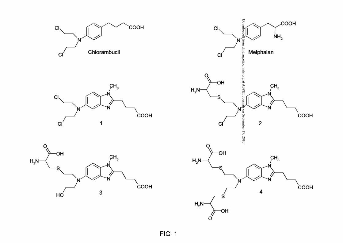
Bendamustine (4-{5-[bis-(2-chloro-ethyl)-amino]-1-methyl-1H-benzoimidazol-2-yl}-butyric
acid, 1, Figure 1), is a promising bifunctional alkylating agent containing a heterocyclic
nucleus that induces more long-lasting DNA double-strand breaks than other alkylating drugs
(Strumberg et al. 1996). The compound was first synthesized in 1963 with anticipated purine
based antimetabolic activity and has been shown preferentially cytotoxic activity in the
treatment of hematological malignancies, including NHL, CLL, and multiple myeloma, as
well as breast cancer (Höffken et al., 1998; Heider and Niederle, 2001; Kath et al., 2001;
Bremer, 2002; Konstantinov et al., 2002; Weidmann et al., 2002; Aivado et al., 2002; Pönisch
and Niederwieser, 2002). Moreover, anticancer activity was reported in advanced small cell
lung cancer and head or neck tumors (Reck et al., 1998; Rahn et al. 2001). 1 has been
approved and used in Germany for about 30 years, but has not been approved outside of
Germany. Currently, it is being studied as an investigational drug in the United States.
There is limited information on the metabolism and disposition of 1. To date, only one
metabolite resulting from biotransformation in humans has been identified as N-demethyl
bendamustine (4-{5-[bis-(2-chloro-ethyl)-amino]-1H-benzoimidazol-2-yl}-butyric acid, 7,
Figure 2) (Matthias et al., 1995; Preiss et al., 1998). An additional metabolite was thought to
be formed via beta-oxidation of the butyric acid moiety as demonstrated by MS data but has
not yet been structurally characterized. Own structural investigation did not confirm
formation of the assumed 2-hydroxy bendamustine. Hence, this metabolite was tentatively
named oxidized bendamustine (8, Figure 2). Similarly to other mustards containing the bis-
chloroethyl moiety, two products of chemical hydrolysis namely monohydroxy and dihydroxy
bendamustine (4-{5-[(2-chloro-ethyl)-(2-hydroxy-ethyl)-amino]-1-methyl-1H-benzoimidazol-
2-yl}butyric acid, 5 and 4-{5-[bis-(2-hydroxy-ethyl)-amino]-1-methyl-1H-benzoimidazol-2-
yl}-butyric acid, 6, Figure 2) have been detected (Preiss et al., 1985). The mean percentage of
the administered dose excreted in urine as the sum of 1, 5, 6, 7, and 8 was 20% as shown in 12
cancer patients with normal renal function (Teichert et al., 2003). This finding provides strong
This article has not been copyedited and formatted. The final version may differ from this version.DMD Fast Forward. Published on April 21, 2005 as DOI: 10.1124/dmd.105.003624
at ASPE
T Journals on Septem
ber 17, 2018dm
d.aspetjournals.orgD
ownloaded from

DMD #3624R
5
evidence that a considerable part of the administered dose is eliminated by nonrenal
mechanism and is compatible with earlier results obtained from animal experiments that
revealed a biliary excretion of nearly 42% for 1 in rats within 2 h after i.v. administration of
radioactively labeled 1 (Bezek et al., 1991). On the other side, 30% of the administered
radioactivity was excreted via urine.
To understand the metabolic fate of 1, we conducted the studies on the metabolism and
disposition of 1 in five patients with cholangiocarcinoma. In particular, we sought to
determine whether the GSH detoxification mechanism plays a role in the metabolism of this
alkylating agent. In vitro spontaneous and GST mediated reaction of melphalan and
chlorambucil both structurally related to 1 (Figure 1) with GSH has been investigated as
reported in numerous articles (Dulik et al., 1986, 1990; Ciaccio et al., 1990, 1991; Meyer et
al., 1992; Horton et al., 1999; Paumi et al., 2001; Zhang et al., 2003; Zhang and Lou, 2003).
However, no papers have been published describing the qualitative or quantitative
determination of these conjugates in humans. In this article, we present the chemical
syntheses of the major biliary metabolites, their characterization and identification in human
bile, urine, and plasma.
This article has not been copyedited and formatted. The final version may differ from this version.DMD Fast Forward. Published on April 21, 2005 as DOI: 10.1124/dmd.105.003624
at ASPE
T Journals on Septem
ber 17, 2018dm
d.aspetjournals.orgD
ownloaded from

DMD #3624R
6
Materials and Methods
Chemicals
L-Cysteine was purchased from Sigma-Aldrich Fine Chemicals Co. (Taufkirchen, Germany).
Acetonitrile, water for HPLC, ammonium acetate, perchloric acid, and acetic acid were
obtained from J. T. Baker (Deventer, The Netherlands). D6-DMSO was obtained from
Chemotrade Chemiehandelsgesellschaft mbh, (Leipzig, Germany). All other chemicals were
obtained from Merck KGaA (Darmstadt, Germany). All reagents were of analytical grade and
the solvents were of HPLC grade. 1 (as hydrochloride), 5, 6, and 7 (as hydrochloride) were
generous gifts from ribosepharm GmbH (Munich, Germany).
Instrumentation and Analytical Conditions
LC-MS
A ConstaMetric 4100 MS Series pump with a SCM 1000 Vacuum Membrane Degasser, an
autosampler AS 3000, and a model spectromonitor 3200 programmable wavelength detector
(Thermo Separation Products, Riviera Beach, Florida, USA) was interfaced to a Finnigan
(Finnigan MAT, Bremen, Germany, now Thermo Electron Corporation) SSQ-7000 single
quadrupole mass spectrometer equipped with an electrospray ionization (ESI) /APCI interface
and coupled to a Digital Personal DEC 5000/25 workstation. The LC was carried out on a
narrow bore column (125 x 2.0 mm i.d.) packed with Ultrasep ES PHARM RP18 5µm
(Separation Service, Berlin, Germany). The mobile phase consisted of two components,
namely solvent A (water with 5 mM ammonium acetate and 0.1% acetic acid, v/v) and solvent
B (acetonitrile with 5 mM ammonium acetate and 0.1% acetic acid, v/v). Samples were
separated using a slow gradient from 5 to 80% B in 60 min at a flow rate of 0.3 ml/min. The
flow was split 5:1 into the mass spectrometer. Positive-ion electrospray-mass spectrometric
analysis was carried out with a capillary temperature of 220°C and a capillary voltage of 4.5
kV. The value of the CID offset voltage was 10.0 V. The sheath gas and auxiliary gas both of
This article has not been copyedited and formatted. The final version may differ from this version.DMD Fast Forward. Published on April 21, 2005 as DOI: 10.1124/dmd.105.003624
at ASPE
T Journals on Septem
ber 17, 2018dm
d.aspetjournals.orgD
ownloaded from

DMD #3624R
7
nitrogen 4.6 (Messer Griesheim GmbH, now Air Liquide Deutschland GmbH, Krefeld,
Germany) were set to 60 psi and 10 psi, respectively. Mass spectra were recorded at an
electron multiplier voltage of 1300V. Peaks were detected either by single ion recording or by
scanning over an appropriate mass range. Data acquisition, reduction, selected ion monitoring
and peak area calculations are performed under software control by Alpha AXP DEC 3000
Data System (Digital Equipment Corp, Maynard, MA, USA).
NMR
1H and 13C NMR spectra were obtained on Varian Gemini 200 (Palo Alto, CA, USA) and
Bruker DRX-600 (Bruker BioSpin GmbH, Rheinstetten, Germany) spectrometers at 26° C,
with DMSO-d6 as the solvent. Residual solvent signals were used as internal chemical shift
references for proton (δDMSO = 2.49 ppm) and carbon (δDMSO = 39.52 ppm) spectra. J values
are given in Hz. Signals were assigned by means of 2D proton-proton (COSY) and proton-
carbon (HMQC, HMBC) shift-correlation spectra.
Preparative HPLC
All conjugates were purified by gradient HPLC procedure using two Shimadzu LC 8 pumps
(Kyoto, Japan) and a Vydac 218 TPB 1520 column (Grace Vydac, Hesperia, CA, USA) 300 x
40 mm i.d. Flow rates were set at 70 ml/min. Mobile phase A consisted of 1 ml 12M HCl in 1
L water and mobile phase B of acetonitrile/water/12M HCl 1000:200:1 (v/v/v). The column
was equilibrated with mobile phase A. From 5 to 50 min, a linear gradient ran from 100% A /
0% B to 100% B. Purity and identity of each conjugate was verified by analytical HPLC and
LC-MS.
Analytical HPLC
Instruments used in this study were: Alliance 2695 and fluorescence detector 2475 (Germany
Waters GmbH, Eschborn), HP 1090 gradient HPLC system with photo diode array (PDA) and
This article has not been copyedited and formatted. The final version may differ from this version.DMD Fast Forward. Published on April 21, 2005 as DOI: 10.1124/dmd.105.003624
at ASPE
T Journals on Septem
ber 17, 2018dm
d.aspetjournals.orgD
ownloaded from
DMD #3624R
8
HP 1046A fluorescence detector (Hewlett Packard, predecessor of Agilent Technologies, Palo
Alto, CA, USA). A column SYNERGI 4 µ MAX-RP 80A, 250 x 2 mm i.d. equipped with a
guard cartridge 4 x 2 mm (Phenomenex, Torrance, CA, USA) was used for identification of
metabolites. The mobile phase consisted of 0.1 ml 12M HCl in 1 L water (A) and
acetonitrile/water/12M HCl 800:200:0.02 (v/v/v) (B). The gradient was 5 - 40% B in 70 min
at a flow rate of 0.3 ml/min. For analysis by PDA detection, UV absorption was recorded at
233 nm. The excitation wavelength of the fluorescence detector was set to 328 nm and the
emission wavelength to 420 nm to monitor eluted components. For quantitative analysis of 1
as well as its metabolites, a six-point calibration curve was constructed for each compound
except 8 according to an internal standard method. Metabolite 8 was tentatively quantitated by
means of the calibration curve of 1. (5-{5-[bis(2-chloro-ethyl)-amino]-1-methyl-1H-
benzoimidazol-2-yl}-pentanoic acid was used as internal standard.
Synthesis of Reference Compounds
Cysteine S-Conjugate of 1
4-{5-[[(2-Amino-2-carboxy-ethylsulfanyl)-ethyl]-(2´-chloro-ethyl)]-amino]-1-methyl-1H-
benzoimidazol-2-yl}-butyric acid, 2, Figure 1, was synthesized following the method reported
by Paumi (16). Accordingly, 0.4 mg/ml L-cysteine and 0.6 mg/ml 1 hydrochloride was
dissolved in 100 ml of a solution of 0.1 M sodium bicarbonate and 0.2 M NaCl. The reaction
mixture was incubated at 25 °C for 1 h and terminated by addition of 2 ml 12M HCl to the
reaction mixture. The product was purified by preparative HPLC followed by lyophilisation.
LC-MS: C19H27ClN4O4S requires [MH]+ 443; found 443.04.
This article has not been copyedited and formatted. The final version may differ from this version.DMD Fast Forward. Published on April 21, 2005 as DOI: 10.1124/dmd.105.003624
at ASPE
T Journals on Septem
ber 17, 2018dm
d.aspetjournals.orgD
ownloaded from
DMD #3624R
9
Cysteine S-Conjugate of 5
4-{5-[[(2-Amino-2-carboxy-ethylsulfanyl)-ethyl]-(2´-hydroxy-ethyl)]-amino]-1-methyl-1H-
benzoimidazol-2-yl}-butyric acid, 3, Figure 1, was synthesized and purified as described for 2
using 5 instead of 1. LC-MS: C19H28N4O5S requires [MH]+ 425; found 425.16.
Dicysteine S-Conjugate Trifluoroacetate of 1
4-(5-{Bis-[2-(2-amino-2-carboxy-ethylsulfanyl)-ethyl]-amino}-1-methyl-1H-benzoimidazol-
2-yl)-butyric acid, 4, Figure 1, was accomplished as described for synthesis of 2. The reaction
was terminated by addition of 2 ml TFA to the reaction mixture. The product was purified as
described for 2. LC-MS: C22H33ClN5O6S2 requires [MH]+ 528; found 528.10.
Sample Collection
Samples were obtained from five patients (2 m, 3 f) with cholangiocarcinoma who have been
on treatment with 1 hydrochloride. The intravenous dose received by each patient on day 1 of
the first of overall 4 cycles was 140 mg/m2, the average age of the patients was 69.0 ± 3.4
years. 20 blood samples (4.5 ml) including a pre-dose blank sample were drawn from the
cubital vein of the arm contra lateral to that used for the administration of 1 hydrochloride up
to 8 hours after starting the i.v. infusion. After centrifugation, the supernatant plasma was
withdrawn and immediately frozen at –70°C. 10 ml aliquots of the urine samples collected
prior dosing and during 6 sampling intervals up to 24 hours postdose were stored in
polypropylene tubes at -70°C. The tubes were pre-treated with HCl and NaCl to prevent
chemical hydrolysis. For bile sampling, two temporary external nasobiliary drainages (7F,
290 cm, 8 holes; Endo-Flex®, Voerde, Germany) were placed into the right and left hepatic
duct each via endoscopic retrograde cholangiography and left in place for the entire collection
period. After placement the complete biliary secretion was collected without any loss during
This article has not been copyedited and formatted. The final version may differ from this version.DMD Fast Forward. Published on April 21, 2005 as DOI: 10.1124/dmd.105.003624
at ASPE
T Journals on Septem
ber 17, 2018dm
d.aspetjournals.orgD
ownloaded from

DMD #3624R
10
the collection period. After collecting the bile samples, the nasobiliary drainages were
removed and permanent endoscopic stenting was performed. Bile aliquots were stored as
described for plasma and urine samples before dosing and during 16 intervals up to 24 hours
after starting the 30-minute infusion. The Ethics Committee of the Faculty of Medicine of the
University of Leipzig issued approval for this study.
Sample Preparation for the Identification and Quantitation of the Metabolites
For the identification and characterization experiments, chromatographic and mass spectral
characteristics of the conjugates from biological fluids were compared with those of the
previously synthesized reference compounds. During shaking on a rotational shaker at 400
min-1, 100 µL 20% perchloric acid were added to 0.2 ml of the plasma samples. The
precipitated proteins were removed by centrifugation at 4°C and 15,000 g for 5 min. The
supernatant was used for chromatographic analysis. For LC-MS experiments, each urine and
bile sample (1 ml) was extracted by SPE procedure after dilution with 2.5 ml water pH 3.0
(adjusted with HCl). For the determination of polar conjugates, urine and bile samples (0.1 ml
each) were diluted with 0.9 ml of the respective mobile phase, centrifuged at 15,000 g for 5
min and filtered through a centrifugal filter device (0.22 µm, Amicon ULTRAFREE®-MC)
before an aliquot was injected into the HPLC system.
This article has not been copyedited and formatted. The final version may differ from this version.DMD Fast Forward. Published on April 21, 2005 as DOI: 10.1124/dmd.105.003624
at ASPE
T Journals on Septem
ber 17, 2018dm
d.aspetjournals.orgD
ownloaded from

DMD #3624R
11
Results
Synthesis and Characterization of Synthetic L-Cysteine S-Conjugates of 1 and 5
The cysteine S-conjugates were synthesized by adaptation of a method for the preparation of
GSH conjugates described for the structurally similar compounds chlorambucil and
melphalan. Sodium phosphate was replaced by sodium bicarbonate to prevent formation of
phosphate adducts. Results of recent pharmacokinetic studies have been indicated that 1 and 5
are the major urinary components following i.v. administration of 1 hydrochloride. Therefore,
we decided to prepare the cysteine S-conjugates of 1 and 5 as well. The reference compound
was purified by preparative HPLC from the reaction mixture of 5 with L-cysteine and
characterized by 1H-NMR, 13C-NMR, and LC-MS analysis. Beside the parent compound 1
(12-20%) and its hydrolysis product 5 (1%), the reaction of 1 with L-cysteine yielded three
peaks of unknown identity separable by reverse-phase HPLC. The minor peak was identified
as 3 by comparison with the reaction product of 5 with L-cysteine. In addition, 3 was obtained
by hydrolysis of 2 with aqueous NaOH. Addition of chloride to the reaction mixture of 1 and
L-cysteine reduces formation of 3 by preventing both, hydrolysis of 1 and 2. Under the chosen
experimental conditions we achieved product yields of 35-40% for 2 and 35-48% for 4.
Hence, two major peaks were obtained from this reaction mixture under the above described
experimental conditions, which were individually collected with preparative HPLC and
characterized by 1H-NMR, 13C-NMR, and LC-MS. LC-MS and HPLC analyses with UV as
well as fluorescence detection revealed a purity of 96.4% for 2, 94.8% for 3, and 92.8% for 4,
respectively. The presence of the cysteine moiety was shown by the detection of characteristic
aliphatic protons and carbon atoms in the NMR spectra. COSY, HMQC, and HMBC spectra
supported this conclusion. The HMBC spectrum of 3 is depicted in Figure 3 showing the
characteristic chemical shifts as well as long-range couplings attributable to the assigned
structure of all cysteine S-conjugates. The NMR data for the synthesized reference standards
This article has not been copyedited and formatted. The final version may differ from this version.DMD Fast Forward. Published on April 21, 2005 as DOI: 10.1124/dmd.105.003624
at ASPE
T Journals on Septem
ber 17, 2018dm
d.aspetjournals.orgD
ownloaded from

DMD #3624R
12
are given in Table 1, and were used along with the MS and HPLC data as basic data for
identification of metabolites.
Identification of 1 and its metabolites in bile, urine, and plasma from cancer patients
An HPLC-method was developed by which both phase I and phase II metabolites of 1 could
be analyzed simultaneously in bile, urine, and plasma samples. The separation of 1, 5, 6, 7,
and 8 and the three cysteine S-conjugates was achieved by gradient elution followed by either
MS or fluorescence detection. Upon HPLC analysis with fluorescence detection of bile from
patients treated with 1 hydrochloride, the 3 peaks at 10.3, 11.5 and 27.3 min were prominently
visible (Figure 4). Further, no background peaks were observed in this elution region of the
blank bile samples collected before drug administration indicating little potential for
interference from background at low concentrations (Figure 4). The cysteine S-conjugates 1,
2, and 3 present in human bile were characterized by LC-MS analyses (Figure 5). In addition,
the expected molecular ions (m/z 425), (m/z 443), and (m/z 528) were detected by MS in
urine as well as bile samples from preparative HPLC (Figure 6). Identical mass spectra were
obtained from the authentic reference compounds previously characterized (not shown).
Quantitative assessment was carried out by HPLC/fluorescence detection. The six-point
calibration curve of each compound showed excellent linearity in the respective concentration
range, which was chosen according to peak intensity of the respective component determined
in preliminary experiments. The lower limits of quantitation for 2, 3, and 4 were 0.1, 0.15,
and 0.2 µg/ml, respectively. The concentration-time profiles indicated that the cysteine
derived adducts were the major drug-related components in all bile samples from the five
subjects. The percentage related to the sum of the parent compound and its metabolites
determined in this study ranged from 90.3 to 98.0 %, with a mean value of 95.8 %. The total
percentage of administered dose recovered in bile ranged from 1.5 to 4.8 %, with a mean
This article has not been copyedited and formatted. The final version may differ from this version.DMD Fast Forward. Published on April 21, 2005 as DOI: 10.1124/dmd.105.003624
at ASPE
T Journals on Septem
ber 17, 2018dm
d.aspetjournals.orgD
ownloaded from

DMD #3624R
13
value of 2.6 %. The sum of the cysteine S-conjugates recovered from urine accounted for 26.0
% of all metabolites including the parent ranging from 14.1 to 41.2 %. On the other side, the
mean percentage of 1 and 5 related to all components determined in urine was 39.0 % and
21.4 %, respectively. We measured a mean total urinary recovery of 8.3 %, including 0.9, 0.5,
and 1.0 % of the dose as 2, 3, and 4, respectively. Figure 7 shows a characteristic
chromatogram of a urine sample as well as a plasma sample from a cancer patient after
administration of 1. Maximum plasma concentrations of 1 ranged from 4.54 to 24.25 µg/ml
with a mean of 16.81 µg/ml. Those for 5, 6, 7, and 8 were in the range 0.12 to 0.77 µg/ml
(mean values). Maximum plasma concentrations of the cysteine S-conjugates were in the
range 0.82 to 2.89 µg/ml (mean values). The mean terminal elimination half-life was 45 min
for 2 and comparable to those for 1 (39 min), whereas prolonged values of 85 and 163 min
were calculated for 3 and 4, respectively. Neither glucuronide and sulfate conjugates nor GSH
conjugates of 1 were detected.
This article has not been copyedited and formatted. The final version may differ from this version.DMD Fast Forward. Published on April 21, 2005 as DOI: 10.1124/dmd.105.003624
at ASPE
T Journals on Septem
ber 17, 2018dm
d.aspetjournals.orgD
ownloaded from

DMD #3624R
14
Discussion
Although 1 is being increasingly used for cancer management, the metabolic profile of this
alkylating agent has been poorly investigated. In this paper, we report the synthesis and
characterization of the monocysteine S-conjugates of 1 as well as 5 and the dicysteine S-
conjugate of 1. The conjugates that were synthesized according to a procedure previously
published for chlorambucil and melphalan yield a characteristic NMR pattern for a cysteine S-
conjugated bendamustine heterocyclus. All NMR spectra were similar to each other except for
the cysteine proton signals of 4, showing twofold intensity indicating two identical cysteine
groups. Broadened signals appearing in the 1H-NMR spectra of 4 indicated the presence of the
trifluoroacetate ion in the molecule, whereas both 2 and 3 occurred in the non-ionic form. The
data described in this manuscript provide the first conclusive characterization of these
cysteine S-conjugates of 1. In the present study, the cysteine S-conjugates were identified in
bile, urine, and plasma samples from cancer patients after intravenous administration of 1
hydrochloride using LC-MS as well as HPLC with fluorescence detection by comparison with
the synthetic standards previously synthesized and characterized. Quantitative determination
of 1 and its metabolites was performed using HPLC with fluorescence detection.
The mean amount of bile collected from the subjects in 24 h was 638 ml. This is in good
accordance with the normal bile production of 600–800 ml per day. The cysteine S-conjugates
presumably formed via the prominent GSH conjugation pathways are excreted into bile in
high concentrations. The cysteine S-conjugate concentrations in the bile sampled directly
from the hepatic bile duct were much higher than the simultaneous plasma concentrations.
This finding provides evidence for efficient biliary excretion of 1 in man, as was found earlier
in an animal experiment (Bezek et al., 1991).
A large variation in total urinary excretion of 1 was seen between patients, with recovery
percentages ranging from 0.7 to 9.5 % of the administered dose. As expected, in all patients
the highest concentrations of 1, 5, 6, 7, and 8 were observed in the first urine samples. For the
This article has not been copyedited and formatted. The final version may differ from this version.DMD Fast Forward. Published on April 21, 2005 as DOI: 10.1124/dmd.105.003624
at ASPE
T Journals on Septem
ber 17, 2018dm
d.aspetjournals.orgD
ownloaded from

DMD #3624R
15
cysteine S-conjugates we observed a mean cumulative urinary excretion of 1.3 – 4.1 % of the
administered dose within 24 h. The major part of 2 was excreted within the first 3 h, whereas 4
was predominantly excreted within 10–24 h after administration. 3 was recovered throughout
the whole sampling period. The individual maximum amount excreted within 24 hours into
urine was 16 % of the administered dose including the parent drug as well as seven
metabolites. Plasma kinetics of 1, 5, 6, 7, and 8 were similar to those reported previously.
There are some unidentified peaks in the chromatograms indicating the presence of unknown
derivatives of 1 in the present study. Therefore, it must be concluded that 1 is excreted into
urine and/or bile in the form of as yet unknown metabolites (e.g. mercapturic acids). This may
be the reason for not being able to account for all of administered parent drug. In general,
glutathione and cysteine conjugates are subjected to further metabolism e.g. N-acetylation to
give mercapturic acid conjugates prior to excretion in mammals. Theoretically, 2 can still
form DNA cross-links via formation of the aziridinium intermediate. Therefore, further
investigations should be targeted to the metabolic pathways of 1.
In this study, three cysteine S-conjugates exemplified by 2, 3, and 4 were detected. The
existence of these S-containing metabolites demonstrates that 1 is conjugated with glutathione
and further metabolized. The results indicate that the detoxifying pathways of 1 in man
primarily involves phase I (and hydrolytic degradation) as well as phase II metabolism
followed by urinary excretion of polar metabolites. Structures of metabolites that have been
identified are shown in Figure 2. However, the major amount of the administered dose was
recovered as parent drug. Further investigations should be address fecal components to assess
the extent that biliary elimination contributes to the overall elimination of 1 in humans.
This article has not been copyedited and formatted. The final version may differ from this version.DMD Fast Forward. Published on April 21, 2005 as DOI: 10.1124/dmd.105.003624
at ASPE
T Journals on Septem
ber 17, 2018dm
d.aspetjournals.orgD
ownloaded from

DMD #3624R
16
Acknowledgments
We thank Dr. Peter Henklein, Charité – Institute of Biochemistry, for support and providing
the preparative HPLC system and Barbara Brecht-Jachan for lyophilisation of the reference
compounds.
This article has not been copyedited and formatted. The final version may differ from this version.DMD Fast Forward. Published on April 21, 2005 as DOI: 10.1124/dmd.105.003624
at ASPE
T Journals on Septem
ber 17, 2018dm
d.aspetjournals.orgD
ownloaded from

DMD #3624R
17
References
Aivado M, Schulte K, Henze L, Burger J, Finke J and Haas R (2002) Bendamustine in the
Treatment of Chronic Lymphocytic Leukemia: Results and Future Perspectives. Sem Oncol
29, Suppl 13:19-22.
Awasthi S, Bajpai KK, Piper JT, Singhal SS, Ballatore A, Seifert Jr WE, Awasthi YC and
Ansari GAS (1996) Interactions of Melphalan with Glutathione and the Role of Glutathione
S-Transferase. Drug Metab Disp 24:371-374.
Bezek Š, Ščasnár V, Trnovec T and Grupe R (1991) Hepatobiliar elimination of bendamustine
(Cytostasan) in rats. Pharmazie 46:810-811.
Bremer K (2002) High rates of long-lasting remissions after 5-day bendamustine
chemotherapy cycles in pre-treated low-grade non-Hodgkin’s lymphomas. J Cancer Res Clin
Oncol 128:603-609.
Ciaccio PJ, Tew KD and LaCreta FP (1990) The Spontaneous and Glutathione S-Transferase-
Mediated Reaction of Chlorambucil with Glutathione. Cancer Commun 2:279-286.
Ciaccio PJ, Tew KD and LaCreta FP (1991) Enzymatic conjugation of chlorambucil with
glutathione by human glutathione S-transferase and inhibition by ethacrynic acid. Biochem
Pharmacol 42:1504-1507.
Dulik DM, Fenselau C and Hilton J (1986) Characterization of melphalan-glutathione adducts
whose formation is catalyzed by glutathione transferases. Biochem Pharmacol 35:3405-3409.
Dulik DM, Colvin OM and Fenselau C (1990) Characterization of Glutathione Conjugates of
Chlorambucil by Fast Atom Bombardment and Thermospray Liquid Chromatography/Mass
Spectrometry. Biomed Eviron Mass Spectrom 19:248-252.
This article has not been copyedited and formatted. The final version may differ from this version.DMD Fast Forward. Published on April 21, 2005 as DOI: 10.1124/dmd.105.003624
at ASPE
T Journals on Septem
ber 17, 2018dm
d.aspetjournals.orgD
ownloaded from

DMD #3624R
18
Hayes JD and Pulford DJ (1995) The glutathione S-transferase supergene family: regulation
of GST and the contribution of the isoenzymes to cancer chemoprotection and drug
resistance. Crit Rev Biochem Mol Biol 30:445-600.
Heider A and Niederle N (2001) Efficacy and toxicity of bendamustine in patients with
relapsed low-grade non-Hodgkin’s lymphomas. Anti-Cancer Drugs 12:725-729.
Höffken K, Merkle Kh, Schönfelder M, Anger G, Brandtner M, Ridwelski K and Seeber S
(1998) Bendamustine as salvage treatment in patients with advanced progressive breast
cancer: aphase II study. J Cancer Res Clin Oncol 124:627-632.
Horton JK, Roy G, Piper JT, Van Houten B, Awasthi YC, Mitra S. Alaoui-Jamali MA,
Boldogh I and Singhal SS (1999) Characterization of a Chlorambucil-Resistant Human
Ovarian Carcinoma Cell Line Overexpressing Glutathione S-Transferase µ. Biochem
Pharmacol 58:693-702.
Kath R, Blumenstengel K, Fricke HJ and Höffken K (2001) Bendamustine monotherapy in
advanced and refractory chronic lymphocytic leukemia. J Cancer Res Clin Oncol 127:48-54.
Konstantinov SM, Kostovski A, Topashka-Ancheva M, Genova M and Berger MR (2002)
Cytotoxic efficacy of bendamustine in human leukemia and breast cancer. J Cancer Res Clin
Oncol 128:271-278.
Matthias M, Preiss R, Sohr R and Possinger K (1995) Pharmacokinetics of bendamustine in
patients with malignant tumors. Proc Annu Meet Am Soc Clin Oncol 14:A1476.
Meyer DJ, Gilmore KS, Harris JM, Hartley JA and Ketterer B (1992) Chlorambucil-
monoglutathionyl conjugate is sequestered by human alpha class glutathione S-transferases.
Br J Cancer 66:433-438.
This article has not been copyedited and formatted. The final version may differ from this version.DMD Fast Forward. Published on April 21, 2005 as DOI: 10.1124/dmd.105.003624
at ASPE
T Journals on Septem
ber 17, 2018dm
d.aspetjournals.orgD
ownloaded from

DMD #3624R
19
Paumi CM, Ledford BG, Smitherman PK, Townsend AJ and Morrow CS (2001) Role of
Multidrug Resistance Protein 1 (MRP1) and Glutathione S-Transferase A1-1 in Alkylating
Agent Resistance. J Biol Chem 276:7952-7956.
Pönisch W and Niederwieser D (2002) Bendamustine in the Treatment of Multiple Myeloma:
Results and Future Perspectives. Sem Oncol 29, Suppl 13:23-26.
Preiss R, Sohr R, Matthias M, Brockmann B and Hüller H (1985) Investigations of the
pharmacokinetics of bendamustine (Cytostasan) in humans [in German]. Pharmazie 40:782-
784.
Preiss R, Matthias M and Merkle KH (1998) Pharmacological and Clinical Date of
Bendamustine, in 17th International Cancer Congress (Moraes M, Brentani R, Bevilacqua R
eds.), pp 1637 – 1640, Monduzzi Editore International Proceedings Division, Milan.
Rahn AN, Schilcher RB, Adamietz IA, Mose S, Bormeth SB and Böttcher HD (2001)
Palliative Radiochemotherapy with Bendamustine in Locally Advanced Recurrent Tumours
of the Head and Neck [in German]. Strahlenther Onkol 4:189-194.
Reck M, Haering B, Koschel G, Kaukel E, von Pawel J and Gatzemeier U (1998)
Chemotherapy of advanced non-small-cell and small-cell bronchial carcinoma with
bendamustine – a phase II study. Pneumologie 52:570-573.
Strumberg D, Harstrick A, Doll K, Hoffmann B and Seeber S (1996) Bendamustine
hydrochloride activity against doxorubicin-resistent breast carcinoma cell lines. Anti-Cancer
Drugs 7:415-421.
Teichert J, Möckel J, Pönisch W, Seidel A, Lotfi M, Matthias M and Preiss R (2003)
Influence of hepatic and renal function on the pharmacokinetics of bendamustine. Int J Clin
Pharmacol Ther 41:564.
This article has not been copyedited and formatted. The final version may differ from this version.DMD Fast Forward. Published on April 21, 2005 as DOI: 10.1124/dmd.105.003624
at ASPE
T Journals on Septem
ber 17, 2018dm
d.aspetjournals.orgD
ownloaded from
DMD #3624R
20
Weber H, Amlacher R, Preiss R and Hoffmann H (1991) Pharmacokinetics of bendamustine
(Cytostasan) in B6D2F1 mice [in German]. Pharmazie 46:589-591.
Weidmann E, Kim SZ, Rost A, Schuppert H, Seipelt G, Hoelzer D and Mitrou PS (2002)
Bendamustine is effective in relapsed or refractory aggressive non-Hodgkin’s lymphoma. Ann
Oncol 13:1285-1289.
Zhang J and Lou YL (2003) Relationship between activation of microsomal glutathione S-
transferase and metabolism behavior of chlorambucil. Pharmacol Res 48:623-630.
Zhang J, Wong KP and Chow P (2003) Conjugation of chlorambucil with GSH by GST
purified from human colon adenocarcinoma cells and its inhibition by plant polyphenols. Life
Sci 72:2629-2640
This article has not been copyedited and formatted. The final version may differ from this version.DMD Fast Forward. Published on April 21, 2005 as DOI: 10.1124/dmd.105.003624
at ASPE
T Journals on Septem
ber 17, 2018dm
d.aspetjournals.orgD
ownloaded from

DMD #3624R
21
Legends for Figures
Figure 1 Structures of the synthesized cysteine S-conjugates used as reference standards
in this study as well as those of the alkylators chlorambucil and melphalan
structurally related to 1.
Figure 2 Proposed metabolic pathways of 1 in humans.
Figure 3 Chemical structure of the cysteine S-conjugates including numbering of carbon
atoms and aromatic protons as well as an example of characteristic long-range
couplings observed in the two-dimensional HMBC spectrum of 2 after
chemical synthesis and purification by preparative HPLC. Numbering of
carbon skeleton was according to Table 1 in comparison to NMR data and does
not correspond to the rules of chemical nomenclature.
Figure 4 HPLC chromatograms of a human (upper panel) bile sample 90-120 min after
administration of 140 mg/m2 1 as hydrochloride and (lower panel) blank bile
sample before drug administration spiked with internal standard.
Figure 5 HPLC-ESI-MS chromatogram (selected mass tracks, m/z = 425, 443, 528; and
reconstructed, or total, ion chromatogram, RIC) of a human bile sample (1.5-2
h) after start of the 1 hydrochloride infusion 140 mg/m2.
Figure 6 Mass spectra of the cysteine S-conjugates 2, 3, and 4 obtained by preparative
HPLC from the human bile sample described in Figure 5.
Figure 7 HPLC chromatograms of a human (upper panel) urine sample 3 – 6 h and
(lower panel) plasma sample 60 min after drug administration.
This article has not been copyedited and formatted. The final version may differ from this version.DMD Fast Forward. Published on April 21, 2005 as DOI: 10.1124/dmd.105.003624
at ASPE
T Journals on Septem
ber 17, 2018dm
d.aspetjournals.orgD
ownloaded from

DMD #3624R
22
Table 1 NMR spectral data of the synthetic reference standards 2, 3, and 4.
(A) 1H NMR Data (200 MHz, 600 MHz, 1H, H,H COSY)a
2 3 4
proton δ (DMSO-d6) J (Hz) δ (DMSO-d6) J (Hz) δ (DMSO-d6) J (Hz)
H3 7.68 (1H, d) 9.1 7.63 (1H, d) 9.2 7.65 (1H, *) *
H2 7.07 (1H, dd) 9.1, 2.1 7.02 (1H, dd) 9.2, 1.6 7.00 (1H, *) *
H1 6.88 (1H, d) 2.1 6.84 (1H, d) 1.6 6.83 (1H, *) *
CH 4.18 (1H, t) 5.3 4.15 (1H, t) 5.4 4.10 (1H, *) *
NCH3 3.89 (3H, s) 3.87 (3H, s) 3.87 (3H, *)
N-CH2-CH2-Cl 3.77 (2H, t)
CH2-Cl 3.77 (2H, t)
N-CH2-CH2-S 3.63 (2H, t) 3.60 (2H, t) 3.60 (4H, *)
CH2-OH 3.58 (2H, t)
N-CH2-CH2-OH 3.48 (2H, t)
CH2-het 3.13 (2H, t) 3.12 (2H, t) 3.12 (2H, *)
S-CH2-CH 3.09 (2H, t) 3.08 (2H, t)
CH2-CH2-S 2.79 (2H, t) 2.78 (2H, t) 2.79 (4H, *)
CH2-COOH 2.40 (2H, t) 2.40 (2H, t) 2.39 (2H, *)
CH2-CH2-CH2 2.00 (2H, m) 1.99 (2H, m) 2.00 (2H, *)
(B) 13C NMR Data (50 MHz, 150 MHz, APT, HMQC, HMBC)
2 3 4
carbon δ (DMSO-d6) δ (DMSO-d6) δ (DMSO-d6)
CH2-COOH 173.73 174.38 173.75
CH-COOH 169.59 170.26 169.88
C-6 151.85 152.07 151.71
This article has not been copyedited and formatted. The final version may differ from this version.DMD Fast Forward. Published on April 21, 2005 as DOI: 10.1124/dmd.105.003624
at ASPE
T Journals on Septem
ber 17, 2018dm
d.aspetjournals.orgD
ownloaded from

DMD #3624R
23
C-2 145.55 147.12 145.64
C-7 132.56 133.08 132.68
C-5 124.85 124.88 124.65
C-4 113.19 113.19 113.17
C-3 112.13 112.63 111.98
C-1 95.03 94.84 94.78
CH2-CH2-OH 58.75
CH2-CH2-Cl 58.75
N-CH2-CH2-OH 53.90
CH-NH2 52.79 52.79 52.25
N-CH2-CH2-Cl 52.35
N-CH2-CH2-S 51.76 51.67 50.89
CH2-COOH 33.21 33.21 32.56
S-CH2-CH 32.39 32.39 31.51
N-CH3 31.47 31.47 30.81
CH2-CH2-S 29.27 29.27 28.49
CH2-het 24.78 24.78 24.17
CH2-CH2-CH2 22.25 22.25 21.60
a s, singlet; d, doublet; dd, doublet-doublet; t, triplet; m, multiplet; *, not observed due to
broadened signals caused by the ionic structure;
This article has not been copyedited and formatted. The final version may differ from this version.DMD Fast Forward. Published on April 21, 2005 as DOI: 10.1124/dmd.105.003624
at ASPE
T Journals on Septem
ber 17, 2018dm
d.aspetjournals.orgD
ownloaded from
This article has not been copyedited and formatted. The final version may differ from this version.DMD Fast Forward. Published on April 21, 2005 as DOI: 10.1124/dmd.105.003624
at ASPE
T Journals on Septem
ber 17, 2018dm
d.aspetjournals.orgD
ownloaded from

This article has not been copyedited and formatted. The final version may differ from this version.DMD Fast Forward. Published on April 21, 2005 as DOI: 10.1124/dmd.105.003624
at ASPE
T Journals on Septem
ber 17, 2018dm
d.aspetjournals.orgD
ownloaded from

This article has not been copyedited and formatted. The final version may differ from this version.DMD Fast Forward. Published on April 21, 2005 as DOI: 10.1124/dmd.105.003624
at ASPE
T Journals on Septem
ber 17, 2018dm
d.aspetjournals.orgD
ownloaded from

This article has not been copyedited and formatted. The final version may differ from this version.DMD Fast Forward. Published on April 21, 2005 as DOI: 10.1124/dmd.105.003624
at ASPE
T Journals on Septem
ber 17, 2018dm
d.aspetjournals.orgD
ownloaded from

This article has not been copyedited and formatted. The final version may differ from this version.DMD Fast Forward. Published on April 21, 2005 as DOI: 10.1124/dmd.105.003624
at ASPE
T Journals on Septem
ber 17, 2018dm
d.aspetjournals.orgD
ownloaded from

This article has not been copyedited and formatted. The final version may differ from this version.DMD Fast Forward. Published on April 21, 2005 as DOI: 10.1124/dmd.105.003624
at ASPE
T Journals on Septem
ber 17, 2018dm
d.aspetjournals.orgD
ownloaded from

This article has not been copyedited and formatted. The final version may differ from this version.DMD Fast Forward. Published on April 21, 2005 as DOI: 10.1124/dmd.105.003624
at ASPE
T Journals on Septem
ber 17, 2018dm
d.aspetjournals.orgD
ownloaded from