Embed Size (px)
Citation preview


BUTTERWORTH-HEINEMANN An imprint of Elsevier Limited
© 2004, Elsevier Limited.All rights reserved.
No part of this publication may be reproduced, stored in a retrieval system, or transmitted in any form or by any means, electronic, mechanical, photocopying, recordingor otherwise, without either the prior permission of the publishers or a licence permitting restricted copying in the United Kingdom issued by the Copyright LicensingAgency, 90 Tottenham Court Road, London W1T 4LP. Permissions may be sought directlyfrom Elsevier’s Health Sciences Rights Department in Philadelphia, USA: phone: (�1)215 238 7869, fax: (�1) 215 238 2239, e-mail: [email protected] mayalso complete your request on-line via the Elsevier homepage (http://www.elsevier.com),by selecting ‘Customer Support’ and then ‘Obtaining Permissions’.
ISBN 0 7506 8772 X
British Library Cataloguing in Publication DataA catalogue record for this book is available from the British Library
Library of Congress Cataloging in Publication DataA catalog record for this book is available from the Library of Congress
Notice Medical knowledge is constantly changing. Standard safety precautions must be followed, but as new research and clinical experience broaden our knowledge, changesin treatment and drug therapy may become necessary or appropriate. Readers areadvised to check the most current product information provided by the manufacturer ofeach drug to be administered to verify the recommended dose, the method and durationof administration, and contraindications. It is the responsibility of the practitioner, relyingon experience and knowledge of the patient, to determine dosages and the best treatment for each individual patient. Neither the Publisher nor the editors and contributors assumes any liability for any injury and/or damage to persons or propertyarising from this publication.
The Publisher
Printed in China

BPhty(Hons) BScApp(HMS-Ex Man)
Division of Physiotherapy, School of Health and Rehabilitation Sciences,The University of Queensland, St Lucia,Australia
BPhty(Hons)
Division of Physiotherapy, School of Health and Rehabilitation Sciences,The University of Queensland, St Lucia,Australia
DipPhysio GradCertPhty(Geriatics)
Private Practitioner, Brisbane,Australia
BPhty(Hons) MPhty
Division of Physiotherapy, School of Health and Rehabilitation Sciences,The University of Queensland, St Lucia,Australia
BPhty MPhty PhD
Division of Physiotherapy, School of Health and Rehabilitation Sciences,The University of Queensland, St Lucia,Australia
BPhty GradCertPhty(Geriatrics)
Division of Physiotherapy, School of Health and Rehabilitation Sciences,The University of Queensland, St Lucia,Australia
AUA DipPhty
Division of Physiotherapy, School of Health and Rehabilitation Sciences,The University of Queensland, St Lucia,Australia
BPhty(Hons) PhD
Division of Physiotherapy, School of Health and Rehabilitation Sciences,The University of Queensland, St Lucia,Australia
vii
Susan R. Hourigan
Venerina Johnston
Diane L. Josephson
Nancy Low Choy
Jennifer C. Nitz
Ann Rahmann
Ruth Sapsford
Tina Souvlis
Contributors

Introduction
Jennifer C. Nitz and Susan R. Hourigan
1
In 2002 there were 2.5 million older people (aged 65�) in Australia, rep-resenting 12.7% of the total Australian population. In 2031 older peoplewill represent 22.3% of the total population (or 5.4 million people) and by 2051 they will represent over one-quarter of our population(Australian Bureau Statistics 2003). The increase in the older old (those85 years�) is even more pronounced, rising from 9.1% of those aged 65years and over in 1996 to 20.1% by 2051. The average age of people livingin residential care homes in Australia is 83 years (AIHW 2002).
In Australia, at the latest census in June 2001, there were 136 608 people accommodated in 2977 residential aged care facilities (RACFs). Twoper cent of these places were devoted to respite care but this accountedfor 43 606 admissions during the financial year ending June 2001. In thattime there were 46 545 admissions for permanent care. Most of the peo-ple admitted to RACFs were women (72%) and just over half of all peopleadmitted were aged over 85 years. Only 6094 (4.5%) people admittedwere less than 65 years of age. Twenty-three per cent of these youngerpeople were admitted to high care compared to 63% of all older peopleadmitted. The remaining admissions were to low care (AIHW 2002).
Prior to admission, 41% lived alone in a house or flat, 21% with a spouseand 12% with family members. Thus most people admitted to RACFs enterfrom the community. When in the community, many would have beenthe recipients of Community Aged Care Packages that were designed toenable older people to live independently in the community with assis-tance from community agencies such as meals on wheels, home help orcommunity nursing (AIHW 2002).
Where does the physiotherapist fit into the residential aged care scene?This book was conceived to enable physiotherapists to define their rolein this area of practice and to educate physiotherapy students (bothundergraduate and postgraduate). It should assist our co-workers in agedcare regarding the skills we bring to the workplace and how collectivelywe might offer best practice procedures that enable residents and carersto attain a quality living environment.
From here on in we will refer to residential aged care facilities eventhough young people are also cared for in these communities. In othercountries the RACFs will be known as nursing homes, rest homes, hostelsand retirement homes.
People entering RACFs often do so after losing the ability to manageindependently in the community. It is often a culmination of events such

as the loss of a partner or parental carer who made life at large in thecommunity possible, or in other circumstances a catastrophic life eventthat has led to physical dependency. In many cases this ultimate loss fol-lows a series of losses including driver’s licence, home, friends throughdeath and pets. To many individuals, entering a care facility means givingup self-determination, dignity and quality of life but this need not be thecase with good management in a well-organized facility. Irrespective ofwhether the resident will spend their final days in the facility or havebeen admitted to provide respite for a carer or even for slow stream rehabili-tation, it is the duty of the care providers to ensure best practice in allaspects of resident management. In many respects care providers arebound to take on an advocatory role to ensure residents are protectedfrom injury and are able to access all necessities for the best quality of lifepossible to them. Physiotherapists possess numerous skills that enablethem to assist residents and carers to achieve this goal.
Identification of the needs and risk factors a resident presents with onadmission to a care facility allows development of the most appropriatecare plan that will enable attainment of the best quality of life afforded bytheir circumstances. If all care staff adopt a clinical reasoning approachwhen drawing up a care plan that incorporates the stated desires of theresident, a harmonious and cooperative environment can be provided.By allowing the new resident and/or their family to participate in theplanning process, trust is generated and communication of doubts andfears are encouraged, alleviating much anxiety.
New residents often come to residential care with preconceived ideasthat are often of a negative nature. The very idea of something new is for-eign to many as they have lived in the same house with the same peoplepossibly all their life and to change brings fear of the unknown. A newenvironment without any of the familiar objects is very confusing tomany older people. Often the change needs interpersonal skills that havenot been developed due to previous isolation. If you consider the intim-ate interaction that occurs when assisting a totally dependent personwith bathing and toileting some idea of the extent of adaptation neces-sary is gained. Not only is there a new physical environment for new resi-dents, but also new carers, new routines, new faces, new procedures andnew timetables. Add difficulty with communication to a physical depend-ency and we can begin to understand the mental turmoil of some newresidents. Some lucky people come to a residential care facility after having mentally prepared themselves for the lifestyle change. However,there will be a level of stress that always accompanies anybody whomoves from one place of residency to another. Limiting the number ofnew experiences can reduce stress. Therefore it is beneficial to have onecarer undertake the admission process for the new resident. They shouldalso continue to care for the needs of the resident at this time. To have afamily member or friend who is accepted by the resident present duringadmission might help transition.
2 Introduction

On admission, an interim care plan is drawn up for each new resident.This will enable a safe level of functioning to prevail until a full assessmentof physical, communication and behavioural status is undertaken. In real-ity, this process takes a few weeks depending on the distress caused bythe admission to the RACF. Therefore the initial care plan should not beseen as unchangeable nor should any care plan be considered finite. Allresidents will be liable to rapid change in their medical condition simplydue to their age and frailty. Every person involved in the care of a residentshould be aware of this and be able to adapt management strategiesimmediately. However, many carers lack the skill to be able to identifywhen the resident’s condition changes let alone know how to adapt acare plan to suit the change. The physiotherapist is the appropriate practitioner to evaluate change in physical ability and to recommendappropriate care plan modification.
Clinical reasoning is the approach used to develop the care plan for aresident. It involves using theoretical knowledge related to ageing and patho-logical processes seen in the elderly, the assessment of these problems andintervention options to enhance function and quality of life for the resi-dents. In this book we will look at physiological changes accompanyingthe ageing process as well as the physiotherapist’s role in assessment, pre-venting residents’ injuries and promoting life satisfaction. Major presentingproblems relating to being immobile, barely mobile, or ambulatory will beexamined physiotherapeutically. Information and strategies to assist withresidents suffering from pain, osteoporosis, incontinence or palliation willbe covered. A special chapter concerned with aquatic physiotherapy is alsoincluded as pools are becoming more prevalent in aged care facilities.
Appendices have been included to cover a number of issues which areimportant to physiotherapy practice. In a number of RACFs funding islinked to proof of efficacy; therefore choosing the appropriate outcomemeasurement that reflects the results of intervention (unique to an indi-vidual resident’s care plan) is vital. The appendices include a table of use-ful outcome measures, assessment case studies, suggestions for successfulcase conferences, electrotherapy considerations in aged care practice andmobility aids.
We hope the ideas and topics contained in our book are informative aswell as thought provoking for our readers. Working in residential agedcare as a physiotherapist is extremely challenging as our skills in clinicalpractice, education, health promotion and research will be recruited andutilized frequently as we fulfil our role.
Introduction 3
References
Australian Bureau Statistics 2003 Australian SocialTrends, Cat 4102.0, p 2
Australian Institute of Health & Welfare (AIHW) 2002Residential aged care services in Australia
2000–01: a statistical overview. AIHW Cat. No.AGE 22. Canberra: AIHW (Aged Care StatisticSeries No. 11). http://www.aihw.gov.au

Physiological changeswith age
Jennifer C. Nitz and Susan R. Hourigan
7
1
This chapteraims to:
It is a common belief that both physical and cognitive function will declinewith ageing and that people must accept these changes. In many respectsthis is true, but compensatory and preventive management can controlthe effects of system decline. An example of this is to use correctivelenses to accommodate for the presbyopia of middle age. Similar adjust-ments to lifestyle can slow or negate the effect of ageing in other systems.In this chapter the changes due to ageing will be investigated in the context of residents in aged care.
People generally reach their peak health and performance abilitiesbetween adolescence and 30 years of age. Functional capacity after thisage then declines throughout the lifespan depending on genetics, lifestylecharacteristics and health. All physiological measures that decline withage do not do so at the same rate. Heart rate at rest does not change dramatically with advancing years; however, heart rate at maximum physical exertion decreases significantly. Nerve conduction velocity slows10–15% from 30 to 80 years of age and maximum breathing capacitydeclines by approximately 60% between 30 and 80 years. The rates ofdecline in the various functions differ and are significantly influenced byvarious factors including the level of physical activity and any associatedpathology. There is no doubt that regular activity and physical trainingenables older persons to retain higher levels of functional capacity(McArdle et al 1996).
Decline in the special senses can be a most disabling aspect of ageing.As will be seen in later chapters, the resulting disability when vision,auditory and vestibular function and somatosensation diminish with ageplays a major role in exposing the older person to an increased risk of
■ overview the changes associated with ageing in all of thesystems important to physical performance and functioning.This information will be referred to throughout this text as weexamine clinical reasoning and physiotherapeutic treatment ofelderly people living in residential aged care facilities.
Introduction

injury from falling. In addition, interaction with the environment isdecreased and opportunities for choosing modalities for entertainmentare curtailed by loss of vision or hearing or the ability to safely mobilizein the community. Thus decline in the special senses can lead to a dramaticchange in lifestyle and possibly a diminished quality of life. Thereforeunderstanding the ageing effects on these organs is important whenworking with the elderly.
VisionAs the eye ages the lens loses clarity owing to an increase in lens density(Jackson et al 2002). It becomes yellowed and crazed (Bron 1997). Inaddition, the ageing lens can form a cataract. Cataract formation isbelieved to be due to lens protein modification and reduction in antioxi-dants (Bron 1997). The retina at the back of the eye is also affected byageing. Within the retina lie the rods that are responsible for vision in dimlight and the cones that respond to bright light and colour. At the centreof the retina lies the macula. This is a specialized area comprising a cen-tral cone-rich fovea that is surrounded by a rod-dominated parafovea. Thephotoreceptor outer segment contains the proteins of the phototrans-duction cascade. The nutritional needs of this component are maintainedby the retinal pigmented layer. This layer is responsible for maintainingphotoreceptor health by helping to maintain ionic balance and hydration,transport and filtration of nutrients, by providing retinoid intermediatesthat replenish photopigment bleached by light exposure and also byabsorbing stray photons (Jackson et al 2002). Blood flow to the retinal pig-mented layer and the photoreceptors is via the choroidal system, whichhas the richest blood flow in the body. Between the choroid capillariesand the retinal pigmented layer lies Bruch’s membrane. Age-related mac-ular degeneration (ARMD) in the early stages is characterized by extra-cellular lesions and changes in the retinal pigmented layer morphologyand damage to Bruch’s membrane in the region of the macula (Bron 1997,Jackson 2002). Later extensive retinal pigment layer atrophy and vascularchanges with in-growth of vessels in Bruch’s membrane and under theretinal pigmented layer accounts for blindness in ARMD (Jackson 2002).
ARMD might be demonstrated in the early stages by an increased timefor recovery of vision after exposure to bright lights. With ageing, darkadaptation after exposure to bright light increases by about 8 secondsper decade. Delays of an average 13 minutes have been seen in peoplewith early ARMD (Owsley 2001). This can lead to a considerable increasein fall risk for older people when moving from light to dark environmentsor from dark to light as the time for clear vision to return increases withage. Thus older people should be made aware of this delay in order toinclude into their lifestyle safety practices that will reduce the risk ofinjury. Older people with early ARMD (Scilley et al 2002) also experiencenight driving problems.
8 Physiological changes with age
Special senses

Eventually with ARMD, there is central visual field loss that affects theability to read and perform activities requiring fine manipulation such as sewing. Distortion of colour and shapes can occur. Together this canaffect edge perception where contrast is minimal. From a functionalviewpoint, the person is placed in danger of not seeing steps or thinkingthere is a step up or down because of colour change or shadow. A doorthat is ajar if it is the same colour as the surrounding walls may not beseen nor the edge of the chair seat or the edge of the bed if contrastingcolours are not used. The older person is therefore at risk of a fall througherrors of judgement induced by decline in visual acuity or depth percep-tion (Carter et al 1997, Pinto et al 1997). Changes in visual acuity canaffect depth perception, especially when stereopsis cannot be attained withcorrective lenses. It can also contribute to visual illusions such as not per-ceiving moving repeated patterns such as on escalators (Harwood 2001).
With ageing, the ciliary muscles are less able to change lens curvature,thereby slowing the ability to rapidly change focus from close work todistance. This becomes a problem when ambulating when near and distant sections of the environment require scanning to navigate safelythrough environmental hazards.
The pupillary light reflex is slowed with age. This means that it takeslonger for the elderly to be able to see clearly when there is a change inlight conditions from dark to light or light to dark. ARMD also plays a role inthis presentation. In the younger person light accommodation takes a fewseconds. As a person ages, the time interval extends and might take as longas 15 minutes to occur. Not allowing for the slowed light accommodationby standing still and waiting for the vision to clear can increase the risk ofa fall. The typical time when this becomes critical is at night when puttingon the light to get up to go to the toilet or during the day when moving fromindoors to outdoors, especially on a bright sunny day (Pinto et al 1997).
Visual acuity changes with ageing. Presbyopia can increase but morecommonly ARMD and lens pathology interfere with the ability to read orperform close work (Pinto et al 1997). The use of coloured print or papershould be limited if using the print medium to communicate to residentsvia notice boards or newsletters. Many of the oldest old residents mayfind it difficult to discern coloured letters on coloured paper due to thecumulative effect of these eye changes and so might become isolatedfrom the community activities. Larger font and bold black print on whitepaper can limit this possibility even if it is not as attractive to the eye.
Pathological conditions that can affect vision are more common in theelderly. These include complications arising after cataract surgery, retinaldetachment and glaucoma. All can contribute to diminished sight orblindness. Conditions such as stroke and multiple sclerosis can also causevisual field loss and are relatively common in residents.
Visual conflict occurs when what the person interprets they are seeingis at odds with proprioception, touch and pressure and vestibular sens-ory input. Difficulty in discerning reality in visual conflict situations is
Special senses 9

often related to decline in the vestibular and proprioceptive systems aswell as central integrative systems within the central nervous system(Harwood 2001). In these instances the older person is at more risk of a fall.
Age-related hearing loss is known as presbyacusis (Seidman et al 2002).Cochlear degeneration occurs with ageing and signs of degenerationbegin with decline in the ability to hear high frequency sound, followedby progressive loss of lower frequency hearing. There is also loss of theability to discriminate between sounds, which leads to speech soundingmuffled, and in some instances whole words become indiscernible.Hearing loss is not usually symmetrical and this causes difficulty in loca-tion of sound source. Functionally this difficulty in sound location candelay avoiding responses to danger such as moving aside to clear a skateboarder on the footpath or an approaching car. Delayed detection of thedirection from which danger is approaching might require faster evasivemovement than can be controlled by the elderly person and a fall caneventuate. Where possible, hearing should be tested and elderly peoplewith hearing loss should be encouraged to wear hearing aids at all times.
Slowed nerve conduction from the ear and within the central nervoussystem can cause auditory response time to increase. This can lead toconfusion in the elderly if multiple questions are fired at them withoutallowing for time to process the input and respond accordingly. Manyprofessionals have been guilty of this and then misinterpreting the lack orinappropriateness of response by the older person as indicating demen-tia. Unfortunately such labelling can stick, with far-reaching consequences.
The vestibular apparatus comprises the utricle, saccule and three semi-circular canals that enable orientation of the head with respect to gravityand enable response and perception of head movement (Guyton 1991).Thus normal vestibular function is needed for a person to maintain headstability during any movement task, to allow conjugate eye movement forreading and the integration of visual and proprioceptive information forenvironmental interaction (Di Fabio & Emasithi 1997). By the age of 70years there has been a reduction of 40% in the number of vestibular hairand nerve cells (Rosenhall & Rubin 1975). Ageing of the components ofthe vestibular apparatus leads to a reduction in vestibular sensory inputthat causes the older person to rely more on vision and less on proprio-ceptive information to continue to move and interact in the environ-ment. The impact of vestibular decline with age will be covered in moredetail in the chapter dealing with balance (Chapter 9).
With ageing there is a decline in ability to taste food. Because of this dietis often changed by the elderly to increase taste needs. Unfortunately thisis often by increasing the sugar and salt content, thereby raising the like-lihood of glucose intolerance and hypertension. The sense of smell alsodeclines. Since the aroma of food and taste are intimately connected to
10 Physiological changes with age
Hearing andvestibular
function
Taste and smell

enhance enjoyment of eating, the elderly often reduce nutritional intakedue to the lack of interest in food that accompanies the decline in thesesenses. A potential danger associated with loss of smell is the inability tosmell smoke. This functional decline might be a factor in the number ofelderly people injured or killed in house fires each year. In a recent studyundertaken in the USA, which examined olfactory decline in people aged53 to 97 years, impaired olfaction was found in 24.5% of the participants.Impairment was more prevalent among men and was present in 62.5% ofpeople over 80 years of age. Self-reported olfactory decline did not equateto test findings and this aspect became worse with age, thus confirmingthe importance of acknowledging the deficit and educating older peopleabout safety precautions to compensate for the loss (Murphy et al 2002).
Table 1.1 summarizes the changes to special senses, the effect of thesechanges on function, and management strategies that might be employedto minimize the impact on daily life.
Special senses 11
Special sense Change due to ageing Functional impact Management strategy
Vision Lens: Decreased ability to Use strongly contrastingyellowed and crazed discern colour, perceive colours in decor so edges are cataract an edge and do close definedARMD vision activities Large black print andGlaucoma increased wattage for lightAcuity bulbs
Encourage use of spectaclesfor close or distance
Slowed reflexes: Increases risk of falls Pause until vision is clearpupillary light reflex when focus or lightlens accommodation conditions change
Remove clutter on the floorVisual conflict Inappropriate balance Identification of the problem
correction leading to and instigation of a strategy increased fall risk suitable for the individual
resident
Hearing Cochlear degeneration: Social isolation Encourage hearing aid useloss of high frequency Increased risk of and help change batteriessound falling Face the deaf person at theirunilateral hearing loss height so they can see you
clearly. Speak slowly andclearly. Do not shout. Beaware your approach mightnot be heard and suddenappearance in line ofsight can disturb balance
Table 1.1Special sense changes with ageing
table continues

Age changes can occur in the gastrointestinal tract from the mouth to theanus. These changes can have a major bearing on the health of the elderlyresident and thereby directly or indirectly affect the role of the physio-therapist.
Nutritional deficiency has been identified as one of the factors con-tributing to frailty (Campbell & Buchner 1997). The diet of elders containsdecreased protein, fruit and vegetables; thus problems relating to vitaminand mineral deficiencies are common. Such deficiencies can adverselyaffect tissue health, with all organs being affected, but the main effects areevident in the skin and neural tissues. Anaemia is common in the elderlyand affects the oxygen-carrying potential of the blood. This in turn can leadto reduced metabolic function in all tissues with particular consequencesof declined cognitive function, muscle weakness and increased falls risk.
A number of factors can contribute to poor nutrition. Inability to shopfor fresh food, lack of financial capacity to buy fresh food as well as lackof the cognitive and physical capability to prepare and cook the food cancompound the physiological changes due to ageing that reduce the nutri-tion gained from food ingested. Residents of care facilities should not bedisadvantaged nutritionally as all the factors identified to contribute tomalnutrition should be countered.
The changes due to ageing of the gastrointestinal system affect the alimentary tract and also the related organs including the liver and pan-creas. It is therefore important to understand the ageing effect on theseorgans as the resident can present with major health ramifications fromthese changes.
Atrophy of the gums and bone that occurs with age and poor dentalhygiene can lead to tooth loss. Many elders have no teeth and often donot wear false teeth. The most common reason for not using dental pros-theses is poor fit because of gum atrophy. The consequence of not hav-ing teeth relates not only to cosmesis but to inability to enunciate words
12 Physiological changes with age
Special sense Change due to ageing Functional impact Management strategy
Vestibular Otolith and endolymph Delayed balance Ensure good lighting andfunction declines response and increased correction of acuity so vision Hyposensitivity to risk of falls can be used efficiently to head position and Greater reliance on vision maintain balancemovement in all planes and somatosensation
for balance
Taste and smell Decline in taste and Lack of interest in Prepare visually appealingsmell eating and consequent food to stimulate the
nutritional lack appetite. Use spices toInability to detect enhance tastea fire Installation of smoke-
detecting fire alarms
Gastrointestinalsystem
The mouth

so that communication is impaired. Most importantly nutrition suffers, asthe first part of digestion when salivary enzymes are mixed with the foodbolus is inefficient. Nutrition is further reduced by reduction in salivaproduction with age. In response to these deficiencies, mastication offood is reduced in efficiency and most elders adapt their diet in response.
During swallowing aspiration and nasal regurgitation can occur due toage-associated decline in the efficiency of the oropharyngeal control ofthe food bolus. The texture and consistency of the food might need to bechanged to prevent these occurrences in order to prevent lethal outcomefrom aspiration pneumonia. If aspiration or nasal regurgitation is sus-pected, a swallow assessment by the speech pathologist should berequested urgently.
Preliminary protein digestion by salivary amylase might be deficient inthe elderly, thus reducing the efficiency of the digestive process in thestomach, duodenum and small intestine. Other factors that can affectdigestion include the delayed gastric emptying that occurs with ageing.This can cause an uncomfortable bloated feeling and cause the elderlyperson to eat less. An advanced thoracic kyphosis can add to this dis-comfort by its effect of reduced abdominal volume capacity.
In general, as the gastrointestinal tract ages it becomes less efficient atabsorbing nutrients and maintaining fluid balance. Apart from the nutri-tional aspect this will also impact on drug and mineral absorption. Inaddition to gut malabsorption, excretion is impaired, especially wherethere is impaired liver and renal function.
The liver is a discrete organ the basic functions of which are related to themetabolism of carbohydrates, fats and proteins. The liver stores vitamins A, Dand B12 as well as iron. The liver has an excretory function that forms bile.Also the liver filters and stores blood (Guyton 1991). As a person ages, theliver loses weight. Between the age of 20 and 80 years this weight loss hasbeen shown to be around 24% in men and 18% in women. However, liverweight loss does not appear related to morphological changes, as liverfunction tests remain normal with ageing (Zeeh & Platt 2002). Change inhepatic blood flow is most likely responsible for decline in liver function.There is a consequent reduction in bile salt formation, bile flow and alter-ation in hepatic enzyme activity. Thus metabolism and excretion of manydrugs is impaired and this can lead to overdose, especially of drugs suchas some antibiotics, tricyclic antidepressants and benzodiazepines. How-ever, drug excretion can be influenced by factors other than reduced liverfunction and bile excretion such as tissue distribution, protein bindingand renal excretion. Hormones are also excreted in the bile after chemicalalteration in the liver. Therefore any reduction in efficiency of this systemcan lead to endocrine dysfunction (Guyton 1991).
The liver 13
Swallow
Digestion
The liver

In addition to excretion of waste products in the bile many are trans-ported in the circulation to be excreted in the urine. It is therefore import-ant to consider renal function alongside liver function.
The kidney functions to maintain fluid balance in the body and to assistin the process of excretion of metabolic waste products. However, withnormal ageing the efficiency of these functions decreases as there is areduction in kidney plasma flow of around 45% by the age of 80 (Guyton1991). Chronic and acute renal failure are relatively common in the eld-erly. There is also a decrease in renal weight due to decline in nephrons.Accompanying this is a decreased filtration rate and impaired excretion,specifically of urea and creatinine, as well as less efficient fluid balancecontrol. Also of importance is the effect of reduced renal function on drugexcretion. This will result in high plasma levels and potential side effectsthat might increase the risk of falls for the resident. An important func-tion of the kidney is the second stage of conversion of vitamin D into1,25-dihydroxycholecalciferol after initial conversion in the liver so thatcalcium can be absorbed from the intestine. Obviously this is important formaintenance of bone mineral density by allowing access to dietary calcium.
Although ageing changes in renal function alone will not need to beconsidered a problem, the prevalence of renal failure in residents in agedcare is such that physiotherapists must understand the implications inthe context of functional capacity. Mechanical problems relating to mic-turition and bladder function are covered in more detail in the chapterthat addresses continence (Chapter 14).
The skin of elderly people undergoes changes that make it more suscep-tible to injury. These include a decrease in strength, and the thickness ofthe dermis declines with loss of subcutaneous fat, blood vessels, hair fol-licles, sebaceous glands and free nerve endings. There is a loss of collagenand elasticity, and the skin becomes fragile and tears rather than stretch-ing and returning to shape as it does in younger people. The epidermiscan, however, become thickened in areas of hyperkeratosis. Fingernailsand toenails can also become hyperkeratotic, making it more difficult tokeep the nails cut. Long toenails can cause problems with footwear, lead-ing to foot pain and gait instability, and in turn to immobility. The elderlyhave difficulty in reaching their toenails and so need assistance in regularpedicure if this cause of mobility problems is to be controlled.
Ultraviolet light damage over a lifetime damages the skin and manyelderly people will have skin cancers or scars where skin cancers havebeen removed. Ageing changes the distribution and amount of melanin inthe skin. Exposure to the sun should be limited in the elderly and sunblock, broad-brimmed hats and sunglasses applied when going outdoorsto reduce skin damage potential and glare.
14 Physiological changes with age
The kidney
The skin

The reduction in blood vessels in the dermis also poses a problem forthermoregulation due to the reduced ability to sweat to reduce body tem-perature, and heat stroke can occur easily in hot weather. Thermoregulationin the elderly is considered related to a reduction in physical activity andfitness (Van Someren et al 2002). There is a normal circadian regulationof body temperature that leads to a core temperature that is maximal inthe late afternoon and minimal in the early morning. Deviations from thiscircadian rhythm come from physical activity and environmental modifi-cation. Age-related changes may affect thermoreception, central integrationand thermogenesis. Thus older people are more at risk of hypothermia orheat stress than younger people. This is attributed to the slower percep-tion of environmental temperature change and a lesser ability to generateheat to warm core temperature through shivering or physical activity or to cool the body by sweating (Van Someren et al 2002). Reduced arterialcompliance has been related to decreased ability of older people to changeskin perfusion to conserve or lose heat. Dehydration also plays a major rolein thermoregulation in the elderly. Adequate fluid intake and environ-mental temperature control are mandatory in hot weather to counteractthis problem. However, hydration and environmental temperature controlare also vital in cold climates for older people.
Skeletal muscle functionMuscle cross-sectional area is generally the largest at around 20–30 yearsof age for both men and women; this coincides with the highest strengthlevels within the lifespan. Thereafter, strength in most muscle groupsdeclines, progressing slowly at first and then more rapidly after middleage. Most people notice the effect of skeletal muscle ageing in middle age when there is a perceived loss of strength and endurance (Tseng et al1995). Strength loss among elderly individuals is directly associated withtheir limited mobility and physical performance as well as to the sequelaeof the increased incidence of accidents suffered by those with muscleweakness and poor balance (Brookes & Faulkner 1994, Chakravarthy et al 2002).
Motor unit remodelling is a continual and natural process in the normalindividual. It occurs by selective degeneration of muscle fibres, followedby terminal sprouting of axons from adjacent motor units. Researchershave hypothesized that with age this process gradually becomes a ‘dener-vation muscle atrophy’ and an irreversible degeneration of muscle fibresand endplate structures, especially in the type II muscle fibres. This leadsto a progressive decrease in cross-sectional area of muscle (Brookes &Faulkner 1994). However, skeletal muscle size is not the only determinantof functional ability. Synchronized motor unit recruitment is vital for effi-cient function (Booth & Criswell 1997). Improvement in both these aspectsof skeletal muscle decline that are found with ageing can be addressed byfunctional resistance activation of the muscles (Booth et al 1998).
Musculoskeletal system 15
Musculoskeletalsystem

A 40–50% reduction in muscle mass between 25 and 80 years of age,due to motor unit losses and muscle fibre atrophy, is the primary factorresponsible for the age-related decline in muscle strength, even amonghealthy active men and women (McArdle et al 1996). The loss of musclefibres becomes particularly apparent around the fifth decade. The loss ofmuscle volume is due to reduced fibre size, especially in the fast twitchfibres in the lower extremities. This results in a proportionate increase in thearea occupied by slow twitch (type I) muscle fibres (Larsson et al 1979).
Decreased strength is the result of several factors that are believed to con-tribute to decreased muscle hypertrophy and diminished function in humanmuscle tissue with ageing. These include a loss of alpha motor neurons,atrophy of type I and II myofibres, diminished oxidative capacity of exercis-ing muscle, and a subsequent reduction in ability to produce torque (Craik1993). These progressive neuromotor responses coupled with a decreasein the daily level of muscle loading lead to a decline in strength (Lexell1993). Lower extremity muscle strength can be reduced by as much as 40%between the ages of 30 and 80 years (Anniansson et al 1986). Weakness iseven more severe in older nursing home residents with a history of falls.Whipple et al (1987) found a marked reduction in measures of mean ankleand knee strength in fallers compared to non-fallers. The importance ofskeletal muscle strength to function is large. Buchner & deLateur (1991)concluded that over 20% of the variance in functional status is explained byrelative strength. Interestingly, very little research is available which reportson eccentric muscle strength and age-related changes although this type ofcontraction is important in maintaining postural stability (Lord et al 1995).
The length of soft tissues including skeletal muscle, fascia, nerves, bloodand lymph vessels in addition to joint capsule and ligamentous structuresdetermines flexibility or suppleness of the body. Adaptive shortening ofall soft tissues ensues if regular lengthening and shortening to the avail-able limits with active movement does not occur. Tissue length and jointrange of movement are lost with ageing.
With advancing age the normal curvature of the spine changes. In thethoracic spine, up to the age of 40 years women have a slightly straightercurve than men but after this age the increase in thoracic curvature issimilar for both men and women. Similarly, there is loss of range of bothflexion and extension in the lumbar spine with age (Loebl 1967). In thecervical spine, for example, the loss of range of active neck extensiondeclines by 5 degrees each decade while flexion, lateral flexion and rota-tion to each side declines by 3 degrees each decade (Youdas et al 1992).
Peripheral joint range is also lost with age. In the ankle loss of rangebecomes of considerable importance when the contribution of ankledorsiflexion and plantarflexion is considered. This ankle range is essen-tial for strategies adopted in balance reactions, sit-to-stand activities andwhen walking on the flat or up and down hills or stairs. A significantdecrease in ankle dorsiflexion range has been demonstrated to occur
16 Physiological changes with age
Flexibility

between the ages of 35 and 85 years in women (Nitz et al 2004,Spanevello & Nitz 1999). The biggest decline was shown between the55–65 age cohort and the 75 years and over cohorts, with no significantdifference in range between the women up to 55 years of age.
A major component in the soft tissues is collagen. With ageing collagenhas been shown to undergo glycation that is due to non-enzymatic cross-linking and is responsible for the loss of elasticity and dysfunction of thistissue with age (Bailey et al 1998). Functionally, these changes lead to jointstiffness and a decrease in tensile strength of tendons (Johnson et al 1994)and thus increased susceptibility to injury (Dressler et al 2002).
An important consideration for physiotherapists working in aged care isadverse neuromechanical tension. Due to changes outlined herein there is a greater susceptibility of aged persons to develop neural tension andneuromechanosensitivity. This may well lead to a syndrome of pain, immo-bility, decreased movement and related postural change (Wright 1998).
Age-related decrements in neuromuscular performance are demonstratedby studies that examine both simple and complex reaction and movementtimes. The decline in performance of these activities with age resultslargely from a 37% decline in the number of spinal cord axons and a 10%decline in nerve conduction velocity (Clarkson 1978). ‘When reactiontime is split into central processing time and muscle action time it is thetime required to detect a stimulus and process the information to pro-duce a response that is affected most by the ageing process’ (McCardle et al 1996, p 641, Spirduso 1975).
Because reflexes (such as the knee jerk response) are controlled at the spinal level these are less affected by ageing than voluntary responsessuch as reaction and movement times. Interestingly in all instances of onestudy at least, the active groups (both young and old) were far swifter thanthose people in the non-active group, irrespective of age (McCardle et al1996).
Slowed reaction time is a usual deficit produced by ageing (Spirduso1975). The time interval between application of a stimulus and initiationof movement is increased (Stelmach & Worringham 1985). This finding isalso linked to degenerative changes in the motor unit. In addition, pre-motortime (time interval between application of a stimulus and initiation ofelectromyographic (EMG) activity) and motor time (time interval betweenonset of EMG activity and initiation of movement) are lengthened with nor-mal ageing (Craik 1993, Welford 1984). Impairments of central sensory pro-cessing and organization may occur with ageing. Although accurate sensoryinputs may be available, this may cause the central nervous system (CNS)not to recognize or incorporate accurate information into determinations ofposition and movement (Allison 1995). The decline in ability to divideattention and accurately perform multiple tasks, whether they are motoror a combination of motor and cognitive tasks, has been demonstrated in
Nervous system 17
Nervous system

older people with no obvious pathology (Brauer et al 2001, 2002, Mulderet al 1993).
Cognitive function is dependent on cerebral oxygenation and glucoselevels which in turn are dependent on cerebral blood flow (Guyton1991). Oestrogen levels are also considered to play a major role in bothneurodegenerative events and non-degenerative events that lead to age-related cognitive and memory decline (Morrison & Hof 1997).
When considering cognitive state, however, we must acknowledge theimportance of sleep and the disturbances to sleep that are encounteredas well as memory loss, confusion and dementia, which are also preva-lent in residents. These problems worry the resident and can lead tostress-related conditions. Care planning and strategies for managing resi-dents with cognition-related disorders depend on understanding aspectsof sleep, memory and confusion that can be improved. Therefore these topics will be considered in more detail.
Disorders of sleep are common in the elderly. Although the reason whywe sleep is still unclear, changes in sleep patterns and the effects of thesechanges are obvious and can be disturbing in the elderly. Most adultsrequire between 7 and 8 hours of sleep each night. Although some adultsroutinely sleep as little as 3 hours with no detrimental effect on functionalcapacity, others need as much as 10 hours nightly. Sleep has two compon-ents, rapid eye movement (REM) and non-rapid eye movement (NREM).On falling asleep there is a period of NREM sleep that progresses to REMsleep. During NREM sleep, muscle relaxation occurs and the breathingand pulse rate drops. REM sleep has been described as having four stagesduring which there is characteristic eye movement and changes in mus-cle tone, respiratory and heart rate. REM sleep occupies about 25% of thesleep cycle. With ageing, the cycle of NREM and REM sleep decreases andmay contribute to nocturnal waking and daytime drowsiness and snoozes.Some researchers have connected this pattern to cognitive decline espe-cially in people over 75 years. Elderly men have been identified as demon-strating this pattern and thus have more cognitive decline. Another sleepdisorder that commonly presents in later life is REM sleep behaviour dis-order, which is distinguished by the sufferer enacting dreams. This disorderhas been connected with nocturnal falls (Morfis et al 1997). Care shouldbe taken not to discount this condition when investigating falls duringthe night. REM sleep behaviour disorder has been connected to vascularlesions in the pontine tegmentum. As vascular lesions become moreprevalent in older people, it is important to be aware of the contributionof such lesions to nocturnal falls as medication management can effec-tively overcome the REM sleep disorder (Morfis et al 1997).
Disruption to the circadian sleep rhythm can be intrinsic or extrinsic.Extrinsic examples of disruption include the effect of jet lag or workingnight shift. In the elderly, an intrinsic circadian rhythm sleep disorder is
18 Physiological changes with age
Cognition
Sleep

the advanced sleep phase syndrome. This presents with excessive waking.This has been related to changes in the hypothalamic-suprachiasmaticnucleic function (Burney-Puckett 1996). Disorders of this area of the brainhave also been linked to the ‘sundown syndrome’, where the elderly personshows increasing agitation in the evening.
Insomnia is another example of an intrinsic disorder of sleep. It is thechronic inability to obtain the amount of sleep needed to maintain func-tional capacity and wellbeing. Primary insomnia describes the disorderthat has been present since childhood. Secondary insomnia is very com-mon and is caused by pain, incontinence problems, stress, depression,alcohol and tobacco abuse, caffeine, dementia and normal ageing.
Sleep is considered within Chapter 4 (‘Life satisfaction’) in some detailas the authors acknowledge the important relationship between sleepand healthy function.
Many elderly people consider memory loss to be an inevitable part of theageing process. This is not necessarily so as the ability to remember isdetermined by many factors. When an event is given undivided attentionand during this process the event is related to some other experiencethat in turn gives the event meaning, a person is more likely to be able torecall the event (Rugg 1998). Memory is often thought of as short or longterm. Durable or long-term memories are mostly related to the hippo-campus and adjacent medial temporal lobe areas of the brain (Knowlton& Fanselow 1998, Rugg 1998). The early concept of short-term memoryis now referred to as working memory. The working memory refers tothe ability to manipulate and maintain information to enable reasoning,learning and understanding (Baddeley 1998). These short-term memoryfunctions, exemplified by remembering a list of words and then recallingthe list, involve the left prefrontal cortex (Baddeley 1998, Rugg 1998),left parahippocampal and fusiform gyri (Wagner et al 1998) for encodingincluding analysis of the meaning of the word and the right prefrontalcortex for recall (Baddeley 1998, Rugg 1998). When the items needing tobe remembered are novel, the parahippocampal cortex is involved. Thisactivity is known to be bilateral where verbal and pictorial memoryencoding takes place (Rugg 1998). Other areas of the brain implicated inpictorial memory are the fusiform regions, caudate and anterior cingulate(Brewer et al 1998).
Decline in memory ability can be related to pathological changes in thespecific areas of the brain that are responsible for the function. A factorthat might contribute to poor memory is lack of use. Stimulation of neural pathways through practice of memory tasks might enhance function.Also the ability to remember is often contextual or requiring relevancy tobe considered worthy of remembering. Residents in aged care often dis-miss the importance of keeping abreast of the news if they believe thereis no relevancy to the current life situation and so no effort is made toremember news items. The same can be said for other information such
Nervous system 19
Memory

as daily routine. If care staff are there to remind the resident when andwhere to go to lunch, then there is no reason to remember at what timelunch is served or how to find the dining room. Encouraging residents tobe self-sufficient in simple memory tasks such as this should help main-tain functional memory.
Identification of the changes in the heart and arterial system that are onlyattributable to ageing and not to pathology is difficult as very few olderpeople are free from pathologies such as hypertension, diabetes, hyper-lipidaemia or a smoking history. With ageing, morphological changes tothe heart include increasing thickness of the ventricular walls, dilationand stretching of the valves, with calcification of the valves and coronaryarteries a common occurrence, as well as increased deposition of epicar-dial fat (Roffe 1998). There is a decrease in myocardial maximal oxygenconsumption. Systolic function is preserved but diastolic functiondeclines. The decline in diastolic function is due to a reduction in rate ofcalcium reuptake after depolarization in the sarcoplasmic reticulum. Thisreuptake is required for relaxation of the myocardium. Structural changessuch as increased fibrosis of the pericardium and myocardium lead toincreased stiffness that also affects diastolic function (Bounhoure 1997).There is reduced sympathoadrenergic responsiveness in the aged to theneed for increased cardiac output. Collectively these lead to a rise in dia-stolic pressure (Roffe 1998). Those older people who undertake regularphysical activity retain a pattern of left ventricular diastolic filling(though attenuated) that is similar to younger people but better thansedentary older people (Petrella et al 1996). The rise in cardiac outputinduced by exercise in the elderly is not achieved by raising the heart rateas occurs in young people. Rather it is due to increased left ventricularfilling and reliance on the Frank–Starling mechanism (Roffe 1998).
The effects of ageing of the arterial system contribute to the changesseen in the heart with ageing. Although there is a reduction in number of small arteries and arterioles with ageing, there is a progressive increasein arterial volume due to the large arteries becoming dilated and tortuous(Roffe 1998). The arterial walls comprise elastin and collagen fibres inaddition to smooth muscle. They become thickened in the media andintima due to deposition of lipid thus decreasing the lumen of the vessels.With ageing the elastin fibres transfer their distensibility to the collagen,which is less elastic, and so the arterial wall becomes stiffer (London &Guérin 1999). Parallel to this change is an increase in systolic blood pres-sure. Hypertension presents commonly with ageing. Blood pressure isusually expressed as the systolic and diastolic pressure. The product ofthe total peripheral resistance and the cardiac output defines the meanblood pressure. With ageing the diastolic blood pressure increases up toabout 70 years of age then decreases due to the increased volume in thelarge central arteries. On the other hand, the systolic blood pressure and
20 Physiological changes with age
Cardiovascularsystem

pulse pressure amplitude continually increase with age in response toincreasing arterial stiffness. The change in stiffness of the aorta causes anincreased pulse wave velocity and increase in amplitude of the compon-ent pressure wave from ventricular ejection and these waves are reflectedback towards the heart faster. The reflected wave tends to reach the heartduring left ventricular ejection rather than after aortic valve closure (asoccurs in younger people with normal arteries), thereby raising centralsystolic pressure. This in turn lowers mean diastolic pressure and cor-onary perfusion instead of the opposite effect seen in young people. Theresult of this in the elderly is an increase in cardiac muscle oxygen con-sumption, ventricular afterload and left ventricular hypertrophy (London &Guérin 1999).
Another consequence of the dilatation of large arteries is a decline inthe baroreceptor responses to change in blood pressure. This contributesto the symptoms of postural hypotension experienced by a number ofolder people when dizziness, lightheadedness and syncope are experi-enced on changing position from lying to sitting or sitting to standing(McLaughlan 2001). Prolonged standing can also precipitate syncopefrom orthostatic hypotension (Carey & Potter 2001). Also at rest and dur-ing exercise the heart rate slows in older people. This can cause dizziness,increase the potential for a fall and generally decrease exercise tolerance.Carotid sinus syndrome is considered to result from altered central pro-cessing of baroreflex responses. It can result in syncope when mechan-ical stimulation of the carotid sinus occurs in situations such as turningthe head or straining (Carey & Potter 2001).
Hypertension will increase the risk of cardiovascular disorders such asmyocardial infarction, disorders of rhythm and arterial disease, strokeand renal dysfunction. Normal ageing in the cardiovascular system isexacerbated by additional pathologies including arteriosclerosis andchronic heart failure (McLaughlan 2001).
Static and dynamic lung measures both deteriorate with age. There is a significant slowing of ventilation and gas exchange kinetics during the transition from rest to exercise, for example. The three most impor-tant changes that occur within this system with age are a gradual increasein the size of the alveoli, the disintegration of the support structure of thelungs, and a weakening of the respiratory muscle. The normal lungenjoys a large reserve capacity that can meet ventilation requirementseven during maximal exercise. This reserve capacity begins to diminishafter the age of 30 and then accelerates after 60 years of age (Brooks et al2000).
Ageing induces intrinsic and extrinsic mechanisms that affect the respira-tory system. The extrinsic mechanisms relate to the chest wall and venti-lation mechanics while the intrinsic mechanisms include the lung tissueand pulmonary circulation.
Respiratory system 21
Respiratorysystem

The ability of the chest to increase in volume, enabling the developmentof negative intrathoracic pressure during inspiration, reduces with age.The contraction of the inspiratory muscles elevates the ribs and sternum,extends the thoracic spine and lowers the diaphragm, thereby increasingthe thoracic volume (Webster & Kadah 1991). Muscle strength and jointrange of movement determine the size of movement. In the older person,the muscles become less efficient due to morphological change and thejoints become stiffer. The thoracic kyphosis is often increased due topoor postural muscle activity or vertebral crush fractures from osteo-porosis so the potential for thoracic extension to increase the vertical vol-ume of the chest in conjunction with rib elevation is diminished. Also theincreased thoracic kyphosis and loss of the lumbar lordosis that occurswith age contributes to the abdominal contents being pushed up underthe diaphragm. This inhibits the amount of diaphragmatic descent avail-able and further reduces respiratory mechanical efficiency. Thus attentionto postural correction in lying, sitting and standing is vitally important forthe older person.
The contribution of inhaled environmental pollutants to the ageingeffects on the lung makes it difficult to identify the true effects of age on the lung. Ageing of the lung should be viewed in conjunction with an individual’s occupation, diet and where they lived during different periods of their life. Normal values of lung function tests might overesti-mate the normal values for the elderly simply from the ‘survivor effect’ orbecause these values might have been extrapolated from normal datafrom younger age cohorts. Acquired lung pathology also interferes withventilation in any age group but with ageing the likelihood of somepathological lung changes being present increases.
The physiological changes that have been identified in lungs fromolder people include a reduction in elasticity that is postulated to reducelung compliance and recoil, with an increase in collapse of small airwaysduring expiration. Thus although total lung volume is considered not tochange, inspired volumes are reduced due to decreased lung elasticity,chest wall stiffness and muscle weakness, and there is an increase inresidual volume (Dyer & Stockley 1999).
Within the lung the large airways maintain their diameter but there isloss of small airways and alveolar dilatation. Thus surface area over whichgaseous exchange can take place is reduced.
Oxygen transport from the inspired air to the tissues is also affected by age. Ventilation–perfusion mismatch increases with age. Pulmonaryarterial wall thickening due to fibrosis and collagen deposition within themedia causes loss of arterial compliance. There does not appear to be aloss of alveolar capillary density but there is a decrease in alveolar area.This might explain the decrease in ventilation–perfusion mismatch thatcauses reduced arterial oxygen pressure and saturation with ageing.
22 Physiological changes with age
Chest wallchanges with
ageing
Age effect on the lung

With ageing there is a reduction in the ventilation response to hypoxiaand hypercapnia. This presentation might cause the older person to notfeel breathless until considerably incapacitated by hypoxia or hyper-capnia. However, there is an increased response to hypercapnia duringexercise in the elderly. This should be considered when undertaking anexercise programme.
Other changes that occur with ageing within the lung include adecline in mucociliary clearance ability that can increase susceptibility ofthe older person to infection (Dyer & Stockley 1999). Thus an increase inretention of lung secretions combined with reduced immunity canaccount for this state. Parkinson’s disease has been associated with anincreased production of lung secretions that can exacerbate the declinein mucociliary clearance ability and further increase the potential forlung infection.
Decline in endocrine function with ageing becomes clinically importantwhen involving the pancreas and the thyroid and indirectly via the effectof hypothalamic-pituitary decline leading to the menopause andandropause (Lamberts et al 1997). Pancreatic, insulin receptor and post-insulin receptor changes that occur with ageing lead to impaired glucosetolerance or diabetes mellitus (DM). DM is present in 40 to 50% of peo-ple over 65 years of age and is responsible for considerable morbidity andmortality. It is often associated with poor diet, decreased activity levels,increased abdominal fat and decreased lean muscle mass. Exercise playsa major role in control of DM, in addition to diet and oral hypoglycaemicdrugs and insulin.
Thyroid dysfunction due to ageing is revealed by a slight decline inpituitary thyroid-stimulating hormone (TSH) release and a decreasedperipheral degradation of thyroxine (T4) which results in a decline in tri-iodothyronine (T3) concentration (Lamberts et al 1997).
Thyroid disorders are relatively common in older people (Finucane &Anderson 1996). Reduced thyroid function (hypothyroidism) is manifestby a number of symptoms including inertia with consequent reduction inactivity levels, cognitive impairment, depression, reduced cold tolerance,constipation, bradycardia, leg cramps, skin changes to name a few thatare most problematic for the older person. Hyperthyroidism is also foundin the elderly. The most significant problems associated with this stateinclude heat intolerance, leg oedema, tremor, tachycardia, anxiety or apathy, cognitive impairment, and psychosis (Finucane & Anderson 1996).Finucane & Anderson (1996) point out that thyroid dysfunction can beoverlooked in the older person as many of these symptoms can be con-fused with normal ageing presentations.
There is a decline with ageing in the level of growth hormone releaseby the pituitary. This leads to a decrease in insulin-like growth factor
Endocrine system 23
Endocrinesystem

(IGF-1) by the liver and other organs. It is implicated in the somatopause,which is associated with decrease in muscle mass and strength, bonemass and ultimately quality of life from the resultant decline in physicalability and possible morbidity related to falls.
Hypothalamic-pituitary decline also leads to menopause in women and andropause in men as well as a decline in adrenal production ofdehydroepiandrosterone (DHEA) and dehydroepiandrosterone sulphate(DHEAS). Adrenal cortisol levels remain unchanged with age. The declinein oestrogen has been shown to increase the incidence of atheroscler-osis, bone mineral loss and decline in cognitive state (Compton et al2002, Lamberts et al 1997). The decline with ageing of DHEA, which is aprecursor for oestrogen or testosterone in both men and women, can berelated to decline in muscle mass and muscle strength. High physicalactivity levels have been associated with maintenance of DHEAS levels in women (Nitz et al 2002) and support the importance of maintainingphysical activity capacity in the elderly.
Hormonal levels under the control of the hypothalamic-pituitary axisand associated with the ageing process are responsible for change in bodycomposition. This is expressed by increased adiposity, especially in theabdominal area, and reduction in lean muscle mass in addition to reductionin bone mass due to loss of bone mineral density. The consequences ofthese changes are a decline in physical ability and bone fragility. Both ofthese are responsive to intervention, the simplest of which is exercise.Physiotherapists should be able to design an exercise programme thataddresses these aspects as well as the problem of balance decline thataccompanies muscle function decline.
The immune system is responsible for defending the body from infection.It is closely associated with the endocrine and nervous systems (Marianiet al 1998) with which it communicates via hormones, cytokines and neuro-transmitters. There are two components, the innate and the adaptive, thatcomprise the immune system. The innate systems aim at lysis and phago-cytosis of the pathogen by the leucocytes and neutrophils, thereby tryingto resolve the infection. The natural killer (NK) cells attack intracellularinfections, such as viruses, by inducing apoptosis of the cell. If the infectionis not resolved within 4–5 days the second immune response, the adaptiveimmune response, has developed. This involves the activation of the T cellsinto Th1 and Th2 effector cells. The Th1 cells help the macrophages clearintracellular bacterial infections and the Th2 cells help the B cells to pro-duce high affinity antibody (Lord et al 2001). With ageing both the innateand adaptive immune responses can decline but owing to the cooperativeinteraction between the two systems at a number of levels, decline in either system compromises the immune response. The increased suscep-tibility of the elderly to bacterial infection is considered to relate to thisdecline.
24 Physiological changes with age
Immune system

Poor nutrition has been identified as a contributing factor fordecreased immunity in the elderly. One result of poor nutrition is lowzinc bioavailability. Zinc is required as a catalyst in the immune responseand reduced immunoresistance in the elderly may be due to low zincbioavailability (Mocchegiani et al 2001). Studies of zinc supplementationfor a short period of 1 to 2 months in the elderly have shown improvedgeneral health and cognition up to a year post intervention (Mocchegianiet al 2001). Also important in the maintenance of homeostasis of theimmune system is the calcium ion regulating hormone 1,25-dihydroxy-vitamin D3 (Mariani et al 1998). Reduced vitamin D availability is commonin the elderly residing in aged care facilities due to dietary deficiency anddecreased sun exposure that facilitates vitamin D production by the skin(Ravaglia et al 1994). Vitamin D levels have been shown to be signifi-cantly associated with NK cell activity. NK cells increase in number withage. This helps to compensate for the decline in the number of T cells inthe immune response (Mariani et al 1998).
Stress is a factor that is known to change immunoregulation. Stress can be physical or psychological and affects the cortisol levels in thehypophyseal-adrenal axis. Acute or chronic stress can have differentimmunological effects.
Humour or laughing has been shown to reduce the effect of stress on immunosuppression (Kamei et al 1997). Therefore incorporation of‘humour therapy’ might be of use in reducing the disease susceptibility.
Absence of depression has been associated with increased physicalability and cognitive function in the oldest-old and depression was not correlated with NK cell numbers or function (Mariani et al 1998).This finding supports the importance of participation by residents inphysical and cognitive activity programmes in RACFs. Physiotherapists in collaboration with occupational therapists and speech pathologists play an important role in the implementation and design of such programmes.
Exacerbation of the ageing effect on many of the body systems can comeabout intrinsically from poor nutrition, hydration and oxygenation abilityand lack of physical activity. Extrinsically environmental pollutants,excess sun exposure, thermal stress and potential for trauma from otherenvironmental influences such as people and machines can hasten theageing effect on the body tissues. Collectively there is a cascade effectfrom intrinsic and extrinsic influences that results in loss of quality of lifefrom acquired morbidity. The ageing responses are also determined tosome extent by the individual’s genetic make-up, thus accounting for thevariability and heterogeneity of older people. These factors also identifypossible avenues for controlling the ageing effect. Physiotherapists canprovide interventions that affect most intrinsic ageing contributors and
Conclusion 25
Conclusion

26 Physiological changes with age
can assist in limiting the effect of environmental influences that mightlead to trauma, for example from a fall that is injurious.
During the ageing process, the maintenance of a positive equilibriumin the intrinsic contributors for good health is crucial. In order to achieveoptimal function for enhanced quality of life and the prevention of physicaland cognitive decline this balance must be preserved. The interactionbetween different intrinsic components of good health may be visualizedas a wheel of good aged health as pictured in Figure 1.1. It is importantto note that no component is more important than any other. Therefore,failure in any component may lead to total collapse of the system, resultingin bad health or an inability to function well.
The functional consequences of the changes in various physiologicalsystems have been summarized in Table 1.2. These changes need to beconsidered when evaluating the status of residents in aged care facilities.Therefore the next chapter, which looks at the physiotherapist’s contri-bution to assessment of the resident, acknowledges these changes andincorporates them into the assessment and shows how the age changesinfluence performance and interpretation of findings. Normal ageing isalso incorporated into Chapter 3, which deals with resident injuries, asmany injuries occur due to increased susceptibility to injury that isrelated to circulatory, neural and tissue structural decline. In this chapter,for example, circulatory changes are discussed in more detail in the con-text of injury and healing in the presence of additional pathologicalchanges, thereby increasing the relevancy of the information.
Healthand
Wellbeing
PhysicalActivity
CognitiveState
EndocrineSystem
Elimination
RespiratorySystem
Emotionalstate
Sleep
CirculatorySystem
NutritionalState
ImmuneSystem
Figure 1.1The wheel of good
aged health: intrinsiccomponents of
healthy ageing.

Conclusion 27
System Functional consequence
Gastrointestinal systemAtrophy of the gums and bones, tooth Impaired mastication, enunciation and communicationloss and poorly fitted denturesReduction in saliva production Reduced preliminary digestionSwallow inefficiency Nasal regurgitation or aspirationDelayed gastric emptying Bloated feeling and decreased appetiteDecline in intestinal absorption ↓ vitamin, mineral, fat, carbohydrate and protein for
normal tissue maintenance and metabolismSlowed gut motility and fluid balance Constipation and poor excretion of waste productsability
LiverReduced hepatic blood flow Reduced bile formation and flow, altered enzyme activityDecreased metabolic and storage Poor drug absorption, mineral, vitamin and glycogencapacity with decreased size storage
KidneyDecreased plasma flow, decreased renal Chronic and acute renal failureweight, decreased number of nephrons,decreased filtration rate and impairedexcretionLess efficient fluid balance control and Drug toxicity and increased risk of fallsdrug excretion
SkinDecreased thickness and strength of the Skin fragility and tears and increased ulcer riskdermis, ↓ collagen elasticity, areas of Nail changeshyperkeratosisDecreased subcutaneous fat, hair follicles, Sensory loss, dryness and poor thermoregulation andblood vessels, sebaceous glands and nerve increased risk of bruisingendings Decreased touch and temperature perception and risk of
burns and shear injuryDecreased melanin Increased risk of sunburn
Muscles and jointsMuscle mass ↓ due to ↓ number and size Decreased strength and postural stabilityof type IIa and IIb fibres and ↓ type Ifibre sizeInsulin sensitivity ↓ Decreased muscle glycogen utilization, weaknessOxidative enzymes ↓, size and number of Decreased muscle metabolic function, weaknessmitochondria ↓ and ↓ enduranceSize of motor units ↓ Decreased strengthStiffness of connective tissues in joints ↑ Decreased joint stability and mobilityWater content in intervertebral cartilage ↓ Atrophy and increased chance of disc protrusion and
compression fractures in spineIncreased cross-linkages in collagen Increased susceptibility of tendon ruptureLoss of soft tissue compliance Decreased flexibility
Increased potential for adverse neural tension presentation
table continues
Table 1.2Summary of the physiological effects of ageing

28 Physiological changes with age
System Functional consequence
Body composition and statureAbdominal fat deposition ↑ Coronary heart disease, hyperlipidaemiaBody fat ↑ Impaired mobility and increased risk of diseaseLean body mass ↓ Decreased metabolic rate and ↓ strength and exercise
tolerance and thermoregulationKyphosis ↑ Loss of height, decreased postural stability, impaired
respiratory mechanics and decreased abdominal volume
Endocrine/BoneBone minerals ↓ Osteoporosis – increased risk of fractureImpaired glucose tolerance and insulin Increased incidence of diabetes mellitussensitivityChanges in thyroid function Confusion, inertia, constipation and leg cramps or cognitive
impairment, heat tolerance, apathy and psychosesGrowth hormone decline Decreased muscle strength and bone massGonadal hormone decline Oestrogen level decline → loss of bone minerals, decline
in cognitive ability and increased atherosclerosisImmune systemInnate and adaptive system decline Increased susceptibility to bacterial infectionPoor nutrition Low vitamin, zinc and iron bioavailability
RespirationIncreased stiffness of the thoracic cage Decreased static and dynamic lung volumesMaximum breathing capacity ↓ Decreased lung volumes, less respiratory reserve and
lower exercise capacityElasticity of lung support tissues ↓ Increased work of breathing, decreased lung elastic recoilSize of alveoli ↑ Decreased diffusion capacity and increased dead spaceNumber of pulmonary capillaries ↓ Decreased ventilation/perfusion ratio and mismatchDecreased mucociliary clearance Increased retention of secretions and increased
susceptibility to infection
Summary ■ Changes do occur in all systems with ageing.■ It is difficult to quantify the effects of ageing on physiological
function and physical performance.■ Disease may complicate our understanding of the ageing
process itself.■ Changes associated with ageing are difficult to extrapolate on
for a number of reasons. Ageing itself is a variable processand individuals vary considerably in their capacity forlongevity and wellness, which makes the elderly quite a non-homogeneous group.
■ Many aspects of ageing are exacerbated by poor nutrition,hydration and oxygen delivery to tissues.
■ Knowledge of changes due to ageing leads to logicalassessment of disabilities related to older people and well-integrated care plans.

References 29
References
Allison L 1995 Balance disorders. In: Umphred D A(ed) Neurological rehabilitation. Mosby-Year Book,St Louis
Anniansson A, Hedberg M, Henning G 1986 Musclemorphology, enzymatic activity and muscle strengthin elderly men: a follow up study. Muscle Nerve9:585–591
Baddeley A 1998 Recent developments in workingmemory. Current Opinion in Neurobiology8:234–238
Bailey A J, Paul R G, Knott L 1998 Mechanisms ofmaturation and ageing of collagen. Mechanisms ofAgeing and Development 106(1–2):1–56
Booth F W, Criswell D S 1997 Molecular events under-lying skeletal muscle atrophy and the develop-ment of effective countermeasures. InternationalJournal of Sports Medicine 18(Suppl 4):S265–S269
Booth F W, Tseng B S, Fluck M, Carson J A 1998Molecular and cellular adaptation of muscle inresponse to physical training. Acta PhysiologicaScandinavica 162(3):343–350
Bounhoure J P 1997 Heart failure in the elderly.Annales de Cardiologie et D’Angiologie 46:473–478
Brauer S G, Woollacott M, Shumway-Cook A 2001The interacting effects of cognitive demand andrecovery of postural stability in balance-impairedelderly persons. Journal of Gerontology MedicalSciences 56(8):M489–496
Brauer S G, Woollacott M, Shumway-Cook A 2002The influence of a concurrent cognitive task onthe compensatory stepping response to a perturb-ation in balance-impaired and healthy elders. Gaitand Posture 15(1):83–93.
Brewer J B, Zhao Z, Desmond J E, Glover G H,Gabriele J D E 1998 Making memories:brain activ-ity that predicts how well visual experience willbe remembered. Science 281:1185–1187
Bron A J 1997 Loss of vision in the ageing eye. Ageand Ageing 26:159–163
Brookes S V, Faulkner J A 1994 Skeletal muscle weak-ness in old age: underlying mechanisms. Medicineand Science in Sports and Exercise 26:432
Brooks G A, Fahey T D, White T P, Baldwin K M 2000Exercise physiology: human bioenergetics and itsapplications. Mayfield Publishing, Mountain View,CA
Buchner D M, deLateur B J 1991 The importance ofskeletal muscle strength to physical function in olderadults. Annals of Behavioural Medicine 13:12–21
Burney-Puckett M 1996 Sundown syndrome:etiologyand management. Journal of Psychological Nursing34:40–43
Campbell A J, Buchner D M 1997 Unstable disability and the fluctuations of frailty. Age and Ageing26:315–319
Carey B J, Potter J F 2001 Cardiovascular causes offalls. Age and Ageing 30(S4):19–24
Carter S E, Campbell E M, Sanson-Fisher R W, Redman S,Gillespie W J 1997 Environmental hazards in thehomes of older people. Age and Ageing 26:195–203
Chakravarthy M V, Joyner M J, Booth F W 2002 An obligation for primary care physicians to pre-scribe physical activity to sedentary patients toreduce the risk of chronic health conditions. MayoClinic Proceedings 77(2):165–173
Clarkson P M 1978 The relationship of age and level of physical activity with the fractionated compo-nents of patellar reflex time. Journal of Gerontology3:650
Compton J, van Amelsvoort T, Murphy D 2002 Mood,cognition and Alzheimer’s disease. Best Practice andResearch in Clinical Obstetrics and Gynaecology16(3):357–370
Craik R L 1993 Sensorimotor changes and adaptationin the older adult. In: Guccione A A (ed) Geriatricphysical therapy. Mosby Year Book, St Louis
Di Fabio R P, Emasithi A 1997 Ageing and the mech-anisms underlying head and postural control duringvoluntary motion. Physical Therapy 77(5):458–475
Dressler M R, Butler D L, Wenstrup R et al 2002 Apotential mechanism for age-related declines inpatellar tendon biomechanics. Journal ofOrthopaedic Research 20(6):1315–1322
Dyer C A E, Stockley R A 1999 The ageing lung.Reviews in Clinical Gerontology 9:103–115
Finucane P, Anderson C 1996 Thyroid disease inolder patients: diagnosis and treatment. CurrentTherapeutics 4:72–79
Guyton A C 1991 Textbook of medical physiology,8th edn. W B Saunders, Philadelphia, p 679–684
Harwood R H 2001 Visual problems and falls. Age andAgeing 30(S4):13–18
Jackson G R, Owsley C, Curcio C A 2002Photoreceptor degeneration and dysfunction inageing and age-related maculopathy. AgeingResearch Reviews 1(3):381–396
Johnson G A, Tramaglini D M, Levine R E et al 1994Tensile and viscoelastic properties of human

30 Physiological changes with age
patellar tendon. Journal of Orthopaedic Research12(6):796–803
Kamei T, Kumano H, Masumura S 1997 Changes inimmunoregulatory cells associated with psycho-logical stress and humor. Perceptual and MotorSkills 84:1296–1298
Knowlton B J, Fanselow M S 1998 The hippocampus,consolidation and on-line memory. CurrentOpinion in Neurobiology 8:293–296
Lamberts S, van den Beld A W, van der Lely A-J 1997The endocrinology of aging. Science 278:419–424
Larsson L, Grimby G, Karlsson J 1979 Musclestrength and speed of movement in relation to ageand muscle morphology. Journal of Applied Physio-logy, Respiratory, Environment, Exercise Physiology46:451–456
Lexell J 1993 Ageing and human skeletal muscle:observations from Sweden. Canadian Journal ofApplied Physiology 18:2–9
Loebl W Y 1967 Measurement of spinal posture and range of spinal movement. Annals of PhysicalMedicine 9(3):103–110.
London G M, Guérin A 1999 Influence of arterialpulse and reflective waves on systolic blood pres-sure and cardiac function. Journal of Hypertension17(Suppl 2):S3–S6
Lord S R, Anstey K J, Williams P, Ward J A 1995Psychoactive medication use, sensori-motor func-tion and falls in older women. British Journal ofClinical Pharmacology 39:227–234
Lord J M, Butcher S, Killampali V, Lascelles D,Salmon M 2001 Neutrophil ageing and immunose-nescence. Mechanisms of Ageing and Development122(14):1521–1535
McArdle W D, Katch F I, Katch V L 1996 Exercisephysiology: energy, nutrition and human perform-ance. Williams & Wilkins, Baltimore
McLaughlan M A 2001 The ageing heart: state-of-the-art prevention and management of cardiac disease.Geriatrics 56:45–49
Mariani E, Ravaglia G, Meneghetti A et al 1998Natural immunity and bone and muscle remodel-ling hormones in the elderly. Mechanisms ofAgeing and Development 102(2–3):279–292
Mocchegiani E, Giacconi R, Muzzioli M, Cipriano C2001 Zinc, infections and immunosenescence.Mechanisms of Ageing and Development 121 (1–3):21–35
Morfis L, Schwartz R S, Cistulli P A 1997 REM sleepbehaviour disorder: a treatable cause of falls in elderly people. Age and Ageing 26:43–44
Morrison J H, Hof P R 1997 Life and death of neurons in the aging brain. Science 278(17 October):412–419
Mulder T, Berndt H, Pauwels J, Nienhuis B 1993Sensorimotor adaptability in the elderly and dis-abled. In: Stelmach G, Homberg V (eds) Sensorimo-tor impairment in the elderly. Kluwer, Dordrecht,p 413–436
Murphy C, Schubert C, Cruickshanks K J et al 2002Prevalence of olfactory impairment in olderadults. JAMA 288:2307–2312
Nitz J C, Low Choy N L et al 2002 How physical activ-ity level affects balance, bone activity markers and some sex hormone levels in women over 40.Abstract, Australasian Menopause Society Confer-ence, Sydney, Australia
Nitz J C, Low Choy N L et al 2004 Ankle dorsiflexionrange in women aged 20 to 85 years. New ZealandJournal of Physiotherapy (In review)
Owsley C, Jackson G R, White M, Feist R, Edwards D J2001 Delays in rod-mediated dark adaptation in early age-related maculopathy. Ophthalmology108:1196–1202
Petrella R J, Cunningham D A, Nichol P M, Paterson D H1996 Effects of regular physical activity on leftventricular filling in the elderly. Cardiology in theElderly 4:201–206
Pinto M R, De Medici S, Zlotnicki A et al 1997Reduced visual acuity in elderly people: the role ofergonomics and gerontechnology. Age and Ageing26:339–345
Ravaglia G, Forti P, Pratelli L et al 1994 The associa-tion of ageing with calcium active hormone statusin men. Age and Ageing 23:127–131
Roffe C 1998 Ageing of the heart. British Journal ofBiomedical Sciences 55:136–149
Rosenhall U, Rubin W 1975 Degenerative changes inhuman vestibular sensory epithelia. Acta Otolaryn-gology 79:67–81.
Rugg MD 1998 Memories are made of this. Science281:1151–1152
Scilley K, Jackson G R, Cideciyan A V et al 2002 Early age-related maculopathy and self-reportedvisual difficulty in daily life. Ophthalmology109(7): 1235–1242

References 31
Seidman M D, Ahmad N, Bai U 2002 Molecular mechanisms of age-related hearing loss. AgeingResearch Reviews 1(3):331–343
Spanevello M, Nitz J C 1999 Ankle dorsiflexion rangeof motion in different aged women. Honours Thesis,Department of Physiotherapy, The University ofQueensland
Spirduso W W 1975 Reaction and movement time as a function of age and physical activity level.Journal of Gerontology 30:435
Stelmach G E, Worringham C J 1985 Sensorimotordeficits related to postural stability. Clinics inGeriatric Medicine 1(3):679–694
Tseng B S, Marsh D R, Hamilton M T, Booth F W 1995Strength and aerobic training attenuate musclewasting and improve resistance to the develop-ment of disability with ageing. Journal ofGerontology Medical Sciences 50:113–119
Van Someren E J W, Raymann R J E M, Scherder E J A,Daanan H A M, Swaab D F 2002 Circadian and age-related modulation of the thermoreception andtemperature regulation;mechanisms and functionalimplications. Ageing Research Reviews 1:721–778
Wagner A D, Schacter D L, Rotte M et al 1998Building memories: remembering and forgettingverbal experiences as predicted by brain activity.Science 281:1188–1191
Webster J R, Kadah H 1991 Unique aspects of respira-tory disease in the aged. Geriatrics 46:31–43
Welford A T 1984 Between bodily changes and per-formance: some possible reasons for slowing withage. Experimental Aging Research 10:73
Whipple R H, Wolfson L I, Amerman P M 1987 Therelationship of knee and ankle weakness to falls in nursing home residents: an isokinetic study.Journal of the American Geriatrics Society 35:13–20
Wright A 1998 A reappraisal of the ‘adverse neuraltension’ phenomenon. In: Adverse neural tensionreconsidered. Australian Journal of PhysiotherapyMonograph No 3
Youdas J W, Garrett T R, Suman V S et al 1992 Normalrange of motion of the cervical spine: an initialgoniometric study. Physical Therapy 2(11):770–780
Zeeh J, Platt D 2002 The ageing liver: structural andfunctional changes and their consequences fordrug treatment. Gerontology 48:121–127

The physiotherapist’scontribution to residentassessment
Jennifer C. Nitz and Susan R. Hourigan
32
2
This chapter aims to:
Ideally all residents should be thoroughly assessed on admission to theRACF. The physiotherapist’s assessment should be undertaken as soon aspossible so that a realistic care plan can be developed. Of most impor-tance is the evaluation of functional ability and potential for falls andinjury from other sources. This information will enable a safe transitionfrom the community to the RACF. A more thorough team assessment canthen be made over the ensuing days and the care plan finalized. All teammembers must realize that this care plan has to be flexible to accommo-date the changes occurring in the resident from day to day. The rate atwhich these changes occur can be swift but at other times the conditionof the resident can remain stable for many years.
The team should consider assessment findings in the context of thesocial, cultural, spiritual and emotional background the resident bringswith them to the RACF.
The diversity of residents in care facilities will be reflected in the prob-lems the physiotherapist will encounter and be required to manage. As a
■ prepare the physiotherapist for the role of assessing the level ofdependency of the resident by identifying how much and whattype of assistance is needed by the resident on admission to the RACF
■ encourage the physiotherapist to assess all aspects of residentfunction and through the process of clinical reasoning developa treatment programme for the individual
■ prepare the physiotherapist for working in a multidisciplinaryteam whose primary aim is to enhance the quality of life of theresidents
■ prepare the physiotherapist to take a leadership role within theteam and to always ensure adequate and appropriateassessment takes place.
Introduction

general rule, most residents will be elderly. There will generally be a smallpercentage of residents who are younger and because of physical or mentaldisability are unable to cope independently in the community. Disabilitiesthroughout the facility range from acquired brain injury, spinal cord injuryor degenerative disease of the nervous system to arthritis, other musculo-skeletal disorders and intellectual disability. Pathologies may be newlyacquired such as a recent stroke, and the resident might not have receivedrehabilitation owing to various circumstances but might benefit from anintense rehabilitation programme. Conversely, the pathologies might belong-standing and the effect of ageing with the disability has led to theinability to cope in the community. Physical disability or intellectualimpairment puts a person of any age at greater risk of injury in any envi-ronment. Physiological age changes augment the level of injury risk in thepresence of physical disability. For these reasons residents of care facilitieshave a high risk for accidental injury. Those that are most traumatic pri-marily occur from a fall. Physiotherapists are often required to identifypotential risk of injury to the resident by initially assessing dependencyand development of the personal care plan and in so doing also addressthe important aspect of injury prevention for personal carers. The otheraspect of assessment by the physiotherapist that is extremely important is the evaluation of disability that is potentially reversible and for which arehabilitation programme should be developed.
Probably the most common physical problems encountered in resi-dents are pain, incontinence, balance and mobility difficulties. Dementiaand intellectual disability are also commonly encountered. Pain, incontin-ence and balance are such specialist areas that they warrant separatechapters devoted only to those topics and only passing reference will be made to their assessment in this chapter because it forms part of theholistic resident evaluation.
It is desirable to have information that relates to the most recent medicalhistory so that all staff members are aware of any precautions for hand-ling management. An example would be a recent total hip replacementwhere a flexed and adducted position of the hip such as might occurwhen rising out of a low chair is likely to facilitate dislocation of the jointand so needs to be avoided. The past medical and surgical history pro-vides important information as it helps determine falls risk as well asother injury risk in addition to identifying a possible reason for pain. Forexample, past femoral-popliteal bypass surgery will indicate the presenceof significant arterial disease and this will increase the potential for skinlaceration and poor healing if minor bumps should occur in the lowerlimbs during transfers. It also indicates there will be less efficient perfu-sion of muscles and this will affect the type of exercise chosen toimprove fitness, balance and function. A history of cardiorespiratory disease will also be a factor used in the clinical reasoning process that
Principles of assessment 33
Principles ofassessment

determines exercise prescription. Most residents will be elderly but resi-dents who are younger generally have physical or mental disability thathas prevented ambulation, so all residents will be likely to have somedegree of osteoporosis. Where there is a history of cancer or surgery forcancer there is always the possibility that bony metastases and pain willbecome a problem. For the physiotherapist this knowledge enablesappropriate choice of pain-relieving treatment after considering the contraindications to some electrotherapy modalities, for example. Wealso know that the greater the number (three or more) of significant med-ical and surgical co-morbidities presenting in the resident the greater therisk of a fall. Some specific conditions alone have been identified as sig-nificant risk factors for falls. These include dementia, with Parkinson’sdisease dementia (Waite et al 2000) being the most likely to lead to falls,neurological and musculoskeletal disorders and hypotension (Tinetti et al1988). All the medications taken by the resident including those pre-scribed by the doctor and those bought over the counter should be iden-tified as 4 or more medications increase the likelihood of recurrent falls(Leipzig et al 1999, Yip & Cumming 1994). Sedatives, psychotropic drugsand opioid analgesics are specific medications that have been shown to increase the risk of a fall (Neutel et al 1996, Weiner et al 1998). Otherdrugs that might contribute to a fall are antihypertensives, antiarrhyth-mics, anti-Parkinson’s disease drugs and hypoglycaemic agents (Yip &Cumming 1994).
If the new resident is taking a course of antibiotics this will usuallyindicate the presence of an acute infection that in older people can causeconfusion, weakness and reduced functional ability. Therefore the level ofdependency shown during the illness might be expected to change oncethe resident becomes well. This exemplifies the need for review of func-tion within the first few weeks of residency so that the true picture of theresident can be found and the care plan adjusted to reflect the presenta-tion. If the new resident is accompanied by a family member or commu-nity carer at admission to the RACF, this person should be asked aboutusual functional status, what the resident likes and dislikes and any needspeculiar to them so as to ease the transition to care. Even if the new resi-dents are able to speak for themselves, it is good to get this informationfrom the relative to corroborate the story as the presence of confusion ordementia often clouds the picture.
The past history will also provide information on newly acquired med-ical conditions that might be reversible with more intensive physiother-apy intervention. Such a situation is the new resident who has suffered arecent stroke and has been slow to demonstrate recovery. These peopleoften respond to slow stream rehabilitation that was not possible in the pri-mary health care facility and the resident should not be denied the oppor-tunity to achieve recovery. In many cases these residents will have thepotential to return home to the community or at least achieve a betterlevel of independence but remain in the RACF.
34 The physiotherapist’s contribution to resident assessment

Communication 35
It is clear there are many factors a thorough assessment will include.Table 2.1 highlights the major factors necessary in an assessment.
Before embarking on a physical assessment of the resident, the physio-therapist needs to determine the most appropriate method of communi-cation. This enables the development of a trusting relationship that cangrow to mutually benefit the resident and care staff. There are times whenthe resident will arrive with comprehensive referral notes and this happycircumstance allows you to proceed with the assessment having a fairly
Communication
Key factors Rationale
1. Immediate medical and surgical Indicates need for special history precautions
2. Past medical and surgical history: Potential for improvement in �3 increase risk of a fall condition
3. Complete medication list: �4 Needed to complete the clinical increase risk of recurrent falls reasoning process for all
physiotherapeutic interventions and recommendations for manual handling issues
4. Communication ability Trust and determination of needs5. Vision and hearing Safety6. Swallowing ability Prevention of aspiration7. Cognitive state Affects communication, memory
and level of anxiety and stress8. Functional level Determines safety and amount
of assistance needed for activities of daily living
9. Posture and any structural Thoracic kyphosis, scoliosis, joint deformities need to be arthrodesis or significant joint identified and measured range loss should be noted and
prevention of further deformity instigated
10. Skin integrity and circulation Presence of pressure areas or status potential for pressure area
formation must be evaluated, peripheral pulses or skin atrophy noted
11. Pain presentation Type and cause of pain needs to identify cancer, osteoporosis, arthritis, vascular and neural causes of pain
12. Continence Some continence problems can be improved by physiotherapy intervention
Table 2.1Key factors in an
assessment

accurate idea of communication ability. This is rarely the case and gener-ally time must be spent in establishing communication. The cognitivestate of the resident will determine whether they can understand instruc-tions and how complex those instructions can be before understandinginterferes with the ability to comply.
Depending on your impression regarding communication, evaluatingthe impact of cognitive decline or dementia might need to be undertakenprior to the physical assessment.
Be aware that the change from home to the RACF can cause confusionand alteration in the behaviour of the new resident. This might present asshortened attention span and the person being easily distracted. Memorymight also appear poor. Re-evaluation needs to be undertaken after ashort adaptation period so that this aspect is accounted for in the defini-tive care plan.
There are times when a comprehensive speech pathology and audi-ology assessment is indicated to determine the factors affecting communi-cation difficulties. In these cases the physiotherapist might instigate thereferral. Simple methods of communication other than with speechinclude writing and picture boards.
The starting point for this assessment will often be directed by where youfind the resident. They could be in bed, sitting in a chair or wheelchair orwalking in the corridor. Begin the assessment by evaluating the func-tional requirements of the situation where you encounter the resident.For ease of covering the items that need to be evaluated, the functionalactivities will be addressed in sequence.
Independence in bed mobility enables the resident to remain comfort-able by changing position during the night to relieve pressure, reduce jointstiffness, change the amount of bed clothes in response to air temperatureand ultimately to achieve a good night’s sleep.
Evaluate rolling ability to each side. This should be done with the bedclothes up and down to see what effect this restriction has on the resi-dent. If unable to roll independently identify what assistance is needed toachieve independence. This could be by supplying a bed pole (Fig. 2.1),bed rope or overhead ring or by elevating the bed rails so the residentcan pull on them to roll. A sturdy bedside table might be a suitable sub-stitute aid. Physical assistance might be needed for more disabled resi-dents. This assistance might only require help to position the lower limbsonce the roll has been achieved to prevent pressure from bone-on-bonecontact or to place pillows under the upper knee to support a total hipreplacement. In some cases full assistance is needed to roll. A slide sheetand one or two carers might be required depending on the individual resi-dent’s needs. Before indicating this level of dependency the physiotherapist
36 The physiotherapist’s contribution to resident assessment
Functional ability and level
of dependence
Bed mobility
Rolling

Functional ability and level of dependence 37
should allow the resident to try out a range of devices that might assistindependent activity to see if they can enable the resident to participatein the roll. This reduces the staffing numbers for this activity and willensure the resident is helped to turn as frequently as is needed. It alsoallows retention of self-efficacy that is so important for life satisfaction.
The transition from lying to sitting on the side of the bed is undertaken byindividuals in many ways. There are no rules regarding the best or appro-priate method of achieving the movement except that safety must be main-tained. Ask the resident to perform the movement and observe the wayit is done. If the resident successfully performs the movement, decide if it could be made easier or safer by supplying an aid such as a bed pole.Your aim should be to enable residents to maintain independence withleast effort expended so they have more energy to impart to other activ-ities. Another aspect that you need to consider when evaluating the move-ment is how the execution of the movement might be aggravating pain or co-morbidities. An example of this would be if the resident had osteo-porosis and sat up through a sit-up manoeuvre. This action applies a strongflexing moment to the thoracic and lumbar spine that could potentiate acrush fracture and increased pain. In such a situation the physiotherapistmight encourage rolling to the side and then moving to sitting from theside lying position. Provision of an aid might help the adoption of a saferpractice. If the resident had a diagnosis of dementia the carer might needto remind the resident to move in the new sequence, so standby supervi-sion and instruction might be added to the care plan. However, do not for-get to identify the specific instructions you want given so all participants(resident and care staff) are informed appropriately.
Residents who need physical assistance to achieve this movement needto be identified and the physiotherapist must nominate how the assistanceshould be given. Examples might include hands-on stability support oncethe sitting position has been achieved to ensure safety when postural
Figure 2.1Bed pole positioned
for a frail elderlyresident.
Moving from lyingto sitting on the side
of the bed

hypotension is a problem and the resident is likely to faint and fall to thefloor. The movement can cause considerable vestibular stimulation andthis too can cause sitting balance to be precarious until the effect has sub-sided, so the resident might need hands-on assistance to maintain stabilityonce they have moved to sitting independently. In each of these situationsresidents should not be left alone until they report they have balance con-trol or a fall will result. More assistance might be needed when a residenthas difficulty controlling the lower limbs during the transfer. In this casethe carer might need to shift the legs for the resident. Care must be takento prevent skin trauma by not bumping or scraping the heels or legs onthe bed or other furniture when moving. A hoist transfer is the chosenoption if the resident is completely unable to perform the movement withhelp and sitting balance is poor or very unreliable, therefore requiring fullassistance.
There are many levels of sitting balance required for daily function. Thephysical problems that can interfere with balance include ataxia, paralysis,spasticity or rigidity, weakness, pain or a combination of these. Often thesafety of balance in sitting is determined by the ability of the resident todivide their attention between balancing and balancing whilst doing a task.It is important to find out whether the resident can sit on the edge of thebed holding on in safety for at least 10 seconds. This time allows the carerto ensure a shower chair or wheelchair is positioned appropriately for thetransfer. This might be the situation with an ataxic resident who is reason-ably strong, stable and able to perform tasks and transfers as long as theycan hold on. Other residents might demonstrate various levels of sitting bal-ance and ability to perform quite complex tasks safely while sitting unsup-ported. Sitting balance should be assessed in other situations such as on the toilet, when dressing or being dressed. Perhaps for safety the residentshould be dressed while lying on the bed if the large perturbations thatoccur during dressing are likely to cause the resident to fall over. Finishingdressing the top half and putting on shoes could be done in the wheelchair.
What effect does having the feet firmly supported on the floor have onmaintenance of balance? Should there be a specific mention in the careplan about bed and chair height to allow foot support and safer inde-pendent or assisted activity. Many residents use the sitting position whenconversing, eating, drinking and participating in recreation or entertain-ment activities. Therefore appropriate support needs to be provided toenable a safe, balanced upright position. This support comes from thebackrest, armrests, the seat and the feet.
In other words you need to identify seating requirements from this sitting assessment so you can recommend which chair is suitable for theresident to use. Which type of chair a resident should use needs to beidentified in the care plan. Further discussion of seating requirements isfound in the chapters dealing with the immobile or barely mobile resi-dent (Chapters 5, 6 and 7).
38 The physiotherapist’s contribution to resident assessment
Sitting balance

Functional ability and level of dependence 39
If the resident is unable to take their weight through the legs and performa standing transfer, their ability to perform a sliding transfer to a wheel-chair independently or with assistance should be evaluated. Slide boardsare commonly used to assist people to transfer to and from a wheelchair.If a resident has good upper limb function and some sitting balance thismight be an option worth trying before settling for a hoisted transfer. Ifthe resident can achieve this degree of ability the potential for outingsand social integration are expanded.
Can the resident do this independently with or without an aid? Do theyneed stand-by instruction to lean forwards and ‘put nose over toes’ to getup without physical help? Does the resident need some physical helpfrom a carer using a walking belt? Is this help to ensure stability and safetydue to the effect on blood pressure or vestibular stimulation from pos-ition change? Are one or two carers needed due to the resident’s size orinstability? Is the resident consistently reliable and not likely to collapseat the knees during the transfer? Can the resident control the sittingmotion? Can they judge where the chair/bed is and safely sit? Does thereappear to be a visual spatial disability, or is a difficulty with depth per-ception interfering with safe judgement? Would the resident benefit fromsome intervention to improve transfer ability? Figure 2.2 illustrates theuse of a walking belt to assist a sit-to-stand transfer with a frail resident.
When maximal assistance is required to enable a standing transfer, astanding hoist might be employed as a safer option for staff and residents.The prerequisites for using a standing hoist are that the resident can takesome weight-bearing through their lower limbs and the upper limb func-tion is not restricted by shoulder pain and loss of range since the residenthas to hold on to the standing hoist during the transfer.
Standing up is a tremendously valuable ability to maintain. In someinstances the only way a resident can achieve this position is by pullingup on a wall bar (Fig. 2.3) or pushing up on some very secure piece of
Seated transfer
Transferring fromsitting to standing
and standing tositting
Figure 2.2Sit-to-stand transferassistance using one
person and a walkbelt for stability.
Standing ability

40 The physiotherapist’s contribution to resident assessment
furniture. The assessment should investigate if the resident can do this andfor how long. Carers need at least 10 seconds to dry the bottom or pull upthe pants. These care tasks can be done on repeated stands. The partici-pation by the resident during such activity provides major benefits thatsupport the continuation of this activity for as long as possible.
It is important to determine the ability to stand unsupported and per-form a simple task. An example of when this would be required is whenwalking using a pick-up walking frame (otherwise known as a hopper orZimmer frame).
The most important aspect of gait to evaluate is safety. If the resident usesa walking aid (Fig. 2.4), observe whether it is being used appropriately.
Figure 2.3Ability to pull to
standing using a wallbar facilitatestransfer from
wheelchair to showerchair, and assists
dressing and bathing,thereby enhancing
life satisfaction.
Figure 2.4Walking using a
wheeled walker forsupport. Note the use
of a walk belt foradditional security.
Gait assessment

Functional ability and level of dependence 41
Are the rubber stoppers worn or safe, are the wheels jamming or runningsmoothly and are the brakes working and able to be applied easily? Ensureurgent maintenance is applied if the equipment is faulty as a fall couldoccur if you delay intervention.
Environmental distraction that divides the attention of the residentfrom the task of walking can cause a fall. Distractions can be people talk-ing to the resident while they are trying to walk (Lundin-Olsson et al1997, Nitz & Thompson 2003), people moving quickly through the visualfield, environmental decor such as paintings, vases of flowers or notices.Brown et al (1999) have shown that decline in cognitive function pre-vents older people from maintaining safe balance when having to dividetheir attention between tasks. Many residents have some level of cogni-tive decline and so are at risk of falls when mobilizing. A simple methodof testing falls potential is to see if the resident has to stop walking to talk.The conversation should involve some recall component as this appearsto be more discriminatory for falls risk (Nitz & Thompson 2003).
The resident should be encouraged to wear supportive and protectiveshoes when walking. This practice will help prevent falls due to slippingand injury to the feet, and might help sensory input to enable better balance. Wearing shoes might rely on ability to put the shoes on easily.A long-handled shoe-horn is very useful for assisting the foot into theshoe and preventing the counter from becoming bent under the heel.Provision of a shoe-horn might prevent a fall or foot injury.
Residents using wheelchairs should be encouraged to maintain or gainindependent mobility. The ability to propel a manual wheelchair in astraight line, turn corners and reverse should be assessed. The samemanoeuvres should be evaluated for motorized wheelchair users. This isimportant for determining user safety as well as protecting other pedes-trians. Efficient wheelchair users should be told to be careful when mov-ing along corridors as walking residents could be at risk of falling if theymove past them too quickly. On the other hand, residents who use wheel-chairs for long distances might be encouraged to propel themselves sothat they benefit from the fitness training this provides.
As with walking aids, the physiotherapist should check the resident’swheelchair for safety. Brakes, tyres, tip-up and removable footrests andremovable armrests should be checked for function and any repairs ormaintenance requirements attended to urgently. This maxim also appliesfor wheelchairs owned by the RACF that are used for resident transfersover long distances.
The cushion used on the wheelchair should be looked at to make sureit is positioned correctly and that extra covers such as plastic sheets arenot being used as ‘protection’ from damage due to incontinence as thiswill negate the pressure-relieving function of the cushion. Care staff mustbe educated about the inadvisability of this habit.
Wheelchair mobility

The physiotherapist should assess the functional ability of the upperlimbs by noting whether the resident can hold a cup of water and drinkwithout spillage, can do up and undo buttons or zips, hold a pen andwrite, turn the pages of a book as well as a magazine. Grip strength andendurance might be useful to test if the resident needs to pull up to stand-ing and hold on firmly to maintain the standing position.
Shoulder range of movement needs to be evaluated for dressing andhygiene, especially in highly dependent residents. Any pain associatedwith arm movements must be noted and treatment implemented by thephysiotherapist as well as due care being taken by carers during activitiesof daily living. Functional loss should also warrant treatment by the physio-therapist, as the potential to participate in care activities is highly relianton retaining upper limb functional capacity.
A number of conditions found in residents can contribute to swallowingproblems that if not identified could cause aspiration pneumonia. Stroke,traumatic brain injury, Parkinson’s disease, motor neuron disease, mul-tiple sclerosis, muscular dystrophy, Huntington’s chorea and Friedreich’sataxia are just a few such medical conditions. Admission notes mightidentify a swallowing problem but sometimes this is overlooked. In somemedical conditions deterioration in motor function can lead to the devel-opment of swallowing problems. Care staff should be advised to reportsigns of dysphagia. When observing swallowing, the ease of swallowshould be noted; also if a cough spontaneously follows the swallow of adrink, inefficiency in airway protection should be suspected. Similarly, ifthe voice sounds ‘wet’ after the swallow, there might be pharyngeal pool-ing of fluid that is then silently aspirated. If any of these presentations areapparent, it is wise to obtain a formal assessment from the speech path-ologist and in the meantime recommend thickened fluids if swallow ofmore solid food boluses is effective.
Poor vision and hearing problems should be identified and how thecarer should accommodate for them noted in the care plan.
Akinesia or ‘freezing’ can interfere with movement in the resident withParkinson’s disease. If medication is used well, this might not be a problembut the occasional occurrence should be pre-empted by providing strate-gies for the carer to follow close at hand, so that regular referral to the careplan reinforces how to intervene to protect the resident from a fall or injury.
Reduced attention-sharing ability must be noted and care staff warnednot to talk to the residents while assisting walks between destinations inthe building so as to reduce the risk of a fall.
Pain can interfere with all aspects of life by affecting mobility, sleep, rest andthe enjoyment of entertainment or hobbies. It may be the only symptomthat precipitates a fall by causing an arthritic knee to catch and give way
42 The physiotherapist’s contribution to resident assessment
Upper limb function
Swallow safety
Factors contributing to functional
difficulties
Pain

during gait. Therefore pain in this instance becomes the primary problemthat needs to be treated but in order to do so you need to assess the resi-dent to determine the factors causing the pain. Avascular necrosis in thearthritic knee joint might have produced a small bone fragment thatbecomes a loose body in the joint. Loose bodies can jam between jointsurfaces and cause acute pain, muscle inhibition and the fall potential.Similarly, if the patella is not tracking correctly due to arthritis affectingthe joint, this too can cause painful catches that could lead to a fall. Allpain from arthritic joints should be assessed and the appropriate treat-ment of the joint instigated. This treatment might include peripheral jointmobilizations, exercise and thermal or electrotherapeutic modalities.Interestingly, exercise and movement are the most beneficial interven-tions (Minor & Lane 1996).
Incontinence is a major physical disability that contributes considerablyto the decision to place a person in an RACF. Consequently a very largeproportion of new residents will have incontinence of urine and faeces.Management programmes for incontinence should be determined byexamination of the medical and surgical history of the resident in con-junction with a thorough examination of the person. In some cases spe-cific strategies can be put into place that will reduce the embarrassmentand care needs of the individual and make them less reliant on a carer toremain dry. The physiotherapist should work with the nursing staff andthe dietitian to develop the continence care plan. The reader is referredto Chapter 14 for more detailed discussion of continence.
There are a number of types of dementia:Alzheimer’s disease (AD), vascu-lar dementia (VaD), dementia associated with Lewy bodies, Parkinson’s dis-ease dementia, AD plus vascular dementia and a miscellaneous group thatmight, for example, include adults ageing with the sequelae of traumaticbrain injury or Down’s syndrome (Fromage & Anglade 2002). To be classi-fied as dementia, cognitive impairment needs to be such that it interfereswith the person’s ability to function independently in daily life. Peoplewith dementia present with impairment to memory, orientation, problem-solving ability, judgement, abstract thinking and the ability to performcomplex physical activities such as personal hygiene independently. Inaddition the personality can change in ways that are evident in socialbehaviour (Jorm 1994). A person suffering mild dementia is still capable offunctioning independently in the community although social interactionmight be slightly impaired. People with moderate or severe dementiarequire care. Those with moderate dementia are unsafe to live independ-ently as they can forget to turn off appliances and fire becomes a signifi-cant risk. In a hot climate forgetting to put food in the refrigerator can leadto food poisoning becoming a major health concern. There might also be
Dementia 43
Continence
Dementia

a risk of poisoning from inappropriate medication use. People withsevere dementia require constant supervision in order to maintain per-sonal hygiene and function safely in the environment ( Jorm 1994). Thusit is usually people with moderate to severe dementia that are admitted toRACFs because they are no longer safe to live in the community. However,examples of mild dementia are encountered where the person also hassome degree of physical disability that interferes with independent livingand has necessitated admission to the RACF.
When assessing the disability imposed by the dementia, three areasrequire attention: personal care ability, instrumental activities of daily liv-ing such as using money or the telephone, and behaviour. A large amountof information regarding these functional areas can be obtained fromfamily and previous carers. However, there are times when this informa-tion is not available and the assessment will rely solely on current obser-vations. The physiotherapist or occupational therapist undertakes thecomponent of assessment relating to spatial perception, attention-dividingability and mobility. The doctor or neuropsychologist best evaluates thecognitive components.
Other things to note are the environmental requirements for the individ-ual resident that enable them to function optimally. For example, theroom of a person who is blind should be set up in the best configurationdictated by the physical and behavioural needs of that resident. Care andcleaning staff should understand that furniture items should not beshifted and obstacles not left in walkways so that the resident maintainssafe independence. In other words, if you move something in the courseof your duty you should immediately replace it in the position you foundit. A photograph of the room layout might facilitate compliance with thisvery important need for the mobile blind resident.
Knowledge of the effects of ageing on all body systems in addition to anunderstanding of the pathological processes of medical conditions and theconsequences of surgical procedures enables the physiotherapist to usethe processes of clinical reasoning to guide assessment, problem identifi-cation, problem-solving, goal-setting and intervention. It enables the physio-therapist to adapt the process to the individual rather than requiring theindividual resident to fit the process. Adaptability and flexibility during theassessment process, due to the skill in movement analysis and interpret-ation of the meaning of movement observations, enables the physiotherapistto bypass tests of basic functional skills when the ability to perform at a par-ticular level has been indicated by observation during the initial interviewphase. We are able to analyse how much assistance is required to achieve aparticular functional level whether it be physical or from adaptation of theenvironment to allow independence. This thinking, reflective, deductive
44 The physiotherapist’s contribution to resident assessment
Recording assessment
findings
Interpretationof assessment
findings andthe clinical reasoning
process

and decision-making process is the clinical reasoning process. It puts thisskill into the context of the individual person by incorporating into theprocess the emotional, spiritual, behavioural and social components of lifethat particularly relate to that person. It also incorporates the individualperson in the problem identification and decision-making processes,thereby encouraging self-efficacy and empowerment in the individual. Inthe case of people entering residential care, the process should include thefamily or previous carers as this enables them to retain worth in a processthat might otherwise carry a feeling of failure. Such feelings of failure tocontinue to care for a family member can have a considerable effect on thehealth of these people. It can also affect the new resident who might havefeelings of letting the carer down by not being able to help as much as wasneeded to enable them to stay at home. Therefore to incorporate family andcarers in the admission process to the RACF and to value and use the inputrelating to their loved ones is vitally important.
Additional knowledge required to use the clinical reasoning processsuccessfully includes a broad arsenal of intervention modalities, knowingwhen and how to use these interventions, how to evaluate the effect ofthe intervention and when it is appropriate to discontinue or change anintervention. Continual surveillance of the resident’s status is needed inorder to maximize the resident’s function but also to enable early identi-fication of change in ability before the person is put at risk of injury. It isoften the untrained carer of the resident who is first to notice a declinein functional motor ability. They might say the resident is getting slowerto walk from bed to shower or is more distracted during a task. Theseobservations should trigger reassessment by the team as they could indi-cate physical or cognitive decline that might be amenable to a burst ofphysiotherapy or psychological intervention.
An important component of the process of assessment is the develop-ment of communication channels with the resident, their family and otherstaff members. This enables the interactive discussion of problems andcare plan decisions. By discussing the assessment findings, the implica-tions of these findings and the possible interventions that might addressthem with all participants, this education, negotiation and collaborativedecision-making process helps all participants to maintain their worth andnot feel excluded (Higgs & Jones 2000). Exclusion from decision-making islikely to cause feelings of dissatisfaction, dismissal and uselessness. In thecase of the resident this can decrease motivation and increase depression.
Examples of cases are provided in Appendix 1 to enable the reader toappreciate more fully the clinical reasoning process through its applica-tion to real life situations.
It is of paramount importance to keep accurate and timely reports of allassessment and treatment (Table 2.2). These notes will be the foundationof best practice and enable clinical reasoning such that the resident
Documentation and accountability 45
Documentationand
accountability

46 The physiotherapist’s contribution to resident assessment
receives the optimum care available. Ongoing progress notes will allowthe therapist to review any changes and adjust the care plan accordingly.For example, on admission the priority for treatment with a given residentmay be to provide assistance with active exercises targeting strength gainin the lower limbs and sit-to-stand practice. As the resident improvesgiven the appropriate environment and treatment, the plan may changeto target mobility and standing balance exercise. Ongoing assessment
Assessment should include:Primary current diagnosisCurrent medical conditionsMedical historySurgical historyMedicationsSocial history/personal factorsReason for admissionResident’s main goal/wishCognitionHearing/visionCommunicationPainSwallowingGlobal assessmentPhysical:
Compliance/cooperationSkin condition – pressure area riskContinenceRespiratory/chestToneStrengthRange of motionBalance – sitting and standingEndurance
Chair prescribedMobility aid prescribedAll other aids used (wheelchair (manual or electric), mobility aids, hoists,slide sheets, bed poles, splints, compression stockings, orthopaedicdevices, catheters, feeding devices/tubes, continuous oxygen, CPAP)Mobility and dexterity assessment (functionally based)Gait assessmentFalls risk assessment
Documentation should include:Results of a thorough assessment as listed above plus:Physiotherapy progress notesMobility/transfer plan (to instruct nursing staff on the method of choice)Individualized exercise programme (usually conducted by the nursingstaff daily)
Table 2.2Essentials of
assessment and documentation

and adjustments enable a system which is sensitive to change and whichensures maximum gain with respect to a resident’s mobility and dexter-ity, thereby contributing to independence and life satisfaction. It is just asimportant to note that changes may reflect the worsening status of a resident. A usual scenario would be the person who has had a suddenchange of status leading to immobility such as a stroke or other neurol-ogical insult. The changes in this case may well lead to a new exercise careplan that incorporates passive exercise, massage and positioning wherepreviously the aim was to maintain mobility or transfer ability.
In today’s society of health care there is a great deal of interest in legalobligations with respect to documentation. Most practitioners are highlyaware and concerned about ensuring they have covered themselves inrespect of litigation. The accuracy and completeness of the residentassessment by the physiotherapist and the manner in which it is recordedin the individual’s file is the professional responsibility of the physiother-apist. All resident files become a legal document that cannot be changedor added to retrospectively. Perhaps the most important details whichneed to be included in notes are as follows: time, date, resident’s name,date of birth, assessment findings, treatment provided, reassessment, andplans. The notes should be signed, with surname printed and designationindicated, for example JDBloggs (BLOGGS) PT. Inclusions should be objec-tive, indicate the source of the information, show goals of intervention,the intervention prescribed, any warnings or results of specific testsundertaken such as skin sensory testing prior to application of a hot packor electrotherapeutic modality and the response to the treatment. Anyadverse occurrence should also be recorded along with any action takenin response to such an occurrence.
Where possible, outcome measurements should be utilized to indicatethe change in status of the resident. It is important, therefore, to have con-sidered which clinical tests are available to monitor your plan. These testsshould be valid, reliable and reproducible and therefore provide goodobjective measurement of how a resident is travelling with respect to theidentified aims and goals of treatment. Some countries have specific record-ing tools that are required by law. In other places it is up to the individualfacility and physiotherapist to choose an outcome measure that is mostappropriate for the individual resident. Appendix 2 identifies some simpleand complex assessment measurement tools, most have been scientificallyvalidated; references to sources have been included where possible.
Perhaps the most important reason for good documentation in themind of facility managers, accreditation bodies and government agencieswill be to provide accurate levels of funding required to fulfil the direc-tions prescribed. In Australia at least, the exercise programmes writtenand prescribed by the physiotherapist will often have to be implemented bynursing staff in the absence of physiotherapy supervision. In these situa-tions exercise programmes need to be very clear and as simple to follow aspossible. The key points which need to be included are as follows:resident
Documentation and accountability 47

48 The physiotherapist’s contribution to resident assessment
name, date of birth, identified aims and goals, frequency of programme,staff assistance required, intensity, type of exercise, clear instructions as to given exercises (perhaps pictures), time or number of repetitionsrequired, and signature. Often another major role of the physiotherapistworking in RACFs will be to train staff in the supervision and implemen-tation of exercises, both passive and active.
Summary ■ The physiotherapist working in RACFs needs a broad range ofassessment skills as residents’ conditions and problems in thisarea are diverse.
■ Functional assessments are very important as they relate toactivities of daily life, useful goals and generate informationrelating to how much assistance a resident may require withdaily tasks.
■ The key factors requiring assessment were highlightedthroughout this chapter along with the rationale for theirinclusion within practice.
■ We discussed the importance of good clinical reasoning andthe interpretation of assessment findings within the contextof the aged care setting.
References
Brown L A, Shumway-Cook A, Woollacott M H 1999Attentional demands and postural recovery: theeffects of aging. Journal of Gerontological MedicalSciences 54A(4):M165–171
Fromage B, Anglade P 2002 The ageing of Down’ssyndrome subjects. Encephale-Revue de Psych-iatrie Clinique Biologique et Therapeutique28(3):212–216
Higgs J, Jones M 2000 Clinical reasoning:an introduc-tion. In: Higgs J, Jones M (eds) Clinical reasoning in the health professions, 2nd edn. ButterworthHeinemann, Oxford, p 3–14
Jorm A F 1994 Disability in dementia: assessment,prevention, and rehabilitation. Disability andRehabilitation 16:98–109
Leipzig M, Cummings R, Tinetti M 1999 Drugs andfalls in older people:a systematic review and meta-analysis: 1. Psychotropic drugs. Journal of theAmerican Geriatrics Society 47:30–39
Lundin-Olsson L, Nyberg L, Gustafson Y 1997 ‘Stopswalking when talking’ as a predictor of falls in theelderly. Lancet 349:617
Minor M A, Lane N E 1996 Recreational exercise inarthritis. Musculoskeletal Medicine 22(3):563–577
Neutel C I, Hirdes J P, Maxwell C J, Patten S B 1996New evidence on benzodiazepine use and falls:the time factor. Age and Ageing 25:273–278
Nitz J C, Thompson K 2003 ‘Stops walking to talk’:A simple measure of predicting falls in the frail elderly. Australasian Journal on Ageing 22(2):97–99
Tinetti M E, Speechley M, Ginter S F 1988 Risk factorsfor falls among elderly persons living in the community. New England Journal of Medicine319:1701–1707
Waite L M, Broe G A, Grayson D A, Creasey H 2000Motor function and disability in the dementias.International Journal of Geriatric Psychiatry 15: 897–903
Weiner D K, Hanlon J T, Studenski S A 1998 Effects of central nervous system polypharmacy on fallsliability in community-dwelling elderly. Gerontology44:217–221
Yip Y B, Cumming R G 1994 The associationbetween medications and falls in Australian nurs-ing-home residents. Medical Journal of Australia160:14–18
Resident injuries
Jennifer C. Nitz
49
3
This chapteraims to:
One of the major problems in RACFs is the potential for the resident tosustain injury. Whilst in most instances injury is preventable, familiaritywith factors that predispose a resident to injury or increase the likelihoodof accidents is paramount for carers. This knowledge will enable prevent-ive action that will enhance quality of life to be implemented. Physio-therapists have a major responsibility to ensure residents are protectedfrom injury but are still able to function at their maximum level of ability.There also is a responsibility to care staff to protect them from injury and to enable confident participation in care procedures that facilitatemaximum participation by the resident. In this way residents and carersmaintain enthusiasm and quality of life.
Every resident is at risk of injury at some stage during his or her stay inthe RACF. For those residents who have retained some mobility a fall ishighly likely to occur. The mobile resident is at additional risk frominjuries such as bumps, shear stress, pressure, maceration and thermalmechanisms which are the common causes of injury seen with immobileresidents. Increased morbidity and mortality is the consequence of any ofthese injuries. Table 3.1 lists mechanisms and consequences of injury.
The definition of a fall is when a person unintentionally comes to rest onthe floor or at a lower level than before (i.e. not when this occurs due to a major intrinsic event such as a stroke or a faint or an overwhelming
■ identify mechanisms and consequences of injury■ identify precipitating factors for injury■ identify factors that interfere with recovery from injury■ describe interventions that might prevent injury■ discuss treatment suitable for injury management.
Introduction
Mechanismsof injury to
residents
Falls

environmental hazard) (Tinetti et al 1988). In the community, around 30%of people over the age of 65 will fall in any one year (Lord et al 1993).
Forty per cent of RACF residents enter because they have becomeunsafe to remain living in the community due to having sustained a fall(Tinetti et al 1988) or have developed a medical condition that predis-poses them to falling. Such an admission often presumes that the personhas entered a safe environment where supervision is available and there-fore the possibility of falling is reduced. Unfortunately this is not the case
50 Resident injuries
Mechanism Consequence or source of injury
Falls Major cause of fractured neck of femurHead injuryPelvic fracturesLacerationsLoss of confidence and transition to immobilityIncreased morbidity and mortality
Bumps Shins by wheelchair foot plates during standingtransfersElbows and forearms during transportation inwheelchairsAny body part during hoisted transfersCan cause lacerations that potentially mightbecome infected and become life-threatening
Shear forces Chair seats and backrests that offer little supportand encourage sliding to gain comfortToilet seatsPoorly applied slings for hoist transfersPotentially allowing infection and life-threatening septicaemia
Unrelieved pressure Wrinkles in socks inside tight shoesWrinkles in bed clothes the resident is lying onUnprotected feet on wheelchair footrestsElbows on inside of chair armrestsOcciput and ears are at risk, especially where theleg of spectacles are pressed against the heador ear due to the inability of the resident to lifttheir headAll can produce pressure areas (decubitus ulcers)that can become life-threatening
Thermal injury Shower and bath water burnsSpills of tea or coffeeHot pack or ice pack treatmentElectrotherapy modalities, e.g. ultraviolet light,infrared, ultrasound, electrical burns from shortwave diathermyPotentially life-threatening if infected
Table 3.1Injury mechanismand consequence

as many extra factors that predispose a person to a fall immediately enterthe equation and increase the potential for a fall. More than 50% of nurs-ing home residents will fall each year (Runge et al 2000).
Falls can be due to factors intrinsic to the person such as postural instabil-ity, weakness, a neurological condition, cardiovascular instability, cog-nitive status, number of medications ingested, sensory decline or morecommonly multifactorial in presentation (Ashburn et al 2000, Tinetti et al1988). Extrinsic factors contributing to falls can be caused by environ-mental hazards such as poor lighting, uneven, wet or icy surfaces, trafficor moving walkways or escalators. The environmental hazards can be inthe home or in the community and generally affect the person in a man-ner that relates to their level of activities of daily living (Nevitt et al 1989).Therefore if a person is able to go to the shopping centre independentlythen they are at greater risk of a community fall than those people whoare unable to venture out. All people who have any of the intrinsic factorsthat can precipitate a fall and are homebound are more at risk of fallingwhen moving around the house, especially if there is clutter, scatter rugs,stairs and poor lighting (Tinetti et al 1988). Many residents in low carefacilities still retain community mobility and look after personal require-ments for activities of daily living, and therefore are subject to the samerisks as those people still living at home. Table 3.2 provides a list of intrin-sic factors associated with increased risk of a fall. Differentiation betweensyncopal and non-syncopal causes of falls is necessary (Nevitt et al 1989,Runge et al 2000) because of the need for a different approach to pre-vention for these residents.
Arthritic conditions affecting lower limb joints and the associated pain,muscle weakness and lack of joint range of movement reduce the effi-ciency of balance and gait to such an extent that they have been impli-cated as a major predictor of falls (Nevitt et al 1989). Often lower limbarthritis is the main contributing factor to the inability to rise from a chairwithout using the armrests with or without physical assistance. Difficultyassociated with chair rising has been shown to predict fallers in the com-munity (Nevitt et al 1989, Tinetti et al 1988), so extrapolation to theRACF situation is not unreasonable since most residents will be more frailthan community dwellers. In fact, falls are very commonly associatedwith residents who are no longer independently mobile and who try toget up and walk by themselves. This is often a dilemma for carers whendeciding how to prevent falls from occurring whilst endeavouring tomaintain mobility and resident autonomy.
Pain from musculoskeletal origins such as arthritis, polymyalgiarheumatica, fibrositis or osteoporosis has a potent muscle inhibitionaction that has the potential to precipitate a fall. Physiotherapeutic inter-ventions can alleviate some if not all of the problems that contribute tothe pain found in the arthritic and soft tissue conditions nominated andshould be instigated, with due regard to concomitant pathologies, in all
Falls 51
The relationshipbetween musculo-skeletal problems
and falls

52 Resident injuries
Musculoskeletal ArthritisPostural abnormalities (kyphosis, scoliosis)Polymyalgia rheumaticaCervical spondylosisFoot disordersLower limb amputationWeakness
Special sense decline CataractMacular degenerationDepth perception difficultiesBlindnessHearing loss
Neurological disease StrokeParkinson’s diseaseMultiple sclerosisPeripheral neuropathyAutonomic dysfunctionMenière’s diseaseVestibular disordersDizzinessSeizures
Nutritional and endocrine Dehydrationdisorders Malnutrition
AnaemiaVitamin B1 and B12 deficiencyAlcohol abuseDiabetes mellitusHypothyroid or hyperthyroid disorder
Cognition Multi-infarct dementiaAlzheimer’s diseaseDepressionDelirium
Cardiovascular disease ArrhythmiasIschaemic heart diseasePeripheral arterial diseasePostural hypotensionVertebrobasilar insufficiency
Medication AntihypertensivesDiureticsCardiac medicationAntipsychoticsTranquillizersSedativesAnti-ParkinsonianBaclofenNarcotic analgesics
Table 3.2Intrinsic factors associated with
increased risk of a fall

residents so that falls from these causes can be reduced. Chapter 15 pro-vides information regarding management options for pain control andthe reader is encouraged to access this information.
Postural deviations are common in the elderly, especially where osteo-porosis has contributed to vertebral crush fractures. A kyphosis from thiscause can drastically change the orientation of the centre of mass of thebody in relation to the feet, thus leading to postural instability and diffi-culty in correcting postural perturbations. With the inevitable pull ofgravity these kyphotic deformities progress further and put the residentat greater risk of losing balance. Ideally intervention would realign pos-ture. However, in these cases this solution is not appropriate and adaptivemethods need to be chosen. Back-strengthening exercises might help but an appropriate walking aid that provides effective support withoutencouraging further forward flexion is indicated.
Muscle weakness has been identified as a major contributor to a fall(Runge et al 2000, Tinetti et al 1988). It rarely occurs independently ofneurological, arthritic or nutritional causes but can be present due to disuse. Examples of acquired weakness in the absence of pathology thatwould directly affect muscle are seen commonly in the person whothrough fear of falling has restricted movement to the extreme (Vellas et al 1997) or in the frail elderly person after an acute illness. Lower limbmuscle weakness is implicated in the decline in function that leads to afall. Research findings generally identify the quadriceps, dorsiflexors,plantarflexors and hip extensor muscles (Hauer et al 2002, Nevitt et al1989). These findings coincide with anteroposterior instability and falls inthese directions. More recently it has been shown that owing to medio-lateral stability requirements for efficient gait and balance, weakness inthe hip abductors and adductors also plays an important role in fallpotential (Low Choy 2002). Trunk and upper limb weakness plays a majorrole in stability and gait performance when a walking aid is utilized, so afrail, generally weak resident is likely to be at risk of sustaining a fall.However, it can be argued that limitations in research protocols have onlyidentified those muscles assessed in the elderly fallers for weakness and
Falls 53
NSAIDsDiabetic medicationAnticonvulsants
Acute illness and other Urinary tract infectionRespiratory infection IncontinenceObesityActivity levelWalking aid usePainHistory of at least one fall
it is most likely that all muscles are weak and could benefit from generalstrengthening programmes that aim to improve functional mobility.
Foot problems that include rigidity of joints, deformities such as halluxvalgus, hammer toes and dropped arches, calluses and corns as well as mus-cle weakness and sensory loss are prevalent in the elderly (Menz & Lord2001a, 2001b). Tinetti et al (1988) identified foot problems as an independ-ent risk factor for falls. Rarely are these foot problems considered in balanceevaluations but they are often the reason for elders wearing inappropri-ate shoes or walking in socks. The safety issues accompanying these habitsare many and all can contribute to a fall. Physiotherapists can contribute tothe management of foot problems by improving foot joint mobility andincreasing muscle strength, thereby allowing better accommodation touneven surfaces and balance reactions that accompany foot–ground con-tact. Close liaison with a podiatrist is also warranted.
This topic has been covered in the chapter dealing with ageing (Chapter 1).Rarely will the problems associated with the decline in vision and hearingbe present without some other risk factors due to pathology. The impor-tant implication for controlling risk associated with vision and hearingdeficits is to ensure where possible that the resident is using spectacles andhearing aids effectively. The physiotherapist should also assess whetherdepth perception and other visual perceptual problems exist and ensurethat care staff are aware of methods of overcoming these problems. Whensupervision or assistance is required for safety in transfers or walking, thecarer can help the resident with judging how far away a chair is before sit-ting down or that the shadow on the floor or a change in floor colour is nota step up or down. These are common occurrences that are misinterpretedby the elderly resident and potential contributors to a fall. Environmentalfall risk factors due to visual perceptual dysfunction are rarely identified inthe literature but play a frequent role in falls in the elderly.
Neurological disorders (Tinetti et al 1988), specifically Parkinson’s disease(Ashburn et al 2000, Nevitt et al 1989), have been identified as increasingthe risk of a fall in the elderly. Seizures, dizziness and vertigo in addition tosyncope and drop attacks have been identified as responsible for around5% of falls (Runge et al 2000). Physiotherapists can assess whether thereare aspects of balance dysfunction that present in residents with any ofthese neurological conditions. Intervention programmes that have beenshown to be effective in improving balance and function (Low Choy et al2003, Nitz & Low Choy 2004) should be instigated so that these residentscan improve their quality of life through fall reduction and enhanced self-efficacy. Information concerning exercise prescription and ideas for appro-priate exercises which may be helpful in designing a programme to suitindividual residents can be found in Chapters 10 and 11.
Autonomic dysfunction plays an important role in increasing the risk ofa fall due to orthostatic hypotension. Orthostatic or postural hypotension
54 Resident injuries
Decline in thespecial senses
and falls
Neurological disease and falls

is a major cause of syncopal falls and is found in residents with diabetes,peripheral vascular disease, heart failure, Parkinson’s disease, stroke anddementia to name a few. We generally think of postural hypotension caus-ing syncope when moving quickly from sitting to standing, a situationthat can be aggravated on a hot day or when slightly dehydrated. The dropin blood pressure can occur in elderly residents when standing up oreven when hoisted from a lying position and sat upright in a chair. To prevent this occurrence position changes of the resident should be doneslowly whilst continually monitoring for effect. A postural drop in systolicblood pressure of 20 mmHg or more is pathological. Consultation withthe doctor managing the resident will assist with determining whetherthe drop in blood pressure is due to over-medication with antihyperten-sives or to reduced cardiac output. Either finding will respond to medi-cation change and possibly reduce the likelihood of a fall.
Dementia has been singled out as a high risk factor for a fall (Nevitt et al1989, Pomeroy 1993, Shaw & Kenny 1998, Tinetti et al 1988). Often residents with dementia will present with multiple pathologies such ascerebral infarcts and cardiac arrhythmias that will contribute to falls risk.Psychotropic drugs and drugs from other classes known to increase fallsrisk are commonly prescribed for residents with dementia (Thapa et al1995), thereby increasing the potential to fall. Therefore in order to reducethe risk of a fall in residents with dementia a multidimensional approach toa falls risk assessment is needed. This should investigate medication admin-istration for drug interactions and whether medications with greater likeli-hood of precipitating a fall can be replaced by ones less likely to do so.Nutrition and hydration need to be controlled so as to ensure they play norole in causing a fall. The physiotherapist and possibly a neuropsychologistwill also assess the balance and attention ability of the resident. Inability to divide the attention between tasks has been shown to reduce balance ability and precipitate a fall (Brown et al 1999). Lundin-Olsson et al (1997)identified a simple assessment of this for residents with dementia: if theyare unable to continue walking when talking, they are at risk of a fall.
Delirium is an acute confusional state common among elderly peopleresiding in RACFs. The delirious resident must be differentiated from theresident with dementia as delirium is often the result of a treatable condi-tion. Residents with delirium will present with reduced ability to concen-trate, sustain focus or be diverted from an activity (perseverance). Oftenthey have a poor awareness of the surrounding environment and theirlevel of consciousness may be changed (Samuels & Evers 2002). The resi-dent might become hypoactive or hyperactive in his or her delirium anddepending on behaviour might be labelled quite erroneously as having agood or bad day. Delirium is often found in the frail elderly with numerousco-morbidities but can also present in the relatively healthy elderly. Themost common causes for this acute confusion are polypharmacy espe-cially when new drugs have been added within the previous few weeks,
Falls 55
Cognitive impairment
and falls

electrolyte abnormalities, dehydration, hypoxia, acute infection, acquiredbrain injury, myocardial complications, urinary retention and faecalimpaction (Samuels & Evers 2002). Physical restraints have also been iden-tified as contributing to delirium. Delirium can be present in a dementedresident due to any of the causes already identified. Any sudden change inbehaviour should be regarded as suspicious. The astute carer will recog-nize delirious behaviour and seek the cause. However, the physiotherapistis often the person to notice the relationship between the signs of heartfailure, hypoxia or pneumonia and decline in cognitive behaviour in a resi-dent and to instigate closer investigation by the doctor. Most of the causesof delirium can be reversed through appropriate medical intervention. Itis the recognition of the cause and effect that enables this action and theultimate prevention of a fatal outcome (Samuels & Evers 2002).
Depression can occasionally be confused with the hypoactive presen-tation of delirium. However, depressive signs in residents who have notbeen clinically diagnosed with depression should have an identifiablecause. Fluctuations in concentration and attention levels, symptoms ofdelirium, are not found in the depressed resident (Samuels & Evers 2002).
Syncopal episodes culminating in a fall are commonly related to cardiacarrhythmias, myocardial infarction, vasovagal attacks, aortic stenosis andvertebrobasilar insufficiency (VBI). Any or all of these pathological prob-lems can be encountered in residents, so should be identified as possiblerisk factors and appropriate measures taken to reduce the contributionthey can make to the resident sustaining a fall. This management mighttake the form of altering medication while closely monitoring responses.
It is important to educate residents and carers regarding the risks asso-ciated with sustained extended neck postures to eliminate contributionto injury from circulatory factors such as vertebrobasilar insufficiency.Also of note is the risk associated with the performance of a Valsalvamanoeuvre during tasks requiring concentration or effort. Older personswith hypertension who use this breath-holding technique risk a danger-ous elevation in blood pressure. It is relatively common for people withchronic lung disease to be in the habit of holding their breath duringfunctional tasks such as stair climbing or dressing. The physiotherapistshould monitor how residents perform difficult tasks and be vigilant withregard to this phenomenon in addition to analysing the movement duringtask performance. Older men with an enlarged prostate tend to holdtheir breath during micturition to assist bladder emptying. This mightlead to a syncopal episode and a fall, so individuals should be made awareof this possibility and appropriate action taken to reduce the fall risk.
Hypotension is also common in residents who have a long history ofhypertension and heart disease (Busby et al 1994). These residents aregenerally frail and in poor health. They are at greater risk of syncope withpositional change. Also they will be more at risk of falls in hot weatherand if they become dehydrated.
56 Resident injuries
Cardiovasculardisorders and falls

Non-compliance or inability to comply with dosage requirements asmight occur with a community-dwelling elder should not be a problemfor residents as care staff usually take responsibility for timely provisionof the prescribed dose to each resident. This allows better control of medi-cation and should reduce the possibility of drug interactions from pre-scribed and off-the-shelf medications. On admission to an RACF, eachresident should have a review of current medication by the doctor tolimit the potential for drug interactions and overdose.
Drug interactions and the slowed metabolism and clearance that leads tohigh serum drug levels are major contributors to falls related to medica-tion. Confusion and hypotension are two most likely consequences arisingfrom medication ingestion that will result in a fall. Drug groups that need tobe carefully monitored because of these effects are identified in Table 3.3.
Falls 57
Medications and falls
Confusion Tricyclic antidepressants:imipramine (Tofranil)doxepin (Sinequan)amitriptyline (Tryptanol)nortiptyline (Allegron)
Phenothiazines:chlorpromazine (Largactil)thioridazine (Melleril)prochlorperazine (Stemetil)
Barbiturates:phenobarbital
Narcotic analgesicsAnticonvulsants
Hypotension Anti-Parkinsonian:L-dopa
Diuretics and other antihypertensivesPhenothiazinesTricyclic antidepressantsBenzodiazepinesAntispasmodics:
baclofenNarcotic analgesics:
codeineMS contin
Table 3.3Drugs commonly
responsible for confusion orhypotension
Nutritional and endocrine disorders and
falls
Dehydration is a major problem for residents who live in warm climates.The confusion and hypotension associated with dehydration are the mainreasons these people fall. Many aspects of the physiotherapist’s practicein RACFs such as management of continence problems, fitness educationand raising carer and resident awareness of the dangers of dehydrationwill work to reduce the prevalence of the problem.

Malnutrition with the consequent anaemia, protein, mineral and vita-min deficiencies will result in musculoskeletal and neurological dysfunc-tion. Muscle weakness and slowed reaction time are the most obviousfunctional deficits from malnutrition. The contribution these deficits maketo falls is obvious. However, what is not as obvious is the need to addressnutritional deficiencies as well as introducing an exercise programmethat is designed to improve balance, muscle strength and reaction timeand prevent a fall.
Diabetes mellitus can contribute to a fall in various ways. Hypoglycaemicattacks (low blood sugar) can lead to drowsiness, incoordination and afall. Long-term diabetes can lead to neurological damage of somato-sensory and autonomic nerves, thereby affecting balance reactions and controlling postural hypotension, both of which can contribute to a fall.Vision is also commonly affected in the end stages of diabetes and thisdeficiency can also increase the risk of a fall.
Only about 5% of falls cause a bone fracture. More commonly theskin is lacerated or bruised. In young people who are healthy, injuriesthat break the skin undergo a healing process that is determined bythe depth and size of the break. Normal wound healing depends on adequate arterial, venous and lymphatic circulation to provide nutrientsto rebuild tissue and to clear metabolites from the area. Most elderlyresidents have deficits in the circulation that inhibit fast wound healingand in some cases precipitate chronic wounds that will never heal. In factit has been suggested that close to 90% of nursing home residents willhave some element of undiagnosed arterial obstructive disease (Paris et al 1988).
Bumps, lacerations, shear forces, unrelieved pressure and thermal injuriescan precipitate chronic wounds. Thus trauma is the initiating factor inthese cases and is potentially preventable. Spontaneous skin breakdowncan occur where the circulation is compromised by arterial or venousdisease. The differentiation between ulcers originating from arterial,venous or pressure causes is vital as this aspect determines the manage-ment (Thomas 2001a). The causative pathology cannot be assumed bysite of occurrence, as more than one pathology can contribute to theulcer and not all treatment modalities are compatible with all patho-logical diagnoses (Thomas 2001a). This section will identify the differencesin cause and the implications for management.
Ulcers are often encountered in people residing in aged care facilities.There are three main types of ulcer, pressure, arterial and venous, namedfor the different origins. Tissue and vascular changes associated with age-ing put elderly and chronically ill people more at risk of developing allthree types of ulcer. Other factors that increase the risk of ulcer develop-ment include nutritional deficiencies, paralysis, suppressed immunity,
58 Resident injuries
Ulcers andchronic wounds
Ulcers of vascularorigin

immobility, dementia, joint contractures and multiple co-morbiditiescausing functional dependency.
In order to understand how to prevent and treat each type of ulcer, itis necessary to explore how the age changes in skin and the blood vesselscan precipitate ulceration.
There are two main causes of ulcer formation. Firstly, failure of the cardiac or venous pump is responsible for arterial or venous ulcers.Occlusion of an end artery is also a factor in arterial ulceration. Pressure,which inhibits circulation locally to the tissues under the area of pres-sure, ultimately leads to necrosis and ulceration.
Peripheral vascular disease (PVD) or occlusive arterial disease (OAD) isthe most common pathology that has the potential to cause arterial ulcer-ation. Depending on the primary pathology large or smaller arteries areaffected. Arteriosclerosis commonly resulting from hypercholesterolaemia(Khan et al 1999) mainly affects arteries of large lumen although it can alsocause atheromatous plaques to stenose and/or occlude smaller diametervessels. Diabetes mellitus generally is responsible for stenosis or occlu-sion of smaller diameter arteries. Renal disease is often a precursor ofOAD. OAD leads to a chronic ischaemic state in the tissues supplied by adiseased artery. This puts these tissues at greater risk in the event ofinjury, where healing is compromised due to impaired arterial circula-tion, and chronic ulceration eventuates.
Pathologies that can present with arterial inflammatory states thathave the potential to cause occlusion and necrosis of the tissues normallysupplied by the artery include rheumatoid arthritis and systemic lupuserythematosus (SLE). The endarteritis seen in these conditions often onlyaffects the nail beds but might on occasion affect any medium to smallperipheral artery in any part of the body such as in the upper or lowerlimbs or gut. Deprived of its blood supply the tissue necroses and is seenas small black patches on the skin that generally progresses to an ulcer.Healing of ulcers of arterial origin is almost impossible without arterialreconstructive surgery. Often infection, presenting as cellulitis, compli-cates the condition and this includes local soft tissue; in some casesosteomyelitis is present. Pain is a major accompaniment to arterial ulcersand can be extreme since it is due to varying causes including ischaemia,inflammation and pressure from oedema. The management of pain fromarterial ulcers is vital for the comfort of the resident. Very strong painrelief is generally required and needs to be constant not ‘as required’.Referral to the chapter in this text that considers pain (Chapter 15) willassist in choosing appropriate physiotherapeutic modalities to treat painfrom vascular origin.
Most elderly people who need to be admitted to an RACF have somedegree of arterial insufficiency and residents who have had lower limbamputations as a result of OAD are often present. A study by Paris et al(1988) supports this statement; they found that 88% of residents had OAD
Ulcers and chronic wounds 59
Arterial ulceration

but only 5% had been previously diagnosed with OAD. They concludedthat elderly residents were at high risk of morbidity related to OAD. Thepresenting signs and symptoms of OAD should be easily identified by carestaff and appropriate risk management measures adopted. In OAD theperipheral pulses are absent or weak, and the skin is shiny, hairless and tissue-paper thin. Evidence of the extent of OAD will be demonstrated on elevation as the limb will blanch quickly and on hanging dependent itwill become engorged and purplish-red. In addition to the skin changes,there is muscle weakness, sensory loss and loss of thermoregulation in the region supplied by a diseased artery. The peripheral neuropathy is notalways clinically obvious in the early stages of ischaemia but nerve con-duction studies have indicated it is an early manifestation in conditionssuch as diabetes mellitus (de Wytt et al 1999). Thus the usual protectiveresponses for noxious stimuli such as heat or pressure are absent or dimin-ished (Table 3.4). Therefore substantial tissue trauma will occur before theperson becomes aware of a problem. Resting pain or claudication is awarning sign of severe OAD. Acute arterial occlusion is also possible dueto thrombosis or embolism. The signs are excruciating pain, pallor, muscleparalysis, paraesthesia and extremely cold skin distal to the site of occlu-sion. This is a situation that should be identified and managed immediately,otherwise irreparable damage will have occurred and limb amputation is the only option to remove the pain and life-threatening toxicity.Occasionally the decision is made to ‘just keep the resident comfortable’in this situation. Usually a decision such as this is made because of theimminence of death from other causes. Large doses of analgesia are thenrequired to control pain to keep the resident comfortable. Unnecessarymoving of the resident out of a comfortable position is not recommendedas at this stage prevention of complications from immobility is not the pri-mary goal. Comfort is paramount in the last hours or days for this resident.
Prevention of injury should be the primary goal in the management ofresidents with OAD or connective tissue diseases. Attention should bepaid to protecting the feet and shins from injury and this should involvethe removal of potential risk factors from the resident’s environment.Figure 3.1 shows the clinical signs of skin fragility in an older adult andthe use of a sheepskin to protect the skin from injury.
It must be remembered that usual first aid treatments for simpleinjuries can be contraindicated and if applied to people with OAD cancause irreversible tissue damage.
A swollen foot or hand should never be elevated above the heart levelin a person with OAD to try and reduce swelling. This only leads to lessarterial perfusion of the tissues that will complicate the cause of theswelling. Clinical reasoning principles must be applied to understandwhy elevation is contraindicated even though it is the usual treatmentchoice for the presenting condition. Peripheral oedema in the elderly canbe due to many aetiologies. Simple assessment principles can indicate the
60 Resident injuries
Injury preventionand risk
identification

Ulcers and chronic wounds 61
Signs and symptoms of Clinical Implicationsarterial insufficiency presentation for management
Absent or diminished Dorsalis pedis and posterior Decreased healing potential ofperipheral pulses tibial arteries in the foot and accidental injury, need to control
popliteal and femoral arteries environmental risk factorsproximally in the leg
Skin changes Tissue paper thin, shiny, Easily traumatized by minimal hairless, necrotic patches, pressure or shear contact.e.g. nail bed necrosis Spontaneous ulceration of necrotic
patches
Autonomic dysfunction Thermoregulation loss, Susceptible to burn injury in bathing.loss of sweating Ice and heat treatments contraindicatedBaroreceptor insensitivity Hypothermia or hyperthermia isleads to poor arterial common in cold or hot weatheradaptation to postural Postural hypotension leading to fallschange causing blood pressure to drop
Sensory neuropathy Diminished light touch; Increased risk of injury from loss ofpressure, pain, hot and cold protective reactionssensation loss; decreased Balance loss and incoordination ofvibration sense and movement, gait problemsproprioceptive loss
Motor neuropathy Absent or diminished Balance loss and inefficient balancetendon reflexes, muscle reactions, increased risk of falls,weakness, slowed reaction difficulty with gait and activities of time daily living
Table 3.4Key points relating to occlusive arterial disease (OAD)
Figure 3.1Skin fragility and the
use of sheepskins for protection against injury.

most likely cause and so point to the most appropriate managementchoice. It should be determined if the oedema is present in one or bothlimbs. If oedema is present only in one limb (arm or leg) the most com-mon precipitants include deep vein thrombosis, lymphoedema, cellulitis,muscle paralysis leading to loss of the muscle pump and circulatory stasis, complex regional pain syndrome or injury such as ankle sprain.Knowledge of the resident’s medical and past surgical history will allowelimination of some options as well as identification of the likely cause.For example, a history of mastectomy might account for a swollen armand further examination will often confirm the presence of lymphoedema.In this case elevation is one management option. On the other hand,palpation and observation of the limb might reveal heat and erythema,which are signs of cellulitis where antibiotic therapy is indicated.
If oedema is present bilaterally, the cause is more likely to be failure of the cardiac or peripheral pump. It can also be a complication of somemedications such as non-steroidal anti-inflammatory drugs. In any caseconsultation with the resident’s doctor should be made to confirm diag-nosis and to allow appropriate treatment to commence.
Never treat a swollen joint or soft tissue injury such as a bruise orsprain with ice or heat when OAD is present. The circulatory responseneeded to cope with the temperature changes is inefficient and tissuedamage will occur. Be particularly careful with residents who haverheumatoid arthritis or SLE as arteritis can be present and will contra-indicate the use of many thermal modalities when treating painfulswollen joints. Another precaution should be considered when prescrib-ing exercise for residents with OAD. Muscle contraction during exerciseor any movement compresses the arteries adjacent or contained withinthe muscle compartment. Complete closure of the arterial bed and inter-ruption of blood flow ensues (Coffman 1988). This compromises thesupply of nutrients to the muscles during contraction. In a situation ofisometric contraction that is maintained for any length of time, thisischaemic state can aggravate tissue ischaemia already present owing tothe reduced circulation due to the OAD. Claudication pain indicates thereis significant arterial insufficiency, with retention of metabolites and tissue irritation and damage occurring. Therefore the duration of musclecontraction and the closer to maximum voluntary contraction demandwill be factors that can increase tissue damage in OAD and must be considered when decisions on exercise programmes are made. Low orno resistance is recommended, and practice of functional tasks such aswalking or moving from sitting to standing in the barely mobile residentare appropriate exercise choices. Exercise training of this nature hasbeen shown to facilitate collateral circulation and inhibit progression ofatherosclerotic disease, changes in blood rheology and metabolic adapta-tions to ischaemia (Remijnse-Tamerius et al 1999). If weights are utilizedin an exercise programme great care should be taken when attachingthem to the limbs, especially round the shin area where skin damage is
62 Resident injuries

likely. You should always weigh up the consequences of skin trauma inthe presence of OAD against the possible strength gains with weighttraining, particularly as in reality the gains might be greater if functionaltasks were practised instead.
Stretches to maintain soft tissue length are commonly included in resi-dent treatment programmes. Certainly, contracture prevention is desir-able to assist hygiene needs but static stretches are not an appropriatemethod to maintain length in residents with OAD. Consider the situationof tight calf muscles. This muscle group commonly becomes shortened in barely mobile residents due to the relaxed plantarflexed positionsadopted for most of the day when the resident is either lying on the bedor sitting in an easy chair with feet raised on a support. If sustained dorsiflexion is prescribed to lengthen the calf, the circulation to calf muscles can be severely compromised due to the compression of thearteries in the posterior compartment by the less inextensible fasciabeing stretched during the manoeuvre, which in turn compresses themuscles and vessels. Soft tissue length can be maintained or gained bypassive, assisted active or active, reciprocal movements to the limits ofrange. This can be attained by positioning the feet on the floor when theresident sits out in a chair for meals or for periods during the day. Activemovement is achieved when the resident is assisted from sitting to stand-ing and during a walk. Utilizing these methods enhances the likelihood of increased tissue length due to stimulation of the muscle pump toimprove circulation, thereby enhancing muscle mutability and resultingin greater length. The added benefits of using this approach to preventcontractures are that the resident maintains the ability to stand even ifunable to mobilize. This makes life easier for carers and less stressful forthe resident.
The most appropriate treatment to improve arterial circulation is gentle exercise. Gentle exercise that is reciprocal in motion stimulatescollateral circulation and enhances tissue nutrition. Walking is a very easyway of achieving this goal. However, many residents are unsafe to walkalone or are unable to walk. In these situations assisted mobility is desir-able for the unsafe resident and time should be set aside by the staff toprovide this treatment. Similarly, treatment time should be provided forimmobile residents when they are assisted with reciprocal movementsthat are prescribed by the physiotherapist to assist circulation.
Two of the main causes of venous hypertension are failure of the cardiacand/or muscle pump that in turn might precede a venous ulcer. In thenormal upright position the venous pressure in the lower limbs is about2 mmHg (Cavorsi 2000). This is insufficient to move the blood againstgravity and back to the heart. Successful venous return to the heart isassisted by the strong leg muscle pump and the valves in the large lowerlimb veins that prevent back flow. In people who have lower limb muscleparalysis this muscle pump is lost and peripheral oedema with venous
Ulcers and chronic wounds 63
Venous ulcers

and lymphatic engorgement ensues. A similar situation occurs in unaf-fected people who sit with their legs dependent for extended periods oftime without any movement.
Diabetes mellitus is a common cause of peripheral neuropathy (de Wyttet al 1999). Therefore residents with insulin- or non-insulin-dependentdiabetes are at risk of ulcer formation due to neuropathy and arterialinsufficiency so should be specially singled out for preventive manage-ment whether ambulant or immobile. There are many other conditionsthat lead to peripheral sensory loss encountered in residents. These con-ditions include stroke, multiple sclerosis, rheumatoid arthritis, systemiclupus erythematosus, alcoholic neuropathy, metabolic neuropathies,traumatic brain injury and spinal cord injury. Many residents will presentwith multiple co-morbidities that make them more likely to suffer injuryfrom any source.
Sensory, motor and autonomic dysfunction is present in neuropathicconditions and all elements must be considered during risk assessmentand preventive and intervention treatments. Muscle weakness or paraly-sis will contribute to the potential to develop ulcers due to the loss of themuscle pump in the lower limbs. This leads to venous pooling andchanged arterial response to thermal and postural change. Neuropathiculcers are most common on the soles of the feet or other aspects of thefoot subjected to pressure from the ground or shoes during gait. Pressureand shear forces cause the tissue damage that goes undetected owing tolack of sensation. Thus the precipitating cause of injury is not removedand a chronic wound develops. Prevention and management approachesfor neuropathic ulcers are similar to those for decubitus ulcers and arecovered in the following section.
The incidence of pressure ulcers occurring in people living in residentialcare facilities is recognized as being around 25% of residents (Bergstromet al 1995). Pressure ulcers are associated with pain, suffering anddecreased quality of life and because pressure areas are more common inthe frail elderly, their contribution to premature death in these residentsis often underrated (Tsokos et al 2000). The physiotherapist should beaware of all factors that might predispose a resident to pressure ulcer formation as we are involved in the education of carers in RACFs and this role should include pressure area prevention.
Pressure areas develop due to the inability of the body to overcomethe effect of pressure load on tissue and thereby maintain circulation tothe area (Bergstrom et al 1995, Brienza et al 2001). Shear stresses on cuta-neous tissue also play a part in tissue breakdown (Bergstrom et al 1995,Sprigle 2000). Repetition of shear stress and time of loading increase thepossibility of tissue damage (Sprigle 2000). The body parts most suscep-tible to pressure are the bony prominences and other poorly vascularizedtissues such as the heels of the feet and ears (Edsberg et al 2001).
64 Resident injuries
Neuropathic ulcers
Pressure or decubitus ulcers
and pressure areas

Aggravating factors include cognitive status, medications, malnutrition,obesity or emaciation, chronic disease or neurological disorder, inconti-nence, moisture, heat and pressure duration (Edsberg et al 2001).
Pressure ulcers are graded on a scale from 1 to 4. Table 3.5 describesthe ulcer presentation and scale. Stage 4 and some stage 3 ulcers are life-threatening owing to the likelihood of deep-seated infection, haemato-genous spread of infection and septicaemia.
Prevention of ulcer development is ideal since the financial and per-sonal cost of pressure ulcers is enormous. Therefore identification of residents who are at most risk of developing a pressure ulcer is vital.
On admission to a residential aged care facility every person should beassessed for risk of acquiring a pressure ulcer. Previous studies haveshown that it is within the first week of admission to a nursing home thatthe resident is most likely to develop a pressure ulcer (Bergstrom et al1995). This underlines the importance of assessing pressure area risk atadmission so that prevention measures can be instituted immediately andnot after skin breakdown has already occurred. A person who is inde-pendently mobile is least at risk. It is the dependent resident who is mostat risk from superficial trauma. This usually results from friction or shearforces applied to the skin which is often exacerbated by maceration fromwetness due to incontinence (Taler 2002).
Two validated and reliable scales have been developed to identify pres-sure area risk. These are the Norton and Braden Scales. Both scales indi-cate highest risk by a low score. The Norton Scale has five domainsevaluated. These include:the physical condition and the presence of acuteor chronic medical or surgical conditions that would affect tissue integrity;mental condition where capacity to respond to discomfort is defined;activity with respect to ambulation; mobility relating to the ability tochange position independently to relieve pressure; and incontinence.The Braden Scale evaluates six domains. These include:sensory perceptionwhere appropriate response to discomfort from pressure is evaluated;frequency of moisture exposure of the skin (including sweat, urine andfaeces); level of independent activity; mobility relating to the ability tochange position independently; nutritional intake including fluids; fric-tion and shear exposure during movement with or without assistance.Each scale uses a severity score of 1 to 4 in each domain assessed where1 means complete dependency or major impairment and 4 indicatescomplete control and independence.
Ulcers and chronic wounds 65
Risk assessment forpressure ulcer
formation
Stage Descriptive criteria
1 Non-blanchable erythema2 Break in skin integrity such as blistering or abrasion3 Break in skin exposing subcutaneous tissue4 Break in skin exposing and or extending into muscle or bone
Table 3.5Pressure ulcer severity scale

A score of 14 or less on the Norton scale (Norton et al 1975) or 16 orless on the Braden Scale (Bergstrom et al 1995) indicates vulnerability topressure ulcer development.
Prevention requires an understanding of the causative mechanisms. Inthe frail elderly who reside in aged care facilities, tissue changes associ-ated with the ageing process play a major role in predisposing the personto a pressure ulcer. Decreased nutritional status, poor circulation, decreaseddermal thickness, decrease in collagen content and loss of elasticity,changes in fat distribution in subcutaneous tissue (Thomas 2001a),decline in sensation and neural conduction velocity, muscle weaknessand loss of bulk, stiff joints and cognitive decline contribute to the effectof ageing on increasing pressure ulcer risk. Many residents have addi-tional physical disabilities such as stroke, motor neuron disease, multiplesclerosis, Parkinson’s disease, amputation or arthritis that have necessi-tated their admission to the care facility and contribute to predisposingthem to pressure ulcers owing to the effect on mobility. Incontinenceaccompanies ageing and many of these disabilities, thus increasing predisposition.
A logical approach to prevention that is easy to understand from thecause and effect viewpoint is most likely to be adopted and followed bycarers. Table 3.6 identifies situations that will cause pressure ulcers if notacted on and methods of alleviating risk. It should be noted that the regu-lar position change is advocated for residents unable to move independ-ently. In acute care facilities position change every 2 hours is advocatedto prevent pressure ulcer formation. In the frail elderly, 2 hours in oneposition is too long. The tissue changes due to age and concomitant con-ditions increase the susceptibility to pressure area development and posi-tion changes should be as regularly as every hour or more frequentlydepending on evaluation of skin condition at every turn. It is importantto keep the skin and bedclothes dry to reduce the heat retention andmaceration that contribute to skin breakdown (Edsberg et al 2001).
The cost of managing pressure ulcers is vast both in staff time and mater-ials, not to mention cost in suffering by the resident. The first and logicalstep should be to remove the causes where possible and provide themost advantageous healing environment. Thus pressure and shear forcesmust be removed. Continence must be controlled. Nutrition must includeprotein, vitamin C and zinc (Thomas 2001b).
The physiotherapist can assist the care staff in pressure ulcer treat-ment by utilizing electrotherapeutic modalities that facilitate healingsuch as high voltage galvanic, direct current electrical stimulation andlow level laser or ultraviolet light therapy. We can also help by encour-aging carers to adhere to the prevention and treatment protocols and byrewarding carers when they are compliant.
Stimulation of circulation is vital in order to promote healing. If theresident is able to mobilize, every opportunity should be taken to encourage
66 Resident injuries
Preventive measuresfor pressure ulcers
Management ofpressure ulcers

movement and walking. All carers should be recruited to the cause tomaximize movement. Passive movements of limbs by the care staff canassist circulation when there is paralysis or loss of self-initiated move-ment. Care must be taken when applying passive movement that thelimbs are fully supported and the heels are not dragged up and down the sheets, thus endangering the skin integrity in this area. Handling of thelimbs should be done carefully where the skin is fragile as skin tears areoften caused by the hand grip. The hand should cradle the limb duringassisted movements. Figures 3.2 and 3.3 show some handling techniques.
Ulcers and chronic wounds 67
Cause Recommended intervention
Pressure – bed Pressure-relieving mattress (ripple, egg shell)A water-bed can relieve the problemRegular position change (at times more frequently than second hourly turns are needed)
Pressure – chair Pressure-relieving cushions and regular position change
Shear stress – bed/chair Inclined backrest often causes the resident to slide down in the bedor chair. Sheepskins under the sacrum, heels and spine can reducethe friction and shear forces. Tilt-in-space chairs also will reducethe forward slide of the buttocks on the seat of a chair
Moisture – sweat Many elderly people have altered sweating and if the sheets are wetshould have regular linen changes
Moisture – incontinence Residents should be checked every hour if incontinent and unable(urine and/or faeces) to communicate. Sheets and/or clothing should be changed regularly
to prevent the resident sitting or lying in wet clothes. The acidity ofurine and faeces irritates the skin more than sweat and willprecipitate skin breakdown. Since wetness from incontinence affectsthe areas most at risk from pressure and shear tissue injury, extra vigilance is necessary if pressure areas are to be avoided
Mental confusion or the These residents are unable to communicate their discomfort and relyunresponsive resident on the carer to check for wetness and change position regularly
Immobility These residents may not be able to communicate and so need regularchecks for wetness and position changes. Residents able to communicateshould be assisted to move and not considered demanding if they are asking for help to change position frequently. Two-hourly turns are not frequent enough to prevent pressure injury in the elderly
Behavioural changes Noisy demented residents are often uncomfortable. Check for wetnessand change to dry clothing as well as assisting to move. Take residentsfor a walk if they are able
Table 3.6Causes or indicators of incipient pressure ulcer development and recommended preventiveinterventions

68 Resident injuries
Figure 3.2Careful handling of
the upper limbs of afrail resident.
Figure 3.3Careful handling ofthe lower limbs of afrail resident. Note
the discoloration ofthe skin in the lowerhalf of the legs due
to circulatorychanges.
Summary ■ All residents are at risk of injury and need to be assessed andtreated to reduce the likelihood of this occurrence.
■ Injury leads to decreased mobility and movement that mayaccelerate morbidity and mortality.
■ Falls are one of the greatest mechanisms of severe injury toolder persons within RACFs. Prevention and risk managementis vital.
■ Ulcers and chronic wounds can be brought about bypreventable injuries. Residents often have poor healingabilities and therefore increased risk of chronic wounds.
■ Differentiation between arterial, venous or pressure ulcers isimperative in order to deliver appropriate treatment.

Ashburn A, Stack E, Pickering R M, Ward C D 2000Predicting fallers in a community-based sample of people with Parkinson’s disease. Gerontology47:277–281
Bergstrom N, Braden B, Boynton P, Bruch S 1995 Usinga research based assessment scale in clinical prac-tice. Nursing Clinics of North America 30:539–551
Bergstrom N, Braden B, Kemp M, Champagne M,Ruby E 1998 Predicting pressure ulcer risk. NursingResearch 47:261–269
Brienza D M, Karg P E, Geyer M J, Kelsey S, Trefler E2001 The relationship between pressure ulcer incidence and buttock–seat cushion interface pres-sure in at-risk elderly wheelchair users. Archives ofPhysical Medicine and Rehabilitation 82:529–533
Brown L A, Shumway-Cook A, Woollacott M H 1999Attentional demands and postural recovery: Theeffects of ageing. Journal of Gerontology: MedicalSciences 54A(4):M165–M171
Busby W J, Campbell J, Robertson M C 1994 Is lowblood pressure in elderly people just a conse-quence of heart disease and frailty? Age and Ageing23:69–74
Cavorsi J P 2000 Venous ulcers of the lower extremi-ties: Current and newer management techniques.Topics in Geriatric Medicine 16:24
Coffman J D 1988 Pathophysiology of arterialobstructive disease. Herz 13:343–350
De Wytt C N, Jackson R V, Hockings G I, Joyner J M,Strakosch C R 1999 Polyneuropathy in Australianoutpatients with type II diabetes mellitus. Journalof Diabetes and its Complications 13:74–78
Edsberg L A, Natiella J R, Baier R E, Earle J 2001Micro-structural characteristics of human skin subjected to static versus cyclic pressures. Journalof Rehabilitation Research and Development38:477–486
Hauer K, Specht N, Schuler M, Bärtsch P, Oster P2002 Intensive physical training in geriatricpatients after severe falls and hip surgery. Age andAgeing 31:49–57
Khan F, Litchfield S J, Stonebridge P A, Belch J J 1999Lipid lowering and skin vascular responses inpatients with hypercholesterolaemia and periph-eral arterial obstructive disease. Vascular Medicine4:233–238
References 69
■ Elevation is not always a treatment of choice with swollenlimbs. You must consider occlusive arterial disease as in thiscase elevation may lead directly to poor perfusion and aworsening of the condition.
■ Similarly, cold treatment or ice application to a body area in a resident who has occlusive arterial disease is an unwiseapplication. Tissue damage might occur due to the circulatoryinsufficiency and inability to deal with thermoregulatorydemands.
■ Any exercise, stretch or position in which compression ofarterial structures is likely or possible should be avoided withresidents who have OAD, e.g. prolonged stretching andisometric muscle contractions.
■ Physiotherapists should be involved in education relating topressure area risk, prevention and treatment. There areseveral main causes of pressure ulcers, and interventions weresuggested concerning how to deal with all of these withinthis chapter.
■ Above and beyond all else – think before you act and teachothers to do the same. Most accidents are preventable.
References

Lord S R, Ward J A, Williams P et al 1993 An epidemiologic-study of falls in older community-dwelling women – the Randwick falls and fracturesstudy. Australian Journal of Public Health 17(3):240–245
Low Choy N L, Isles R C, Barker R, Nitz J C 2003 Theefficacy of a work-station intervention program toimprove functional ability, flexibility and fitness inageing clients with cerebral palsy. Disability andRehabilitation 25:1201–1207
Lundin-Olsson L, Nyberg L, Gustafson Y 1997 ‘Stopswalking when talking’ as a predictor of falls in theelderly. Lancet 349:617
Menz H B, Lord S R 2001a The contribution of footproblems to mobility and falls in community-dwelling older people. Journal of the AmericanGeriatrics Society 49:1651–1656
Menz H B, Lord S R 2001b Foot pain impairs balanceand functional ability in community-dwellingolder people. Journal of the American PodiatricMedicine Association 91:222–229
Nevitt M C, Cummings S R, Kidd S, Black D 1989 Riskfactors for recurrent nonsyncopal falls. JAMA261:2663–2668
Nitz J C, Low Choy N L 2004 The efficacy of a specific balance strategy training program for pre-venting falls among the frail elderly. A pilot RCT.Age and Ageing 33:52–58
Norton D, McLaren R, Exton-Smith A N 1975 Aninvestigation of geriatric nursing problems in hos-pitals. Churchill-Livingstone, Edinburgh
Paris B E, Libow L S, Halperin J L, Mulvihill M N 1988The prevalence and one-year outcome of limbarterial obstructive disease in a nursing home pop-ulation. Journal of the American Geriatrics Society36:607–612
Pomeroy V M 1993 The effect of physiotherapy inputon mobility skills of elderly people with severedementing illness. Clinical Rehabilitation 7:163–170
Remijnse-Tamerius H C, Duprez D, DeBuyzere M,Oeseburg B, Clement D L 1999 Why is training
effective in the treatment of patients with inter-mittent claudication? International Angiology18:103–112
Runge M, Rehfeld G, Resnicek E 2000 Balance train-ing and exercise in geriatric patients. Journal ofMusculoskeletal Interaction 1:54–58
Samuels S C, Evers M M 2002 Delirium: pragmaticguidance for managing a common confounding,and sometimes lethal condition. Geriatrics57:33–38
Shaw F E, Kenny R A 1998 Can falls in patients with dementia be prevented? Age and Ageing27:7–10
Sprigle S 2000 Effects of forces and the selection of support surfaces. Topics in Geriatric Medicine16:47
Taler G 2002 What do prevalence studies of pressureulcers in nursing homes really tell us? Journal ofthe American Geriatrics Society 50:773–774
Thapa P B, Gideon P, Fought R L, Ray W A 1995Psychotropic drugs and risk of recurrent falls inambulatory nursing home residents. AmericanJournal of Epidemiology 142:202–211
Thomas D 2001a Age related changes in wound heal-ing. Drugs and Aging 18:607–620
Thomas D 2001b Prevention and management ofpressure ulcers. Reviews in Clinical Gerontology11:115–130
Tinetti M E, Speechley M, Ginter S E 1988 Risk factorsfor falls among elderly persons living in the community. New England Journal of Medicine319:1701–1707
Tsokos M, Heinemann A, Puschel K 2000 Pressuresores: epidemiology, medico-legal implicationsand forensic argumentation concerning causality.International Journal of Legal Medicine113:283–287
Vellas B J, Wayne S J, Romero L J, Baumgartner R,Garry P J 1997 Fear of falling and reduction ofmobility in elderly fallers. Age and Ageing26:189–194
70 Resident injuries

Life satisfaction
Jennifer C. Nitz
71
4
This chapteraims to:
Happiness and contentment are a priority for life satisfaction. Residents living in an RACF might find that life satisfaction is related to both quality ofcare and quality of life. Kane (2001) identified 11 aspects of quality of lifethat require consideration if the resident is to feel happy and content withliving in the RACF environment. Feeling safe and secure where peoplearound the resident can be trusted can be considered by many older peopleto outweigh personal comfort in importance. Social interaction aspectsincluding enjoyment, meaningful activity and the ability to develop a rela-tionship with other residents or staff were other quality of life needs iden-tified. Autonomy was seen to be very important for the resident in order toenable the individual to attain their maximal functional competence, bothphysical and mental. Dignity, privacy, individuality and spiritual wellbeingall relate to the autonomy allowed to the resident. Collectively theseaspects of life should be integrated into mission statements of RACFs. Thedifficult task for the facility then comes when the individual differencesthat each resident and staff member brings to the RACF community needto be accounted for in developing the care programmes within the builtenvironment available in the facility.
In order to achieve this ideal, Kane (2001) identified the need for a ‘cul-ture change’ from a care approach based on hospital routine to more appre-ciation of residents as people and the satisfaction of the mind and soul aswell as physical needs of the individual cared for. Inclusion of the residentand families in decision-making for care plans is a starting point. Allow-ing favourite pieces of furniture to be used as well as pictures and otherdecor items brought from home help in the deinstitutionalization of theRACF. Physiotherapists might be required to enable continued use of
■ raise the awareness of readers to the needs of residents toobtain life satisfaction
■ identify factors that can impact on attaining a liveable life style■ illustrate some examples of the physiotherapist’s role in
advocacy and attainment of residents’ wishes.
Introduction

favourite furniture that is inappropriate in height, for example, by pro-viding a safe method of raising height to assist with resident transfers aswell as showing care staff how best to encourage independence or assistthese transfers.
Most older people will at some stage need assistance with activities ofdaily living, basic nursing procedures such as wound dressings and super-vision of medication administration. To the older person this aspect ofcare is of varying importance. Kane (2001) points out that some residentsidentify their care plans as ‘instruments of terror’ owing to lack of dignity,handling care, comfort and personal preferences. Furthermore, unless thesocial, intellectual and leisure needs are met ‘life is not worth living’ forresidents. Depression is a very real problem for residents in RACFs andmight be related to lack of attention to the basic needs for life satisfaction.
Paramount to happiness is being able to participate in leisure activitiesthat we like such as reading, listening to the music we enjoy, choosingsport, theatrical and movie entertainment of our own liking and keepingcompany with people with whom we want to associate. Leisure pursuitsthat involve active participation and are chosen pastimes for many elderlypeople include sport such as lawn bowls, swimming and fishing, as well ascards, handcrafts and gardening. Cultural background and religious per-suasion might also determine daily activity. All aspects of life have varyingdegrees of importance for all people and not to be able to continue a life-time pursuit can lead to discontent, which might be expressed by mooddisturbances and through uncooperative or disruptive behaviour. If the resi-dent is unable or constrained in the ability to communicate this frustra-tion and unhappiness, behavioural problems are more likely to be present.
Lack of privacy has been identified as a problem for residents (Ronnberg1998). This lack of privacy or ability to be alone when desired can be asource of irritation for a resident who is unable to change his or her envir-onment independently by moving to another room to get away fromothers or is unable to turn off the television or radio. In such circumstancesfrustration might be expressed by unsocial behaviour. Privacy should alsobe considered along with maintenance of dignity during care procedures,where adequate draping and screening from outside view is extremelyimportant. Some of the older residents might not have undressed in frontof their spouse, so can be quite traumatized by complete strangersundressing them during care activities. Respect for these sensibilities isvital if the resident is to feel safe and that the carer still respects them asa person.
Residential care has been considered to isolate people from the out-side world (Ronnberg 1998). Boredom through lack of stimulation alsocauses restlessness and depression (Kane 2001). Residents who are blindor deaf need different sensory stimuli to prevent boredom. Similarly, res-idents who have acquired brain injuries that have affected sensory inputin such a way that they have lost all reference to their surroundings andare unable to interpret where they are in the environment require a
72 Life satisfaction

further variation in the stimulation approach chosen. These residentsmight benefit from increased touch and handling. This helps them toorientate with the environment, thereby reducing the continual, restlessnon-directional movement that can place them at risk of fall injury werethey to wriggle out of bed or a chair. Davies (1994, p 57–58) discussesthis problem and possible interventions that might be applied to relievethe aimless movement.
Most people identify the maintenance of autonomy over decisionsrelating to how they spend their waking hours and, when possible, activ-ities undertaken as important for contentment. Such autonomy is oftenlost when older people move into a residential care facility where hospital-like routines are commonly followed by care staff (Schroll et al 1997).Physical and cognitive capabilities and the consequent level of depend-ency most often determines the amount of independence that a personretains regarding decisions about when to bathe, eat or go for a walk. Ifthe resident requires assistance in some activity of daily living, medica-tion administration or nursing procedure, care staff availability and timeconstraints enter the picture. The more dependent the resident the moretheir day is likely to be determined by staff routines. Conformity might bedifficult for many new residents who have been autonomous up to RACFentry. The result of loss of control and autonomy might cause the resi-dent to lack motivation, be passive during care activities and lose thecapacity to learn new skills (Ronnberg 1998). To counteract this learnedhelplessness, the physiotherapist should work with the resident and carestaff to ensure maximal functional independence is maintained, and alsoto ensure access to stimulating activities that the resident finds interest-ing. Discontent can arise if residents are not consulted regarding partici-pation in activity programmes and are only assisted to attend thosefunctions the staff member considers will interest the person. It is impor-tant to find out the resident’s interests and facilitate integration into pro-grammes appropriate for these interests run by the facility or outside inthe community. The willingness of the carers to transport the resident toa venue in order for them to participate in an activity has been identifiedas a major determinant of how much social engagement is possible fordependent residents (Schroll et al 1997). The physiotherapist might needto encourage the care staff to assist individuals to achieve this activityinteraction by identifying for the care staff the positive outcomes for theresident and the likely decline in physical and cognitive function in theresident if they are unable to access the activity.
Life satisfaction is such an individual state that there is no prescriptionthat can be applied to suit all residents. The demographic make-up of theRACF population has to be considered when developing resources andeducating carers. Once programmes are in place they need to be flexibleas the population of the RACF changes with the turnover of residents. Asa rule there will be some core factors that are common to varying pro-portions of the resident population that will need to be addressed by care
Introduction 73

staff in order to control behavioural problems and to ensure a better quality of life for residents and staff.
Environmental factorsEasy access to toilet facilities is very important for residents with contin-ence problems. Kane (2001) suggests that toilets should be situated nearthe front door, communal activity and dining rooms and other places fre-quented by residents. Having to return long distances to their room to goto the toilet can cause episodes of incontinence that otherwise would beavoided. Mirrors also should be at wheelchair height so grooming can beundertaken independently. Light switches at an accessible height andremote controls for televisions and sound systems can all make life morecomfortable for residents who are able to cope independently. By notproviding these simple environmental facilities, the resident is forced toassume a level of helplessness that defies independence and ultimatelycauses increased disability and loss of quality of life. It also increases theworkload for staff from both aspects.
Many residents will present with cognitive dysfunction. The cognitivedysfunction can be due to acquired brain injury or degenerative pathologysuch as stroke, closed head injury, multiple sclerosis, Parkinson’s disease,Huntington’s chorea, Creutzfeld–Jakob disease and Alzheimer’s disease toname a few. In some RACFs there will be residents who have aged withlow IQ in addition to multiple physical disabilities such as cerebral palsy,Down’s syndrome, auditory loss and blindness. Many of these latter residents will have spent a considerable proportion of their life in carefacilities. They can present with acquired disability from contractures andsensory deprivation not to mention pressure areas and nutritional prob-lems. Irrespective of the presenting cognitive and physical problems, theaim of the carers should be to maximize quality of life. Where possible thisshould provide a living environment that ensures prevention of the add-itional disability often associated with immobility, poor nutritional intake,reduced respiratory function and lack of stimulation.
Cognitive function is dependent on cerebral oxygenation and glucoselevels (Guyton 1991). Physiotherapists can enhance respiratory functionthrough positioning the resident for sleeping and during the day such thatchest movement and ventilation capacity is maximized. The safest positionfor taking sustenance should also be identified by the physiotherapist foreach resident in order to prevent aspiration as well as to enhance enjoy-ment from eating. The reader is directed to the section on the immobileresident (Section 2) for further detail on these aspects of management.
A lower level of cognitive function is more likely to be correlated witha higher frequency of behavioural disturbance (Jagger & Lindesay 1997)and specifically disruptive vocalization (Burgio et al 2001). Between 10and 30% of residents exhibit disruptive vocalization (DV) in any or all
74 Life satisfaction
Factors thatcontribute to
dissatisfactionwith life
Residents

forms such as screaming, cursing, complaining, calling out for attention,perseveration and moaning (Burgio et al 2001). DV can occur continuallyor in bursts at various times during the day. Precursors related to circa-dian rhythm such as changes in body temperature and sleep–wake cyclesor of an environmental nature such as lighting levels and care staff shiftchanges or busy times (Burgio et al 2001, Evans 1987) have been hypothe-sized to precipitate DV. Peaking of DV has been related to the ‘sundown-ing syndrome’, when other agitated behaviours such as wandering andconfusion (Duckett 1993) are more frequent. DV is most disturbing forstaff and other residents and might be considered a major factor affectinglife satisfaction for all. There is, however, a strong correlation betweenagitation and other disruptive behaviours and medication, light exposureand sleep (Martin et al 2000).
We must acknowledge the importance of sleep and the disturbances tosleep that are encountered in RACFs. Depression, memory loss and con-fusion are also prevalent in residents. These problems worry the residentand can lead to stress-related behaviours or medical conditions such asgastric ulcers. Care planning and strategies for managing residents withcognition-related behaviour disorders depends on understanding aspectsof sleep, depression and confusion that can be improved. Thereforethese topics will be considered in more detail.
Secondary insomnia is very common in residents and the elderly in general and is contributed to by intrinsic problems such as pain, incon-tinence problems, stress, depression, alcohol and tobacco abuse, caffeine,dementia and normal ageing. Many of the causes of insomnia present inelderly residents can be treated. Pain in particular can be better managedby a multidisciplinary team approach. The physiotherapist can offer manytreatment interventions that can modulate pain from musculoskeletaland neural origin. The reader is referred to the chapter later in this textthat addresses pain and its management (Chapter 15).
An extrinsic cause of sleep problems in residents is a noisy environ-ment. Disruptive and noisy residents might contribute but so too canstaff if they need to attend to a resident in a shared room during sleephours. Unfortunately, the first avenue for sleep disorder management inthe over 65-year-olds tends to be via prescription of benzodiazepines andhypnotic drugs. It is well documented that these drugs contribute to balance disorders and have been linked to falls (Adunsky & Hershkowitz1993, Lord et al 1995, Nevitt et al 1989) and these falls often result inmajor injury such as fractured neck of femur. Neutel et al (1996) reportedthat the highest risk of a fall resulting in serious injury occurred within 15days of commencing benzodiazepine medication. Benzodiazepines andhypnotic drugs have also been linked to changes in cognitive function,with confusion and memory impairment commonly found (Samuels &Evers 2002). Other less drastic side effects of these medications can begrogginess and daytime somnolence.
Factors that contribute to dissatisfaction with life 75
Sleep

76 Life satisfaction
Therefore logic should prevail and non-toxic modalities for assistingsleep such as relieving pain and discomfort should be attempted first.Conservative methods that might be used to achieve better sleep pat-terns in residents where there is no underlying concomitant problemsuch as pain are listed in Table 4.1. However, these ideas should be imple-mented as a general rule and in addition to addressing other symptoms.
Consideration should be given to the wishes of residents regardinglodging in an individual room or shared room where the facility providesa choice of accommodation. Loneliness at night, if unable to sleep, can bedisturbing for residents. Just having another person nearby when awake atnight might reduce the confusion that comes about when the resident hasno immediate reference regarding what they should be doing or wherethey are (Milward 2001). Wandering can be a problem in residents withnight-time confusion and this in turn is a causative factor in falls. In add-ition, it is important to be aware of REM sleep behaviour disorder and itsassociation with nocturnal falls (Morfis et al 1997). In some instances thissleep behaviour disorder is responsible for violently aggressive behaviourthat might lead to resident or carer injury (Portet & Touchon 2002). Atother times the resident might act out a dream, and if this involves walk-ing or running, the resident might get out of bed and consequently have afall. Sleep studies might be indicated if falls risk is great or violent behav-iour has occurred or when there is excessive daytime drowsiness. Anothercause of daytime drowsiness and poor sleep is sleep apnoea.
Sleep apnoea is most common in men who are obese and hypertensiveand can be exacerbated by excessive alcohol ingestion. Sleep apnoea is
Environment controlEnsure adequate warmth of the room and lightweight bedclothes tomake bed mobility easierComfortable support from pillows of the desired height and a mattressdesigned to reduce pressure are mandatory. Sheepskins might also beused to protect bony prominences from excess pressureControl noise by raising staff awareness and relocating disruptive residentsConsider lighting and provide easy access to night light switch ratherthan leaving the light on all nightProvide devices that give assistance to enable independence in bedmobility
Individual interventionEncourage participation in physical and/or cognitive activities during theday that will lead to natural tiredness and desire for sleepDiscourage daytime nappingEncourage relaxation with relaxation techniques, massage, aromatherapy or a night-cap (hot milk or nip of alcohol)
Table 4.1Conservative inter-
ventions to enhancesleep patterns
Sleep apnoea

also seen in people with myotonic dystrophy and other disorders affectingthe oropharynx. During sleep, breathing ceases, most likely due to upperairway obstruction that can be contributed to by the resident lying supineto sleep. Spontaneous respiration returns after variable lengths of timeand usually after a stronger inspiratory effort. The symptoms include day-time drowsiness, morning headaches and cardiac arrhythmias, which arecaused by low arterial oxygen concentration due to the apnoeic periods.In residents with other cognitive problems, function can be furtherimpaired by the presence of sleep apnoea. Treatment is dependent ondiagnosis. Since residents can present with similar symptoms that are dueto other causes, investigation for sleep apnoea is relatively uncommon in this population. When diagnosed, sleep apnoea is generally treated byweight loss, surgery or nasal continuous positive airways pressure (CPAP).The physiotherapist might be consulted regarding the use of CPAP. Allthese treatments aim to improve oxygenation and relieve the symptoms.Patients generally report feeling rested after a night’s sleep, as havingmore energy and not having their usual headache. Cognitively, they maybe brighter, able to concentrate and remember recent events better.
Schnelle et al (2001) found that the prevalence of depression amongpeople in long-term care was underestimated owing to the care staff con-sidering the symptoms as those associated with normal ageing. Between30 and 75% of residents in nursing homes have some degree of depression(Ames 1993). Some of the factors contributing to depression have beenidentified as an awareness of imminence of death, loss of confidence andself-esteem, decreased opportunity for self-expression and the opportun-ity to cope with new challenges, reduced conversational opportunities,isolation and loneliness (Higgs et al 1998, Ronnberg 1998). A lack of funand laughter is often present in RACFs as many are run on the same prin-ciples as hospital wards where light-heartedness is misplaced. However,if an RACF is to equate to a ‘home’ environment then fun times are essen-tial. Laughter has been shown to improve the immune response and togenerally relieve stress levels (Kamei et al 1997). Care staff should beencouraged to share funny stories and life experiences with the residentsso all can have a good laugh. Participation in interactive programmes suchas story-telling has been shown to reduce depression, improve spon-taneous communication and generally improve quality of life (Ronnberg1998). Similar improvements in quality of life have been demonstratedwhen residents participated in exercise and sensory stimulation pro-grammes (Fiatarone 1994, Orsulic-Jeras et al 2000).
Confusion is common among residents. The underlying cause of the con-fusion might be (early) dementia, a urinary tract or chest infection, dehy-dration, hypoglycaemia, medication related or chronic anaemia. All of theseare common problems encountered in RAFCs and all except dementia arereversible by appropriate interventions if identified. Thus a confused
Factors that contribute to dissatisfaction with life 77
Depression
Confusion

resident should be assessed and not ignored owing to the care staff’sassumption that the behaviour is due to the ageing process.
When confusion is due to an irreversible condition such as Alzheimer’sdisease or dementia of another cause, the resident often has retainedsome insight into their own behaviour and becomes worried about suchthings as forgetting appointments, people’s names or to groom them-selves properly, and where they are going or what they intended to do.Feil (1993) classified dementia into four stages. These were the period ofmalorientation, time confusion, repetitive motion and vegetation. Rarelyare the stages clear but usually overlap. In order to assist family and carersto understand and cope with the odd behaviour and statements made bypeople with dementia validation therapy was developed to assist withcommunication (Feil 1993). Feil contends that using validation meansshowing empathy for the disorientated older person through under-standing the stages of dementia, thereby rationalizing behaviours andallowing communication. This allows family members to put the behav-iour into context by connecting it with life experiences of the elder;using the connection to validate and empathize with the elder might helpcoping with otherwise distressing situations. If successful managementof the demented resident’s behaviour is achieved through validation it isa well worthwhile approach to use in working for life satisfaction for resident, family and carers.
The inability to talk or understand the spoken word is common amongresidents. Hearing loss without additional pathology is the easiest com-munication difficulty to overcome if the resident can read. However, addvisual impairment or illiteracy and more creative methods of communi-cation need to be developed between the care staff and the resident.Mime or demonstration of the physical task the carer wishes the residentto perform or participate in generally results in an appropriate response.If the resident has a cognitive deficit or aphasia after a stroke, communi-cation is more difficult and specific management strategies need to beincorporated in the care plan. Residents with expressive and/or recep-tive aphasia often become frustrated in their attempts to communicate.Ensuring a relaxed atmosphere that engenders a supportive and caringfeeling between the resident and carer (Sundin & Jansson 2003) canachieve communication. Often there is no need for words if an empath-etic relationship has developed between resident and carer who ultim-ately develops an anticipation of the resident’s needs.
Various methods of communication have been developed for peoplewithout speech. Boards with pictorial representations of physical needssuch as a drink, toilet, comb or toothbrush enable residents to attain some independence and autonomy regarding when activities relating to the pictured task occur. Computers have been programmed to communi-cate the thoughts of some disabled people to carers. Staff should ensurethe resident is not denied access to the method of communication they
78 Life satisfaction
Communication

use by inadvertently putting the communication board out of reach duringcare tasks and then not returning them to within reach.
A speech pathologist should be consulted when communication diffi-culties arise with new or old residents so that the most effective methodof communication is achieved. Life satisfaction is drastically affected whenisolation occurs through inability to communicate.
Sexuality and intimacy are most important aspects contributing to life sat-isfaction. There are many myths and misconceptions regarding sexualityin older people and specifically those elders who reside in RACFs. Sexualexpression and intimacy do not decline with ageing. More commonly anydecline is related to medication, illness, loss of a life-long partner, lack ofopportunity, or social expectations and culture (Kaye 1993). For residentsmany of these situations are pertinent. However, institutionalization shouldnot preclude the development and continuation of a relationship betweenresidents. Unfortunately, not all RACFs cater for couples by providingdouble rooms, nor do they always provide privacy and support for part-ners who are separated by the care needs of one. Care staff should beaware that sexual activity is normal throughout life and should not lookon older residents as being abnormal or depraved if they participate insexual activity (Kaye 1993). All people feel the need for caring, sharing,loving and intimacy. These emotions and actions are reciprocal betweenpeople and do not necessarily involve a sexual relationship. Thereforecare staff and residents incorporate many of these aspects in day-to-dayinteractions where privacy, trust and respect are probably more likelywords used to describe the relationship. It is interesting to note that Kane(2001) did not identify sexuality as a major determinant of life satisfactionand the needs of people in long-term care. Nor was the topic identifiedovertly or discreetly in the study by Chou et al (2002) that investigatedresident satisfaction in RACFs.
Establishing a relationship with other residents was found by Lee et al(2002) to be a challenge for new residents and cultural considerationsmight interfere. Kaye (1993) identified the need to define the ‘rules’ for newresidents so that self-determination could determine interpersonal wishes.
On occasion when dementia or confusion is present in a resident, sexu-ally overt behaviours are found. Such actions might be related to the needto urinate and an inability to communicate that need in any other way.Similarly, undressing in public might indicate the person is too hot. Onthe other hand, ‘patting a carer on the bottom’ might be construed asuninhibited sexual behaviour between an elderly male resident andfemale carer when in reality the action was meant to be one of appreci-ation for care received. There is the situation when such an action is theresult of disinhibited behaviour and appropriate behaviour modificationintervention is indicated.
The paucity of research that addresses sexuality in the elderly residingin aged care indicates the need to raise awareness of this aspect of life
Factors that contribute to dissatisfaction with life 79
Sexuality

80 Life satisfaction
satisfaction with providers of RACFs as well as care staff. Incorporation ofitems related to sexuality and intimacy in quality of life questionnairesand evaluations of care seems highly appropriate.
We would all love to live in utopia and to be able to provide an RACF thatapproached this ideal. The Eden Alternative is one example of a philoso-phy that embraces the search for improvement in RACFs. It aims at elim-inating loneliness, helplessness and boredom by modifying the social andphysical environment. The core concept is to see the environment as a habitat for human beings rather than facilities for the frail and elderly.The Eden Alternative uses companion animals and gives residents theopportunity to give meaningful care to other living creatures. (For moreinformation go to www.edenalt.com.) Some ideas that might be usefulsuggestions for enhancing life satisfaction for residents are listed inTable 4.2 to enable the reader to work towards this goal.
Interventionsthat might
enhance lifesatisfaction
Built environmentHeated swimming pool and spa will encourage increased activity ortherapy possibilitiesLandscaped gardens with seats, shade, contemplation areas, fish-pondsand water featuresReady access to toilets with disabled accessGymnasium and community recreation roomsBistro area where residents can have coffee or an alcoholic beveragewhile socializingProvision of a designated hairdresser room with hair washing basin, mirrors and hair drierProvision of a dedicated room or building where residents can fulfil theirreligious or spiritual needsProvision of double room for couples
Activity varietyProvision of computers with internet access so residents can contactfriends and family by email as well as access the World Wide WebBuild up a facility borrowing library with large print books, magazines,films on video and music compact discsA garden area with raised garden beds where residents can plant annuals or vegetables and look after the crop is most relaxing forresidents who love gardeningA woodwork shop where residents can make toys or furniture (undersupervision if needed) is also possibleRegular exercise classes, relaxation training and brain-teasing sessionssuch as debating or ‘trivial pursuit’
Table 4.2Suggestions
for enhancing lifesatisfaction for
residents

Conclusion 81
Life satisfaction should be the ultimate goal of physiotherapy manage-ment with residents living in RACFs. This goal should be achievedthrough a clear understanding of the issues involved and consultationwith individual residents to establish their aspirations and needs.
Social activitiesBarbecue area for residents to have family events as well as residentgatheringsRegular outings to theatre, cinema and sporting events on the wheel-chair accessible facility busEncourage involvement of family and friends with care planning andvisiting as well as taking the resident out on day excursionsResident committees can be encouraged and given responsibility forarranging outings, visits by entertainment groups, and publication of afacility newsletter if the resident demographics includes cognitivelycapable older people
CaringA cat or a dog can provide companionship for residents who are used tohaving a petA small aviary or fish tank can also be conducive to relaxation and canencourage communication among residentsRegular hairdresser attendance at the facilityCarers to take time to listen and talk with residents and their familiesand friendsEncourage volunteer groups to visit residents to talk, teach new skillssuch as how to use a computer, play contract bridge or bonsai a tree.The list could be as long as lateral thinking allowsManicures, pedicures or facials can be very soothing interventions thatmany residents enjoy and want to continue to receiveEnable couples to stay together when in residential careUnderstand and accommodate in a tasteful manner sexuality needs ofresidents, especially if young or living in a stable relationship
Conclusion
Summary ■ Life satisfaction relies on both quality of care and quality of life.■ Safety, trust, comfort, happiness, contentment, social
interaction and autonomy are all important considerations indetermining life satisfaction.
■ Dignity, privacy, individuality and spiritual factors all play anintegral part in a resident’s perception of satisfaction intheir life.
■ The most important features of life satisfaction will dependon that particular resident. Life satisfaction is individual –different things will be important to different people!

82 Life satisfaction
■ Individual consideration (or assessment) is of greatimportance. The involvement of the resident and their familyin decision-making should be a priority wherever feasible.
■ Participation in chosen leisure activities and outings should bemade possible.
■ Diversional therapists and activities officers have an integralpart to play in ensuring stimulation through a wide range ofactivities, outings and concerts.
■ This chapter examined areas which may contribute todissatisfaction with life such as: environmental factors; otherresidents; poor sleep and sleep apnoea; depression; confusion;poor communication; and sexuality issues.
■ Intervention ideas have been suggested which may beconsidered for use in practice (Table 4.2).
References
Adunsky A, Hershkowitz M 1993 The role of psy-chotropic drugs in the elderly with hip fractures.Clinical Rehabilitation 7:135–138
Ames D 1993 Depressive disorders among elderly people in long-term institutional care. Australian andNew Zealand Journal of Psychiatry 27:379–391
Burgio L D, Scilley K, Hardin M, Hsu C 2001Temporal patterns of disruptive vocalization inelderly nursing home residents. InternationalJournal of Geriatric Psychiatry 16:378–386
Burney-Puckett M 1996 Sundown syndrome:etiologyand management. Journal of PsychologicalNursing 34:40–43
Chou S-C, Boldy D P, Lee A H 2002 Resident satisfac-tion and its components in residential aged care.The Gerontologist 42(2):188–198
Davies P M 1994 Starting again: early rehabilitationafter traumatic brain injury or other severe brainlesion. Berlin, Springer-Verlag, p 57–58
Duckett S 1993 Managing the sundowning patient.Journal of Rehabilitation 59(1):24–29
Evans L K 1987 Sundown syndrome in institutional-ized elderly. Journal of the American GeriatricsSociety 35:101–108
Feil N 1993 The validation breakthrough: simpletechniques for communicating with people withAlzheimer’s-type dementia. Maclennan & Petty,Sydney
Fiatarone M A 1994 Exercise training and nutritionalsupplementation for physical frailty in very elderlypeople. New England Journal of Medicine 330:1769–1774
Guyton A C 1991 Textbook of medical physiology,8th edn. W B Saunders, Philadelphia, p 679–684
Higgs P F D, MacDonald L D, MacDonald J S, Ward M C 1998 Home from home: residents’ opinion ofnursing homes and long stay wards. Age andAgeing 27(2):199–206
Jagger C, Lindesay J 1997 Residential care for elderlypeople: the prevalence of cognitive impairmentand behavioural problems. Age and Ageing 26(6):475–481
Kamei T, Kumano H, Masumura S 1997 Changes inimmunoregulatory cells associated with psycho-logical stress and humor. Perceptual and MotorSkills 84:1296–1298
Kane R A 2001 Long-term care and a good quality oflife:Bringing them closer together. The Gerontolo-gist 41(3):293–302
Kaye R A 1993 Sexuality in later years. Ageing andSociety 13:415–426
Lee D T F, Woo J, Mackenzie A E 2002 The cultural con-text of adjusting to nursing home life: Chinese eld-ers’ perspectives. The Gerontologist 42(5):667–675
Lord S R, Anstey K J, Williams P, Ward J A 1995Psychoactive medication use, sensori-motor

References 83
function and falls in older women. British Journalof Clinical Pharmacology 39:227–234
Martin J, Marler M, Shochat T, Ancoli-Israel S 2000Circadian rhythms of agitation in institutionalizedpatients with Alzheimer’s disease. ChronobiologyInternational 17(3):405–418
Milward D V 2001 Lonely nights in long-term care.Age and Ageing 30:271–272
Morfis L, Schwartz R S, Cistulli P A 1997 REM sleepbehaviour disorder: a treatable cause of falls in elderly people. Age and Ageing 26:43–44
Neutel C I, Hirdes J P, Maxwell C J, Patten S B 1996New evidence on benzodiazepine use and falls:the time factor. Age and Ageing 25:273–278
Nevitt M C, Cummings S R, Kidd S, Black D 1989 Riskfactors for recurrent nonsyncopal falls. JAMA261:2663–2668
Orsulic-Jeras S, Schneider N M, Camp C J 2000 Specialfeature: Montessori-based activities for long-termcare residents with dementia. Topics in GeriatricRehabilitation 16(1):78–89
Portet F, Touchon J 2002 REM sleep behavioural dis-order. Revue Neurologique 158(11):1049–1056
Ronnberg L 1998 Quality of life in nursing-home residents: an intervention study of the effect ofmental stimulation through an audiovisual pro-gramme. Age and Ageing 27(3):393–398
Samuels S C, Evers M M 2002 Delirium: pragmaticguidance for managing a common confounding,and sometimes lethal condition. Geriatrics 57:33–38
Schnelle J F, Wood S, Schnelle E R, Simmons S F 2001Measurement sensitivity and the minimum dataset depression quality indicator. The Gerontologist41(3):401–405
Schroll M, Jonsson P V, Mor V, Berg K, Sherwood S1997 An international study of social engagementamong nursing home residents. Age and Ageing26(6):S55–S60
Sundin K, Jansson L 2003 ‘Understanding and beingunderstood’ as a creative caring phenomenon – incare of patients with stroke and aphasia. Journalof Clinical Nursing 12(1):107–116

The complexity of theimmobile or barelymobile resident
Jennifer C. Nitz
87
5
This chapteraims to:
The information contained in Section 2 is of pivotal importance to the careof immobile or barely mobile residents. These residents will be encounteredin centres providing homes for people who are highly dependent owing tothe severity or multiplicity of their structural or functional disabilities. It isvery difficult to implement care plans for these very dependent residents ifthe carer has little or no understanding of the whys and wherefores regard-ing instructions. Lack of this aspect of knowledge in carers has been identified as one of the main reasons for non-compliance with care planinstructions and the consequent quality of life issues that arise for resi-dents. We therefore aim to present a very complex problem situation in acause, effect, consequence and prevention model.
The extremely frail elderly resident who is unable to move or changeposition efficiently is typical of a large proportion of people in aged carefacilities. There are, however, some younger adults residing in RACFs as aresult of acquired disability.
There are five major categories of resident with limited mobility:
1. residents who are able to communicate their need to change positionbut need assistance to change position and with activities of daily living(ADLs), e.g. tetraplegics
■ identify residents at risk of succumbing to complications ofimmobility
■ identify the consequences of immobility■ discuss the implementation of management using a clinical
reasoning process that incorporates evidence-based practice aswell as identifying where evidence is lacking and the need forclinical studies
■ stress at all times the quality of life of the resident with respectto rationale for suggested interventions that could beincorporated into care plans.
Introduction
The resident

2. residents who can move independently and might need some assistancewith ADLs, e.g. bilateral lower limb amputees
3. residents who can move independently and be independent in ADLsbut cannot verbally communicate, e.g. cerebrovascular accident (CVA)with expressive aphasia
4. residents who are unable to communicate their need to change positionand need assistance to change position and with ADLs, e.g. acquiredbrain injury or end-stage dementia
5. residents who lack safety awareness while retaining independent move-ment and ADLs but may or may not be able to communicate reliably,e.g. CVA or dementia.
For each of these categories of resident there is a different degree of assist-ance required from care staff. Often the care staff will need to anticipatewhen the resident will need help to move or maintain safety. Such antic-ipation cannot be regimented into a care plan but will be determined bythe vagaries of the resident and the ability of the care staff to identify whenintervention is pertinent. This situation is especially important when demen-tia or anosognosia prevents the resident from appreciating the potentialfor falling and supervision or assistance is needed to ensure safety whenmoving. Caring for a resident who has intact cognition and is able to com-municate their needs clearly is easy but a relatively rare occurrence inaged care. The effect of medication as well as emotional and behaviouralstates such as depression also need to be considered. Physiotherapistsalso have to assess whether the cause of immobility is reversible or not.It is possible that immobility has been precipitated, for example, by medi-cation usage to control wandering in a person with dementia or volun-tarily through fear of falling prior to RACF admission. Such situationsshould be identified at the admission assessment and an appropriateintervention initiated so that the potential for improved quality of lifethrough some independence in ADLs is maximized for the resident.
It is often the utter frailty of the resident that has rendered them immobile.No one cause is usually identified but a multitude of causes that have con-tributed. In most instances immobility has been acquired in later life butsome residents will have congenital or acquired disability from birth or earlyin life. These residents might not be very elderly but will present with manyof the physiological and pathological problems of frail elders. Residents withsevere cerebral palsy, intellectual and physical impairment might present inthis fashion. Other not so old residents might include people with acquiredbrain or high cervical spinal cord injury, multiple sclerosis, Alzheimer’sdisease, motor neuron disease, severe arthritis and cancer. Then there are theelderly who have multiple systemic pathology, stroke, dementia, Parkinson’sdisease or all of the above. All of these groups of residents will have reducedmobility and most will eventually become immobile towards the end of life.It is therefore important to maintain the ability to move or assist with
88 The complexity of the immobile or barely mobile resident
Factorscontributing to
immobility

transfers or bed mobility for as long as possible. To this end, all residentsshould be encouraged to participate in functional activities.
All residents should be encouraged to participate in care activitiesand to do as much as possible for themselves. ‘Hands off’ can bemore caring and beneficial than all the help in the world.
Some residents will lose social skills because they are reliant on othersto move them from one venue to another in order to participate in activ-ities. Interaction and communication will be affected by this situation,with the result that potential to participate in physically and mentallystimulating activities will be decreased and this will have an impact onmotivation to make the most of life. Where possible, residents should beassessed to determine potential to mobilize independently or with assist-ance in using a wheelchair. Many younger residents would benefit fromspecialized seating consultations and provision of electric powered ormanual wheelchairs. Unfortunately in many facilities there are no wheel-chairs available for residents to use for independent mobility since theRACF has to purchase such wheelchairs and funding is generally insuffi-cient. Residents are required to purchase their own wheelchair. Often themost inappropriate wheelchair is chosen through ignorance and becauseof price. The outcome then is discomfort, injury to the resident when per-forming transfers and at the other extreme, unavailability as the staff haveborrowed it to move another resident.
Depression often accompanies chronic illness or disability and is rela-tively common in older people (Jorm et al 2000). It can aggravate thedegree of immobility found in a resident due to the apathy and lack ofinterest in life that are symptoms of the condition. Depression should be identified and treated so that functional potential is maximized. Unfor-tunately immobility can be a result of polypharmacy or poor excretion ofdrugs with sedative effects. If sedation is suspected as the cause of decreasedarousal, medical evaluation of the drug regime should be sought. Iner-tia and depression due to thyroid disease can be treated in many casesand should be suspected in some residents (Finucane & Anderson 1996).
Another more sinister cause of acquired immobility has been emergingwith the advent of the ‘no lift’ policy in manual handling of residents.Many residents have been deemed hoist transfers because of an acuteepisode such as a respiratory tract infection that has affected balance orstrength and this change in care plan had been made to prevent a fall orinjury to a carer during assisted transfers. In most cases this debility can bereversed with the instigation of a judicious exercise programme developedby the physiotherapist. Similar situations occur when a resident puts onweight and independent transferring ability declines to a point whereassistance from a carer is needed. In this situation the size of the residentmight be considered a risk factor for carer injury and hoist transfers
Factors contributing to immobility 89
Key point
commenced. A better management would be for the physiotherapist anddietitian to assess the resident and instigate an appropriate exercise andeating programme that the resident can undertake that aims to strengthen,reduce weight and maintain independence. Decline in ability is reversiblein many cases, especially those involving younger, less frail residents, andshould be considered thus. Incorporation of the resident and their fami-lies in all phases of development of an intervention designed to retainindependence should always occur. This enables the resident to retainautonomy and enhances compliance.
When you go to the theatre or watch the cricket or a football match youdo not sit in one spot without moving. You wriggle, move the position of your legs by crossing and uncrossing them, lean your trunk forwards,twist in your seat and even stand up to see more clearly. Why do you thinkyou move so much? Consider the situation if you could not perform anyof these movements. You would feel stiff, get a numb or sore bottom, loseinterest in the entertainment and become focused on your discomfortand generally become grumpy. This is exactly what happens every day to many residents who are left sitting out in a chair all morning or after-noon. These residents are often given ‘entertainment’ by positioning themin front of the television. Furthermore, if they ask to be moved or put in adifferent chair, they might be considered a bother and left to sit in dis-comfort in wet pants for even longer. So it is no wonder they try to standup and fall over, or cry out and become disruptive to other residents.
Therefore the most obvious outcomes of immobility are physical dis-comfort and sensory and emotional deprivation. This often leads to theneed for increased pain relief. Feelings of hopelessness and lack of worthcan ensue that may progress to depression. In many situations behav-ioural problems or aggression can present and these might be managedinappropriately with sedation or restraint, both of which further aggra-vate the problem of immobility. Each resident should be evaluated andappropriate diversions supplied. Find out from the relatives what theindividual likes. Talking books and classical music provided through head-phones might fix the behaviour problem in the intellectually inclined resident who had previously been ‘forced’ to endure contemporary musicand children’s television shows. Sometimes residents will find dictatingtheir life stories fulfilling and provision of a Dictaphone might providehours of relief by enabling this activity.
Speaking generally, all parts of the body which have a function, ifused in moderation and exercised in labours to which each isaccustomed, become thereby healthy and well developed, and age slowly; but if left unused and left idle, they become liable todisease, defective in growth, and age quickly.
Hippocrates
90 The complexity of the immobile or barely mobile resident
Implications ofimmobility

Physiological problems associated with immobility include circulatorystasis, decreased ventilation and hypoxia, decreased gut motility and consti-pation, urinary drainage problems and decline in the ability to integratemovement and sensory input, thus increasing balance problems. These resi-dents get very little vestibular stimulation owing to the reclined positionsin bed and chair that are interchanged throughout the day. It is no wonder that hoist transfers, which can involve swift movements in many planes of movement, can cause the resident to cry out and becomeagitated through fear and confusion.
If we analyse resident responses and behaviours using a clinical rea-soning process it is often obvious what is causing discomfort and byextending the reasoning, solutions should be sought and implemented inorder to improve quality of life for the resident.
The visible evidence of immobility that includes pressure areas, skin macer-ation and venous ulcers are only a few of the complications arising fromnot being able to move independently. Many residents have the potentialfor venous stasis and deep vein thrombosis (DVT) formation due to inef-ficient cardiac and muscle pumps. However, cancer is a common diagno-sis and alone can increase the potential for DVT development. The readeris referred to Chapter 3 devoted to resident injuries where circulatoryand pressure aspects were addressed.
The not so obvious complications that might affect the immobile resident can also contribute to system decline and are possibly lethal.
Swallowing problems and inability to guard the airway are not solved bynasogastric tube or PEG (percutaneous endoscopic gastrostomy) feedingexcept to maintain nutrition and hydration of the resident. Saliva is stillsecreted and if not swallowed or dribbled is likely to be aspirated into thelungs. This can lead to hypoxia from repeated chest infections or pneu-monia. Even when this scenario is not present, respiratory function isdiminished by the habitual reclined position care staff favour for the immo-bile resident. Ventilation is impaired when reclining in ‘geri-chairs’ (seatingis discussed in detail in Chapter 7). The supine position is similar to thereclined sitting-out position and it has been shown that such a positioncauses an increase in the functional residual capacity as well as a decreasein tidal volume (Vitacca et al 1996). This position also results in the inhibitionof diaphragm movement and a change to the uniformity of inspired gas andpulmonary gas flow distribution for healthy subjects. Therefore the effectthis position has on the lungs of frail elderly people who already demon-strate structural changes such as kyphosis, reduction in alveolar volume,reduced compliance and altered gas exchange due to the ageing process(Webster & Kadah 1991) needs to be considered. In the frail elderly thereis a reduced cardiac output that further limits tissue oxygenation. Cumu-latively these lowered oxygen levels might lead to reduction in cognitionand arousal, delayed wound healing, and delirium (Samuels & Evers 2002).
Consequences and complications arising from immobility 91
Consequencesand
complicationsarising from
immobility

When the ability for independent movement is lost, joints and muscleslose the ability to change position. The joint capsule becomes tight andmuscles can no longer lengthen sufficiently to allow full joint range of move-ment. Muscle weakness and spasticity that limit the amount of movementavailable are other factors that might contribute to joint contracture(Farmer & James 2001). However, physiotherapists must remember thatmuscle, neural and vascular tissue has also shortened. Entrapment ofperipheral nerves might have occurred and this is often complicated byadhesion formation, thereby causing neuromechanosensitivity (Butler2000). Injudicious stretching during passive movement by untrained carerscan cause pain from tissue tears or increase in neural tension from entrap-ment or adhesion. The pain resulting from these outcomes can increasethe loss of range of movement and exacerbate the disability imposed by thecontractures. When range of movement is lost, difficulties arise in findingsitting positions that are supported and comfortable for the resident andvariations on a theme of lying are adopted to ‘sit out’ the resident. Personalcare becomes difficult when hip adduction and shoulder adduction con-tractures prevent the perineum and axillae from being washed and driedwell. The hands of some residents with increased tone in the upper limbalso pose problems for personal hygiene. Skin maceration occurs in theseareas due to sweating and incontinence, and ulceration is common.Ultimately this can lead to a lethal outcome from infection.
Not moving also leads to sensory deprivation. Vestibular, propriocep-tion, touch, pressure, hearing and visual input is lacking and this leads tointegrative problems such as perceptual dysfunction, visual conflict andallodynia. Many residents also have vision and hearing loss, while othershave decrease in touch, pressure and thermal sensation that can furtherinterfere with the integration of sensory input. Ultimately the residentmight react to these altered perceptions with disruptive behaviour suchas crying out as most sensations become quite frightening and uncom-fortable to endure. The physiotherapist should be able to identify resi-dents who are at risk of developing or who already suffer these problemsand assist the carers with strategies to reduce the resident’s discomfort.
Vision and hearing loss can lead to isolation of the resident. Together theycan contribute to an inability to communicate with care staff or otherresidents and so reduce the opportunities for entertainment and interactiveactivities. The consequences of immobility are summarized in Table 5.1.
The barely mobile resident is one who might only be able to help thecarer roll them. Others might be able to help when moving from lying tositting or with a standing transfer from bed to chair and possibly taking afew steps. Assisting when bathing, dressing, drinking or eating might alsobe possible. These little abilities can easily be lost if the resident is notencouraged to continue to participate in care activities. Care staff oftendiscourage participation for reasons such as time constraints or even a
92 The complexity of the immobile or barely mobile resident
The barelymobile resident

misguided sense of the role a carer should take, so that everything isdone for the resident rather than enabling the resident to assist. Part ofthe role of the physiotherapist is to educate the care staff regarding thedisabling effect the practice of ‘helping too much’ has on the resident’sretention of functional motor ability. Considerable discussion is generallyneeded with carers to ensure the resident retains their rights to continueperforming functional motor tasks. The common argument put forwardby the care staff is that the carer does not have the time to wait for theresident to perform the activity as they take a long time and carers havelimited time to devote to that individual. Negotiating time managementstrategies can sometimes alleviate the workload situation but there needs tobe considerable willingness to change usual practice before this approach issuccessful. This aspect has a huge impact on life satisfaction issues for theresident.
The barely mobile resident 93
Problem Implication
Injury to the skin Maceration, tear, pressure, ulcer, infection
Reduced respiratory Decreased air entry, decreased oxygenation,capacity increased functional residual capacity,
decreased cough, increased retention ofsecretions
Swallowing problems Decreased guarding of airway, increased potential for aspiration of saliva or food
Inability to move Joint contractures are acquired, fear ofmovement develops, pain on movement occurs,difficulty in hygiene and care activities
Pain Altered perception of sensory input might causethe resident to perceive the feeling of movement or touch as pain
Sensory deprivation Reduced visual and auditory stimulation,decreased and/or habitual vestibular stimulation,decreased touch, altered perception of sensoryinputs
Decline in Inability to make eye contact, project voice, seecommunication ability person they are talking to, observe facial
expressions and emotion in others
Bladder and bowel Increased residual volumes and likelihood ofproblems urinary tract infection. Constipation and dis-
comfort from bloating that often accompaniesaperient use. Incontinence increases the potential for skin irritation, infection and pressure areas
Reduced quality of life Depression, apathy and loss of worth as a person
Table 5.1Consequences of
immobility

The physiotherapist can organize the provision of assistive devicessuch as bed poles or overhead rings or a low bed that allows the feet totouch the floor for a standing or slide board transfer to facilitate residentparticipation. Self-esteem is enhanced when a resident can achieve someindependence. It encourages a less passive acceptance and more desireto participate in activities that will enhance quality of life. By practising a functional task, the resident can improve the level of proficiency andpossibly reduce the need for assistance from care staff. This approach canassist with reducing the workload of carers.
Enabling the resident who can weight-bear to stand up from a raisedbed with the assistance of two carers and a walk belt and thus assist inperforming a standing transfer might be evaluated and implemented ifthe physiotherapist considers it safe. Retention of the ability to weight-bear can have far-reaching benefits for the resident.
Thus the barely mobile resident should be fully assessed by the physio-therapist and encouraged to keep active by participation in restorative ormaintenance exercise programmes specifically designed for that resident.Team conferences can ensure medications are not contributing to immo-bility and diversional programmes should be utilized to improve or main-tain cognitive function and sensory stimulation and integration. Thoseresidents who have some degree of physical dependency but retain cog-nitive capacity should be given special assistance to maintain their capabil-ities so that they can stay in low care situations for as long as possible.
Assumptions of permanency of immobility can be made for residentsentering the RACF from acute hospital care. It is relatively common forpeople who have suffered a stroke and have considerable sensorimotoror perceptual deficit to be listed for RACFs before any recovery hasoccurred or if recovery is slow. Many of these residents will improve withappropriate physiotherapeutic intervention to facilitate movement recov-ery and functional task ability. Often depression overlies the physicalpresentation in people after a stroke and it is not until this inertia is over-come that positive progress towards recovery can be made. The physio-therapist should always fully assess the new stroke resident and instigateappropriate treatment so that potential is maximized. Advice to the staffregarding management of specific problems presenting in the individualresident is also important. Such an example would be the resident with astroke presenting as a ‘pusher’ (Ashburn 1995). Most carers would auto-matically try to hold the resident upright but would not be aware thatpushing against the side to which the resident is leaning exacerbates theproblem. In these cases the carers should be taught to use a structuredenvironment that enables the resident to use sensory and verbal cues tomove away from the direction to which they ‘push’. It is only the repeatedexperience of moving into this space that normalizes the sensory andmotor integration; this ultimately allows confidence in interpretation ofsensory input and leads to safer movement (Pedersen et al 1996).
94 The complexity of the immobile or barely mobile resident
The potentiallymobile resident

The initial care plan meeting should involve the resident, the family, physio-therapist, carers, medical officer and pharmacist. All aspects of the admis-sion assessments will be discussed, with identification of the main problemsrelating to care needs considered. At the same time, the goals and aspir-ations of the new resident should be incorporated in the care plan. Allmembers of the team must realize that the care plan that eventuates atthis initial meeting remains flexible. It should reflect the individual dif-ferences possessed by the resident and his or her family and potential forchange in the condition and functional abilities of the resident. Rememberthe important components that contribute to life satisfaction. These con-siderations should not be denied to residents who are terminally ill or in a vegetative state from brain injury just because it is easier to followprotocols rather than comply with stated desires and needs.
An extremely simple way of increasing the resident’s ability to move inde-pendently is to provide assisting devices. Work with the resident. If giventhe potential to solve a personal problem of mobility many residents, youngor old, will be encouraged to do more. In other words, the resident is shownrespect and there is value placed on their opinion and ideas. Self-esteemimmediately rises. They might have devised individual methods of under-taking a task that is not ‘usual practice’. The role of the physiotherapist inthese instances might be to ensure safety is preserved in the new environ-ment of the RACF or to suggest a more efficient method of achievingthe same independence in the task. We should be careful not to ridiculeinnovative though unsafe methods the resident has devised.
When potential for functional improvement is evident the physiother-apist must immediately instigate a programme that is most effective inrestoring function. Goal-oriented task practice (Dean et al 2000) is prob-ably the most useful approach in these circumstances. It allows the resi-dent, family and carers to see the meaning of the task being practised inrelation to self-care, communication and functional independence.
If the resident is not able to improve their functional status, theresponsibility of the carers is to prevent complications. Thus the safest,most comfortable and supportive as well as stimulating environmentshould be provided to ensure quality of life to the end. Therefore pain andincontinence need consideration in addition to the furniture and environ-ment to fulfil these needs. The succeeding chapters will look at theseaspects in considerable detail.
Insufficient staff can lead to complaints that there is insufficient timeavailable to assist with mobility. The most common instance where carestaff can assist with maintenance of mobility is in helping residents walkto meals. Accompanying the resident to provide direction when the abilityto find the venue is lost or to ensure safety if balance is a problem is thecare task that is considered too time-consuming as resident walking paceis very slow. Thus to speed up the process residents are placed in wheel-chairs and sped from their room to the dining room. A novel solution that
The tragic outcomes of cost cutting and ignorance 95
Strategies forpreventing
complicationsarising from
immobility
The tragicoutcomes ofcost cutting
and ignorance

would assist with such a problem would be to team up cognitively cap-able residents who can mobilize independently with residents who getlost and encourage them to help by performing the guide role. Of coursethis only works if there are residents with the capability to assist. On theother hand, the carer might ask the disorientated mobile resident toaccompany him or her while assisting the resident with poor balance.Thus two residents are delivered to the dining room and time is saved.Simple time-saving strategies might need to be identified for staff, withsuitable resident combinations also identified by the physiotherapist.
Unfortunately, if all residents are treated in the same manner withoutregard for their individual abilities and personal preferences, they willsoon assume the same non-responsive immobile persona. Lack of will-ingness to change the care approach, on the grounds that it is too hardand time-consuming to do so, should not be acceptable for residents,families or RACF management. The argument that the change might costmore and no funds are available should not be acceptable. Inflexibilityborn of ignorance can be overcome by judicious discussion, problem-solving sessions and utilization of the skills the physiotherapist has ineducation and negotiation to assist care staff and management to changepractices in order to improve life satisfaction for everybody.
96 The complexity of the immobile or barely mobile resident
Summary ■ Frail, fully dependent, immobile residents require specialattention within RACFs. Because of their condition, complexproblems often arise which require a preventative,educational approach to management.
■ Communication, cognition and safety are key factors thatinfluence the management of immobile residents.Physiotherapists and other staff need to use a great deal of anticipation in order to prevent poor or risky situations and therefore avoid negative consequences.
■ Assessment initially needs to establish whether the resident’simmobility is reversible. If this may be possible, treatmentshould target mobility as a priority in order to prevent thecascade of negative factors associated with the inability tomove and walk.
■ Many factors may contribute to immobility. One of the mostpreventable features is ‘learned disuse’, or ‘learnedhelplessness’ caused by staff assisting a resident too much. It is crucial that staff learn to encourage the resident to do as much as possible for themselves – ‘hands off’, unlessnecessary.
■ The implications of immobility are vast. They include physicaland emotional problems, and discomfort. The consequences

Ashburn A 1995 Behavioural deficits associated withthe ‘pusher’ syndrome. Proceedings of World Con-gress of Physiotherapists, Washington DC, p 819
Butler D S 2000 The sensitive nervous system.Noigroup Publications, Adelaide
Dean C, Richards C, Malouin F 2000 Task-related circuit training improves the performance of loco-motor tasks in chronic stroke: a randomised, con-trolled pilot trial. Archives of Physical Medicineand Rehabilitation 81:409–417
Farmer S E, James M 2001 Contractures in orthopaedicand neurological conditions: a review of causesand treatment. Disability and Rehabilitation23(13):549–558
Finucane P, Anderson C 1996 Thyroid disease in olderpatients: diagnosis and treatment. Current Thera-peutics 4:72–79
Jorm A F, Grayson D, Creasey H, Waite L, Broe G A 2000Long-term benzodiazepine use by elderly people
living in the community. Australian and NewZealand Journal of Public Health 24(1):7–10
Pedersen P M, Wandel A, Jorgensen H S et al 1996Ipsilateral pushing in stroke: incidence, relation to neuropsychological symptoms, and impact onrehabilitation. The Copenhagen Stroke Study.Archives of Physical Medicine and Rehabilitation77(1):25–28
Rappl L, Jones D A 2000 Seating evaluation: specialproblems and interventions for older adults.Topics in Geriatric Rehabilitation 16(2):63–71
Samuels S C, Evers M M 2002 Delirium: pragmaticguidance for managing a common confounding,and sometimes lethal condition. Geriatrics 57:33–38
Vitacca M, Clini E, Spassini W et al 1996 Does thesupine position worsen respiratory function inelderly subjects? Gerontology 42:46–53
Webster J R, Kadah H 1991 Unique aspects of respi-ratory disease in the aged. Geriatrics 46(7):31–43
References 97
of this can be increased pain, morbidity and mortality due tomany problems (see Table 5.1).
■ Good management is essential and does not just includepressure area care alone. Therapeutic exercise to prevent softtissue shortening and joint contractures, respiratory exercise,vestibular stimulation, diversional therapy and ongoingreview are all important.
■ Swallowing ability and appropriate positioning or techniquesfor assisting with meals should be documented by thephysiotherapist (in association with the speech therapist),whenever concern regarding a particular resident is raised.
■ ‘No lift’ policies are important in terms of workplace healthand safety. The decision to use mechanical lifters with aresident is not usually an easy one, but physiotherapists needto remember to review their decisions in light of new abilities.Residents may improve and no longer require hoist transfersafter acute illnesses or periods of deterioration.
References

Managing problemsencountered in immobileor barely mobileresidents
Jennifer C. Nitz
98
6
This chapter aims to:
There are a number of problems that the physiotherapist and carersencounter in residents who are immobile or barely mobile. These prob-lems are related to the medical conditions presenting in the resident aswell as problems that have been acquired through immobility. In the previous chapter the consequences of immobility were identified. Nowmethods of managing these problems are discussed using clinical reason-ing and evidence-based arguments. In many instances the physiothera-pist relies on care staff to assist with implementation of an interventionprogramme as part of the care plan. Carers often ask physiotherapistswhy they should be asked to assist residents to move or to perform pas-sive movements of limbs. In many instances these questions are posedbecause of a care plan order and the carer’s perception that complianceis a waste of time or an imposition upon an already tight schedule. Unfor-tunately, the need for untrained carers to implement physiotherapeuticintervention programmes stems from cost cutting by managements and the lack of understanding that these interventions require continualreassessment and adaptation during application in order to accommo-date to the responses gained from the resident. This skill is only providedby physiotherapists and is the reason why response to intervention is
■ identify why and when movement is important for residentsunable to move themselves
■ discuss methods of maintaining movement potential bymaintenance of joint range, control of abnormal tone andprevention of deformities
■ discuss the factors that might change from time to time that couldimpact on ability to maintain physical performance in residents
■ raise the carer’s awareness of the potential for resident or carerinjury when assisting movement
■ discuss maximizing respiratory function through preventativeintervention and treatment of acute or chronic chest conditions.
Introduction

better if the treatment is undertaken by them. Quality of care is deter-mined by the quality of service.
The life expectancy for residents who are fully dependent for all caredepends on the reason for immobility. The resident might be in a vegetativestate from an acquired brain injury. In these instances the age and pres-ence of co-morbidities often determine length of survival. Or the residentmight have suffered a high spinal cord injury. In this instance the poten-tial for communication and cognition is normal and the person is trappedin their body. Often these residents are young and their life expectancymight be a further 50 years. At the other end of the spectrum the fullydependent state might only last hours or a few days, as might be the casefor some cancer sufferers. In all instances the rights of the resident mustbe respected. They and their families should expect and receive the higheststandard of care. The residents are still individuals and must have assess-ments that identify their own problems, and care plans that set goals thatfit their needs and wishes, not some outdated and inappropriate protocolthat was originally designed to treat the ‘fully dependent resident’.
Problems likely to be encountered in the fully dependent resident canvary widely. All have immobility and the need to rely on others for move-ment, eating, drinking, hygiene and control of continence. When move-ment is not undertaken, tissues and joints lose their extensibility andcontractures form (Johnson et al 1992). Some of these residents also willhave lost skin sensation and are therefore at greater risk of trauma fromfriction, pressure and shear forces. Communication might be possiblewith some residents but difficult, variable or impossible with others.Other problems might include joint damage, abnormal muscle tone,oedema, poor respiratory function and pain. Ultimately many of theseproblems can be alleviated to some extent through maintaining move-ment ability either with the individual’s volition or with full assistance.There are three levels of movement.
■ Active movement describes the movement that a resident can initiateand control independently.
■ Assisted active movement utilizes some effort to initiate and controlmovement on the part of the resident, with the carer assistingperformance so that a complete movement is possible.
■ Passive movement describes the situation where all movement isperformed by the carer for the resident.
Movement of body parts may occur from volition on the part of the indi-vidual or involuntary activation of motor activity due to central nervoussystem pathology, or it is imposed on the body by an outside force such as gravity. Once the resident loses the ability to voluntarily control
The importance of maintaining movement ability 99
The fullydependent
resident
The importanceof maintaining
movement ability

or initiate movement and has lost postural stability, potential for safe independence in activities of daily living is lost. Additionally the residentacquires an increased risk of developing co-morbidities that might be life-threatening as they are now reliant on carers for prevention. Table 6.1identifies the consequence of immobility and the possible implicationsfor the resident and the carers.
Remedial measures that might be taken to limit the development of co-morbidities from immobility commonly include passive movement of limbs. Passive movements appear to the observer to require little skill. This is not so. The use of passive movements of body parts to prevent shortening of soft tissues, maintain joint range of movement andprovide sensory stimuli requires extensive knowledge in the areas ofanatomy, the pathologies of ageing and abnormal tone if injury is to beavoided.
Why is there the need for anatomical knowledge when passively movinga limb? Most joints are complex and allow for more than one plane ofmovement. Flexion and extension of a joint is commonly seen by unskilledcarers as the only direction that needs to be addressed when passivemovements are performed, thereby neglecting abduction and adduction,rotation and glides. Carers without physiotherapy training do not generallypossess sufficient anatomical or kinesiological knowledge to incorporatethe complex interaction between adjacent joints that allows movementssuch as arm elevation above the head to occur. Stiffness in the vertebraljoints through lack of trunk movement also impacts on costovertebraljoints and respiratory function, scapular mobility and upper limb function.
Passive movements should move the joints through the available rangeof movement in a reciprocal fashion. Repeated rhythmical movementassists with improving blood supply to the muscles, lubricates joints andslides neural and vascular tissue inside the fascial sheaths. This increasesthe extensibility of the tissues and enables slightly more movement tooccur with repetition. Changing the length of muscle with passive move-ments also stimulates the muscle spindles (Lewis & Byblow 2002, Seiss et al 2002) and Golgi tendon organs as well as joint mechanoreceptors,thereby increasing proprioceptive input. In addition the manual handlingof the body facilitates touch and pressure nerve endings in the skin. Thecombined sensory inputs produce considerable central nervous systemstimulation (Radovanic et al 2002). Thus these sensory stimuli are vitalfor preventing situations of decreased sensorimotor integration that contribute to sensory deprivation and an agitated frightened resident.Residents who might benefit from passive movements include those who are fully dependent and those who are unable to move a limb orlimbs through full range independently such as after a stroke or spinalcord injury.
100 Managing problems encountered in immobile or barely mobile residents
Passive movement

Passive movement 101
When passive movements are performed by an operator on the residentthe aim is generally to maintain length of soft tissues and range of joints.To this end movement might be limited to a single joint or to multiplejoints and the surrounding soft tissues. The more complex the movementproduced the less control the operator has over the movement and there
Table 6.1Consequences of immobility
Precautions andcontraindications
for passive movement
Consequences of immobility Implications for residents
Shortening of soft tissues including Pain and loss of range of movementmuscle, neurovascular bundles and joint structures
Altered perception of movement Fear and reduced desire to move
Loss of joint range of movement in the:Hip and shoulder joint Difficulty in maintaining perineal and axillary hygiene
and dressingHand Skin breakdown from trapped moisture
Untrimmed fingernails growing into the fleshHip, knee and ankle Lower limb joint range loss can make provision of
appropriate seating difficult and expensive to obtain
Inability to relieve pressure Increased potential for skin trauma and ulcer development
Decreased sensory stimulation Loss of efficiency of sensorimotor integration causing further decline in postural stability, muscle strength andpotential to assist in physical activitiesGreater likelihood of adverse reactions to movement,especially during hoisted transfers when sudden vestibularstimulation might cause fear and anxiety because of inability to integrate the sensory inputAllodynia might present owing to altered sensory input
Any painful stimulus such as a pressure Exaggerates increased toneulcer, sore joint, full bladder or bowel
Accelerated loss of bone mineral density The resident becomes more susceptible to bone fracture and worsening of osteoporosis from trivial trauma such as unskilled passive movements of
limbs affected by spasticity
Inefficient muscle pump Dependency oedema
Decreased options for mobility if the Reduced possibility for communication, participationresident does not possess a wheelchair in entertainment activities and for excursions outside
the RACF
Increased isolation Increased depression, decreased motivation and decline incognitive function
Decreased bladder and bowel function Lack of movement can reduce gut motility and cause constipation or gut obstructionBladder drainage is limited
102 Managing problems encountered in immobile or barely mobile residents
is a greater risk of unintentional injury. Thus if the physiotherapist isinstructing unskilled carers in the art of passive movement it is advisableto keep the movements simple and localized to a small area.
Residents who might benefit from passive movements often have med-ical conditions that present with problems that increase the risk of injuryduring passive movement application. The physiotherapist should be theperson who determines suitability for having passive movements appliedby persons other than trained physiotherapists. A thorough assessmentincorporating an understanding of the past medical history in the contextof the presenting problems in the resident should allow the physiothera-pist to make a clinically reasoned decision regarding whether passivemovements are indicated. From this process the physiotherapist will deter-mine who should apply them and any precautions that need to be takenduring application. All this reasoning process must be entered in the resident’s file with specific reference to rationale for contraindications toapplication or when caution and specific care must be taken. Table 6.2 liststhe conditions and the problems that indicate that passive movements arecontraindicated or that extreme care must be taken during application.
Nurses in general do not have the knowledge needed to make informeddecisions regarding passive movement prescription. Care should betaken to ensure that nursing procedures for the management of immobileresidents do not include passive limb movement as a routine entry. Suchroutine practices risk causing injury to residents and might open thefacility to litigation.
Once the decision is made that passive movements are appropriate forthe individual resident, those movements that are suitable for the residentshould be entered in the care plan and the person who is to apply thetechnique identified.
The following section discusses the rationale and possible applicationsof passive movements for residents.
Head and neck movementMany residents will retain the ability to move their head and they shouldbe encouraged to move as often as possible. Carers can draw the resi-dent’s attention to interesting objects so that they are stimulated to turntheir head and look. Residents should also be encouraged to turn theirheads and talk to other residents. Head movement not only maintainsjoint range but also assists in increasing vestibular stimulation and theintegration of vision and vestibular input with proprioception from theneck joints and muscles. Some residents will need assistance to move tothe limits of range, and in these cases gentle assistance should be given toenter that range. Care should be taken not to hold extremes of range asvertebrobasilar insufficiency is very common in the older person. Thespeed of movement should also be moderated so as to be tolerated by the person. Some residents will be unable to move their own head.
Where to startencouraging
movement orapplying passive
movements

Passive movement 103
Condition Rationale for contraindicationor caution
Osteoporosis Potential for pathological fracture Elderly residents is great. Force applied at the endConnective tissue diseases (RA, SLE) of range might cause fracture. Steroid drug use (organ transplant, Rotational forces are most likelyCOPD, CTD) to cause fracture of long bones SCI, ABI, MS and other and vertebrae. Caution should be non-ambulant residents used at all times
Cancer and possibility for Any forced movements might bone metastases cause a pathological fracture.
Contraindicated for non-physiotherapy staff
Joint instability Lack of capsular and ligament RA, Charcot joint, polio support prevents accurate ‘feel’ ofHip or knee joint replacement end of joint range and the correct Girdlestone’s procedure plane of movement thus trauma to
structures is likely. Caution is needed
Pain Pain from any cause indicates movement is contraindicated untilthe cause is identified and treated
Abnormal muscle tone As many of the conditions that Spasticity (SCI, MS, stroke, TBI) present in older people and lead toRigidity (Parkinson’s disease, CP) increased tone or decreased tone Athetosis (CP) also cause limited mobility, these Flaccidity (polio, peripheral nerve residents will be most likely to lesion, diabetic neuropathy, present with osteoporosis and/or muscular dystrophy) joint instability that require caution
when considering passive movement application
Rheumatoid arthritis The neck vertebrae of people withRA can be very unstable betweenmany segments thus increasing thepotential for spinal cord injury.Passive neck movement is contraindicated
Table 6.2Contraindications and precautions for application of passive movements
SLE, systemic lupus erythematosus; RA, rheumatoid arthritis; COPD, chronic
obstructive pulmonary disease; CTD, connective tissue disease; SCI, spinal cord injury;
ABI, acquired brain injury; MS, multiple sclerosis; CP, cerebral palsy; TBI, traumatic
brain injury.

Sometimes this is due to an increase in muscle tone that is limiting all orsome movement directions. The presence of medical conditions such asrheumatoid disease must be identified before any passive movements ofthe head and neck are initiated for the reasons identified in Table 6.2.
Gentle rotational movements between the upper and lower trunkapplied while the resident is lying on the side will assist in maintainingrib movement range as well as reducing joint stiffness that might be con-tributing to the resident’s pain. Trunk-on-trunk rotation can also be gainedby gently rocking the bent lower limbs from side to side while the resi-dent is lying in supine. The speed of movement should be slow with caretaken not to force into extra range at the available movement extremes.Force can precipitate fracture of vertebrae and femurs affected by osteo-porosis. Encouragement to participate in the movements should also bemandatory for residents able to cooperate and produce some muscleactivity. If the ability of the resident to assist with trunk rotation isimproved, they are more able to assist with rolling from side to side andless effort is then needed from the carer. The movement also may increasecomfort for the resident by improving joint nutrition and flexibilitythroughout the skeletal system. Discomfort is often brought about by pro-longed periods with little movement in beds or chairs.
Spasticity affecting neck and trunk musculature also can be reduced tosome extent with the application of slow rhythmical rotational movements.
The movement of the scapula around the rib cage might be limited byjoint stiffness or muscle tightness. The muscle tightness can be due tohabitual posture and adaptive shortening of muscles such as the pec-torals and parts of serratus anterior. An increased thoracic kyphosis dueto osteoporotic collapse of vertebrae will precipitate this postural adap-tation. Generally the opposing muscle groups will then be maintained ina lengthened position induced by the posture and any contractile abilitywill be inhibited, which increases the postural deformity.
Increased muscle tone from central nervous system pathology com-monly affects the muscles that elevate and retract the scapula and intern-ally rotate the glenohumeral joint. These muscles include the pectoral,teres major, subscapularis, rhomboid and latissumus dorsi muscles(Bohannon et al 1986, Mayer et al 1997, Van Langenberghe & Hogan1988). Tightness in these muscles may cause limitation of scapular move-ment into depression, protraction and lateral rotation and may also limitglenohumeral joint movements of flexion, abduction and external rota-tion. These movements are necessary to direct the glenoid cavity upwardsand allow full glenohumeral movements that enable elevation of the armabove the head.
The side lying position is the best for the resident when scapularmovements are being performed. It is important to support the weight ofthe arm while moving the scapula. This might be done on pillows or by
104 Managing problems encountered in immobile or barely mobile residents
Scapula movement
Trunk movement

the person applying the movement. The scapula is grasped by bothhands, one placed near the glenohumeral joint the other inferiorly on themedial border. These hand positions allow the operator to control themovement well. When applying the passive movement the operatorshould be cognizant of the need to move around the curve of the rib cageand not grind the scapula across the ribs during the movement, as thiscould be uncomfortable for the resident. Movements can also be com-bined. Moving the scapula in the two diagonal patterns of movement isrecommended. One pattern combines retraction with elevation thenmovement towards protraction and depression and the second patternprotraction and elevation combined followed by movement towardsretraction and depression. The principle of combining movement com-ponents was emphasized by the proprioceptive neuromuscular facilita-tion philosophy (Adler et al 1993).
Any passive movement that involves elevation of the arm above the headshould incorporate scapular movement. Thus the operator should movethe arm with one hand while ensuring appropriate scapular positioningwith the other. This is a complex skill that needs coordination betweenthe hands and knowledge of scapulohumeral rhythm. Maximal range ofmovement is more easily obtained if the operator holds the humerusclose to the glenohumeral joint in the axilla. This hold allows incorpora-tion of external rotation of the humerus with humeral flexion so thatimpingement of the greater tubercle of the humerus and rotator cuffdoes not occur under the acromial process (Tyson & Chissim 2002). Ifthese aspects of movement are not undertaken, injury will occur and painand further loss of joint range will eventuate. Adherence to these prin-ciples is advised when handling residents who have paralysis of the armsuch as occurs after a stroke.
Loss of external rotation of the glenohumeral joint has been identifiedas a contributing factor for shoulder pain in a number of pathologiesincluding tetraplegia from spinal cord injury (Salisbury et al 2003) andstroke (Bohannon et al 1986, Joynt 1992, Zorowitz et al 1995). The latis-simus dorsi muscle plays an important part in limiting trunk-on-trunkrotation, trunk flexion, scapular protraction and abduction and gleno-humeral external rotation, abduction and flexion (Nitz 1982). Thus whenmoving any of the body parts addressed so far, the influence of tightnessin this large muscle should be considered and special attention paid tomaintaining length and associated joint ranges of movement.
Combining elbow flexion with supination and extension with pronationof the forearm allows movement of joint structures as well as maximumlengthening of the muscles crossing the joints. It also utilizes the normalmuscle synergy combinations, thus enhancing the sensorimotor effect.Commonly the biceps, brachialis and brachioradialis and forearm prona-tors as well as long wrist and finger and thumb flexors are affected by
Passive movement 105
Upper limb movement
The elbow andsuperior radio-
ulnar joint

106 Managing problems encountered in immobile or barely mobile residents
spasticity (Mayer et al 1997). Slow rhythmical movement of the limb inthis manner provides the additional advantage of reducing this spasticity.
Gentle wrist movements might be incorporated into the elbow movement.However, in the presence of tight muscles, loss of muscle length often pre-vents the combining of these passive movements. Wrist movements shouldinclude flexion, extension, radial and ulnar deviation. The tenodesis effectof enabling finger extension with the wrist in flexion might be a usefulmethod of maintaining finger joint extension range when shortness and/orspasticity is present in flexor digitorum superficialis and flexor digitorumprofundus muscles (Mayer et al 1997). Carpometacarpal glides will helpmaintain the ability of the hand to ‘cup’ so that thumb opposition remainseffective and hand function is retained. It is equally important to open outthe hand so it can be flattened on a surface so it can be used as a prop toassist with sitting balance or for stabilizing an object. Spasticity commonlyaffects the thenar muscles of the thumb (Mayer et al 1997). Passive move-ments of the thumb should be undertaken carefully, with the operatorgrasping the first metacarpal during opposition, extension, abduction,flexion and adduction movements. Otherwise, if the phalanges are held,abnormal stresses might be imparted to the metacarpophalangeal (MCP)joint causing trauma. Figure 6.1 shows the correct method of handling thethumb for passive movement application and Figure 6.2 shows the effectof poor handling. Movements of the MCP joints should include flexion andextension combined with finger flexion or with the fingers in extension.Abduction and adduction movement should also be undertaken with theMCP joints in extension. If the MCP joints cannot be extended, thesemovements should not be done because anatomically abduction andadduction are not possible in the flexed MCP joint.
Specific attention should be given to maintaining hip abduction andexternal rotation range so that perineal hygiene can be easily attended to.Before any large limb movements are carried out, gentle hip rotations withthe limb supported flat on the bed help to reduce stiffness of arthriticjoints and improve the result of subsequent movements. The speed of
Wrist and handmovements
Figure 6.1Correct method of
handling the thumbfor passive movement
application.
Lower limb movements

Passive movement 107
passive rotation mobilizations will be determined by the presence ofincreased tone from central nervous system pathology but slow move-ments still are effective. This procedure can still be done if there is a hipflexion contracture. Support the leg on pillows then gently and slowlyrock the femur to obtain the rotations. Always make sure the heel is notrubbing on the sheet and being damaged by friction during this manoeuvre.Passive movement of the hip is contraindicated when protrusio acetabulior severe osteoporosis is known to be present in the femoral neck. Neverforce a movement. After a few rotations, gentle hip abduction and adduc-tion, flexion and extension should be undertaken. Internal and externalrotation plus hip abduction and adduction with the hip being held in aflexed position might be considered. Caution is advised especially whena hip replacement is present. These movements will help reduce spastic-ity in the affected muscles including hip adductors, hamstrings and hipflexors (Mayer et al 1997).
Knee joint movement might be obtained most easily when the residentis sitting in a chair or by supporting the hip in some flexion then movingthe shank. Again gentle slow rotation of the tibia on the femur when theknee is in about 60 degrees of flexion will help maintain cartilage healthand hamstring length. Combining hip extension with knee flexion is alsonecessary to stretch the quadriceps muscle where spasticity and tight-ness is common (Mayer et al 1997).
It is extremely important to maintain the plantigrade position of thefoot. Ankle dorsiflexion movements that stretch soleus performed with theknee flexed will allow this. Calf stretches that are applied with the knee inextension certainly maintain gastrocnemius muscle and neurovascularbundle length, but possible problems caused by applying passive dorsi-flexion movement in this position include pain from neural tension, muscleor tendon tears or posterior knee joint capsule injury. Passive movementsthat stretch muscles such as gastrocnemius and tibialis anterior with longtendons have been shown to maximally lengthen the muscle-tendonunits (Herbert et al 2002). With the response to stretch focused in this
Figure 6.2The effect of poor
handling.
region extra care should be exercised during application, as it is knownthat changes in this tissue due to ageing might increase the risk of tears.The reader is referred to Chapter 1, where the age-related changes to tis-sues were identified.
Maintenance of the length of the longitudinal ligaments of the feet canhelp reduce the possibility of varus deformities that can interfere withfootwear in younger disabled residents. Gentle distraction of the calca-neus from the anterior foot structures can help.
Passive movements can be applied when the resident is on the bed, sit-ting in a chair or in the bath. Many residents respond well to gentlemovement while in a warm bath. Some advantages include the warmthincreasing extensibility of soft tissues as well as general relaxation andpain reduction. The warm bath can also make the passive movements apleasurable sensory experience that might have a calming effect for agi-tated residents. In some instances the warmth of the bath might reducemuscle tone.
Many residents suffer medical conditions that have resulted in abnor-mal tone. It is therefore appropriate to consider management proceduresfor coping with abnormal tone other than passive movement.
SpasticitySpasticity presents in well-recognized synergic patterns. When spasticity is due to conditions such as stroke, multiple sclerosis (MS) and spinal cordinjury, the forces acting on joints and muscles determine the prevailing pat-terns. In both the upper and lower limbs, gravity produces a distractingforce and is imposed on a limb that is dependent and not bearing anyweight. This force will stretch muscles and result in a flexion abductionand externally rotated posture of the hip with the knee flexed in the lowerlimb. In the upper limb, the elbow is flexed, shoulder adducted and intern-ally rotated, the forearm pronated and the wrist and hand flexed. Theseflexion synergies are perpetuated if continually stimulated by the effect ofgravity or a noxious stimulus. In other residents with atherothromboticbrain infarct (ABI), extensor synergies of the upper or lower limbs mightpredominate. In the lower limbs, knee extension, equinovarus and greattoe extension accompany hip extension, adduction and internal rotation.The upper limb extensor synergy includes shoulder extension, adduction,internal rotation, elbow extension, pronation and wrist and fingerflexion. Variations of these spastic synergic presentations are found. Factorsthat facilitate the abnormal tone include gravitational pull, head position,pressure under the ball of the foot, or on the toes, pain and irritatingstimuli such as tight bedclothes, socks, shoes and unrelieved pressure.Less obvious causes include a full bladder (possibly due to a blockedcatheter) or a full bowel or wind, abdominal pathology commonly includesdiverticulitis, gut obstruction, gall bladder or renal stones or bile duct orureter obstruction. Differential diagnosis of medical or surgical conditions
108 Managing problems encountered in immobile or barely mobile residents
Managing theresident with
abnormal tone

is the realm of the doctor but the physiotherapist should be able to deter-mine aggravating causes of the spasticity and instigate management practices that help the resident and carers to lead a satisfactory life. Anychange in the presentation of spasticity or an increase in isolated musclespasm should be investigated, with all possible aggravating factors consid-ered. Often the solution is simple. For example, the unexplained increasein spasms in the lower limbs of a resident with complete spinal cord injurymight be due to new shoes that are too tight across the toes and causingpressure injury. In this instance the noxious stimulus from unrelieved pres-sure mediates the flexor withdrawal response, which is a spinal reflex(Bannister 1978). Inspection of the skin of the feet will show redness oreven skin breakdown in the area of irritation and changing the shoes easilysolves the problem.
When spasticity interferes with the life satisfaction of the resident bycausing pain, disturbing sleep and causing major difficulties for carers,the physiotherapist and doctor might discuss treatment options with theresident and other team members. At this meeting clear goals for reliev-ing spasticity need to be identified and the methods of measuring the out-come of the interventions determined (Barnes 1998, Pierson 1997). Someof the goals are very simple and pertain to increasing the ease with whichcarers can attend to hygiene, control continence, dress or transfer theresident. The potential for carer injury should also be considered as a rea-son for instigating additional spasticity control. The muscle spasms experi-enced by residents with spinal cord injury are often so strong that theymight cause the resident to ‘jack-knife’ during a hoisted or slide boardassisted transfer or when seated in their wheelchair. If the carer tried to‘catch’ the resident to prevent a fall, personal injury might be sustained.Prescription of oral anti-spastic medication is usually the first managementchoice. Many of these medications cause drowsiness or in some instancesare liver toxic, so the lowest effective dose is preferred (Barnes 1998). Thephysiotherapist might be required to monitor the response to medicationthrough applying a suitable outcome measurement so that titration of themost effective dose is achieved easily.
More recently control of annoying local muscle spasm has beenachieved through injection with botulinum toxin into individual musclesthat have particularly troublesome spasms (Ward 2002). In some verysevere cases intrathecal medication delivered at a continual rate via a pumpmight be indicated to control spasms (Barnes 1998). Residents might alsobenefit from phenol injection into selected peripheral nerves. This blocksthe neural excitation of the muscle and stops the spasms. It can also causesensory loss, damage to other local tissues and pain (Barnes 1998).
Adjunct management initiated by the physiotherapist or occupationaltherapist such as special positioning, seating and splinting or casting isuseful. Bed and seating considerations are addressed in the followingchapter, which includes quite complex discussion of furniture compon-ents and rationale for choosing specific chairs and attachments.
Managing the resident with abnormal tone 109

110 Managing problems encountered in immobile or barely mobile residents
Orthotic devices such as static splints or serial casts have been used toprevent contracture development or to lengthen tissues when spasticityis present (Barnes 1998). It is likely that both biomechanical and neuro-physiological mechanisms are contributing to the gaining or mainten-ance of tissue length (Lannin et al 2003). Most therapists agree that theuse of orthotics is not indicated when active movement is present. Thusresidents who are immobile and at risk of developing contractures fromhabitual postures and/or spasticity of muscle groups might benefit fromthe use of orthotics for co-morbidity prevention. It is very important tostress that movement must still be provided for the resident. An orthosisdoes not negate this activity. However, some residents come to the facilitywith established contractures. The cause of the contracture must be estab-lished so that the most appropriate method of overcoming the problemcan be chosen. When spasticity is a major contributor anti-spastic medica-tion needs to be considered so that the adjunct treatment using orthoticscan have the best chance of being effective. Lengthening of soft tissues andjoint structures will involve stretch being applied. This is often painfuland if spasticity is present the noxious stimulus will increase the spasm.This will increase pressure between the skin and orthotic interface andthe possibility of tissue damage. Therefore any splinting should be delayeduntil spasm is controlled. If this control is unsatisfactory with oral media-tion, botulinum toxin injection might be deemed appropriate.
The most troublesome and potentially the most harmful contracturesare those affecting the hand, where the fingers and thumb are clenchedin a fist and there is the potential for maceration, infection and skin break-down (Wilton 1997). Spacers placed between the fingers and palm areused to prevent this problem. Wilton (1997) recommends that the spaceris placed adjacent to the proximal and middle phalanges so that theamount of flexion at the MCP and proximal interphalangeal (PIP) joints iscontrolled. Adding more rigid materials to the centre of the rolled spacermight increase range. Ideally the spacer should be washable so that odourfrom sweat is removed. The spacer should be removed daily so that thehands can be washed and thoroughly dried and passive movementsundertaken to assist with gaining more range and comfort. Inflatable plas-tic spacers are another way of providing this type of stretch in the hand.
Serial casting might be considered to increase knee or elbow jointrange of movement. It is advisable for a physiotherapist, occupationaltherapist or orthotist skilled in their application and conversant with theproblems specific to the older person to apply these casts. Skin traumafrom excessive interface pressure is a high risk and the consequences ofthis eventuality should be considered fully before deciding to use thistechnique to gain range or when deciding to use any splint to maintainrange in the presence of spasticity.
Other interventions with relatively short-lasting effect include cold orheat application. Cold in the form of ice packs or ice baths has been util-ized by physiotherapists for reducing muscle spasm so that facilitation of

normal movement is possible (Price et al 1993). Tone might be reducedsomewhat for up to one hour but then gradually returns. In two of thesubjects in the study by Price et al (1993) spasticity was aggravated bythe application of ice. However, ice treatments are generally contraindi-cated for older people owing to the prevalence of obstructive arterial dis-ease (OAD). Since over 80% of residents in RACFs have been identified as having OAD (Paris et al 1988), only the occasional resident with MS mightbe suitable for this method of reducing tone. The physiotherapist is cau-tioned that although young the resident with MS might have circulationproblems and should be assessed for signs of arterial and venous insuffi-ciency and autonomic dysfunction prior to introducing this treatment.
Gentle warmth is possibly more appropriate and can be combinedwith passive movement application in a bath for more effect. Strongerheat treatments including hot packs are not recommended because ofthe need for efficient circulation responses as were needed for cold tol-erance. Also the presence of skin sensory loss is common in the residentswith muscle spasms and so contraindicates their use.
Electrical stimulation has also been used to reduce spasticity and enhancethe potential for motor activation of opposing muscle groups in peopleafter stroke (Alfieri 1982). This concept of functional electrical stimulation(FES) has been utilized by physiotherapists for motor retraining in manyneurological conditions. There has been very little use for specifically reduc-ing muscle tone without the added advantage of stimulating contractionof opposing muscles. The proposed neurophysiological mechanism thatinhibits spasticity in the antagonists to the muscles being stimulated isthe stimulation of Golgi tendon organs in these antagonists as a result ofthe contraction of the stimulated muscles (Alfieri 1982). This principle hasbeen shown to reduce spasticity when applied in both the upper and lowerlimbs (Alfieri 1982, Seib et al 1994) and is well worth trying as the stimula-tion can be applied using the burst mode available on many TENS units.
Measurement of response to intervention should be an integral partof spasticity treatment. Although there are some scales that measure spas-ticity such as the Ashworth Scale (Bohannon et al 1986), they lack mean-ing when considering the stated goal for using anti-spastic medication oradjunct therapies. Pierson (1997) recommends that functional improve-ment be measured. Depending on the ability of the resident the func-tional measure chosen might include many items such as the FunctionalIndependence Measure (Rankin 1993) or the Clinical Outcomes VariableScale (Seaby & Torrance 1989). In other situations where the resident isimmobile and the goal of treatments is to make it easier for carers to pro-vide hygiene assistance, carer satisfaction might be the better measure.
Rigidity and akinesia are major problems for residents with Parkinson’sdisease and other conditions affecting the extrapyramidal system.Medication is the main method of controlling the movement disorder ofParkinson’s disease. It is vitally important for the dose to be adjusted so
Managing the resident with abnormal tone 111
Rigidity

that maximal benefit is obtained from the lowest dose. Carers must beaware of the need to provide medication on time. It is also recommendedthat the early morning dose be taken one hour before the resident isexpected to start the activities of the day. This allows time for the medica-tion to reach its maximal effect on movement control (Morris et al 1995).When the resident is able to participate in movement, but is affected by‘freezing’, a number of strategies might be used to control movement. Theresident should be encouraged to rehearse the movement in their mindand break the sequence of movements into simple steps before com-mencing the task. While performing the task, the resident should concen-trate fully on the movement. It is not recommended that more that onetask is undertaken at any time. For example, the resident should concen-trate on rolling over in bed and not talking to the carer at the same time.Sometimes visual or auditory cues can be useful in assisting initiation of amovement such as a step. When a carer needs to help initiate movement,gently rocking the resident’s weight to each side of the buttocks, or for-wards and backwards, can help initiate movement (Morris et al 1995).
Flaccidity or low muscle tone is found in residents who have musculardystrophy or peripheral neuropathy such as post-polio or Guillain–Barrésyndrome. The major consequence of flaccidity is the inability to protectjoints from injury from abnormal movement or positioning and the diffi-culty in controlling body position during hoisted transfers and when sit-ting upright. Particular care should be taken during passive movementapplication. Also the sling chosen for a hoist transfer must be very sup-portive as the flaccid resident will easily slide out of the support if thesize or application of the sling does not match the resident’s needs. Thephysiotherapist should identify the most appropriate sling to use withhoisting and ensure that carers are aware of the potential dangers if thesling is not applied carefully on every occasion.
Peripheral oedema is relatively common in older people and is generallymultifactorial in origin (Smith 1996). Because it is often mild or not caus-ing particular distress, diagnosis of the causative factors and initiation ofmanagement procedures is often overlooked. Haemodynamic factorsplay a major role in the development of peripheral oedema, with cardiacand muscle pump inefficiency the most common. Other contributing fac-tors include renal failure (Menon et al 2001), liver failure (Forrest et al1996), deep venous thrombosis, critical limb ischaemia (Khiabani et al2000a), low serum albumin (Khiabani et al 2000b, Waterlow 1984),calcium channel blocker medication (Borrild 1997) or non-steroidal anti-inflammatory drugs, proximal lymphadenopathy or disruption ofproximal lymphatic vessels (Berlin et al 1999, Uher et al 2000) and ortho-static oedema from dependency (Streeten 1975). Thus it is important todetermine which factors are contributing to the peripheral oedema that
112 Managing problems encountered in immobile or barely mobile residents
Flaccidity
Managingoedema

presents in the resident as certain management methods will be contra-indicated in some instances. Although often present in the lower limbs,peripheral oedema can be present in the hands and upper limbs as wellas in dependent parts of the body. When the oedema is determined to bedue to fluid retention that is associated with renal and liver failure, hyper-tension or chronic heart failure, management is by reduction of the fluidoverload (Anand & Florea 2001, Forrest et al 1996, Menon et al 2001).Medication change might be considered when medication is thought tobe contributing to the presentation. A previous history of surgery orradiotherapy for the treatment of cancer will often identify lymphoedemaas the cause of swelling. In these instances complex regional lymphaticdrainage consisting of massage, compression bandaging, active exerciseand skin care is indicated to decrease the swelling, control pain andimprove functional capacity (Mason 1993). Compression therapy usingmanual compression, intermittent compression pumps, bandaging, com-pression stockings or sleeves can be very effective in reducing theswelling (Berlin et al 1999). Adding muscle pumping exercise enhancesthe effect of these interventions (Simons et al 1996). However, many ofthe immobile residents with swelling are unable to perform these exer-cises effectively. Assisted active or passive movement of the limbs willhave some compressive effect on the venous pump systems and assistwith oedema clearance when added to manual or pump compression,bandaging or compression garments. Segers et al (2002) found that the pressure in the chambers of multi-chambered pressure cuffs used for sequential pressure therapy did not conform to the manufacturer-predicted pressures. They cautioned that pressures should be less than30 mmHg as tissue damage was likely if this upper limit were used. Whenswelling in the legs has been determined to be caused by venous stasis,compression stockings have been useful in improving the venous haemo-dynamics but mainly when the person was still able to mobilize(Ibegbuna et al 2003). In residents who are immobile and also have fragileskin, application of compression stockings might cause additional skintrauma that ulcerates. Therefore this method of controlling oedema dueto venous insufficiency is not an appropriate choice.
The physiotherapist is urged to use caution if recommending elevationof the feet to control lower limb oedema. Possible problems other thancausing further circulation problems include the possibility of acquiringa footdrop and injury to the skin on the calf due to pressure from thelegrest. Alternation between elevation and dependency is important sothat plantigrade ankle range can be maintained and the calf pressure canbe relieved. When the legs are elevated it is important to support thethigh as well as the lower leg so that the knees are not stressed.
When peripheral oedema is present in a limb with chronic arterialinsufficiency, microscopic changes such as wide gaps between cells inthe capillary walls have been demonstrated (Anvar et al 2000). This struc-tural change prevents any oedema control method from being effective
Managing oedema 113

and if, for example, compression or elevation was applied it might pre-cipitate more tissue damage and pain.
Hand oedema is often seen in residents who have suffered a stroke andhave developed a complex regional pain syndrome (CRPS) or shoulder–hand syndrome. Roy et al (1995) found 57% of their group of 76 patientswith stroke had signs of CRPS. CRPS has been reported as appearingbetween the second and fourth month post stroke and has been asso-ciated with spasticity (Braus et al 1994). It was also suggested by Braus et al (1994), on the basis of post-mortem findings, that peripheral nerveinjuries were responsible for CRPS appearing. How these injuries cameabout remains controversial. Possible causes include direct internal orexternal trauma to a flaccid and/or insensate upper limb (Corrigan &Maitland 1995). Another possibility is that there was direct damage to thecentral components of the autonomic nervous system at the time of stroke.It would appear that trauma is not the only contributing cause of CRPS inthe stroke patient. A correlation was also found between CRPS and severeparalysis, spasticity and glenohumeral subluxation (Braus et al 1994). Inaddition to a swollen hand, the resident will have pain on passive move-ments of the limb, reduced or absent voluntary movement in the limb andobvious arterial blood flow changes. Gentle massage comprising effleuragestroking can assist with drainage of the swelling. Encouragement of activeor assisted active movement augments the effect of massage (Uher et al2000). However, Geurts et al (2000) debate whether any method of man-agement is superior or even effective in treating this problem after stroke.
The resident who falls into this category is one who is at major risk of los-ing what little mobility remains. These residents might be able to standwith the assistance of carers, wall bars or with a standing hoist (Fig. 6.3).The benefits gained from weight-bearing through the legs – the visual,vestibular and somatosensory stimulation, muscle contraction and jointmovement not to mention the satisfaction of being able to achieve thislevel of function – should not be underestimated. Furthermore, the mainte-nance or improvement in this level of function should be a major aim for theresident, physiotherapist and carers. That these residents are at risk of fallingmust also be acknowledged. This risk is due to the physical ability level butalso increased when cognitive deterioration is present. If the resident canunderstand that they should wait for assistance before trying to stand thensafety is not a major problem. However, if the resident cannot remember towait for assistance or is impulsive, then sustaining a fall while trying to standis most likely. This situation is most disturbing for the carers as any injuriessustained might be seen to reflect on their competency. In this situation ateam meeting should be called that includes all stakeholders and a solutiondecided on that is then entered in the care plan for the resident.
Interventions that might improve functional ability are similar to thoseused by more mobile residents and are discussed in the following chapters.
114 Managing problems encountered in immobile or barely mobile residents
Considerationsrelating to thebarely mobile
resident

Factors that can affect performance 115
As a rule, barely mobile residents need more experienced people to assistwith an exercise programme so that the input is individually tailored totheir problems and the response to intervention can be continually moni-tored for effect. Any adverse responses can then be identified and theintervention immediately modified. Thus physiotherapists are the mostappropriate people to provide treatment for this category of resident.
Decline in physical ability might occur due to an acute episode or insidi-ously over time. Some causes of fluctuating or acute change in perform-ance ability are identified in Table 6.3. An acute change in ability mightput both resident and carer at risk of injury if simple evaluation of resi-dent competency is not checked at each transfer.
The important consideration in determining the management of thechanged physical functional level of the resident in these circumstancesis whether the problem causing the change is identifiable and whether itis reversible to an extent that will enable the resident to regain the loss.For this reason changes to care plans should be flexible, and appropriateintervention from the physiotherapist should be instigated once the med-ical management has controlled the condition. The physiotherapist willusually work closely with the doctor as the medication dosage is titratedto suit the individual resident. For example, the physiotherapist’s rolewhen the resident with Parkinson’s disease is having their medicationadjusted is to measure the effect on function and the duration of thiseffect so that timing of medication and dose can be optimal.
Figure 6.3Standing hoist in use.
Factors thatcan affect
performance

Table 6.3Factors that can change the ability of residents to perform at theirusual level of participation in activities of daily living
116 Managing problems encountered in immobile or barely mobile residents
In many circumstances it is easy to decide that a resident requires fullhoisting for transfers. These residents will include those who are unable tocommunicate or physically participate in a transfer. Residents who are in thelate stages of dementia or in a vegetative state from ABI might be examples.Other residents might be unable to physically stand or weight-bear such
How to decidewhen full hoistis necessary for
transfers
Causes of change in physical ability Consequence of change in ability
Acute neurological insult, e.g. Loss of motor and/or sensory transient ischaemic attack or stroke function – increased risk of falling
Acute infection, e.g. chest, urinary Increased muscle weakness and tract, cellulitis or septicaemia confusion
Cardiac insult, e.g. myocardial Confusion, muscle weakness and infarction, chronic heart failure shortness of breath
Fluctuation in performance associated with:Medication dose, e.g. ‘Freezing’Parkinson’s diseaseFatigue Inability to weight-bear in
afternoonMuscle spasm Too stiff or knees ‘give way’; often
related to timing of medication
Change in medication Decreased level of arousalIncreased confusionLethargyDecreased motor planningDecreased strength
Pain Decreased strength, avoidance ofweight-bearing through the painful limb or knees ‘give way’
Peripheral oedema from decreased If severe, oedema in the feet and efficiency of the cardiac or muscle lower legs can interfere with the pump, cellulitis, lymphoedema, ability of the resident to dorsiflex medication or low serum albumin the feet, which makes stand trans-
fers more difficult, increases the amount of assistance needed fromthe carer to move to standing, andreduces the sensory input from the feet and efficiency of musclefunction, thereby reducing balance ability

as those with bilateral lower limb amputation, spinal cord injury or othersevere neurological dysfunction.
Progression of medical conditions or acquisition of new pathologygenerally precipitates the progression to full hoist. These residents mighthave previously been using a standing hoist, pulling to stand at a wall baror depending on assistance from carers to stand for transfer. The mostimportant deciding factor will be inability to continue to transfer withsafety for the resident or the carer. Safety will be compromised when:
■ lower limb weakness has progressed to the point where the resident isunable to weight-bear or to take weight reliably
■ a flexor withdrawal response is stimulated by weight being takenthrough the balls of the feet because a plantigrade position is no longerpossible for the resident
■ the resident has become too heavy■ joint stability has declined to the point where trauma is occurring dur-
ing the stand■ the resident can no longer use a standing hoist as they slip through the
support■ shoulder or knee pain, arthritis and/or lack of range of movement
prevents the use of the standing hoist■ multiple factors have contributed to decline in ability.
Some residents will have been independent or requiring some assistancewith slide seated transfers. This ability might be lost when upper limbpain, reduced strength or loss of range of movement interferes with usingthe upper limbs to effect the transfer. Every effort should be made tosolve the problem by changing the point of weight-bearing from thehands and wrists to the forearms but often this is not successful and fullhoisting is the only solution. Assessment of joints, muscle function andidentification of the cause of loss by the physiotherapist might enablespecific treatment interventions to be undertaken to preserve independ-ence but in the elderly disabled resident this is often only a temporaryreprieve.
When confusion compounds the increased difficulty in transferringsafely, full hoist might be used for transfers but in the absence of staffsupervision some of these residents might try to stand. In these circum-stances restraints have been employed to prevent injury.
Residents who are at risk of falling, wandering off or inflicting self-injuryowing to their physical frailty and/or cognitive dysfunction pose a majorproblem for carers who are responsible for their safety. In many countriesrestraints are employed to prevent movement that might lead to injury.Their use is controlled by ethical and legal issues that have led to formallegislation regarding usage and who is allowed to direct restraint imple-mentation and determine type of restraint (Ljunggren et al 1997). However,
Restraints 117
Restraints

there is much controversy surrounding restraint use, which has arisen fromthe adverse occurrences that have been recorded during usage. These com-plications include increased agitation, pressure areas, skin tears and bruis-ing, sedation and cognitive decline, strangulation and death (Shorr et al2002). Restraints can be physical in the form of belts, straps attached to thetrunk or limbs or pharmaceutical in the form of sedatives or tranquillizers.Chair design can be such that it prevents egress and so has a restrainingeffect on independent mobility (Rappl & Jones 2000). Unfortunately,injuries still occur during restraint use. Confused residents are most likelyto struggle free and fall or be injured by the restraints (Ljunggren et al 1997,Shorr et al 2002). Thus the argument that staff numbers are insufficient toensure resident safety and so restraint is necessary is not supported byevidence of incident decline when restraints are used. Change in theapproach to caring for these at-risk residents might reduce the number ofinjuries. Problem-based solutions address the underlying reasons for theactivity undertaken by the resident that precipitated the fall.
All residents will be affected by age-related decline in respiratory func-tion. The ability to maintain normal oxygen levels is reduced further bypoor posture or position. Intrinsic differences specific to the residentsuch as obesity or chronic lung pathology will also affect respiratoryfunction. Thus it is important to control for these effects in order to maxi-mize functional and/or cognitive potential.
The habitual positions for the immobile resident in lying or reclinedsitting have been compared to the upright sitting position. This has shownthat recumbent or reclined positions reduce forced vital capacity, forcedexpiratory volume at one second, peak expiratory flow rate, dynamiclung compliance and tidal volume but increase the respiratory rate totidal volume ratio (Vitacca et al 1996). The study by Nitz et al (2004) sup-ported these findings by showing that in frail immobile residents thereclined sitting position led to poorer oxygen saturation than when pos-itioned in upright sitting. Maintenance of optimal oxygenation is desirableso that cognitive state is not further compromised. Therefore when ‘sit-ting out’, these frail immobile residents would benefit from being pos-itioned in a more upright posture in the chair. If the resident is very obesethe more upright position is also preferred, as Ferretti et al (2001) demon-strated that the supine position significantly affected the expiratory flowrate and contributed to dyspnoea.
It is important that the posture of the resident is optimal in the uprightsitting position. The resident needs to be able to sustain an erect spinalposture, and not collapse into a kyphotic hunched posture that will com-press the abdomen and chest, in order to obtain the benefit of the pos-ition. Tilting the chair in space through 25 degrees, as discussed in thenext chapter, can alleviate this problem while maintaining the benefits ofimproved respiratory function.
118 Managing problems encountered in immobile or barely mobile residents
Maintaining ormaximizing respiratory
function

Other intrinsic factors encountered in the residents relate to the levelof fitness or habitual activity. Residents who have been immobile orrestricted in mobility for any length of time will have reduced respiratoryfunction and will be at greater risk of acquiring problems related to poorventilation. Those residents with chronic lung pathologies have an evengreater risk of infection and hypoxaemia-related disorders. However, mildto moderate chronic airways disease where the older person can main-tain relatively normal blood oxygen levels has not been implicated in cog-nitive decline (Incalzi et al 2002) if the person is still mobile and living in the community. Therefore maintaining mobility in the resident withchronic lung disease should be a primary aim for physiotherapists. At alltimes residents should be encouraged to participate in the discussion anddecision process that determines the medical, pharmaceutical and physio-therapy management of their condition. This active participation willencourage more active self-participation in all aspects of management oftheir condition and enhance quality of life (Oliver 2001). Even when theresident is no longer able to perform activities of daily living independ-ently, they should be encouraged to participate to the best of their abilityby helping to perform rolling or dressing activities, for example. The physiotherapist should observe the manner in which residents withadvanced lung disease perform activities. In a number of cases bad habitswill have been acquired such as breath holding while moving. This habitreduces the oxygen delivery levels during a time of oxygen demand fromthe muscles and increases the perceived exertion and dyspnoea with theattendant prolongation of recovery time. Simply pointing out the habitand the detrimental effect on ability to function might be beneficial.
Some residents might also need specific interventions that aim atimproving ventilation and assist with secretion removal. Prior to com-mencing any active treatment for airway clearance or to improve ventila-tion, the resident should be assisted into the best position for attaining thedesired response. This might be the upright sitting position. Ensure thatthe upper limbs are supported, thereby controlling accessory muscleactivity and reducing the work of breathing. Many of these residents willalso have pain, neuromuscular disorders, a weak cough and abnormal air-ways so might benefit from airway clearance techniques such as autogenicdrainage and active cycle of breathing (Hardy & Anderson 1996). Whenthe resident is unable to participate in airway clearance techniques suchas when confused due to a chest infection, facilitation of movement suchas rolling or just changing position might stimulate a cough. On the rareoccasion when osteoporosis is not a problem, stretch facilitation appliedto the chest wall will assist in increasing ventilation volumes (Adler et al1993, Nitz & Burke 2002) and address the problems of atelectasis andretention of secretions. Passive movement of the upper limb can assistwith increasing inspiratory volumes when the resident is unable to par-ticipate with treatment. When the physiotherapist has access to equip-ment that might be used to assist with secretion removal, these might be
Maintaining or maximizing respiratory function 119

120 Managing problems encountered in immobile or barely mobile residents
used after due consideration of indications and contraindications for use.One simple and low cost piece of equipment is a bubble blowing kit. If theresident can cooperate, encouragement to blow bubbles will increase tidalvolumes and increase the ability to remove secretions or limit atelectasis.
The physiotherapist should also ensure that supplementary oxygen or delivery of medication by nebulization is being undertaken safely andappropriately. Carers might not understand that increasing the flow rateand therefore the oxygen concentration being delivered to a residentwith chronic airflow limitation should not be done if the resident is shortof breath. Similarly oxygen should not be used to drive the nebulizerwhen delivering medication to these residents, as the flow rate needed toproduce nebulization is much higher than that used to deliver 24 or 28%oxygen – the likely safe limit for these residents. Medical air or a com-pressor pump is recommended instead.
The immobile or barely mobile resident poses a major challenge for physio-therapists and carers, who need to work together to maintain comfort andprevent the complications of immobility whilst encouraging continuedsocial interaction. In this chapter we have looked at the importance ofmaintaining movement and methods of assisting the resident to achievethis, as well as methods of controlling abnormal muscle tone, peripheraloedema and maximizing respiratory function. When to decide that a resi-dent needs full hoisting for transfers and the use of restraints has alsobeen discussed.
Conclusion
Summary ■ Maintenance of movement through assisted standing, passiveor assisted active movement is vital in order to preventco-morbidities associated with immobility.
■ Control of spasticity with medication and physiotherapeuticmodalities enhances resident comfort and function.
■ Rigidity associated with Parkinson’s disease and extra-pyramidal pathology can be managed to a degree withmedication and motor control strategies.
■ Hypotonicity needs to be managed with appropriate supportfrom chairs and poses special problems during transfers.
■ Peripheral oedema seen in residents has many aetiologies andthe causes must be determined before using elevation orother physiotherapeutic modalities for management.
■ Often the decision to use a hoist (standing or full) with aresident is a difficult one. There are many factors that must betaken into account but the primary decision is based on safetyand the ability of a resident to weight-bear.

References 121
Adler S S, Beckers D, Buck M 1993 PNF in practice:anillustrated guide. Springer-Verlag, Berlin
Alfieri V 1982 Electrical treatment of spasticity.Scandinavian Journal of Rehabilitation Medicine14:177–182
Anand I S, Florea V G 2001 Diuretics in chronic heartfailure – benefits and hazards. European HeartJournal Supplements 3:G8–G18
Anvar M D, Khiabani H Z, Nesland J M, Stranden E,Kroese A J 2000 Vascular and stromal features inthe skin of the lower limb in patients with chroniccritical limb ischaemia (CLI) and oedema. EuropeanJournal of Vascular and Endovascular Surgery20(2):125–131
Bannister R 1978 Brain’s clinical neurology, 5th edn.Oxford University Press, New York, p 307
Barnes M P 1998 Management of spasticity. Age andAgeing 27(2):239–246
Berlin E, Gjores J E, Ivarsson C et al 1999 Post-mastectomy lymphoedema – treatment and a five-year follow-up study. International Angiology18(4):294–298
Bohannon R W, Larkin P A, Smith M B, Horton M G1986 Shoulder pain in hemiplegia: statistical rela-tionship with five variables. Archives of PhysicalMedicine and Rehabilitation 67(8):514–516
Borrild N J 1997 Patients’ experiences of antihyper-tensive drugs in routine use:results of a Danish gen-eral practice survey. Blood Pressure Suppl 1:23–25
Braus D, Krauss J, Strobel J 1994 The shoulder handsyndrome after stroke: a prospective clinical trial.Annals of Neurology 36(5):728–733
Corrigan B, Maitland G D 1995 Practical orthopaedicmedicine. Butterworth Heinemann, Oxford
Ferretti A, Giampiccolo P, Cavalli A, Milic-Emili J,Tantucci C 2001 Expiratory flow limitation andorthopnoea in massively obese subjects. Chest119(5):1401–1408
Forrest E H, Jalan R, Hayes P C 1996 Review article:renal circulatory changes in cirrhosis:pathogenesisand therapeutic prospects. Alimentary Pharmacol-ogy and Therapeutics 10(3):219–231
Geurts A C, Visschers B A J T, van Limbeek J,Ribbers G M 2000 Systematic review of aetiology
■ Standing hoist use relies on weight-bearing ability, a degreeof joint stability, cooperation and the ability to maintain anerect posture. Shoulder or knee joint pain, deformity orlimitation can inhibit the use of a standing hoist.
■ Method of toileting and continence must be considered asthe use of a full hoist with a resident requiring or requestingfrequent toileting does have major implications for nursingmanagement.
■ Restraint use is a contentious issue and in some areas isprohibited. Where restraints are in use, sound rationaleshould be the basis of choice regarding type, time, applicationand monitoring. Restraints used poorly can cause injury, andeven death. It is imperative to control their use as they can lullcarers into a false sense of security regarding accidents. Theuse of restraints does not remove the necessity for regularposition changes, pressure area care and toiletingrequirements.
■ Exercises, positioning and stimulation all contribute tomaintaining respiratory function. This is clearly important inoptimizing functional and cognitive potential.
References

122 Managing problems encountered in immobile or barely mobile residents
and treatment of post-stroke hand oedema andshoulder-hand syndrome. Scandinavian Journal ofRehabilitation Medicine 32:4–10
Hardy K A, Anderson B D 1996 Noninvasive clear-ance of airway secretions. Respiratory Care Clinicsof North America 2(2):323–345
Herbert R D, Mosely A M, Butler J E, Gandevia S C2002 Change in length of relaxed muscle fasci-cules and tendons with knee and ankle movementin humans. Journal of Physiology 539(2):637–645
Ibegbuna V, Delis K T, Nicolaides A N, Aina O 2003Effect of elastic compression stockings on venoushemodynamics during walking. Journal ofVascular Surgery 37(2):420–425
Incalzi R A, Bellia V, Maggi S et al 2002 Mild to mod-erate chronic airways disease does not carry anexcess risk of cognitive dysfunction. Aging Clinicaland Experimental Research 14(5):395–401
Johnson E R, Fowler W M, Lieberman J S 1992Contractures in neuromuscular disease. Archivesof Physical Medicine and Rehabilitation 73(9):809–810
Joynt R L 1992 The source of shoulder pain in hemi-plegia. Archives of Physical Medicine and Rehabilita-tion 73(5):409–413
Khiabani H Z, Anvar M D, Kroese A J, Stranden E2000a The role of veno-arteriolar reflex (VAR) inthe pathogenesis of peripheral oedema in patientswith chronic critical limb ischaemia (CLI). Annalesof Chirurgiae et Gynaecologiae 89(2):93–98
Khiabani H Z, Anvar M D, Kroese A J, Stranden E2000b Transcapillary forces and the developmentof oedema in the lower limb of patients with chroniccritical limb ischaemia (CLI). European Journal ofVascular and Endovascular Surgery 19(6):598–604
Lannin N A, Horsley S A, Herbert R, McCluskey A,Cusick A 2003 Splinting the hand in the functionalposition after brain impairment: a randomized,controlled trial. Archives of Physical Medicine andRehabilitation 84(2):297–302
Lewis G N, Byblow W D 2002 Modulations in cortico-motor excitability during passive upper-limb move-ment: is there a cortical influence? Brain Research943(2):263–275
Ljunggren G, Phillips C D, Sgdari A 1997 Comparisonsof restraint use in nursing homes in eight coun-tries. Age and Ageing 26(6):S43–S48
Mason M 1993 The treatment of lymphoedema bycomplex physical therapy. Australian Journal ofPhysiotherapy 39(1):41–45
Mayer N H, Esquenazi A, Childers M K 1997 Commonpatterns of clinical motor dysfunction. Muscle andNerve Suppl 6:S21–S35
Menon M K, Naimark D M, Bargman J M, Vas S I,Oreopoulos D G 2001 Long-term blood pressurecontrol in a cohort of peritoneal dialysis patientsand its association with residual renal function.Nephrology, Dialysis Transplant 16(11):2207–2213
Morris M, Iansek R, Kirkwood B 1995 Moving aheadwith Parkinson’s. Kingston Centre, Australia
Nitz J C 1982 Latissimus dorsi: the muscle often neglected when spasticity is present. AustralianJournal of Physiotherapy 28(6):22–24
Nitz J C, Burke B 2002 Facilitation of respiration inmyotonic dystrophy. Physiotherapy Research Inter-national 7(4):228–238
Nitz J C, Steer M E, Hourigan S R 2004 A study of twositting positions in frail, elderly, non-mobile andtotally dependent residents of aged care facilities.(in preparation)
Oliver S M 2001 Living with failing lungs: thedoctor–patient relationship. Family Practice18(4):430–439
Paris B E, Libow L S, Halperin J L, Mulvihill M N 1988The prevalence and one-year outcome of limbarterial obstructive disease in a nursing home popu-lation. Journal of the American Geriatrics Society36:607–612
Pierson S H 1997 Outcome measures in spasticitymanagement. Muscle and Nerve Suppl 6:S36–S48
Price R, Lehmann J F, Boswell-Bessette S, Burleigh A,de Lateur B J 1993 Influence of cryotherapy onspasticity at the human ankle. Archives of PhysicalMedicine and Rehabilitation 74(3):300–304
Radovanovic S, Korotkov A, Ljubisavljic M et al 2002Comparison of brain activity during different typesof proprioceptive inputs: a positron emission tomography study. Experimental Brain Research143(3):275–285
Rankin A 1993 The functional independence meas-ure. Physiotherapy 79(12):184
Rappl L, Jones D A 2000 Seating evaluation: specialproblems and interventions for older adults. Topicsin Geriatric Rehabilitation 16(2):63–72
Roy C W, Sands M R, Hill L D, Harrison A, Marshall S1995 The effect of shoulder pain on outcomeof acute hemiplegia. Clinical Rehabilitation9:21–27
Salisbury S K, Low Choy N L, Nitz J C 2003 Shoulderpain, range of movement and functional motor

References 123
skills after acute tetraplegia. Archives of PhysicalMedicine and Rehabilitation 84(10):1480–1485
Seaby L, Torrance G 1989 Reliability of a physiother-apy functional assessment used in a rehabilitationsetting. Physiotherapy Canada 41:264–271
Segers P, Belgrado J P, Leduc O, Verdonck P 2002Excessive pressure in multichambered cuffs usedfor sequential compression therapy. PhysicalTherapy 82(10):1000–1008
Seib T P, Price R, Reyes M R, Lehmann J F 1994A quantitative measurement of spasticity: effectof cutaneous electrical stimulation. Archives ofPhysical Medicine and Rehabilitation 75(7):746–750
Seiss E, Hesse C W, Drane S et al 2002 Proprioception-related evoked potentials: origin and sensitivity tomovement parameters. NeuroImage 17(1):461–468
Shorr R I, Guillen M K, Rosenblatt L C et al 2002Restraint use, restraint orders, and the risk of fallsin hospitalized patients. Journal of the AmericanGeriatrics Society 50(3):526–529
Simons P, Smith P C, Lees W R, McGrouther D A 1996Venous pumps of the hand – their clinical import-ance. Journal of Hand Surgery – British andEuropean Volume 21B(5):595–599
Smith E 1996 A survey of peripheral oedema in eld-erly patients admitted to a geriatric ward. BritishJournal of Clinical Practice 50(1):20–21
Streeten D H 1975 The role of posture in idiopathicoedema. South African Medical Journal 49(13):462–464
Tyson S F, Chissim C 2002 The immediate effect ofhandling technique on range of movement in thehemiplegic shoulder. Clinical Rehabilitation 16(2):137–140
Uher E M, Vacariu G, Schneider B, Fialka V 2000Comparison of manual lymph drainage with exer-cise therapy in the complex regional pain syn-drome 1. A randomised controlled trial. WienerKlinische Wochenschrift 112(3):133–137
Van Langenberghe H V K, Hogan B M 1988 Degree of pain and grade of shoulder subluxation in thepainful hemiplegic shoulder. Scandinavian Journalof Rehabilitation Medicine 20:161–166
Vitacca M, Clini E, Spassini W et al 1996 Does thesupine position worsen respiratory function inelderly subjects? Gerontology 42(1):46–53
Ward A B 2002 A summary of spasticity management –a treatment algorithm. European Journal ofNeurology 9(Suppl 1):48–52
Waterlow J C 1984 Kwashiorkor revisited: the patho-genesis of oedema in kwashiorkor and its signifi-cance. Transactions of the Royal Society ofTropical Medicine and Hygiene 78(4):436–441
Wilton J C 1997 Hand splinting. Principles of designand fabrication. W B Saunders, London, p 186–187
Zorowitz R D, Hughes M B, Idank D, Ikai T,Johnson M V 1995 Shoulder pain and subluxationafter stroke: correlation or coincidence? AmericanJournal of Occupational Therapy 50(3):194–201

What about beds and chairs?
Jennifer C. Nitz
124
7
This chapter aims to:
Immobile or barely mobile residents will spend the whole 24 hours ofthe day either in their bed or sitting in a chair of some design. Over 60%of older adults living in residential aged care have difficulty transferringin and out of bed or a chair (Alexander et al 1997). It is therefore vitallyimportant that these items of furniture are chosen appropriately so thatthey maximize the remaining function of the resident and do not con-tribute to co-morbidity problems.
As a general rule the beds supplied in RACFs are single. Only a few resi-dences provide double beds for couples or allow new residents to bringtheir own beds from home. RACFs that accommodate low care residentscould easily allow any bed that the resident prefers, as many of these resi-dents have the potential to be independent in bed mobility and transfers.Their need for care is more likely due to cognitive impairment or theneed for supervision and assistance in bathing and dressing. Thus spe-cific features needed on beds in RACFs mainly apply to those housingmore dependent high care residents.
The bed features can be divided into those relating to the resident andthose for the carer. Table 7.1 lists these features.
Mattresses should be firm and supportive so that optimal posture can be maintained. Waterproof coverings that are commonly used to protect
■ identify the desirable features of beds and bed equipment thatbenefit residents and carers
■ discuss devices that improve comfort and prevent or minimizeinjury to the resident whilst in bed
■ identify the desirable features in chairs and identify howindividual differences and needs are catered for by the design
■ discuss the advantages of the seated position for eating,communication and mobilizing.
Introduction
Beds
Mattresses

Beds 125
them from damage due to incontinence can affect the supportive poten-tial of the mattress. These plastic covers also increase the possibility forskin damage by trapping moisture. The combination of shear forces, pres-sure and moisture that is acidic have been identified as potent factors inthe development of pressure ulcers (Taler 2000). Rather than protectingthe mattress the focus should be on protecting the resident. A number ofabsorbent draw sheets or two-way stretch waterproof covers are availablethat act to draw moisture away from the skin while providing mattressprotection. Using these sheets with incontinence protective clothing is asuitable though fairly expensive solution.
Many mattresses used to relieve pressure and assist in the fight againstpressure ulcers are so soft and squashy that the bed occupant requiresconsiderable strength and ability to sit up in order to move laterally in thebed and turn over. Thus if such a mattress is used, the resident might loseindependence in bed mobility if weakness or level of disability is severeenough. In such cases the pressure-relieving mattress might have beenconsidered because the resident had muscle paralysis or severe arthritis
Resident Carer
Ability to lower the bed to allow the Ability to raise the bed for more feet to easily touch the floor for ergonomic posture during care safe sitting balance and transfers to tasks,cleaning and linen changesstanding or a wheelchair. Low bed height might also minimize injury in the case of a fall from the bed
Ability to elevate the head of the An elevating bed head enables bed for relief of breathlessness or easier attainment of an upright to read or watch television more posture for drinking and feedingcomfortably. It also assists those residents who take meals in bed
Easy addition of assisting devices Wheels with efficient brakes to such as bed poles, overhead ring, allow movement of the bed bed rails, bed ladder or bed rope. in an emergency and to ensure These should be detached if not safe manual handlingneeded as any addition increases the chance of injury to the resident from bumps or scrapes
Space under the bed for motors to Electronic controls are better drive ripple mattresses and still than manual wind-up controls, allow the bed to be low enough which can pose an injury risk for for independent seated transfers the lumbar spine when the carer
stoops to wind the control and from bumps and knocks on the protruding winding handle
Table 7.1Resident and carer
based bed needs
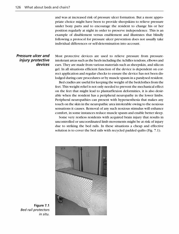
126 What about beds and chairs?
and was at increased risk of pressure ulcer formation. But a more appro-priate choice might have been to provide sheepskins to relieve pressureunder bony parts and to encourage the resident to change his or her position regularly at night in order to preserve independence. This is anexample of disablement versus enablement and illustrates that blindly following a protocol for pressure ulcer prevention does not usually takeindividual differences or self-determination into account.
Most protective devices are used to relieve pressure from pressure-intolerant areas such as the heels including the Achilles tendons, elbows andears. They are made from various materials such as sheepskin, and silicongel. In all situations efficient function of the device is dependent on cor-rect application and regular checks to ensure the device has not been dis-lodged during care procedures or by muscle spasm in a paralysed resident.
Bed cradles are useful for keeping the weight of the bedclothes from thefeet. This weight relief is not only needed to prevent the mechanical effecton the feet that might lead to plantarflexion deformities, it is also desir-able when the resident has a peripheral neuropathy in the lower limbs.Peripheral neuropathies can present with hyperaesthesia that makes anytouch on the skin in the neuropathic area intolerable owing to the noxioussensations it causes. Removal of any such noxious stimulus will enhancecomfort, in some instances reduce muscle spasm and enable better sleep.
Some very restless residents with acquired brain injury that results inuncontrolled or uncoordinated limb movements might be at risk of injurydue to striking the bed rails. In these situations a cheap and effectivesolution is to cover the bed rails with recycled padded quilts (Fig. 7.1).
Pressure ulcer andinjury protective
devices
Figure 7.1Bed rail protectors
in situ.

Residents can in some situations bring a favourite chair from home. Oftenthese chairs are comfortable and enable the new resident to retain a famil-iar object that links them tangibly and through memories back to thecommunity. When a resident has to give up all their possessions on enter-ing an RACF, there is likely to be greater stress surrounding the move.
Chairs occupied by residents often need to provide more than a com-fortable place to sit. They might be required to give support to the bodyand head when muscles are paralysed, assist in the control of musclespasm and prevent muscle contractures as well as enable easy access andegress. Furthermore, the chair might need to provide support to enablecommunication and social interaction, increase personal independenceto perform activities of daily living and assist with mobility. Thus functionis paramount and since the residents demonstrate many different needs avariety of chairs should be available to meet these individual needs.Physiotherapists often need to be proactive in this regard by consultingwith and providing documentation to support chair choice to RACF managements when new furniture is being bought. They will also assistfamilies to invest in wheelchairs or lounge chairs that suit the individualneed of the resident who is a family member.
In many instances the purchase of chairs for residents in RACFs has beenundertaken by interior designers or architects, who as a rule do notunderstand the importance of fitting the chair to the user and their indi-vidual needs. Pedersen (2000) comments that long-term care facilitiestend to buy wheelchairs and lounge chairs in bulk, thereby taking advan-tage of discount prices. This practice allows few if any choice of size orfeature options such as removable footrests. Only rarely will a chair besuitable for a resident without considerable modification. When choosingthe chair, size dimensions, the extent of support needed, flexibility toaccommodate joint range loss, how the resident will transfer in and out ofthe chair and potential for independent mobility whilst seated all requireconsideration. Only when these aspects are provided should upholsterymaterial and colour be chosen. The material used in the upholstery isimportant, as the skin-to-upholstery interface is a site of trauma in manyfrail older people. The upholstery should ideally be soft and pliable, nothold moisture and not stick to the skin (Rappl & Jones 2000).
A comprehensive assessment of the physical and functional status shouldbe undertaken for each individual needing specific seating. This will ensurethat all support, protection and social requirements can be considered.However, the cost of the ideal chair or wheelchair can be prohibitive sosome compromise might have to be made. In these situations, the pros andcons of various aspects of the chair should be discussed with the residentand their family and the best option purchased. Specific note should bemade of the physical and pathological aspects identified in Table 7.2.
All chairs have a seat and backrest. Additional features include arm-rests, tray-tables, swing-away and removable and/or elevating legrests and
Chairs 127
Chairs
Choosing a chair

128 What about beds and chairs?
Aspect Rationale
Skin Poor or absent sensation will increase risk ofpressure areasPoor circulation increases risk of injury and pressure areasScar tissue can increase risk of shear force injury
Posture Older people often have increased thoracickyphosis and decreased lumbar lordosis. Determineif deformity is fixed or flexible and if the residentcan actively straighten the spine. The chosen chairshould limit progression of the deformity
Joint range of Available hip ROM will determine seat-to-backrestmovement (ROM) angle
Tight hamstrings will influence leg elevationpotentialReduced shoulder ROM will influence the height ofarmrests and wheel access for residents usingmanual wheelchairs
Neuromuscular Increased muscle tone due to acquired brain injurystatus or spinal cord injury can be controlled to some extent
by positioningTone can be increased by poor head and bodyposition, discomfort, increased pressure causing skin trauma and anxiety caused by fear of falling and lack of sensory inputResidents with low tone or muscular dystrophy needsupport to protect them against the effect ofgravity on posture
Respiratory Residents with chronic respiratory disease might need function a wheelchair for ambulation that allows the upper
limb to be supported while the trunk is in the forward leaning position, as this position enhances ventilation capacityOther residents who have poor muscle function and find it difficult to maintain the upright positionagainst gravity might need a well-supported,slightly reclined position to maximize ventilationcapacity
Communication Eye contact is vital for communication. Inability toand eating raise the head due to thoracic kyphosis, muscle
weakness or vertebrobasilar insufficiency should benoted and a slightly reclined position adopted,preferably using a ‘tilt-in-space’ option (maximumtilt 25 degrees). Note that the head also needssupport if recliningThis position is also optimal for eating and drinking
Table 7.2Assessment aspects
essential for chairprescription

Chairs 129
wheels. Thus chairs can be adapted to offer assisted or independentmobility. These components of chairs will be discussed in detail as theyare of considerable importance when matching the chair to the individ-ual needs of the resident. One aspect that might be useful to consider iswhether the residents using particular chairs are ambulant or not.Ambulant residents need chairs that they can get up from without toomuch difficulty. ‘Geri-chairs’ or recliners do not allow the feet to be pos-itioned close to the front of the chair, as is needed to stand up, becauseof interference from the folded legrests that extend during the reclinemode. Therefore ‘geri-chairs’ might prevent independent egress from thechair or increase the risk of a fall during egress. This safety issue shouldbe carefully explained to residents, family and carers so that appropriatechoices can be made regarding chair usage (Pedersen 2000).
The depth and width dimensions of the seat will determine the ease ofaccess and egress as well as the ability of the backrest to provide support.Ideally, the seat should be no deeper than the measurement from the
Aspect Rationale
Mobility If the resident is independently mobile or walks with assistance then access and egress are an issuewith respect to seat height, depth, armrest access,foot room under the front of the chair and whetheror not the resident is cognitively safe to mobilizeindependentlyIf the resident uses his or her feet to propel the chair,the seat height and under-chair obstructions areimportant considerations for minimizing leg traumaand increasing efficiencyResidents who use manual wheelchairs need tohave the method of transfer considered to ensuresafety from leg trauma, sit-to-stand efficiency, slideboard position, need for armrest or legrest removalor hoist access. They also need to have wheel accessconsidered, push posture, upper limb function andventilation capacity optimizedResidents who use motorized wheelchairs forindependent mobility often use a hoist for transfers,so the chair can be slightly higher and/or ‘tilt-in-space’to enhance communication, eating, ventilation and function. A confounding problem arises whentransfers are performed using a standing hoist. Inthese instances the seat of the motorized wheelchairis often too high and there is difficulty in positioningthe bottom far enough back on the seat. In thesecases a lower seat height should be prescribed
Seat
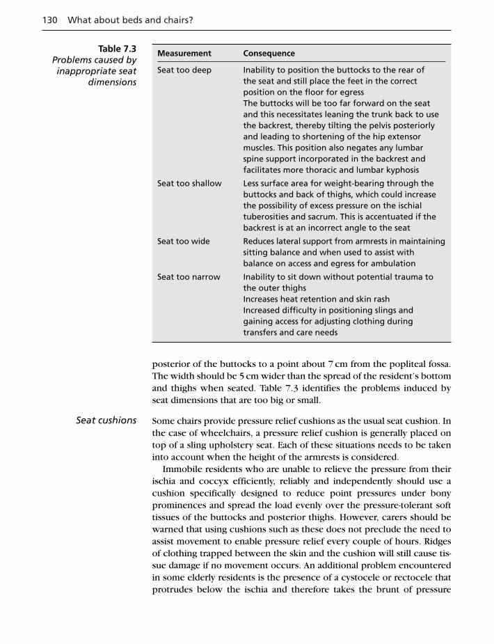
130 What about beds and chairs?
posterior of the buttocks to a point about 7 cm from the popliteal fossa.The width should be 5 cm wider than the spread of the resident’s bottomand thighs when seated. Table 7.3 identifies the problems induced byseat dimensions that are too big or small.
Some chairs provide pressure relief cushions as the usual seat cushion. Inthe case of wheelchairs, a pressure relief cushion is generally placed ontop of a sling upholstery seat. Each of these situations needs to be takeninto account when the height of the armrests is considered.
Immobile residents who are unable to relieve the pressure from theirischia and coccyx efficiently, reliably and independently should use acushion specifically designed to reduce point pressures under bonyprominences and spread the load evenly over the pressure-tolerant softtissues of the buttocks and posterior thighs. However, carers should bewarned that using cushions such as these does not preclude the need toassist movement to enable pressure relief every couple of hours. Ridgesof clothing trapped between the skin and the cushion will still cause tis-sue damage if no movement occurs. An additional problem encounteredin some elderly residents is the presence of a cystocele or rectocele thatprotrudes below the ischia and therefore takes the brunt of pressure
Seat cushions
Measurement Consequence
Seat too deep Inability to position the buttocks to the rear ofthe seat and still place the feet in the correctposition on the floor for egressThe buttocks will be too far forward on the seatand this necessitates leaning the trunk back to usethe backrest, thereby tilting the pelvis posteriorlyand leading to shortening of the hip extensormuscles. This position also negates any lumbarspine support incorporated in the backrest andfacilitates more thoracic and lumbar kyphosis
Seat too shallow Less surface area for weight-bearing through thebuttocks and back of thighs, which could increasethe possibility of excess pressure on the ischialtuberosities and sacrum. This is accentuated if thebackrest is at an incorrect angle to the seat
Seat too wide Reduces lateral support from armrests in maintainingsitting balance and when used to assist withbalance on access and egress for ambulation
Seat too narrow Inability to sit down without potential trauma tothe outer thighsIncreases heat retention and skin rashIncreased difficulty in positioning slings andgaining access for adjusting clothing duringtransfers and care needs
Table 7.3Problems caused byinappropriate seat
dimensions

Chairs 131
when the person is sitting. Should this be the case, special cushions thatcan be contoured to accommodate for the deformity and still spread pres-sure on surrounding pressure-tolerant areas should be used so as to pre-vent tissue injury and necrosis. Contouring can be achieved with air-filledcompartments, silicon gel, foam inserts or cut-outs. Often the determin-ing factor for choosing one over another is the financial cost to the resi-dent. However, the physical suffering imposed by not endeavouring toprevent pressure and shear injury must also enter the equation.
Some chairs utilized by older residents have water cushions for pres-sure relief. Like waterbeds they can be very cold and so can increase thepossibility of the user becoming hypothermic in the colder months or inair-conditioned RACFs. They also promote the fetal position and reducethe potential for active movements, as they are too soft.
It is easiest to rise from a chair if the seat-to-floor height is equivalent toknee-to-floor height (Alexander et al 1996, Sweeney & Clarke 1991). Ifthe seat is lower, the occupant needs to use faster and larger trunk move-ments to stand (Alexander et al 1996) and needs increased leg strengthand range of motion in all lower limb joints. This puts them at greater riskof losing control and suffering a fall due to compromised stability (Nitz2000). Raised seat heights are supposed to make egress easier for olderpeople and those with weakness and arthritis. Alexander et al (1996)found that unimpaired elders preferred knee height chairs to high chairs.Older people using higher chairs were more fearful standing up fromthese chairs than from knee high chairs, possibly due to having to scootthe buttocks forwards and reach down to the floor. This task is essentiallyunstable and the concern expressed by the older people in the study byAlexander et al is quite understandable. This problem arises in addition todifficulties the elderly face in assuming a safe elimination position byinstallation of raised toilet pedestals. Provision of foot stools onlyaddresses the ability to assume a good elimination position. Foot stoolsbecome a safety issue for residents when accessing the toilet by intro-ducing an obstacle in the area.
Most backrests are too high for residents as the chairs are designed using‘normal’ anthropometric measurements that do not take into account‘shrinkage’ due to loss of disc height, osteoporosis and vertebral crushfractures. The backrest also should provide support that assists by keepingthe pelvis in a neutral tilt. Consequently contours of standard backrests tendnot to equate to the older spinal curves and often accentuate thoracickyphoses and forward head position. Any potential for head support is thusnegated and well-meaning care staff often compound the problem by fill-ing the gaps with pillows that further push the head and trunk into moreflexion. The result of this is less ventilation capacity, communication abilityand discomfort, which can lead to agitation and calling for help if the res-ident is demented or unable to move into a more comfortable position.
Seat-to-floorheight
Backrest height

Lateral support of the trunk is desirable for a number of residents whohave neurological or musculoskeletal conditions that affect the ability toperceive or maintain the vertical position. In these instances the width ofthe backrest needs to be close to the shoulder and hip width of the occu-pant. Little if any support is provided if the backrest is too wide or toonarrow. Changing the contours of the backrest with foam inserts cut to theshape of the resident is a cost-effective method of alleviating this problem.
Choosing the correct seat-to-backrest angle is also vital for addressingthe support needs of the resident.
The ideal seat-to-back angle is around 95 degrees (Alexander et al 1996).Sweeney & Clarke (1991) proposed that the most appropriate support wasprovided to the occupant of the chair if the whole chair was tilted back6 degrees, thus providing the chair tilt of around 100 degrees found byAlexander et al (1996) to be most comfortable for older adults. Unfortu-nately the negative aspect of posteriorly angling the seat is that egress ismade more difficult. Older people find it more difficult to move to thefront of the seat to prepare for standing up, and if reduced hip flexionrange is present the forward lean of the trunk achieved by flexing at the hipsis inhibited, thus making egress less efficient and safe. In these circum-stances the seat should be kept horizontal to the floor and at a height thatdoes not exceed knee height while inclining the back around 5 degrees.
Reduced range of movement at the hip can be encountered in manyelderly residents. Arthritis, joint arthroplasty and muscle contractures arecommon causes. Other less common findings include heterotrophic ossi-fication or joint arthrodesis. Ultimately further loss of range in the jointsis undesirable so where possible the limbs and trunk should be sup-ported in a comfortable position that encourages optimal joint positionfor hygiene and function. Foam wedges, ‘bean bags’ and correctly fittingchair dimensions can assist with maintaining range.
Increased extensor tone in the trunk and lower limbs can lead to theresident sliding forwards and out of the chair. Generally, if the hips can bekept flexed to around 90 degrees by the seat-to-backrest angle this willcontrol the slide. However, the bottom must be kept at the back of theseat so that the support provided by the chair keeps the hips at thisangle. If the buttocks are positioned forwards on the seat or the occupanthas slid forwards due to slippery clothing, control will be lost. Also, if thehead is allowed to flex forwards, this can stimulate an extensor responsein the trunk and hips that slides the occupant forwards in the seat. Usingthe ‘tilt-in-space’ idea can control these responses. Good foot supportwith the feet in the plantigrade position assists in maintaining control. Ifthe pressure through the feet acts through the balls of the feet an exten-sor response or clonus might be facilitated and forward slippage can occur.Thus combining good postural support, position in space and a seat cush-ion to maintain comfort and relieve pressure is essential to prevent theresident sliding out of the chair and suffering injury.
132 What about beds and chairs?
Seat-to-backrestangle

There are times when loss of flexion range at the hips prevents thesemeasures from controlling the forward slip. If the loss is unilateral theseat cushion might be split between the right and left sides under thethighs so that the more extended hip is positioned in as much flexion asis available and the other hip flexed to 90 degrees. Thus the seat-to-backangle is essentially around 90 degrees and is then ‘tilted-in-space’ to anangle of around 25 degrees to the upright. A tilt of 25 degrees to the ver-tical still allows for maximal function. It also reduces shear forces at theinterface between the body and cushion to almost zero, thereby reducingthe risk of tissue injury. On the other hand, if the seat-to-back angle isopened to 115 degrees the shear forces increase by 25% (Hobson 1992).
In order to maintain the potential for a resident to stand and walk theankles and feet should be able to achieve a plantigrade position. Thereforelounge chairs should have a seat height that allows the feet to be positionedflat on the floor without having to adjust the position of the buttocks whilestill providing postural support. If the chair occupant is no longer ambu-lant, plantigrade feet are still needed to assist with maintaining a stable sit-ting position. This enables more efficient manual wheelchair propulsion,control of spasticity and prevention of falls from the toilet. The increasedstability of the feet on the footplates afforded by the plantigrade positionalso helps prevent the feet falling off the footrests during activities such asshowering and outings and thus prevent foot and leg trauma from bumpsor scrapes. The consequence of injuries to the feet and lower limbs in frailresidents, especially when arterial insufficiency is present, can be as dras-tic as amputation and death and at the very least pain and sleep loss.
The height of the footrests should be related to knee height so that theseat of the chair can support the posterior thighs and distribute the weightover a larger support surface. If the footrests are too low, excessive pressuremight be applied to the popliteal area, causing discomfort and potentialtissue damage. If the footrests are too high, more pressure is transferred tothe ischial tuberosities, coccyx and sacrum and this can increase the risk ofpressure ulcer formation in these pressure-intolerant areas. In Figure 7.2 wesee a resident whose lower limbs and feet have been elevated without duesupport and with added pressure being placed on the calves. Positioningsuch as this can lead to poor outcomes such as foot drop, ankle contrac-tures, calf deep vein thrombosis, impaired circulation and discomfort.
The armrests should be at a height that allows the occupant to rest theirforearms without having to slump to the side or forwards or for the shoul-ders to be hunched whilst receiving the support. The individual differ-ences encountered in different residents’ body sizes means that the correctheight will rarely be found in chairs shared by residents. Chairs belong-ing to individuals should be fitted to provide this degree of support.
Other considerations relate to the chairs used by residents who walk inde-pendently with or without an aid or need assistance from carers to transfer
Chairs 133
Footrests andlegrests
Armrest heightand size

134 What about beds and chairs?
to and from standing safely. In these instances the chair armrests shouldextend to the front of the chair seat so that good leverage can be gained fromthe resident to control the transfer. This help from the chair also reduces thephysical assistance the carer needs to provide. Thus the resident maintainsstrength and stability and the carer is at less risk of injury during the task.
Full seat depth armrests also allow the option of a desktop to be attached.This addition helps reduce the fear of falling forward out of the chair. It alsoprovides a surface to prop the forearms to achieve the supported forwardlean position useful for occupants who suffer shortness of breath as well asa place to put a communication board for residents who are unable to speak.Desktops also allow independent access to a water bottle with a straw, andsupport for a book or remote control for entertainment and for meals. Thisprovides self-determination and independence with some basic activities forresidents ageing with high cervical level spinal cord injuries or with condi-tions such as motor neuron disease, multiple sclerosis or cerebral palsy.
In some circumstances the armrests can be shorter (desk arms) toallow closer table access. This set-up might be found on a wheelchairwhen the occupant transfers using a hoist or slide board. In these casesremovable armrests are also desirable wheelchair attributes. Another use-ful addition is the flip-down armrests found on some lounge chairs. Thesemight assist in hoist or lateral slide transfers using a slide board and slidesheet with the resident in sitting or supine lying.
The armrests should be padded to prevent pressure areas should theoccupant use them for support in order to remain upright. Chairs thatthe resident transfers to and from by standing should provide armreststhat afford firm support and also reduce the risk of the hands slippingduring the transfer to increase safety.
These are often chosen for residents as they provide variable seat-to-backrest angles with a built-in elevating legrest and as such are presumedto be the best option. Other designs only provide the leg elevation option,
Figure 7.2Poor leg positioning.
Recliners or ‘geri-chairs’

Chairs 135
water cushion seat and no ability to change the seat–backrest angle. These‘geri chairs’ are favoured by care staff as they decrease the work of the carerand allow the resident to be pushed around as they are on wheels. They alsoenable elevation of the legs to control ankle oedema and are perceived to provide comfort, pressure area prevention and control of the effects ofabnormal tone. However, Rappl & Jones (2000) consider ‘geri-chairs’ orthe fully reclined chair position to be a restraint. They also consider thatthis use of the chairs should only be a last resort decision by caregiversowing to the disabling result for the older person. The supposed benefitsof using such chairs, listed above, are now discussed in more detail, so thatreaders can draw their own conclusions from the arguments given.
Dependency is rarely the only cause of the foot and leg oedema encoun-tered in residents and so mandatory elevation of the legs of non-mobile resi-dents when ‘sitting out’ has little support from clinical rationale. Some of thefactors that preclude leg elevation without a considerable transfer of weight-bearing surfaces from the buttocks and thighs to the sacrum are:shortenedposterior and lateral thigh muscles especially the hamstrings, adductorlongus, tensor fasciae latae, gracilis, neurovascular bundle and joint capsuleof the knee (Pedersen 2000). Stretch of these shortened tissues can lead topain felt locally and from pain referred distally or proximally and emanatingfrom the neural tissue on stretch. When the range of movement is insuffi-cient to fulfil the needs of the chair-to-legrest angle, an undesirable increasein pressure applied to the calves or heels will also ensue. The discomfort socaused might lead to the resident sliding the buttocks forward in the seat torelieve the pain and pressure. If this occurs then any support of the lowerback curves that had been provided by the chair is lost. Thus the resident islikely to assume a flexed posture that in turn will affect gut motility, respira-tion, circulation and cause joint and muscle pain. The change in weight-bearing surfaces will also increase the risk of pressure ulcer developmenton the sacrum, spinous processes, Achilles tendons or heels of the feet.
Many of the residents positioned in ‘geri-chairs’ for comfort would benefitfrom being positioned in upright well-supporting lounge chairs. This isespecially so when contractures of soft tissues and the hip and knee jointsprevent the resident assuming the reclined positions with legs elevated.Thus, comfort is rarely achieved for a resident placed in a ‘geri-chair’unless they have full range of movement available and are able to changeposition independently.
Some aspects relating to the manufacture of ‘geri-chairs’ have pressure-relieving properties. However, the frail immobile resident will still needto be assisted to change position frequently to achieve this goal throughthe chair. Many of the arguments cited previously support the proposalthat the ‘geri-chair’ does not prevent pressure ulcer formation.
The most distressing result of hypertonicity for care staff is the residentwho continually slides the buttocks to the front of the chair seat and on
Elevation of the feetand legs to prevent
ankle oedema
Perceived comfort
Pressure area prevention
Control of spasticity

136 What about beds and chairs?
to the floor. These residents often have acquired brain injury from various injuries or pathologies and in many instances cannot communi-cate. Therefore gaining cooperation for maintaining a particular seatedposition is unlikely to succeed.
There are a number of factors that might contribute to the facilitationof increased tone in these residents. Noxious stimuli such as pain from apressure ulcer, full bladder or constipation can increase tone in peoplewith central nervous system pathologies (Bannister 1978). Just feeling stiffand aching will induce movement to relieve the discomfort in unaffectedpeople so can be the reason for residents with neurological conditions towriggle also. The movement, however, occurs in the only way possibleand that is through combinations of movement that are representative ofspastic synergies. These synergic patterns might be symmetrical or asym-metrical, thus accounting for the difficulty in maintaining the positionfirst achieved on being placed in the chair. The first requirement whenfinding the right chair for the resident with a neurological problem thatpresents with muscle hypertonicity is to undertake a thorough assess-ment. This assessment should describe the distribution and presentationof the hypertonus. Factors that aggravate the tone such as head or hipposition, the presence of pressure ulcers, sore joints or muscles must beidentified so that the chair does not compound the problem. Once theseaspects have been addressed, then any loss of limb range of movementresulting from the hypertonicity can be considered. The presentation ofmuscle spasticity (flexor or extensor) will determine the loss of rangeand therefore the specific requirements of a chair. In some cases the chairseat configuration might assist in controlling unwanted responses. Ingeneral this control is achieved by keeping the pelvis in a position of neutral tilt that maintains slight lumbar lordosis and the normal thoracickyphosis and normal cervical position. This position enables goodweight distribution between the buttocks, thighs and feet and therebyincreases the potential for the resident to function at their maximalpotential for upper limb movement and for communication (Pedersen2000). The physiotherapist might consider the introduction of a move-ment programme that aims to regain lumbar mobility so that the optimalpelvic position can be obtained for residents who are very stiff aroundthe hips and pelvic area. Some strategies that might be employed werediscussed in the previous chapter.
The reclining ‘geri-chair’ obviously does not support the pelvis in thedesired position to control the tonal response. It acts as a restraint andmight stop the resident from sliding out of the chair but it is also likely toincrease the hypertonicity problem and the acquisition of limb contrac-tures through the constant stimulation or reinforcement of the spasticpatterns of movement. Independence and function in activities of dailyliving will also be decreased. Thus the ideal chair for a resident with theseproblems is one that controls the pelvic position and supports the feet,back and head as well as optimizing function.

Chairs 137
The other distressing presentation for carers is the resident who ‘jack-knifes’ out of their chair. This is most common in the spinal injured resi-dent. A strap around the chest fastened to the backrest is usually necessaryto control these spasms as anti-spastic medication does not always pre-vent their occurrence. This strap should not be considered a restraint asthe context of usage is to enhance functional capacity and possible inde-pendence if the resident is mobilizing in an electric powered wheelchair.
The aim of specifically supplying equipment and/or furniture should beto maximize function. The factors that are advantageous to this are manyand each item varies in importance depending on the needs of the indi-vidual using the chair. Thus the advantages of a good chair include theprovision of the following.
■ Easy access and egress that is provided by the seat height being at kneeheight. This means chairs of varying heights or adjustable leg lengthsshould be available in any facility so tall and short residents are cateredfor. Armrests that extend to the front of the chair seat enhance safetyduring these transfers in addition to assisting the movement by makingthe forward trunk lean safer. The result of providing chairs that makestanding up easy is that residents maintain independent or assistedmobility for longer even if the sit-to-stand transfer is only used to assistwith hygiene needs.
■ Maximization of communication and social interaction occurs when theresident is sitting upright and able to see the person talking with them.
■ Optimal postural support through adequate control of pelvic positionand the spinal curves or support of spinal deformities so as to reducethe progression of deformity can control the onset of pain.
■ Good support assists in obtaining and maintaining postural stabilitywhich in turn improves the ability to use the upper limbs and retainmaximal functional ability.
■ Comfort is provided through good support. When residents are comfortable they will be less agitated and more able to participate inphysical care and social activities (Rappl & Jones 2000).
■ When the chair enables the resident to assume the upright positionsafely, vestibular input and integration is improved. This reduces thetendency for the resident to push into extension and lessens the likeli-hood of the resident falling backwards when standing during careactivities or when being assisted to mobilize (Nitz 2000).
■ Maintains verticality and possibly visuospatial normality.■ To enable adequate nutritional and fluid intake the resident needs to
be positioned upright (Steele et al 1997). Unless this position is usedthere is a danger of the resident aspirating fluids and developing aspiration pneumonia.
■ The upright position maximizes respiratory function, especially whenthe spinal curvature is well supported. This improves the potential for
Advantages of agood chair

138 What about beds and chairs?
cerebral oxygenation, thus controlling agitation and aggression thatmight result from cerebral hypoxia (Nitz 2000).
The chair design that suits most requirements is one that has a seat-to-back angle of no more than 95 degrees. The chair should support the pelvisin a position that allows slight lumbar lordosis, and also the spinal curves,length of the thighs and the feet in a plantigrade position on the floor orfootplate. Upper limb support from armrests should not elevate the shoul-ders and cause the occupant to slouch or lean laterally to gain support.Adding a ‘tilt-in-space’ element to this configuration enables improved for-wards stability for residents who have difficulty in maintaining the uprightposition due to weakness or conscious control. A backwards tilt alsochanges the direction of gravitation forces acting on the thoracic spine ofthe resident who has a progressive kyphotic deformity due to vertebralcollapse caused by osteoporosis. The ‘tilt-in-space’ option can also controlmuscle tone by maintaining pressure and touch through the feet and thighsand keeping the head position in neutral flexion–extension. It also allowsthe resident to rest out of bed. However, where possible the resident shouldbe put on the bed for a rest if staffing numbers allow. This enables the limbsto be straightened and moved to reduce joint stiffness and discomfort butalso to relieve pressure from the seated weight-bearing surfaces. Recentlya new chair design has become available that allows the backrest to belowered flat and the legrests to be elevated so that the occupant can lieflat. This option allows the limbs to be straightened out and the residentnursed side-to-side for maintenance of joint range and pressure reliefwhile using the chair. A change of scenery is possible for immobile resi-dents by using these chairs as they also have wheels for easy relocation.These chairs also have drop-down sides that allow slide transfers frombed to chair or from bed-bath to chair so hoisting can be eliminated.
Concludingcomments
Summary ■ It is imperative to evaluate furniture and ensure it is appropriate for the given resident. Good equipment prescription helps to optimize resident function and assists in preventing likely co-morbidities and injuries.
■ Both residents’ and carers’ needs should be taken intoaccount when deciding upon suitable beds.
■ Numerous devices are available to help with injury preventionincluding pressure-relieving cushions, bed cradles and paddedbed rail protectors.
■ Appropriate chair choice is based on good rationale, and allcomponents of the chair must be considered to match the individual. Desired outcomes often relate to comfort, tone control, posture, joint positioning and functional outcomes.
■ The advantages of a correctly fitted chair are numerous.

References 139
Alexander N B, Koester D J, Grunawalt J A 1996 Chairdesign affects how older adults rise from a chair.Journal of the American Geriatrics Society44:356–362
Alexander N B, Schultz A B, Ashton-Miller J A, GrossM, Giordani B 1997 Muscle strength and risingfrom a chair in older adults. Muscle and Nerve Suppl5:S56–S59
Hobson D A 1992 Comparative effects of posture onpressure and shear at the body–seat interface.Journal of Rehabilitation Research and Develop-ment 29:21–31
Nitz J C 2000 The seating dilemma in aged care.Australian Journal of Physiotherapy 46:53–58
Pedersen J P 2000 Functional impact of seating modifications for older adults: an occupational
therapist perspective. Topics in GeriatricRehabilitation 16(2):73–83
Rappl L, Jones D A 2000 Seating evaluation: specialproblems and interventions for older adults.Topics in Geriatric Rehabilitation 16(2):63–72
Steele C M, Greenwood C, Ens I, Robertson C,Seidman-Carlson R 1997 Mealtime difficulties in ahome for the aged: not just dysphagia. Dysphagia12:43–50
Sweeney G M, Clarke A K 1991 Selecting easy chairsfor people with arthritis and low back pain.Physiotherapy 77:509–511
Taler G 2002 What do prevalence studies of pressureulcers in nursing homes really tell us? Journal ofthe American Geriatrics Society 50:773–774
References

The profile of the mobileresident and how toprotect such residentsfrom falls
Jennifer C. Nitz and Susan R. Hourigan
143
8
This chapter aims to:
There is rarely a resident in an RACF who has no limitations at all tomobility. At some time or another most residents will have suffered someinjury or illness or have other issues which make their mobility limitedand their functional performance variable. Most mobile residents willrequire the use of a mobility aid for support and/or confidence, and at thevery least the physiotherapist should ensure proper use and maintenanceof this equipment.
The most obvious issues which spring to mind for mobile residents arebalance, falls prevention and relevant exercise prescription. Other issuesrelate to mobility aids, and principles of assessment and treatment (clinicalreasoning). As balance and falls prevention are such topical issues we havedesigned a chapter to deal with this subject individually; see Chapter 9.
This chapter therefore will profile the mobile resident and identifytheir needs in order to remain safe and mobile. The mobile residentincludes any person who can manage to walk with or without physicalassistance from carers and/or walking aids. The distance that the residentcan walk might only be a few metres although some residents will be ableto walk many kilometres. Cognitive function and physical disability arethe main determining factors for safe ambulation. These residents mightbe housed in low care or high care facilities depending on the amount ofassistance or supervision that is required to achieve a safe standard of living.When transferring, these residents will not rely on mechanical devices,although they might need some assistance from a carer to rise from a lowchair and to walk short distances.
■ identify the abilities and potential problems of residents whoare mobile
■ discuss the needs of these residents to maintain safe mobility andreduce the potential for losing functional capacity or beinginjured by a fall.
Introduction
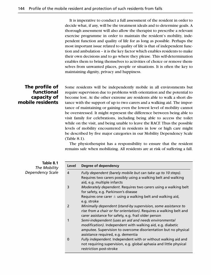
It is imperative to conduct a full assessment of the resident in order todecide what, if any, will be the treatment ideals and to determine goals. Athorough assessment will also allow the therapist to prescribe a relevantexercise programme in order to maintain the resident’s mobility, inde-pendent function and quality of life for as long as possible. Perhaps themost important issue related to quality of life is that of independent func-tion and ambulation – it is the key factor which enables residents to maketheir own decisions and to go where they please. This self-determinationenables them to bring themselves to activities of choice or remove them-selves from unwanted places, people or situations. It is often the key tomaintaining dignity, privacy and happiness.
Some residents will be independently mobile in all environments butrequire supervision due to problems with orientation and the potential tobecome lost. At the other extreme are residents able to walk a short dis-tance with the support of up to two carers and a walking aid. The impor-tance of maintaining or gaining even the lowest level of mobility cannotbe overstressed. It might represent the difference between being able tovisit family for celebrations, including being able to access the toiletwhile on the visit, and being unable to leave the RACF. Thus the possiblelevels of mobility encountered in residents in low or high care might be described by five major categories in our Mobility Dependency Scale(Table 8.1).
The physiotherapist has a responsibility to ensure that the residentremains safe when mobilizing. All residents are at risk of suffering a fall.
144 Profile of the mobile resident and protection of such residents from falls
The profile offunctional
capacity ofmobile residents
Level Degree of dependency
4 Fully dependent (barely mobile but can take up to 10 steps).Requires two carers possibly using a walking belt and walkingaid, e.g. multiple infarcts
3 Moderately dependent. Requires two carers using a walking beltfor safety, e.g. Parkinson’s diseaseRequires one carer � using a walking belt and walking aid, e.g. stroke
2 Minimally dependent (stand-by supervision, some assistance torise from a chair or for orientation). Requires a walking belt andcarer assistance for safety, e.g. frail older person
1 Semi-independent (uses an aid and needs environmental modification). Independent with walking aid, e.g. diabeticamputee. Supervision to overcome disorientation but no physicalassistance required, e.g. dementia
0 Fully independent. Independent with or without walking aid andnot requiring supervision, e.g. global aphasia and little physicalrestriction post-stroke
Table 8.1The Mobility
Dependency Scale

Even the most seemingly capable resident is at risk as in many instancesthey have age related balance decline or have cognitive dysfunction thatinterferes with their ability to move safely in the environment.
The role of the physiotherapist is to maintain or maximize functionand this generally entails regular assessment of functional capacity andbalance ability and the prescription of an exercise programme. The find-ings from the assessment should also be used to identify for the residentand the carers any specific problem such as visual perceptual, motorplanning dysfunction or loss of muscle strength that might be putting the resident at increased risk of a fall. A careful explanation needs to be given so that all parties understand the mechanism that increases the risk.
The resident is never totally protected from suffering a fall but the risk ofan injurious fall might be reduced if some adjustment to current practiceis made. All over the world RACFs have some form of falls prevention programme in place but residents still suffer falls at an alarming rate.We believe that the content of these programmes is essentially correctand theoretically should reduce the prevalence of falls. However, it is the lack of explanation given to residents and carers regarding the whysand wherefores of the components of the falls prevention programmethat leads, through ignorance, to choices being made that reduce compliance.
In the following pages we will explain some of the items included infalls prevention programmes. We will identify factors that increase thelikelihood of falls and examine related problems encountered in olderpeople that are due to ageing and acquired pathologies. Enabling all parties to understand the clinical reasoning behind the components offalls prevention programmes will make it more logical to comply withrecommendations. Most falls prevention programmes are multifacetedand include components such as environmental adaptations, residentassessment and exercise interventions, education and information to residents and staff and consultation on medication and assistive devices(Becker et al 2003). Even if fall prevention programmes are comprehen-sive and cover all these aspects, their effectiveness is only as good as the completeness of the coverage of all risk factors for falling as well as the understanding of how the risk factors interact with the older person. Carers, family members and residents all need to understandhow the individual will be affected by the risk if strategies to control or avoid these risk factors are to be fully exploited. Thus any educa-tional package must include explanations of cause and effect so thatinformed decisions, warnings or directions can be made or given by all concerned. The following information is provided to enable readers to understand the mechanics and interpretation of risk assessment findings.
Protection from falls 145
Protection from falls

The assessment of falls risk must include environmental influences, thoseattributable to the carer, assistive equipment and the intrinsic risk factorsfound in the resident. A number of processes can then be put in place to reduce the risk but the human factor associated with the potential for minor to catastrophic change in residents or carers must not be discounted.
Changes to depth perception, visual acuity in low contrast situations aswell as edge perception difficulties (Lord & Dayhew 2001, Tinetti 2001)that are normal ageing changes will be amplified in some environmentalsituations. These influences might be limited if changed by refurbishmentor if the carer interprets the visually conflicting situation and reassuresthe resident regarding the interpretation of the environmental situation.The independently mobile resident will not be able to benefit from carerinterpretive information and so will still be at major risk in some visuallyconfounding situations.
Small rooms with a number of pieces of furniture reduce the area inwhich the residents and carers can manipulate walking aids and safelyturn to reach the bed or a chair. Poor depth perception or visual fieldlosses put residents at risk of a fall. Items of personal equipment or shoesleft lying around increase the risk of tripping. Furthermore, a carer cannotefficiently assist a resident to walk if they are stepping over or around fur-niture or clutter on the floor and are themselves at risk of a fall. Corridorsand common areas must be kept clear of clutter so independently mobileresidents are not put at risk.
Furniture can pose major risk factors for falling. The beds used by resi-dents contribute a number of potential fall precipitators. Side rails are oftenraised bilaterally in the belief that the resident will be protected from a fallfrom the bed. When the resident is cognitively able, raised side rails mightserve as a reminder to call for assistance when getting up or to enable inde-pendent rolling during the night by providing a solid point to pull on when rolling and security from rolling out of bed. However, if the residenthas some cognitive deficit the rails might only be seen as a barrier to climb over and so cause a fall from a greater height resulting in injury or death from becoming wedged between the rails (Capezuti et al 2002). Great careshould be exercised when using bed rails to ‘protect’ the resident from afall and each case must be evaluated individually taking all aspects of theresident’s physical and mental status into account. If safety is ultimatelycompromised putting the mattress on the floor for the resident to sleepon will eliminate the potential for a fall, although it makes nursing moredifficult.
Some falls from bed can be related to the height of the bed from the floor. If the bed is too high and the resident is unable to easily reachthe floor with their feet after swinging their legs over the side of the bed,stability is lost and a fall might follow. Similarly, if the resident has to slide
146 Profile of the mobile resident and protection of such residents from falls
Risk assessment
Environmentalfalls risk
assessment

the bottom forwards to the edge of the bed before their feet touch thefloor, stability might be lost and a fall ensue. Therefore the bed should belowered to chair height so that the feet easily touch the floor for egress.New beds can usually be lowered to about 50 cm above the floor. This isstill higher than most dining room chairs (45 cm) so will still pose theproblems outlined previously but not to as great an extent as some of theolder design of beds.
Care must be taken to apply all the brakes on the bed wheels so thatthe bed does not ‘run away’ from the resident during a transfer in or outof bed. This aspect is also very important for preventing carer injury atany time when the carer is helping a resident who is in bed. Often thethighs of the carer are leant against the bed for extra support and stabil-ity during the task. If the bed moves at any time serious back, neck orother injuries are likely to be suffered. Similarly, if a standing or full hoistis used to transfer a resident, the brakes should be applied during anyprocedure when the equipment is stationary. This ensures added stabilityand safety.
Floor materials can pose major problems for mobile residents. Theyshould be non-shiny and non-slip. Floors that are shiny even if not slip-pery will change the gait of all people walking on the surface. The per-ception that the floor is slippery will slow the cadence, reduce the steplength, broaden the base and in so doing might raise the strengthdemands by reducing the effective use of momentum for forward propul-sion. This might interfere with postural stability and balance enough toincrease the risk of a fall.
Carpet can become worn and patchy, causing variability in height ofthe pile. In some places carpet tiles are used so that heavy wear areas can be controlled. However, the edges of carpet squares or carpet of vari-able height pile can cause the resident to trip when they have difficultyin lifting their feet when walking.
Colour changes or floor-covering changes between rooms or corri-dors can lead to visual conflict and increase the potential for a fall. Allolder people have some degree of acuity loss, age-related macular degen-eration or cataracts that reduce the efficiency of their vision and are putat increased risk of a fall by misinterpreting what they are seeing. On theother hand, colour contrast between chair upholstery and floor cover-ings can enhance the ability of the resident to see the edge of the chairand therefore be able to transfer from standing to sitting with more safetythan if the colours of the decor blended.
Shadows falling across corridors due to sunlight streaming throughwindows can produce such light and dark effects that residents withproblems of visual conflict might perceive this light effect as a step up or step down. This might cause hesitation or the resident might try tonegotiate a step only to cause a fall through losing balance.
The outside environment also can pose a major fall risk. Automaticallyopening doors are useful to prevent the problem of losing balance while
Risk assessment 147

trying to pull or push open heavy doors. However, automatically openingdoors can also affect postural stability through the effect of the movementof the door through the visual field and the stimulation of the vestibularoptic reflex changing postural muscle activity. Care should be taken toensure the doorway is not visually confounding and the door runners arenot causing hesitation or obstruction.
Paving material that is not slippery when wet should be chosen. Smallpavers that might become uneven or poorly maintained should beavoided so that trips and falls are averted. The same should be said forramps. They should be at the correct incline (readers are encouraged toconsult the Building Standards for their part of the world to find the legis-lated requirements) and have solid railings. Outside communal areaswhere residents might congregate for social occasions or for time withfamilies should be protected from the sun, rain and wind. Being able tospend time outdoors is important for vitamin D synthesis, so should bemade possible for the resident. If a grassed area is accessible it should bevery flat and kept mown short. Thick, though manicured lawns are quiteunsafe for residents with poor balance as the proprioceptive input fromthe lower limbs is confounded and more reliance on vision and vestibu-lar input is demanded. Muscles also need to be stronger and reaction timefaster to maintain balance on thick grass. Many residents enjoy gardeningand if raised beds accessed by a suitable pathway are available, then pro-vision of a safe area such as this where they can potter is very calmingand good exercise. Two heights of raised garden bed are advisable.Garden beds should be at chair height for those residents who gardenfrom a wheelchair and higher for the walking gardeners. Garden pathsshould always be swept clean as dropped leaves and flower petals can bevery slippery if damp. Other hazards include gutters and stormwatergrates, so where possible alternative solutions to removal of water thatkeep the ground flat and non-slippery should be employed.
Walking aidsVery few residents will walk without the benefit of an assistive device.These walking aids fall into two major categories, those with wheels andthose without, and all have some inherent safety issue that relates to effi-cient maintenance of the equipment. Wheeled devices might have solidtyres or pneumatic inner tubes. If pneumatic tyres are used then theyshould always have equal pressure in the tubes, otherwise they becomeunstable and can tip the resident off balance and thus precipitate a fall.All wheeled devices need to have some braking mechanism so that theresident can control the speed of progression. Many have bicycle-typebrakes that require the resident to have reasonably strong hand grip andspeed of coordination as well as sufficient cognitive ability to apply thebrakes to enable safe function. If these requirements are not present inthe resident then safety becomes a major issue. Other wheeled walking
148 Profile of the mobile resident and protection of such residents from falls
Resident equipment risk
factors

frames utilize the application of downward pressure on the handles toengage the brakes. This is quite useful if the resident loses balance in aforward direction, which will apply the brakes. Still other wheeled assis-tive devices have no brakes at all and are only safe for use by residentswith good balance or with maximal assistance being given to the residentby carers who then control the movement of the walking aid. Wheeledwalking aids can be very useful but also can introduce additional safety considerations, so all assistive devices should be evaluated for levelof maintenance as well as appropriateness of prescription for the resi-dent, and the physiotherapist is the professional most able to performthis task.
Walking sticks or canes, whether they are single-point or four-point,are generally fitted with rubber stoppers on the feet. When the treads onthese stoppers become worn, they are liable to slip on the floor and causethe user to have a fall. Therefore the stoppers should always be checkedfor sufficient tread and the stopper replaced if it is showing wear.
The height of the walking assistive device must be correct so that itcan provide the appropriate level of support. Walking aids that are toohigh or too short will affect the posture of the user and contribute toinstability and might precipitate a fall.
Painful feet, foot deformity and inability to put on one’s own shoes can bereasons for wearing no shoes or ill-fitting shoes or slippers when walking.Painful or deformed feet alone can increase the risk of a fall (Menz & Lord1999) but if a supportive, well-fitted shoe is worn the risk of falling shouldnot be increased. On the other hand, if the resident wears shoes that aretoo big because of trying to accommodate deformities or are poorlysecured to the feet, the risk of tripping on the shoes or walking out of theshoe and tripping is increased. This mechanical risk is added to by slowand inefficient balance reactions. Slipping due to decreased frictionbetween the shoes and the support surface is another problem attributa-ble to footwear that can precipitate a fall. Menz et al (2001) showed thatno shoe could be considered safe from slipping on wet surfaces. Thedynamic coefficient of friction at the shoe–surface interface was differentbetween types of shoe, application of a textured or leather sole and differentheel geometry, but there was still need to control manufacturing standardsfor casual footwear to enhance safety, especially for older people.
Even if the shoes are supportive and secure on the feet, heel soles thatare unevenly worn can produce postural instability that might contributeto a fall. Care should therefore be taken to ensure that excessive solewear is repaired or new shoes bought so that the resident is not put indanger from this cause.
Many older people lose height due to osteoporosis or weight from dis-ease or illness and as a consequence can find it difficult to obtain clothingthat is short or small enough that it does not trail on the floor and pose atripping hazard. In some cases, the resident does not consider trailing
Risk assessment 149
Shoes
Long or trailingclothing

clothing as a fall risk and this hazard should be identified and a solutionsuch as raising the hemline or tying the dressing gown cord implemented.
Poor vision has been identified as a major contributing factor for falls. Manyresidents have vision deficits that will benefit from wearing glasses thatimprove acuity and stereoscopic vision, as this will enhance the ability of theresident to judge distance and distinguish visually confounding situations.Thus by encouraging residents to use their glasses appropriately, safetywhen mobilizing might be enhanced. Carers should be aware that someresidents might need to be reminded to put their glasses on when dress-ing as well as having them cleaned so that the lenses are fully functional.
Lord et al (2002) showed that multifocal glasses increased the poten-tial for falling in older people with depth perception and edged contrastsensitivity deficiencies as they blurred the visual field when the personwalked. This came about when the visual line coincided with the changein lens focal length. It is recommended that older people use separatereading and distance glasses rather than multifocals to reduce the potentialfor this cause of falling.
Hearing loss has been linked to instability and increased potential to suf-fer a fall in nursing home residents (Bumin et al 2002). Hearing and soundlocation ability is vital for safety in congested areas and to prevent the resi-dent from being startled by a person or vehicle suddenly appearing in thevisual field. If balance is poor these situations might precipitate a fall.Using a hearing aid or bilateral hearing aids might improve hearing to an extent that reduces the risk of a fall and so all staff should encouragethis practice. Often residents lack the fine manual skills needed to replacethe batteries in hearing aids and this task then falls to the carers or doesnot get done, so residents discontinue hearing aid use, thus putting them-selves at risk of a fall. Carers should be ready and able to assist with hearing aid battery replacement and application to residents. Regulareducation sessions provided by an audiologist to residents, their familiesand carers might increase confidence in using and assisting residents touse hearing aids, thereby improving safety and reducing the risk of a fall.
Education regarding procedures for prolonging battery life by turningoff the hearing aid or opening the battery case to save the charge mightassist in reducing the need for maintenance and frequent battery change.This in turn will reduce frustration from a useless aid and improve residentsafety and quality of life.
More than three co-morbidities or in some cases the presence of specificmedical conditions such as stroke, Parkinson’s disease, arthritis, depres-sion and dementia are all identified, intrinsic resident risk factors for falls(Tinetti et al 1988). Muscle weakness and pain have also been included ascontributing factors.
Postural hypotension can be a major cause of falls in the elderly. Itmight be related to medication or the actual pathology that the resident
150 Profile of the mobile resident and protection of such residents from falls
Glasses
Hearing aids
Resident intrinsicrisk factors

presents with such as Shy–Drager syndrome. On the other hand, it mightbe due only to the effect of ageing on the sympathetic nervous system,heart and cardiac output. A thorough physical examination by the med-ical practitioner and consultation with the pharmacist should help iden-tify the factors that are contributing to the hypotension so that if possiblesome remedial action can be undertaken. If there is no treatment then all carers and the resident must be made aware of the risk of a fall due to postural hypotension and of steps that might be taken to reducethis risk, such as pausing after moving from lying to sitting and afterstanding up.
Another risk factor common in the elderly is vertebrobasilar insuffi-ciency (VBI). Many older people have extreme thoracic kyphoses thatrequire them to extend their upper cervical spine to see where they aregoing when walking. This position might cause dizziness or loss of con-sciousness if sustained so residents and carers should be warned of therisk associated with this movement. A similar situation of sustained uppercervical extension is assumed if the resident has to look up to the carerto talk. This is another reason why the carer should always get down tothe same level as the resident when conversing.
The physiotherapist will also perform a full functional and balanceassessment that should identify physical fall risk factors. These assess-ments are discussed in Chapter 2 and also in more detail in the next chapter, which deals with balance exclusively.
Medications and the problem of polypharmacy have been identified asmajor contributors to increased risk of a fall. All the medications taken bythe resident including those prescribed by the doctor and those boughtover the counter should be identified, as four or more medications increasethe likelihood of recurrent falls (Leipzig et al 1999, Yip & Cumming 1994).Sedatives, psychotropic drugs and opioid analgesics are specific medica-tions that have been shown to increase the risk of a fall (Neutel et al 1996,Weiner et al 1998). Other drugs that might contribute to a fall are: anti-hypertensives, antiarrhythmics, anti-Parkinson’s disease drugs and hypo-glycaemic agents (Yip & Cumming 1994). In addition, drug interactionsare common in the elderly and a pharmacist is the person best suited toidentify the presence of potential drug interactions in a resident. Anotherproblem encountered in older people is the change in half-life and excre-tion of many drugs owing to liver and renal dysfunction.
Primarily physiotherapists will perform the most efficient physical exam-ination to determine the physical factors that contribute to increased riskof falling. In collaboration with the nursing staff they will determine thelevel of assistance or supervision required during activities of daily livingand for special situations such as walking in the garden. The physiother-apist will identify physical problems that might be improved in functionwith an exercise intervention programme that specifically targets these
The physiotherapist’s role in protecting residents from a fall 151
The physio-therapist’s role
in protecting residents from
a fall

problems, thereby improving the physical function and safety of the resi-dent. Assessment and treatment of balance problems are covered in detailin the following chapter. Thus the physiotherapist has an important rolein identifying the resident who is at risk of falling. Physiotherapists arealso responsible for monitoring the status of residents and noting changesthat might indicate more assistance or intervention is required as the resi-dent ages or acquires further disabilities, as might occur should they suffera stroke.
Physiotherapists should also be involved in conferences to determinethe contributions to falls risk from medication or personal equipment,especially when intervention is needed and the team needs to decide on a mode of action with the consent and cooperation of the resident andtheir family. Therefore it is usually important to include the resident and/orfamily member in this conference.
Physiotherapists are usually responsible for the manual handling train-ing of carers and as such have an ideal situation to educate the carers aboutthe risk factors for falls during these sessions. Manual handling is intimatelyrelated to falls prevention as it considers the risks for the resident as well asthe carer. Explaining when to be most vigilant when walking a resident andwhy this is important will increase carers’ compliance with instructions asrelevancy is emphasized. This enables the logical reasoning behind theinstructions to be considered by both carers and residents and so enhanceswillingness from all participants to apply the instruction.
Patient handling refers to the moving or ‘handling’ of people. The tasksinvolved are those activities which require the use of force exerted by a carer to hold, support, transfer (lift, lower, carry, push, pull, slide),or restrain a person at the workplace.
‘No lift’ policies aim to protect both residents and staff members byensuring that the manual lifting of residents is eliminated in all but excep-tional or life-threatening circumstances (Royal College of Nursing 1996).Such policies require that residents who are able to assist are encouragedto do so, whereas mechanical devices such as hoists are used for thoseresidents who are unable to assist in order to minimize the strain on theworker and lessen the likelihood of injury to the resident.
It is the physiotherapist’s responsibility to individually assess residents’mobility and ability to transfer on admission and as regularly as is neces-sary. The physiotherapist is the professional staff member most appropri-ately trained to undertake this assessment. The assessment will attemptto ascertain the resident’s rehabilitation needs and their ability to partic-ipate whilst always considering the needs of other staff members. The aimis to protect both workers’ health and residents’ health, and the goal is primarily to protect both parties. The contribution provided by thephysiotherapist is discussed in detail in Chapter 2.
Safe patient handling systems are introduced to facilitate a safe environ-ment, as mentioned previously. Other goals of a patient handling ‘no lift’
152 Profile of the mobile resident and protection of such residents from falls
The physio-therapist’s role
in educatingstaff regarding
safe patient handling

system which should be focused upon include reducing the numbers ofinjuries and injury severity, complying with organizational policy, meet-ing union imperatives, complying with workplace health and safety leg-islation, assisting with quality improvement systems, increasing the skillsand knowledge of staff members and improving patient care. With thesegoals in mind it is easy to realize the extreme importance of safe patienthandling. It is also easy to realize that this job requires an enormousamount of organizational skill, educational prowess and the ability to assessand teach on the part of the physiotherapist in the RACF. Developing and continuing patient handling programmes is arguably the biggestresponsibility an RACF physiotherapist has.
Workplace health and safety legislation requires both employers andworkers to meet various obligations. The RACF must ensure the healthand safety of workers by providing equipment, space and training toemployees. Workers must ensure they follow instructions given in rela-tion to manual tasks and not wilfully place themselves or another’s healthor safety at risk. Staff members must follow instructions given in trainingprogrammes and use patient handling equipment that is provided by thefacility. Carers need to check the resident mobility plan which is designedafter assessment by the physiotherapist, communicate with the residentand other staff involved, refer to senior staff for assistance if required, andbe responsible for reporting risks within the workplace.
A risk management system must be developed. This should ensureequipment maintenance requirements are met and any problems arereported in a timely and efficient manner. Incident and accident reportsshould be formalized in order to enable statistics to be recorded and riskmanagement strategies to be developed and refined. Staff should beempowered to be involved in processes which contribute to maintainingbest practice in the area of resident care, health and safety managementand quality improvement strategies. Figure 8.1 shows how the risk man-agement process is continuous.
The physiotherapist’s role in educating staff regarding safe patient handling 153
Monitorand
Review
RiskIdentification
ImplementControl
Measures
RiskControl
RiskAssessment
Figure 8.1The risk
management process.

The individual resident’s risk assessment is part of the initial and ongoing physiotherapy review. This assessment and the development of the residents’ mobility plans are the first and most important processesin selecting safe handling techniques to be utilized by the residents andstaff. This fundamental assessment will lead to safety, best practice andfalls prevention. Refer to Chapter 2 that deals with assessment for moredetailed information. However, the assessment should specifically includethe following:
1. medical diagnosis2. mobility history3. physical examination (pain, range, balance, strength)4. mental status and cognition5. communication6. weight7. other relevant features (e.g. diurnal variation, extra medical equip-
ment – PEG feeds, urinary catheters and oxygen supplementation).
Perhaps the simplest strategies are sometimes the hardest to convey tostaff. It is never superfluous to repeat the basics and to spell out somerules relating to resident handling. Some of the most important points toteach and assess in patient handling programmes are related in point format below. Educate staff to do the following:
■ Refer to the mobility plan (especially if they do not know residentswell or might have been away on leave and not current with residentstatus), get a good handover in order to be up-to-date, read any appro-priate communication books/boards.
■ Encourage residents to assist themselves as much as possible (this helps carers and residents). Sometimes the less help given by staff the better, even if it does take a little longer for the task to be completed.
■ Emphasize good communication (get eye contact, tell the resident whatthe aim is and get their concurrence if possible).
■ Adjust the environment for safety (manipulate bed height, ensurebrakes are on equipment, clear the pathway and surrounds as necessary).
■ Refer to senior staff if a resident is on the floor or seems changed or ill.
■ Utilize principles of back care and joint protection in all work practices (good posture, using their legs and abdominal muscles well).
■ Plan the move, confirm it verbally with the resident and their carepartner if applicable and initiate the transfer on the count of three sothat all parties are ready and assisting.
154 Profile of the mobile resident and protection of such residents from falls

Becker C, Kron M, Lindemann U et al 2003 Effective-ness of a multifaceted intervention on falls in nursing home residents. Journal of the AmericanGeriatrics Society 51(3):306–313
Bumin G, Uyanik M, Aki E, Kayihan H 2002 An inves-tigation of risk factors for falls in elderly people ina Turkish rest home: A pilot study. Aging Clinicaland Experimental Research 14(3):192–196
Capezuti E, Maislin G, Strumpf N, Evans L K 2002Side rail use and bed-related fall outcomes amongnursing home residents. Journal of the AmericanGeriatrics Society 50(1):90–96
Leipzig M, Cummings R, Tinetti M 1999 Drugs andfalls in older people: a systematic review and meta-analysis:1. Psychotropic drugs. Journal of theAmerican Geriatrics Society 47:30–39
Lord S R, Dayhew J 2001 Visual risk factors for falls in older people. Journal of the American GeriatricsSociety 49(4):508–515
Lord S R, Dayhew J, Howland A 2002 Multifocalglasses impair edge-contrast sensitivity and depthperception and increase the risk of falls in olderpeople. Journal of the American Geriatrics Society50(11):1760–1766
Menz H B, Lord S R 1999 Foot problems, functionalimpairment, and falls in older people. Journal
of the American Podiatric Association 89(9):458–467
Menz H B, Lord S R, McIntosh A S 2001 Slip resist-ance of casual footwear: implications for falls inolder adults. Gerontology 47(3):145–149
Neutel C I, Hirdes J P, Maxwell C J, Patten S B 1996New evidence on benzodiazepine use and falls:the time factor. Age and Ageing 25:273–278
Royal College of Nursing 1996 Code of Practice forPatient Handling, London
Tinetti M E 2001 Where is the vision for fall preven-tion? Journal of the American Geriatrics Society49(5):676–677
Tinetti M E, Speechley M, Ginter S F 1988 Risk factorsfor falls among elderly persons living in the com-munity. New England Journal of Medicine 319:1701–1707
Weiner D K, Hanlon J T, Studenski S A 1998 Effects of central nervous system polypharmacy on fallsliability in community-dwelling elderly. Gerontology44:217–221
Yip Y B, Cumming R G 1994 The association betweenmedications and falls in Australian nursing-homeresidents. Medical Journal of Australia 160:14–18
References 155
Summary ■ The role of the physiotherapist is to assess the physical andcognitive state of the resident to determine the presence ofrisk factors for a fall.
■ The physiotherapist also needs to educate the resident, familyand carers about the environmental, personal and age-relatedfactors that might contribute to a fall and relate thisinformation to manual handling and caring tasks.
■ Case conferences that include a pharmacist, medicalpractitioner, carers, residents, family and the physiotherapistmay need to be initiated by the physiotherapist. All aspects offalls prevention might then be addressed and the residentand family are incorporated in the decision-making processesso all will be happy to act on decisions regarding care.(Appendix 3 discusses case conferences in more detail.)
■ Physiotherapists should provide carer education regardingsafe resident handling procedures.
References

A theoretical frameworkfor the assessment andtreatment of balance and mobility deficits in the elderly
Nancy Low Choy
156
9
This chapteraims to:
Falls and injury prevention is a topical area of research in the elderly withprogrammes aiming to reduce the incidence of falls and related injuriesin men and women over the age of 65 years (Hinman 1998, Lord et al1994, 1995, 1996, Speechley & Tinetti 1991, Steinberg et al 2000, Tinettiet al 1994). This focus has been the result of studies that show fallsoccurring in one in three people aged over 65 years in any given 12-monthperiod (Cho & Kamen 1998, Lord et al 1993, Speechley & Tinetti 1990,Woollacott & Tang 1997). One of the contributing elements to the rise inprevalence of falls in elders is the decline that may occur in the multiplemechanisms involved in postural stability and balance. The physiother-apist is ideally equipped to manage dysfunction in the postural mechanismand can use acquired knowledge, skills and experiences to address theneeds of the frail aged in residential aged care along with older people wholive in supervised care or independently in the community. An under-standing of the theoretical basis of ‘balance’ provides the physiotherapistwith the knowledge base from which an informed assessment can bedeveloped. A thorough assessment of the elements contributing to balanceand safe mobility identifies those aspects of balance, along with underlying
■ review the theoretical elements that contribute to balance andmobility as a basis for assessing balance and mobility in theelderly, and presenting a tailored but multidimensional approachto assessing and retraining balance and mobility deficits in the elderly
■ describe a process of assessing balance and mobility in theelderly that uses observational and problem-solving skills whileanalysing balance and mobility tasks, identifies and measuresimpairments, and monitors progress and outcomes for elders ofvarying functional motor ability.
Introduction

Theoretical basis for assessment and interventions 157
impairments, that need to be addressed for each resident. The use oftools that monitor progress and outcomes and the ability to implementtargeted intervention programmes on an individualized basis or effec-tively in a group provide the physiotherapist with the specialized skillsrequired to meet the variable needs of older people.
The presenting balance and mobility of an individual (resident or com-munity ambulant elder) is dependent on the integrity of the sensorimotorsystems, the given task and the environment in which the individual isfunctioning. This triad has been given prominence by the works ofShumway-Cooke & Woollacott (2001). For the ageing adult, this meansunderstanding the individual with varying impairments (abilities anddeficits), while performing a range of tasks that require stability anddemand mobility in a functioning environment such as found within anRACF, an individual’s home and local community (Table 9.1). A grasp ofthis concept ensures that a comprehensive and multidimensional approachto both assessment and intervention for optimal management of the elderly client is offered and clearly underpins the functional and multi-dimensional approach to retraining balance and mobility.
A large body of research informs us of the elements that an individualrequires for efficient balance and mobility. These include the role andquality of sensory input, central processing and integration of sensorycues, reaction time to stimulus response and cognitive (attention) demandsduring the execution of tasks. There is also the need for flexibility in themusculoskeletal system, selective activation and timing of motor units fortask execution as well as strength and endurance of recruited skeletal mus-cle fibres. Deficits in any of these elements could contribute to decreasedstability, mobility, endurance and efficiency of movement. Thus each ofthese changes within the individual needs to be considered as a basis fordeveloping the practical implications for retraining balance and mobility.
Theoreticalbasis for
assessment and interventions
The client groupFrail aged with multiple impairments who is dependentElder requiring supervised careElder who is independent
Balance and mobility tasksSitting balanceCapacity to stand up, balance and walkCapacity to transfer and be wheelchair ambulant
The settingsResidential aged care facilitySupervised care within a hostel/homeLiving independently at home within the community
Table 9.1The contextualframework for
assessment and intervention

The visual, somatosensory and vestibular systems are important compon-ents of postural control. These systems are used to monitor the relation-ship between the position and movement of the body in space and theforces acting upon the body that may cause displacement (Shumway-Cooke & Woollacott 2001). Older people frequently have impaired vision,vestibular function, and somatic sensation in the legs and feet. A declinein the sensory systems may cause decreased perception, integration, reac-tion time and feedback during tasks under varying environmental condi-tions. Such impairments reduce postural stability, particularly when there isconcomitant impairment of central processing within the nervous system.
Decreased visual acuity adversely affects balance ability. It is recog-nized that a loss of visual acuity can result from cataracts, macular degen-eration, and loss of peripheral vision due to ischaemic retinal or braindisease (Lord et al 2001, Shumway-Cook & Woollacott 2001). Distanceacuity of less than 20/50 will have a significant effect on postural stabil-ity. Because of multiple changes within the structure of the eye itself, lesslight is transmitted to the retina. With a decrease in contrast sensitivity,this causes loss of visual contour and depth perception. Understanding ofthe type of changes in the visual system that cause decreased postural stability has been advanced by the work of Tanaka et al (1995), Wade &Jones (1997) and Lord & Menz (2000). Studies by Tanaka et al (1995)have shown that the combined effects of age, insufficiency of musclecontrol, and reduced vision and tactile sensitivity are associated with pos-tural instability. The work of Lord and colleagues has demonstrated theimportance of considering contrast sensitivity and depth perception, as adecline in these functions with ageing has been linked to an increasedrisk of slips, trips and falls (Lord & Menz 2000). Further, the issue of thetype of corrective lenses has been raised, with multifocal lenses attrib-uted with a higher incidence of falls than bifocal or distance only lenses(Lord et al 2001, 2002).
These issues are important as elders are considered to be more relianton vision for balance than younger persons. This reliance has been demon-strated in healthy elders (Alhanti et al 1997, Manchester et al 1989, Newton1995, Tanaka et al 1995, Teasdale et al 1991) and fallers aged over 65 years(Anacker & Di Fabio 1992, Cho & Kamen 1998, Lord et al 1991, 1992,Maki et al 1994, Tobis et al 1985, Whitney et al 2000). The somatosensoryand vestibular system changes that have been documented with ageingmay be important contributors to this reliance on vision.
Somatosensory changes with ageing have been extensively researchedin recent decades with a number of parameters requiring consideration.Hurley et al (1998) give emphasis to the combined effect of age, strength andjoint position sense on postural steadiness and function, while othershave demonstrated decreased tactile sensitivity and dynamic joint posi-tioning ability with ageing (Manchester et al 1989, Tanaka et al 1995). Thework of Anacker & Di Fabio (1992) demonstrated the importance of thequality of orienting input from the ankle receptors to postural stability
158 Assessment and treatment of balance and mobility deficits in the elderly
Sensory systemdecline

when balancing on firm and compliant surfaces, while Ring et al (1989)showed the relationship of foot position awareness, age and stability.Earlier researchers (Whanger & Wang 1974) had demonstrated that cuta-neous vibratory sensation could best be detected at the knee in elders as reduced ankle sensation had occurred with ageing. In general, thereduced function of the proprioceptive system is considered a contribu-tor to the rising incidence of falls with elders over 65 (Anacker & Di Fabio1992, Brocklehurst et al 1982, Horak et al 1990, Manchester et al 1989,Tanaka et al 1995). It is clear that a decline in somatosensory functioncould contribute to a reduced ability to balance with changes in tactileacuity, vibration sense and joint repositioning ability needing to be con-sidered. Other researchers give emphasis to the combined effect of adecline in somatosensory function and vestibular function on posturalinstability in elders (Alhanti et al 1997, Horak et al 1990, Newton 1995,Peterka & Black 1990, Teasdale et al 1991, Whitney et al 2000).
In the aged, a reduction in function of the vestibular system is evidencedby a loss of vestibular hair and nerve cells by 60 years of age (Paige 1992)but the precise contribution to unsteadiness and falls is difficult to measure(Lord et al 2001). Researchers have demonstrated the importance of effi-cient vestibular function while balancing on a compliant surface withoutvision and that postural stability is significantly different in young and olderadults when ankle joint and visual input are distorted or absent (Baloh et al1987, Horak et al 1989, 1990, Peterka & Black 1990, Teasdale et al 1991,Woollacott et al 1986). This means that older people are more prone to fallswhen visual conflict or poor vision presents in conjunction with a changeto the ground or support surface or when deficits are present in either theproprioceptive or vestibular systems when vision is occluded.
Thus the vestibular system is an important source of information forstability during movements in ‘busy’ environs or when tasks are executedon stable or more challenging, unstable surfaces. It provides the individ-ual with the capacity to know when the environment is stable or moving,thus resolving any conflict for the visual system. In a similar way thevestibular system assists the somatosensory systems to maintain balanceduring tasks that are faster paced, when tasks require the individual tobalance over a narrower base or while working at height, and when tasksare executed on an unstable base (e.g. grass) or if lighting is poor (e.g.shaded conditions or twilight), when vision is less able to be used or notavailable (e.g. night-time/darkness).
Other researchers have drawn attention to the contribution of dizzinessand gaze instability to unsteadiness and falls. With ageing, the otolith maybecome hardened, fragment and detach, setting up abnormal signal pat-terns in the hair cells (Baloh et al 1987). An imbalance of signals from onevestibular apparatus may contribute to the increased incidence of dizzi-ness with unsteadiness reported in the elderly. The most common causeof dizziness with unsteadiness in the elderly is benign paroxysmal posi-tional vertigo (BPPV) where hardened otoconia dislodge and float in one
Theoretical basis for assessment and interventions 159

of the semicircular canals, usually the posterior canal (Baloh et al 1987,Baloh & Halmagyi 1996, Herdman 2000). A bilateral loss of vestibular cellsis less likely to cause an imbalance of signals, such that unsteadiness maypresent without accompanying dizziness, although complaints of a shift-ing visual field (oscillopsia) remain common (Hillman et al 1999). Gazestability is controlled through the vestibular ocular reflex (VOR) whichprovides a coordinated action between eye and head movement duringmotor tasks so that objects in the visual field remain clear during move-ment. With a decline in peripheral vestibular receptor function with age-ing, the capacity of the VOR to maintain gaze stability may be reduced andthe visual field appears to ‘shift’ or ‘blur’ with head movement (Hillman et al 1999). In addition to such degenerative changes within the vestibularapparatus, dizziness may be caused by medication and a number ofpathologies implicating the vestibular system (Baloh & Halmagyi 1996,Herdman 2000). As dizziness may contribute to instability, unsteadinessand imbalance during gait and negatively affect functional mobility, it isimportant to identify the cause of dizziness. If the vestibular system isimplicated it may be amenable to physiotherapy intervention. This meansthat an assessment of balance ability in the elderly needs to consider thestatus of the vestibular system and determine if gaze instability and/ordizziness are contributing to the balance difficulties experienced as wellas assessing balance under environmental conditions (removal of visionand a change in the support base) that challenge vestibular function.
In summary, there is some redundancy in each of the sensory systemswhen the systems are efficiently functioning. This means that balanceshould be able to be maintained without vision, on unstable surfaces, or insensory conflict situations. However, if more than one system is deficient,balance control is challenged and falls may result (Horak et al 1990). Thismay occur with pathology (Baloh & Halmagyi 1996, Herdman 2000), but inthe elderly a general decline in multiple systems has been demonstrated andthese changes summate in the aged to cause balance difficulties in a numberof situations (Herdman 2000, Lord et al 1991, 1993, Woollacott 2000). Forthe elderly, situations of sensory conflict (e.g. a busy visual environment ora change in the support surface being negotiated) may tax an alreadyimpaired sensorimotor system and contribute to a loss of balance. Althoughit is important to assess the integrity of the sensory systems, the decline inbalance is recognized in part as a problem of central processing of sensoryinformation as much as a primary change in sensory system function.
Multiple aspects of central nervous system (CNS) function are suscep-tible to changes with ageing. Consideration needs to be given to: thereduced ability to process and re-weight sensory information; slowerreaction times and delayed anticipatory postural responses in prepar-ation for voluntary movement; and the decreased capacity to allocate
160 Assessment and treatment of balance and mobility deficits in the elderly
Changes in central nervous system function associated with a decline in balance

sufficient attention to postural control during the performance of dual ormultiple tasks.
Slower processing and organization of sensory information may occurwith ageing and contribute to postural dyscontrol. Even if accurate sens-ory input is available, the CNS is less able to use accurate information indeterminations of position and movement (Horak et al 1989). Elders findsituations with reduced or conflicting somatosensory and visual inputsparticularly challenging and when they are asked to balance in thesedemanding situations increased postural sway or loss of balance may occur(Alhanti et al 1997, Horak et al 1990, Newton 1995, Peterka & Black 1990,Teasdale et al 1991, Whitney et al 2000). In addition, many elders whohave had a fall have impaired visuospatial perception. This change in abil-ity to correctly perceive the vertical and other orientations may be par-ticularly important in the anticipatory formulation of postural adjustmentsfor a specific environmental hazard (Tobis et al 1985, Wade & Jones 1997).
Slowed reaction time is a frequent deficit produced by ageing (McIlroy &Maki 1996), with an increased time interval between application of a stimu-lus and initiation of movement. In addition pre-motor time (time intervalbetween application of a stimulus and initiation of electromyographic(EMG) activity), and motor time (time interval between onset of EMGactivity and initiation of movement) is lengthened with normal ageing(Studenski et al 1991). Thus both anticipatory postural responses andthose adjustments required during movement may need to be consideredwith ageing.
In certain dynamic situations the body uses anticipatory (proactive)postural responses to stabilize the body both during and before initiatingvoluntary movement. Some research has studied the age-related changesassociated with these anticipatory responses in order to better under-stand these issues for balance. Older adults have been shown to haveslowed muscle response latencies compared to younger individuals forboth postural and prime-mover muscle groups in a simple single-leg flex-ion task. The same research found a decreased correlation between thetwo muscle groups and a decrease in time period between onset of thetwo groups when the task was increased in speed. A loss of balance wasassociated with prime-movers and postural muscles being activated atthe same time in the very old group, which may be a factor in decreasingpostural control in the elderly (Studenski et al 1991, Woollacott 2000).Subsequent research has identified slower voluntary reaction times andpostural muscle reaction times, disruption in the organization of musclesynergists, increased variability in muscle response latencies and acti-vation, and an increased incidence of co-contraction of muscles in elders.These changes serve to decrease the efficiency of the anticipatory pos-tural response system, which clearly may cause instability and lead to fallsin the elderly (Frank et al 1987, Inglin & Woollacott 1988, Stelmach &Worringham 1985).
Theoretical basis for assessment and interventions 161
Processing andorganization of
sensory information
Anticipatory postural responses
and strategy selection

Age-related changes in automatic postural response synergies werestudied by Woollacott et al (1986). These researchers examined the mus-cle response synergies and strategies of older adults (61–78 years) in orderto make a comparison with young adults (19–38 years). Fundamentallythe older group recorded similar response organization patterns to theyounger group (muscle synergies began in the ankle and moved proxi-mally), yet differences were evident in some response characteristics. Asignificantly slower onset latency in ankle dorsiflexors was shown forankle strategies in a backward sway perturbation. In some older adultsthe muscle response organization was upset, with response activationoccurring proximally first and radiating downwards, a direct reversal ofthe normal pattern shown in earlier research. Co-activation in the antag-onist muscles along with the agonists tended to occur more often in theolder group than in the younger group, resulting in a general stiffening ofjoints in response to perturbation. This impaired response amongst syn-ergistic muscles activated in response to instability is an important find-ing in relation to age-related changes in the elderly and balance control.
The elderly have been shown to use a hip movement strategy moreoften than younger controls. This may be due to related pathology suchas ankle muscle weakness or stiffness, or peripheral sensory systemdecline. This increased usage of the hip strategy may alter movementsmade within the new internally mapped limits of stability and generallylimit the boundaries for the use of discrete movement strategies (Horaket al 1989, Manchester et al 1989). These researchers suggested that thepreferred use of the hip strategy predominantly in older adults may beimplicated in falls in certain situations, for example when ambulating ondifficult narrower surfaces where the shear forces generated by the feetcannot be resisted adequately to prevent falling. These findings amongstelderly subjects show that there is limited ability to adapt movements forbalance in response to changing tasks and environmental demands.
More recent research (Brauer et al 2000) demonstrated a delay in glu-teus medius activation during a stepping task with increasing age whichwas predictive of prospective fallers. Impaired control of lateral steppingreactions in older adults has also been demonstrated (Maki et al 2000).Other research has also demonstrated delayed responses and reducedability to step maximally and rapidly, with displacement in all directionsin elders compared to younger adults, and this was also significantlymore impaired in those elders who had had a fall (Medell & Alexander2000). This decline in ability to activate muscles efficiently during tasks and to control stepping responses when displaced is critical given theincreased reliance of elders on the hip strategy as a mechanism for balanc-ing when displaced. Together these findings demonstrate the vulnerabilityof elders when required to balance in challenging or unstable situations.
A growing area of research relates to the cognitive aspect of postural con-trol, i.e. the allocation of attention to balance during the performance of
162 Assessment and treatment of balance and mobility deficits in the elderly
Attention demandsand balance

Theoretical basis for assessment and interventions 163
additional tasks. Changes to postural steadiness while standing on varyingsurfaces and executing a second task have been demonstrated in elderlyfallers compared to young and older people who have not had a fall (Braueret al 1999a, 1999b, Shumway-Cook & Woollacott 2000, Shumway-Cook et al1997). Other research has shown the influence of a second task while walk-ing (Brauer et al 2000, Lundin-Olsson et al 1997, 1998, Shumway-Cook et al2000). Rankin et al (2000) have shown that a decline in muscle activityoccurs when performing a secondary (dual) task while Shumway-Cook &Woollacott (2000) give emphasis to the increasing attention demands forpostural control with ageing, with elders unable to allocate sufficient atten-tion to postural control during the performance of multiple tasks.
Cognitive control may strongly impact on the ability of the elderly tobalance. Dual-task situations, decreased confidence and a fear of fallingare important aspects to examine in the elderly and more research isneeded in this area. Fear of falling or apprehension about movement is alikely result of difficulty in maintaining balance. This may result in the vol-untary decrease in movement often seen in the aged. Often it is not aninability to move that is witnessed but a fear of moving. A pattern of dis-use may result with a negative cycle of events leading to further disability.Some research has been directed at the impact of fear of falling on posturalcontrol and falls risk (Maki et al 1991) but further work is warranted.
An understanding of how the decline in joint range of movement, musclestrength and endurance contributes to the deterioration of balance andpostural stability is important in directing the management of aged clients.
Age-related deterioration in sensorimotor function of muscle has beenestablished (Aniansson et al 1980, Hyatt et al 1990, Lexell 1993, Murray et al 1985, Vandervoort & McComas 1986). Decreased strength is the resultof several factors that may contribute to decreased muscle hypertrophyand subsequent decline in function with ageing. These include a lossof alpha motor neurons, atrophy of type I and II myofibres, diminishedoxidative capacity of exercising muscle, and a subsequent reduction inability to produce torque (Grimby & Saltin 1983, Murray et al 1985,Rutherford & Jones 1992). Such changes in muscle morphology, togetherwith a decrease in the daily level of muscle loading, leads to a decline instrength (Lexell 1993). Lower extremity muscle strength can be reducedby as much as 40% between the ages of 30 and 80 years with weaknesseven more severe in older nursing home residents with a history of falls(Whipple et al 1987). This research found a marked reduction in measuresof mean ankle and knee strength in fallers compared to non-fallers.
Skeletal muscle strength has been shown to be very important forfunction, with Buchner & de Lateur (1991) demonstrating that over 20%of the variance in functional status is explained by relative strength in thelower limb muscles. Other studies support this view and have shown thatage is correlated negatively with strength, postural stability and mobility
Musculoskeletalsystem decline
Strength changeswith age

164 Assessment and treatment of balance and mobility deficits in the elderly
tasks (Hurley et al 1998, Lord & Castell 1994). Quadriceps strength has beenemphasized in most of the research linking lower limb strength, posturalstability and mobility tasks (Hurley et al 1998, Lord et al 1991, 1994).Limited research has been carried out on the strength of the hip muscles.Further work associated with hip muscle strength, particularly the hipabductor muscles, and balance performance with ageing is required,given the work of Brauer et al (2000) that demonstrated the delay in glu-teus medius activation in prospective fallers. In addition, very little atten-tion has been paid to eccentric muscle strength and age-related changesalthough this type of contraction is important in maintaining postural sta-bility and while performing tasks such as sitting down from standing.
The compliance of tissues with ageing has received some attention fromresearchers. Increased tightness of muscle and stiffness of joints is par-ticularly evident toward the end range of motion and may influence theoverall skill in coordinated movement (Studenski et al 1991). Loss offlexibility has been linked to degenerative changes in collagen fibres,dietary deficiencies, general paucity of movement, and arthritic jointchanges (Lewis & Phillippi 1993).
Diminished strength and flexibility have been considered precursorsto poor postural alignment, and the importance of joint range of move-ment, muscle strength and endurance for postural stability has been documented (Hurley et al 1998, Skelton et al 1994). Decreased range ofmotion and loss of spinal flexibility may lead to a characteristic flexed orstooped posture. Faulty posture is further influenced by inactivity andprolonged sitting. Of particular importance is the potential loss, owing tostiffness, of ability to efficiently accomplish preparatory postural adjust-ments prior to execution of a movement.
In summary, unsteadiness and the increased potential for a fall may bea consequence of the multiple changes that could occur with ageing.Challenging surface and visual environments, dual-task situations, ineffi-cient responses when tasks or situations impose speed demands alongwith reduced strength and flexibility may collectively contribute to poorbalance and decreased confidence with fear of falling emerging in certainsituations (Tinetti et al 1990).
Multiple problems may underlie decreased balance ability with ageing,which clearly reflects the complexity and multisystem control of posturalstability and balance. A growing body of evidence guides our under-standing of these changes and supports a holistic and comprehensiveapproach to assessment which links the individual deficits with specifictasks and environmental situations. In this way the physiotherapist canconfidently demonstrate the problems as they relate to each individualresident or community ambulant elder, identify which tasks are problem-atic or ably executed and in which environmental conditions the task canbe performed safely and independently, or when supervision or assis-tance is required for safety.
Soft tissue compliance and
ageing

Assessment of balance and mobility 165
The assessment process used by physiotherapists considers these multiple elements during the performance of motor tasks in a variety ofenvironments. In turn, interventions can provide an opportunity to integrateall aspects to improve balance and safe mobility in relevant environmentsfor each individual resident. This would also include a need to identifythose tasks and environmental conditions that require supervision orassistance from care staff for safe execution.
The resident who has a fall or who is at risk of a fall requires a compre-hensive evaluation to determine the causal factors and identify thoseaspects amenable to physiotherapy intervention. Multiple factors havebeen shown to increase the risk of a fall and associated injuries in eldersover 65 years. These factors include environmental challenges, posturalhypotension, multiple pathologies, four or more medications, depres-sion, declining sensorimotor function as well as the difficulty elders havewith dual tasks and the external environment (Brauer et al 1999a, 1999b,Hinman 1998, Shumway-Cooke et al 1997, 2000, Speechley & Tinetti1991, Tinetti et al 1988, Woollacott & Tang 1997).
Key points Multiple elements need to be assessed in an examination of balance to determine those aspects that require attention during intervention. Factors relating to the individual include:
capacity to move from positions with larger base of support tothose with smaller base of support (e.g. sitting ability; standingup; standing balance; weight shift and step)
quality of alignment and ability to hold a position with a decreasing base of support (e.g. standing with standard base of support; feet together; stride stand; tandem stand)
ability to hold varying positions under a variety of conditionsinvolving visual/proprioceptive conflict (e.g. eyes open/closed;firm/soft surface)
ability to control internally generated displacement (e.g. weightshift; reach and step) and determine limits of stability
ability to control responses to external forces: quality ofresponses (ankle; hip and stepping strategies) and responsetime to displacement (efficient; delayed; absent)
capacity to manage dual/multi-taskscontribution of specific sensory and motor systems to balance:
sensory system function (visual, somatosensory and vestibularsystem function) and visuospatial perception; functionalstrength and flexibility; musculoskeletal/cardiovascular-respiratory endurance.
Assessment ofbalance and
mobility

166 Assessment and treatment of balance and mobility deficits in the elderly
A medical review provides the physiotherapist with a profile of generalhealth, medical and surgical history, co-morbidities and pharmacologicalmanagement as a basis for interpreting the presenting balance and functionalmobility. From a physiotherapy assessment the contribution of sensory/perceptual, cognitive (attention to dual task) and motor dysfunction to theperformance of balance/mobility tasks within the functioning environmentcan be determined. The environmental considerations include ability towalk over a variety of floor surfaces;ability to manage from a variety of seat-ing and negotiate chair/table access; indoor and outdoor surface challenges;management of stairs, slopes, uneven surfaces; and capacity to manageunder conditions of variable lighting (sunlight and shadows) or poorly litconditions. The clinical reasoning skills of the physiotherapist enable theidentified problems to be interpreted and prioritized into a managementplan that addresses the reduced postural control evident during balance andmobility tasks in a variety of environmental and lighting demands.
Understanding the conditions in which the balance performance iscompromised provides the basis for intervention and, as required, formodifying the environment to minimize ‘falls risk’. Essentially the physio-therapist seeks the reasons for poor balance and increased falls risk andidentifies the tasks and environmental conditions required for safe exe-cution of tasks in positions such as sitting, standing or walking. Reliableand valid clinical measures can be used to identify the underlying impair-ments contributing to reduced stability and mobility and to objectivelymonitor the performance of the different aspects of balance and mobility.Such steps provide us with the information required for a future reviewof the resident and enable us to determine if interventions have beeneffective as well as the need for assistance during activities of daily living.
As physiotherapists we frequently commence the evaluation of balanceand mobility tasks using our observational skills while analysing move-ment. Subsequently we document the quality of the movement controlduring the performance of the selected motor task. An examination ofsitting and standing balance as well as walking is critical but it may alsobe necessary to consider bed mobility tasks for the resident functioningat a lower level. During the analysis of tasks, clinical reasoning skills willbe used to consider the likely problems that could be disturbing balanceand mobility. The impairments are confirmed by applying specific tests,and a summary of suitable measures is provided. An objective measure ofperformance of the task completes the review of the resident.
(i) Observe, describe and palpate sitting posture and alignment■ Are the feet supported on the floor or dangling? Does support change
all further observations?■ Is weight evenly distributed; adequate anterior pelvic tilt; adequate
trunk and neck extension; head balanced on level shoulders?
Analysis of balance and mobility tasks: use of observational skills
1. Balanced sitting:consider the effect
of the type ofchair/bed surface on sitting ability

Assessment of balance and mobility 167
■ If resident leans or falls – why? Which direction?■ Is he/she aware of falling? Sensory loss? Spatial neglect? Verticality
problems?■ Is he/she afraid?■ Is the resident visually dependent?■ Is dizziness a problem – reason?
(ii) Observe resident-initiated displacements in sittingInternal displacement These include movement of head and trunk,reaching forward, to side, back, overhead and to the floor. Note thecues/assistance required and the ability of the resident to move outsidethe base and return, to determine if the resident can be safely left unsuper-vised in sitting.
■ Head/trunk movements while moving in a position.■ Postural adjustments during reaching tasks forward/side/back direc-
tions and return to a stable position.■ Forward reach towards floor and return; the ability to pick an object
up from the floor.■ Ability to reach sideways towards floor and return to upright or need
to sit in chair with arms.■ Ability to balance in sitting while lifting up right/left legs alternately;
ability to put on shoes.
(iii) Observe responses to external displacements■ Note the quality of trunk and limb responses to external displacement
applied in anterior-posterior and lateral directions. Are the reactionsreliable? Delayed/slow? Or efficiently used to enable the client to safelyundertake tasks in sitting?
■ For more able residents the ability of clients to balance, move andrespond to displacement while sitting on a softer surface (e.g. over theside of a bed) could be made. Is the resident able to control this seatedposition with feet on/off the floor?
(iv) Clinical measures of sitting ability■ Timed sitting for 30 seconds (eyes open/eyes closed).■ Seated reach: measure the distance reached forward/sideways in
unsupported sitting.■ Use the relevant component of a functional motor scale, e.g. Motor
Assessment Scale (MAS) (Carr et al 1985), Clinical Outcomes VariableScale (COVS) (Seaby & Torrance 1989) or Physical Mobility Scale (PMS)(Nitz & Brown, in preparation).
In summary, the analysis of sitting ability will determine if the resident issafe and independently able to sit unsupervised and their capacity to carryout tasks in sitting with or without supervision; the assessment needs toconsider the type of seating required and the instructions required forindividual carers and other staff.

168 Assessment and treatment of balance and mobility deficits in the elderly
If the resident is independent, observe the task of sit to stand and returnto sitting, or provide the close supervision/assistance needed to com-plete these tasks. Analyse the tasks and identify the likely causes of insta-bility or inability associated with the performance of these tasks. Ifassistance is required, consider why it is necessary so that appropriaterecommendations for care staff to provide assistance will ensure safetyfor residents and the carers.
(i) Consider the following during standing up from a chair/bed(varying heights) as a sole task and then while carrying out asecond task■ Is the foot placement appropriate with feet positioned under/behind
knees?■ Is the calf/ankle flexible to enable heel contact with the floor when
feet are correctly positioned?■ Is the base (foot position) too wide, too narrow or appropriate?■ Is forward inclination of the trunk controlled with appropriate anterior
pelvic tilt?■ Is anterior translation of knees, with passive dorsiflexion of the ankle
present to prepare for weight acceptance over the base (feet) prior tobuttock off?
■ Is force generation in the plantarflexors (foot stability)/quadriceps sufficient for buttock off?
■ Is extension of hips and knees over the base (feet) appropriate for theupright position to be attained?
(ii) Observe sitting down and consider the control/problemswhile performing this task alone and then while completing asecond task, e.g. holding an object■ Forward trunk inclination with sufficient anterior pelvic tilt.■ Initial anterior translation of knees, with passive dorsiflexion of the
ankle during controlled lowering towards new base (buttock/thigh).■ Quality of weight distribution during sitting down to prepare for
weight acceptance at buttock-on.■ Posterior translation of weight from feet to buttock/thigh.■ Realignment of trunk to upright.
(iii) Clinical measures of standing up and sitting down■ Use the relevant component of the MAS, COVS or PMS.■ Use the time taken to complete a number of cycles of sit to stand to sit,
e.g. five repetitions with and without holding an object.
In summary, the analysis of standing up and return to sitting will determineif the resident is safe and independent;whether supervision or assistanceis required; the effect of surface on this performance; and whether thepatient has the capacity for multiple stands (functional strength andendurance) and whether a second task can be executed while standingup and sitting down.
2. Standing up andsitting down:
consider the effectof the type/height
of chair/bed onstanding and sitting ability

Assessment of balance and mobility 169
(i) Standing alignment and weight distribution■ Can the patient stand unsupported or do they require assistance? Why?
What type of assistance?■ Is base of support wide or narrow?■ Is weight evenly distributed or asymmetrical?■ If the resident is unstable, note direction and reason for losing balance.■ Is the resident afraid of falling?■ Does range or muscle length affect alignment/balance?
(ii) Internal displacement – self-generated movements in standingObserve movements and quality of control as the resident moves instanding using a standard base; progress the more able resident to thestride position or narrow the base during the following tasks.
■ Stand and turn head and body to the right and then to the left. Is dizzi-ness a problem?
■ Weight shift from right to left leg.■ Ability to stand, reach outside base of support and return to a stable
position (forward/side/back/across midline and return).■ Ability to step and touch/pick up objects and shift to a new position.■ Ability to lower towards floor/pick up an object.■ Ability to reach overhead/adding toe stand during reach.■ Speed of execution of these tasks.■ Measure using appropriate objective tools.
(iii) External displacement in standing Residents who are inde-pendently ambulating are frequently challenged by surface changes,uneven surfaces as well as unexpected forces such as jostling in a crowdor while standing in a moving bus or train. For such higher level clients,the capacity of an individual to manage an external force may be assessedby the physiotherapist identifying the quality of response to displace-ment and then grading this response.
The physiotherapist gently displaces the resident forward/laterally andbackward and records the quality of ankle, hip and stepping strategies tothis displacement (Shumway-Cooke et al 2001). This level of challenge tobalance is carried out if the resident is able to step and is a critical aspectto include when the resident is mobile. The responses may be absent, ora gentle force may elicit a reliable or unreliable response. Without reliablestepping responses there is limited capacity for an older person to safelywalk without supervision and their reliance on assistance from a personor walking aid is apparent. Use of an objective scale to record theresponse to displacement helps to quantify the response.
■ Pastor, Day & Marsden Scale (1993). The examiner displaces the resident backwards and grades the response. (Always have a reliableperson behind the resident to prevent a fall.)
■ The quality of stepping response to lateral displacement is graded(Maki et al 2000).
3. Balanced standing

In summary, the analysis of standing balance will determine if the residentis safe and independent in standing; whether supervision or assistanceis required; the effect of support base and surface challenge on this performance; and whether the resident has the capacity to stand andcarry out dual/multiple tasks in standing. The functional strength andendurance for ongoing work in standing can be identified. Guidelines forcarers can then be established.
During the analysis of gait, the physiotherapist determines whether theresident can walk alone, requires physical assistance and/or use of awalking aid. You will have determined the ability of the resident to stepas part of your balance assessment and thus will know how best to sup-port the resident during the initial assessment of gait. The stability at mid-stance, where momentary single limb stance is required during stepping,is of particular interest and will help you to decide on the amount of support required or the capacity of the resident to manage unaided. Ourfocus is on whether the resident can safely mobilize or whether there isa falls risk during walking. Stability during turning is of particular interestas people turn frequently whilst walking (Dite & Temple 2002, Thigpin etal 2000) and falling while turning is common. The level of independenceand use of aids and/or orthoses needs to be recorded. As appropriate, anobjective measure of gait is used.
(i) General observations Note the speed of gait (measure this object-ively); cadence; rhythm/symmetry; step length; arm swing; trunk rotation.Record the type of surfaces walked on and progression to different floorsurfaces (e.g. carpet), stairs, ramps and outside (pavers, road, grass, gravel).
(ii) Analyse the gait cycle to identify the specific component(s)that require attentionStance phase Observation of the movements and muscle control atheel-strike, foot flat, mid-stance, heel rise and toe-off provides the clin-ician with a strategy to analyse gait and observe:
■ anterior-posterior trunk/pelvic/hip/knee control■ mediolateral trunk/pelvic/hip/ankle control■ knee control at heel strike, mid-stance and heel rise to toe off
controlled knee flexion from heel strike to mid stanceknee extension in mid-stance, then controlled flexion to toe off
■ control of the foot during:heel strike and while lowering foot to floorrollover with development of leverage for push-off during heel rise push off.
Swing phase Observations of propulsion for toe off and control of thetrunk, pelvis, hip, knee and foot during mid-swing and late swing deter-mines the quality of:
■ hip, knee flexion and dorsiflexion to enable adequate ground clearancein early to mid swing
170 Assessment and treatment of balance and mobility deficits in the elderly
4. Gait

Assessment of balance and mobility 171
■ rotation of pelvis during swing■ knee extension with dorsiflexion of the ankle during late swing as the
foot reaches towards the floor.
Turning during walking An inherent aspect of walking is the needto turn. Impaired turning ability has been linked to falls. Preliminaryresearch demonstrates that dependent elderly are unable to pivot safely,take more steps to turn, stagger on turning and frequently pause or hesi-tate during turning (Thigpin et al 2000). Dite & Temple (2002) recom-mend assessing four aspects of turning:
■ steadiness throughout the turn■ fluency of movement between the turn and walk when exiting the
turn – no hesitancy■ the number of steps taken to turn■ the time taken to turn.
(iii) Clinical measures of balance and walking in an indoorenvironment For those residents who are able to ambulate with theassistance of a walking aid or independently, additional tools and specificmeasures are available:
■ the Timed ‘Up and Go’ test (TUG) (Podsiadlo & Richardson 1991)■ the TUG(manual) and TUG(cognitive) (Shumway-Cooke et al 2000)■ timed 10 metre walk (Wade et al 1987)■ the 10 metre walk with head rotation (Herdman 2000) (the time to walk
10 metres is recorded and the ability to keep within a 25 cm wide walk-way when the head is actively rotated every three to four steps is noted).
(iv) Higher level activities/mobility disability An analysis of tasksin which the more independent resident may be participating is import-ant to ensure safe execution within the RACF or general community.A review of tasks at this level enables the mobility disability to be deter-mined. The research of Patla & Shumway-Cooke (1999) has increased theawareness of the level of disability associated with community ambula-tion. These researchers define mobility disability as a product of the inter-action between the individual and critical environment factors thatimpact on the individual’s ability to safely and independently managemobility tasks critical to that individual. At this level the environment(extrinsic factors) is increasingly contributing to the challenge of walking.
Tasks within a residential environment■ Can the elder walk indoors and safely change from a firm to a softer
surface?■ Can the elder walk throughout the residential environment, accessing
bathroom/toilet; dining table/chair etc.?■ Can the elder change speed safely, slowing for obstacles or hurrying
to meet a time constraint, i.e. efficiently respond to environmental con-straints or task demands?

172 Assessment and treatment of balance and mobility deficits in the elderly
■ Can the elder walk over a thick mat?■ Can the elder walk up stairs? Use of rail/s? Use of a walking aid?■ Can the elder walk on firm outdoor surfaces?■ Can the elder change from daylight to shade safely while walking
outdoors?■ Can the elder manage a ramp/incline?■ Can the elder walk outside over uneven surfaces.■ Can the elder open a door (both pushing the door away and pulling it
towards them)?■ Can the elder turn 360 degrees to left/right? Is dizziness provoked?■ Can the elder safely walk in poor/dim/light? Is a night-light required?■ Can the elder safely walk while carrying an object or while talking?■ Can the elder run safely for short periods in response to a task demand?
Tasks outside the RACF■ Can the elder walk on firm outdoor surfaces?■ Can the elder change from daylight to shade safely?■ Can the elder manage a ramp or incline?■ Can the elder walk outside on uneven surfaces?■ Can the elder manage time constraints for traffic crossings?■ Can the elder manage shopping environments?■ Can the elder manage crowds?■ Can the elder manage the effect of traffic?
In summary, the analysis of walking will determine if the resident is safeand independent or whether supervision or assistance is required;the effect of a variety of surfaces and walking tasks on this performance;therole of lighting and visual conditions; and whether the resident has thecapacity to carry out a sole or dual task. Guidelines for carers can then be developed.
Identify impairments that may contribute to balance and mobility problemsThere are a number of impairments that may compromise the perform-ance of functional motor tasks and in particular the ability to balance andambulate. Research findings may provide a basis for prioritizing the test-ing of some impairments, particularly when the predictive capacity ofthe impairment can be demonstrated. To date variable results have beendemonstrated with strength of lower limb muscles as a predictor of gaitoutcome (Nadeau et al 1997, Perry et al 1995). While the strength of theplantarflexors and knee extensors correlates with some gait parameters,the quality of sensory function has been shown to correlate more consist-ently with gait parameters in elders and after stroke (Lord et al 1996,Nadeau et al 1997, Perry et al 1995). Recent research associates changesin visual acuity, contrast sensitivity and depth perception with slips, tripsand falls (Lord & Menz 2000, Lord et al 2001). In the absence of vestibu-lar pathology, the link between gaze instability and falls is less clear (Hillmanet al 1999, Lord et al 2001, Whitney et al 2000).

Assessment of balance and mobility 173
Measures of motor and sensory function provide the clinician withobjective baseline data of the cause(s) of the balance/mobility problem.These findings are generally used by the clinician to target interventions.The clinician frequently grades muscle strength and records sensoryfunction as intact, impaired or absent. New clinical measurement toolsare increasingly available at moderate cost to provide the clinician withthe capacity to objectively measure impairments and monitor changeswith intervention. An outline of the available objective clinical measuresof impairments is provided with the reliability of the measures identified.
(i) A spring gauge or a handheld dynamometer This could beused to record the strength of the lower limb muscles as an option insteadof the use of manual muscle tests. The average of three maximal contrac-tions is determined by setting the spring gauge or handheld dynamom-eter at right angles to particular muscle groups such as the hip abductorsand extensors, the quadriceps and foot dorsiflexors/plantarflexors. Bothtools provide repeatable and reliable measures of strength.
(ii) Functional strength■ The wall squat test: the time is recorded before fatigue of quadriceps is
apparent (only 60% of this time is used during training).■ The number of sit to stands that could be carried out in 10 or 30 seconds
could also be used as a functional strength measure.
Length of all muscles can be checked but an emphasis is placed on calfflexibility in relation to balance.
(i) An inclinometer measure of ankle range This is made by pos-itioning an inclinometer on the shin about 15 cm below the tibial tuberosity.
■ Clients who are unable to participate in a standing measure could havethe ankle flexibility measured in sitting. The heel is positioned at 90degrees under the knee (inclinometer at zero) and then the heel ismoved back while maintaining heel contact with the floor (inclino-meter measures the available range).
■ More able clients can perform a lunge/squat test (while supportingthemselves on a rail or table) where the heel of the posterior limb iskept flat on the floor (Bennell et al 1998).
(ii) Distance of the big toe from a wall Another method of measuringthe calf is the distance of the big toe from a wall with the knee flexed totouch the wall and the heel flat on the floor (Collins et al 2003).
Elders are heavily reliant on their vision for balance and mobility and thusit is important to understand the quality of available vision. The use ofcorrected lenses should be noted and any optometry/ophthalmologyreports should be reviewed. Several tests are available and two importanttests that allow the quality of visual acuity and contrast sensitivity to bemeasured have been developed (Lord et al 1996). These aspects need to
1. Measures of muscle strength
2. Measures of lower limb
flexibility/ROM
3. Measures of visual function

174 Assessment and treatment of balance and mobility deficits in the elderly
be considered as one of the impairments that may contribute to reducedability to interact with the functioning environment, e.g. cracks in footpathsor edges of curbs/gutters. Tests for visual field loss are more likely to be car-ried out when CNS dysfunction is being considered or has been diagnosed.
(i) Visual acuity■ High and low contrast sensitivity tests. This test is undertaken
with or without corrected lenses depending on normality of vision.The line of greatest clarity is recorded while reading from a Snellenchart which has high and low contrast sensitivity sections (Lord &Clark 1996). Elders with poor visual acuity have more than two linesdifference between the high/low contrast sensitivity tests. These olderpeople have an increased falls risk as the subtle differences in floor surfaces may not be detected (Lord & Menz 2000).
■ Contrast sensitivity. Normal vision or use of corrected lenses is usedduring the Melbourne Edge Test, which assesses contrast sensitivityand depth perception (Lord et al 1996). A score of 20 to 24 is normalbut scores progressively less than 20 correlate with an increased risk ofa fall from hesitating/tripping at surface changes or curbs/gutters wherea height change occurs. Fallers typically score less than 16 on this test(Lord & Menz 2000, Lord et al 2001).
(ii) Tests of ocular control – observational tests■ Smooth pursuit. Note the control of eye movements in all directions
(side to side and up/down in periphery). The quality of smooth pur-suit is observed and any nystagmus crossing the midline or with achange of direction suggesting CNS dysfunction.
■ Saccadic movements. Ask the resident to look repeatedly betweentwo objects in the visual field and note the ability to do the task withone corrective saccadic eye movement – additional beats to fixate onthe object suggest vestibular dysfunction.
■ The optokinetic response. Observe the ocular response when astriped fabric is moved across the visual field. The observed nystagmusdemonstrates the re-fixing ability of the oculomotor system, whichmay be reduced in elders and could contribute to reduced stability ofelders while walking in a dynamic visual field, e.g. when people ortraffic are moving past the elder who is walking.
■ Observe end-point nystagmus. This is a normal fatigue response tothe eyes being held at the end of ocular range.
(iii) Visual field Testing for visual field loss determines the functioningvisual field in which the elder can interact – definitive field losses areassociated with specific sites of CNS lesions, although elders often havereduced peripheral vision.
(iv) Perception of upright When balance loss is associated with poororientation, fear of movement towards the midline or loss of balance,

Assessment of balance and mobility 175
then difficulties with perception of the upright may need to be considered.Vestibular hypo-function is prevalent with advanced age and could con-tribute to this perceptual problem. In addition, the effect of reclined seat-ing may also need to be considered as frequent resting in this positionmay increase the fear of moving towards the upright/midline. If a centrallesion interferes with the relay of vestibular data, possibly as a conse-quence of lacunar infarcts and as part of a pattern of early dementia, orwhen the lesion interferes with interpretation of sensory cues in the parietalcortex more severe ipsi-propulsion/‘pushing’ may present (Davies 1990).
■ Normal perception of upright � error range 5–7 degrees.■ Perception of upright with vestibular dysfunction � 7 degrees.
A range of portable and clinical tests have been developed to measurevibration sensitivity, joint repositioning ability and tactile sensitivity(Lord & Clark 1996).
(i) Measurement of lower limb joint repositioning abilityPhysiotherapists can use standing methods of measuring joint positionsense and passive movement appreciation or make use of the reposition-ing unit developed by Lord & Clark (1996). This test measures the abilityof the resident to match the lower limb position either side of a largeacrylic protractor while the eyes are closed or when blindfolded. Theperson is seated in front of a 60 cm by 60 cm acrylic sheet inscribed witha protractor which stands perpendicular to the floor. The person isrequired to position and reposition the knee by flexing and extendingthe knees and opposing the big toes in the same position either side ofthe acrylic sheet. Two practice trials are followed by five experimentaltrials while the eyes are closed. The score is the mean error in knee posi-tion by comparing the position of the big toes in degrees.
(ii) Tactile sensitivity Light touch can be assessed using touch local-ization with the client rating the intensity from 1 to 10. Lord & Clark(1996) have developed a pressure aesthesiometry testing kit where thesize of the monofilament is recorded once the tactile pressure is per-ceived. Filaments can be applied to the medial and lateral malleoli; thecentre of the heel; the plantar aspect of the big and little toe; and underthe head of the first and fifth metatarsal heads. The client indicates whenand where the stimulus is felt and the size of the filament is recorded. Theprotocol was established by Holewski et al (1988) and quantified byKumar et al (1995). Assessment usually commences with the 5.7 monofila-ment, which is the level at which protective sensation has been estab-lished, and then monofilaments on either side of this level (e.g. 4.17 and6.10 monofilaments etc.) are progressively used to document the preciselevel of tactile sensitivity (Kelly 2000).
(iii) Measurement of lower limb vibration sensitivity A tuning forkcan be used to determine the integrity of vibration sense. Alternatively,
4. Measures ofsomatosensory
function

vibration perception can be detected using an electronic device that gen-erates a 120 Hz vibration of varying amplitude (Lord & Clark 1996). Thevibration is applied to the tibial tuberosity and is measured in microns ofmotion. Subjects are asked to indicate when they first feel the vibrationand then it is turned down to the threshold level and the amplituderecorded.
In addition to balance tests that implicate the vestibular system, dizzinessand gaze stability need to be assessed. A range of clinical tests of gazestability can be used to test the integrity of the vestibular ocular reflex(VOR). Observable nystagmus at rest or during head movements suggestsa peripheral vestibular disorder or CNS dysfunction.
A more objective measure of gaze stability can be made by using theDynamic Visual Acuity (DVA) test (Herdman 2000).
(i) Tests of gaze stability establish the integrity of vestibularocular reflex (VOR) Testing for gaze instability is usually carried outif the client complains of difficulty reading or of a shifting visual fielddespite having corrected lenses and intact eye movement control(Hirvonen et al 1997). It is possible that instability of the VOR couldcause the words to shift on the page while reading, or labels to move oncans/objects while elders are shopping. These experiences can causepostural instability and add to the falls risk while ambulating.
■ Gaze stability during slow side to side and vertical headmovements. Observe the ability to maintain gaze on the examiner’snose during slow, passive head rotations and vertical movements.Difficulties with fixation (positive response) while moving the headslowly may occur in chronic vestibular disorders (e.g. bilateral hypo-function with ageing) but the ability to fixate is usually retained duringslow movements with unilateral loss of vestibular function.
■ Halmagyi impulse test. Observe VOR instability in response to a sin-gle rapid head thrust from about 30 degrees cervical rotation towardsthe midline (Baloh & Halmagyi 1996). This test is a test of dynamicVOR stability and is usually positive with peripheral vestibular disor-ders when a single catch-up saccades follows the single head thrust tothe midline. The status of the VOR in elders may be normal or com-promised depending on the degree of unilateral/bilateral degenerationof receptor function.
■ The Dynamic Visual Acuity (DVA) test is a more objective clinicalmeasure of gaze stability. In this test the resident reads from the top ofa Snellen chart until the smallest correct line is determined. The headis kept still while reading initially and then the reading task is repeatedwhile the head is gently but passively rotated, at a rate of about 2 Hz/second – a metronome can be used to standardize speed (Herdman2000). A normal response causes a drop of one to two lines while agreater drop is evidence of VOR instability. Uncompensated unilateral
176 Assessment and treatment of balance and mobility deficits in the elderly
5. Tests of vestibular function
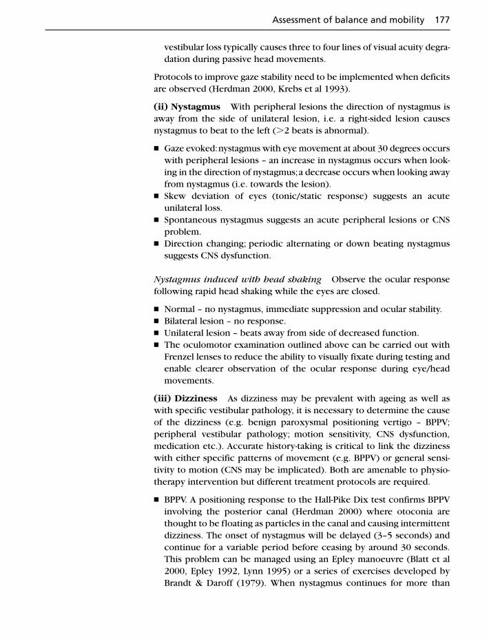
vestibular loss typically causes three to four lines of visual acuity degra-dation during passive head movements.
Protocols to improve gaze stability need to be implemented when deficitsare observed (Herdman 2000, Krebs et al 1993).
(ii) Nystagmus With peripheral lesions the direction of nystagmus isaway from the side of unilateral lesion, i.e. a right-sided lesion causesnystagmus to beat to the left (�2 beats is abnormal).
■ Gaze evoked:nystagmus with eye movement at about 30 degrees occurswith peripheral lesions – an increase in nystagmus occurs when look-ing in the direction of nystagmus;a decrease occurs when looking awayfrom nystagmus (i.e. towards the lesion).
■ Skew deviation of eyes (tonic/static response) suggests an acuteunilateral loss.
■ Spontaneous nystagmus suggests an acute peripheral lesions or CNSproblem.
■ Direction changing; periodic alternating or down beating nystagmussuggests CNS dysfunction.
Nystagmus induced with head shaking Observe the ocular responsefollowing rapid head shaking while the eyes are closed.
■ Normal – no nystagmus, immediate suppression and ocular stability.■ Bilateral lesion – no response.■ Unilateral lesion – beats away from side of decreased function.■ The oculomotor examination outlined above can be carried out with
Frenzel lenses to reduce the ability to visually fixate during testing andenable clearer observation of the ocular response during eye/headmovements.
(iii) Dizziness As dizziness may be prevalent with ageing as well aswith specific vestibular pathology, it is necessary to determine the causeof the dizziness (e.g. benign paroxysmal positioning vertigo – BPPV;peripheral vestibular pathology; motion sensitivity, CNS dysfunction,medication etc.). Accurate history-taking is critical to link the dizzinesswith either specific patterns of movement (e.g. BPPV) or general sensi-tivity to motion (CNS may be implicated). Both are amenable to physio-therapy intervention but different treatment protocols are required.
■ BPPV. A positioning response to the Hall-Pike Dix test confirms BPPVinvolving the posterior canal (Herdman 2000) where otoconia arethought to be floating as particles in the canal and causing intermittentdizziness. The onset of nystagmus will be delayed (3–5 seconds) andcontinue for a variable period before ceasing by around 30 seconds.This problem can be managed using an Epley manoeuvre (Blatt et al2000, Epley 1992, Lynn 1995) or a series of exercises developed byBrandt & Daroff (1979). When nystagmus continues for more than
Assessment of balance and mobility 177

178 Assessment and treatment of balance and mobility deficits in the elderly
30 seconds and up to several minutes, particles may be more adherentto the cupula and a Semont Libatory manoeuvre may be more appro-priate treatment (Herdman 2000).
■ General sensitivity to motion will present with less specificity and anumber of functional motor activities can be disturbed. The intensityand duration of dizziness is recorded against each motor task to deter-mine which movements should be targeted to foster habituation andan improved tolerance to motion (Herdman 2000). An habituation pro-gramme is the protocol of choice in order to desensitize the responseto motion. The tasks that are least problematic are addressed initiallyand as the tolerance to motion improves then the more problematicmovements are targeted.
The impact of dizziness on postural stability as well as on the quality oflife can be determined by using either the Dizziness Handicap Inventory(Jacobson & Newman 1990) or the short form of this tool (Tesio et al1999).
In summary, identification of the underlying impairments enables thephysiotherapist to understand the causes contributing to the balance andmovement disorder and to include these aspects in a management pro-gramme. The perceived needs of each resident along with the physio-therapist’s problem-solving skills are used to develop priorities formanagement. Objective recordings of appropriate balance and mobilitytasks are made to provide a baseline from which progress can be monitored.
A large range of tools are available to monitor physical mobility, with thephysiotherapist responsible for selecting the tool(s) that monitor thephysical ability/difficulty of the client group. The selected tool is used toprovide a baseline measure, assist with goal setting, and monitor progressand outcome after treatment intervention.
Objective measures of
functionalmotor tasks
1. Clinical measuresof sitting ability
Measures of postural stability and dynamic balance and functional mobilityFor the client who cannot stand, timed measures of sitting ability canbe used.
(i) Timed sitting Note the quality of alignment while holding theseated position for 30 seconds.
(ii) Seated reach Measures of the distance reached can be made in sit-ting when the client is unable to stand unsupported.
(iii) MAS, COVS or PMS The relevant component of the MotorAssessment Scale (MAS) (Carr et al 1985), the Clinical Outcomes VariableScale (COVS) (Seaby & Torrance 1989) or Physical Mobility Scale (PMS)(Nitz & Brown, in preparation) may be used.

Objective measures of functional motor tasks 179
A number of tests are available to monitor the ability to stand on firm andsoft surfaces (postural stability) as well as measure dynamic balance andfunctional mobility. The tests will be applicable for the resident who canstand unsupported, and progressively available for those able to take atleast one step alone, or able to mobilize with or without a walking aid.The information provided by the test conditions will be summarized todevelop the implications for intervention programmes.
(i) Timed standing on a firm surface For the resident who can standunsupported it is appropriate to introduce a timed stand with feet apart asan objective measure (Bohannon et al 1984, Newton 1989). For those resi-dents who are able to stand for 30 seconds and take at least one step, stand-ing ability can be challenged by either changing the base of support (feettogether or stride) or by closing the eyes. Some clients may be able to bechallenged by introducing the single limb stance test. The time the residentcan hold the position without support is recorded. Normative data for ageis available (Bohannon et al 1984, Newton 1989) to determine if the balanceability of the individual resident is typical for age or if they are less able andrequiring attention to ensure added stability in standing. These tests requireboth anteroposterior control and considerable mediolateral stability at boththe hip and ankle. During preliminary observations of stance you will notethe control of hip and knee muscles, in addition to the ankle musculature,to allow you to judge when it is safe to introduce these tests.
(ii) Responses to changing sensory cues during timed standingShumway-Cooke & Horak (1986) developed the Clinical Test for theSensory Integration of Balance (CTSIB) to monitor the capacity of the indi-vidual to process and use the information from the somatosensory andvestibular systems during standing. The test involves standing on a firm,then a high density foam surface. For those residents who are able to man-age standing on a firm floor for 30 seconds, particularly if the test can bemanaged with eyes closed, then it is appropriate to introduce the completeCTSIB and apply the six conditions (eyes open, eyes closed and use of a con-flict dome on a firm, then foam surface). The introduction of the foam sur-face will challenge the somatosensory receptors even with eyes open andwill help you to predict the capacity of the resident to change surfacessafely. A further progression to managing with the eyes closed will deter-mine those able/unable to rely on their vestibular function under dark conditions when a softer surface is underfoot (e.g. carpet). A fall frequentlypresents with elders when testing on foam, particularly with the eyes closed,and should only be undertaken if additional stand-by support is available.
Implications of test findings for management There are a numberof implications for residents who cannot manage these test conditions.Standing in dim/dark conditions may compromise balance ability and thusit is important to stand, transfer and mobilize these elders on firm surfacesin well-lit environs. For the mobile resident, advice regarding standing
2. Clinical measuresof standing ability(postural stability)

when showering (e.g. eyes closed while washing hair), while walking oncarpeted surfaces and the need for a light for safe mobility at night couldbe outcomes of this test. For residents who are more independent inter-ventions may need to consider challenging the individual and introducinga variety of surfaces to balance on and/or negotiate while walking duringtreatment sessions. Dynamic visual environs could also be gradually intro-duced (e.g. engaging with a balloon floating through the air; interactingwith multiple obstacles;walking past/around a number of people who arealso moving; pass/bounce/throw/catch a light ball). For these residents,the physiotherapist is aiming to prevent the decline in the sensory systemsthat are typically associated with advancing age.
Initially a self-initiated weight shift can be observed with the selectedstrategy (ankle/hip strategy) noted during the weight shift. A well-controlled ankle strategy is appropriate during weight shift while thoseclients with trunk/pelvic instability are more likely to demonstrate a com-bined ankle/hip movement during weight shift (Herdman 2000). The fol-lowing tests are appropriate for the resident who can stand unsupportedand initiate reaching outside the base of support or is able to take a stepwithout support.
(i) Functional reach The capacity to reach forward outside the base ofsupport can be measured using the protocol established by Duncan et al(1990) while the ability to reach sideways outside the base of support canbe measured using the guidelines set down by Brauer et al (1999c). Bothtests have established normative data which can be used to predict the riskof a fall (Duncan et al 1992, Brauer et al 2000). For those clients whose abil-ity to reach is compromised (forward �25 cm;laterally �18 cm), there is anincreased risk of a fall. For forward reach a significant difference betweenfallers and non-fallers has been demonstrated, with healthy elderly able toreach more than 25 cm while frequent fallers reach less than 15 cm.
Implications for intervention■ When compromised forward and lateral reach is identified, initially in
the treatment objects should be located so that the distance to bereached is reduced for safe performance of tasks.
■ Interventions will aim to introduce tasks that gradually and safely chal-lenge these limits and restore the capacity to reach outside the supportbase.
■ Underlying impairments should also be identified and addressed.
(ii) The Step Test (Hill et al 1996) This test requires the resident torepeatedly place the whole foot up on to a 7.5 cm block and then back tothe floor as many times as possible in 15 seconds. The number of timesthe stepping leg is placed on to the block in 15 seconds is recorded. Theresident should not be supported but requires close supervision to ensuresafety. The test challenges mediolateral stability and requires efficient
180 Assessment and treatment of balance and mobility deficits in the elderly
3. Clinical measures of internal
displacement

Objective measures of functional motor tasks 181
strength in the stance leg, particularly the hip abductors. Normative datais available that shows that healthy elders (�60) take about 16–18 steps in15 seconds (Hill et al 1996), although Isles et al (2004) provide a differ-ential stepping ability for elders (�60 years: right � 15.6, left � 15.9;�70 years: right � 13.7, left � 14.1).
Implications of test findings Performances under these norms for agesuggest that mediolateral stability may require attention, with interven-tions targeting strength/flexibility and endurance of the musculature aboutthe trunk/pelvic/hip/knee regions to improve the performance of this test.
A recent addition to the available battery of tests has emerged with thedevelopment of the rapid step test: the time to complete 24 random stepsforward, backward and to the side in response to a verbal command ismeasured (Medell & Alexander 2000). This test determines the maximumstep distance for an individual, then uses 80% of this distance to set tar-get points to which the individual steps during the execution of the test.The test is considered to be a measure of the protective elements of bal-ance, with increased time recorded for elders compared to young adultsto initiate (react to a command)/prepare to step and execute repeatedsteps. The test also discriminates between healthy elders and impairedelders. The test is repeatable and reliable and performance correlateswith single limb stance ability as well as strength of the quadriceps andcalf muscles.
Clients who are independently ambulating are frequently challenged by surface changes, uneven surfaces as well as unexpected forces such asjostling in a crowd or while standing in a moving bus/train. For suchhigher level clients, the capacity of an individual to manage an externalforce may be gained by applying the following tests.
(i) Pastor, Day and Marsden Scale (Harburn et al 1995, Pastor et al1993) The examiner stands behind the person and gives a brief tugbackwards at the shoulders. The patient has eyes open and is told whatto expect and is instructed to resist the pull in order to prevent backwardmovement. The response is rated according to a scale (0–4). Inter-raterreliability is low and the force applied by the tester may be problematic.This means that several repeated tests are recommended to rate theresponse to displacement to improve the reliability (Harburn et al 1995).
(ii) Stepping response to lateral displacement (Maki et al 2000)The lateral stepping response to displacement is graded according to thecontrol of the stepping response and the pattern of stepping response – acrossover response is typical of a young adult while multiple steps andincreased use of arms is frequently associated with the response in elders(Maki et al 2000).
(iii) Quality of ankle/hip/stepping responses (Shumway-Cook &Woollacott 2001) These also encourage the clinician to note the ankle,
4. A measure ofreaction time: the
rapid step test
5. Response toexternal
displacement

182 Assessment and treatment of balance and mobility deficits in the elderly
hip, combination ankle/hip, stepping strategy or the inability to activate aresponse resulting in a fall without support during the displacement. Inaddition, the relationship of force applied to the chest/thorax to the qual-ity/type of response yielded should be noted. For example, does a mildforce yield an ankle strategy to maintain balance or does the resident stepor over-balance, which is more typically associated with a moderateforce? With the application of a moderate/vigorous force the residentwould be expected to use a hip/stepping response but may be unable torespond and may fall.
(a) Tools that monitor balance and mobility within an indoorenvironment For those residents who are able to ambulate with orwithout a walking aid, additional tools are available to evaluate and measure performance.
(i) Berg Balance Scale (Berg et al 1989) This is used to monitor balancewith elderly clients.
(ii) TUG (Podsiadlo & Richardson 1991) The test may be carried outusing a walking aid, which makes it more suited to those residents who needto use an aid. The resident walks at a safe speed to complete the set task.
Normative data for the TUG:
■ healthy elders (75 years): 8.5 seconds■ fallers: �20 seconds.
Shumway-Cooke et al (1997) recommend that the test be carried out asfast as the person can safely walk to improve the discrimination betweenfallers and non-fallers.
(iii) TUG(manual) and TUG(cognitive) (Shumway-Cooke et al 2000)For more able residents, the dual-task Timed ‘Up and Go’ test allows theeffect of a second task (manual or cognitive) to be monitored during thetest. An increase in time by 2 seconds is considered significant.
(iv) Timed 10 metre walk (Bohannon 1997, Wade et al 1987, Wolfson et al 1990) The test is usually performed without an aid but could beadapted to the functional level of the resident. A study by Dean et al(2001) demonstrated that walking speed over 10 metres overestimateslocomotor capacity after stroke.
A further modification of the test could be the time to wheel 10 metersin a manual wheelchair. This has been used to monitor the speed ofwheeling a wheelchair for ageing clients with cerebral palsy (Low Choyet al 2003) and could be applied to any client who is independentlymobile in a wheelchair.
(v) 10 metre walk with head rotation (Herdman 2000) For those res-idents who can ambulate without an aid this test challenges the vestibularfunction. The time to walk 10 metres is recorded and the ability to keep
6. Measures of balance, gait and
endurance

within a 25 cm wide walkway when the head is actively rotated everythree to four steps is noted.
(vi) The Modified Elderly Mobility Scale (Prosser & Canby 1997, Smith1994) This scale is used with older people with balance and mobilityproblems.
(vii) The gait items of the MAS (Carr et al 1985) and COVS (Seaby &Torrance 1989) These items can be selected and used independently ofthe total scales.
Implications of findings for management A number of elementsare associated with these tests and require consideration in balance pro-grammes. Functional strength is required to stand and during walking,with mediolateral control particularly challenged during the turningcomponents of this task. Linear motion and particularly rotational motionis experienced during turning, which adds a vestibular challenge to thetask. In addition, as the test is performed as fast as the client can safelymanage, reaction time, the capacity to remain stable during attemptedincreased speed and the added functional strength and control requiredwith added speed need to be considered. As appropriate, these elementscan be progressively introduced during retraining programmes.
(b) Ambulation measures associated with challenges encounteredin community environments Patla & Shumway-Cook (1999) giveemphasis to the importance of tools being applied in less predictablecommunity environs and conditions. In the absence of adverse condi-tions, it is recommended that the examiner interpret the performance of community ambulant clients with caution as the performance in idealconditions may underestimate the ability of community ambulant clients.
(i) Dynamic Gait Index Eight Gait Items are scored (Shumway-Cookeet al 1997). The test has good inter-rater and test–retest reliability and canbe used to predict falls among the elderly (Shumway-Cooke et al 1997):
■ scores for healthy aged: 21 � 3■ scores for aged fallers: 11 � 4.
(ii) Fukuda Stepping Test This test was developed to monitor vestibularfunction in the ambulant client (Uttenberger’s test). The patient takes 50 high steps on the spot with the eyes closed and the arms held forwardat shoulder height. The test lacks sensitivity, with a normal response allow-ing rotation up to 45 degrees and travel up to 1 metre with an abnormalresponse being rotation �45 degrees in the direction of the peripheralvestibular problem and/or travel �1 metre (Bonanni & Newton 1998).
(iii) Singleton’s test A test that challenges balance following a 360degree turn. The elder is asked to walk 3 metres towards you, turnquickly and hold steady with eyes open, then repeat the test and holdsteady with eyes closed (Herdman 2000).
Objective measures of functional motor tasks 183

(iv) Dual tasks while walking These include: time to execute an obstacle course/perform dual/multiple tasks while manipulating objects;throw/catch ball; picking up objects from floor (Herdman 2000).
(v) Duke Mobility Skills Profile This was developed to measure thegait performance of older adults. The test has good inter-rater and test–retest reliability (Duncan et al 1993).
(vi) Rivermead Mobility Index (Colleen et al 1991) Monitors a rangeof tasks associated with indoor/outdoor activities.
(c) Tinetti Fear of Falling Scale Fear of falling can also be monitoredto determine the anxiety/perceived risk associated with a variety ofhome- and community-based tasks (Manning et al 1997, Tinetti et al 1990).
(d) Endurance The functional endurance of each client could be meas-ured using the 3- or 6-minute walk test (Butland et al 1982, Schenkman et al 1997, Shumway-Cooke et al 1997). The baseline measure could beused to determine the benefits of an intervention programme (e.g. walk-ing programme, or use of treadmill/ergometer).
Management for the frail elderly often means a focus on bed mobility, sit-ting ability, transfers, wheelchair skills and/or gait training. It will thus beimportant to select a tool that includes this range of functional tasks forthe more dependent resident. Several tools are available and include thefollowing.
■ The Physical Mobility Scale (PMS) currently being investigated forreliability and validity for use in residential aged care facilities (Nitz &Brown, in preparation).
■ The Physical Performance and Mobility Examination (PPME)developed to monitor physical function and mobility in frail olderadults (Winograd et al 1994).
■ The Mobility Scale for Acute Stroke Patients (Simondson et al 1996).■ The Motor Assessment Scale (MAS) (Carr et al 1985) developed for
use with stroke clients.■ The Clinical Outcomes Variable Scale (COVS) (Seaby & Torrance
1989) developed for use with ortho-geriatric and neurological clients.
Each of these scales includes measures of bed mobility; sitting ability;standing and walking ability. The MAS and COVS also include upper limbfunction while the COVS and PMS provide scores for transfers and wheel-chair management. These latter tools also offer a greater capacity tomeasure the amount of assistance required within each task. This is con-sidered an advantage for use with clients in residential aged care facilitieswhere some clients will require assistance for all tasks. Such scales alsoenable the burden of care to be monitored, an important component toconsider for those physiotherapists working in residential care facilities(Low Choy et al 2003).
184 Assessment and treatment of balance and mobility deficits in the elderly
Tools that monitorgeneral physical
performance andmobility

Objective measures of functional motor tasks 185
Summary ■ The information gleaned from the assessment process and theselected tests will help you to decide on the stability and safemobility of each elder.
■ There will be a need to identify the safe level of mobility foreach resident/elder and for those in an RACF it will benecessary to document clearly the level of supervision orassistance required for all balance and mobility tasks.
■ The information should also be used to identify situationswhen residents are at risk of a fall. An example might bewhen the resident is unable to divide attention betweenwalking and talking while remaining stable. In this instancecare plan instructions should document that the carer shouldnot ask questions or carry out a conversation with theresident as this could compromise the safety of the residentby increasing the risk of a fall.
■ The assessment process also enables targeted interventions tobe offered and the education of elders about the limits totheir safe mobility.
■ More specific findings from the assessment will havedetermined the time the resident could participate instanding activities, the visual and support conditions thatshould form part of the set-up for task practice and thecapacity of the resident to be worked in standing. This latteraspect has implications for use of tasks where residents canmove and reach safely within their ability and then bechallenged (under supervision) to progress towards a newlimit of stability.
■ This may mean working the elder while moving over astationary base (e.g. reach and weight shift with the feet aparton a firm surface) or use of a dynamic base (e.g. working on asoft surface or stepping while reaching for an object).
■ Finally, the number of steps that could be encouraged, thespeed at which tasks could be safely practised, the distancesthat could be walked, the obstacles/surfaces that could beintroduced during a walking task and the capacity to managedual tasks could be determined from some of the moreadvanced tests discussed in this section.
■ Thus the assessment process determines those aspects relatedto the elder that are amenable to intervention, the motortasks that require attention in any given environment and theway the environment impacts on task performance. Theobservational analysis of balance and mobility tasksdetermines the quality of the performance and identifiespossible impairments using problem-solving skills; the

186 Assessment and treatment of balance and mobility deficits in the elderly
Alhanti B, Bruder L A, Creese W et al 1997 Balanceabilities of community dwelling older adults underaltered visual and support surface conditions.Physical and Occupational Therapy in Geriatrics15:37–52
Anacker S L, Di Fabio R P 1992 Influence of sensoryinputs on standing balance in community-dwelling elders with a recent history of falling.Physical Therapy 72:575–584
Aniansson A, Rundgren A, Sperling L 1980 Evaluationof functional capacity in activities of daily living in70 year old men and women. Scandinavian JournalRehabilitation Medicine 12:145–154
Baloh R W, Halmagyi G M 1996 Disorders of the vesti-bular system. Oxford University Press, New York
Baloh R W, Hornrubia V, Jacobson K 1987 Benignpositional vertigo: clinical and oculographicfeatures in 240 cases. Neurology 37:371–378
Bennell K, Talbot R, Wajswelner H, Techovanich W,Kelly D 1998 Intra-rater and inter-tester reliability ofa weight-bearing lunge measure of ankle dorsiflex-ion. Australian Journal of Physiotherapy 44:175–180
Berg K, Wood-Dauphine S, Williams J I 1989 Meas-uring balance in the elderly: preliminary develop-ment of an instrument. Physiotherapy Canada41:304–311
Blatt P J, Georgakakis G A, Herdman S J, Clendaniel R A,Tusa R J 2000 The effect of the canalith reposition-ing maneuver on resolving postural instability inpatients with benign paroxysmal positional vertigo.American Journal of Otology 21:356–363
Bohannon R W 1997 Comfortable and maximum walk-ing speed of adults aged 20–79 years: reference values and determinants. Age and Ageing 26:15–19
Bohannon R, Larkin P, Cook A, Gear J, Singer J 1984Decrease in balance scores with ageing. PhysicalTherapy 64:1067–1070
Bonanni M, Newton R 1998 Test–retest reliability ofthe Fukuda Stepping Test. Physiotherapy ResearchInternational 3(1):58–68
Brandt T, Daroff R B 1980 Physical therapy for benignparoxysmal positional vertigo. Archives ofOtolaryngology 106:484–485
Brauer S, Woollacott M, Shumway-Cook A 1999aBalance impaired elderly: secondary task influ-ences EMG response to perturbation. Gait andPosture 9(1):S33
Brauer S, Woollacott M, Shumway-Cook A 1999bBalance impaired elderly change response strategywith cognitive load. Society Neuroscience Abstracts25(1):108
Brauer S, Burns Y, Galley P 1999c Lateral reach:a newclinical measure of medio-lateral balance. Physio-therapy Research International 4(2):81–88
Brauer S, Burns Y, Galley P 2000 Postural stabilitymeasures predict falls in community-dwelling elderly. Journal of Gerontology Medical Science55(8):M469–M476
Brocklehurst J C, Robertson D, James-Gordon P 1982Clinical correlates of sway in old age: sensorymodalities. Age and Ageing 11:1–10
Buchner D M, de Lateur B J 1991 The importance ofskeletal muscle strength to physical function inolder adults. Annals of Behavioral Medicine13:91–98
Butland R J A, Pang J, Gross E R, Woodcock A A,Geddes D M 1982 Two, six and 12 minute walkingtests in respiratory disease. British Medical Journal284:1607–1608
Carr J, Shepherd R, Nordholm L, Lynne D 1985A Motor Assessment Scale for stroke. PhysicalTherapy 65:175–180
Cho C, Kamen G 1998 Detecting balance deficits infrequent fallers using clinical quantitative evaluation
objective examination and use of tools to measure theimpairments confirms the contribution of the impairment tobalance and mobility dysfunction; and objective recordings ofappropriate balance and mobility tasks are made to provide abaseline from which progress can be monitored.
■ The perceived needs of each elder/resident along with theassessment process using your problem-solving skills are usedto develop priorities for management.
References

References 187
tools. Journal of the American Geriatrics Society46:426–430
Colleen F M, Wade D T, Robb G F, Bradshaw C M 1991The Rivermead Mobility Index: a further develop-ment of the Rivermead Motor Assessment. Inter-national Disabilities Study 13:50–54
Collins N, Vicenzino B, Teys P 2003 The initial effectsof a Mulligan mobilisation with movement tech-nique on dorsiflexion and pain in subacute anklesprains. Manual Therapy (in press)
Davies P M 1990 Right in the middle. Springer, BerlinDean C M, Richards C L, Malouin F 2001 Walking speed
over 10 metres overestimates locomotor capacityafter stroke. Clinical Rehabilitation 15:415–421
Dite W, Temple V A 2002 Development of a clinicalmeasure of turning for older adults. AmericanJournal of Physical Medicine and Rehabilitation81(11):857–866
Duncan P, Weiner D K, Chandler J 1990 Functionalreach: a new clinical measure of balance. Journalof Gerontology 45:M192–M197
Duncan P W, Studenski S, Chandler J, Prescott B 1992Functional reach: predictive validity in a sampleof elderly male veterans. Journal of Gerontology47:M93–M98
Duncan P W, Shumway-Cook A, Tinetti M E et al 1993Is there one simple measure for balance? PTMagazine 1:74–81
Epley J M 1992 The canalith repositioning procedurefor treatment of benign paroxysmal positional vertigo. Otolaryngology Head Neck Surgery 107:399–404
Frank J S, Patla A E, Brown J E 1987 Characteristics of postural control accompanying voluntary armmovement in the elderly. Society for NeuroscienceAbstracts 13:335
Grimby G, Saltin B 1983 The ageing muscle. ClinicalPhysiology 3:209–218
Harburn K, Hill K, Kramer J et al 1995 Clinical applic-ability and test–retest reliability of an external perturbation test of balance in stroke subjects.Archives of Physical Medicine and Rehabilitation76(4):317–323
Herdman S J 2000 Vestibular rehabilitation. F A Davis,New York
Hill K, Bernhardt J, McGann A, Maltese D, Berkovits D1996 A new test of dynamic standing balance forstroke patients: reliability, validity and comparisonwith healthy elderly. Physiotherapy Canada 48:257–262
Hillman E J, Bloomberg J J, McDonald V P, Cohen H S1999 Dynamic visual acuity while walking in normals and labyrinthine deficit patients: a meas-ure of oscillopsia. Journal of Vestibular Research9(1):49–57
Hinman M R 1998 Causal attributions of falls in olderadults. Physical and Occupational Therapy in Geri-atrics 15:71–84
Hirvonen T P, Aalto H, Pykko I, Juhola M, Jantil P 1997Changes in VOR of elderly people. Acta Otolaryn-gology (Stockholm) Supplementary 529:108–110
Holewski J, Stress R, Graf P et al 1988 Aesthesiometry:quantification of cutaneous pressure sensation indiabetic peripheral neuropathy. Journal of Rehabi-litation Research and Development 25:1–10
Horak F, Schupert C, Mirka A 1989 Components ofpostural dyscontrol in the elderly:a review. Neuro-biology and Aging 10:727–745
Horak F, Nashner L M, Diener H C 1990 Posturalstrategies associated with somatosensory andvestibular loss. Experimental Brain Research 82:167–177
Hurley M, Rees J, Newham D 1998 Quadriceps func-tion, proprioceptive acuity and functional perform-ance in healthy young, middle-aged and elderlysubjects. Age and Ageing 27:55–67
Hyatt R, Whitelaw M, Bhat A, Scott S, Maxwell J 1990Association of muscle strength and functional sta-tus in elderly people. Age and Ageing 19:330–336
Inglin B, Woollacott M H 1988 Age-related changes in anticipatory postural adjustments associatedwith arm movements. Journal of Gerontology 43:M105–M113
Isles R C, Nitz J C, Low Choy N L 2004 Normativedata for clinical balance measures in women aged20 to 80. Journal of the American GeriatricsSociety (in press)
Jacobson G P, Newman C W 1990 The developmentof the dizziness handicap inventory. Archives ofOtolaryngology Head and Neck Surgery116:424–427
Kelly C B 2000 Conservative management of vulner-able feet. In: Lusardi M M, Nielsen C (eds)Orthotics and prosthetics in rehabilitation.Butterworth Heinemann, Boston
Krebs D E, Gill-Body K M, Riley P O, Shumway-CookA 1993 Double-blind, placebo-controlled trial ofrehabilitation for bilateral vestibular hypo-func-tion:preliminary report. Otolaryngology Head andNeck Surgery 109:735–741

188 Assessment and treatment of balance and mobility deficits in the elderly
Kumar S, Fernando D, Veves A et al 1995 Semmes-Weinstein monofilaments: a simple, effective andinexpensive screening device for identifying diabetic patients at risk for foot ulceration.Diabetes Research Clinic Practitioner 13:63–68
Lewis C, Phillippi L 1993 Postural changes with age andsoft tissue treatment. Physical Therapy Forum 9:4–6
Lexell J 1993 Ageing and human skeletal muscle:observations from Sweden. Canadian Journal ofApplied Physiology 18:2–9
Lord S, Castell S 1994 Physical activity programmefor older persons: effect on balance, strength,neuromuscular control and balance reaction time.Archives of Physical Medicine and Rehabilitation75:648–652
Lord S R, Clark R D 1996 Simple physiological andclinical tests for the accurate prediction of fallingin older people. Gerontology 42:199–203
Lord S R, Menz H B 2000 Visual contributions topostural stability in older adults. Gerontology 46:306–310
Lord S R, Webster I W 1990 Visual field dependence inelderly fallers and non-fallers. International Journalof Ageing and Human Development 31:267–277
Lord S R, Clark R D, Webster I W 1991 Physiologicalfactors associated with falls in an elderly popula-tion. Journal of the American Geriatrics Society39:1194–1200
Lord S R, McLean D, Strathers G 1992 Physiologicalfactors associated with injurious falls in older peopleliving in the community. Gerontology 38:338–346
Lord S R, Caplan G A, Ward J A 1993 Balance, reactiontime and muscle strength in exercising and non-exercising older women: a pilot study. Archives ofPhysical Medicine and Rehabilitation 74:837–839
Lord S R, Sambrook P N, Gilbert C 1994 Posturalstability, falls and fractures in the elderly: resultsfrom the Dubbo osteoporosis epidemiology study.Medical Journal of Australia 160:684–691
Lord S R, Ward J A, Williams P, Strudwick M 1995 Theeffect of a 12 month exercise trial on balance,strength and falls in older women. Journal of theAmerican Geriatrics Society 43:1198–1206
Lord S R, Ward J A, Williams P 1996 Exercise effect ondynamic stability in older women: a randomisedcontrolled trial. Archives of Physical Medicine andRehabilitation 77:232–236
Lord S R, Sherrington C, Menz H 2001 Falls in olderpeople: risk factors and strategies for prevention.Cambridge University Press, Cambridge
Lord S R, Dayhew J, Howland A 2002 Multifocalglasses impair edge-contrast sensitivity and depthperception and increase the risk of falls in olderpeople. Journal of the American Geriatrics Society50(11):1760–1766
Lundin-Olsson L, Nyberg L, Gustafson Y 1997 ‘Stopswalking when talking’ as a predictor of falls inelderly people. Lancet 349:617
Lundin-Olsson L, Nyberg L, Gustafson Y 1998 Atten-tion, frailty and falls: the effect of a manual task onbasic mobility. Journal of the American GeriatricsSociety 46:758–761
Lynn S, Pool A, Rose D, Brey R, Suman V 1995 Ran-domised trial of the canalith repositioning pro-cedure. Otolaryngology Head Neck Surgery 113:712–720
McIlroy W E, Maki B E 1996 Age-related changes in compensatory stepping in response to unpre-dictable perturbations. Journal of Geriatrics 51:M289–M296
Maki B E, Holliday P J, Topper A K 1991 Fear of fallingand postural performance in the elderly. Journal ofGerontology Medical Science 46:M123–M131
Maki B E, Holliday P J, Topper A K 1994 A prospectivestudy of postural balance and risk of falling in anambulatory and independent elderly population.Journal of Gerontology Medical Science 49:M72–M84
Maki B E, Edmonstone M A, McIlroy W E 2000 Age-related differences in laterally directed compensa-tory stepping behaviour. Journal of GerontologyMedical Science 55A:M270–M277
Manchester D, Woollacott M, Zederbauer-Hylton N,Marin O 1989 Visual, vestibular and somatosen-sory contributions to balance control in the olderadult. Journal of Gerontology Medical Science 44:M118–M127
Manning J, Neistadt M, Parker S 1997 The relation-ship between fear of falling and balance and gaitabilities in elderly adults in a subacute rehabilita-tion facility. Physical and Occupational Therapyand Geriatrics 15(2):33–47
Medell J L, Alexander N B 2000 A clinical measure ofmaximal and rapid stepping in older women.Journal of Gerontology Medical Science 55A:M429–M433
Murray M P, Duthie E H, Gambert S R, Sepic S B,Mollinger L A 1985 Age-related differences in kneemuscle strength in normal women. Journal ofGerontology 40:275–280

References 189
Nadeau S, Gravel D, Arsenalt A, Bourbonnais D,Goyette M 1997 Dynamometric assessment of theplantarflexors in hemiparetic subjects: relationsbetween muscular, gait and clinical parameters.Scandinavian Journal of Rehabilitation Medicine29:137–146
Newton R 1989 Review of tests of standing balanceabilities. Brain Injury 3:335–336
Newton R A 1995 Standing balance abilities of elderlysubjects under altered visual and support condi-tions. Physiotherapy Canada 47:25–29
Nitz J C, Brown A The physical mobility scale toshow dependency in frail elderly people:a reliabil-ity and validity study (in preparation)
Paige G D 1992 Senescence of human visual-vestibu-lar interactions with ageing. Journal of VestibularResearch 2:133–151
Pastor M A, Day B L, Marsden C D 1993 Vestibularinduced postural responses in Parkinson’s Disease.Brain 116:11–17
Patla A E, Shumway-Cook A 1999 Dimensions ofmobility: defining the complexity and difficultyassociated with community mobility. Journal ofAging and Physical Activity 7:7–19
Perry J, Garrett M, Gronley J K, Mulroy S J 1995Classification of walking handicap in the strokepopulation. Stroke 26:982–989
Peterka R, Black F 1990 Age related changes in humanposture control: sensory organisation tests.Journal of Vestibular Research 1:73–85
Podsiadlo D, Richardson S 1991 The timed ‘Up andGo’: a test of basic functional mobility for frail per-sons. Journal of the American Geriatrics Society39(2):142–148
Prosser L, Canby A 1997 Further validation of theElderly Mobility Scale for measurement of mobilityof hospitalised elderly people. Clinical Rehabilita-tion 11:338–343
Rankin J K, Woollacott M H, Shumway-Cook A,Brown L A 2000 Cognitive influence on posturalstability: a neuromuscular analysis in young andolder adults. Journal of Gerontology MedicalScience 55A:M112–M119
Ring C, Nayak U S L, Isaacs B 1989 The effect of visualdeprivation and proprioceptive change on posturalsway in healthy elders. Journal of the AmericanGeriatrics Society 37:745–749
Rutherford O M, Jones D A 1992 The relationshipbetween muscle and bone loss and activity levelswith age in women. Age and Ageing 21:286–293
Schenkman M, Cutson T M, Kuchibhatla M, ChandlerJ, Pieper C 1997 Reliability of impairment andphysical performance measures for persons withParkinson’s disease. Physical Therapy 70:19–27
Seaby L, Torrance G 1989 Reliability of a physio-therapy functional assessment in a rehabilitationsetting. Physiotherapy Canada 41(5):264–271
Shumway-Cook A, Horak F 1986 Assessing the influ-ence of sensory interaction on balance:suggestionfrom the field. Physical Therapy 66:1548–1550
Shumway-Cook A, Woollacott M 2000 Attentionaldemands and postural control:The effects of sensorycontext. Journal of Gerontology Medical Science55A:M10–M16
Shumway-Cook A, Woollacott M 2001 Motor control:theory and practical applications. Williams &Wilkins, Baltimore
Shumway-Cook A, Woollacott M, Kerns K A, BaldwinM 1997 The effects of two types of cognitive taskson postural stability in older adults with and without a history of falls. Journal of GerontologyMedical Science 52A:M232–M240
Shumway-Cook A, Brauer S, Woollacott M 2000Predicting the probability for falls in the commu-nity-dwelling adults using the Timed Up & GoTest. Physical Therapy 80(9):896–903
Simondson J, Goldie P, Brock K, Nosworthy J 1996The Mobility Scale for acute stroke patients: intra-rater and inter-rater reliability. Clinical Rehabilitation10:295–300
Skelton D, Greig C, Davies J, Young A 1994 Strength,power and related functional ability of healthyaged 65–89 years. Age and Ageing 23:371–377
Smith R 1994 Validation and reliability of the ElderlyMobility Scale. Physiotherapy 80(11):744–747
Speechley M, Tinetti M 1991 Falls and injuries in frailand vigorous community elderly persons. Journalof the American Geriatrics Society 39:46–52
Steinberg M, Cartwright C, Peel N, Williams G 2000A sustainable programme to prevent falls and nearfalls in community dwelling older people: resultsof a randomised trial. Journal of Epidemiology andCommunity Health 54:227–232
Stelmach G E, Worringham C 1985 Sensorimotordeficits related to postural stability: implicationsfor falling in the elderly. Clinical Geriatric Medicine1:679–694
Studenski S, Duncan P W, Chandler J 1991 Posturalresponses and effector factors in persons withunexplained falls: results and methodological

190 Assessment and treatment of balance and mobility deficits in the elderly
issues. Journal of the American Geriatrics Society39:229–234
Tanaka T, Hashimoto N, Noriyasu S et al 1995 Ageingand postural stability: changes in sensorimotorfunction. Physical and Occupational Therapy inGeriatrics 13:1–16
Teasdale N, Stelmach G, Breunig A 1991 Posturalsway characteristics of the elderly under normaland altered visual and support surface conditions.Journal of Gerontology 46:B238–B244
Tesio L, Alpini D, Cesarani A, Perucca M 1999 Shortform of the dizziness handicap inventory.American Journal of Physical Medicine andRehabilitation 78:233–241
Thigpin M T, Light K E, Creel G L, Flynn S M 2000Turning difficulty characteristics of adults aged 65years and older. Physical Therapy 80(12):1174–1187
Tinetti M E, Speechley M, Ginter S E 1988 Risk factorsfor falls among elderly persons living in the com-munity. New England Journal of Medicine 319:1701–1707
Tinetti M E, Richman D, Powell L 1990 Falls efficacyas a measure of fear of falling. Journal of Gerontol-ogy Medical Science 45:239–243
Tinetti M, Baker D, McAcay 1994 A multifactorialintervention to reduce the risk of falling amongelderly people living in the community. NewEngland Journal of Medicine 331:821–827
Tobis J S, Reinsch D, Swanson J M, Byrd M, Scharf T1985 Visual perception dominance of falls amongcommunity dwelling older adults. Journal of theAmerican Geriatrics Society 33:330–333
Vandervoort A, McComas A J 1986 Contractilechanges in opposing muscles of the human anklejoint with ageing. Journal of Applied Physiology61:361–367
Wade M, Jones G 1997 The role of vision and spatialorientation in the maintenance of posture. Phys-ical Therapy 77(6):619–627
Wade D, Wood V, Heller A, Maggs J, Hewer R 1987Walking after stroke. Scandinavian Journal ofRehabilitation Medicine 19:25–30
Whanger A D, Wang H S 1974 Clinical correlates ofthe vibratory sense in elderly psychiatric patients.Journal of Gerontology 4:39–45
Whipple R H, Wolfson L I, Amerman P M 1987 Therelationship of knee and ankle weakness to fallsin nursing home residents: an isokinetic study.Journal of the American Geriatrics Society 35:13–20
Whitney S L, Hudak M T, Marchetti G F 2000 Theassociation between observed gait instability andfall history in persons with vestibular dysfunction.Journal of Vestibular Research 10:99–105
Winograd C H, Lemsky C M, Nevitt M C 1994Development of a physical performance and mobil-ity examination. Journal of the American GeriatricsSociety 42:743–749
Wolfson L, Whipple R, Amerman P 1990 Gait assess-ment in the elderly: a gait abnormality rating scaleand its relation to falls. Journal of Gerontology45:M12–M19
Woollacott M H 2000 Systems contributing to bal-ance disorders in older adults. Editorial. Journal ofGerontology Medical Science 55A(8):M424–M428
Woollacott M, Tang P 1997 Balance control duringwalking in the older adult: research and implica-tions. Physical Therapy 77:646–659
Woollacott M H, Shumway-Cook A, Nashner L 1986Ageing and posture control: changes in sensoryorganization and muscular coordination. Inter-national Journal of Aging and Human Development23:97–114

Retraining balance usingtask-focused workstations
Nancy Low Choy
191
10
This chapteraims to:
Management of balance dysfunction in the older person requires an holis-tic approach to which physiotherapy makes an important contributionthrough falls prevention programmes as well as effective remedial inter-vention when falls and injuries occur. An assessment by a physiotherapistidentifies those causal factors amenable to intervention and identifies andprovides strategies to minimize future falls risk. It has been argued that themanagement of balance dysfunction in older people requires multiplesystems to be considered and that addressing the impairments inherent ineach individual in conjunction with varied task practice and environmentsenables elders to maintain and regain the ability to mobilize safely. Wherethe ability to mobilize safely is compromised it is critical that the limitationsto balance and the need for supervision or assistance for safe mobility areidentified.
A task-oriented approach to retraining balance and mobility has emergedas an effective model of intervention over the last decade with a range of basic sciences used to provide the theoretical foundation for thismodel. Carr & Shepherd (1987, 1998, 2000, 2003) and Shumway-Cook &Woollacott (2001) are strong advocates of the need to consider a broadscience basis for practice and believe that functional task training offers
■ present a task-oriented approach to retraining balance andmobility using workstations as an effective mode of deliveringtailored interventions to individual or small groups of residentsor community-based elders who are: frail aged; elders whoambulate with supervision within residential care facilities; orelders who are independently ambulant in the community
■ provide an outline of workstations that illustrate varying levelsof challenge but address the multiple aspects of balance andmobility that need to be considered while retraining balanceand mobility in elders.
Introduction
Task-orientedapproach to
retraining balance and
mobility

an efficient mode of regaining motor control by integrating the capacityof the brain to reorganize after injury (Johansson 2000, Nudo & Friel1999, Nudo et al 2001, Shepherd 2001) as well as through application ofprinciples of skill acquisition (Gentile 2000, Magill 2001). Such a modelfosters specificity of task training, with muscle actions practised in thecontext of function, repeated task practice using different forms of feed-back that can gradually be withdrawn, and varied task practice to enabletransfer of learning to meet the functional demands of different environ-ments. The challenge for the physiotherapist has been to know when toprovide more direct assistance for the practice of motor tasks, when tooperate more as a ‘coach or trainer’ and when to effectively incorporatepart practice or ‘sub-tasks’ (Carr & Shepherd 2003).
Task-oriented training is promoted by Carr & Shepherd (2003) andShumway-Cook & Woollacott (2001) and the efficacy of such training hasbeen evaluated in a number of studies. The efficacy of task-related traininghas been established after stroke (Dean et al 2000, Dean & Shepherd1997, Richards et al 1993), when retraining balance and mobility in fallers(Nitz & Low Choy 2004, Shumway-Cook et al 1997b) and for retrainingfunctional motor tasks in cerebral palsy clients who are ageing (Low Choyet al 2003). The triad of addressing individual needs during varied taskpractice and in different environments is integrated within each of theseprogrammes. Consideration of this triad is recommended when deliver-ing balance and mobility programmes to other clients with neurologicaldisorders and to those elders requiring intervention within residentialcare facilities and other community-based environments.
A review of intervention programmes that have been delivered toimprove balance and mobility and decrease the falls risk of elders livingin the community reveals that while an appropriate emphasis has beenplaced on strength, flexibility and resistance training (Buchner et al 1997,Chandler et al 1998, Lord & Castell 1994, Lord et al 1993, 1995, 1996,Rooks et al 1997, Sauvage et al 1992, Skelton et al 1995) few programmeshave addressed the sensory processing and challenges that the environ-ment (visual and surface challenges) places on balance systems or thecognitive demand inherent in many functional motor tasks requiringboth balance and mobility (Brauer et al 2001, Lundin-Olssen et al 1997,1998, Nitz & Thompson 2003, Shumway-Cook et al 1997a, 1997b,Shumway-Cook & Woollacott 2000). Most programmes include an exer-cise regime that can be delivered to groups of elders in community facil-ities although some programmes have been offered within the home(Campbell et al 1997, Jette et al 1996, McMurdo & Johnstone 1995,Sherrington & Lord 1997) with variable levels of supervision and supportfor the participants in the programmes. Less prevalent in the literature are programmes that provide a comprehensive, multidimensional, task-oriented approach to upgrading balance and mobility in elderly fallers.Such programmes have been successfully delivered to individual clients(Shumway-Cook et al 1997a) and to groups of elders (Nitz & Low Choy
192 Retraining balance using task-focused workstations

2004) who had a falls history. In the latter study, workstations were usedto provide a comprehensive programme that addressed all aspects of balance and mobility. A significant reduction in falls was achieved for the intervention group. It is possible that the added attention to balancestrategy training, progressively increasing sensory challenges to tasks invarying environs and the added demand of dual tasks while practisingmotor tasks could have provided additional capacity for these elders tomanage in community environs without recurrent falls. The delivery ofsuch programmes within a residential care facility is advocated and con-sidered cost-effective given the prevalence of falls in such facilities(Lipsitz et al 1991, Liu et al 1995, Lord et al 1991, Rubenstein et al 1988)and the high cost of care associated with falls and subsequent injury.With the prevalence of falls persisting in the general community despite the attempts to increase participation rates in activity programmes (Hillet al 2002), it is important to invest in strategies that might reduce therecurrence of falls.
Workstations are considered the optimal mode for delivering multi-dimensional task-focused programmes that are specific to the client’s need.Proponents of the workstation mode of intervention (Carr & Shepherd2003, Dean & Shepherd 1997, Dean et al 2000, Low Choy et al 2003, Nitz &Low Choy 2004) recognize that task training can be delivered in an integrated and holistic programme, that the intensity and duration ofrehabilitation programmes can be increased without adding to the cost ofdelivering a physiotherapy programme, and that the group dynamics gen-erated may enhance motivation and compliance to training (Ada et al 1999).While this form of intervention is also considered to reduce the depend-ency on physiotherapists, the functional ability of individual clients willneed to be considered, particularly in the context of frail older people living in residential care facilities.
The challenge for physiotherapists working in residential or community-based aged care is to deliver interventions that integrate the multiple elements of balance, target the needs of individual clients yet ensure acost-effective mode of delivery. Clinicians could work with individual residents at their functional level and work capacity or overlap residentswho could feasibly be managed in a small group. An appropriate assess-ment would provide the clinician with the necessary information to decideon the feasibility of overlapping residents or the need to work on an indi-vidualized basis. Once the focus of a workstation has been determined itis feasible to adapt the station to the activities of daily living encounteredby a resident so that the individual can practise the functional task duringcare activities under the supervision of the carer. In addition, other moreambulant residents or community-dwelling elders could reasonably beworked in a small group under supervision, with respective workstationsadjusted to safely challenge these clients. The need to include training for
Using workstations as a mode to deliver balance and mobility programmes 193
Using workstationsas a mode to
deliver balanceand mobility programmes

carers who work with frail aged or other highly dependent residents toensure effective carryover of functional gains made with training wassupported by the findings of a study with cerebral palsy residents whoare ageing with a disability (Low Choy et al 2003).
Workstations set up in the form of a circuit would appear to provide aneffective option for the physiotherapist to deliver variable task experi-ences while addressing the elements of balance under different environ-mental conditions. The multiple aspects of balance that may need to beaddressed when delivering such a balance and mobility programme to anindividual within a residential care facility or community environmentwere discussed in the preceding chapter. Interventions may need to:
■ improve functional strength and flexibility■ ensure specificity of practice to meet task and environmental demands■ increase control at the limits of stability in all planes with particular
attention to mediolateral stability for safe mobility during walking andparticularly turning
■ decrease reaction times by increasing speed of response to stimuliwhile balancing and walking
■ develop reliable postural responses and varied strategies according todirection/force of displacement
■ activate, foster integration and use sensory information/challenges forimproved balance during task execution and/or conditions involvingaltered visual/proprioceptive input or conflict
■ increase ability to manage dual/multiple tasks or recognize the inabil-ity of residents to safely manage multiple tasks
■ improve endurance by increasing musculoskeletal and cardiovascular-respiratory capacity.
There are four practical steps to apply when planning a workstationmode of intervention for an individual resident or for a number of olderpeople who could participate in a small group intervention:
■ assess each resident to determine the aspects of balance and mobilitythat require attention and use tools that can objectively measure theimpairments and functional motor abilities/limitations of the residentprior to the programme being implemented
■ set up workstations that ensure all elements of balance and mobilityare addressed and can be tailored to meet the specific problems of theindividual
■ plan how to make each station easier or more difficult to cater for the individual abilities that could present in the group through use of variable tasks which can be practised under different environmentalconditions at each station
■ reassess residents at the end of the programme or at intervals that aredetermined by their changing medical status to determine improve-ment and further areas for intervention.
194 Retraining balance using task-focused workstations

In developing a workstations programme, a number of key principlesneed to be considered. The programme is driven by the individual needsof each participant and their active participation rather than beingdependent on the physiotherapist being available for active practice offunctional tasks. It is important therefore that each participant is pro-vided with sufficient knowledge so that they are able to maximally benefit from their own programme. This is just as applicable for moreindependent/able residents as for those with a high level of functionaldependence. Each participant therefore needs to be able to understandthe purpose of each individual station and the relevant aim and goal forthemselves. The relevance of the activity to specific functional tasksneeds to be clear, as do the means of varying the activity. This shouldinclude not only making the task easier or harder (i.e. progressions orregressions) but also task variations so as to assist with transference oflearning and promote interest during the repeated practice critical to asuccessful outcome.
By providing the resident with such knowledge and skills, the likeli-hood of carryover beyond the therapy setting is increased. This is further enhanced by the functional context of activities which are similar to work in real life and thus addressing the issue of specificity oftraining.
Each workstation should have a clear, easily understood instructionsheet. Key words, photos or diagrams may be used to name and give pur-pose to the workstation (e.g. sit-to-stand station, reaching station). Theactivity is best placed into a relevant functional context (e.g. to make iteasier to reach into the bedside cupboard). The purpose needs to be clearlyexplained. The means of varying the task, making it easier or harder needto be clearly illustrated with words or pictures.
A second important principle is that the therapist acts as a ‘coach’ or‘trainer’. The supervising therapist is responsible for the initial detailedassessment and subsequent reassessment. This is vital so as to allow a tar-geted, individualized approach. Based upon the assessment findings thetherapist is then responsible for developing workstation tasks to addressspecific identified issues. The therapist is responsible for training eachparticipant in the task to be practised and for providing strategies forsolving everyday functional problems and so place the training activityinto a functional context which increases the likelihood of carryover.Safety remains a primary issue and all tasks need to be designed so thateach individual is challenged but is able to perform the activities safely.This will often provide challenges to the therapist when designing theprogramme.
Residents at different functional levels may have different goals fromindividual workstations. More independent residents are able to takeresponsibility for learning the tasks and are able to work their way inde-pendently through the stations. Such individuals may gradually be able toindependently adapt, modify and progress each task so as to optimally
Using workstations as a mode to deliver balance and mobility programmes 195

challenge themselves. Residents at a lower functional level will be morereliant upon the physiotherapist to set up each task to suit individual needs.They may be more reliant on equipment and/or the physiotherapist/carerto ensure safety. This is clearly the case for those residents with impairedcognitive function, perceptual deficits, behavioural problems or poor sit-ting or standing balance. Tasks and activities for these residents will needto be carefully evaluated and participation in challenging aspects of aworkstation programme will require individualized attention. For example,residents with very poor sitting balance may perform preparatory tasks inlying, actively move and reach while sitting in a chair but practise sittingtasks without such supportive seating when the physiotherapist is avail-able. Use of a tilt table with modified support to enable the upper bodyto actively participate in reaching activities, or standing with bodyweight support (if available), may allow such residents to be challengedin a safe and supported environment.
Thus workstations can be used to deliver comprehensive, multidimen-sional and targeted interventions to improve balance and mobility in eld-erly residents, with the content and level of delivery adjusted to meet thefunctional level of each individual. This means that the more dependentresident, those who can be worked under supervision and those who can be progressively extended and challenged are able to benefit fromthe same workstation. Thus the frail elderly living in residential care areas able to participate in such programmes as are those living in super-vised care or those living independently in the community. The challengeis for physiotherapists to apply their acquired knowledge and skills in aflexible and effective programme to meet the needs of participating elders.
With the aim of these interventions being an improved ability to balanceand mobilize the workstations need to address:
■ flexibility of the trunk and limbs with specific attention to the lowerlimbs
■ functional strength training using tasks that involve the lower limbs(sit to stand; stepping activities and walking tasks)
■ internal perturbations through reaching tasks that require variableweight shift, in lying at the lowest level or load/de-load of the lowerlimbs in both sitting and standing, and stepping if working in standing
■ challenges at the limits of stability with particular attention to medio-lateral stability during walking and turning
■ introduction of dual tasks that increase the motor and cognitivedemands
■ preparing for external perturbations by retraining the ankle, hip, sus-pension and stepping strategies
■ introducing external perturbations (changing surfaces, visual conflict,external forces)
196 Retraining balance using task-focused workstations
Specific work-station foci
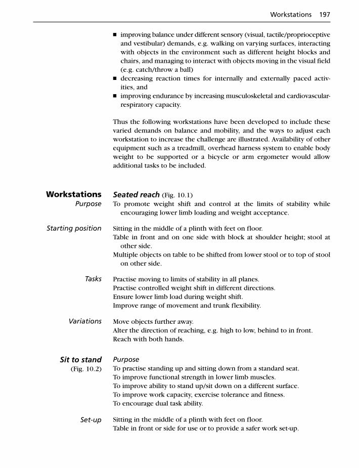
■ improving balance under different sensory (visual, tactile/proprioceptiveand vestibular) demands, e.g. walking on varying surfaces, interactingwith objects in the environment such as different height blocks andchairs, and managing to interact with objects moving in the visual field(e.g. catch/throw a ball)
■ decreasing reaction times for internally and externally paced activ-ities, and
■ improving endurance by increasing musculoskeletal and cardiovascular-respiratory capacity.
Thus the following workstations have been developed to include thesevaried demands on balance and mobility, and the ways to adjust eachworkstation to increase the challenge are illustrated. Availability of otherequipment such as a treadmill, overhead harness system to enable bodyweight to be supported or a bicycle or arm ergometer would allow additional tasks to be included.
Seated reach (Fig. 10.1)To promote weight shift and control at the limits of stability while
encouraging lower limb loading and weight acceptance.
Sitting in the middle of a plinth with feet on floor.Table in front and on one side with block at shoulder height; stool at
other side.Multiple objects on table to be shifted from lower stool or to top of stool
on other side.
Practise moving to limits of stability in all planes.Practise controlled weight shift in different directions.Ensure lower limb load during weight shift.Improve range of movement and trunk flexibility.
Move objects further away.Alter the direction of reaching, e.g. high to low, behind to in front.Reach with both hands.
PurposeTo practise standing up and sitting down from a standard seat.To improve functional strength in lower limb muscles.To improve ability to stand up/sit down on a different surface.To improve work capacity, exercise tolerance and fitness.To encourage dual task ability.
Sitting in the middle of a plinth with feet on floor.Table in front or side for use or to provide a safer work set-up.
Workstations 197
WorkstationsPurpose
Starting position
Tasks
Variations
Sit to stand (Fig. 10.2)
Set-up

198 Retraining balance using task-focused workstations
ab
c d
e
Figure 10.1a–eExamples of the progression of exercises that may be utilized in a seated reach workstation. (a) Forward reach using both arms to gain trunk extension. (b) Lateral reach to encourageweight shift and trunk mobility. (c) Reach to floor for trunk control and flexibility. (d) Reachand participate in problem-solving task. (e) Use of less stable base to challenge balance control during reach.

Change the height of the bed/chair (lower to make it harder).Stand/sit without using hands for support.Stand/sit while holding objects, e.g. cup of water on a tray; objects of
varying size/weights to add the demand of a second task.Stand/sit with softer surface (e.g. mat) under feet.Stand from a chair/table arrangement.Stand/sit with one leg in front of the other.Stand/step towards object on one side.
Workstations 199
a b c
d
e f
Figure 10.2a–fExamples of the progression of exercises that may be utilized in a sit-to-stand workstation.
Variations

200 Retraining balance using task-focused workstations
a b
c d
e
Figure 10.3a–eExamples of the
progression ofexercises that may
be utilized in areach/step and reach
workstation.

PurposeTo improve the ability to reach and shift objects in standing and while
stepping and reaching.To promote weight shift in all planes and towards the limit of stability.To promote mediolateral stability.To improve functional strength (eccentric/concentric control) of the
lower limbs.To improve ability to manage multiple tasks.
Standing at a table/bench with a lower plinth behind for safety.Trolley with shelving to one side, table/stool 1–2 metres away on other side.Firm/mat as floor surface related to ability level.
Move objects further away, higher, lower.Alter the direction of reaching, e.g. high to low, behind to in front.Reach with both hands.Shift light/heavy objects.Add a softer surface.
PurposeTo increase functional strength, concentric and eccentric control of the
lower limb extensors, abductors and adductors.To improve mediolateral stability by stepping up and over blocks of vary-
ing heights and walking sideways over the blocks.To improve ability to carry out a dual task.
Two small blocks between parallel bars.
Walk forward, sideways and backwards with care.Introduce a variety of blocks with added height challenges.Add a soft foam square between the blocks.Carry an object while walking forward/sideways and backwards.
Workstations 201
Set-up
Variations
Block work (Fig. 10.4)
Set-up
Variations
ba
Figure 10.4a–dExamples of the progression of exercises that may be utilized in a block work workstation.
Reach/step andreach in standing
(Fig. 10.3)

PurposeTo improve functional strength, added flexibility and controlled force
generation of the plantarflexors by lowering heels over stair edge,then carrying out a controlled toe-stand.
To promote concentric/eccentric control of the lower limb extensors with reciprocal stair walking.
Small set of stairs with rails.
202 Retraining balance using task-focused workstations
c
d
a b c
Figure 10.5a–cExamples of the progression of exercises that may be utilized in a stairs workstation.
Set-up
Figure 10.4a–dContinued.
Stairs (Fig. 10.5)

Number of times the stairs are practised with use of rails.Number of heel drops/toe-stands.Walking slowly up and down stairs without use of rails.Walking at a comfortable speed up and down stairs without use of rails.
PurposeStepping out of the square to promote weight shift and an efficient step-
ping response.To promote the ability to efficiently plan a step.
Square marked on floor with tape.Markers set around the square at about 60% of the client’s maximum
ability to step.
Stepping to targets within a set time.Use of targets with random number sequence to introduce a second com-
ponent to this task.
Workstations 203
Variations
a b
c
Figure 10.6a–cExamples of the
progression ofexercises that may beutilized in a stepping
out of the squareworkstation.
Stepping out ofthe square
(Fig. 10.6)
Set-up
Variations

PurposeTo promote efficient use of ankle and hip strategies in response to dis-
placement backwards.
Use of a blank wall to lean on when moving backwards.Feet set out from wall.Table in front of patient.
Leaning on wall, reach to table and stand up without pulling on the table.Stand out from the wall and move hips backwards to touch the wall, then
reach forwards/pick up toes and stand up.Start out from wall and lower hips towards wall but pull up toes and
stand up again without touching wall.
204 Retraining balance using task-focused workstations
Variations
Set-up
a b
c
Figure 10.7a–cExamples of the
progression ofexercises that may be
utilized in an ankleand hip strategy
workstation.
Ankle/hip strategy training
(Fig. 10.7)

PurposeTo promote efficient walking over a variety of surfaces and environmental
demands.
Set out blocks, rubber mat/foam square, within parallel bars for lower level client.
Set out blocks, rubber mat/foam square, small tilt board within parallel bars for more able client.
Set up equipment in open space for higher level client.
Walk over different surfaces (stable/unstable).Turning around an obstacle.Search for a series of objects, e.g. find a card series in sequence while
executing a walking circuit, Walk and tap a balloon/while catching/ throwing a ball.
Workstations 205
Walking (Fig. 10.8)
a bc
d e f
Figure 10.8a–fExamples of the progression of exercises that may be utilized in a walking workstation.
Set-up
Variations

206 Retraining balance using task-focused workstations
Summary ■ Physiotherapists play an important role in the field ofbalance, mobility and falls prevention.
■ An effective assessment identifies causal factors leading toimpaired mobility and balance and identifies factors whichmay be amenable to treatment.
■ There are multiple systems which may be involved in balancedysfunction. By addressing impairments in variedenvironments and task-oriented activities we may enableelders to maintain or regain the ability to safely mobilize.
■ Workstations are an efficacious mode of deliveringmultidimensional task-focused programmes. These can bedelivered through individual or small group situations.
References
Ada L, Mackay F, Heard R, Adams R 1999 Stroke rehabilitation: does the therapy area provide aphysical challenge? Australian Journal of Physio-therapy 45:33–38
Brauer S, Woollacott M, Shumway-Cook A 2001 The interacting effect of cognitive demand andrecovery of postural stability in balance impairedelderly. Journal of Gerontology Medical Science56(8):M489–M496
Buchner D M, Cress M E, de Lateur B J et al 1997 The effect of strength and endurance training on gait, balance, fall risk, and health service use incommunity-living older adults. Journal of Gerontol-ogy Medical Science 52(4):M218–M224
Campbell A J, Robertson M C, Gardner M M et al 1997Randomised controlled trial of a general practiceprogramme of home based exercise to preventfalls in elderly women. British Medical Journal315:1065–1069
Carr J H, Shepherd R B 1987 A motor relearning programme for stroke, 2nd edn. Butterworth-Heinemann, Oxford
Carr J H, Shepherd R B 1998 Neurological rehabilita-tion:optimizing motor performance. Butterworth-Heinemann, Oxford
Carr J H, Shepherd R B 2000 A motor learning modelfor rehabilitation. In: Carr J H, Shepherd R B (eds) Movement science foundations for physicaltherapy in rehabilitation. PPO-ED, Austin, TX,p 33–110
Carr J H, Shepherd R B 2003 Stroke rehabilitation:guidelines for exercise and training to optimizemotor skill. Butterworth-Heinemann, Oxford
Chandler J M, Duncan P W, Kochersberger G,Studenski S 1998 Is lower extremity strength gainassociated with improvement in physical perform-ance and disability in frail, community-dwellingelders? Archives of Physical Medicine andRehabilitation 79:24–30
Dean C, Shepherd R 1997 Task-related trainingimproves performance of seated reaching tasksafter stroke: a randomized controlled trial. Stroke28:722–728
Dean C M, Richards C L, Malouin F 2000 Task-relatedtraining improves performance of locomotor tasksin chronic stroke: a randomised controlled pilottrail. Journal of the American Geriatrics Society81(4):409–417
Gentile A M 2000 Skill acquisition: action,movement and neuromotor processes. In:Carr J H,Shepherd R B (eds) Movement science foundationsfor physical therapy in rehabilitation. PPO-ED,Austin, TX, p 111–187
Hill K, Kerse N, Lentini F et al 2002 Falls: a compari-son of trends in community, hospital and mortalitydata in older Australians. Aging 14:18–27
Jette A M, Harris B A, Sleeper L 1996 A home-based exercise programme for non-disabled olderadults. Journal of the American Geriatrics Society44:644–649

References 207
Johansson B B 2000 Brain plasticity and stroke rehabil-itation. The Willis lecture. Stroke 31(1):223–230
Lipsitz L A, Jonsson P V, Kelley M M, Koestner J S 1991 Causes and correlates of recurrent falls inambulatory frail elderly. Journal of Gerontology46:114–122
Liu B A, Topper A K, Reeves R A, Gryfe C, Maki B E 1995Falls among older people – relationship to medica-tion use and orthostatic hypotension. Journal ofthe American Geriatrics Society 43(10):1141–1145
Lord S, Castell S 1994 Physical activity program forolder persons: effect on balance, strength, neuro-muscular control and balance reaction time.Archives of Physical Medicine and Rehabilitation75:648–652
Lord S R, Clark R D, Webster I W 1991 Physiologicalfactors associated with falls in an elderly popula-tion. Journal of the American Geriatrics Society39:1194–1200
Lord S R, Caplan G A, Ward J A 1993 Balance, reactiontime and muscle strength in exercising and non-exercising older women: a pilot study. Archives of Physical Medicine and Rehabilitation 74:837–839
Lord S R, Ward J A, Williams P, Strudwick M 1995 Theeffect of a 12-month exercise trial on balance,strength and falls in older women. Journal of theAmerican Geriatrics Society 43:1198–1206
Lord S R, Ward J A, Williams P 1996 Exercise effect ondynamic stability in older women: a randomisedcontrolled trial. Archives of Physical Medicine andRehabilitation 77:232–236
Low Choy N, Isles R, Barker R, Nitz J 2003 The effi-cacy of a work-station intervention program toimprove functional ability and flexibility in ageingclients with cerebral palsy. Journal of Disabilityand Rehabilitation 25:1201–1207
Lundin-Olsson L, Nyberg L, Gustafson Y 1997 ‘Stopswalking when talking’ as a predictor of falls in elderly people. Lancet 349:617
Lundin-Olsson L, Nyberg L, Gustafson Y 1998Attention, frailty and falls: The effect of a manualtask on basic mobility. Journal of the AmericanGeriatrics Society 46:758–761
Magill R A 2001 Motor learning concepts and appli-cations, 6th edn. McGraw-Hill, New York
McMurdo M E, Johnstone R 1995 A randomized con-trolled trial of a home exercise programme for eld-erly people with poor mobility. Age and Ageing24:425–438
Nitz J C, Low Choy N L 2004 The efficacy of a specific balance-strategy training programme forpreventing falls among older people: a pilot randomised controlled trial. Age and Ageing33:52–58
Nitz J C, Thompson K 2003 ‘Stops walking to talk’:A simple measure of predicting falls in the frail elderly. Australasian Journal on Ageing 22(2):97–99
Nudo R J, Friel K M 1999 Cortical plasticity afterstroke: implications for rehabilitation. ReviewNeurology 9:713–717
Nudo R J, Plautz E J, Frost S B 2001 Role of adaptiveplasticity in recovery of function after damage tomotor cortex. Muscle and Nerve 8:1000–1019
Richards C L, Malouin F, Wood-Dauphine S 1993 Task-specific physical therapy for optimization ofgait recovery in acute stroke patients. Archives of Physical Medicine and Rehabilitation74:612–620
Rooks D S, Kiel D P, Parsons C, Hayes W C 1997 Self-paced resistance training and walking exercise in community-dwelling older adults: effects onneuromotor performance. Journal of GerontologyMedical Science 52:M161–M168
Rubenstein L Z, Robbins A S, Schulman B L et al 1988Falls and instability in the elderly. Journal of theAmerican Geriatrics Society 44:273–278
Sauvage L R, Myklebust B M, Crow-Pan J 1992 A clin-ical trial of strengthening and aerobic exercise toimprove gait and balance in elderly male nursinghome residents. American Journal of PhysicalMedicine and Rehabilitation 71:333–342
Shepherd R 2001 Exercise and training to optimizefunctional motor performance in stroke: drivingneural reorganization? Neural Plasticity 8:121–129
Sherrington C, Lord S R 1997 Home exercise toimprove strength and walking velocity after hipfracture: a randomized controlled trial. Archives ofPhysical Medicine and Rehabilitation 78:208–212
Shumway-Cook A, Woollacott M 2000 Attentionaldemands and postural control:the effects of sensorycontext. Journal of Gerontology Medical Science55A:M10–M16
Shumway-Cook A, Woollacott M 2001 Motor control:theory and practical applications. Williams &Wilkins, Baltimore
Shumway-Cook A, Gruber W, Baldwin M, Liao S 1997aThe effect of multi-dimensional exercises on bal-ance, mobility and fall risk in community-dwellingolder adults. Physical Therapy 77(1):46–57

208 Retraining balance using task-focused workstations
Shumway-Cook A, Woollacott M, Kerns K A, Baldwin M1997b The effects of two types of cognitive tasks onpostural stability in older adults with and withouta history of falls. Journal of Gerontology MedicalScience 52A:M232–M240
Skelton D A, Young A, Grieg C A, Malbut K E 1995Effects of resistance training on strength, power
and selected functional abilities of women aged75 and older. Journal of the American GeriatricsSociety 43:1081–1087
Tinetti M E, Speechley M, Ginter S F 1988 Risk factorsfor falls among elderly persons living in the community. New England Journal of Medicine319:1701–1707

Exercise prescription in residential aged care facilities
Susan R. Hourigan
209
11
This chapter aims to:
One of the main roles of the physiotherapist working in residential agedcare is to prescribe exercise to residents. The physiotherapist individuallyassesses the need for therapy amongst the residents and through clinicalreasoning develops personalized therapy plans. Other staff such as physio-therapy, nursing or activities assistants, who are under the physiother-apist’s direction, often implement the exercise programmes. The ongoingrole of the physiotherapist, in regard to exercise programming, includesregular re-evaluation to determine the effectiveness of the given regimen.It is important that objective measurements are used so that outcome meas-ures can be recorded for accountability and documentation requirements.
With employees other than physiotherapists carrying out the prescribedexercise programmes to varying degrees of effectiveness this system isclearly less than ideal, yet it does enable a broader access to limited physio-therapy assessment and treatment than would otherwise be available.
■ outline the benefits of exercise in the residential aged carepopulation
■ reiterate the importance of good clinical reasoning by means ofthorough and appropriate assessment, treatment, exerciseprescription and re-evaluation
■ overview exercise prescription and how it applies to physio-therapists working with residents living in RACFs
■ show how exercise programming and exercise protocols may beintegrated into the nursing and activities systems of facilities
■ suggest issues that will apply to both mobile and immobileresidents (although the chapter is aimed more specificallytowards those residents who are mobile and have a higher levelof physical functioning than the extremely frail or fullydependent RACF resident)
■ relate principles of exercise prescription to the olderpopulation, various issues encountered in RACFs, theoreticalbackground and treatment ideas for this special group.
Introduction

In Australia at least, the lack of physiotherapy services stems from gov-ernment funding constraints within the aged care sector, the appropri-ation of available funding to other services by management and a genuineworkforce shortage of physiotherapists.
Physiotherapists working in aged care need to have a solid understand-ing of exercise physiology, exercise prescription and clinical reasoning inorder to make judgements regarding best practice in relation to exerciseprogramming. Physiotherapists understand the risks and benefits of vari-ous modalities and doses of exercise in relation to specific pathology and health-related goals. They are the best equipped professionals toincorporate exercise into the health care of residents within RACFs.
The value of exercise in geriatric health care has been extensively ques-tioned. Some reviews have positively hailed exercise as a means of pre-venting and treating diseases associated with old age whilst others havequestioned whether there are any benefits to exercise in late-life andwhether exercise prescription has been over-sold (Fiatarone Singh 2002).It seems critical to point out that there are numerous studies which rep-resent benefits associated with activity and health status in older adults.It is equally as important to highlight that as there are many different people,diseases and health issues;there are as many different modalities of exercise,methods of programming, doses of activity and lengths of programmes.The literature is scattered with both positive and negative research resultspertaining to the usefulness of different exercise regimens.
Advancing age is associated with a decline in both functional abilitiesand fitness component measures. Changes to the cardiovascular system,respiratory system, musculoskeletal system, central and peripheral nervoussystems, posture and gait in the elderly population have been well docu-mented and were summarized in Chapter 1 of this text (McArdle et al 1996,Shephard 1997, Shumway-Cook & Woollacott 1995, Trew & Everett 1997).The ability to exercise and efficiency of movement inevitably decreaseswith the passage of years. Effects on lifestyle and quality of life of this reduction in movement capabilities and functional activities may be cata-strophic. The maintenance of physical function and mobility in the elderlyis of great importance. Perhaps the most important issue related to qualityof life is that of independent function and ambulation – it is the key factorwhich enables residents to make their own decisions and to go where theyplease. This self-determination enables them to bring themselves to activ-ities of choice or remove themselves from unwanted places, people or situ-ations. It is often the key to maintaining dignity, privacy and happiness.
It is clear that a number of age-induced changes can be prevented,slowed down or even reversed with appropriate activity (Trew & Everett1997). Some research has suggested that half of the loss of functionaldecline associated with ageing is a result of disuse, which is easily modi-fiable with regular exercise (Pardini 1984). It is generally accepted thatregular exercise enhances physical adaptability, mobility and coordin-ation, all of which contribute to an enriched life in old age (Rissel 1987).
210 Exercise prescription in residential aged care facilities

Evidence does exist which supports the notion that exercise can beused as therapy to:
■ minimize the physiological changes associated with ageing■ contribute to psychological health and wellbeing■ increase longevity and decrease the risks of many common chronic
diseases, e.g. diabetes and cardiac disease■ primarily treat, or adjunctively assist certain chronic diseases (espe-
cially in regard to ‘disuse’ syndromes)■ counteract specific side effects of standard medical care (iatrogenic
complications)■ prevent and treat disability (Fiatarone Singh 2002, Pardini 1984, Rissel
1987, Trew & Everett 1997).
The benefits of exercise are many, and include but are not limited to:
■ improved strength ■ decreased falls risk■ increased range of motion ■ increased fun and laughter■ improved flexibility ■ stress release/endorphin release■ improved functional mobility ■ improved immunity■ improved cardiac status ■ improved skin integrity■ improved respiratory status ■ improved posture■ better sleep ■ improved gait■ improved gastrointestinal ■ improved efficiency of movement
function ■ increased exercise tolerance■ increased self-esteem ■ decreased fear of falling■ improved tone management ■ increased endurance■ improved postural control ■ improved quality of life■ improved social interaction ■ improved dexterity.
Several other chapters in this text relate specifically to diseases or condi-tions experienced in the age strata residing in RACFs. We have illustratedthe likely benefits of exercise throughout this text in relation to manysyndromes or diseases. Exercise therapy may be utilized in the treatmentof the following conditions, including but not limited to:
■ frailty ■ cancer■ mobility impairment ■ ischaemic heart disease■ functional disability ■ cardiomyopathy■ falls ■ congestive cardiac failure■ osteoporosis ■ respiratory disease■ palliative care ■ chronic renal failure■ diabetes ■ insomnia■ incontinence ■ hypertension■ arthritis ■ peripheral vascular disease■ gout ■ venous stasis■ stroke ■ obesity
Introduction 211

■ constipation ■ dementia■ depression ■ neurological disorders and diseases.
Changes associated with the above conditions occur in physical indicessuch as body weight, strength, cartilage integrity, blood pressure, anxiety,muscle and bone mass, and balance. Once problems are identified we areable to recommend exercise which assists in counteracting the givenchanges, for example prescribing resistance or aerobic training for suffer-ers of osteoarthritis, combined with balance training for those with estab-lished osteoporosis.
It is imperative to conduct a full assessment of the resident in order todecide what, if any, will be the treatment ideals and to determine theirgoals. Chapters 2, 8 and 9 deal in more detail with various aspects of res-ident assessment. A thorough assessment will allow the therapist to pre-scribe a relevant exercise programme in order to improve or maintain theresident’s mobility, independent function and quality of life, for as long aspossible.
A good understanding of different disease processes is necessary inorder to modify exercise given certain considerations. Education at theundergraduate physiotherapy level provides for students to learn how toestablish frames and principles in which to base further knowledge andexperience. It is not within the scope of this chapter to examine condi-tions separately and state the given rationale for different exercises; it isexpected that practising clinicians will be able to do this with prior gainedknowledge or reading of other material as self-directed learning. However,the principles of exercise prescription as related to residents within RACFswill be addressed in the next section. Firstly, we examine the ability of theolder person to train and benefit from exercise.
Traditionally, the view has been held that older individuals were not ableto improve their strength and endurance capacity to the same extent asyounger people. Reasons provided for this ‘blunted’ trainability wereattributed to a decline in neuromuscular function and an age-relatedimpairment in the cellular capability for protein synthesis and chemical reg-ulation. However, significant research in the past two decades has enableda modified version of the traditional view to be made.
‘When a healthy person, young or old is given an appropriate trainingstimulus, large and rapid improvements occur in physiologic function,often at a rate and magnitude that is independent of the person’s age’(McArdle et al 1997, p 643). Regardless of age, regular physical activity pro-duces measurable physiological improvements. The magnitude of theseimprovements depends on many factors, including initial fitness status,medical history, age, and type and amount of training (McArdle et al 1996).
Trainability studies in the elderly concentrating on strength developmentreveal findings clearly indicating an impressive plasticity in physiological,
212 Exercise prescription in residential aged care facilities
Trainability in the aged population

structural, and performance characteristics well into the ninth decadeof life (Fiatarone et al 1990). Healthy older adults show no negativemetabolic or hormonal responses or any adaptive problems to regularexercise that would contraindicate participation in a standard trainingprogramme.
Exercise prescription principles in the mobile elderly 213
Exercise prescriptionprinciples in
the mobile elderly
Exercise prescription is the process whereby a person’s recommendedregimen of physical activity is designed in a systematic and individualizedmanner (American College of Sports Medicine 1991). In all cases, carefulconsideration is given to the resident’s health history, risk factor profile,behavioural characteristics, and personal goals and preferences. Specificpurposes of an exercise prescription will vary according to needs, inter-ests, health status and background. Generally the aim is to enhance phys-ical fitness (maintain or improve functional abilities), promote health (byreducing risk factors and instilling confidence), and ensure safety duringparticipation. Clearly these aims will vary depending on the individualand an exercise programme should reflect the specific outcomes that aresought by a particular resident and the involved physiotherapist. Import-antly exercise prescription is as much an art as it is a science. The sciencebehind exercise programming needs to be applied flexibly in order to reachthe desired outcome of increasing a resident’s habitual physical activity.Behaviours, likes, interests and motivation are therefore key considerationsin the implementation of a successful programme.
The exercise programme designed for the mobile resident will be anintegration of all of the principles we have discussed within the dimensionsof not only this chapter but also Chapter 10 relating to balance training.The ‘whole’ (the programme) will be less than the sum of its parts suchthat exercises prescribed should target multidimensional factors within asimplistic task-oriented focus. For example, if a therapist wishes to trainstrength, balance, vestibular function, flexibility and praxis because a resi-dent suffers from recurrent falls, osteoarthritis and has had previous neuro-logical insult they may wisely choose a sit-to-stand exercise. This is atask-oriented approach which maximizes integration; it uses the principleof specificity to optimize independence and functional activity whilst train-ing many different subsystems all important for the resident’s outcome.Some residents will be capable of independent practice of task-focusedexercise. Where possible the potential for self-management of chronic disease or prevention of the progressive effects of ageing should be identi-fied and encouraged in residents. Independence will be rewarded by anincrease in the feeling of self-worth concerning physical ability. In someinstances residents might be recruited as motivators and leaders of smallexercise groups. They should not be encumbered with the safety ofothers;this should be a staff responsibility. These residents may be able toassist, however, through organizing the attendance of other members atthe exercise venue by providing reminders or by physically accompanying

participants to the venue (this is particularly useful if poor vision pre-cludes safe passage of another resident to the exercise session).
In addition to the maintenance of safety, there are five principles thatare the basis of exercise prescription and must be considered when writingexercise programmes for residents.
1. Principle of specificity – the programme must be specific to achievedesired outcomes – ‘it’s no good doing weight training to increaseone’s flexibility’.
2. Principle of overload – to effect change the training stimulus must exceedthe demands placed on an individual by tasks normally undertaken. Theremust be an additional functional demand to initiate adaptive change.
3. Principle of reversibility – once a programme is discontinued positivegains will be lost. As the body adapts to increased demands, it also adaptsto decreased demands (this is the basis for the negative aspects associ-ated with ‘disuse’ atrophy).
4. Principle of initial values – depending on starting levels some peoplewith low levels of ability will have a greater capacity for percentageimprovement than other residents who begin with average or abovemeasures.
5. Principle of diminishing returns – each person has an individual bio-logical ceiling that determines the extent of a possible training effect.As this ceiling is approached, physical gains will slow and eventuallyplateau. Some residents will have greater scope to improve than others.
It is useful to remember the principles of exercise prescription whenwriting an holistic programme which targets all systems. Perhaps the easiestmethod of remembering what needs to be defined within a programmeis the ‘FITT’ principle of exercise prescription:
F: FrequencyI: IntensityT:TypeT:Time
The frequency parameter relates to the question ‘how often’, i.e. howmany repetitions, how many times per day or week? Intensity relates to‘how hard’, i.e. what target heart rate is required, what level of perceivedexertion and what speed or weight? The type points to which exercise isrequired, ‘what type?’ meaning aerobic training like walking or swim-ming, resistance training like hand weights or Theraband™, or flexibilitywork with stretching or yoga. The time of exercise relates to the question‘how long?’, i.e. a 5 minute walk, 10 second stretch, 5 second hold.
Exercise programmes which are developed for residents must be basedon good clinical reasoning. Once the assessment of the resident has beenundertaken it is advisable to develop a problem list and comment on the
214 Exercise prescription in residential aged care facilities
Individualizedprogrammes

aims and goals of the therapy and exercise to be completed. This is achievedby prioritizing the importance of different objectives. Finally, the physio-therapist should state which outcome measures will be utilized to object-ively review the programme. See Chapters 2, 8 and 9 for information onphysiotherapeutic assessment, and Appendix 2.
A balanced exercise programme in aged care may include the following:
■ cardiorespiratory endurance training – aerobic conditioning■ flexibility training■ resistance training – muscle strength, power and endurance (repe-
tition over time)■ balance and postural control training.
This encompasses all activities which use large muscle groups over a sus-tained duration and is rhythmic and aerobic in nature (e.g. walking, swim-ming and cycling). The intensity generally equates to 55–90% of maximumheart rate, although this may be lower in a group such as those in RACFsdue to the sedentary status and low fitness levels often found in the eld-erly population. The duration of activity required ranges from anywherebetween 15 and 60 minutes of continuous or discontinuous aerobic activity.Again, even 5 minutes may have considerable health and functional bene-fits amongst residents in RACFs. Usually the activity will need to take place3 to 5 days each week. In many cases conditioning effects, most notedover the first 6–8 weeks, will mean the exercise should be progressed bymanipulating duration or intensity, or both. In the elderly, it is extremelyimportant to individualize exercise prescription. In order to modify pro-grammes correctly it is important to do a risk factor assessment and haveknowledge of any pre-existing medical conditions.
Perhaps the most difficult part of exercise prescription is to determineand apply the appropriate intensity. This requires individualization and moni-toring to ensure safety and the correct level of exertion. Usually exerciseintensity is prescribed as a percentage of functional capacity and measuredusing heart rate (HR), rates of perceived exertion (RPE), METs, or estimatedenergy expenditure (VO2). One MET is equivalent to an oxygen uptake(or VO2) of 3.5 ml/kg/min (this represents the approximate rate of oxygenconsumption of a seated individual at rest). It is conventional in clinicalexercise testing to express VO2max in METs. Typically healthy young adultsmay be prescribed exercise at a level which equates to 60–70% of theirfunctional capacity. Within RACFs, however, where many residents have a lower functional capacity, training may be initiated at a level of about40–60% of functional capacity.
One of the easiest ways to determine exercise intensity for the olderperson is through the ‘talk’ test. As the name implies, a person should beable to carry on light conversation whilst exercising to ensure they are not
Exercise intensity 215
Cardio-respiratory
training
Exercise intensity

exercising too hard. Utilizing this easy strategy will ensure a resident isexercising at a light to moderate intensity and staying within a safe levelof exertion. Additionally it may also train dual-tasking by giving a cogni-tive demand through conversation.
Another relatively simple method to utilize that corresponds highly towhat is actually going on in the body is to measure intensity levels usingBorg’s Rating of Perceived Exertion Scale (RPE) (Borg 1982) (Table 11.1 –the category-ratio RPE scale (in the right hand column) is a newer adap-tation of the RPE scale which equates the levels to a 0–10 scale; eitherscale can be used in practice but both have been included for the reader’sinformation). By using and becoming familiar with the scale, some resi-dents will be able to keep themselves within the desired training levelsprescribed by their physiotherapist. This is a particularly useful scale asthere is a strong correlation between maximal oxygen uptake percent-ages, heart rate reserve percentages, minute ventilation, and blood lactatelevels with RPE. This may be even more important and useful with resi-dents taking medications that restrict heart rate responses to exercise,which applies to many residents living within RACFs.
Numerous clinical studies have shown that the category RPE scale is areproducible measure of exertion within a wide variety of individualsregardless of age, gender or cultural origin. After some training using thisscale, it is possible to get residents to keep themselves around differentlevels of perceived exertion during exercise. The range of 10–14 (Light toSomewhat Hard) equates to a usual light to moderate training load (50–75%of heart rate range) and enables the therapist to have control over exerciseintensity due to approximation of RPEs with other exercise intensitymeasures of cardiorespiratory and metabolic variables mentioned previ-ously, such as METs, VO2, heart rate and ventilation.
216 Exercise prescription in residential aged care facilities
Category RPE Scale Category-Ratio RPE Scale
6 0 Nothing at all7 Very, Very Light 0.5 Very, Very Weak8 1 Very Weak9 Very Light 2 Weak
10 3 Moderate11 Fairly Light 4 Somewhat Strong12 5 Strong13 Somewhat Hard 614 7 Very Strong15 Hard 816 917 Very Hard 10 Very, Very Strong18 Maximal19 Very, Very Hard20
Table 11.1RPE Scales
(Borg 1982)

Another method of prescribing exercise intensity relies on heart ratemeasurements and target heart rate ranges as a percentage of maximal heartrate (MHR). MHR can be estimated using the following equation:
MHR � 220 � age (in years)
This relies on the linear relationship between heart rate and exerciseintensity; 70–85% of maximal heart rate is equal to about 60–80% offunctional capacity. As workload increases so does heart rate so that thebody can pump extra oxygen around for the tissues which are in demand;this occurs to a point that is referred to as maximal heart rate where theheart rate cannot increase further. Therefore functional capacity levelsmay be estimated by percentages of MHR and exercise intensity can beprescribed as such.
Example: Mrs X (80 years old):
MHR � 220 � age (80) � 14070–85% of her MHR (140) � 98–119 bpm (beats per minute)
98–119 bpm � 60–80% of functional capacity
An indicator of 40–60% of her functional capacity would be a heart rateof approximately 60–98 bpm. As this may well be around the level of herresting heart rate value, we can see that many residents of advancing agewithin RACFs are already utilizing much of their functional capacity atrest. Often exercise demands do not need to be great to allow sufficienttraining stimulus in the elderly population.
The use of heart rate measurements and percentages is a moreinvolved method of prescribing exercise intensity and may be seen assuperfluous and unnecessary within RACFs. It is important to note thattarget heart rate range is a guideline only and professionals must usetheir decision-making skills to assess how an individual is responding toexercise with consideration to safety and comfort. Some medicationsalter heart rate responses to exercise, so it is important as noted previ-ously to assess what medications a resident is on and how this will affectthem physiologically.
There are many signs and symptoms physiotherapists need to monitorduring exercise training to ensure safety and effectiveness. A number ofmeasurements can also be taken to provide feedback on how a residentis coping physiologically and psychologically and whether exercise shouldbe continued. These include, but again are not limited to:
■ respiratory rate ■ angina■ oxygen saturation levels ■ gait pattern■ pain ■ perceived exertion■ skin colour ■ perceived fatigue■ claudication ■ movement patterns/biomechanics■ shortness of breath on ■ quality of movement
exertion (SOBOE) ■ safety.
Exercise intensity 217

The ability to function normally requires that all joints are able to movethrough an adequate range of motion. Often inflexibility will create difficul-ties with performance of activities of daily living in the elderly. Emphasisgenerally should be on flexibility training in the upper and lower trunk,neck, hip and ankle regions. Importantly, dorsiflexion range in the anklejoint is of particular importance for mobility, more specifically in the per-formance of a sit to stand or in ascending or descending stairs (Bohannonet al 1991, James & Parker 1989).
It is important to perform stretches slowly with a gradual progressionto greater ranges of motion. A slow dynamic movement may be followed bya static stretch that is maintained for 5–10 seconds. The reader is remindedthat a 20 second stretch, although sometimes prescribed in the youngerpopulation, is not advisable for older people owing to the compressionof arterial blood supply that occurs when the fascial planes separatingmuscles and compartments are compressed during the stretch process. Thiscompression may reduce blood supply as suggested and cause tissue injury.Thus, only hold tissues in a lengthened position for around 5 seconds at amaximum and consider the use of other techniques such as slow reciprocalmovements to extremes of range. Other methods may achieve the bestresults in range gain. Proprioceptive neuromuscular facilitation (PNF) techniques such as hold–relax or contract–relax (Adler et al 1993) areadditional active methods of increasing range of movement that are veryeffective for cognitively sound residents.
Ensure the whole programme targets the given aims and assessed prob-lems. Think through what each exercise achieves. What systems does ituse and how could it be modified to adopt a more functional task?
1. Choose low impact activities with basic movement patterns (e.g.walking).
2. Choose enjoyable and non-competitive activities (e.g. dancing orswimming).
3. Do not forget a gentle warm-up and warm-down when needed.4. Include flexibility exercises – teach stretches – do not let residents
bounce into their stretch.5. Include resistance exercises for major muscle groups.6. Start at low levels and progress slowly – this way the body has time
to adapt. Take it easy!7. Progress gradually by increasing time, frequency and intensity.8. Older individuals only need light to moderate intensity exercise – do
not overdo it.9. Concentrate on breathing and postural control.
10. Encourage regular drinks of water, especially in hot weather.11. Be careful of extremes in weather. Avoid exercising in very hot or
very cold temperatures.
218 Exercise prescription in residential aged care facilities
Flexibility
Programmegoals

12. Use positive messages around the facility and get staff to encourageresidents as much as possible.
13. Consider using music – it usually enhances enjoyment when exercising.
Never has there been such an emphasis on thorough written records asnow. The demand for what seems at times a copious amount of paper-work reflects several current factors. At once there is a consumer call forappropriate and excellent services, a government demand for account-ability and proof of funding usages and a society becoming more litigiousin nature. No doubt, record keeping is of high importance;it allows othersclear access to the clinical reasoning and steps which have taken place inthe minds and hands of physiotherapists when formulating programmes.It also allows for more comprehensive reviews by providing baseline dataand allows for evidence-based research to be conducted in some instances.Apart from external pressures the demand illustrates the internal desireamongst physiotherapists to advance their profession and to demonstratetheir autonomy and worth.
The exercise programme (or therapy care plan) itself needs to includethe following information:
■ resident details (name, date of birth, major diagnosis)■ date programme written and initiated■ programme aims – problem list and/or desired outcomes■ assistance required by the resident, e.g. two staff assist/daily■ importantly, details of the exercises, including:
– frequency – (how often) how many repetitions, times per day orweek
– intensity – (how hard) target heart rate, speed, weight– type – (what type) resistance, flexibility, aerobic– time – (how long), i.e. 5 minute walk– date of review/s and details of such (outcome measures, changes,
comments on progress)– appropriate pictures or diagrams which illustrate the exercises –
stick figures may be useful.
This therapy care plan must be included in the total resident care planand be reviewed regularly by the physiotherapist. The question of howfrequently a care plan must be reviewed varies widely depending on thestatus of the resident; this should be determined by the physiotherapist.Physiotherapists should use their professional skills to determine the necessary evaluation which should take place and how often this shouldoccur; this will depend mainly on the resident’s health status and anychanges to this.
Demands for review times are often placed upon physiotherapists byexternal bodies or persons; this may vary with different management, facil-ities or agencies. A brief and recent survey of Australian physiotherapists
Documentation 219
Documentation

determined that generally review periods vary tremendously and are com-pleted anywhere between every 2 and 12 months. The regularity requiredfor reviews has been a contentious issue recently within Australia as dif-ferent stakeholders lay their claims; certainly a consensus has not beendefinitively reached as yet. Best practice would stipulate that decisionsregarding appropriate timeframes for review of exercise programmes arepart of the clinical decision-making process skills needed by the physio-therapist.
Staff involved in the implementation of the therapy care plan will requireeducation and training. Guidance will be needed to ensure the safety ofboth residents and staff. Good handling skills must be taught and empha-sized in order to ensure the effectiveness of therapy programmes. Ensurein-service education sessions are conducted where necessary. Someappropriate topics will include: how best to assist residents with exer-cises; active and passive movements (these are not straightforward andrequire a moderate amount of skill to be implemented safely); massage;positioning; patient handling/manual handling; the benefits of exercise;falls prevention; and how to run group exercise sessions.
The job descriptions of all staff that assist with therapy programmesshould include the need for this to occur. Staff must be empowered toassist residents with exercise and be responsible for ensuring that it isdone. Exercise sessions and resultant resident achievements can becomethe source of a great deal of satisfaction and enjoyment for all concerned.Exercises can assist in the development of a positive, supportive, healthyand happy environment for residents and staff. In order for this to occurthere needs to be team enthusiasm, a fostering environment and the support of management in the programmes offered.
Often time will be an issue for nursing staff; usually it will be the lackthereof or the requirement for other jobs such as hygiene needs, meals or paperwork. It is important to emphasize the benefits of exercise andeducate staff regarding the needs of the residents in this regard. In orderto assist with the daily routine of nursing staff it may be wise to suggestwhen exercises should be conducted with the resident so the workloadis achievable. The programme will no doubt benefit from being writteninto the daily work routine, i.e. state which time of day it needs to occur(between 2:00 and 2:15 pm) or specify when during the hygiene routinethe exercises may be completed (e.g. 10 assisted arm lifts with underarmwashing in the shower in the evening). Involving evening staff in theexercise care plan will share the load more evenly and ultimately may suitsome residents better.
The use of documentation folders for the daily recording of exercisescompleted should be a requirement for all facilities. This enables thephysiotherapist to review what is being done and allows a feedbackmechanism for staff to communicate with the therapist.
220 Exercise prescription in residential aged care facilities
Other staffinvolvement/
education

It is important that staff approach exercise in a positive way. At anyage, many people need some encouragement and assistance to exercise,especially those who may not have done so for many years. Importantly,above all else it is the residents’ choice that matters most when decidingwhether to do exercises or not. Like people of any age they can feelunmotivated when it comes to activity. With encouragement and supportto participate they often feel much better for having done so. Successfuloutcomes in exercise programmes are often determined by the staffmember’s initial approach towards the older person. Good communica-tion, a positive approach and a general willingness to assist are thekey factors which gain the resident’s compliance and willingness tocooperate.
Time, fatigue and pain are all reasons stated by residents for not exercis-ing – amongst others. We shall look briefly at the myriad of excusesgiven for not wanting to participate in exercise and organized activitiesand provide some methods of counteracting the age-old avoidancestrategies.
■ Some residents feel they are beyond help. It may pay to write out a listof the positives and negatives of participation and compare the two;residents will then surely see that it will be worthwhile to fit dailyactivity into their lives.
■ Lack of time? If this is the concern perhaps the person could keep adiary for a week; they will no doubt be surprised at the amount of timespent in pursuits such as waiting for meals or watching television, andrevise the timetable.
■ Perhaps fatigue is a worrying factor? Poor sleep habits leave peoplephysically and mentally sluggish. If difficulty initiating or maintainingsleep is an issue then they should ensure elimination of daytime nap-ping, go to bed at the same time each night, gradually decrease andstop caffeine intake (coffee and tea), use the bed only for sleeping (no reading or television watching), and have a light meal a few hours before retiring. See the section on sleep in Chapter 4 for furtherinformation.
■ Chronic pain? This may be due to multiple medical causes such asosteoporosis, osteoarthritis or even fracture. Residents or staff shouldconsult with the doctor and/or physiotherapist for a thorough assess-ment. Often aches and pains can be reduced by appropriate exercise.Physical activity specific to some complaints (such as joint or boneconditions) can contribute to improved mobility and function, andimproved energy level and a sense of wellbeing (Minor & Lane 1996).
■ ‘Too old’! We are never too old to want to feel better and have animproved quality of life. Often the best motivation for older persons isthe knowledge that there is truth in the adage ‘move it or lose it’.
Behaviour modification 221
Behaviour modification

In order to illustrate the principles outlined in this chapter we haveincluded examples of some exercises commonly prescribed for residents.Related information is included to highlight the systems involved andbenefits to the resident. Numerous variations can be made to the basicexercises and are done so as to achieve slightly different responses or toprioritize particular aims. This list is by no means exhaustive and is onlymeant to illustrate some of the principles previously outlined.
Arm raises (Fig. 11.1)Arm raises can be combined with deep breathing exercise in most indi-viduals. This can be performed in lying, sitting or standing. Weights maybe added.
To improve upper limb strength, active range, thoracic cage expansion,trunk extension, upper limb function, upper limb strengthening.
Muscle strength, power and endurance.Flexibility and joint range training.Balance training.
222 Exercise prescription in residential aged care facilities
Examples ofexercises
1. Exercise:
a bFigure 11.1a & b
Arm raises.
Variations
Goal
Systems
2. Exercise:Variations
Goal
Systems
Leg extension (Fig. 11.2)Sitting, standing, lying, �weights.
To improve lower limb strength; assist in maintaining or improving func-tional mobility, balance.
Muscle strength, power and endurance.Flexibility and joint range training.

Foot and ankle exercises (Fig. 11.3)Sitting, lying, standing, �weights.
To maintain available and necessary range in the ankle joint, improve flexi-bility, circulation, integrate sensory systems and responses from the feet.Maintain or improve functional mobility.
Muscle strength, power and endurance.Flexibility and joint range training.Balance and postural control.
Prayer position to traffic control (policeman) position(Fig. 11.4)This may be completed combined with deep breathing exercises,�weights.
To maintain or improve upper limb activities, scapular retraction,posture training, respiratory exercise, thoracic cage expansion,relaxation.
Flexibility and joint range training.Respiratory system training/enhancement.Muscle strength, power and endurance.Balance and postural control.
Examples of exercises 223
a bFigure 11.2a & b
Leg extension.
3. Exercise:Variations
Goal
Systems
4. Exercise:
Variations
Goal
Systems

224 Exercise prescription in residential aged care facilities
a b
c d
e
Figure 11.3a–eFoot and ankle
exercises.

Trunk rotation (Fig. 11.5)Sitting, standing, in water (aquatic physiotherapy). On a bed, chair, ball.
To maintain flexibility within the trunk, to assist relaxation of tone, toimprove gait pattern, to allow movement for activities of daily living,improve sitting balance.
Flexibility and joint range training.Postural control, balance.Muscle strength, power and endurance.
Examples of exercises 225
a b
Figure 11.4a & bPrayer position to
traffic control(policeman) position.
a bFigure 11.5a & b
Trunk rotation.
Goal
Systems
6. Exercise:Variations
Goal
5. Exercise:Variations
Passive arm and leg exercises (Fig. 11.6)Position (sitting, lying), in water. Often these exercises are combinedwith massage to good effect. One or two persons assist.
To maintain joint range, flexibility, skin integrity, positioning, comfort. Tofacilitate requirements for hygiene assistance and other activities of dailyliving. To control tone and spasticity. To assist in the management of pain

and neuromechanosensitivity and allow smooth movement of neural tis-sues within other soft tissues. Relaxation.
Flexibility and joint range training.
226 Exercise prescription in residential aged care facilities
a b
Figure 11.6a & bPassive arm and leg exercises.
Figure 11.7Sit-to-stand practice.
Systems
7. Exercise:Variations
Goal
Systems
Sit-to-stand practice (Fig. 11.7)At a rail, in front of a mirror, in water, from a chair, bed, or ball.
To maintain or improve lower limb strength, functional mobility, motorplanning, balance, sensory integration.
Flexibility and joint range training.Balance and postural control.Muscle strength, power and endurance.Vestibular system.Cardiorespiratory endurance training.

Ball games (Fig. 11.8)Different sized, shaped or weighted balls (e.g. balloons, Theraballs™,beach balls, footballs). Sitting or standing. Throwing, catching, kicking.Combined with cognitive tasks. One-on-one or group exercise. Differenttextured or noisy balls.
Stimulation, enjoyment. Upper limb function, dexterity and shoulder ranging exercise. Coordination, motor planning, and balance training.Maintain or improve functional mobility, movement.
Flexibility and joint range training.Balance and postural control.Muscle strength, power and endurance.Cardiorespiratory endurance training.
Examples of exercises 227
8. Exercise:Variations
a bFigure 11.8a & b
Ball games.
Goal
Systems
9. Exercise:Variations
Goal
Systems
Walking (Fig. 11.9)Indoors/outdoors, obstacle courses, different levels and surfaces.�Weights. Individual or group training sessions.
To maintain or improve fitness and functional mobility. Enjoyment.Relaxation. Rehabilitation.
Cardiorespiratory endurance training.Balance and postural control.Muscle strength, power and endurance.

Reaching for objects on a wall (Fig. 11.10)High, low and wide placements. Add sit to stand before reaching. Combinewith deep breathing.
To maintain or improve functional mobility. Falls prevention. Upper limbfunction training.
Balance and postural control.Muscle strength, power and endurance.Flexibility and joint range training.Coordination.Cardiorespiratory training.
228 Exercise prescription in residential aged care facilities
a bFigure 11.9a & b
Walking.
Figure 11.10Reaching for objects
on a wall.
Systems
10. Exercise:
Goal
Variations

Sitting balance exercises (Fig. 11.11)Forward reaching in chair – ‘toe touches’/sitting bowls.
From a ball, bed or chair.
To maintain or improve sitting balance, flexibility, trunk movement.Sensory integration. Trunk and abdominal muscle exercise.
Balance and postural control.Muscle strength, power and endurance.Flexibility and joint range training.Coordination.Cardiorespiratory training.
Arm weights (Fig. 11.12)Theraband™, dumbbells, elastic tubing, cans, pulleys, sandbags. Sitting,lying, standing. Combined with walking.
Examples of exercises 229
11. Exercise:
Variations
Goal
a b
c d
Figure 11.11a–dSitting balance
exercises.
Systems
12. Exercise:Variations

To improve upper limb strength and function. Bone health.
Muscle strength, power and endurance.Balance and postural control.Flexibility and joint range training.Coordination.Cardiorespiratory training.
230 Exercise prescription in residential aged care facilities
a bFigure 11.12a & b
Arm weights.
a b c
Figure 11.13a–cNeck exercises/shoulder shrugs.
13. Exercise:Variations
Goal
Goal
Systems
Neck exercises/shoulder shrugs (Fig. 11.13)In water, on land. Standing, sitting or lying.
To maintain or improve head and neck posture and movement; facilitateimproved ergonomics and functioning;relieve postural tension, headaches.Relaxation.

Flexibility and joint range training.Balance and postural control.Muscle strength and endurance.Vestibular system training.
Hand exercises (Fig. 11.14)In water. Combined with wax bathing treatment.
To maintain hand dexterity and function. To maintain independence withmeals, crafts, writing. Coordination.
Flexibility and joint range training.Muscle strength, power and endurance.
Examples of exercises 231
Systems
a b
c dFigure 11.14a–dHand exercises.
14. Exercise:Variations
Goal
Systems
15. Exercise:Variations
Goal
Systems
Squats/Lunges (Fig. 11.15)In water or on land. �Weights. �Hand support (rail).
To maintain or improve lower limb strength and function.
Muscle strength, power and endurance.Balance and postural control.Flexibility training.Cardiorespiratory training.

Step-ups (Fig. 11.16)In water or on land. �Weights. Different heights. �Hand support.
To maintain lower limb strength and function. To improve or maintainfunctional mobility, balance and postural control.
Muscle strength, power and endurance.Balance and postural control.Flexibility and joint range training.Cardiorespiratory training.Coordination.
232 Exercise prescription in residential aged care facilities
a bFigure 11.15a & bSquats/Lunges.
a bFigure 11.16a & b
Step-ups.
Goal
Systems
16. Exercise:Variations

Sideways walking (Fig. 11.17)�Hand support. In water or on land, feet together or crossing like a‘grapevine’ step.
To improve functional mobility. Falls prevention. To improve medial andlateral hip control. Gait training.
Balance and postural control.Coordination.Muscle strength, power and endurance.Cardiorespiratory training.
Stirring the pot (Fig. 11.18)Standing, sitting. �Weights. Can do this similarly by asking the residentto write letters of the alphabet using imagery on the floor.
Examples of exercises 233
17. Exercise:
a b c
Figure 11.17a–cSideways walking.
Figure 11.18Stirring the pot.
Goal
Systems
18. Exercise:Variations
Variations

To maintain or improve upper limb function and dexterity. Useful postfractured humerus to assist in ranging of the glenohumeral joint; and alsoto facilitate hygiene (cleaning under the arms).
Flexibility and joint range training.Muscle strength, power and endurance.Balance and postural control.Coordination.
Writing exercises – island hopping/drawing (Fig. 11.19)Different sized and weighted pens/pencils.
Maintain or improve writing ability, coordination and motor planning andcontrol.
Fine motor control, dexterity.Muscle strength and endurance.Coordination.
234 Exercise prescription in residential aged care facilities
a
b
c
Figure 11.19a–cWriting exercises – island hopping/drawing.
Systems
19. Exercise:Variations
Goal
Systems
Goal

Timber exercise (against wall) (Fig. 11.20)Vary distance from the wall, eyes open, eyes closed.
To maintain or improve standing balance, mobility and function. Fallsprevention. Sensory integration.
Balance and postural control.Vestibular training.Flexibility and joint range training.Coordination.Muscle strength and endurance.
Cycling (Fig. 11.21)Pedals as shown, stationary bike, bicycle.
To maintain or improve cardiorespiratory fitness, lower limb function,mobility and postural control. Enjoyment.
Cardiorespiratory training.Muscle strength, power and endurance.Balance and postural control.Flexibility and joint range training.Coordination.
■ Parallel bar activities.■ Mini-tramp.■ Wipe on/wipe off.
The environment in which exercise is undertaken often enhances theresponses. Exercise in water or on land indoors or outdoors will all havevarying appeal to participants.
Examples of exercises 235
Figure 11.20Timber exercise
(against wall).
Goal
Systems
21. Exercise:Variations
Goal
Systems
Other ideas
20. Exercise:Variations

Aquatic physiotherapy has been shown to benefit older people livingin residential aged care in improving balance and functional ability(Josephson et al 2001). Other benefits attained with aquatic therapyinclude increased socialization and behaviour modification. See Chapter 13on aquatic physiotherapy for further information.
Speaking generally, all parts of the body which have a function, ifused in moderation and exercised in labours to which each isaccustomed, become thereby healthy and well developed, and age slowly; but if left unused and left idle, they become liable todisease, defective in growth, and age quickly.
Hippocrates
236 Exercise prescription in residential aged care facilities
Figure 11.21Cycling.
Summary ■ Exercise can minimize the physiological changes associatedwith ageing in a sedentary individual.
■ Exercise can contribute to psychological health and wellbeing.■ Residents in RACFs retain the ability to train and improve
physically – with appropriate challenges in exercise they mayhave improvements in all areas of fitness similar to youngerpeople.
■ Exercise prescription must be individualized – programming isconsidered after careful consideration of disease processes,health status and functional goals.
■ Exercise prescription must be specific; it should includemodality, dose, duration of exposure and consider complianceissues and behaviour modification strategies. Following the‘FITT’ principles for writing exercise programmes ensures alldetails are included.

References
References 237
■ Exercise programming should be simplistic for most elderlypersons; it is an art and a science. Usually the more straightforward (and therefore easy to follow) the programmeis, the better. Often older persons benefit greatly from a small training demand – exercise does not need to be very hard.
■ Consider all of the resident’s needs (cardiorespiratory training,muscle strength, power and endurance, flexibility, balanceand postural control).
■ Documentation is a necessary part of good clinical practice.Notes must be timely, accurate, detailed, and easy to read.
■ Ensure other staff members who may be involved in theimplementation of exercise programmes are considered. Theyshould be well trained, enthused, and compliant.
■ Changing behaviours such as inactivity (especially those whichmay have been formed over many years) is often a difficultprocess. Be supportive, encouraging, persistent andunderstanding when assisting people with exerciseprogramming and training.
■ There are literally millions of exercises which can beperformed. Do not be limited by your imagination. There aremany ways to reach the same end-point in training and themore varied it is, the more interesting to both the therapistand the resident.
Adler S S, Beckers D, Buck M 1993 PNF in practice:anillustrated guide. Springer-Verlag, Berlin
American College of Sports Medicine 1991 Guidelinesfor exercise testing and prescription, 4th edn.Lea and Febiger, Philadelphia
Bohannon R W, Tiberio D, Waters G 1991 Motionmeasured from forefoot and hindfoot landmarksduring passive ankle dorsiflexion range of motion.Journal of Orthopaedic and Sport Physical Therapy13(1):20–22
Borg G A 1982 Psychophysical bases of perceivedexertion. Medicine and Science in Sports andExercise 14:377–387
Fiatarone Singh M A 2002 Exercise comes of age:rationale and recommendations for a geriatricexercise prescription. Journal of Gerontology57A(5): M262–M282
Fiatarone M A, Marks E C, Ryan N D et al 1990.High-intensity strength training in nonagenarians:effects on skeletal muscle. Journal of the AmericanMedical Association 263:3029–3034
James B, Parker A W 1989 Active and passive mobilityof lower limb joints in elderly men and women.American Journal of Physical Medicine andRehabilitation 68(4):162–167
Josephson S R, Josephson D L, Nitz J 2001 Evaluationof a long-term water exercise program for the elderly: focusing on balance. Australasian Journalon Ageing 20(3):147–152
McArdle W D, Katch F I, Katch V L 1996 Exercisephysiology – energy, nutrition, and human per-formance. Williams & Wilkins, Philadelphia
Minor M A, Lane N E 1996 Recreational exercise inarthritis. Musculoskeletal Medicine 2(3): 563–577

Pardini A 1984 Exercise, vitality and ageing. Ageing344:19–29
Rissel C 1987 Water exercises for the frail elderly: apilot programme. Australian Journal of Physiother-apy 33(4):226–232
Shephard R J 1997 Aging, physical activity andhealth. Human Kinetics Publishers, Champaign. IL
Shumway-Cook A, Woollacott M H 1995 Motor con-trol: theory and practical applications. Williams &Wilkins, Baltimore
Trew M, Everett T 1997 Human movement. ChurchillLivingstone, London
238 Exercise prescription in residential aged care facilities
Further reading
Dishman R K 1994 Motivating older adults to exer-cise. Southern Medical Journal 87(5):79–82
Duda J L, Tappe M K 1988 Predictors of personal invest-ment in physical activity among middle-aged andolder adults. Perceptual and Motor Skills 66:543–549
Evans W, Rosenberg I H 1991 Biomarkers – The 10 keysto prolonging vitality. Simon & Schuster, New York
Gingold R 1992 Successful ageing. Oxford UniversityPress, Melbourne
Howell R A, Howell M 1987 Foundations of health.Brooks Waterloo Publishers, Albion, Queensland
Jones J M, Jones K D 1997 Promoting physical activ-ity in the senior years. Journal of GerontologicalNursing July:41–48
Kenton L 1985 Ageless ageing. Cox & Wyman,Reading, UK
Laura R S, Johnston B B 1997 Fit after fifty. Simon &Schuster Australia, East Roseville, NSW
Norstrom J A, Conroy W 1995 The activity pyramidand the new physical activity recommendations.The Bulletin 39:107–111
Pollock M L, Wilmore J H 1990 Exercise in health and disease – evaluation and prescription for prevention and rehabilitation. W B Saunders,Philadelphia
Pollock M L, Graves J E, Swart D L, Lowenthal D L1994 Exercise training and prescription for theelderly. Southern Medical Journal 87(5):88–95
Siscovick D S, Weiss N S, Fletcher R H, Lasky T 1984The incidence of primary cardiac arrest duringvigorous exercise. New England Journal of Medicine311(14):874–877

Osteoporosis
Susan R. Hourigan and Jennifer C. Nitz
239
12
This chapter aims to:
Osteoporosis is a worldwide health issue. It is an increasing public healthproblem that causes loss of life and reduced quality of life in sufferers. Wehave dedicated a chapter to this important topic because it is so wide-spread amongst people living in RACFs and physiotherapists play such animportant part in treatment. The prevention of fracture and injury is aparamount goal of the physiotherapist working in an RACF, as outlined inprevious chapters. Osteoporosis increases a person’s likelihood of injurythrough increased risk of fracture. It is estimated that in their lifetime, 50%of women and 33% of men will suffer a fracture related to osteoporosis.
Osteoporosis is defined as a reduction in the quantity of bone or atro-phy of skeletal tissue; resulting in bone trabeculae that are scanty, thin,and without osteoclastic resorption (Stedman’s Medical Dictionary, 26thedn, 1995). Harrison’s Internal Medicine text defines osteoporosis as areduction of bone mass (or density) or the presence of a fragility fracture(Braunwald et al 2002). This reduction in bone tissue is accompanied bydeterioration in the architecture of the skeleton, leading to a markedlyincreased risk of fracture. It is a disease characterized by low bone massand micro-architectural deterioration of bone tissue leading to enhancedbone fragility and a consequent increase in fracture risk.
Osteoporosis is defined operationally as a bone mass density that falls2.5 standard deviations below the young adult reference mean, also referredto as a T-score of �2.5 or less.
■ define osteoporosis and identify factors likely to impact onpresentation
■ identify risk factors for development of osteoporosis and waysto modify risks
■ consider the physiotherapy assessment aspects peculiar toosteoporosis, and
■ consider exercise intervention appropriate for prevention ofosteoporotic decline.
Introduction

Osteopenia is variously defined as decreased calcification or density ofbone, due to inadequate osteoid synthesis. It is a descriptive term appli-cable to all skeletal systems in which such a condition is noted;it carries noimplication about causality. The relevant T-score referred to as osteopenicis the range between �1.0 to �2.5.
Strategies to improve bone density and reduce the likelihood of fallsare important in the prevention of osteoporosis. Physiotherapists have animportant role to play in this condition through exercise prescription,therapeutic modalities, specific techniques and education. Appropriatetreatment goals can be established following a thorough assessment ofsigns and symptoms, risk factors for osteoporosis and functional status.
In children the related goal is to optimize bone health and growth:approximately 50% of bone accrual occurs during puberty. Maximizingpeak bone mass and establishing a high baseline before the onset of age-related bone loss in adulthood is important. Children should be encour-aged to participate in a variety of high-impact activities in their early years.In adults the goal is to maintain bone mass and health with less emphasison increasing bone mass. However, small increases in bone mass may beachieved by structured weight-training and weight-bearing exercise.
In older adults within RACFs, the goal is to conserve bone mass and avoidfracture. The prevention of injury and maintenance of function is of primaryconcern to the treating physiotherapist. Recognizing the risks of osteo-porosis, modifying risk factors where possible and integrating manage-ment with the treating doctor and pharmacist is of importance. Anymanagement of residents with osteoporosis will overlap other treatmentideals associated with risk assessment, falls prevention, balance trainingand holistic functional goal setting. In RACFs, the aim of physiotherapeutictreatment with residents suffering from osteoporosis or osteopenia intheir older adult years is to:
■ conserve bone mass■ utilize falls prevention strategies and risk reduction techniques to
reduce the likelihood of falls and fractures■ promote good posture and positioning■ manage and reduce pain■ improve mobility and function■ facilitate independence in function, activities of daily living and other
activities.
The major determinants of fracture risk are outlined in Table 12.1. It isimportant to note that each individual will require a case-based, person-alized assessment and treatment programme. Single risk factors alone donot reliably predict fracture risk or point to particular treatment methods.Bone mineral density should only be measured if the given result willinfluence management; it is not used as an ongoing outcome measure-ment scale to date for residents in aged care.
240 Osteoporosis
Recognizingrisks of
osteoporoticfractures

The main strategies used to alter the risk factors in osteoporosis manage-ment involve exercise, nutrition, lifestyle, falls prevention, the use of hipprotectors and drug therapy. Where possible it is obviously beneficial toeliminate or modify risks. Osteoporosis therapy should aim to optimizebone mass, preserve skeletal integrity and prevent falls.
Good nutrition and a balanced dietary intake are important for normalgrowth and health irrespective of age. Calcium and vitamin D are the twonutritional factors most referred to in relation to osteoporosis althoughadequate protein intake is also important in frail elderly individuals.Adequate vitamin D and calcium are required for healthy bones. Vitamin Dis essential for the intestinal absorption of calcium; blood levels of bothdecline with age. Supplements will only alleviate deficiency states; there is no current evidence which suggests doses above daily requirementsreduce fracture risk. The daily requirements for vitamin D and calcium areindicated in Table 12.2.
Stopping smoking, moderating consumption of alcohol and increasingphysical activity can all improve bone health and reduce the risk of fallsand fractures. Exercise will be examined in greater detail following thegeneral details in this section. It is important to note that some researchsuggests that little is known about the effects of physical activity on the riskof hip fracture among those living in care homes or those over 80 years of
Modifying risks 241
Modifying risks
Nutrition
Lifestyle/Exercise
Risk factor Modifiable
Postmenopausal women No/Yes – ?hormone replacementLow bone mineral density YesCalcium deficiency YesVitamin D deficiency YesPrevious minimal trauma fracture or No/Yes (?early assessment and family history of such prevention)Long-term (�3 months) oral No/Yes – avoid if possiblecorticosteroid therapySedentary lifestyle/propensity to fall YesAgeing NoCigarette smoking YesLow body weight Yes
Table 12.1Major determinantsof fracture risk and
risk modificationability
Calcium Vitamin D
Adolescents: 1200–1500 mg/day Adolescents and adults: 200 IU/daily (when sun exposure is low)
Adults up to 65 years of age: 1000 mg/day Elderly 600 IUPostmenopausal women (not on oestrogen) 1 �g vitamin D � 40 IUand the elderly: 1500 mg/day
Table 12.2Daily requirements ofcalcium and vitamin D
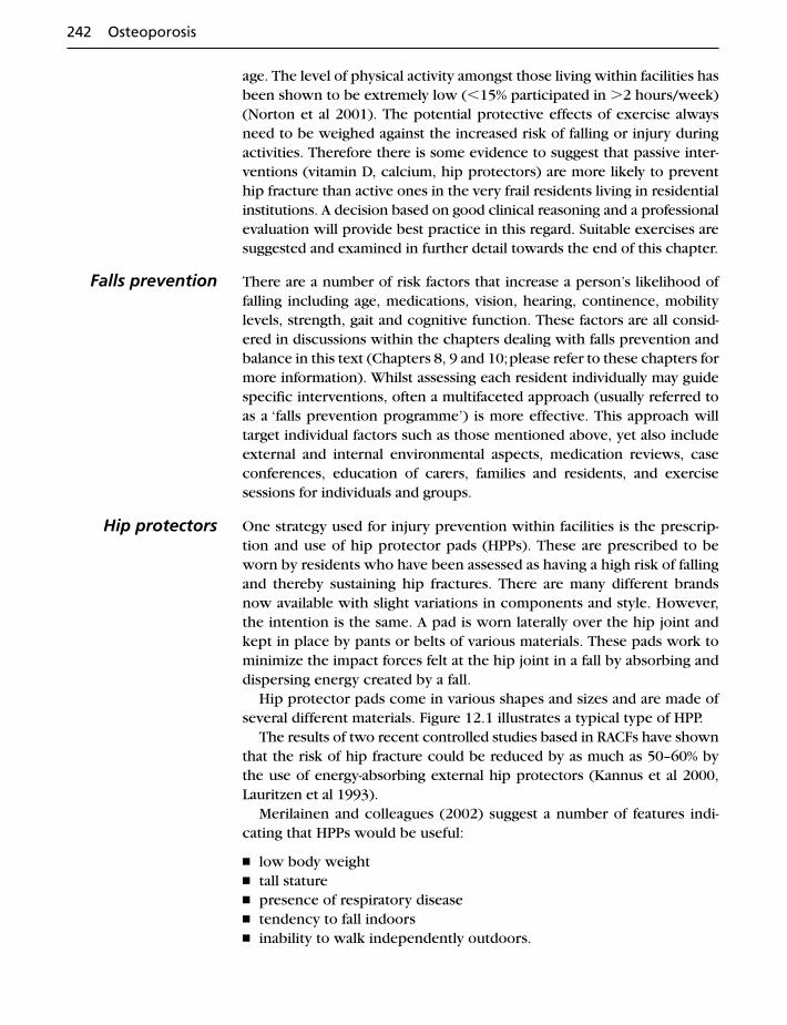
age. The level of physical activity amongst those living within facilities hasbeen shown to be extremely low (�15% participated in �2 hours/week)(Norton et al 2001). The potential protective effects of exercise alwaysneed to be weighed against the increased risk of falling or injury duringactivities. Therefore there is some evidence to suggest that passive inter-ventions (vitamin D, calcium, hip protectors) are more likely to preventhip fracture than active ones in the very frail residents living in residentialinstitutions. A decision based on good clinical reasoning and a professionalevaluation will provide best practice in this regard. Suitable exercises aresuggested and examined in further detail towards the end of this chapter.
There are a number of risk factors that increase a person’s likelihood offalling including age, medications, vision, hearing, continence, mobilitylevels, strength, gait and cognitive function. These factors are all consid-ered in discussions within the chapters dealing with falls prevention andbalance in this text (Chapters 8, 9 and 10;please refer to these chapters formore information). Whilst assessing each resident individually may guidespecific interventions, often a multifaceted approach (usually referred toas a ‘falls prevention programme’) is more effective. This approach will target individual factors such as those mentioned above, yet also includeexternal and internal environmental aspects, medication reviews, caseconferences, education of carers, families and residents, and exercise sessions for individuals and groups.
One strategy used for injury prevention within facilities is the prescrip-tion and use of hip protector pads (HPPs). These are prescribed to beworn by residents who have been assessed as having a high risk of fallingand thereby sustaining hip fractures. There are many different brandsnow available with slight variations in components and style. However,the intention is the same. A pad is worn laterally over the hip joint andkept in place by pants or belts of various materials. These pads work tominimize the impact forces felt at the hip joint in a fall by absorbing anddispersing energy created by a fall.
Hip protector pads come in various shapes and sizes and are made ofseveral different materials. Figure 12.1 illustrates a typical type of HPP.
The results of two recent controlled studies based in RACFs have shownthat the risk of hip fracture could be reduced by as much as 50–60% by the use of energy-absorbing external hip protectors (Kannus et al 2000,Lauritzen et al 1993).
Merilainen and colleagues (2002) suggest a number of features indi-cating that HPPs would be useful:
■ low body weight■ tall stature■ presence of respiratory disease■ tendency to fall indoors■ inability to walk independently outdoors.
242 Osteoporosis
Falls prevention
Hip protectors

These are common features in many older persons living in RACFs, lend-ing support to the argument for mobile residents using hip protectors.However, patient acceptability remains a distinct problem due to skin irri-tations, lack of insight, and a perception that the pads are uncomfortable(Cameron et al 2001, Hubacher & Wettstein 2001). Other problems iden-tified relate to laundering difficulties, cost, fit, style, infection control andmovement of the pad in situ. Education of both residents and staff wouldseem highly appropriate due to scepticism shown by both key stake-holder groups.
Colon-Emeric et al (2003) have shown the cost-effectiveness of provid-ing hip protectors by comparing the cost of this to the financial expenseof medical care needed in the event of hip fracture. This provides a goodargument for some residents to use hip protectors as part of their injuryprevention strategy.
The drugs which have been most rigorously tested and shown to reducespinal fractures in osteoporotic/osteopenic individuals include alendronate,risedronate and raloxifene (Delmas 2002). The most thoroughly testeddrugs reported to reduce non-vertebral fractures are calcium and vitamin Dfor residents in RACFs, and alendronate and risedronate in the community-dwelling older population. Alendronate reduces bone turnover, increasingbone mass by 8% in the spine and 6% in the hip versus placebo in both(Lieberman et al 1995). Parathyroid hormone (PTH) has an ability to reduceboth vertebral and non-vertebral fractures and should also provide an alter-native intervention approach for patients with severe osteoporosis once itbecomes readily available. PTH is normally responsible for controlling theextracellular calcium ion (Ca2�) concentration. It does this by controllingthe Ca2� absorption from the gut, renal excretion of Ca2� and release ofCa2� from the bones (Guyton 1991).
Hormone therapy (HT) has associated benefits and risks and the deci-sion to use this therapy, like all others, should be weighed in relation tocurrent research and with consideration to the individual case at hand.
Modifying risks 243
a b
Figure 12.1a & bA typical hip
protector pad.
Drug therapy

Early evidence was made available suggesting that HT may prevent boneloss in postmenopausal women, and also fractures and chronic heart dis-ease. There is retrospective information suggesting a possible increase incognitive function and prevention of Alzheimer’s disease. The risks of HTinclude vaginal bleeding, breast tenderness, deep vein thrombosis, pul-monary embolism, as well as a moderate increased risk of breast cancer,endometrial cancer and ovarian cancer. Another recent study whichreceived considerable media coverage showed increased risk of coronaryartery disease and ischaemic heart disease; the trial concluded that HTwas not the treatment of choice for primary prevention of coronary heartdisease and should not be initiated or continued for this effect (WHI 2002).Recent debate has questioned the results in light of the statistical ana-lyses and parameters utilized. As is often the case, further research will needto be undertaken to provide more concrete conclusions, although ethicalapproval would no doubt be of concern given early indications of increasedrisk of side effects. Some practitioners believe HT should only be used tocontrol menopausal symptoms and that osteoporosis should be managedby medications such as alendronate, risedronate and raloxifene.
The technique of choice to measure bone density is currently dual energyX-ray absorptiometry (DEXA). Levels of bone density measured from DEXAscans can help to guide patient management and intervention althoughthis is more often done within the community and not prioritized withresidents in RACFs, especially those residing within a high-care setting.The two-dimensional image which is developed allows the measurementof both cortical and trabecular bone density. Repeat scans are performedat intervals of not less than 12 months as changes in bone density havebeen shown to require some time for adaptation to be illustrated.
Interpretation usually relies on a comparison of the resident to others inthe same matched age group (Z-score) and to the mean peak density of a young, healthy, sex-matched group (T-score). Bone mineral density(BMD) scores recorded have been shown clinically to predict fracture riskwithin the vertebrae and hip. With each standard deviation below the meandecrease in BMD, there is a 1.9-fold increase in risk of fracture within thevertebrae and a 2.6-fold increase in risk of hip fracture (Cummings et al1993).
As always, a comprehensive assessment precedes treatment and exerciseprescription. In regard to osteoporosis more specifically it is important toassess posture and range of motion, muscle strength and endurance, aer-obic capacity, balance, pain and function (Bennell et al 2000). Appropriatetests for these measures were outlined in previous chapters. Howeverexamples are illustrated below which may provide more useful data for theresident with osteoporosis. Some useful assessment tools appropriate formeasuring presentation aspects of osteoporosis are shown in Table 12.3.
244 Osteoporosis
Radiography
Physiotherapyassessment

Physiotherapy assessment 245
Factor Measurement tool References
Posture and Serial height measurements Gordon et al 1991range of motion Distance between tragus Laurent et al 1991
and wallShoulder range and elevation Bennell et al 1998Cervical spine movementAnkle range (especially dorsiflexion)Spinal kyphosis (kyphometer, or flexicurve ruler)Photos
Strength/ Sit to standEndurance Spring gauge
Handheld dynamometer Bohannon 1986
Aerobic capacity 6 minute walk Steele 19966 metre walk Hageman &
Blanke 1986Timed ‘up and go’ Podsiadlo &
Richardson 1991
Balance Step test Hill et al 1996Functional reach Duncan et al 1990Clinical test of sensory Shumway-Cook & interaction Horak 1986
Pain and function Timed ‘up and go’ Podsiadlo & Richardson 1991
Timed 6 metre walk Hageman & Blanke 1986
Visual Analogue Scale (VAS) Gagliese & for pain Melzack 1997McGill Pain Questionnaire
Quality of life Osteoporosis Functional Helmes et al 1995Disability Questionnaire Quality of Life Questionnaire Lips et al 1999 of the European Foundation of Osteoporosis (QUALEFFO)Osteoporosis Targeted Quality Lydick et al 1997of Life Survey Instrument for use in the community (OPTQoL)Medical Outcomes Study Ware & Short Form 36-item Sherbourne 1992Questionnaire (SF-36)
Table 12.3Useful tools for the
assessment ofresidents with
osteoporosis

Exercise is increasingly recognized as a means of reducing risk of osteo-porotic fracture and for managing the condition of osteoporosis even inold age. However, the correct choices must be made since not all forms ofexercise are suitable; indeed some actually may increase the risk of frac-ture (Bassey 2001). A general activity programme emphasizing strength,flexibility, coordination and cardiovascular fitness may indirectly reducethe risk of osteoporotic fractures by decreasing the risk of falling and byenabling older persons to remain active, thereby reducing the loss of bonethrough inactivity (ACSM 1995). The primary benefit of exercise for adultbones is conservation, not acquisition. In elderly individuals, improved fit-ness and muscle strength contribute to the prevention of falls and a lowerrisk of fracture. Physical activity may also slow the rate of bone loss.Exercise goals for osteoporosis should include pain reduction, increasedmobility and improvements in muscle endurance, balance and stability.These are worthwhile end-points because not only may they prevent fallsbut they may also improve quality of life. In conjunction with advice toincrease dietary calcium, exercise plays a significant part in a lifestyle prescription for reducing fractures in later life.
Although the best exercises for building bones are those involving impactloading of the skeleton (in children), some studies suggest that this is notvery effective in adults, especially given the negative effects of increasingrisk of osteoarthritis in the older adult and the need for injury prevention.A study on mice further suggested that the ability of bone to respond tomechanical loading is under genetic control (Kodama et al 1999).
The skeleton appears to remain trainable (i.e. responsive to the appro-priate stimuli) into at least the 8th decade. Osteogenic loading activitiesneed to be site-specific; for example, jumping has been used as an activityto improve hip BMD. There is no reason to believe that jumping wouldassist wrist BMD but every reason to be concerned about prescribing thisexercise to the elderly, who are already at risk of injury and who often haveestablished disease processes such as osteoarthritis initially. Resistance pro-grammes need to be carefully prescribed and taught well to the elderly. Theprinciples of exercise prescription outlined in the chapter dealing withprogramming (Chapter 11) should always be followed. A graded resist-ance programme which begins gently and targets muscle groups sur-rounding risk areas such as the hip, back and shoulders should be utilized.The evidence regarding load and repetition regimes leans more towardsusing higher loads with fewer repetitions – this should be kept in mindwhen developing the target programme. Closed chain exercises are oftenthe safest form of weight training and programmes which use the person’sown body weight as resistance are good examples of this, e.g. yoga,Pilates, tai chi. Other aids such as elastic bands and free weights may alsobe used. The focus should sensibly be on the development of low-impactexercises which improve strength and balance and reduce the risk of fallsand subsequent fractures (Turner 2000).
246 Osteoporosis
Prescribedexercise
programmes/suggested
physiotherapyexercises
Resistancetraining/weights

Prescribed exercise programmes/suggested physiotherapy exercises 247
Strategy Examples
Aerobic exercise Walking 3� per week, 15–60 minutesCycling, swimming
Strengthening Walking with weightsexercises Resistance band exercises
Closed chain resistanceusing own body weightLower limb: Protocol (lower and hamstring curls upper limbs):squats 8–10 repslunges 40–60% 1 RM, progress toleg extensions 80% of 1 RMgluteal press 8–10 exercisesquadriceps exercise major muscle groupship abduction 3 times a weekhip adductioncalf press (NB: care must be taken to rotations* ensure posture is erect and Upper limb: that a flexing moment is not biceps curls being transferred to the tricep push-ups thoracic and lumbar spine latissimus dorsi during any of these pectoral squeezes exercises)arm raisespull backswrist curlsball games
Postural Arm raises with deep breathing, trunk extensionexercises/thoracic Chin retraction, deep neck flexors recruitmentextension Lower trapezius setting, scapular retractor training
Postural taping (lower trapezius)Stretching and flexibility
Balance exercises Walking forwards/backwards/sideways/grapevine stepping/heel and toe walking/skipping/standing on one leg (eyes open/closed) (see Chapter 10 for more ideas)
Pain TENSmanagement Gentle heat
Hydrotherapy/aquatic physiotherapyMassageGentle joint mobilization (only applicable in some cases)
Education Lifting advicePostureHealth promotion (smoking advice, nutrition advice)Information on osteoporosis itself
Table 12.4Suggested exercises
and strategies forresidents with
osteoporosis
table continues

There is some evidence that the increase in activity needed to achieve thehighest level of protection is only very slight. This is encouraging for thefrail elderly and their carers. Some research suggests a 5 minute walk dailymay be the key to the conservation of bone mass, with levels in excess ofthis not providing increased protection (Krall & Dawson-Hughes 1994). Ingeneral, however, results from research examining walking interventionprogrammes alone demonstrate that this activity, commonly prescribed topostmenopausal women, does not prevent bone loss alone and needs tobe combined with other management strategies such as medication, otherexercises such as resistance training and balance activities, and education.While walking has numerous health benefits, it is not primarily prescribedas a skeletal loading (resistance) exercise, probably due to its low magni-tude, repetitive and customary qualities. Interestingly, research into its use in this manner in a population with restricted mobility (such as in anRACF) has not been undertaken and may well show positive skeletaleffects related to increased loading. Table 12.4 provides some suggestionsfor exercises suitable for residents with osteoporosis.
See Chapter 11 for more specific principles related to the type, inten-sity and duration of exercises prescribed.
Any exercise which uses large amounts of force pivoting around weaksupport structures (bony areas with fracture risk), especially with a longlever, should be avoided in order to prevent injury and fracture. Fastmovements, risk-taking activities, excessively ballistic patterns of move-ment and jumping may all be well avoided. Dynamic abdominal exercisesshould also be avoided in the aged population due to the increased risk ofmusculoskeletal damage. Some specific exercises to avoid include sit-ups,twisting exercises, trunk flexion exercises, abrupt or explosive loadingand high impact loading (Forwood & Larsen 2000).
248 Osteoporosis
Walking
Exercises toavoid when
osteoporosis isestablished
Strategy Examples
Falls risk Visual and hearing checksassessment and Pharmaceutical reviewmanagement Medical review
Environmental assessmentShoe prescriptionHip protector padsMobility aid prescription, training and maintenance
* Rotational exercises may be important in order to strengthen bony trabeculae
in all directions for improved microarchitectural bony strength. A focus on multi-
directional and rotationally based exercises may be of more benefit for both
function and injury prevention.
1 RM � repetition maximum, maximum weight which can be lifted once only.

References 249
Summary ■ Osteoporosis is characterized by a reduction in bone mass andsubsequent fragility of bone.
■ Osteoporosis is one of a number of factors which increase thelikelihood of a fracture amongst elderly people.
■ There are a number of risk factors for osteoporosis andosteoporotic fractures most of which are modifiable.
■ Nutrition plays an integral role in bone maintenance.■ Lifestyle factors can improve bone health and reduce the
likelihood of injury.■ Falls protection is of paramount importance.■ Hip protectors may prevent hip fractures; recent studies show
positive results.■ Treatment aims to conserve bone mass, reduce the risk of falls,
promote an extended posture, reduce pain and improvemobility and function – thereby preventing fractures and improving quality of life.
■ In the elderly prevention of fracture is most important. Thebest advice and expertise will ensure residents keep activeand take all steps necessary to avoid falls. Exercises andmanagement suggestions were explored to aid in thetreatment given to residents of RACFs with identified orsuspected osteopenia and/or osteoporosis.
References
ACSM 1995 ACSM Position Stand on Osteoporosisand Exercise. Medicine and Science in Sports andExercise 27(4):i–vii
Bassey E J 2001 Exercise for prevention of osteo-porotic fracture. Age and Ageing 30-S4:29–31
Bennell K, Talbot R, Wajswelner H, Techovanich W,Kelly D 1998 Intra-rater and inter-rater reliabil-ity of a weight-bearing lunge measure of ankledorsiflexion. Australian Journal of Physiotherapy44:175–180
Bennell K, Khan K, McKay H 2000 The role of physio-therapy in the treatment and prevention of osteo-porosis. Manual Therapy 5(4):198–213
Bohannon R W 1986 Test retest reliability of hand-helddynamometry during a single session of strengthassessment. Physical Therapy 66(2): February.
Braunwald E, Fauci A S, Kasper D L et al (eds) 2002Harrison’s principles of internal medicine, 15thedn, vol 2. McGraw-Hill, New York, p 2226–2227
Cameron I D, Venman J, Kurrle S E et al 2001 Hip protectors in aged care facilities: a randomisedtrial of use by individual high-risk residents. Ageand Ageing 30(6):447–481
Colon-Emeric C S, Datta S K, Matchar D B 2003 Aneconomic analysis of external hip protector use in ambulatory nursing facility residents. Age andAgeing 32(1):47–52
Cummings S R, Black D M, Nevitt M C et al 1993Bone density at various sites for prediction of hipfractures. Lancet 341:72–75
Delmas P D 2002 Treatment of postmenopausal osteo-porosis. Lancet 359:2018–2026
Duncan P, Weiner K, Chandler J, Studenski S 1990Functional reach:a new clinical measure of balance.Journal of Gerontology 45:M192–M197
Forwood M, Larsen J 2000 Exercise recommenda-tions for osteoporosis. A position statement of the Australian and New Zealand Bone and

250 Osteoporosis
Mineral Society. Australian Family Physician29(8):761–764
Gagliese L, Melzack R 1997 Chronic pain in elderlypeople. Pain 70:3–14
Gordon C C, Cameron Chumlea W C, Roche A F 1991 Stature, recumbent length, and weight. In:Lohman T G, Roche A F, Martorell R (eds) Anthro-pometric standardisation reference manual. HumanKinetics, Champaign, p 3–8
Guyton A C 1991 Textbook of medical physiology,8th edn. W B Saunders, Philadelphia
Hageman P, Blanke 1986 Comparison of gait of young women and elderly women. PhysicalTherapy 66:1382–1387
Helmes E, Hodsman A, Lazowski D et al 1995 A questionnaire to evaluate disability in osteoporoticpatients with vertebral compression fractures.Journals of Gerontology Series A – BiologicalSciences and Medical Sciences 50:M91–M98
Hill K, Bernhardt J, McGann A, Maltese D, BerkovitisD 1996 A new test of dynamic standing balance for stroke patients: reliability, validity, and compari-sons with healthy elderly. Physiotherapy Canada48:257–262
Hubacher M, Wettstein A 2001 Acceptance of hipprotectors for hip fracture prevention in nursinghomes. Osteoporosis International 12(9):794–799
Kannus P, Parkkari J, Niemi S 2000 Prevention of hip fracture in elderly people with use of a hipprotector. New England Journal of Medicine 343:1506–1513
Kodama Y, Dimai H P, Wergedal J et al 1999 Corticaltibial bone volume in two strains of mice:effects ofsciatic neurectomy and genetic regulation of boneresponse to mechanical loading. Bone 25:183–190
Krall E A, Dawson-Hughes B 1994 Walking is relatedto bone density and rates of bone loss. AmericanJournal of Medicine 96:20–26
Laurent M R, Buchanon W W, Ballamy N 1991 Methodsof assessment used in ankylosing spondylitis clini-cal trials. A review. British Journal of Rheumatology30:326–329
Lauritzen J B, Petersen M M, Lund B 1993 Effect ofexternal hip protectors on hip fractures. Lancet341:11–13
Lieberman U A, Weiss S R, Bröll J et al 1995 Effect of oral Alendronate on bone mineral density andthe incidence of fractures in postmenopausalosteoporosis. New England Journal of Medicine.333(22):1437–1444
Lips P, Cooper C, Agnusdei D et al 1999 Quality of life in patients with vertebral fractures: validation of the Quality of Life questionnaire of the EuropeanFoundation for Osteoporosis (QUALEFFO). Osteo-porosis International 19:150–160
Lydick E, Yawn B, Love B et al 1997 Development and validation of a discriminative quality of lifequestionnaire for osteoporosis (OPTQoL). Journalof Bone and Mineral Research 12:456–463
Merilainen S, Nevalainen T, Luukinen H, Jalovaara P2002 Risk factors for cervical and trochanteric hipfracture during a fall on the hip. ScandinavianJournal of Primary Health Care 20(3):188–192
Norton R, Galgali G, Campbell A J et al 2001 Is phys-ical activity protective against hip fracture in frailolder people? Age and Ageing 30:262–264
Podsiadlo D, Richardson S 1991 The timed ‘up andgo’: a test of basic functional mobility for frail elderly persons. Journal of the American GeriatricSociety 39:142–148
Shumway-Cook A, Horak F 1986 Assessing the influ-ence of sensory interaction on balance. PhysicalTherapy 66:1548–1550
Stedman’s Medical Dictionary, 26th edn. 1995.Williams & Wilkins, Baltimore
Steele B 1996 Timed walking tests of exercise cap-acity in chronic cardiopulmonary illness. Journal ofCardiopulmonary Rehabilitation 16:25–33
Turner C H 2000 Exercising the skeleton: beneficialeffects of mechanical loading on bone structure.The Endocrinologist 10:164–169
Ware J E, Sherbourne C D 1992 The MOS 36-itemShort Form Health Survey (SF-36). 1: Conceptualframework and item selection. Medical Care 30:473–483
WHI 2002 Women’s Health Initiative Investigators.Writing Group. Risks and benefits of estrogen plusprogestin in healthy postmenopausal women.Principal results from the Women’s Health Initiativerandomised controlled trial. JAMA 288(3):321–333

Aquatic physiotherapyfor residents in agedcare facilities
Ann Rahmann
251
13
This chapteraims to:
The therapeutic benefits of immersion in water have been known forthousands of years (Edlich 1987). By using a clinical reasoning approach,the positive advantages of exercise in water can be combined with thebest of current scientific knowledge to maximize the outcomes for olderpeople from this enjoyable activity.
It is useful to first consider the different types of water activity cur-rently available and safety issues related to the use of aquatic therapywith elderly people.
Recent guidelines published by the Australian Physiotherapy Association(APA) clearly describe the different types of exercise in water (Larsen et al2002).
■ Hydrotherapy – is used as a general description and covers the widevariety of activities undertaken in a heated pool.
■ Aquatic physiotherapy – has been defined by the APA as the specificpractice of physiotherapy in water carried out by a physiotherapist.This can include individual treatments, group programmes, aquatic physio-therapy classes and physiotherapy prescribed exercise programmes.
■ Water exercise – is exercise carried out in water to maintain andupgrade body strength, flexibility, conditioning and general fitness andto promote a sense of wellbeing, and is usually performed or instructedby exercise professionals. This includes such activities as community-runwater exercise classes.
■ identify the types of exercise undertaken in water■ consider the indications and contraindications for exercise in
water■ discuss the hydrodynamic principles and physiological responses
utilized when exercising in water■ discuss the benefits of managing various impairments with
water exercise.
Introduction
Types of exer-cise in water

■ Aquatic fitness activities – covers aqua fitness, aquarobics, deep waterrunning and should be run by an exercise professional with suitabletraining, e.g. exercise physiologist or aquatic fitness leader.
■ Swimming activities – includes learn to swim, therapeutic swimmingand swimming for those with disabilities.
Because of the potential for multiple health problems in the elderly, mostpeople living in residential care facilities will require specific aquatic physio-therapy programmes rather than the more general approach of waterexercise.
Prior to entering the pool, everyone including helpers should completea screening questionnaire in order to determine if they suffer from anyhealth conditions that may be affected by immersion. This should coveraspects such as the current status of:
■ cardiovascular and respiratory systems■ central nervous system – particularly epileptic fits■ gastrointestinal and genitourinary system■ infectious conditions (tinea and plantar warts)■ eyes and ears (vision or hearing problems might reduce safety due to
reduced ability to communicate)■ skin integrity■ psychiatric conditions, behavioural or cognitive problems■ acute inflammatory conditions■ swimming ability or fear of water■ general mobility, assistance required to walk or dress■ physical ability to determine mode of pool entry/exit.
The APA guidelines clearly explain that each potential client must be individually assessed. The assessment findings should then be used todetermine treatment goals and procedures through a clinically reasonedprocess that takes the physiological effects of immersion into consider-ation. Factors that contraindicate or require extra precautions to be takenby the physiotherapist when taking a particular resident into the poolmust be considered and acted on. Consultation with peers working withsimilar clients or other informed professionals may be required. In particu-lar, regarding infection control, staff and medical specialists might beconsulted. Never put yourself or the client at risk if unsure of safety(Larsen et al 2002).
There are few absolute contraindications for hydrotherapy. Examples ofsituations where entry to the pool is contraindicated include:
■ unreliable faecal incontinence■ skin tears or wounds that cannot be covered with a waterproof dressing
or sleeve (e.g. Seal-Tight, Brisbane, Australia)
252 Aquatic physiotherapy for residents in aged care facilities
Contraindica-tions and
precautions
Contraindications

■ infected discharges■ methicillin-resistant Staphylococcus aureus (MRSA) in an open wound
or present in the gastrointestinal or urinary tract.
However, many medical conditions common in older people require careand caution when the person is being treated in the pool. For example,there is an increased risk of a hypotensive episode when exiting the poolbecause of the effect of immersion on the cardiovascular system. Peoplewith low blood pressure need to be carefully supervised when exitingand showering after a pool session. A water temperature of above 35.5°Ccan also increase the risk of a hypotensive episode. For the elderly, it istherefore necessary to consider not only the effect of immersion on thecardiovascular system but also the temperature of the pool water itself.
Another important factor to consider with the elderly is fragile skin.It is necessary to make sure any skin tears have appropriate dressingsapplied before entering the water. Some types of waterproof dressingsneed to be applied several hours before immersion to ensure the bestadhesion and sealing. In addition, skin tends to soften in the water so it iseasy for fragile skin to be damaged without the person realizing. Potentialsources of damage can be equipment or even the pool surfaces them-selves. Footwear such as foot socks or surf shoes can be worn in the poolas a preventive measure, particularly for people with poor distal sensa-tion such as diabetics with peripheral neuropathy. The physiotherapistmust be aware of potential problems and take precautions accordingly.
A thorough and clearly explained list of conditions and precautions iscontained in The APA Guidelines, Appendix II:Management of precautionsfor activity/treatment in a hydrotherapy pool (Larsen et al 2002). It isimportant for all therapists working in hydrotherapy, particularly with olderpeople, to familiarize themselves with these conditions and managementsuggestions.
Just because a person cannot swim, does not mean he or she is unable to safely exercise independently in the pool. To assess water safety, thephysiotherapist should check that the person can:
■ enter the water independently■ roll from prone to supine and regain a safe breathing position■ if unable to walk or touch the bottom (i.e. if they are out of their depth),
propel themselves to the edge of the pool and a position of safety whilemaintaining a safe breathing position
■ exit the pool safely or get into pool hoist independently (Larsen et al2002).
If the person is not safe and competent in completing these tasks inde-pendently, the physiotherapist should recommend that the resident attendindividual sessions or have a trained carer who can come into the pooland assist them. It is important to note that ‘independent’ means that theperson does not rely on a buoyancy device to achieve the competencies.
Contraindications and precautions 253
Non-swimmersand independent
pool exercise

There are few absolute contraindications to aquatic therapy forolder people. However, because the elderly in residential careoften have multiple medical problems, the physiotherapist mustcarefully consider safety issues when deciding who is appropriatefor pool therapy and whether a group or individual setting is thebest option. Safety of all concerned should be the paramountconsideration. The physiotherapist also needs to decide whether aparticular facility has sufficient staff, access and safety equipmentto meet all the needs of their particular clients.
An understanding of hydrodynamic principles and the way they affectmovement in water is important in designing an effective water-basedexercise programme (Thein & Thein Brody 1998). The basic concepts aremost easily understood by considering what occurs when water is stilland then when it is moving (Becker 1997).
When water is still, the main principles are:
■ density■ buoyancy■ hydrostatic pressure.
When water is moving, they are:
■ water flow – laminar and turbulent■ drag forces.
These concepts are thoroughly discussed in most general hydrotherapytexts to which the reader is referred but will be briefly outlined here to high-light the clinical considerations when taking elderly people into the pool.
The density of an object is simply its mass per unit volume and is com-monly reported in kilograms per cubic metre. Relative density or specificgravity is the ratio of an object’s density compared to water (Becker 1997). Therelative density of the human body compared to water varies throughout lifeand is dependent on the amount of lean muscle and adipose tissue. On averagethe body’s relative density is 0.974. That is, the body is a little less dense thanwater and will tend to float (Becker 1997). The gradual decrease in musclemass later in life means that for older people, the relative density drops toabout 0.86 (Reid Campion 1997a). This means that older people will floatmore easily and also that generally women float more easily than men.Clinically, it is important to consider a person’s density in the water. Forexample, someone who is plump and short or with a higher body massindex (BMI) may tend to float more easily. If the person also cannot swim,he/she will require close supervision to ensure the risks associated withlosing footing or getting out of his/her depth are minimized.
When a person is in water, two opposite forces are acting. Gravity acts ina downward direction and the force of buoyancy acts upwards, with the
254 Aquatic physiotherapy for residents in aged care facilities
Hydrodynamicprinciples
Density
Buoyancy
Key points

buoyant force being equal to volume of water displaced by the submergedpart (Edlich 1987). For the clinician, this simply means that people weighless in water (Harrison et al 1992). This can be extremely beneficial in situ-ations where, for example, reduced weight-bearing is medically neces-sary such as healing pelvic fractures or a recent total hip replacement.
Safety in the water is also related to the metacentric principle. When theopposing forces of gravity and buoyancy are the same, the person is stableand balanced in the water. If the forces are unequal, for example if a personis floating and lifts one arm out of the water, rotation of the body will occuruntil balance is again achieved (Reid Campion 1997b). Because of thisprinciple, it will be much more difficult for a person who has an asymmet-rical posture due for example to hemiplegia to maintain balance in the pool,particularly when floating. Fluctuating muscle tone can also make it difficultfor a person to control his/her position in the water independently. Appro-priate supervision is a necessity for these people when in the water.
Water exerts a constant pressure at the same depth on a submergedobject. This pressure also increases with depth and with increasing densityof the liquid (Reid Campion 1997b). In practical terms, this means that thewater pressure around the feet of a person standing in waist deep water isabout 89 mmHg (Becker 1997). This is probably one of the ways immersioncan help to reduce swelling in the tissues although there is controversy inthe literature about the exact mechanism. People with respiratory limitationsuch as asthma or chronic obstructive pulmonary disease may actually findthat the water pressure around their chest is uncomfortable and increasesbreathlessness. However, an interesting study involving emphysema patientswas reported recently. Subjects breathed in while their head was out ofwater, and then squatted so their nose was under water and breathed out.This was done for 30 minutes, three times a week for 2 months. The resultsshowed significant improvements in both cardiac output and FEV1
(forced expiratory volume in 1 second) (Kurabayashi et al 1998). Positivepressure breathing is often used in land-based physiotherapy for respira-tory patients so there is no reason to suppose that the water-based equiva-lent should be any less beneficial. People with shortness of breath need tobe carefully monitored while they are in the pool and the treatment positionand depth of water carefully chosen. Physiotherapists should also remem-ber that there can be significant benefits from activities in the pool forolder people with these conditions.
Objects moving through water are affected by the water flow. Laminarflow is where an object is streamlined through the water. Turbulent flow,as the name suggests, is where the water makes currents and eddies andsmooth movement is more difficult (Becker 1997). For the clinician, theseforces can be used to make exercises in water easier or harder. Streamlinedmovements with little turbulence in the water are smooth and effortless.Turbulence created by moving more quickly can make movement through
Hydrodynamic principles 255
Hydrostaticpressure
Laminar andturbulent water
flow

the water more difficult and is an easy way to increase the difficulty of anexercise (Reid Campion 1997b). However, turbulence can be a problem forolder people in the pool. A frail elderly person whose balance in the wateris poor may be almost swept off his/her feet either by someone walkingpast quickly or if there are a lot of people in the pool and the water ismoving a lot.
Drag forces are related to the shape of an object and the speed of its move-ment through water and so are less with laminar flow and greater withturbulent water flow (Becker 1997). Two types of drag forces are relevantto hydrotherapy. Pressure drag is created by the turbulence behind a moving object and surface drag is the resistance of the water flowingover the surface of an object (Poyhonen et al 2000).
The effect of pressure drag means that the larger and less streamlined anobject is, the more difficult it will be to move through the water. The fasterthe speed of the movement, the greater the drag forces will be becausethere will be more turbulence in front of the object and greater drag forcesbehind it (Hillman et al 1987). Clinically this effect can be used to makeexercises harder by adding paddles or flippers to increase the forces theneuromuscular system has to overcome in order to move in the water.
It is harder to move through water than air due to surface drag (Poyhonenet al 2000). Because of the density of water, it is 790 times more difficultto move through water than air (Reid Campion 1997b). The benefit of thisresistance is obvious because, for example, it takes about four times asmuch energy to swim than to run an equal distance (Edlich 1987). Anotherless well-known advantage is that if someone loses balance in the pool, he/she will not fall as quickly as on land and may have time to regain balanceusing appropriate strategies (Josephson et al 2001).
Keeping a constant core temperature is important and the body uses acomplex combination of mechanisms to maintain it (Becker 1997). Whenexercising in a heated hydrotherapy pool, it is more difficult for the bodyto maintain homeostasis (Reid Campion 1997d). As Reid Campion (1997d)highlights, there is considerable controversy in the literature about whatis a suitable temperature for a hydrotherapy pool. Temperatures of around28 to 30°C are suitable for active exercise such as swimming or aquaro-bics (Edlich 1987), whereas lower temperatures of 26 to 28°C are moresuitable for the intensive training of elite athletes (Thein & Thein Brody1998). Thermoneutral water is at skin temperature, or around 33 to 34°C(Reid Campion 1997d). This is the most comfortable temperature for lessactive exercise and older people because it minimizes stress on thebody’s thermoregulatory system (Golland 1981). Selection of a facilitywith a pool at the appropriate temperature is particularly important withthe elderly because they find it more difficult than younger people tocope with temperature changes (Evans et al 1993).
256 Aquatic physiotherapy for residents in aged care facilities
Thermodynamicprinciples
Drag forces

A basic understanding of the physiological effects of exercising and beingin water is necessary because of the multiple health problems common inthe elderly. It must be noted that most research has used young adults assubjects and only a few studies have examined the effect of immersion inthe elderly. There are also differing views in the literature about the natureof the changes with immersion and exercise in water. Much depends onthe water temperature and intensity of exercise. In older people, the twosystems most affected by immersion are the cardiovascular and the renalsystems and they will be considered here.
Immersion in water increases the amount of blood centrally in the thoraxby about 700 ml and an increase in stroke volume and cardiac outputresults (Pendergast et al 1993). This mechanism is thought to be one ofthe reasons why heart rate at a given intensity of exercise is lower inwater than on land. However, exercise intensity and type as well as waterdepth and pool temperature can all affect the relationship between heartrate and oxygen consumption (Cureton 1997). Bradycardia occurs onimmersion. This can be greater in people taking beta-blockers, a commonmedication given to elderly patients (Levin 1997). It is also important toremember that hydrostatic pressure is equal to venous pressure in thelegs so that, on getting out of the water, venous pooling in the limbs may rapidly occur, causing a drop in blood pressure (Bookspan 1997).Residents whose ability to move about on land is very limited should alsobe carefully supervised until accustomed to activities in the pool, simplybecause they are able to do more exercise in the water than their cardiovascular system is used to. While elderly people may be quite safeexercising in the pool, the therapist must be aware of the variety of cardiovascular changes that can occur when exercising in water, as wellas when entering and exiting the pool.
The overall effect of immersion in warm water is diuresis but it is a resultof the interaction of many factors. Bookspan (1997) summarized themechanisms of immersion diuresis as follows:
■ Hydrostatic pressure – causes blood to be shunted centrally in thethorax, increasing cardiac output.
■ Temperature – colder water increases the effect, less important in thermoneutral water.
■ Hydration – if already dehydrated, the body decreases output to conserve fluids.
■ Age – diuresis occurs more quickly and in a greater volume in the elderly.
In older people, the positive effect of this diuresis is that it is a very efficientmechanism for reducing oedema (Reid Campion 1997b). However, caremust be taken to ensure the elderly drink fluids during and after hydrother-apy treatment because they are often already dehydrated. The treatingphysiotherapist must also be aware if anyone has kidney dysfunction
Physiological effects of immersion and exercise in water 257
Physiologicaleffects of
immersion andexercise in
water
Cardiovasculareffects
Renal effects

because, as mentioned previously, water temperature is an important factoraffecting the degree of diuresis from immersion. Careful monitoring ofthe time spent in the pool may be also necessary initially until the toleranceto immersion is determined. Staff must ensure that everyone adequatelyrehydrates by drinking water after getting out of the pool to minimize theadverse effects of dehydration, such as headaches.
A variety of physical principles and physiological effects need tobe understood in order for older people to safely utilize the pool.Moving and being in water provide a unique environment for theelderly to exercise. Using clinical reasoning to combine both thehydrodynamic principles and goals for the individual client, a stimu-lating and beneficial treatment programme can be developed.
Over the years, anecdotal evidence has increased regarding the benefitsof water-based therapy. A systematic review of the literature was recentlypublished (Geytenbeek 2002). The outcome was that, although muchresearch remains to be done, there is evidence to support its use in painmanagement, improvement of functional ability, flexibility, strength andbalance as well as self-efficacy. Diagnostic groups that benefit includerheumatic conditions and chronic low back pain in people of all ages.
The following discussion will use a clinical reasoning focus to discussthe practical aspects of aquatic physiotherapy for elderly people withproblems related to:
■ postural stability and balance■ strengthening■ maintaining functional abilities■ pain and psychological factors■ general fitness■ neurological conditions.
Balance involves postural stability and the dynamic control of the bodywhile moving to achieve a functional goal. Maintenance of postural align-ment and stability of the trunk is important for function and balance inthe elderly, particularly those with osteoporosis (Larsen 1998). The struct-ural changes occurring with age that affect posture include an alterationof the normal spinal curves with generally an increase in thoracic kyphosisand a loss of the lumbar lordosis (Kirkegard et al 1998). These changesare likely to be more marked in residents of aged care facilities who aregenerally less mobile than older people still living in the community.
The ability to independently move and balance tends to deterioratewith age and this results in an increased risk of falling (Woollacott 1993).Maintenance of independent and functional balance relies on the inte-gration of multiple sensory and motor systems (Josephson et al 2001).
258 Aquatic physiotherapy for residents in aged care facilities
Postural stabilityand balance
Aquaticphysiotherapy
Key point

However, the ability to control balance is complex and is dependent onthe nature of the task as well as the environment in which the activity isto be undertaken (Huxham et al 2001). Elderly people who have fallen orfear falling can enter a downward spiral. They move less to try and pre-vent falls, which then leads to weakness and reduced ability to balanceand then they are more reluctant and less able to move and the cycle con-tinues (Simmons & Hansen 1996).
Balance is integral to all movement; it is therefore important to train bydeveloping successive skill levels and then the skills learned are able to betransferred across into different activities (Huxham et al 2001). The waterenvironment is an ideal place to retrain or maintain the ability to balance,particularly for those who have entered the downward spiral of decreasedactivity. The benefits of exercise in water to improve balance have beenshown in several studies (Alexander et al 2001, Josephson et al 2001, Lordet al 1993, Simmons & Hansen 1996). The positive effect may be due to anumber of factors. A wide variety of activities at various speeds and rangesof movement can be easily included. Also, the effect of surface drag in thewater means that dynamic movements, such as reaching to the limits ofstability, can be done more slowly and with greater confidence in waterthan on land. This allows those residents whose reactions are not as rapidas they should be to practise without the fear of falling. As a caution though,it is important to remember that frail elderly people can easily lose theirfooting in the pool if the water becomes too turbulent. People who havepoor balance and a history of falls usually require close supervision in thepool. Combinations of activities in a group setting can be easily organized,but must take into account the varying functional abilities and water safetyof all participants. The speed and type of exercise as well as the depth ofwater should be tailored to suit each person’s abilities.
A clinically important but often overlooked advantage of balance activ-ities in water is the fact that people exercise in water with bare feet. Theankle joint tends to lose range with ageing and this can have a markedeffect on balance (Vandervoort 1999). Josephson and colleagues (2001)noted that subjects in their water exercise group had greater range of ankledorsiflexion than those in the non-water treatment group. In younger athletes with a sprained ankle, the addition of aquatic exercise improvedsingle leg balance more than land-based exercise alone (Geigle et al 2001).Clinically it seems logical that balancing and exercising in bare feet in thewater could help the older person to activate muscles around the anklemore effectively, improve movement in stiff joints of the feet and alsoincrease the proprioceptive input from the feet and ankles.
Land-based exercise has been shown to improve both strength and bal-ance in older people (Lord & Castell 1994) . Similarly, regular aquatic exer-cise has been shown to significantly improve functional mobility, flexibilityand balance in the elderly ( Josephson et al 2001, Lord et al 1993). Poolexercise is an evidence-based alternative method of balance training andpostural re-education for the elderly.
Aquatic physiotherapy 259

An understanding of the importance of maintaining and improving mus-cle strength in the elderly to retain function and quality of life has beengrowing in recent years (Latham et al 2002). However, it appears that notjust any exercise will do. The most important factor in gaining musclestrength is the intensity of the exercise and not the age or frailty of theperson (Fiatarone & Evans 1993). A second crucial factor is that in orderto continue to improve strength, the resistance of exercises needs to beprogressed over time (Barrett & Smerdely 2002). Both principles are justas important in water-based activities as they are for land-based exercises(Thein & McNamara 1997). There has been little published research regard-ing the effect of aquatic exercise on strength in the elderly. One recentstudy showed significant improvements in strength and fitness amongolder women after 12 weeks of water exercise (Takeshima et al 2002).
Strengthening activities in the aquatic environment can start in therange and at the rate that the person can comfortably manage (Speer et al1993). This is an obvious advantage for the less physically able elderlypeople from residential aged care settings who may be quite restricted intheir ability to move and therefore exercise on land. Exercises can be mademore difficult using the principles of hydrodynamics. Working againstbuoyancy, moving faster or moving something larger like paddles or flip-pers will all increase the work of an exercise. A metronome can be a veryuseful piece of equipment to encourage people to progress the speed ofmovement in the water. Rates should be set with both the individual’sability and the specific exercise in mind because a set rate will not besuitable for all exercises. The physiotherapist skilled in aquatic treatmentwill be able to select the most appropriate way to progress particularexercises for each individual.
The pool is also an excellent environment for beginning to activateand strengthen very weakened muscles, such as in the frail elderly, earlyafter surgery or after an injury. Buoyancy in the water allows more gentleactive movements than conventional dry-land therapy and can buildstrength to enable a safe transition to active dry-land therapeutics (Speeret al 1993). As these authors highlight, exercises in the water can be an extremely beneficial adjunct to more traditional physiotherapeutictechniques.
With a thorough understanding of the principles of exercise in water,effective strengthening programmes can be undertaken in the pool, par-ticularly for the very frail and debilitated.
Difficulty living independently due to declining balance, strength or mobil-ity is commonly the reason elderly people will move into a residentialcare setting (Kirkegard et al 1998). Physiotherapists who work with theelderly know that, for example, the ease of getting out of a chair often hasmore to do with the height of the chair than with the person’s specificstatic quadriceps strength. What is often overlooked when consideringthe functional abilities of the elderly in research is that the relationship
260 Aquatic physiotherapy for residents in aged care facilities
Strengthening
Maintainingfunctional
abilities

between strength and function is task specific (Chandler et al 1997).Movement normally takes place at a variety of speeds depending on thecontext of the activity (Moy 2003). Without realizing it, elderly peoplewill often slow down their voluntary movements in order to try and con-trol their balance and over time this leads to a reduced ability to movequickly (Woollacott & Manchester 1993). When looking at muscle activa-tion patterns during a stepping task, it was found that patterns variedboth within and between subjects (Mercer & Sahrmann 1999). This high-lights the need for elderly people to practise balance and functional tasksin a variety of settings and at a range of different speeds (Carr et al 2002).The pool is an ideal environment where strength and balance activitiescan be easily combined into functional activities that may not be possibleon land for the frail aged (Levin 1997). Sit to stand and stepping up anddown are two examples of exercises that can be practised easily in thewater. The types of activities and the variations of speed and the controlrequired are only limited by the physiotherapist’s imagination.
Negotiating stairs is a particularly difficult locomotor task for many eld-erly people and they will often have moved into residential care becauseof problems with this activity (Cavanagh et al 1997). The importance ofeccentric muscle strength in such functional activities is well known (Carret al 2002). However, the effect of ageing on eccentric muscle strength hasnot often been investigated. One small study found that women seemed to have a significantly greater loss of eccentric strength with age than men(Bellew et al 1998). Most aquatic exercises involve predominantly con-centric muscle activity (Thorpe & Reilly 2000). Eccentric exercise is pos-sible with an understanding of hydrodynamics by using muscle activationto counteract the effect of buoyancy.
Functional activities such as walking are extremely beneficial activitiesfor the elderly but those in residential care are often limited in their abil-ity to walk any distance or at speed. Walking in the pool can be a way ofretraining or maintaining this ability in those who normally find walkingdifficult (Simmons & Hansen 1996). When walking through water at alevel that is 50% of a person’s height, the patterns of electromyographicmuscle activation were found to be the same as when walking on land(Petrofsky et al 2002). The authors found that muscle activity changed withincreasing depth of water so the optimum depth of water for re-educatinga normal gait pattern is 50% of the person’s height. Different speeds andchanges in directions add to the challenge and variety of the task. Thetherapist needs to weigh up the advantages of normal muscle activationpatterns in shallower water, with the pain relief gained in deeper water.A suitable depth for each person should be chosen, depending on his/hergoals, abilities and treatment aims.
Speer and colleagues (1993) in discussing shoulder rehabilitation iden-tified the contribution hydrotherapy plays in restoring muscle activationand coordination to enable normal function of the entire pectoral girdle.Considering the often multiple health problems of the elderly, the ability
Aquatic physiotherapy 261

to move and function normally in the water is an important benefit, notonly around the shoulder girdle but for the whole neuromuscular system.Complex functional activities not possible on land can also be accom-plished more independently in the water environment.
Back pain is a common problem encountered in elderly residents and isoften associated with joint degeneration and osteoporosis. Efficient trunkstability and good posture is needed to prevent pain. This is provided bya combination of action from global, torque-producing muscles such aslatissimus dorsi and local muscles such as multifidus that attach closelyonto the spinal vertebra to provide stability for the spinal segments(Richardson & Jull 1995). The ability of the motor system to control pos-ture and stability appropriately is dependent on an efficient central ner-vous system (Hodges 2000). Alterations to the normal structure of thespine and trunk in the elderly must affect the ease with which global andlocal muscles can be activated. Pain is known to alter the activation of the muscles which normally provide stability for the trunk (Hodges &Richardson 1996). In an elderly person with poor posture, the addition ofpain from a vertebral crush fracture must further compromise the normalpatterns of activity of these stabilizing muscles. Reducing pain to allownormal muscle patterns to be reactivated is crucial in rehabilitation aftersuch an event and the support provided by water is useful in attainingthis aim (Fig. 13.1).
Relief of pain is often reported as a benefit of aquatic therapy(Alexander et al 2001, Lee 1997, Smit & Harrison 1991). The mechanismfor this is considered to be simply the reduction in weight-bearing due tobuoyancy, allowing the unloading of painful structures (Wyatt et al 2001).
262 Aquatic physiotherapy for residents in aged care facilities
Figure 13.1Supported supine float is a useful position for people with problemssuch as back pain or crush fractures. Pain is significantly reduced andthis allows facilitation of postural and deep stabilizing trunk muscles.
Pain andpsychological
factors

However, current research from the field of chronic pain neurobiologyprovides a much broader and potentially more exciting explanation forthe effect of immersion for someone who is in pain. Pain processing isoften thought of as a simple input/output mechanism but this view isbeing increasingly challenged as brain imaging techniques improve. Themature organism model considers the way, in a painful state, the brainsamples input not only from the tissues but also from the environment as well as thoughts and previous experiences (Gifford 1998). What thismeans in clinical terms is that pain should not be considered simply a‘tissue-based’ process but one that can be influenced by many differentfactors. ‘Movements that are feared, avoided and context dependent willhave to be presented to the brain in different ways, for example in differ-ent environments, or the movement “broken down” or paced … Clearly,the more functional the movement and the more it links into the desiredactivity and achievable goals, the better’ (Butler 2000). The pool is there-fore a powerful rehabilitation tool for people with pain because they areable to accomplish functional movements without the overlay of pain.Areas within the brain that would normally be activated by the thoughtof or commencement of a painful activity can be modified or ‘turneddown’ and the perception of pain altered (Butler 2003). Normal patternsof muscle activity are then, logically, easier to achieve.
Frequent pain is reported by one-third of older adults and 20% restricttheir activities because of significant levels of pain (Reyes-Gibby et al2002). The study also showed that higher levels of pain were closelyrelated to poorer self-perceived health. However, other studies have foundthat the elderly tend to underestimate their pain levels so the real figuremay be closer to 50% (Weiner 2002). Interestingly, the ‘old-old’ also tendto rate their health more favourably than the ‘young-old’ despite having afar greater risk of dying simply because of their age (Idler 1993). What thismeans in relation to the aged in residential care is that more than half are likely to have significant pain, but in spite of this, they are likely to bequite positive about their general state of health.
For those who are less happy about their health, exercise in the pool canhave additional benefits. Older people who regularly swam were found tohave a more positive body image, better bone density and felt they werestronger than community-centre attendees who did not exercise regularly(Benedict & Freeman 1993). Exercise is thought to help in the manage-ment of anxiety and depression through its effect on brain monoamines(Chaouloff 1989) and pain through the stimulation of beta-endorphinrelease (Schwarz & Kindermann 1992). Several studies looking at arthriticconditions have demonstrated reduced pain and increased self-efficacywith aquatic exercise programmes (Ahern et al 1995, Wyatt et al 2001).Another important psychological influence to consider is the positivesocial nature of pool treatment. People having aquatic physiotherapy arenot treated alone in a single cubicle but rather in a pool with other peopleof varying abilities. The importance of support and positive feedback
Aquatic physiotherapy 263

from others in facilitating improvement in chronic pain patients hasrecently been highlighted (Gifford 2002, Muncey 2002). It has been sug-gested that the positive reinforcement of exercising in a class with othersmay be even more important than the content of the class itself (Minoret al 1989). Physiotherapists should not underestimate the benefit of thisparticular aspect of aquatic therapy.
Dementia and impaired cognition should briefly be mentioned becausethey are often thought to be a contraindication to aquatic exercise. Safetyin the water is the predominant issue because people with these condi-tions will often lack the insight and concentration to exercise with a group(Levin 1997). As discussed earlier, it is the responsibility of the supervis-ing physiotherapist to decide whether the person is safe or not to attenda group programme. If group activities are not suitable, the most appro-priate form of water activity would then be either individual ‘one on one’sessions with the physiotherapist or to train a carer to specifically lookafter that one person in the pool. It is also important to remember that itmay be more beneficial for people with cognitive impairments to cometo the pool when it is relatively quiet rather than when it is busy.
When the current knowledge of pain processing is combined with theease of movement in water, as well as the realization that about half of allelderly people have significant pain, the benefits of aquatic therapy forolder people with pain are clear. The psychological benefits for peopleshould also be remembered.
It is being realized that, particularly for the elderly, some activity is betterthan none at all. A walking programme just twice a week was shown toimprove fitness in elderly, sedentary women (Hamdorf et al 1992). Becauseit is possible to move more easily in the water, general fitness and cardio-vascular exercise is made possible for the elderly who cannot easily moveon land. Many studies have been undertaken on younger subjects thatshow it is possible to maintain cardiovascular fitness through pool exercise(Bishop et al 1989, Cassady & Nielsen 1992, Kirby et al 1984, Whitley &Schoene 1987). Positive improvements have also been shown in the fitnessof older people by using water exercise (Danneskiold-Samsoe et al 1987,Ruoti et al 1994, Sanford Smith et al 1998, Takeshima et al 2002). Evenpeople with late-onset poliomyelitis were able to reduce their trainingheart rate after a twice weekly, 8-month water exercise programme (Willenet al 2001).
Basic outlines of aquatic fitness classes for older people are common(Levin 1997). Water-based exercise can be a particularly effective way toimprove the cardiovascular fitness of the elderly from all levels of resi-dential aged care settings, provided the activities are tailored to suit theabilities of the particular people involved and adequate supervision andassistance is available. One important point to remember is that due tothe hydrodynamic effects of immersion, when exercising in water, theheart rate can be between six and fifteen beats per minute lower than
264 Aquatic physiotherapy for residents in aged care facilities
General fitness

when exercising on land (Hamer & Morton 1990). The relationshipbetween heart rate and VO2 (oxygen consumption) is dependent on theexercise intensity and type, as well as the water depth and temperature,so it can be confusing to try and determine appropriate levels from theliterature. Using heart rate is probably a less reliable method of measuringexercise intensity, particularly with the elderly. A convenient and easy-to-use alternative is Borg’s perceived rate of exertion (PRE) where the per-son simply rates on a scale up to 20 how hard they feel they are working(Borg 1982). The moderately hard level of around 12 to 14 has been cor-related with training heart rates of 70% of maximum training heart rate(Hamer & Slocombe 1997). However, while many of the elderly in resi-dential aged care may not be able to manage the amount of exercise thatyounger more able people can, the perceived rate of exertion is still a use-ful measure clinically to encourage them to work a little harder in thewater. As they get stronger and fitter, they will be able to achieve morerepetitions or a longer period of exercise at the same level of exertion.
Maintaining cardiovascular fitness is no less important for the residentsof aged care facilities than for the well elderly living in the community.Effective pool programmes can be developed using the combination ofhydrodynamic principles and clinical reasoning to benefit even the mostdebilitated of people because they can move more easily in the water thanon land.
Treatment in the water of people with neurological impairments is con-sidered controversial. ‘Many neurological physiotherapists are wary oftreating their patients in the water, reserving hydrotherapy for the laterstages when not too much harm can be done and the patient enjoysdoing something a bit different’ (Gray 1997). Most of the debate withinthe literature is related to the varying approaches used to treat neuro-logical patients (Reid Campion 1997c). One point of view is that the unusualenvironment and way muscles work in water is not the best way torelearn function. The opposing view is that functional activities are possible in the pool with less manual assistance and can therefore provideearlier normal movement input and motivation for the patient. Much ofthe success of aquatic treatment for these patients depends on the clin-ical skills of the treating physiotherapist in understanding hydrodynamicprinciples and how they can affect or assist the neurologically impairedperson. The benefits of aquatic exercise for this group of patients havebeen tone reduction, maintenance of joint range, static and dynamic bal-ance, earlier and more effective strengthening, cardiovascular benefits,motivation, recreation and social integration (Morris 1997).
Principles of aquatic treatment for acute neurological conditions willnot be covered here as residents are usually into the subacute or chronicphase when they reach aged care settings. The rehabilitation goals of bal-ance re-education, cardiovascular fitness, functional activity and strength-ening previously discussed are just as relevant for those with neurological
Aquatic physiotherapy 265
Neurologicalconditions

conditions (Carr et al 2002). Several aspects specific to treating neuro-logical dysfunction in the elderly in the pool will be briefly reviewed here.
There are few published studies looking at the effect of aquatic exercise inneurological subjects. One study showed that the gait characteristics ofpeople with spastic hemiplegia improved after aquatic exercise (Zamparo &Pagliaro 1998). It was thought this could be due to a reduction in spas-ticity. A subsequent study then showed that hypertonicity was no worseafter the pool treatment (Pagliaro & Zamparo 1999). Subjects for the sec-ond study included 26 people from age 26 to 81, with diagnoses of hemi-plegia, paraplegia, tetraplegia or multiple sclerosis. Although this studydid not demonstrate reduction in tone, the fact that tone was not madeworse in any of the diverse groups is important to note.
Maintaining or regaining normal muscle length is often a treatment aimin people with increased tone. Standing in the shallow end of the pool withweight down through the feet or walking up a pool ramp are effective waysto regain or retain ankle flexibility where there is increased tone in the calf.Those who are unable to stand easily out of the water can often accomplishthis in the pool (Fig. 13.2). The benefits of stretching can then be easilycombined with functional activities such as sit to stand or step-ups as aprogression in the water, just as it would be in a land-based programme.
266 Aquatic physiotherapy for residents in aged care facilities
Abnormal tone
Figure 13.2This woman who is unable to stand independently out of the pool, can stand at the plinth with even weight-bearing through her legs. Thisposition also allows weight-bearing and positioning of her affected leftarm at a more normal muscle length. She has minimal active movementin the left arm but this position also allows her to do modified push-upsindependently while still maintaining pelvic and trunk stability.

Clinically, the most important issue to be aware of with someone whohas increased tone is that a sudden change in tone can make him/her rapidly roll or lose balance and therefore risk drowning if unable to returnto a safe breathing position. A cough or sneeze or even getting cold canbe enough to cause a rapid increase in tone. Close supervision by a physio-therapist or carer is generally always the safest option for those withunpredictable hypertonicity.
Hypotonicity or low muscle tone presents a different problem. Forexample, someone with a dense hemiplegia can find that his/her legtends to float due to the effect of buoyancy combined with limited activemovement. Using an ankle weight can provide enough proprioceptionand ‘counterbalance’ to the effect of buoyancy to allow more functionalactivities such as standing and walking. Particular care should be takenthat buoyancy is not carrying the limb into a painful range at the gleno-humeral joint even though a painful, hemiplegic shoulder can be com-fortably supported and moved in the water, often more easily than onland. Muscles unable to work against gravity can begin to move a limb inthe supported environment of the water.
Exercises to address the rigidity of Parkinson’s disease can be easilyincorporated into a water-based programme. Because movement in wateris slower, people are able to reach to the limits of their stability and rangecomfortably in the pool and therefore often feel safer going further intorange. Activities such as those focusing on trunk rotation in standing androlling from prone to supine can be particularly effective in the waterenvironment.
It has been suggested that hydrostatic pressure surrounding the limbs inthe water can provide proprioceptive input that enhances active move-ment (Speer et al 1993). Many activities in the water can be used toincrease proprioceptive input. For example, lying prone on elbows on apool plinth while kicking with the legs provides proprioceptive input forthe shoulder girdle and upper limbs, as well as trunk and lower limb stability and strengthening (Fig. 13.3). Standing and pushing down on akickboard is another example.
These examples highlight the fact that one water-based exercise canachieve several different treatment aims so treatment can be very effi-cient. Walking in water reduces the amount of weight-bearing throughthe feet but adding ankle weights can increase the proprioceptive inputand improve walking ability of those with poor control. Residents withtruncal ataxia also find a scuba-diving weight-belt worn around the waistcan give added stability and input when walking in the pool. The amountof weight can be varied and reduced when control improves.
Recently, the use of walking on a treadmill with bodyweight support on landhas become a common therapeutic technique for neurological patients(Hesse et al 1995, Shepherd & Carr 1999). Treadmill walking with 30% of
Aquatic physiotherapy 267
Proprioceptivedysfunction
Walking in water

the body weight supported demonstrated significant improvements in acti-vation of pelvic stabilizing muscles as well as providing a dynamic stimulusto the hemiplegic gait pattern (Hesse et al 1997). In the pool, immersion to30% body weight occurs at about the level of the xiphisternum in quietstanding (Reid Campion 1997a). There is no reason to presume that walk-ing in the pool at that level is any less beneficial than walking on a tread-mill. In addition, in the pool it is possible to include functional tasks suchas stepping and squats which cannot be done safely on a treadmill. It isimportant to remember that when walking through water, increasing thespeed of walking can enhance the strengthening, cardiovascular andweight-bearing benefits of the activity (Harrison et al 1992).
One of the common reasons given for not walking people with neuro-logical problems in the pool is that it is more difficult to facilitate the correctgait pattern, e.g. foot placement. A recent study (Petrofsky et al 2002)demonstrated that when walking in water at 50% of body height, patternsof muscle activation were the same as land-based walking. It is thereforepossible to re-educate normal gait patterns in the water. There is no rea-son why such things as straps that loop around the foot and ankle and arethen held by the therapist or plastic splints such as ankle-foot orthosescannot be used with an old pair of sandshoes in the pool to facilitate gaitre-education and aerobic endurance. Walking people with neurologicalproblems in the pool has many more advantages than disadvantages, not
268 Aquatic physiotherapy for residents in aged care facilities
Figure 13.3This photograph shows how one activity in the water can achieveseveral therapeutic aims at the same time. This woman has had a stroke and is wheelchair dependent out of the pool. Whilestrengthening her knee and hip extensors using flippers, she is also getting proprioceptive input through her affected left arm as well as activating trunk and neck extensors.

the least of which is that fewer people are needed to walk a heavilydependent person in the water than on land.
With careful thought about the individual’s problems and howthey may be affected by moving in water, combined with the bestof current neurology practice, creative and therapeutic aquaticprogrammes can be developed. Gray (1997) gives an excellentoverview of practical treatment ideas and aquatic therapiststreating people with neurological conditions are encouragedto read her suggestions.
Aquatic physiotherapy is an exciting tool for physiotherapists caring for older people. Movements or exercises that are impossible on land canoften be performed in water. As the treatments are performed in smallgroups, the opportunity to communicate with other people is beneficialand the treatment is enjoyable and relaxing. Residents gain confidence intheir ability to cope both physically and socially as a result of participa-tion in a pool programme (Jackson 1996). Surely these positive outcomessummarize the goals of physiotherapy intervention for the elderly in residential aged care settings.
Conclusion 269
Conclusion
Summary ■ Exercise in water has a unique set of risks and physiologicaleffects that must be considered before commencing aprogramme of activities.
■ Safety issues relating to therapist/patient numbers in the pool,adequate supervision, each individual’s competence in the waterand the suitability of a particular facility must be carefullyassessed and documented.
■ By understanding and applying the hydrodynamic principles ofdensity, buoyancy, hydrostatic pressure, water flow and dragforces, the variety of exercises possible in water is virtuallylimitless.
■ A particular advantage of aquatic exercise for the elderly is itsefficiency because a single exercise can address a variety oftreatment goals at the same time.
■ Just as on land, exercises must be tailored to an individual’sneeds and details of depth of water, speed and range specified.
■ The enjoyment and social environment of aquatic exercisemakes it an exciting addition to the treatment options forelderly residents of aged care facilities. People with almost alltypes of medical conditions are able to take part in some formof activity in the pool.
Key point

270 Aquatic physiotherapy for residents in aged care facilities
References
Ahern M, Nicholls E, Simionato E, Clark M, Bond M1995 Clinical and psychological effects of hydrother-apy in rheumatic diseases. Clinical Rehabilitation9:204–212
Alexander M J L, Butcher J E, MacDonald P B 2001Effect of a water exercise program on walkinggait, flexibility, strength, self-reported disabilityand other psycho-social measures of older individ-uals with arthritis. Physiotherapy Canada Summer:203–211
Barrett C, Smerdely P 2002 A comparison ofcommunity-based resistance exercise and flexibilityexercise for seniors. Australian Journal of Physio-therapy 48(3):215–219
Becker B E 1997 Aquatic physics. In: Ruoti R G,Morris D M, Cole A J (eds) Aquatic rehabilitation.Lippincott, Philadelphia
Bellew J M, Nitz A, Hart A 1998 Gender specificityin the age-related decline of strength: concentricversus eccentric. Isokinetics and Exercise Science7:1–9
Benedict A, Freeman R 1993 The effect of aquaticexercise on aged person’s bone density, bodyimage, and morale. Activities, Adaptation andAging 17(3):67–85
Bishop P A, Frazier S, Smith J, Jacobs D 1989Physiologic responses to treadmill and water run-ning. The Physician and Sports Medicine 17(2):87–94
Bookspan J 1997 Physiologic effects of immersion atrest. In: Ruoti R G, Morris D M, Cole A J (eds)Aquatic rehabilitation. Lippincott, Philadelphia
Borg G V 1982 Psychophysical basis of perceivedexertion. Medical Science and Sports Exercise14:377
Butler D S 2000 The sensitive nervous system.Noigroup Publications, Adelaide
Butler D S 2003 The representation – a new paradigmin rehabilitation? Paper presented at the 2nd AnnualGerontology Conference, Brisbane, Australia
Carr J H, Ow J E G, Shepherd R B 2002 Some biome-chanical characteristics of standing up at three dif-ferent speeds: implications for functional training.Physiotherapy Theory and Practice 18:47–53
Cassady S, Nielsen D 1992 Cardiorespiratory responsesof healthy subjects to calisthenics performed onland versus in water. Physical Therapy 72(7):532–538
Cavanagh P R, Mulfinger L M, Owens D A 1997 Howdo the elderly negotiate stairs? Muscle and NerveSupplement 5:S52–S55
Chandler J, Duncan P, Studenski S 1997 Choosing the best strength measure in frail older persons:Importance of task specificity. Muscle and NerveSupplement 5:S47–S51
Chaouloff F 1989 Physical exercise and brain mono-amines: a review. Acta Physiologica Scandinavica137:1–13
Cureton K J 1997 Physiologic responses to exercisein water. In: Ruoti R G, Morris D M, Cole A J (eds)Aquatic rehabilitation. Lippincott, Philadelphia
Danneskiold-Samsoe B, Lyngberg K, Risum T, TellingM 1987 The effect of water exercise therapy givento patients with rheumatoid arthritis. ScandinavianJournal of Rehabilitation Medicine 19:31–35
Edlich R F 1987 Burn Rehabilitation Forum Editorial.Journal of Burn Care and Rehabilitation 8(6):579
Evans E, Rendell M, Bartek J et al 1993 Thermally-induced cutaneous vasodilation in aging. Journalof Gerontology 48(2):M53–M57
Fiatarone M, Evans W 1993 The etiology and reversi-bility of muscle dysfunction in the aged. Journal ofGerontology 48:77–83
Geigle P, Daddona K, Finken K et al 2001 The effectsof supplemental aquatic physical therapy on bal-ance and girth for NCAA Division 3 athletes with agrade 1 or 2 lateral ankle sprain. Journal of AquaticPhysiotherapy 9(1):13–20
Geytenbeek J 2002 Evidence for effective hydro-therapy. Physiotherapy 88(9):514–529
Gifford L 1998 Pain, the tissues and the nervous system:a conceptual model. Physiotherapy 84(1):27–36
Gifford L 2002 An introduction to evolutionary reasoning: diets, discs, fevers and the placebo. In:Gifford L (ed) Topical issues in pain, vol 4. CNSPress, Falmouth, Cornwall
Golland A 1981 Basic hydrotherapy. Physiotherapy67(9):258–262
Gray S 1997 Neurological rehabilitation. In: ReidCampion M (ed) Hydrotherapy: principles andpractice. Butterworth-Heinemann, Oxford
Hamdorf P A, Withers R T, Penhall R K, Haslam M V1992 Physical training effects on the fitness andhabitual activity patterns of elderly women.Archives of Physical Medicine and Rehabilitation73:603–608

Hamer P, Morton A 1990 Water-running: Trainingeffects and specificity of aerobic, anaerobic andmuscular parameters following an eight-weekinterval training programme. Australian Journal ofScience and Medicine in Sport 22(1):13–22
Hamer P, Slocombe B 1997 The psychophysical andheart rate relationship between treadmill and deepwater running. Australian Journal of Physiotherapy43: 265–271
Harrison R A, Hillman M, Bulstrode S 1992 Loading ofthe lower limb when walking partially immersed:implications for clinical practice. Physiotherapy78(3):164–166
Hesse S, Bertlet C, Jahnke M et al 1995 Treadmill train-ing with partial body weight support comparedwith physiotherapy in nonambulatory hemipareticpatients. Stroke 26(6):976–981
Hesse S, Helm B, Krajnik J, Gregoric M, Mauritz K1997 Treadmill training with partial body weightsupport: influence of body weight release on thegait of hemiparetic patients. Journal of NeurologicalRehabilitation 11:15–20
Hillman M R, Matthews L, Pope J M 1987 Theresistance to motion through water of hydro-therapy table-tennis bats. Physiotherapy 73(10):570–572
Hodges P 2000 The role of the motor system in spinalpain: Implications for rehabilitation of the athletefollowing lower back pain. Journal of Science andMedicine in Sport 3(3):243–253
Hodges P W, Richardson C A 1996 Inefficient muscu-lar stabilization of the lumbar spine associatedwith low back pain. Spine 21(22):2640–2650
Huxham F E, Goldie P A, Patla A 2001 Theoretical con-siderations in balance assessment. Australian Journalof Physiotherapy 47(2):89–100
Idler E L 1993 Age differences in self-assessments ofhealth:age changes, cohort differences or survivor-ship? Journal of Gerontology 48(6):S289–S300
Jackson A 1996 Hydrotherapy as experienced by out-patients in a general hospital. British Journal ofTherapy and Rehabilitation 3(11):601–608
Josephson S R, Josephson D L, Nitz J C 2001 Evalu-ation of a long-term water exercise program forthe elderly: focusing on balance. AustralasianJournal on Aging 20(3):147–152
Kirby R L, Sacamano J T, Balch D E 1984 Oxygen consumption during exercise in a heated pool.Archives of Physical Medicine and Rehabilitation65:21–23
Kirkegard Y, Sapsford R, Howard J, Markwell S 1998The aged. In: Sapsford R, Bullock-Saxton J,Markwell S (eds) Women’s health: A textbook forphysiotherapists. W B Saunders, London
Kurabayashi H, Machida I, Kubota K 1998 Improve-ment in ejection fraction by hydrotherapy as rehabilitation in patients with chronic pulmonaryemphysema. Physiotherapy Research International3(4):284–291
Larsen J 1998 Osteoporosis. In: Sapsford R, Bullock-Saxton J, Markwell S (eds) Women’s health:A text-book for physiotherapists. W B Saunders, London
Larsen J, Pryce M, Harrison J et al 2002 Guidelines for physiotherapists working in and/or managinghydrotherapy pools. Australian PhysiotherapyAssociation Policy Documents
Latham N, Anderson C, Bennett D, Stretton C 2002Progressive resistance strength training for phys-ical disability in older people (Cochrane Review).The Cochrane Library, Issue 2, 2003. Oxford:Update Software
Lee C 1997 Orthopaedics. In: Reid Campion M (ed)Hydrotherapy:principles and practice. Butterworth-Heinemann, Oxford
Levin A 1997 Water fitness for the older adult andfrail aged. In: Reid Campion M (ed) Hydrotherapy:principles and practice. Butterworth-Heinemann,Oxford
Lord S, Castell S 1994 Effect of exercise onbalance, strength and reaction time in olderpeople. Australian Journal of Physiotherapy 40(2):83–88
Lord S, Mitchell D, Williams P 1993 Effect of waterexercise on balance and related factors in olderpeople. Australian Journal of Physiotherapy39(3):217–222
Mercer V S, Sahrmann S A 1999 Postural synergiesassociated with a stepping task. Physical Therapy79(12):1142–1152
Minor M A, Hewett J E, Webel R R, Anderson S K,Kay D R 1989 Efficacy of physical conditioningexercise in patients with rheumatoid arthritis andosteoarthritis. Arthritis and Rheumatism 32(11):1396–1405
Morris D M 1997 Aquatic rehabilitation of the neuro-logically impaired client. In:Ruoti R G, Morris D M,Cole A J (eds) Aquatic rehabilitation. Lippincott,Philadelphia
Moy D 2003 The science behind functional training.Network February:65–67
References 271

Muncey H 2002 Explaining pain to patients. InGifford L (ed) Topical issues in pain, vol 4. CNSPress, Falmouth, Cornwall
Pagliaro P, Zamparo P 1999 Quantitative evaluation of the stretch reflex before and after hydro kinesytherapy in patients affected by spastic paresis.Journal of Electromyography and Kinesiology9:141–148
Pendergast D R, Fisher N M, Calkins E 1993 Cardio-vascular, neuromuscular and metabolic alterationswith age leading to frailty. Journal of Gerontology48(Special Issue):61–67
Petrofsky J S, Connel M, Parrish C, Lohman E, LaymonM 2002 Muscle use during gait on land and in water.British Journal of Therapy and Rehabilitation9(1):6–14
Poyhonen T, Keskinen K, Hautala A, Malkia E2000 Determination of hydrodynamic drag forcesand drag coefficients on human leg/foot modelduring knee exercises. Clinical Biomechanics15:256–260
Reid Campion M 1997a Assessment and recording.In: Reid Campion M (ed) Hydrotherapy: principlesand practice. Butterworth-Heinemann, Oxford
Reid Campion M 1997b Basic physics: shape and density. In: Reid Campion M (ed) Hydrotherapy:principles and practice. Butterworth-Heinemann,Oxford
Reid Campion M 1997c Neurosurgical and neurologicalconditions. In:Reid Campion M (ed) Hydrotherapy:principles and practice. Butterworth-Heinemann,Oxford
Reid Campion 1997d Key features of pool design and caring for the pool. In: Reid Campion M (ed)Hydrotherapy:principles and practice. Butterworth-Heinemann, Oxford
Reyes-Gibby C, Aday L, Cleeland C 2002 Impact ofpain on self-rated health in the community-dwellingolder adults. Pain 95(1–2):75–82
Richardson C A, Jull G A 1995 Muscle control – paincontrol. What exercises would you prescribe?Manual Therapy 1:2–10
Ruoti R G, Troup J T, Berger R A 1994 The effects ofnon-swimming water exercises on older adults.Journal of Orthopaedic and Sports PhysicalTherapy 19(3):140–145
Sanford Smith S, MacKay-Lyons M, Nunes-Clement S1998 Therapeutic benefit of aquaerobics for indi-viduals with rheumatoid arthritis. PhysiotherapyCanada Winter:40–46
Schwarz L, Kindermann W 1992 Changes in�-endorphin levels in response to aerobicand anaerobic exercise. Sports Medicine 13(1):25–36
Shepherd R, Carr J 1999 Treadmill walking in neuro-rehabilitation. Neurorehabilitation and NeuralRepair 13:171–173
Simmons V, Hansen P 1996 Effectiveness of waterexercise on postural mobility in the well elderly:an experimental study on balance enhancement.Journal of Gerontology 51A(5):M233–M238
Smit T E, Harrison R 1991 Hydrotherapy and chroniclower back pain:a pilot study. Australian Journal ofPhysiotherapy 37(4):229–234
Speer K P, Cavanaugh J T, Warren R F, Day L,Wickiewicz T L 1993 A role for hydrotherapy inshoulder rehabilitation. American Journal ofSports Medicine 21(6):850–853
Takeshima N, Rogers M E, Watanabe E et al 2002Water-based exercise improves health-relatedaspects of fitness in older women. Medicine andScience in Sports and Exercise 34(3):544–551
Thein J M, Thein Brody L 1998 Aquatic-based rehabili-tation and training for the elite athlete. Journal of Orthopaedic and Sports Physical Therapy27(1):32–41
Thein L, McNamara C 1997 Aquatic rehabilitationof clients with musculoskeletal conditions ofthe extremities. In: Ruoti R G, Morris D M,Cole A J (eds) Aquatic rehabilitation. Lippincott,Philadelphia
Thorpe D E, Reilly M 2000 The effect of an aquaticresistive exercise program on lower extremitystrength, energy expenditure, functional mobility,balance and self-perception in an adult with cere-bral palsy: a retrospective case report. Journal ofAquatic Physiotherapy 8(2):18–24
Vandervoort A A 1999 Ankle mobility and posturalstability. Physiotherapy Theory and Practice 15(2):91–103
Weiner D K 2002 Improving pain management forolder adults: an urgent agenda for the educator,investigator, and practitioner. Pain 97(1–2):1–4
Whitley J, Schoene L 1987 Comparison of heart rateresponses: water walking versus treadmill walk-ing. Physical Therapy 67(10):1501–1504
Willen C, Stibrant Sunnerhagen K, Grimby G 2001Dynamic water exercise in individuals with latepoliomyelitis. Archives of Physical Medicine andRehabilitation 82:66–72
272 Aquatic physiotherapy for residents in aged care facilities

Woollacott M H 1993 Age-related changes in postureand movement. Journal of Gerontology 48(Special Issue):56–60
Woollacott M H, Manchester D L 1993 Anticipatorypostural adjustments in older adults: are changesin response characteristics due to changes in strategy? Journal of Gerontology 48(2):M64–M70
Wyatt F B, Milam S, Manske R C, Deere R 2001 Theeffects of aquatic and traditional exercise programs
on persons with knee osteoarthritis. Journal ofStrength and Conditioning Research 15(3):337–340
Zamparo P, Pagliaro P 1998 The energy cost of levelwalking before and after hydro-kinesy therapy inpatients with spastic paresis. Scandinavian Journalof Medicine and Science in Sport 8:222–228
References 273

Physiotherapy for pelvicfloor dysfunction in theaged care setting
Ruth Sapsford
274
14
This chapter aims to:
Pelvic organ function, like all body systems, deteriorates with age. However,the recent onset of symptoms may be the result of changes in a patient’smedical condition, or pharmacological or dietary intake. Physiotherapistsneed to be cognizant of this and ensure that these factors are addressedbefore instituting treatment.
The pelvic floor must provide support for the pelvic organs at rest andwith activity. It contributes to urinary and anal continence and releasesfor elimination. The predominance of slow twitch muscle fibres enablescontinuous tonic muscle activity. It is a musculoskeletal unit with the passive subsystem limiting range of organ descent, the active subsystemproviding muscular support, outlet closure and release, and the neuralsubsystem ensuring that the tonic muscles are active at rest with recruit-ment of strength occurring at the appropriate time and speed. Details ofthe muscle complex are provided in Table 14.1.
It has now been established that the PFMs are part of the local abdominalmuscle capsular system. Like other components – the diaphragm, trans-versus abdominis and the deep fibres of multifidus – the PFMs have beenshown to have an anticipatory or feedforward activation prior to a rapid
■ give an overview of pelvic organ and pelvic floor muscle (PFM)function
■ explain the normal age-related changes in these organ systems■ describe the conditions, causes and symptoms of bladder and
bowel dysfunction that commonly occur in the elderly■ outline the management of each condition, and indicate what
physiotherapy intervention is appropriate■ provide a detailed description of physiotherapy techniques
suggested for the specific conditions.
Introduction
Pelvic floormuscles and
their function
Pelvic floor and abdominal
muscle co-activation

Pelvic floor and abdominal muscle co-activation 275
arm movement (Hodges & Richardson 1996, Hodges et al 1997, 2002,Moseley et al 2002). Co-activation of the abdominal and pelvic floormuscles during voluntary exercise has been shown in recent research.During a maximal PFM contraction the abdominal obliques and transver-sus abdominis are activated (Neumann & Gill 2002, Sapsford et al 2001).The abdominal muscle response varied depending on the position of thelumbar spine – whether in flexion, extension or a neutral position. Thereverse response has also been demonstrated with pubococcygeus (themain PFM) and the external anal sphincter contracting during isometricabdominal holds. As the strength of the abdominal hold increased, thePFM activity increased (Sapsford & Hodges 2001).
Relaxation of the abdominal wall also affects the activity in the PFM,with pubococcygeal EMG activity falling below its resting baseline when
Layers Muscles Attachments Function
Superficial Bulbospongiosus Perineal body to clitoris Sexual arousalPerineal body to penile Urethral emptying and bulb sexual arousal
Ischiocavernosus Ischial tub. to clitoris Sexual arousalIschial tub. to penile Penile erectionbody
Superficial transverse Ischial tub. to perineal Perineal stabilityperineum bodyExternal anal sphincter Coccyx to perineal body Anal continence
Intermediate (NB: Intrinsic urethral Intramural urethra Resting urethral pressure gender variations sphincter and rapid urethraloccur) Compressor urethrae Ischiopubic ramus to closure before ↑
urethra intra-abdominal pressureUrethrovaginal Urethra to perinealsphincter body
Deep (levator ani) Puborectalis U-shaped muscle from Maintains anorectalpubic bone around angle for continenceanorectal junction
Pubococcygeus (PC) Pubic bone and arcuate Compresses and supportsline to tip of coccyx vagina and rectum
Iliococcygeus Arcuate line and ischial Rectal supportspine to sides of coccyx
(Ischio)coccygeus Ischial spine and Rectal supportsacrospinous ligament tosides of lower sacralsegments
Table 14.1The pelvic floor muscles

the abdominal wall was allowed to relax and sag forward (Sapsford &Hodges 2001).
The bladder is the only smooth muscle hollow organ under voluntarycontrol. Its interlacing muscle layers, which constitute the detrusor muscle, allow extensibility during filling with a minimal increase in pres-sure. The pressure in the filling phase is generally �10 cmH2O. The blad-der has a capacity of 500–600 ml in healthy people, though there is nofinite limit and larger volumes are frequently noted. Emptying generallyoccurs at 70–75% of capacity, leaving a residual of zero. The rate of filling,which is a continuous process, depends on fluid intake, ambient temperature, alcohol and certain drugs. Fluid intake is extremely variableand so influences voiding frequency. Urine is produced in a circadian pattern with antidiuretic hormone, produced by the posterior pituitarygland, reducing production at night. Thus the frequency of daytime voiding with a fluid intake of 1.5–2 litres is between 5 and 8 times.Nocturia, defined as waking from sleep to void, occurs once in 22% offemales and 12% of males aged between 14 and 69 years (Boedker et al1989). The term polyuria is applied to urine production �2500 ml in 24 hours.
The bladder neck, at the apex of the bladder base (the trigone), shouldbe closed during filling. The bladder neck is the proximal part of the urethra and is embedded within the detrusor muscle. In males it is thesite of the smooth muscle internal urethral sphincter, which prevents retrograde ejaculation. This sphincter is under autonomic control andhas no equivalent in the female.
The female urethra is 3–4 cm in length. Its closing pressure at rest isdependent on the mucosa, which is oestrogen sensitive, the vascular sub-mucosa, also oestrogen sensitive, elastic fibres which resist distension,smooth muscle layers and the intrinsic striated muscle urethral sphincter.The urethra has no direct attachment of the PFM, but is embedded in thelower third of the anterior vaginal wall, which provides its support. Thuswhen the vagina loses its support so does the urethra. In the male the urethral smooth muscle component extends through the prostate withthe striated sphincter distal to the prostatic apex.
Urethral pressure increases occur with voluntary pelvic floor contrac-tions and with strong isometric abdominal contractions (Sapsford et al1998). However, the urethral pressure must increase automatically priorto increases in intra-abdominal pressure that occur with lifting, sneezing,coughing and laughing. Increases in urethral closing pressure during acough occur in the distal part of the urethra and precede the increase inbladder pressure by 200–250 milliseconds (Constantinou & Govan 1982,Thind et al 1990). These changes, measured in females, are attributed tothe periurethral sphincter muscles in the intermediate layer of the pelvicfloor (DeLancy 1990).
276 Physiotherapy for pelvic floor dysfunction in the aged care setting
Urethrovesicalfunction

Urethrovesical function 277
Continence depends on a number of factors and like other systemswithin the human body can still function effectively even if there is somedegree of impairment. Urethral pressure must be greater than bladderpressure except during micturition. This is dependent on:
■ bladder pressure that remains low during filling■ urethral closing pressure that is stable■ appropriate recruitment of the periurethral sphincter mechanism
during increases in intra-abdominal pressure.
Urethral instability has been noted in 25% of incontinent women referredto an assessment clinic. With the sound of running water urethral pres-sure was noted to fall by 30% or more in these women, giving a sensationof urge (Skehan et al 1990). Appropriate urethral pressure increases,prior to the bladder pressure increase, did not occur in women withstress urinary incontinence (Thind et al 1990).
Voiding generally occurs in sitting in females and standing in males.
■ On sitting the pelvic floor and the abdominal muscles relax.■ The PFM, bladder neck and vagina descend.■ Vaginal dorsal movement with descent allows the urethrovesical angle
to widen and the bladder neck to open.■ Urine can then flow into the proximal urethra.■ This triggers the detrusor to contract, thus shortening and widening
the urethra.■ The bladder keeps contracting until it is empty.■ At the end of voiding the urethra closes and the pelvic floor is elevated
into its resting position.■ In men the abdominal wall must be relaxed in standing to allow pelvic
floor relaxation and urethral opening.
The control of voiding occurs in the pons and is relayed through thesacral micturition reflex centre (S2–4). Studies have shown increasedregional cerebral blood flow in the right dorsomedial pontine tegmen-tum, the periaqueductal grey matter, the hypothalamus and the rightinferior frontal gyrus during voiding (Blok et al 1997a). These areas con-trol detrusor activity. The forebrain area may be associated with the deci-sion to commence voiding. In study subjects who tried vigorously to voidbut were unable to do so despite a full bladder, blood flow increased inthe right ventral pontine tegmentum. It seemed that this area controlled the urethral sphincter and pelvic floor muscles and may play a role in stor-age of urine. The area may also be involved in tonic pelvic floor muscleactivity. Further studies on cerebral blood flow showed that the supero-medial precentral gyrus, the most medial part of the motor cortex, wasactivated during voluntary pelvic floor muscle exercises. The area
Urinary continence – what
maintains it?
Micturition – theprocess
Neurological control of voiding

seemed to be involved in conscious withholding of urine, but had noconnection with the pontine area responsible for automatic control ofmicturition (Blok et al 1997b).
The following changes may affect bladder function as people age.
■ Bladder capacity.■ Detrusor contractility.■ Urine flow rate.■ Urethral closing pressure at rest.■ PFM support.■ Oestrogen.■ Fluid intake (old people frequently lose their sense of thirst).■ Response to antidiuretic hormone.■ Cerebral inhibition of voiding.
■ Frequency of daytime voiding.■ Nocturia.■ Bladder overactivity.■ Residual urine.■ Urethral and vaginal mobility related to parity and straining at stool.■ Atrophic urethritis and vaginitis.■ Benign prostatic hypertrophy.■ Night-time urine production.
These changes of bladder overactivity, decreased bladder contractility andincreased residual urine have been shown in approximately half of a healthy,continent 80- and 90-year-old mixed gender population group (Resnick et al 1995).
Pelvic floor functional systems are divided into urethrovesical, anorectaland in females uterovaginal. While changes with ageing occur in all func-tional and support systems, it is vaginal delivery in women, with stretchto passive structures and partial pelvic floor neuropathy, which is gener-ally considered the catalyst for problems in later life. These problems maybe latent for many years.
Menopausal effects with the withdrawal of oestrogen compound theproblems. However, problems in old age are not confined to parouswomen, so other muscle factors, e.g. abdominal muscle recruitment,may also be implicated. In men, benign prostatic hypertrophy (BPH) isuniversal with ageing and contributes to urinary dysfunction.
While the prevalence of all types of pelvic floor dysfunction increaseswith age, a recent community-based survey across all ages (MacLennan et al 2000) showed that 46.2% of women and 11.1% of men acknowl-edged some form of major pelvic floor dysfunction at the time of the
278 Physiotherapy for pelvic floor dysfunction in the aged care setting
Normal changeswith ageing
Decreases
Increases
Problems of urinary control
with ageing

Urethrovesical function 279
survey. Urinary incontinence was the most common condition. Womenin the 70–74-year age bracket had a prevalence of 51.9% and men over 75 years 17.6%. Significant associations with pelvic floor dysfunctionincluded increasing age, higher parity and the mode of delivery, increas-ing body mass index, respiratory problems and coughing, diabetes,osteoporosis and arthritis. Neurological conditions present their own setof problems.
Prevalence of frequent urinary incontinence in elderly nursing homepatients was 50% (Ouslander et al 1982), with 16–25% amongst those inresidential homes for the elderly (Tobin & Brocklehurst 1986a). Faecalincontinence also occurred in 41% of those with urinary incontinence inthose residential homes. These figures increase with increased depend-ency of the resident.
The main types of problems related to the urethrovesical system as theyaffect the ageing population are outlined below. Definitions are based onthe ‘The standardisation of terminology of lower urinary tract function:report from the standardisation sub-committee of the International Con-tinence Society’ (Abrams et al 2002). In older literature different termin-ology will be found.
These include slow stream, difficulty in commencing, stream starts andstops, need to strain, post-micturition dribble, incomplete emptying(with resultant urinary tract infections).
■ Increased daytime frequency: complaint of voiding too often.■ Nocturia: being woken from sleep to void.■ Urgency: sudden compelling desire to pass urine.■ Stress urinary incontinence: involuntary leakage on effort or exertion,
sneeze or cough.■ Urge urinary incontinence: involuntary leakage accompanied by or
preceded by urgency.■ Mixed urinary incontinence: a combination of both stress and urge
incontinence.■ Nocturnal enuresis: loss of urine occurring during sleep.■ Continuous urinary incontinence: continuous leakage.
Conditions of urinary dysfunction are explained in more detail in the following sections. Advice and some management strategies are alsoincluded. Physiotherapeutic interventions are described in detail later inthe chapter.
Voiding dysfunction implies that there is a reduced urine flow rate orreduction of volume voided. Voiding frequency may be increased as thebladder is only partially emptied each time.
Lower urinarytract dysfunction
Voiding-relatedproblems
Storage-relatedproblems
Voiding dysfunction

Causes1. Underactive detrusor contractility occurs in many older persons
(Resnick et al 1995). In elderly women (mean age � 80 years), normalbladder contractility was associated with a vaginal oestrogen effect inalmost 70% of those studied. In those with impaired bladder contract-ility an oestrogen effect was found in only half as many. The authorssuggested that oestrogen deficiency should be considered as a potentialfactor in impaired bladder contractility in women (Kuchel & Resnick1997).
2. Outlet obstruction. There is a strong relationship between large cysto-celes and urodynamically proven voiding dysfunction in females.However, there was no evidence that other forms of uterovaginal prolapse caused voiding dysfunction (Manning et al 1998). In malesurethral outlet obstruction is related to prostatic enlargement.
3. Urinary tract infections (UTI) are another consequence of residualurine and are common amongst elderly women in retirement commu-nities. If, however, the bacteriuria is asymptomatic there seems to belittle benefit from investigation and treatment (Abrutyn et al 1994).Bacteriuria occurs in 25–40% of non-catheterized patients in long-termresidential care. Pyuria is common in this group, and may not be asso-ciated with bacteriuria (McMurdo & Gillespie 2000). Overuse of anti-biotic medication provides opportunities for antimicrobial resistanceto develop. Thus judicious use is advised.
4. Post-micturition dribble describes an involuntary loss of urine immedi-ately after finishing passing urine. In men it usually occurs after leavingthe toilet and is associated with pooling of urine in the bulbar part ofthe urethra. In women it occurs after rising from the toilet and may beassociated with a cystocele. Management for men includes ‘milking’ ofthe residual urine from the bulbar urethra at the end of the void. Upwardpressure with three or four fingers behind the scrotum and draggingthe fingers forward should help to empty the residual urine.
Physiotherapy interventions for voiding dysfunction include use of avoiding position and pattern and double voiding PFM exercise.
‘While it is often stated that urinary incontinence is a major factor inreferral for institutional care, there are few convincing data to supportthis assertion’ (Fonda et al 1998, p 280). Thom et al (1997), though,determined that the risk of hospitalization was 30% higher in women and50% higher in men following medically recognized urinary incontinence.It also substantially increased the risk of admission to a nursing home.
Older frail people include those in long-term care, with dementia, andthe house-bound who need assistance for some aspects of daily living.Co-morbid conditions with urinary incontinence within this group includefaecal impaction, confusional states, functional disabilities and medica-tion. While the types of problems amongst this group of people have beendocumented, validated research into the long-term efficacy of treatment
280 Physiotherapy for pelvic floor dysfunction in the aged care setting
Urinary incontinence
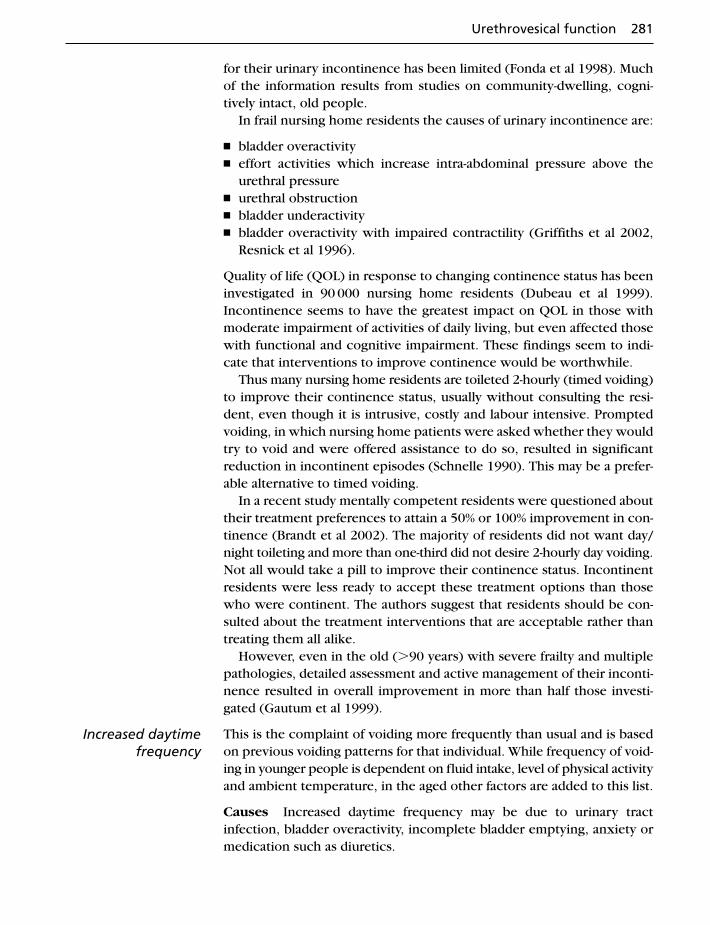
for their urinary incontinence has been limited (Fonda et al 1998). Muchof the information results from studies on community-dwelling, cogni-tively intact, old people.
In frail nursing home residents the causes of urinary incontinence are:
■ bladder overactivity■ effort activities which increase intra-abdominal pressure above the
urethral pressure■ urethral obstruction■ bladder underactivity■ bladder overactivity with impaired contractility (Griffiths et al 2002,
Resnick et al 1996).
Quality of life (QOL) in response to changing continence status has beeninvestigated in 90 000 nursing home residents (Dubeau et al 1999).Incontinence seems to have the greatest impact on QOL in those withmoderate impairment of activities of daily living, but even affected thosewith functional and cognitive impairment. These findings seem to indi-cate that interventions to improve continence would be worthwhile.
Thus many nursing home residents are toileted 2-hourly (timed voiding)to improve their continence status, usually without consulting the resi-dent, even though it is intrusive, costly and labour intensive. Promptedvoiding, in which nursing home patients were asked whether they wouldtry to void and were offered assistance to do so, resulted in significantreduction in incontinent episodes (Schnelle 1990). This may be a prefer-able alternative to timed voiding.
In a recent study mentally competent residents were questioned abouttheir treatment preferences to attain a 50% or 100% improvement in con-tinence (Brandt et al 2002). The majority of residents did not want day/night toileting and more than one-third did not desire 2-hourly day voiding.Not all would take a pill to improve their continence status. Incontinentresidents were less ready to accept these treatment options than thosewho were continent. The authors suggest that residents should be con-sulted about the treatment interventions that are acceptable rather thantreating them all alike.
However, even in the old (�90 years) with severe frailty and multiplepathologies, detailed assessment and active management of their inconti-nence resulted in overall improvement in more than half those investi-gated (Gautum et al 1999).
This is the complaint of voiding more frequently than usual and is basedon previous voiding patterns for that individual. While frequency of void-ing in younger people is dependent on fluid intake, level of physical activityand ambient temperature, in the aged other factors are added to this list.
Causes Increased daytime frequency may be due to urinary tract infection, bladder overactivity, incomplete bladder emptying, anxiety ormedication such as diuretics.
Urethrovesical function 281
Increased daytimefrequency

Management The traditional method of assessment uses a frequency/volume chart in which the number of voids and the volume per void arerecorded. This record needs to be kept for at least 24 hours and prefer-ably 48 hours. Fluid intake and types of fluid should be recorded at thesame time. However, many community-based patients find such monitoringtedious and this would be compounded in the aged care setting. Recordingthe number of voids per 24 hours and the times of voiding as well as thefluid intake (mugs of fluid per day) can be an easier alternative.
Advice regarding modifying fluid intake and strategies to defer voidingcan then be provided.
Physiotherapy interventions include use of a voiding position andpattern, deferred voiding, and tonic PFM re-education.
This is defined as the number of voids preceded and followed by sleep. Itis considered pathological if it occurs more than twice a night. This fre-quency has been noted in 50% of those older than 80 years (Swithinbanket al 1998a).
Causes1. Nocturnal polyuria This occurs when �35% of total 24-hour urineoutput is produced in the sleeping hours. This was noted in 71% of 80-year-olds with nocturia (Swithinbank et al 1998a). This change in theusual circadian pattern of urine production occurs in normal, continentand urodynamically assessed elders (Morgan et al 2000).
Factors associated with disordered circadian urine output (Weiss et al1998) include:
■ diuretic medication, alcohol, caffeine, and excessive fluid intake latebefore retiring
■ congestive heart disease, peripheral oedema (accumulation of water inextracellular space)
■ stroke, diabetes■ renal insufficiency■ decreased plasma vasopression levels.
An increased oedema ratio before sleep has been noted in the elderlywith nocturia – that is the ratio of extracellular water to total body watervolume (Sugaya et al 2001).
2. Sleep apnoea Significant associations have been found between nocturia and sleep apnoea, irritable bladder symptoms and apnoea, andnocturia and body mass index in adults of both genders older than 55 years (Umlauf et al 1999).
3. Nocturnal detrusor overactivity (NDO) This was the most commoncause of nocturia – in more than half the subjects studied. Mixed noctur-nal polyuria and NDO occurs in about one-third of the cases (Weiss et al1998).
282 Physiotherapy for pelvic floor dysfunction in the aged care setting
Nocturia

Effects Nocturia causes sleep disturbance in the frail and increasedrisk of traumatic injury from falls. Many women consider it bothersomeif it occurs more than two times a night.
Management Strategies suggested for nocturia, after medical aspectshave been addressed, include:
■ Evening fluid restriction. Those who complain of thirst during thenight may ease this by sucking an ice cube rather than consuming a fullglass of water.
■ Timed diuretics. These may be taken in the late afternoon and this mayhelp clear peripheral oedema accumulated during the day.
■ An afternoon nap. Resting with legs horizontal or elevated for an houror two may assist extracellular fluid return, rather than all of thisoccurring at night.
■ Topical vaginal oestrogen. This is more effective than systemic oestro-gen in decreasing urgency and frequency (Cardozo et al 2001).
Oral desmopressin (vasopressin), an antidiuretic agent, has been used inthe treatment of nocturnal polyuria. However, side effects of headache,nausea, dizziness and abdominal pain have occurred. In patients over 65years, hyponatraemia, where the serum sodium levels fall below normal,is a significant problem and generally rules out this form of treatment inthe older age groups of both genders (Abrams et al 2001).
Physiotherapy interventions include PFM tonic re-education, useof a voiding position and pattern and daytime deferment to increase bladder capacity.
This is experienced as a strong desire to void that cannot be deferred. It fre-quently results in immediate urine loss at the onset of urge, or as approach-ing or undressing at the toilet. Loss is often a large volume that necessitatesa change of clothing. In many cases it is perceived as a penile or perinealsensation which can trigger a response of perineal pressure by hand orcrossing of legs in females. Males may compress the penis. These actionsmimic those used by young children who are gaining bladder control.These strategies have been shown to have an inhibitory effect on bladderpressure, via afferents of the dorsal nerve of penis and clitoris and afferentsfrom the hip adductor muscles (Lindstrom & Sudsuang 1989). Urge urinaryincontinence, but not stress urinary incontinence, has also been impli-cated in non-spinal, non-traumatic falls and fractures (Edwards 2000).
Symptoms■ Perineal or penile sensation of urge to void.■ Inability to defer voiding, worse as the person nears the toilet.■ Increased day frequency.■ Nocturia.■ Large volume urine loss.■ Urge often occurs on rising from bed or chair.
Urethrovesical function 283
Urgency and urgeincontinence

■ Urge can be triggered by the sound of running water, homecoming,key in the door.
Prevalence Urge incontinence occurs in 23.9% of males and 36.4% offemales over 70 years.
Those with weak or intermittent stream are more likely to report asense of urge. Age and female gender increase the risk of urge with orwithout incontinence (Nuotio et al 2001).
Causes1. Decreased bladder inhibition due to:
■ ↓cerebral blood flow, particularly underperfusion of the right superior frontal lobe (Griffiths et al 1994)
■ neurological conditions (stroke, parkinsonism), which can interferewith normal inhibitory pathways
■ ↓PFM support of the bladder (Gunnarsson & Mattiasson 1999).2. Decreased urethral closing pressure:
■ ↓periurethral oestrogen results in ↓urethral closing pressure(oestrogen nourishes the urethral mucosa and vascular submucosa)
■ ↓periurethral muscle activity■ spontaneous fall in maximal urethral pressure (urethral instability)
prior to unprovoked detrusor contraction (Wise et al 1993).3. Bladder overactivity due to mechanical factors:
■ prolapse drag � urethral kinking■ uterine fibroid pressure■ BPH outlet obstruction (Rosier et al 1995)■ Bladder stones, tumour.
4. Decreased bladder contractility with or without bladder overactivity(Griffiths et al 2002).
5. Medication – diuretics, anticholinergics, antipsychotics, sedatives,hypnotics (Kirschner-Hermanns et al 1997).
6. Anxiety.
Management As existing medication can contribute to urgency andurge incontinence, a review of medication is important before any treat-ing medications are added. Response to treatment of urinary urge incon-tinence in the elderly varies.
Use of oxybutynin chloride, an antispasmodic, in those with bladderoveractivity � impaired detrusor contractility (mean age of 79 years)resulted in side effects of a dry mouth and increased post-void residuals.Constipation occurred rarely. Treatment was successful in both groups,but cure rates were less in those with impaired contractility (Miller et al2000). Outcome measures of treatment in women �75 years using oxybutynin � bladder retraining for 6–8 weeks resulted in reduction of dayfrequency and incontinent episodes (Wagg et al 1999). Topical vaginaloestrogen is also helpful (Cardozo et al 2001).
284 Physiotherapy for pelvic floor dysfunction in the aged care setting

Urethrovesical function 285
Physiotherapy interventions include monitoring of urinary output,via a frequency/volume chart, and fluid intake, urge control techniquesand retraining tonic and strong PFM support.
Factors predicting urine loss prior to treatment in the elderly includefluid intake, voiding frequency, impaired orientation in time on cognitivetesting. Factors that predicted persistent loss, after treatment, wereunderperfusion of the cerebral cortex, reduced bladder sensation, andimpaired orientation in time on cognitive testing. The latter two are con-sistent with underperfusion of the right superior frontal lobes (Griffithset al 1994, 1996).
Stress incontinence occurs when the bladder pressure (due to an increasein abdominal pressure) is greater than the urethral closing pressure. Itaffects women predominantly, but may be less of a problem in the agedthan urge incontinence. This is particularly so if the person coughs orsneezes infrequently. It can occur in men following radical prostatectomyfor cancer of the prostate. Urine loss is generally a spot loss at the time of increased intra-abdominal pressure. Also continuous seepage of urinecan occur with walking and moving if the resting urethral pressure is low.
Symptoms A spot loss of urine associated with sneeze, cough, laugh,lift, bend.
Causes■ Weak PFM support of the urethra and vagina.■ Weak periurethral sphincter muscles, which are responsible for
urethral closure prior to increases in intra-abdominal pressure (IAP)with effort.
■ Increased vaginal, urethral and bladder neck mobility due to vaginaldeliveries, regular straining at defecation, chronic coughing and obesity.
■ Obesity generates higher resting abdominal pressures and higher pres-sures on coughing than occur in those within a normal weight range(Sullivan et al 2000).
■ Weak abdominal tonic and phasic activity. When women lean forwardto bend or lift, the abdominal wall can sag forward. It seems that trans-versus abdominis does not hold automatically. When the abdomensags, pubococcygeal resting activity falls below baseline (Sapsford &Hodges 2001). It has been observed clinically in those with weakabdominal muscles, that the abdomen bulges instead of drawing induring coughing. This results in decreased PFM recruitment.
Associated conditions Elderly women with stress urinary incontinenceare more likely to have:
■ atrophic vaginitis■ cystocele■ rectocele■ hysterectomy
Stress urinary incontinence

286 Physiotherapy for pelvic floor dysfunction in the aged care setting
■ oestrogen replacement therapy■ greater body mass index■ increased parity■ decreased vaginal muscle tone■ decreased sustained PFM contraction (Burns et al 1993).
Treatment Options for women include a conservative approach of PFMrehabilitation (Berghmans et al 1998), or a surgical intervention such asBurch colposuspension or tension free vaginal tape. Treatment programmesfor post-radical prostatectomy incontinence that included pelvic floorexercise � neuromuscular stimulation found no difference in outcomes32 weeks post surgery (Moore et al 1998). Recent clinical observationsindicate that increasing intensity of abdominal exercise, with pelvic floorholds, has resulted in a marked decrease in urine loss in men with stressincontinence following radical prostatectomy. The added demands ofuphill running (which requires increased tonic abdominal activity) havefurther enhanced continence (P. Dornan 2002, personal communica-tion). Some of those in the aged care setting will be able to cope withincreased abdominal muscle activity.
Physiotherapy intervention focuses on PFM rehabilitation.
This is the term for loss of urine during sleep.
Causes It was noted in many older subjects (mean age 79 years) thatenuresis occurred in close time relationship to an episode of sleep apnoeaor hypopnoea (Ouslander et al 1999). Specific sleep disorders may beimplicated in the pathophysiology of night-time incontinence. Associatedconditions include nocturia �2, urgency and urge incontinence and stressurinary incontinence, but enuresis is not always related to age (Swithinbanket al 1998b).
Management Strategies must first deal with other medical problems.In those who do not wake to void, arousal to bladder sensation does notoccur, though bladder sensation may be impaired even in waking hours.People who are mentally competent may be prepared to use an alarm tobe woken after 4 hours of sleep to void.
This tends to be a continuous loss of urine both day and night. Loss that only occurs in the upright position is likely to be due to low urethralresting pressure.
Causes1. Non contracting bladder. In the elderly this is likely to be related to
some form of autonomic neuropathy, such as can occur in diabetes.Urine loss occurs when the pressure of urine is greater than the restingurethral pressure. Voiding can occur with an increase in intra-abdominalpressure, e.g. with straining, but seepage will occur in the absence ofbladder awareness and regular emptying. Management options includeintermittent clean self-catheterization or an indwelling catheter.
Nocturnal enuresis
Continuous urinaryincontinence

2. Bladder outlet obstruction. Prostatic enlargement can obstruct the urethra, leading to straining and overflow. Bladder volumes may reach high levels. This may also result from a cystocele, which kinks theurethra and restricts flow. However, reduction of the cystocele, eitherby a pessary or surgery, can unmask an incompetent urethral sphincterand this leads to further incontinence.
Pelvic organs are supported by passive and active systems – fascia and ligaments and striated muscle. Failure in either of these systems can leadto prolapse. However, anterior pubococcygeus histological examinationshowed no difference in muscle fibre type percentage and fibre diameterin patients with prolapse and/or urinary incontinence when comparedto asymptomatic women. Fibre types were 66% slow twitch fibres incomparison with 48% in other human female muscle (Heit et al 1996).
Prolapse is a protrusion of a pelvic organ or structure distal to its nor-mal anatomical level. It can involve the bladder (cystocele), the urethraand bladder (cystourethrocele), the uterus and vagina (uterine descent,vaginal vault prolapse, enterocele) or the rectum (rectocele). Any organdescent within the vagina is termed a 1st degree prolapse. A 2nd degreeprolapse reaches the introitus, and a 3rd degree protrudes beyond theintroitus. While a cystourethrocele (the anterior compartment) is the mostcommon type of prolapse, the majority of patients demonstrate problemsin the middle and posterior compartments as well. These have beendemonstrated on dynamic cystoproctography (Maglinte et al 1999).Significant associations have been found between large rectoceles andanal incontinence.
This is very difficult to ascertain as many prolapses are asymptomatic. Ina younger population, 20–59 years, 30.8% of the women had a prolapse,but only 2% were 2nd degree. Associated conditions in this group wereparity, PFM strength and maximum birth weight (Samuelsson et al 1999).By the age of 80 years the lifetime risk of undergoing surgery for prolapsein America was 11.1% (Olsen et al 1997).
■ Congenital collagen deficits – a decrease in the total collagen contenthas been observed. Women with these types of problems have beennoted to have joint hypermobility (Norton et al 1995).
■ Increasing parity plus larger birth weight babies.■ Increases in intra-abdominal pressure such as occur with frequent cough-
ing in chronic obstructive pulmonary disease and smoking. Frequentstraining at stool in those with difficult evacuation can contribute toprolapse (Spence-Jones et al 1994).
■ Vaginal vault prolapse may occur following hysterectomy, or bladderneck suspension.
Urogenital prolapse 287
Urogenital prolapse
Prevalence
Causes

■ Dragging discomfort and/or a sense of heaviness due to abnormal tension on the stretched passive tissues. This is worse after prolongedstanding or at the end of the day.
■ A lump protruding from the vagina.■ Urinary symptoms, e.g. stress urinary incontinence, urgency and
frequency.■ Incomplete bladder emptying, resulting from urethral kinking, with
frequency and urinary tract infection.■ Uterine prolapse is often accompanied by low backache.■ A rectocele often results in incomplete bowel emptying.
Stretched passive structures will not shorten with pelvic floor exercise.However, in cases of heaviness and dragging, improving tonic PFM sup-port can relieve these symptoms as well as those of low backache.Symptomatic prolapses generally require surgical repair. Uterine pro-lapse can be supported with a pessary that elevates the cervix. Thisrequires adequate PFM tissue to maintain the pessary in position. A pes-sary can be a long-term option in some women. However, those who strainto evacuate will be likely to push the pessary out. Women with a recto-cele may use upward perineal body pressure, or backward pressure onthe rectocele per vaginam, to assist evacuation. Upward perineal pres-sure increases rectal tone (Gosselink & Schouten 2002).
Physiotherapy interventions for these conditions include tonicPFM rehabilitation, and teaching of voiding and defecation positions andpatterns. These are beneficial taught prior to surgery to enable optimalfunction postoperatively.
Normal anorectal function results in defecation between three times aday and three times a week in more than 90% of a healthy working popu-lation (Drossman et al 1982). Ease of emptying depends on fibre and fluidintake, colonic motility, rectal sensitivity and contractility, pelvic floorsupport and anal release.
Liquid contents pass into the colon via the ileocaecal valve and traversethe ascending, transverse, descending and sigmoid colon into the rectum.Different types of colonic activity either move the contents back and forthto allow absorption of water and electrolytes, or propel them onwards.This peristalsis is stimulated by food ingestion – gastrocolic reflex – andin some people by moving and walking. Coffee has been observed to trig-ger rectosigmoid motility, and a subsequent desire to evacuate, in somepeople, and this occurs with or without caffeine (Brown et al 1990).Ingestion of hot water does not produce the same effect as coffee in thispopulation. A similar stimulating effect of coffee, either with or withoutcaffeine, has been reported recently on blood pressure and nervous systemactivity (Corti et al 2002). In healthy people increasing physical activity
288 Physiotherapy for pelvic floor dysfunction in the aged care setting
Symptoms
Management
Anorectal function
Colonic motility

Anorectal function 289
does not generally change the rate of passage of intestinal contents, butthe effect in the poorly mobile aged population is likely to differ.
The rectum begins at the sacral convexity and ends at the anorectal junc-tion just anterior to the tip of the coccyx. The rectal smooth musclewalls, inner circular and outer longitudinal, extend into the anal canal.Here the inner circular muscle thickens to form the internal anal sphinc-ter (IAS), which is under autonomic control and is responsible for 85% ofanal resting pressure. The striated external anal sphincter (EAS) encirclesthe IAS, contributing 15% to the anal resting pressure. Puborectalis isincorporated into the deep part of EAS and acts as one with it. Voluntarycontraction of the anal sphincter – squeeze pressure – can double theresting pressure but cannot be maintained for long.
Initial awareness of rectal filling occurs with �30 ml of contents (sen-sory threshold volume). This awareness triggers the IAS to relax, and thereis a simultaneous increase in EAS activity to maintain continence. ThisIAS relaxation allows the very sensitive anal mucosa to sample the rectalcontents, but defecation can still be deferred. The IAS activity recoversquickly with a consequent reduction in EAS activity back to resting levels.Further intermittent rectal filling will result in a desire to evacuate ataround 60–100 ml of contents (defecation threshold volume). At thistime the IAS remains relaxed and EAS activity is increased to maintaincontinence until evacuation is imminent. This reflex is termed the rectoanal inhibitory reflex (RAIR) and is mediated by receptors of theenteric nervous system within the rectal wall. If evacuation does not pro-ceed, the rectum accommodates to the contents, the sensation of rectalfullness dissipates, and the IAS and EAS return to their normal restingpressure levels. The desire to empty may not occur again until the nextbolus of faecal matter enters the rectum, possibly on the next day. Such adefinite RAIR response to rectal filling may not occur if contents pass intothe rectum very gradually (Rasmussen 1994).
For a person to be continent the anal pressure must be greater than therectal pressure whenever contents are present. Resting rectal pressureis �5 cmH2O, whereas resting anal pressure may be 75–80 cmH2O(Akervall et al 1990).
The following factors contribute to anal continence:
■ anal pressure that is greater than rectal pressure■ puborectalis maintains the anorectal angle at or �90 degrees■ awareness of rectal contents■ RAIR relaxes the IAS, but EAS activity compensates to maintain
continence■ rectal compliance and capacity can accommodate contents■ rate of filling, consistency and volume of contents must be within
reasonable limits.
Rectal filling
Anal continence –what maintains it?

290 Physiotherapy for pelvic floor dysfunction in the aged care setting
The person sits or squats and bowel emptying proceeds in the followingorder.
■ Relaxation of the pelvic floor occurs, in particular puborectalis andthe EAS, and the abdominal muscles.
■ The pelvic floor descends, the anorectal angle widens and the analcanal widens and shortens.
■ The rectum empties due to its intrinsic contractions.■ If effort is required to commence or continue evacuation, the waist
widens sideways (bracing) due to the combined action of diaphrag-matic descent and activation of the transverse, internal oblique andexternal oblique abdominal muscles. This increases IAP.
■ The PFM hold isometrically in the lowered position to support the rectum against the increased IAP (Lubowski et al 1992).
■ The abdominal wall is bulged forward by further descent of thediaphragm and lengthening of the held abdominal muscles (Chiarelli &Markwell 1992, Markwell & Sapsford 1995). This bulging allows releaseof puborectalis and EAS.
■ Evacuation proceeds until the rectum is empty. However, completeemptying does not always occur.
■ The pressure in IAS and EAS is restored once the rectum is empty andthe pelvic floor ascends to its resting position (Markwell 1998). This istermed the closing reflex.
■ Maximal rectal volume.■ Resting anal pressure.■ Maximal anal squeeze pressure.■ Volume of contents to trigger RAIR.■ Pelvic floor support.
■ Threshold pressure to trigger RAIR.■ Threshold pressure for perception of rectal filling.
Rectal contractions were observed in 78% of the normal sampled popu-lation. There was an association with gender (less in females), but notage. While increasing parity did not show any effect on anal function inwomen in the study by Akervall et al (1990), sphincter integrity and pres-sures, both IAS and EAS, can be affected by vaginal delivery (Sultan et al1994, Wynne et al 1996). Age seems to affect maximal squeeze pressuremore than the resting pressure.
There are also changes in intestinal motility that influence function.These include decreased colonic contractions and decreased gastrocolicreflex. Immobility contributes to delayed colon transit.
Defecation – theprocess
Changes in anorectal function with ageing (Akervall et al 1990)
Increases
Decreases

Constipation, faecal incontinence, faecal impaction and obstructed defe-cation are the most common problems facing those in aged care. They donot seem to be separate entities as they can be in younger people, forconstipation and obstructed defecation lead to faecal impaction in whichcontents then bypass the impacted faecal mass and result in soiling andincontinence.
In true constipation intestinal contents are slow to reach the rectum,hence there is a decreased frequency of evacuation. Slow colonic transitoccurs throughout the life spectrum, but in the aged other factors maycause or exacerbate slow transit. The more common causes are listed.
Causes■ Decreased fibre and fluid intake.■ Megacolon/megarectum.■ Decreased neurological functioning affecting autonomic and enteric
systems.■ Endocrine changes such as hypothyroidism.■ Pharmacological ingestion – e.g. opiates, antidepressants, diuretics, iron,
anticholinergics.■ Immobility.■ Irritable bowel syndrome.■ Faecal impaction and outlet obstruction.
Symptoms (Talley et al 1993)■ Straining �25% of time.■ Hard stool �25% of time.■ Feeling of incomplete evacuation �25% of time.■ Fewer than 2–3 stools per week.■ Postprandial abdominal bloating.■ Haemorrhoids.■ Rectal mucosal prolapse.■ Laxative and enema use are indicative of constipation.
Faecal impaction with decreased frequency of evacuation and conse-quent faecal incontinence is particularly common in demented patientsand those with severe mobility impairments in the nursing home popu-lation (Chassagne et al 2000).
Management This is a continual process in this group. Use of osmoticagents, e.g. lactulose, combined with rectal stimulants and weekly ene-mas resulted in fewer episodes of faecal incontinence in those patientswho had complete rectal emptying (Chassagne et al 2000). This resultedin a reduced workload for the carers. In those who participated inprompted voiding for urinary function, there was also an increase in the fre-quency of bowel movements. Those whose urinary control improved onthe prompted voiding regimen also had a greater proportion of continent
Anorectal function 291
Constipation
Problems ofanorectal function
with ageing

292 Physiotherapy for pelvic floor dysfunction in the aged care setting
bowel movements (Ouslander et al 1996). Suggested positive contributingfactors were increased mobility for voiding, increased fluid intake at theprompted void (everyone was given fluid at the time of prompting) andpossible positive response of faecal impaction to this programme.
Physiotherapy intervention in the management of constipation innursing home patients is likely to be very limited. Less dependent, motiv-ated residents will benefit from improving tonic PFM activity prior todefecation retraining.
This is the term used when a patient reports the desire to evacuate but isunable to get the contents out. There is a feeling of anal blockage, incom-plete emptying and a prolonged defecatory attempt. Use of upward digital perineal body pressure and digital disimpaction are also noted (Talleyet al 1993). Obstructed defecation and slow colonic transit often coexist.
Signs noted on defecography are rectocele, enterocele, rectal pro-lapse, anal incontinence and excessive pelvic floor descent. Those withmore vaginal deliveries and a hysterectomy are likely to be at greater risk.A gastrorectal reflex resulting in increased rectal tone occurs in healthycontrols after the ingestion of food. It has also been noted in people withobstructed defecation who have normal colon transit times, but isimpaired in those with prolonged transit (Gosselink & Schouten 2001).
Causes Poor rectal support results in excessive pelvic floor descentduring attempted defecation (Markwell 1998). A rectocele allows rectalcontents to be pushed into the lax rectal tissue and a bulge is noted in thevagina. Both of these conditions result in regular straining with later devel-opment of haemorrhoids as well as the signs mentioned earlier. Pain, dueto repeated pudendal nerve stretch, may develop later.
Management As with constipation, less dependent motivated residentswill benefit from:
■ encouragement to respond to the desire to defecate after food inges-tion – particularly ingestion after a prolonged period without food, e.g.in the morning
■ adequate dietary fibre intake, frequently with a fibre supplement aswell as fruit and vegetables; those people with coexisting slow colonictransit may find bloating is aggravated by wheat grain fibre productsand other high fibre foods
■ use of digital perineal support.
Physiotherapy intervention should improve tonic PFM activity priorto defecation retraining.
More recently the term anal incontinence has been used, instead of faecal incontinence (FI), as this includes accidental loss of gas, liquid orsolid contents. While vaginal delivery has been stated as the precipitatingfactor in most cases of anal incontinence in a younger population, those
Obstructed defecation
Anal incontinence

in nursing home care present with two main problems. They either havefaecal impaction with bypassing of the mass by liquid contents or a neurogenic cause. Symptoms such as diarrhoea must be investigated.
Prevalence In those �75 years living at home 3% reported regular FI(Edwards & Jones 2001). Figures for women were higher than for men.Prevalence figures vary depending on the criteria used – whether loss isoccasional or daily or any stated frequency in between. Significant associ-ations with FI were anxiety, depression and increasing disability.Amongst those in residential homes for the elderly 10.3% were inconti-nent one or more times a week (Tobin & Brocklehurst 1986b). There wasan even distribution of those with faecal impaction and neurogenicincontinence. While the majority affected suffered from dementia, poormobility was also a factor. Higher figures are reported for nursing homes.
Causes Pudendal nerve stretching during vaginal delivery may be the pre-cursor of later neurogenic problems. Occult anal sphincter tears inducedby vaginal delivery result in immediate problems of control, but neuro-genic damage does not manifest itself until later in life, and is com-pounded by ageing and disease. These patients tend to pass formed stool.Repair of torn anal sphincters, as diagnosed by anal endosonography, canhave benefit even in the elderly.
Faecal impaction and bypassing result from an accumulation of faecalmatter in the rectum. These patients tend to have continual soiling.Decreased colon motility and gastrocolic reflexes, as well as an increasedthreshold for perception of rectal contents, mean that filling does nottrigger a defecation response as it did in younger years. If then there is little pelvic floor support of the rectum on attempted evacuation in thepresence of reduced rectal contractility, emptying will be infrequent andincomplete, and further rectal filling will add to the mass.
Incomplete rectal emptying with a small loss of residual contents, notassociated with impaction, may occur in many of those in all areas of careand in the community. Contributing factors to incomplete emptying areconsistency of faecal contents and poor rectal support during evacuation.
Management For nursing home residents with faecal impaction treat-ment involves ongoing care to relieve the impaction and prevent recur-rence. Enemas to clear the impaction, followed by regular osmoticlaxatives and enemas were used to keep the rectum empty. Residents withneurogenic incontinence were constipated with codeine phosphate andgiven twice weekly enemas (Tobin & Brocklehurst 1986b). The cure ratein residents who complied with the chosen regimen was 87% (Tobin &Brocklehurst 1986b).
Physiotherapy intervention of defecation retraining is helpful forthose with incomplete emptying and loss some time later. The ingestionof a stool-bulking agent, such as psyllium, creates a bigger stool whichempties more completely and this complements the retraining.
Anorectal function 293
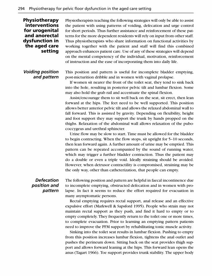
Physiotherapists teaching the following strategies will only be able to assistthe patient with using patterns of voiding, defecation and urge controlfor short periods. Thus further assistance and reinforcement of these pat-terns for the more dependent residents will rely on input from other staff.Thus physiotherapists who share information on functional activities byworking together with the patient and staff will find this combinedapproach enhances patient care. Use of any of these strategies will dependon the mental competency of the individual, motivation, reinforcementof instruction and the ease of incorporating them into daily life.
This position and pattern is useful for incomplete bladder emptying,post-micturition dribble and in women with vaginal prolapse.
If women sit nearer the front of the toilet seat, they tend to sink backinto the hole, resulting in posterior pelvic tilt and lumbar flexion. Somemay also hold the grab rail and accentuate the spinal flexion.
Assist/encourage them to sit well back on the seat, sit erect, then leanforward at the hips. The feet need to be well supported. This positionallows better anterior pelvic tilt and allows the relaxed abdominal wall tofall forward. This is assisted by gravity. Depending on flexibility, heightand foot support they may support the trunk by hands propped on thethighs. Relaxation of the abdominal wall allows relaxation of the pubo-coccygeus and urethral sphincter.
Urine flow may be slow to start. Time must be allowed for the bladderto begin contracting. When the flow stops, sit upright for 5–10 seconds,then lean forward again. A further amount of urine may be emptied. Thispattern can be repeated accompanied by the sound of running water,which may trigger a further bladder contraction. Thus the patient may do a double or even a triple void. Ideally straining should be avoided.However, when detrusor contractility is compromised, straining may bethe only way, other than catheterization, that people can empty.
The following position and pattern are helpful in faecal incontinence dueto incomplete emptying, obstructed defecation and in women with pro-lapse. In fact it seems to reduce the effort required for evacuation inmany asymptomatic persons.
Rectal emptying requires rectal support, anal release and an effectiveexpulsive effort (Markwell & Sapsford 1995). People who strain may notmaintain rectal support as they push, and find it hard to empty or toempty completely. They frequently return to the toilet one or more times,to complete evacuation. Prior to learning an emptying pattern patientsneed to improve the PFM support by rehabilitating tonic muscle activity.
Sinking into the toilet seat results in lumbar flexion. Pushing to emptyfrom this position increases lumbar flexion, tightens the anal outlet andpushes the perineum down. Sitting back on the seat provides thigh sup-port and allows forward leaning at the hips. This forward lean opens theanus (Tagart 1966). Toe support provides trunk stability. The upper body
294 Physiotherapy for pelvic floor dysfunction in the aged care setting
Physiotherapyinterventionsfor urogenitaland anorectal
dysfunction inthe aged care
setting
Voiding positionand pattern
Defecation position and
pattern

Physiotherapy interventions for urogenital and anorectal dysfunction in the aged 295
can be supported through the arms with hands or forearms propped onthe thighs. A good lumbar curve encourages a better pushing pattern.Assuming this position may be difficult for those who use a raised toiletseat. See Figures 14.1, 14.2, 14.3 and 14.4.
As the person pushes, the spine should not move, the lower ribswiden sideways (this action provides rectal support) and the abdomenbulges forward (this action opens the anal outlet) (Markwell & Sapsford1995). If there is pelvic floor laxity, perineal descent will still occur usingthis pattern. Digital support of the perineum or ischiorectal fossa region(between the anus and the ischial tuberosity) may help in these cases.Digital support of the perineal body region during defecation has beenshown to increase rectal tone in healthy controls and to a lesser extent inthose with obstructed defecation (Gosselink & Schouten 2002). Upwardpressure in the ischiorectal fossa region supplements or provides rectalsupport against the increased IAP. This support is normally provided byiliococcygeus. Frequently, initial emptying requires effort or digital sup-port, then the rest of the contents are passed easily. This is more likely tooccur if adequate fibre in the diet results in a soft, formed and texturedstool. Contents that are pasty are harder to empty completely and requireprolonged wiping attempts. Use of this defecation pattern, at the end of
Figure 14.1A flexed lumbar spine andslumped sitting position can leadto incomplete bladder and bowelemptying. Many older patientsmay be perched on a high seatwith legs dangling. Foot supportis needed for trunk stability.
Figure 14.2In forward lean sitting, whilemaintaining a good lumbar curve,it is easy to relax the abdominalwall for voiding. This is useful forall women, but is particularlyhelpful for those who suffer frompost-micturition dribble.

296 Physiotherapy for pelvic floor dysfunction in the aged care setting
the initial part of evacuation when the person feels that emptying is notcomplete, may empty any residual faecal matter, and avoid soiling.
Retraining, involving activation of support first followed by anal releasewhile the support is maintained, can result in more effective emptying,when there is perineal descent. However, this is a complex pattern andmay be beyond the ability of this group of patients to achieve. Referral toa physiotherapist who works with pelvic floor dysfunction may be anoption for suitable patients.
Urgency is often perceived as a perineal or penile sensation, which isprobably related to urethral relaxation. Urge is more of a problem whenmoving from lying and sitting into standing, or when walking and bend-ing. In forward lean the abdominal wall often sags forward with conse-quent relaxation of the PFM. To prevent urge occurring in the abovesituations the lower abdominal wall should be held gently firm before
Figure 14.3The position for defecation issimilar to that used for voiding.Forward leaning at the hips opensthe anal outlet. Forearms restingon thighs (one arm has beenstraightened for the purposes ofthe photograph), heels raisedwith weight resting on toes andeyes looking ahead, not down, allassist in maintaining a lumbarcurve. This lumbar curve facilitatesan effective evacuation pattern ofabdominal bulging (see text).
Figure 14.4Those people using a raisedtoilet seat due to limited hipflexion after hip replacementmay still be able to assume aforward lean position if theaffected hip is in a moreextended position with eitherknee flexion or extension.
Urge preventionand urge control

Physiotherapy interventions for urogenital and anorectal dysfunction in the aged 297
moving (Sapsford & Hodges 2001). The hold should be sustained whilewalking to the toilet or until the bladder overactivity quietens. Gentle lowerabdominal holds should be used during vacuuming or bending, but shouldnever impact on breathing. See section on tonic PFM activity, below.
Some women have found that wearing an abdominal support garmentis beneficial in inhibiting urgency (Pietzsch et al 2001). Lifting the laxlower abdomen by hand and holding it lifted as they walk to the toilet hasbeen helpful in maintaining bladder control for some women. These twostrategies are probably more beneficial in those with an obese abdomen.
When the urge begins certain strategies can be used to quieten it.However, the earlier the strategy is implemented the more successful itwill be. Once the detrusor muscle is contracting and urine is escaping,there is little hope of controlling it. The following actions have beenfound helpful:
■ hand pressure over clitoris and to give vaginal support■ pressure on the glans or compressing the penis■ sitting on the edge of a firm chair with pressure over the vaginal/
perineal region■ crossing legs – hip adductor afferents have an inhibitory effect on the
bladder when standing■ a firm PFM hold – though this does not have an inhibitory effect on the
bladder (Lindstrom & Sudsuang 1989)■ strong gluteal contraction (buttock squeeze) combined with curling
the toes down when standing.
Each of these interventions needs to be maintained until the urge sensa-tion fades. It may take 10–12 seconds. The last two are the only ones thatare socially acceptable. When the urge quietens, the person can thenwalk to the toilet to void. However, the urge often returns while walkingunless the person shifts their mental focus to something other than thebladder. Counting every step as they walk and while undressing can pro-vide another focus. Gentle lower abdominal holds, which result in tonicPFM activity, while walking and undressing may help too.
Once the patient has learnt to subdue the urge they should be encour-aged to defer voiding. This deferment should commence with one or twominutes and should gradually increase. This is called bladder training.The bladder slowly adapts to greater volumes and does not signal the needto empty so soon. This retraining may take 3 to 6 months and is oftenconducted with help from an antispasmodic medication, e.g. oxybutyninchloride. However, many older people may never be able to defer, butthey may reach the toilet without loss. Daytime deferment with conse-quent increase in bladder capacity may benefit those with nocturia.
This strategy is useful in preventing urinary urgency, to decrease the dis-comfort of a prolapse, to initiate pelvic organ support prior to lifting, cough-ing and sneezing, and as the initial phase of a PFM rehabilitation programme.
Tonic pelvic floormuscle activity

The pelvic floor muscles have a predominance of slow twitch fibres to maintain tonic activity for sustained antigravity support and outlet closure. Loss of tonic activity has been demonstrated in some womenwith stress urinary incontinence, and even in some nulliparous females(Deindl et al 1994). Thus retraining of tonic activity prior to strengthen-ing seems warranted. Activation of slow twitch PFM fibres to retraintonic activity requires a slow and gentle onset and a sustained hold at lowintensity. This can be approached in two ways.
Gentle isometric lower abdominal muscle holds have been shown to acti-vate pubococcygeus and the external anal sphincter (Sapsford & Hodges2001). In standing with a normal lumbar curve and without moving thespine, the patient very slowly and gently draws in the lower abdomentowards the spine, without using any respiratory effort. The muscle is thenheld, and light breathing recommences. A subjective awareness of slightperineal tension, either urethral, vaginal or anal, should be reported onabdominal in-drawing or release of tension on relaxation. This abdominalaction can be palpated and observed, and is more obvious on release.Women who have had vaginal repair surgery may have limited or no per-ineal awareness in response to lower abdominal activation. Sustained holdsshould be built up to 10, 15, 20 and more seconds, repeating five in a row,five times a day. When the action has been mastered they can then be incor-porated into daily activities in standing, e.g. cleaning teeth, showering, iron-ing, talking and later walking and sitting. Use before and during bending andlifting activities can prevent loss of urine in many women.
An alternative approach is to use a very gentle cognitive periurethralmuscle hold. In upright unsupported sitting with a good lumbar curve,the patient gently holds an imagined flow of urine. As the flow is held,lower abdominal palpation will note an increase in tension, or a slightdrawing-in of the deep abdominal muscles. Release of the abdomen on release of the imagined urine flow may be more obvious than the tensioning effect.
Once the tonic activity has been mastered, strengthening of the PFM canbe added. However, tonic exercise should not cease when strength workbegins. Coughing and sneezing require a stronger PFM activation. Strength-ening can be achieved either by cognitive PFM holds, or by strong isometric abdominal holds (Sapsford & Hodges 2001). Even withoutbuilding up much strength a cognitive PFM hold prior to and during acough has been shown to significantly decrease urine loss in womenwith mild to moderate stress urinary incontinence (Miller et al 1998).Focusing the hold around the urethral and vaginal area is more beneficial.A correct cognitive PFM hold can be detected by placing the hand overthe vagina and perineum. A correct action should draw the perineum upand the vagina up and closed a little. An incorrect action is felt as a down-ward and outward bulging. Patients can check this for themselvesthrough their underwear.
298 Physiotherapy for pelvic floor dysfunction in the aged care setting
Pelvic floor musclestrengthening

When the abdominal route is to be used for PFM strengthening inwomen with a perineal awareness of activation, the same pattern is usedto initiate the hold as is used for tonic activity. The focus is on the lowerabdomen, beginning with a slow and gentle action and then continuingto draw the lower abdomen in towards the spine and hold it there hardfor up to 6 seconds. The spine should not move. There should be anawareness of increasing vaginal or perineal tension with the increasedabdominal hold. Breathing patterns are somewhat restricted during thestrong abdominal hold, but the breath should not be held. Usual strength-ening programmes may include 5–8 repetitions twice a day. Feedbackfrom patients has indicated that many women find it easier to performand remember to do the abdominal holds both for tonic and strengthwork, than to do a formal pelvic floor exercise programme. PFMs do not gain their optimum strength unless the abdominal muscles are alsostrengthened.
These actions require a rapid and strong PFM response. They are alsomore respiratorily effective if the abdominal muscles produce a strong andrapid in-drawing action (DeTroyer & Estenne 1988, Estenne & Gorini 1992).Strong abdominal isometric activation induces strong pubococcygealactivation (Sapsford & Hodges 2001). In many women with lax musclesthe abdomen bulges out as they cough. If the abdomen bulges pubococ-cygeus activity is decreased (Sapsford & Hodges 2001). A stronger musclecontraction can be achieved if muscles are put on a stretch prior to con-traction. Diaphragmatic breathing as physiotherapists know it will putthe abdominal muscles on more of a stretch than if inspiration occurs asa rib cage widening and elevation action. Those people using the strongabdominal activation for increasing PFM strength may find it easier todevelop continent cough and sneeze patterns.
Prior to teaching coughing and sneezing patterns diaphragmaticbreathing should be checked and corrected as necessary. Lying should be the first position used, progressing to sitting and standing. Practice ofnose blowing can precede coughing. The pattern is practised initially insitting and later in standing using a diaphragmatic breath in, followedimmediately by the nose blow or cough with a strong cognitive abdom-inal in-drawing. Viewing the action in a mirror can be very helpful. Thesepatterns are practised daily. Activation of the nose blow and cough pat-terns should give a subjective awareness of vaginal or perineal support orlift. There should not be a sense of vaginal bulging. It may be that regularpractice of the correct pattern develops an automatic response as occursin motor relearning. Similar retraining can be applied to sneeze and laughpatterns. However, when respiratory conditions or infections causerepeated severe coughing which fatigues muscle support, firm hand sup-port over the vagina and perineum will restrict downward bulging andthis reduces urine loss. Obviously this can only be used in appropriate situations.
Physiotherapy interventions for urogenital and anorectal dysfunction in the aged 299
Cough and sneezepatterns

300 Physiotherapy for pelvic floor dysfunction in the aged care setting
Almost all physiotherapy interventions require a self-help approach fromthe patient to achieve a successful outcome. Community-dwelling elderlypeople are often well motivated to maintain or improve their quality of lifeand are prepared to undertake an exercise programme. But old people,even in the community, find it harder and harder to make the effortrequired. Urinary and faecal incontinence are distressing conditions thatshould spur the sufferer to participate in a programme. But taking a pill orwearing a pad are easier options in many cases. And this is more so for thosein residential aged care. Hence exercise programmes must be simple andinvolve the least number of the most effective exercises, and be functionallybased. If patients cannot see some benefit within a very short time they willbe deterred. Exercises such as gentle lower abdominal holds to preventurge on rising from sitting or urine loss on forward bending are very easy toachieve. All that is then needed is to remember to use them at the appro-priate time. Coughing patterns require a more dedicated approach.
It is hoped that the treatment strategies outlined in this chapter providesome interventions that physiotherapists can use for the benefit of residentsin the aged care setting.
Conclusion
Summary ■ Signs and symptoms can indicate the type of problemaffecting the resident.
■ Other medical conditions and prescribed medication canimpact on bladder and bowel function.
■ Medical investigations are required for some conditions.■ Each problem requires a specific physiotherapy intervention.■ Use the simplest strategy first.■ Keep the exercise programme to a minimum.■ Incorporate exercises and strategies into functional tasks and
positions.■ Refer more complex problems to pelvic floor physiotherapists.
References
Abrams P, Weiss J, Mattiasson et al 2001 The efficacyand safety of oral desmopressin in the treatment ofnocturia in men. Neurourology and Urodynamics20:456–457
Abrams P, Cardozo L, Fall M et al 2002 The standard-isation of terminology of lower urinary tract func-tion. Neurourology and Urodynamics 21:167–178
Abrutyn E, Mossey J, Berlin J A et al 1994 Doesasymptomatic bacteriuria predict mortality anddoes antimicrobial treatment reduce mortality inelderly ambulatory women? Annals of InternalMedicine 120:827–833
Akervall S, Nordgren S, Fasth S et al 1990 The effectsof age, gender, and parity on rectoanal function inadults. Scandinavian Journal of Gastroenterology25:1247–1256
Berghmans L C M, Hendriks H J M, Bo K et al 1998Conservative treatment of stress urinary incon-tinence in women: a systematic review of ran-domised clinical trials. British Journal of Urology82:181–191
Blok B F M, Willemsen A T M, Holstege G 1997a A PET study on brain control of micturition inhumans. Brain 120:111–121

References 301
Blok B F M, Sturms L M, Holstege G 1997b A PETstudy on cortical and subcortical pelvic floor musculature in women. Journal of Comparative Neurology 389:535–544
Boedker A, Lendorf A, H-Neilsen A et al 1989 Mictur-ition pattern assessed by frequency/volume chartin a healthy population of men and women.Neurourology and Urodynamics 8:421–422
Brandt L, Griffiths D, Resnick N 2002 Nursing homeresidents’ preferences for urinary continence treat-ment: what are they and are they consistent? Neurourology and Urodynamics 21:430–431
Brown S R, Cann P A, Read N W 1990 Effect of coffeeon distal colon function. Gut 31:450–453
Burns P A, Nochajski T H, Pranikoff K 1993Predictors of incontinence in elderly women.Neurourology and Urodynamics 12:432–434
Cardozo L, Lose G, McClish D et al 2001 Oestrogentreatment for symptoms of an overactive bladder:results of a meta-analysis. Proceedings of IUGAconference abstract 67
Chassagne P, Jego A, Gloc P et al 2000 Does treatmentof constipation improve faecal incontinence ininstitutionalised elderly patients? Age and Ageing29:159–164
Chiarelli P, Markwell S 1992 Let’s get things moving.Gore and Osment, Sydney
Constantinou C E, Govan D E 1982 Spatial distribu-tion and timing of transmitted and reflexly gener-ated urethral pressures in healthy women. Journalof Urology 127:964–969
Corti R, Binggeli C, Sudano I et al 2002 Coffee acutelyincreases sympathetic nerve activity and bloodpressure independently of caffeine content. Cir-culation 106:2935–2940
Deindl F M, Vodusek D B, Hesse U et al 1994 Pelvicfloor activity patterns: comparison of nulliparouscontinent and parous urinary stress incontinentwomen. A kinesiological EMG study. BritishJournal of Urology 73:413–417
DeLancey J O L 1990 Anatomy and physiology of urin-ary continence. Clinical Obstetrics and Gynecology33:298–306
DeTroyer A, Estenne M 1988 Functional anatomy ofthe respiratory muscles. Clinics in Chest Medicine9:175–193
Drossman D A, Sandler R S, McKee D C et al 1982Bowel patterns among subjects not seeking healthcare. Gastroenterology 83:529–534
Dubeau C E, Simon S, Morris J N 1999 The impact of urinary incontinence and changing continence
status on quality of life in nursing home residents.Neurourology and Urodynamics 18:389
Edwards N 2000 Urinary incontinence:does it increaserisk of falls? Journal of the American GeriatricsSociety 48:721–725
Edwards N I, Jones D 2001 The prevalence of faecalincontinence in older people living at home. Ageand Ageing 30:503–507
Estenne M, Gorini M 1992 Action of the diaphragmduring cough in tetraplegic subjects. Journal ofApplied Physiology 72:1074–1080
Fonda D, Resnick N M, Colling J et al 1998 Outcomemeasures for research of lower urinary tract dys-function in frail older people. Neurourology andUrodynamics 17:273–281
Gautam P C, Jamieson A D, Donald S 1999 Do the veryfrail, oldest old benefit from active management of urinary incontinence? Neurourology and Uro-dynamics 18:390
Gosselink M J, Schouten W R 2001 The gastrorectalreflex in women with obstructed defecation. Inter-national Journal of Colorectal Disease 16:112–118
Gosselink M J, Schouten W R 2002 The perineorectalreflex in health and obstructed defecation.Diseases of Colon and Rectum 45:370–376
Griffiths D J, McCracken P N, Harrison G M et al 1994Cerebral aetiology of geriatric urge incontinence.Age and Ageing 23:246–250
Griffiths D J, McCracken P N, Harrison G M et al 1996Urge incontinence in elderly people: factors pre-dicting severity of urine loss before and after pharmacological treatment. Neurourology andUrodynamics 15:53–57
Griffiths D J, McCracken P N, Harrison G M et al 2002Urge incontinence and impaired detrusor contractil-ity in the elderly. Neurourology and Urodynamics21:126–131
Gunnarsson M, Mattiasson A 1999 Female stress,urge, and mixed urinary incontinence are associ-ated with a chronic and progressive pelvic floor/vaginal neuromuscular disorder. Neurourology andUrodynamics 18:613–621
Heit M, Benson J T, Russell B et al 1996 Levator animuscle in women with genitourinary prolapse:indirect assessment by muscle histopathology.Neurourology and Urodynamics 15:17–29
Hodges P W, Richardson C A 1996 Inefficient muscu-lar stabilization of the lumbar spine associatedwith low back pain. Spine 21:2640–2650
Hodges P W, Butler J E, McKenzie D K et al 1997Contraction of the human diaphragm during rapid

302 Physiotherapy for pelvic floor dysfunction in the aged care setting
postural adjustments. Journal of Physiology 505:539–548
Hodges P W, Sapsford R, Pengel L 2002 Feedforwardactivity of the pelvic floor muscles precedes rapidupper limb movements. Australian PhysiotherapyAssociation Conference, Sydney, abstract 21
Kirschner-Hermanns R, Scherr P, Resnick N M 1997Relationship of medication use to urinary urgencyand incontinence in the elderly: a populationbased study. Neurourology and Urodynamics 16:437–438
Kuchel G A, Resnick N M 1997 Does estrogen defi-ciency contribute to impaired bladder contractilityin older women? Neurourology and Urodynamics16:443–444
Lindstrom S, Sudsuang R 1989 Functionally specificbladder reflexes from pelvic and pudendal nervebranches:an experimental study in the cat. Neuro-urology and Urodynamics 9:393
Lubowski D Z, King D W, Finlay I G 1992 Electromyo-graphy of the pubococcygeus muscle in patientswith obstructed defecation. International Journalof Colorectal Disease 7:184–187
MacLennan A H, Taylor A W, Wilson D H et al 2000The prevalence of pelvic floor disorders and theirrelationship to gender, age, parity and mode of deliv-ery. British Journal of Obstetrics and Gynaecology107:1460–1470
McMurdo M E T, Gillespie N D 2000 Urinary tractinfection in old age: over-diagnosed and over-treated. Age and Ageing 29:297–298
Maglinte D D, Kelvin F M, Fitzgerald K 1999 Associ-ation of compartment defects in pelvic floor dys-function. American Journal of Roentgenology 172:439–444
Manning J, Korda A, Benness C 1998 Does uterovaginalprolapse cause voiding dysfunction? Neurourologyand Urodynamics 18:327
Markwell S 1998 Functional disorders of the anorec-tum and pain syndromes. In: Sapsford R, Bullock-Saxton J, Markwell S (eds) Women’s health: atextbook for physiotherapists. W B Saunders,London, p 366
Markwell S J, Sapsford R R 1995 Physiotherapy management of obstructed defecation. AustralianJournal of Physiotherapy 41:279–283
Miller J M, Ashton-Miller J A, DeLancey J O L 1998 Apelvic muscle precontraction can reduce cough-related urine loss in selected women with mildSUI. Journal of the American Geriatrics Society 46:870–874
Miller K L, DuBeau C E, Bergmann M A et al 2000Dose titration key to oxybutynin efficacy for geri-atric incontinence even with DHIC. Neurourologyand Urodynamics 19:538–539
Moore K N, Griffiths D J, Hughton A 1998 A ran-domised controlled trial of pelvic muscle exercisesor pelvic muscle exercises plus electrical stimula-tion for post radical prostatectomy urinary inconti-nence. Neurourology and Urodynamics 17:424–426
Morgan K, Bergmann M, Kiely D et al 2000 The void-ing pattern of normal elders. Neurourology andUrodynamics 19:536–537
Moseley G L, Hodges P W, Gandevia S C 2002 Deepand superficial fibres of lumbar multifidus muscleare differently active during voluntary arm move-ments. Spine 27(2):E29–E36
Neumann P, Gill V 2002 Pelvic floor and abdominalmuscle interaction: EMG activity and intra-abdominal pressure. International UrogynecologyJournal 13:125–132
Norton P A, Baker J E, Sharp H C et al 1995 Genitouri-nary prolapse and joint hypermobility in women.Obstetrics and Gynecology 85:225–228
Nuotio M, Jylha M, Luukkaala T et al 2001 Urgencyand urge incontinence and voiding symptoms inmen and women aged 70 and over. Neurourologyand Urodynamics 20:496–497
Olsen A L, Smith V J, Bergstrom J O et al 1997 Epi-demiology of surgically managed pelvic organ prolapse and urinary incontinence. Obstetrics andGynecology 89:501–506
Ouslander J G, Kane R L, Abrass I B 1982 Urinaryincontinence in elderly nursing home patients.Journal of the American Medical Association 248:1194–1198
Ouslander J G, Simmons S, Schnelle J et al 1996Effects of prompted voiding on fecal incontinenceamong nursing home residents. Journal of theAmerican Geriatrics Society 44:424–428
Ouslander J, Adelman C, Khan N et al 1999 Doesnight-time incontinence disrupt sleep in geriatricpatients? Neurourology and Urodynamics 18:386
Pietzsch A, Sapsford R, Bullock-Saxton J 2001 Use of anabdominal support garment in the management ofurgency and urge incontinence. Physiotherapy Hon-ours research project. The University of Queensland
Rasmussen O O 1994 Anorectal function. Diseases ofColon and Rectum 37:386–403
Resnick N M, Elbadawi A, Yalla S V 1995 Age and thelower urinary tract. What is normal? Neurourologyand Urodynamics 14:577–579

References 303
Resnick N M, Brandeis G H, Baumann MM et al 1996Misdiagnosis of urinary incontinence in nursinghome women: prevalence and a proposed solu-tion. Neurourology and Urodynamics 15:599–618
Rosier P F W M, de la Rosette J J M C H, Wijkstra H et al 1995 Is detrusor instability in elderly malesrelated to the grade of obstruction? Neurourologyand Urodynamics 14:625–633
Samuelsson E C, Victor A F T et al 1999 Signs of geni-tal prolapse in a Swedish population of women 20 to 50 years of age and possible related factors.American Journal of Obstetrics and Gynecology180:299–305
Sapsford R R, Hodges P W 2001 Contraction of thepelvic floor muscles during abdominal maneuvers.Archives of Physical Medicine and Rehabilitation82:1081–1088
Sapsford R R, Markwell S J, Clarke B 1998 The relation-ship between urethral pressure and abdominalmuscle activity. Abstracts from 7th National CFAConference on Incontinence, Australian Contin-ence Journal 4:102–110
Sapsford R R, Hodges P W, Richardson C A et al 2001Co-activation of the abdominal and pelvic floormuscles during voluntary exercises. Neurourologyand Urodynamics 20:31–42
Schnelle J F 1990 Treatment of urinary incontinencein nursing home patients by prompted voiding.Journal of the American Geriatrics Society 38:356–360
Skehan R, Moore K H, Richmond D H 1990 The audi-tory stimulus of running water: its effect on urethral pressure. Neurourology and Urodynamics9:351–353
Spence-Jones C, Kamm M A, Henry M M et al 1994Bowel dysfunction:a pathogenic factor in uterovagi-nal prolapse and stress urinary incontinence. BritishJournal of Obstetrics and Gynaecology 101:147–152
Sugaya K, Nishijima S, Oda N et al 2001 Biochemicalanalysis of nocturia in the elderly. Neurourologyand Urodynamics 20:458–460
Sullivan J, Lewis P, Howell S et al 2000 Abdominalpressure, obesity and incontinence in women.International Continence Society conference,Tampere, Abstract 170
Sultan A H, Kamm M A, Hudson C 1994 Pudendalnerve damage during labour: prospective study
before and after childbirth. British Journal ofObstetrics and Gynaecology 101:22–28
Swithinbank L V, Vestey S, Abrams P 1998a Nocturnalpolyuria in community dwelling women. Neuro-urology and Urodynamics 17:314–315
Swithinbank L V, Donovan J, Rogers C et al 1998bNocturnal enuresis in adult women:a hidden prob-lem. Neurourology and Urodynamics 17:315–316
Tagart R E B 1966 The anal canal and rectum: theirvarying relationship and its effect on anal contin-ence. Diseases of Colon and Rectum 9:449–452
Talley N J, Weaver A L, Zinsmeister A R et al 1993Functional constipation and outlet delay: a popu-lation-based study. Gastroenterology 105:781–790
Thind P, Lose G, Jorgensen L et al 1990 Variations inurethral and bladder pressure during stress episodesin healthy women. British Journal of Urology 66:389–392
Thom D H, Haan M N, Van Den Eeden S K 1997Medically recognised urinary incontinence andrisks of hospitalisation, nursing home admissionand mortality. Age and Ageing 26:367–374
Tobin G W, Brocklehurst J C 1986a The managementof urinary incontinence in local authority residentialhomes for the elderly. Age and Ageing 15:292–298
Tobin G W, Brocklehurst J C 1986b Faecal incontin-ence in residential homes for the elderly: preva-lence, aetiology and management. Age and Ageing15:41–46
Umlauf M, Burgio K, Bliwise D et al 1999Mechanisms of nocturia in older adults: prelimi-nary results of clinical interviews. Neurourologyand Urodynamics 18:385
Wagg A, Cumming K, Malone-Lee J G 1999 Age andthe measurement of outcome of treatment of theoveractive bladder. Neurourology and Urodynamics18:388
Weiss J P, Blavais J G, Stember D S et al 1998 Nocturiain adults:aetiology and classification. Neurourologyand Urodynamics 17:467–472
Wise B G, Cardozo L D, Cutner A et al 1993Prevalence and significance of urethral instabilityin women with detrusor instability. British Journalof Urology 72:26–29
Wynne J M, Myles J L, Jones I et al 1996 Disturbedanal sphincter function following vaginal delivery.Gut 39:120–124

Pain in the elderly
Tina Souvlis and Venerina Johnston
307
15
This chapteraims to:
Chronic pain can be a disabling problem for the older adult. It is oftenthought that pain may be a natural consequence of ageing. However, painis most often related to disease and pathology and not solely due to theprocess of ageing. The incidence of diseases associated with pain and thelikelihood of having multiple health problems increases with age (Farrellet al 1996). The definition of pain that has been developed by theInternational Association for the Study of Pain (IASP) is ‘an unpleasant sens-ory and emotional experience associated with actual or potential tissuedamage, or described in terms of such damage’. They note that ‘the inabil-ity to communicate verbally does not negate the possibility that an indi-vidual is experiencing pain and is in need of appropriate pain relievingtreatment’ (Merskey & Bogduk 1994). Thus pain is a complex phenom-enon encompassing physiological and emotional elements. It is recog-nized that special problems such as depression, anxiety, decreasedmobility and increased risk of drug interactions may influence the poormanagement of pain (Ferrell 1991, Popp & Portenoy 1996).
Physiotherapists play a key role in the management of pain for the olderpatient in residential care as the prevalence of pain of musculoskeletal origin increases with age. As well, physiotherapists manage patients whohave pain and movement impairments as a result of disease that is not ofmusculoskeletal origin. Therefore, knowledge of the physiology of pain aswell as assessment and management strategies are necessary to provideoptimal care.
■ outline the prevalence of pain in the older population■ provide an understanding of neurophysiological mechanisms
of nociception, development of chronic pain and modulation ofpain
■ provide an understanding of musculoskeletal pain mechanisms■ provide an overview of pain assessment methods■ provide an overview of management of pain by physiotherapy
techniques.
Introduction

In this chapter, the prevalence of pain in the elderly will be discussed.A section on the neurophysiology of nociceptive processing and thechanges that occur in the nervous system that contribute to the develop-ment of chronic pain will provide a background for discussion of assess-ment and management strategies for this patient group.
In Australia, the majority of people aged 65 years and over (66%) ratedtheir own health as being good, very good or excellent. However, thepercentage of people who describe their health as fair or poor increaseswith age. Ten per cent of people aged 65–75 years and 13.5% of peopleaged 75 years and over describe their health as poor (Department ofFamilies 2003). The prevalence of physical conditions also increases withage, with 21% of adults between 25 and 34 years and 77% of adults over65 years reporting a physical condition such as visual impairment, hear-ing impairment or arthritis. Many of these impairments are of a chronicnature, and cause functional deficits and pain. Some authors believe thatas many as 85% of people over 65 years have at least one health problemwhich predisposes to pain (Harkins et al 1994).
Many studies have investigated the overall frequency of pain in society.The estimates show a great variability from 7% (Bowsher et al 1991) to40% (Brattberg et al 1989). Among institutionalized elderly, the preva-lence of pain has been reported to range from 49 to 83% (Fox et al 1999).These disparities in reported prevalence may be due to methodologicalvariations.
■ There were variations in the definitions of pain and nature of questionsasked (Bassols et al 1999, Gagliese et al 1999, Helme & Gibson 1999).Some researchers assessed for any type of pain in the previous 2 weeks(Bredkjaer 1991), pain ‘on and off’ for more than 3 months (Bowsheret al 1991), some for fleeting, minor or ‘obvious pain’ (Brattberg et al1989) or ‘often troubled by pain’ (Crook et al 1984).
■ The samples from which the study population was drawn could beelectoral roles, telephone books, workers, nursing homes or medicalpractices (Gagliese et al 1999, Simmonds & Scudds 2001).
■ The method of assessment varied from telephone interviews (Bassolset al 1999, Bowsher et al 1991), postal questionnaires (Bergman et al2001, Roy & Thomas 1986) to physical examination by a rheumatolo-gist (Carmona et al 2001).
Acute painAcute pain is primarily a symptom of disease or injury whereas chronicpain represents a disease in itself (Sternbach 1986). It was said by Harkinset al (1994) that ‘acute pain associated with cutaneous structures, mostfractures and muscle, joint and tendon strain probably do not change in a clinically significant manner with age in otherwise healthy, elderly
308 Pain in the elderly
Prevalence of pain in the adult
population
Types of pain

people’. Helme & Gibson (1999) likewise believe that acute pain occursat much the same rate across all age groups but older people experiencemore chronic pain.
Harkins et al (1994) define chronic pain as pain persisting at least onemonth beyond the usual course of an acute disease. It is considered thatolder people are more susceptible to the experience of pain than othersectors of the population (Cowan et al 2003). This is considered relatedto the high prevalence of musculoskeletal disorders, phantom pain andcancer pain in this age group (Ferrell 1991). Although it is believed thatage does not decrease pain perception, there is evidence that emotionalsuffering related to pain may be less in the older than younger chronicpain patient (Harkins et al 1994).
A study by Crook et al (1984) was one of the first to show that pain preva-lence increased with increasing age for persistent pain described aslonger than 2 weeks’ duration. The prevalence of temporary pain wasfound to be unrelated to age. Other studies suggest the prevalence ofpain peaks in the middle years (Bergman et al 2001, Brattberg et al 1989,Helme & Gibson 1999) and declines thereafter to a steady rate. However,Roy & Thomas (1986) found 83% of nursing home residents aged over 65years had reported musculoskeletal pain with 18% saying is was at a highto intolerable level.
A longitudinal study by Brattberg et al (1997) spanning 24 years included321 subjects with a baseline age of 53 years, and the authors found thatonly 6.2% of the sample did not report any pain in any location over theentire period. They found that the prevalence of pain differs with loca-tion, with chest and abdominal pain increasing with age (Brattberg et al1997). The increase in chest pain was thought to be due to cardiovascu-lar disease. They also found the prevalence of musculoskeletal pain in theback and hips decreased with age, thought to be due to retirement fromwork-related risk factors. Harkins et al (1994) found the prevalence ofheadaches and low back pain decreased with age after peaking in middleyears of 45–50 (Gagliese et al 1999, Helme & Gibson 1999).
However, the majority of studies indicate that musculoskeletal pain isa significant problem for the older population, with musculoskeletal ill-ness representing 50% of all painful aetiologies in subjects over 70 yearsof age (Bassols et al 1999) and every fifth individual over 60 years of agereported a musculoskeletal illness (Bredkjaer 1991). Other studies havefound pain of musculoskeletal origin is the most common type of pain inolder adults (Afable & Ettinger 1993, Bowsher et al 1991, Brattberg et al1997, Bredkjaer 1991, Gagliese et al 1999), with Simmonds & Scudds(2001) reporting that prevalence of articular joint pain in the over-65s istwice that of younger adults.
Types of pain 309
Chronic pain
Chronic pain inthe elderly varies
with location, ageand gender

Most epidemiological studies have found that women have a signifi-cantly higher prevalence of pain than men of similar age (Crook et al1984) but Brattberg et al (1997) found no significant gender differencesregarding the number of occasions pain was reported over 24 years.However, for musculoskeletal pain, women reported more severe painmore often and in more locations than men.
It is thought that pain remains a neglected phenomenon in olderpeople (Closs 1994) due to under-reporting (Gagliese et al 1999, Herr2002a) and inadequate assessment. Hence, pain in the elderly is consid-ered to be under-treated and inadequately managed (Chibnall & Tait 2001,Cowan et al 2003, Crook et al 1984, Simmonds & Scudds 2001).
What factors contribute to the under-reporting of pain in the elderly?
■ The elderly may de-emphasize pain due to other significant life eventssuch as death of a spouse or loss of independence (Helme & Gibson1999). This phenomenon was highlighted by Seitsamo & Klockars(1997), who studied 30- to 64-year-old workers over 11 years. Theyfound that although the number of diseases increased with age, theworkers considered themselves in good health. This was explained bythe reference group theory, whereby people assess their own situationby comparing themselves with a peer group.
■ Many older adults may fear the meaning of pain (Simmonds & Scudds2001), believing it may be related to impending death or loss of inde-pendence or need for hospitalization (Herr 2002a) or the presence of co-morbidities that compete for the attention of health care staff(Chibnall & Tait 2001).
■ Cultural variations in pain reporting may lead some elderly to under-report pain if stoicism is part of their cultural background (Helme &Gibson 1999).
■ There is a tendency for health care personnel, family and olderpatients themselves to discount the levels of reported pain due to ageistattitudes. Very often, pain is accepted as a natural part of the ageingprocess and hence not reported (Chibnall & Tait 2001, Cowan et al 2003).
What factors contribute to the inadequate assessment of pain in theelderly?
■ Tools used for pain assessment have been mainly validated and used inyoung populations and extrapolation to the elderly is difficult and maynot be valid (Chibnall & Tait 2001).
■ Variability in the cognitive awareness and communication levels in theelderly are major barriers to adequate assessment and management ofpain (Cohen-Mansfield & Lipson 2002, Ferrell 1995, Herr 2002b).
■ There is significant discussion in the literature about the lack of knowl-edge of health care providers in the assessment of pain (Cowan et al2003, Simmonds & Scudds 2001).
310 Pain in the elderly

What factors contribute to the under-treatment of pain in the elderly?
■ Physicians’ and nurses’ fears regarding the use of drugs is a significantreason for the under-treatment of pain (Herr 2002a). Their fears sur-round the possible side effects of respiratory depression, increasedrisk of falling and addiction (Ardery et al 2003, Chibnall & Tait 2001).
■ It has been suggested that older people feel less pain than younger adultsdue to altered perception of pain (Harkins et al 1994). However, a reviewby Gagliese et al (1999) and a study by Harkins & Price (1992) found nosignificant age differences in pain intensity or unpleasantness ratings.
The nociceptive system normally becomes activated to signal damage orimpending damage to the body. However, there exists a variable linkbetween injury, disease and pain. This is evident following ankle sprainwhen pain, swelling and inability to walk on the leg rapidly follow dam-age to the ligament. In contrast, patients with severe damage to lungs dueto cancer may not experience any pain. Pain can be referred to remotesites, e.g. angina pain may also be experienced as left arm pain. Phantomlimb pain following limb amputation can be severe and debilitating.Whilst the value of pain following an injury can be understood – todecrease behaviours that may cause further injury – the value of chronicand referred pain is less obvious. It does illustrate, however, that changesthat occur following injury and disease can upregulate the activity withinthe nervous system to produce chronic pain.
Nociceptors are receptors that are responsive to noxious or damagingstimuli. The skin has a dense innervation of nociceptors (Galea 2002).There are also nociceptors in tissues including muscle, bone, joint cap-sules, viscera and blood vessels and in the nervous system (e.g. meningesand peripheral nerve sheaths). In contrast, no nociceptors have beenlocated in the brain and spinal cord, articular cartilages, synovium, lungparenchyma and visceral pleura (Galea 2002). The receptor has beendescribed as a free nerve ending which subserves the reception of noxiousand other stimuli (Bessou & Perl 1969, Burgess & Perl 1967).
Two types of afferent fibres are associated with the conduction of thenociceptive stimuli (Bessou & Perl 1969, Burgess & Perl 1967). Small thinlymyelinated fibres (A�) conduct at a velocity of 5–30 metres/second and pro-vide well-localized sharp sensation or ‘first pain’ whereas non-myelinated (C)fibres conduct more slowly (0.5–2 metres/second) and provide a dull, lesswell-localized sensation also known as ‘second pain’ (Melzack & Wall 1996).
Cutaneous nociceptors may have a specific response to mechanicalstimuli such as strong pressure or they may be polymodal, responding toa variety of stimuli including mechanical, heat, cold and chemical. Thereare also a group of mechanically insensitive or ‘silent’ nociceptors whichrespond to chemical stimuli and may become sensitized to mechanicalstimulation in the presence of injury and inflammation (Galea 2002).
Neurophysiology of pain 311
Neurophysiol-ogy of pain
Nociception

Nociceptors from deep structures such as muscles and joints sharesimilar properties to cutaneous receptors in that they can be described asmechanosensitive or polymodal. The classification of these afferents isgroup III fibres for the small diameter, thinly myelinated fibres whilegroup IV fibres are non-myelinated. In muscle, nociceptors respond toexcessive stretch, rapid contraction (group III) and during contraction inthe presence of ischaemia (group IV) (Mense 1993). In the joint, noxiouspressure or extremes of joint movement can stimulate the nociceptors(Schaible & Grubb 1993). As in cutaneous structures, silent nociceptorscan be activated in the presence of inflammation. For viscera, inflamma-tion and distension of muscular walled organs such as the bladder arestimuli that are capable of producing pain. Inflammation of organs suchas the uterus can lead to the development of referred pain and distalmuscle changes (Giamberardino et al 1999).
Afferent fibres cluster to form spinal nerves to convey informationfrom the periphery to the central nervous system. The cell bodies of theafferent fibres are located in the dorsal root ganglia where there exists acorrelation between the size of the cell body and the afferent fibre.In general, large cell bodies are associated with larger diameter fibrestransmitting predominantly non-noxious mechanical information, withsmaller cells and their fibres relaying nociceptive stimuli (Galea 2002).
The spinal nerves split into dorsal and ventral roots prior to entering thespinal cord. The dorsal horn is the area where primary processing of sen-sory information takes place. There exists a functional differentiation forprocessing depending on where afferents terminate within the dorsal horn.It is generally accepted that the nociceptive afferents terminate predom-inantly in laminae I and II with some termination of the myelinated fibresonly going into lamina V (Rexed 1954). Lamina II, also known as the ‘sub-stantia gelatinosa’, is the site of potential modulation or ‘gating’ of noci-ceptive information by inhibitory influences of large myelinated afferentsand inhibitory interneurons described in the ‘Gate Control Theory’ pos-tulated by Melzack & Wall (1965). Large myelinated afferent fibres termi-nate in laminae III, IV and V (Galea 2002, Melzack & Wall 1996).
The afferent fibres can synapse with dorsal horn neurons of the noci-ceptive specific (N-S) type or with wide dynamic range (WDR) neurons.N-S cells are activated by high threshold noxious stimuli whereas WDRcells are activated by both innocuous and noxious stimuli, with the firingrate of the cells dependent on the stimulus intensity. Lamina I containsboth types of neurons. However, lamina II and V cells are primarily of theWDR type (Galea 2002, Sluka & Rees 1997).
Each of the laminae, except for lamina II, sends projections supraspinallyto centres such as the thalamus, the reticular activating system and themidbrain. The anterolateral system consisting of the spinothalamic, spino-reticular and spinomesencephalic tracts relays information about noci-ception and temperature. The dorsal column pathways, which convey
312 Pain in the elderly
Ascending transmission

information about touch and proprioception, have also been reported torelay information from unmyelinated afferent fibres that synapse withdorsal column nuclei (Galea 2002).
In the thalamus, there are two subdivisions of nuclei that receive noci-ceptive input from the projection neurons. These divisions are the lateraland medial groups. There is a direct projection from the primarysomatosensory cortex for localization of sensory stimuli. Projectionsfrom the medial nuclei appear to have a wider distribution networkincluding the anterior cingulate gyrus. This network is thought to beassociated with arousal and affective states related to the painful stimulus(Galea 2002, Sluka & Rees 1997). Projections to the midbrain periaque-ductal grey area, basal ganglia, limbic system and of course the cortexhave also been described and all have a role in the discrimination andmodulation of nociception (Coghill et al 1994).
While the mechanisms described above illustrate the physiologicalprocess of nociception, the experience of pain is a subjective one. It isthe net result of ascending input, and the influence of descending con-trols. Nociceptive system function can be upregulated in the presence ofinjury or disease so that the system is said to exhibit plasticity, that ischanges depending on a number of influences rather than being static in nature. Chronic pain may be the result of changes to the processing ofsensory information by the nociceptive system in addition to the ongoingdisease and pathology.
There are a number of peripheral and central nervous system mech-anisms that contribute to the development of chronic or persistent pain.Altered sensitivity within the nociceptive system can lead to the devel-opment of hyperalgesia, defined as an increased response to a stimulusthat is normally painful, or allodynia, described as pain due to a stimulusthat does not normally provoke pain (Merskey & Bogduk 1994).
Hyperalgesia may be related directly to the tissue involved in theinjury – primary hyperalgesia – or may develop at a distant site. This isknown as secondary hyperalgesia and may be a mechanism for referredpain. Primary hyperalgesia develops as a result of peripheral sensitization ofthe nervous system. Following injury, hyperalgesia, allodynia and sponta-neous pain can become apparent. For example, in the simple case of asprained ankle, there is increased tenderness around the ankle, pain at restand pain on movement. A number of mechanisms can contribute to thedevelopment of peripheral sensitization. In inflammation after injury, thereis a release of chemicals such as prostaglandins, bradykinin, serotonin andhistamine causing a change in the pH of the tissues, which in turn, causesstimulation of the polymodal nociceptors – particularly the C or group IVfibres. In addition, activation of the C-fibre afferents also causes the releaseof substance P and calcitonin gene related peptide (CGRP) which furtherincrease the release of inflammatory mediators (Dray 1996, Sluka & Rees1997, Sluka et al 1995). This process is known as neurogenic inflammation.
Neurophysiology of pain 313
Pain

Changes in sodium and potassium ion channel permeability of the cellcan result in the increased response to stimulus that characterizes hyper-algesia (Dray 1995). Hyperpolarization of the nerve fibre that normallyoccurs after nerve firing can be inhibited by the action of ‘inflammatorysoup’ chemicals such as bradykinin, serotonin and prostaglandins. Thisallows the afferents to fire more frequently. Recruitment of ‘silent noci-ceptors’ that then become sensitized and responsive to mechanical stimu-lation occurs following tissue injury, thereby further increasing afferentbarrage to the central nervous system.
Central sensitization also occurs in the spinal cord and supraspinalcells after injury. It is a complex phenomenon and changes in the spinalcord dorsal horn, particularly in the superficial laminae, are broughtabout by tonic input from sensitized nociceptors (Mayer et al 1999).Central mechanisms subserving this process involve the NMDA receptor,which consists of an ion channel and several sites for activation by a num-ber of substances. These include excitatory amino acids such as glutam-ate, which is co-released with substance P from peripheral afferents toproduce a cascade of intracellular effects that increase the excitability ofthe cell and cause spread sensitization in spinal cord neurons (Mao et al1995, Mayer et al 1999, Price et al 2000).
Another mechanism that is important particularly in the presence ofnerve injury is the reorganization that can occur at the spinal cord level.Myelinated afferents that would normally terminate in laminae III or IVnow grow into lamina II, where there is potential for these axons tosynapse with neurons involved in the transmission of nociceptive stimu-lation (Woolf & Mannion 1999). Thus a number of changes in the organ-ization and function of the central nervous system are brought about byafferent barrage following injury in the periphery (Melzack & Wall 1996).
These changes in spinal cord neuron excitability can also alter the out-put of the motor system. There are a number of theories that describe the effect of pain on the motor function. Enhanced flexor withdrawalreflexes are evident as a consequence of pain and may persist followingresolution of injury (Wall & Woolf 1984). The ‘vicious cycle’ model or thepain/spasm/pain cycle proposed by Johansson & Sojka (1991) and the‘pain adaptation’ model (Lund et al 1991) are examples of such hypoth-eses. Limitations to both these theories have been highlighted in that insome cases, a decrease in muscle activity can be observed in the pres-ence of acute muscle pain which may in turn lead to a coordinationchange in the performance of a motor task (i.e. pain adaptation). In othersituations, accumulation of metabolites due to overactivity in motor unitsmay sensitize the nociceptive system, producing a pain/spasm/pain cycle.Although there are anecdotal clinical reports in support of this ‘viciouscycle’ model, little conclusive evidence currently exists. More recentlyanother model has emerged which describes changes in motor recruit-ment patterns between larger global muscles and smaller intersegmentalmuscles that might more adequately explain the complex relationship
314 Pain in the elderly

between pain and motor control (Hides et al 1994, Hodges & Richardson1996, Hodges 2001). Sterling et al (2001) suggest that a key effect of painis to disrupt control of synergistic muscle functions in addition to poten-tial influences on agonist and antagonist functions as suggested by thepain adaptation theory.
There is also evidence of the influence of altered nociceptive process-ing on the autonomic nervous system. Changes in the structure of thesympathetic fibres in the dorsal root ganglion have been demonstratedfollowing nerve constriction injury (Janig & McLachlan 1992) and devel-opment of sensitivity by afferents to circulating noradrenaline releasedfrom sympathetic fibres may increase pain ( Janig et al 1996). Develop-ment of complex regional pain syndrome following injury demonstrates, atthe extreme, the possible interrelationship of the sympathetic nervousand nociceptive systems (Stanton-Hicks et al 1998).
Thus change in nociceptive processing can have wide-ranging andimportant influences on behavioural responses to pain-provoking stim-uli. Evident as increased withdrawal reflexes or alterations in motor con-trol, changes in the motor system are the key signs that a therapist canuse in the assessment of painful conditions and the sequelae. Severedebilitating symptoms can result from changes to autonomic (particu-larly sympathetic nervous system) system changes.
Activation of pain inhibitory systems can also influence the nature ofpain following injury. As mentioned above, the pain experience is com-plex and dependent on the state of the nociceptive system and the activ-ity within the descending pain inhibitory system. It is evident that bothdirect and external stimulation of the central nervous system can causeactivation of the descending inhibitory circuitry. The premise thatendogenous mechanisms exist within the nervous system to inhibit ormodulate pain has been the subject of extensive research using brainstimulation, stress and morphine induced analgesia models. These studies are relevant and significant in that they have provided us with anumber of important observations that have formed the broad theoreti-cal construct upon which continuing research into analgesia related tophysiotherapy treatment modalities is based.
The demonstration that focal stimulation of the midbrain periaque-ductal grey area (PAG) could produce profound analgesia (Reynolds 1969)initiated investigation of stimulation, stress and drug induced analgesiaand the methods by which it occurs (Cannon et al 1982, Lewis & Gebhart1977, Lutfy et al 1993, Nichols & Thorn 1990, Terman et al 1984).Electrical and chemical stimulation of the PAG produces potent analgesiawithout producing deficits in other sensory systems (Mayer 1979,Terman et al 1984). It can inhibit the nociception produced by a diversityof stimuli in laboratory animals such as paw pinch, electric shock andheat (Lewis & Gebhart 1977), and Hosobuchi et al (1977) also demon-strated its effectiveness in human pain relief. The functional significance
Neurophysiology of pain 315
Descendinginhibitory systems

of the PAG in analgesia is now well established (Lovick 1991). PAG isinvolved in both ascending and descending transmission of impulses(Behbehani 1995) and receives afferent input from spinal cord nocicep-tors and relays projections to and from the cortex, thalamus, amygdalaand rostral ventromedial medulla (RVM) as well as the dorsolateral pons(Fields & Basbaum 1994). There are few direct PAG projections to thespinal cord as information is relayed from the PAG to the RVM to thespinal cord via the dorsolateral funiculus. As the RVM receives input fromboth serotonin (e.g. dorsal raphe nucleus) and noradrenaline-containingneurons (e.g. A5 and A7 cells in the pontine nuclei), this network utilizesboth neurotransmitters (Cui et al 1999). Opioid peptides are also locatedwithin the PAG. Enkephalin and dynorphin containing cells originatewithin the PAG. However, endorphinergic cells project to PAG from thehypothalamus (Fields & Basbaum 1994).
The PAG is not a homogeneous structure and according to Morgan(1991), the characteristics of analgesia produced following electrical stim-ulation of PAG in the rat are dependent on the area stimulated. The termslateral PAG (lPAG) and ventrolateral PAG (vlPAG) describe functionallydistinct columns and these have been the most extensively researchedcolumns. However, it is recognized that there may be overlap in structureand function between these columns and the other parts of the dorsaland ventral regions of the PAG (Bandler et al 1991, Jansen et al 1998).
In addition to the analgesic effects, Lovick (1991) describes a set ofcomplex responses following stimulation of the PAG subdivisions. Shenoted that stimulation of vlPAG in rodents produces a tripartite effectincluding inhibition of the sympathetic nervous system, freezing ofmovement and analgesia which is opioid in nature. Stimulation of lPAGappears to result in a non-opioid form of analgesia accompanied by sym-pathoexcitation and movement facilitation. Hence she suggested thatlPAG coordinates a ‘flight or fight’ response to threatening or nociceptivestimuli, whereas vlPAG activates more recuperative behaviour involvingthe opioid system. The ‘flight’ response usually occurs first and its activ-ity inhibits the vlPAG neurons (Behbehani 1995). A study by Jansen et al(1998) has also confirmed the presence of local connections betweenthe columns of the PAG. As such, it appears that PAG is responsible forcoordinating responses which ensure survival rather than solely modu-lating pain perception (Fanselow 1991).
In addition, connections from higher centres also modulate the activity ofthe PAG so that learning and memory can influence the responsiveness ofthe PAG to sensory stimulation (Bandler & Keay 1996). This descending sys-tem plays a role in the control of pain in acute inflammatory states (Sluka &Rees 1997) and dysfunction of this system is thought to contribute to wide-spread pain conditions such as fibromyalgia (Graven-Nielsen et al 1999).
The IASP definition refers to the pain experience as a sensory andemotional one. Although this section of the chapter has predominantlyreviewed the neurophysiological mechanisms involved with pain
316 Pain in the elderly

perception, the role of psychological dimensions on pain cannot beunderestimated. Attitudes and beliefs about pain can influence the waythat pain is perceived and managed by the patient (Unruh et al 1999).Anxiety and distress can be associated with pain and disease and conse-quent loss of mobility and function (Farrell et al 1996).
As mentioned earlier, there are high rates of musculoskeletal pain cited inthe literature for aged care residents (Bassols et al 1999). The mechanismsdescribed in the preceding sections underpin the development of chronicor persistent musculoskeletal pain syndromes. For example, in osteo-arthritis, breakdown of cartilage and bone can cause the release ofinflammatory mediators, in turn causing sensitization of peripheral affer-ent fibres, spinal and supraspinal centres involved in nociceptive pro-cessing. Activation of silent nociceptors and pressure increases within thejoint activating high threshold mechanoreceptors summate to increasenociceptive input to the nervous system (Schaible & Grubb 1993).Sensitization of the C-fibre afferents, due to the process of neurogenicinflammation, can cause plasma extravasation and increased joint oedema(Zimmerman 1989).
Central sensitization and convergence of cutaneous, muscle jointinput are also mechanisms involved with the perception of joint pain(Schaible & Grubb 1993). Therefore, second order neurons within thedorsal horn would respond to input from these cutaneous and muscularstructures to explain superficial tenderness and pain perceived withinthe muscles surrounding the joint (hyperalgesia). Decreased thresholdand increased response to stimulation and larger receptive fields are now evident and as such, previously innocuous movement and touch inareas around and outside the involved joint now promote the perceptionof pain (Farrell et al 2000).
There is evidence that supraspinal activity counteracts to some extentthe increase in spinal and peripheral excitability (Sluka & Rees 1997) astonic descending inhibitory influences are increased during acute inflam-mation. In addition to this centrally mediated effect, Stein et al (1999)suggest that there is an increase in peripheral receptors for opioids,which mediate pain relief in response to inflammation. These authorshave demonstrated that opioids are released from inflamed tissue andactivate opioid receptors located in synovia to decrease the level of pain.Thus both central and peripheral mechanisms are present to decreasethe amount of pain experienced in the inflamed arthritic joint.
In addition to the pain involved in osteoarthritis, there is evidence ofdysfunction of the muscle and sensorimotor systems. Due to pain, thepatient may adopt pain-relieving postures and refrain from painful activ-ity. This may be in part a conscious effort by the patient but may also indi-cate the effect of pain on motor reflexes as described above. Ferrell et al(1988) have demonstrated that intra-articular inflammation can producean increase of the flexor withdrawal response possibly responsible for
Neurophysiology of pain 317
Musculoskeletalpain – mechanismsinvolved in osteo-
arthritis pain

the characteristic flexion deformity of the arthritic knee. Inhibition ofthe quadriceps muscles might reflect the reduced capacity of the muscleto contract in the presence of pain and joint swelling (Lund et al 1991).Garsden & Bullock-Saxton (1999) demonstrated bilateral deficits in pro-prioception in unilateral osteoarthritic knees, suggesting the involve-ment of central control mechanisms of joint proprioception (Sharma et al1997). There is a potential for deficits in motor control to further com-pound the situation, adding to the loss of joint control that occurs as aresult of pain and injury (Hodges & Richardson 1996).
A surge in research into mechanisms of pain has provided us withimportant information about the neuroplasticity of the nociceptive sys-tem in the presence of injury and disease. Peripheral sensitization cancause an increase in the afferent barrage to the spinal cord as the sensi-tivity and number of afferent fibres activated is increased in the presenceof inflammatory mediators. This increase in input from the peripherydrives changes in the spinal cord and higher centres within the centralnervous system. Central sensitization may be responsible for the devel-opment of referred pain and changes within the motor and autonomicnervous systems that accompany pain. Development of chronic pain isdependent on the relationship between the afferent system, changes inthe cellular mechanisms at the level of the spinal cord and the modula-tion of nociceptive stimuli by the descending inhibitory system. Changesin the motor and sympathetic nervous systems can also contribute todevelopment of chronic pain states, particularly in the case of musculo-skeletal pain. Knowledge of how the system changes with injury and themechanisms involved in pain modulation underpin the strategies fortreatment of patients in pain using physiotherapeutic modalities.
In the next sections, assessment and management of pain are covered.Appropriate assessment and the use of relevant outcome measures areessential in the development of a sound management plan for patients in pain.
Pain, especially chronic pain, can profoundly impact on an older person’squality of life. Some of the consequences of pain in the elderly popula-tion include:
■ decrease in function and quality of life (Chibnall & Tait 2001, Ferrell1995, Simmonds & Scudds 2001, Weiner et al 1999)
■ increase in agitation (Chibnall & Tait 2001)■ decrease in mobility and independence (Cowan et al 2003, Herr &
Mobily 1991)■ increase in emotional distress, depression and anxiety (Chibnall & Tait
2001, Cowan et al 2003)■ increased risk of mortality (Chibnall & Tait 2001)■ disturbed sleeping patterns (Bassols et al 1999, Cowan et al 2003)■ impaired posture and appetite (Cowan et al 2003).
318 Pain in the elderly
Assessment ofpain in the
elderly

Activity restriction due to pain in the elderly has been found to be asgreat as that reported by middle-aged people (Brattberg et al 1989). YetBowsher (1991) found 55% of people surveyed stated they were unableto lead a normal life because of pain, with the majority of these beingover 45 years. This is in contrast to Roy & Thomas (1986) who found thatactivity levels and use of health care services among elderly people withand without chronic pain do not differ.
Accurate assessment of pain is critical for the identification of appro-priate interventions and evaluation of the effectiveness of such interven-tions (Fig. 15.1) (Herr & Garand 2001). This would require more than asimple question or description of pain intensity (Herr 2002a). The fol-lowing key points/steps for the assessment of pain in the elderly patientare based on the work of Herr (2000a) and Herr & Mobily (1991):
■ a thorough physical evaluation and history, in particular history of painmedication
■ detailed assessment of pain – location, quality, intensity, onset, dura-tion, pattern of radiation or variation, manner of expressing pain, rela-tionship to movement or position, time of occurrence, related motoror sensory complaints
■ assessment or screening for cognitive impairment, sensorimotorimpairment, language, cultural or educational needs
■ assessment of any changes in behaviour – vocalizations, body move-ment or activities of daily living – which will provide clues to the presence of pain
■ assessment of functional status, nutrition, sleep patterns, social activityand self-care.
Assessment of pain in the elderly 319
Is pain present?
No
No
Yes
Yes
Acute
Chronic
Seek Diagnosis
Is the problem remedial?
Fully assess levelof suffering
Provide symptom reliefand/or Curative treatment
Tailor response- palliation- rehabilitation- education- MTP
PAIN
MOOD
ACTIVITY
Figure 15.1Flow diagram of pain assessment and treatment process. MTP, multidisciplinary treatment programme. Adapted from Helme & Gibson (1998) with permission.

Prior to selecting a pain scale, the following considerations are needed.
■ Demographics of the individual, their educational level, ethnicity andlanguage. Many pain scales are in English and require a level of readingand writing skills.
■ If cognitive status or memory is impaired, then the assessment needsto be simple with clear explanations, examples used and demonstra-tions given. This will ensure the person is attentive and understandswhat is being asked. It may be necessary to collect all the necessarydata over several short sessions rather than one long one.
■ Sensory function such as vision and psychomotor skills need to beconsidered as even mild hearing loss can cause problems with con-centration and understanding. If visual impairments are present, it maybe necessary to alter the pain scales by enlarging the print, print thequestions on one side only to prevent any omissions, insert adequatespacing between questions, and use non-glare paper, simple ratherthan decorative drawings so as not to confuse, page tabs for those withfine motor coordination problems (Herr & Mobily 1991).
■ Consider the person’s preference, as some may prefer a pain diagramrather than a pain descriptor scale, and ensure the pain scale has beenvalidated for use with older adults.
Visual Analogue Scale (VAS)/Verbal Descriptor Scale (VDS)These consist of a set of numbers with words representing differentlevels of pain. Patients select the word or number that best representstheir intensity of pain. The VAS can be presented horizontally or verti-cally. The Pain Thermometer is a variation of the vertical VAS. A study ofthe VAS, VDS, Pain Thermometer and Numerical Rating Scale (NRS)amongst people over 65 years found the failure rate was comparable withthe general population. However, the subjects in this study were cogni-tively alert patients with pain and preferred the VDS of all the scalesoffered (Herr & Mobily 1993). Gagliese et al (1999) reported that as manyas 30% of cognitively intact elderly may be unable to complete the VAS,perhaps due to deficits in abstract reasoning. Kremer et al (1981) alsofound that increasing age was associated with a higher frequency ofincomplete or unscoreable responses on the VAS. In summary, althoughthe VAS is a simple, easy and quick tool to administer, it has not been val-idated in older adults and may be too abstract for them to use reliably.
These are the most widely used multidimensional pain inventories andmeasures of pain intensity as well as quality (Herr 2002a, Weiner et al1998). They are appropriate for use with older adults and those with cog-nitive impairment. Ferrell et al (1995) assessed pain in 325 nursing homeresidents with a high prevalence of cognitive impairment. They used fivedifferent pain scales – the McGill Present Pain Intensity (PPI), VAS, Verbal0–10 scale, Memorial Pain Card and the Rand COOP Chart. They found
320 Pain in the elderly
Tools
McGill PainQuestionnaire (MPQ)
and the Short Form-MPQ
Most commonlyused tools forassessing pain in the elderly

that 83% of the residents could complete at least one of the scales withthe highest being the PPI subscale of the MPQ (65%). The MPQ containsa body chart on which the person can indicate the location and type ofpain experienced. This is useful for those who have difficulty verballyexpressing pain.
This is an 8-point facial expression scale depicting varying levels of dis-comfort and has been shown to correlate well with the VAS and the NRSin patients with osteoarthritis of the lower limb joints (Frank et al 1982).It was suggested it could be useful for the elderly with language or men-tal capacity difficulties. Herr et al (1998) studied the use of a modifiedversion of the Face Pain Scale developed for children. They used 168cognitively intact adults aged over 65 years. They established preliminarysupport for its validity and test–retest reliability. This scale was trialledwith cognitively impaired adults by Kamel et al (2001). They comparedtwo methods of pain assessment in nursing home residents. In onegroup, pain was assessed by asking the question ‘Do you have pain?’ Inthe second group, pain was assessed with a combination of the VAS,Faces Scale and a Pain Descriptor Scale. The study showed that utilizationof the combined scales resulted in an increased frequency of pain report-ing. Also, the frequency of diagnosing pain correlated with the level ofcognitive functioning. In other words, the less impaired the resident themore likely the pain report. The simple question did not yield the sameresult as the combination scales as it was considered that older adultsmay use different terminology to describe pain with terms of ‘discom-fort’ or ‘aching’ rather than ‘pain’. This was clearly demonstrated byWeiner et al (1999) in their pilot study of 158 nursing home residentswho responded ‘no’ when asked if they had pain but would respond tofollow-up questions with ‘aching or soreness’.
This tool was developed by Weiner et al (1999) who found that cognitiveperformance had no influence on the stability of self-reported pain.Hence, their study does not support the notion that pain is more difficultto assess in cognitively impaired persons. Chibnall & Tait (2001) likewisefound that the amount of pain experienced by older patients did not varysignificantly as a function of cognitive impairment. The SPI has beenadvocated for use with older people (Herr & Garand 2001) with the 0–10scale (zero means ‘no pain’ and 10 is ‘maximum pain’) of limited value ina cognitively impaired population due to its abstract nature.
The SPI basically consists of the questions ‘Do you have some pain ordiscomfort every day or almost every day?’ and ‘What about aching orsoreness?’ A positive response to either question is regarded as a positiveresponse to the SPI. Subjects are then asked, ‘What part of your bodyhurts the most today?’ and a pain map is shown. The study by Weiner et al (1999) demonstrated considerable miscommunication about painbetween residents and staff of the nursing homes investigated. Thus theSPI is a feasible tool for examining pain in the nursing home setting.
Assessment of pain in the elderly 321
Structured PainInterview (SPI)
Faces Pain Scales

It has been shown that persons with mild to moderate cognitive impair-ment can respond to pain scales and provide adequate information abouttheir pain. When the person is unable to use standard self-report measures,information gathered by observing the person’s behaviour is useful (Herr2002b, Herr & Garand 2001). There is much debate in the literature as to whether behavioural observations are sensitive and specific for painassessment among older persons with dementia. One tool, the Checklistfor Nonverbal Pain Indicators, shows preliminary validity and modestreliability with cognitively impaired persons in the acute care setting.This tool requires the clinician to observe the patient both at rest andduring movement and to evaluate six pain-related behaviours.Information from family and carers about changes in physical or socialbehaviours and the impact of pain on activities of daily living is very use-ful. There have been documented inaccuracies between the assessmentof pain by carers and patients, so surrogate reports should not be useduntil it is determined that the patient’s account is unreliable (Cowan et al2003, Herr 2002b).
The behaviours that may be observed to indicate the presence of paininclude vocalizations, facial expressions, restlessness or agitation, groan-ing and moaning, sleep disturbance, changes in posture as limping orguarding, changes in mobility or uptime or self-care or eating habits. Ithas been said that clinical observations of facial expressions and vocal-izations are accurate means of assessing for the presence of pain but notits intensity (Manfredi et al 2003).
Once the presence of pain has been established in the cognitivelyimpaired person, the next challenge is to identify the source of the pain.Patients may express discomfort from constipation, emotional distress,cold, hunger and fatigue in the same ways – agitation, grimacing and rest-lessness (Cohen-Mansfield & Lipson 2002). The choice of intervention isthus limited by the inability of the person to cooperate or indicate theeffectiveness of the intervention.
Changes to nociceptive processing associated with injury or disease cancontribute to pain states and as such, it is important for therapists tounderstand and acknowledge these mechanisms when considering themanagement of the patient in pain. Treatment of the patient in pain mustbe tailored to the findings of the physical examination, an understandingof the underlying pathology and knowledge of pain mechanisms. It isimportant to understand that it is rarely considered appropriate to exacer-bate a patient’s pain during therapy as this can increase the barrage to thenociceptive system and maintain upregulation and thereby contribute tothe painful condition. Addressing sensorimotor and motor deficits is alsoimportant to the long-term management of pain and injury. While therehas been long-standing anecdotal evidence for a number of physiother-apy modalities for the management of pain conditions, evidence of the
322 Pain in the elderly
Physiotherapyin the
management of pain
Observationaland behavioural
measures
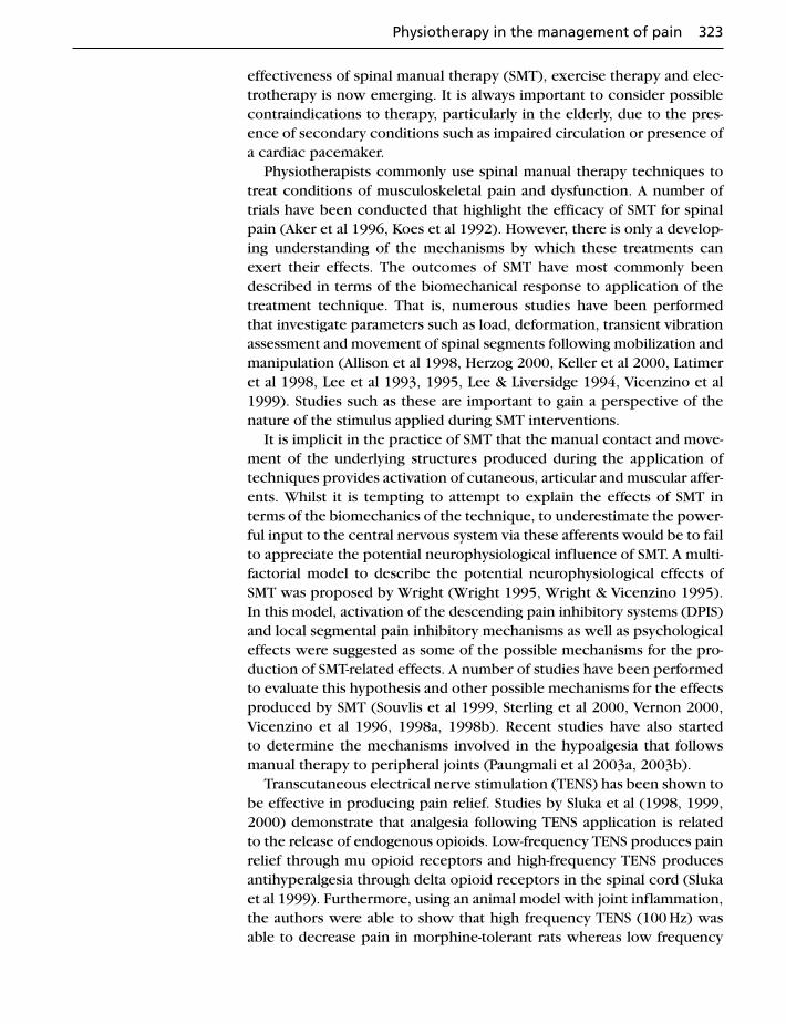
effectiveness of spinal manual therapy (SMT), exercise therapy and elec-trotherapy is now emerging. It is always important to consider possiblecontraindications to therapy, particularly in the elderly, due to the pres-ence of secondary conditions such as impaired circulation or presence ofa cardiac pacemaker.
Physiotherapists commonly use spinal manual therapy techniques totreat conditions of musculoskeletal pain and dysfunction. A number oftrials have been conducted that highlight the efficacy of SMT for spinalpain (Aker et al 1996, Koes et al 1992). However, there is only a develop-ing understanding of the mechanisms by which these treatments canexert their effects. The outcomes of SMT have most commonly beendescribed in terms of the biomechanical response to application of thetreatment technique. That is, numerous studies have been performedthat investigate parameters such as load, deformation, transient vibrationassessment and movement of spinal segments following mobilization andmanipulation (Allison et al 1998, Herzog 2000, Keller et al 2000, Latimeret al 1998, Lee et al 1993, 1995, Lee & Liversidge 1994, Vicenzino et al1999). Studies such as these are important to gain a perspective of thenature of the stimulus applied during SMT interventions.
It is implicit in the practice of SMT that the manual contact and move-ment of the underlying structures produced during the application oftechniques provides activation of cutaneous, articular and muscular affer-ents. Whilst it is tempting to attempt to explain the effects of SMT interms of the biomechanics of the technique, to underestimate the power-ful input to the central nervous system via these afferents would be to failto appreciate the potential neurophysiological influence of SMT. A multi-factorial model to describe the potential neurophysiological effects ofSMT was proposed by Wright (Wright 1995, Wright & Vicenzino 1995).In this model, activation of the descending pain inhibitory systems (DPIS)and local segmental pain inhibitory mechanisms as well as psychologicaleffects were suggested as some of the possible mechanisms for the pro-duction of SMT-related effects. A number of studies have been performedto evaluate this hypothesis and other possible mechanisms for the effectsproduced by SMT (Souvlis et al 1999, Sterling et al 2000, Vernon 2000,Vicenzino et al 1996, 1998a, 1998b). Recent studies have also started to determine the mechanisms involved in the hypoalgesia that followsmanual therapy to peripheral joints (Paungmali et al 2003a, 2003b).
Transcutaneous electrical nerve stimulation (TENS) has been shown tobe effective in producing pain relief. Studies by Sluka et al (1998, 1999,2000) demonstrate that analgesia following TENS application is related to the release of endogenous opioids. Low-frequency TENS produces painrelief through mu opioid receptors and high-frequency TENS producesantihyperalgesia through delta opioid receptors in the spinal cord (Slukaet al 1999). Furthermore, using an animal model with joint inflammation,the authors were able to show that high frequency TENS (100 Hz) wasable to decrease pain in morphine-tolerant rats whereas low frequency
Physiotherapy in the management of pain 323

TENS (4 Hz) was not (Sluka 2000). This illustrates that in patients takingopioids for pain relief high frequency TENS may be of more use. In a clin-ical study by Cheing et al (2002) comparing TENS (100 Hz), isometricexercise and a combination of both to a placebo condition, the authorsdemonstrated a cumulative reduction in knee pain in chronic kneeosteoarthritis patients following 4 weeks of TENS compared to the placebocondition. This pain relief was also demonstrated in a group given TENSand exercise. In a follow-up study, the authors showed that the optimalstimulation period was 40 minutes (Cheing et al 2003).
Osiri et al (2003) in a review for the Cochrane Library concluded thatanalgesia from high frequency TENS and acupuncture-like TENS treatmentwas significantly better than placebo treatment. Knee stiffness alsoimproved significantly in the active treatment group compared to placebo. Asin the study by Cheing et al (2002), the reviewers concluded that repeatedsimulation produced a stronger effect (Osiri et al 2003). Electrical stimulationfor shoulder pain following stroke was also found to be of value byCochrane Library evaluation (Price & Pandyan 2003). The authors reviewedfour randomized controlled trials (RCTs) including use of TENS and func-tional electrical stimulation (FES) and concluded that there was no signifi-cant change in pain incidence. However, there was a significant treatmenteffect for FES for improvement in pain-free range of passive humeral lateral rotation. In a review of TENS for rheumatoid arthritis pain in thehand, a review of three RCTs revealed that administration of 15 minutesof acupuncture-like TENS (low frequency, high intensity) per week, for 3weeks resulted in a significant decrease in rest pain (Brosseau et al 2003).
It is evident that TENS has a therapeutic effect for pain relief in chronicmusculoskeletal conditions such as osteoarthritis and rheumatoid arth-ritis. The use of high or low frequency is dependent on factors such asmedication taken by the patient and type of condition.
Application of radiant heat sources such as hot packs and wax bathsgives superficial heat to provide relief from pain, muscle spasm and jointstiffness. This type of application is considered to be an effective form ofpain relief. However care needs to be taken in the application of heat inpatients with impaired sensation or who are cognitively impaired (Ferrell1996). Appendix 4 provides an overview of the contraindications andprecautions in the use of electrophysical agents for pain relief.
Cryotherapy or use of cold can be beneficial for pain relief. It provideseffective analgesia by decreasing activity in afferent nerves. However,although effective, it is probably not as well accepted or tolerated in theolder patient as heat therapy.
Massage therapy is used frequently to relieve musculoskeletal pain,reduce swelling and for relaxation. In a study by Ferrell et al (1989), theauthors were able to demonstrate reduced anxiety and pain perceptionin cancer patients following massage. Specific trigger point massage maybe useful in the presence of myofascial pain syndrome. It has the benefitthat it is easily administered by family or caregivers.
324 Pain in the elderly

Exercise is considered to be an essential tool for use by physiother-apists in the prevention and management of musculoskeletal pain. In theearlier section on neurophysiology of pain, it was highlighted thatchanges occur in the motor system as a result of both acute and chronicpain. Pain that is not of musculoskeletal origin can produce referredhyperalgesia and change in reactivity in muscles distant from the pain site(Giamberardino et al 1999). Therefore it is essential to assess and treatthe motor system to maintain active support and stabilization of joints inpain (Richardson & Hides 2002). Decreasing stress on joint structuresmay decrease the incidence of pain and improve function. Recentresearch has demonstrated that there is a decrease in recurrence of painfollowing the use of specific therapeutic exercise (Hides & Richardson1996, O’Sullivan et al 1997, 1998). In addition to the supporting or biome-chanical effect, there is some evidence that activation of the motor sys-tem can provide inhibition of dorsal horn via descending projectionsfrom the corticospinal tracts (Galea & Darian-Smith 1995, Galea 2002). Ingeneral, exercise is thought to produce many other benefits as well aspain relief such as increasing cardiovascular fitness. Aquatic therapyprovides an excellent combination of warmth and exercise and the fur-ther therapeutic value and other benefits of aquatic therapy are coveredelsewhere in this text. Ahmad & Goucke (2002) advocate the use of ‘non-drug treatment options’ in the management of patients with neuropathicpain disorders. They consider that adoption of an exercise programme,lifestyle changes and environmental modification together with mainte-nance of mobility and independence where possible are important adjunctsto drug therapy for pain management.
Physiotherapy in the management of pain 325
Summary ■ The perception of pain is a complex sensory and emotionalexperience involving multiple levels of the nervous system.
■ In the presence of injury and inflammation, there isupregulation of the nociceptive system so that an increasedsensitivity to previously noxious stimuli (hyperalgesia) anddecrease in threshold to non-noxious stimulation (allodynia)develop.
■ Peripheral and central sensitization of the nervous systemoccurs to produce these responses, and these mechanisms arealso implicated in the development of referred pain. They alsounderline the transition from acute to chronic pain states.
■ Motor and autonomic nervous system changes may occurfollowing injury and these changes can also contribute to thedevelopment of chronic or persistent pain syndromes.
■ Inhibitory mechanisms mediated by descending pathwaysfrom the midbrain are important in modulating spinalprocessing of nociceptive information. However, psychological

326 Pain in the elderly
influences play a significant role in the overall pain processingand perception.
■ The management of pain by the physiotherapist should bepreceded by a thorough assessment of pain, the patient’soverall physical presentation and their environment.
■ Use of manual therapy techniques such as spinal manualtherapy and both specific and general exercise programmescan be valuable to decrease pain.
■ Use of adjunct therapies such as massage, heat and coldshould be considered in the absence of any contraindications.Research has revealed that TENS can be particularly usefuland that the benefits of its use are cumulative over time.
■ Further research into the mechanisms of common pain problemsin the elderly patient will provide us with the backgroundnecessary to develop the most effective therapeuticprogrammes to manage and hopefully prevent pain.
References
Afable R F, Ettinger W H 1993 Musculoskeletal dis-ease in the aged: diagnosis and management.Drugs and Aging 3(1):49–59
Ahmad M, Goucke C R 2002 Management strategiesfor the treatment of neuropathic pain in the eld-erly. Drugs and Aging 19:929–945
Aker P D, Gross A R, Goldsmith C H, Peloso P 1996Conservative management of mechanical neckpain: systematic overview and meta-analysis.British Medical Journal 313:1291–1296
Allison G, Edmondston S, Roe C et al 1998 Influenceof load orientation on the posteroanterior stiffnessof the lumbar spine. Journal of Manipulative andPhysiological Therapeutics 21:534–538
Ardery G, Herr K A, Titler M G, Sorofman B A, SchmittM B 2003 Assessing and managing acute pain inolder adults: a research base to guide practice.Medsurgical Nursing 12(1):7–19
Bandler R, Keay K A 1996 Columnar organization inthe midbrain periaqueductal gray and the integra-tion of emotional expression. Emotional MotorSystem 107:285–300
Bandler R, Carrive P, Depaulis A 1991 Emerging prin-ciples of organisation in the midbrain periaque-ductal gray matter. In: Depaulis A, Bandler R (eds)The midbrain periaqueductal gray matter. PlenumPress, New York, p 1–8
Bassols A, Bosch F, Campillo M, Canellas M, Banos J E1999 An epidemiological comparison of pain com-plaints in the general population of Catalonia(Spain). Pain 83(1):9–16
Behbehani M M 1995 Functional characteristics ofthe midbrain periaqueductal gray. Progress inNeurobiology 46:575–605
Bergman S, Herrstrom P, Hogstrom K et al 2001Chronic musculoskeletal pain, prevalence rates,and sociodemographic associations in a Swedishpopulation study. Journal of Rheumatology 28(6):1369–1377
Bessou P, Perl E R 1969 Response of cutaneous sensory units with unmyelinated fibres to noxiousstimuli. Journal of Neurophysiology 32:1025–1043
Bowsher D, Rigge M, Sopp L 1991 Prevalence ofchronic pain in the British population:a telephonesurvey of 1037 households. The Pain Clinic4(4):223–230
Brattberg G, Thorslund M, Wikman A 1989 Theprevalence of pain in a general population. Theresults of a postal survey in a county of Sweden.Pain 37(2):215–222
Brattberg G, Parker M G, Thorslund M A 1997Longitudinal study of pain: reported pain frommiddle age to old age. Clinical Journal of Pain13(2):144–149

References 327
Bredkjaer S R 1991 Musculoskeletal disease inDenmark. Acta Orthopaedica Scandinavica 62(Supplement 241):10–12
Brosseau L, Yonge K A, Robinson V et al 2003Transcutaneous electrical nerve stimulation (TENS)for the treatment of rheumatoid arthritis in thehand (Cochrane Review). The Cochrane Library
Burgess P R, Perl E R 1967 Myelinated afferent fibresresponding specifically to noxious stimulation of the skin. Journal of Physiology, London 190:541–562
Cannon J T, Prieto G J, Lee A, Liebeskind J C 1982Evidence for opioid and non-opioid forms of stimulation produced analgesia in the rat. BrainResearch 243:315–321
Carmona L, Ballina J, Gabriel R, Laffon A 2001 Theburden of musculoskeletal diseases in the generalpopulation of Spain:results from a national survey.Annals of the Rheumatic Diseases 60(11):1040–1045
Cheing G L, Hui-Chan C W Y, Chan K M 2002 Doesfour weeks of TENS and/or isometric exercise pro-duce cumulative reduction in osteoarthritic kneepain. Clinical Rehabilitation 16:749–760
Cheing G L, Tsui A Y, Lo S K, HuiChan C W Y 2003Optimal stimulation duration of TENS in the management of osteoarthritic knee pain. Journalof Rehabilitation Medicine 35:62–68
Chibnall J T, Tait R C 2001 Pain assessment in cogni-tively impaired and unimpaired older adults: acomparison of four scales. Pain 92(1–2):173–186
Closs S J 1994 Pain in elderly patients: a neglectedphenomenon? Journal of Advanced Nursing 19(6):1072–1081
Coghill R C, Talbot J D, Evans A C et al 1994Distributed processing of pain and vibration bythe human brain. Journal of Neuroscience 14:4095–4108
Cohen-Mansfield J, Lipson S 2002 Pain in cognitivelyimpaired nursing home residents: how well arephysicians diagnosing it? Journal of the AmericanGeriatric Society 50(6):1039–1044
Cowan D T, Fitzpatrick J M, Roberts J D, While A E,Baldwin J 2003 The assessment and managementof pain among older people in care homes:currentstatus and future directions. International Journalof Nursing Studies 40(3):291–298
Crook J, Rideout E, Browne G 1984 The prevalenceof pain complaints in a general population. Pain18(3):299–314
Cui M, Feng Y, McAdoo D J, Willis W D 1999Periaqueductal gray stimulation-induced inhibitionof nociceptive dorsal horn neurons in rats is asso-ciated with the release of norepinephrine, sero-tonin and amino acids. Journal of Pharmacologyand Experimental Therapeutics 289:868–876
Department of Families Queensland Government2003 Profile of Older People in Queensland:Health,from www.families.qld.gov.au/seniors/index
Dray A 1995 Inflammatory mediators of pain. BritishJournal of Anaesthesia 75:125–131
Dray A 1996 Neurogenic mechanisms and neuropep-tides in chronic pain. Progress in Brain Research110:85–94
Fanselow M S 1991 The midbrain periaqueductalgray as a coordinator of action in response to fearand anxiety. In: Depaulis A, Bandler R (eds) Themidbrain periaqueductal gray matter. Plenum Press,New York, p 151–173
Farrell M, Gibson S, Helme R 1996 Chronic nonmalig-nant pain in older people. In:Ferrell B R, Ferrell B A(eds) Pain in the elderly. IASP Press, Seattle, p 81–89
Farrell M, Gibson S, McMeeken J, Helme R 2000 Painand hyperalgesia in osteoarthritis of the hands.Journal of Rheumatology 27:441–447
Ferrell B A 1991 Pain management in elderly people.Journal of the American Geriatrics Society 39:64–73
Ferrell B A 1995 Pain evaluation and management inthe nursing home. Annals of Internal Medicine123(9):681–687
Ferrell B A, Ferrell B R, Rivera L 1995 Pain in cogni-tively impaired nursing home patients. Journal ofPain and Symptom Management 10(8):591–598
Ferrell B R 1996 Patient education and non-druginterventions. In: Ferrell B R, Ferrell B A (eds) Painin the elderly. IASP Press, Seattle, p 35–44
Ferrell B R, Wisdom C, Wenzl C 1989 Quality of life asan outcome variable in the management of cancerpain. Cancer 63:2321–2327
Ferrell W, Wood L, Baxendale R 1988 The effect ofacute joint inflammation on flexion reflexexcitability in the decerebrate, low spinal cat.Quarterly Journal of Experimental Physiology73:95–102
Fields H L, Basbaum A I 1994 Central nervous systemmechanisms of pain modulation. In: Wall P D,Melzack R (eds) Textbook of pain. ChurchillLivingstone, Edinburgh, p 243–260
Fox P L, Raina P, Jadad A R 1999 Prevalence and treat-ment of pain in older adults in nursing homes and

328 Pain in the elderly
other long-term care institutions: a systematicreview. Canadian Medical Association Journal160(3):329–333
Frank A J M, Moll J M H, Hort J F 1982 A comparisonof three ways of measuring pain. Rheumatologyand Rehabilitation 21:211–217
Gagliese L, Katz J, Melzack R 1999 Pain in the elderly.In: Melzack R, Wall P (eds) Textbook of pain, 4thedn. Harcourt, London, p 991–1006.
Galea M P 2002 Neuroanatomy of the nervous sys-tem. In: Strong J, Unruh A, Wright A, Baxter G D(eds) Pain: a textbook for therapists. ChurchillLivingstone, Edinburgh, p 13–41
Galea M P, Darian-Smith I 1995 Voluntary movementand pain: focussing on action rather than percep-tion. Moving in on Pain, Adelaide
Garsden L R, Bullock-Saxton J E 1999 Joint repositionsense in subjects with unilateral osteoarthritis ofthe knee. Clinical Rehabilitation 3:148–155
Giamberardino M-A, Affaitati G, Iezzi S, Vecchiet L1999 Referred muscle pain and hyperalgesiafrom viscera. Journal of Musculoskeletal Pain7:61–69
Graven-Nielsen T, Sorenson J, Henriksson K G,Bengtsson M, Arendt-Nielsen L 1999 Central hyper-excitability in fibromyalgia. Journal of Musculo-skeletal Pain 7:261–271
Harkins S W, Price D D 1992 Assessment of pain inthe elderly. In:Turk D C, Melzack R (eds) Handbookof pain assessment. Guilford Press, New York,p 315–331
Harkins S W, Price D D, Bush F M, Small R E 1994Geriatric pain. In: Wall P D, Melzack R (eds)Textbook of pain, 3rd edn. Churchill Livingstone,Edinburgh, p 769–784
Helme R D, Gibson S J 1998 Measurement and man-agement of pain in older people. AustralasianJournal on Ageing 17(1):5–9
Helme R D, Gibson S J 1999 Pain in older people. In:Crombie I K, Croft P R (eds) Epidemiology of pain.IASP Press, Seattle, p 103–112
Herr K 2002a Chronic pain: challenges and assess-ment strategies. Journal of Gerontological Nursing28(1):20–27
Herr K 2002b Pain assessment in cognitivelyimpaired older adults. American Journal of Nursing102(12):65–67
Herr K A, Garand L 2001 Assessment and measure-ment of pain in older adults. Clinics in GeriatricMedicine 17(3):457–478
Herr K A, Mobily P R 1991 Complexities of painassessment in the elderly: clinical considerations.Journal of Gerontological Nursing 17(4):12–19
Herr K A, Mobily P R 1993 Comparison of selectedpain assessment tools for use with the elderly.Applied Nursing Research 6(1):39–46
Herr K A, Mobily P R, Kohout F J, Wagenaar D 1998Evaluation of the Faces Pain Scale for use with theelderly. Clinical Journal of Pain 14(1):29–38
Herzog W 2000 Clinical biomechanics of spinalmanipulation. Churchill Livingstone, Edinburgh
Hides J, Richardson C 1996 Multifidus muscle recov-ery is not automatic after resolution of acute, firstepisode low back pain. Spine 21:2763–2769
Hides J, Stokes M J, Saide M, Jull G A, Cooper D H1994 Evidence of lumbar multifidus wasting ipsi-lateral to symptoms in patients with acute/sub-acute low back pain. Spine 19:165–172
Hodges P W 2001 Changes in motor planning of feed-forward postural responses of trunk muscles inlow back pain. Experimental Brain Research141:261–266
Hodges P, Richardson C 1996 Inefficient muscularstabilisation of the lumbar spine associated withlow back pain. A motor control evaluation oftransversus abdominis. Spine 21:2640–2650
Hosobuchi Y, Adams J E, Linchitz R 1977 Pain reliefby electrical stimulation of the central gray matterin human and its reversal by naloxone. Science197:183–186
Janig W, McLachlan E 1992 Characteristics offunction-specific pathways in the sympatheticnervous system. Trends in Neuroscience 15:475–481
Janig W, Levine J, Michaelis M 1996 Interactions ofsympathetic and primary afferent neurons follow-ing nerve injury and tissue trauma. Progress inBrain Research 113:161–184
Jansen A S P, Farkas E, Sams J M, Loewy A D 1998Local connections between the columns of theperiaqueductal gray matter: a case for intrinsicneuromodulation. Brain Research 784:329–336
Johansson H, Sojka P 1991 Pathophysiological mech-anisms involved in genesis and spread of musculartension in occupational muscle pain and inchronic musculoskeletal pain syndromes. MedicalHypotheses 35:196–203
Kamel H K, Phlavan M, Malekgoudarzi B, Gogel P,Morley J E 2001 Utilizing pain assessment scalesincreases the frequency of diagnosing pain among
References 329
elderly nursing home residents. Journal of Painand Symptom Management 21(6):450–455
Keller T S, Colloca C J 2000 In vivo transient vibrationassessment of the normal human thoracolumbarspine. Journal of Manipulative and PhysiologicalTherapeutics 23:521–530
Koes B W, Bouter L M, Vanmameren H et al 1992Randomized clinical trial of manipulative therapyand physiotherapy for persistent back and neckcomplaints – results of one year follow-up. BritishMedical Journal 304:601–605
Kremer E, Atkinson H J, Ignelzi R L 1981Measurement of pain: patient preference does notconfound pain measurement. Pain 10:241–248
Latimer J, Lee M, Adams RD 1998 The effects of highand low loading forces on measured values of lumbar stiffness. Journal of Manipulative andPhysiological Therapeutics 21:157–163
Lee M, Liversidge K 1994 Posteroanterior stiffness atthree locations in the lumbar spine. Journal of Manip-ulative and Physiological Therapeutics 17:511–516
Lee M, Latimer J, Maher C 1993 Manipulation: investi-gation of a proposed mechanism. Clinical Bio-mechanics 8:302–306
Lee M, Maher C, Simmonds M J, Kumar S, Lechelt E1995 Spinal models:use of a spinal model to quan-tify the forces and motion that occur during thera-pists’ test of spinal motion. Physical Therapy75:638–641
Lewis V A, Gebhart G F 1977 Morphine-induced and stimulation produced analgesias at coincidentperiaqueductal central gray loci: evaluation of anal-gesic congruence, tolerance, and cross-tolerance.Experimental Neurology 57:934–955
Lovick T A 1991 Interactions between descendingpathways from the dorsal and ventrolateral peri-aqueductal gray matter in rats. In: Depaulis A,Bandler R (eds) The midbrain periaqueductal graymatter. Plenum Press, New York
Lund J P, Donga R, Widmer G, Stohler C 1991 Thepain-adaptation model: a discussion of the rela-tionship between chronic musculoskeletal painand motor activity. Canadian Journal of Physiologyand Pharmacology 69:683–694
Lutfy K, Hurlbut D E, Weber E 1993 Blockade of mor-phine-induced analgesia and tolerance in mice byMK-801. Brain Research 616: 83–88
Manfredi P L, Breuer B, Meier D E, Libow L 2003 Painassessment in elderly patients with severe demen-tia. Journal of Symptom Management 25(1):48–52
Mao J, Price D D, Mayer D J 1995 Mechanisms ofhyperalgesia and morphine tolerance: a currentview of their possible interactions. Pain 62:259–274
Mayer D J 1979 Endogenous analgesia systems:neuraland behavioral mechanisms. In: Bonica J J (ed)Advances in pain research and therapy. 3. RavenPress, New York, p 385–410
Mayer D J, Mao J, Holt J, Price D D 1999 Cellularmechanisms of neuropathic pain, morphine toler-ance and their interactions. Proceedings of theNational Academy of Science – USA 96:7731–7736
Melzack R, Wall P D 1965 Pain mechanisms: a newtheory. Science 150:171–179
Melzack R, Wall P D 1996 The challenge of pain.Penguin Books, Harmondsworth, Middlesex
Mense S 1993 Nociception from skeletal-muscle inrelation to clinical muscle pain. Pain 54:241–289
Merskey H, Bogduk N 1994 Classification of chronicpain: descriptors of chronic pain syndromes anddefinitions of pain terms. IASP Press, Seattle
Morgan M M 1991 Differences in antinociceptionevoked from dorsal and ventral regions of the caudal periaqueductal gray matter. In: Depaulis A,Bandler R (eds) The midbrain periaqueductal graymatter. Plenum Press, New York
Nichols D S, Thorn B E 1990 Stimulation-producedanalgesia and its cross tolerance between dorsaland ventral PAG loci. Pain 57:347–352
Osiri M, Welch V, Brosseau L et al 2003Transcutaneous electrical nerve stimulation for kneeosteoarthritis (Cochrane Review). The CochraneLibrary, Update Software
O’Sullivan P, Twomey L, Allison G 1997 Evaluation of specific stabilising exercise in the treatment ofchronic low back pain with radiologic diagnosis of spondylosis or spondylolisthesis. Spine 22:2959–2967
O’Sullivan P, Twomey L, Alison G 1998 Alteredabdominal muscle recruitment in patients withchronic back pain following a specific exerciseintervention. Journal of Orthopaedic and SportsPhysical Therapy 27:114–124
Paungmali A, O’Leary S, Souvlis T, Vicenzino B 2003aHypoalgesia and sympathoexcitatory effects ofmobilisation with movement for lateral epicondyl-algia. Physical Therapy 83(4):374–383
Paungmali A, O’Leary S, Souvlis T, Vicenzino B 2003bNaloxone fails to antagonise initial hypoalgesiceffect of a manual therapy treatment for lateral

330 Pain in the elderly
epicondylalgia. Journal of Manipulative andPhysiological Therapeutics (in press)
Popp B, Portenoy R K 1996 Management of pain inthe elderly: pharmacology of opioids and otheranalgesic drugs. In: Ferrell B R, Ferrell B A (eds)Pain in the elderly. IASP Press, Seattle, p 21–34
Price C I M, Pandyan A D 2003 Electrical stimulationfor preventing and treating post-stroke shoulderpain (Cochrane Review). The Cochrane Library
Price D D, Mayer D J, Mao J R, Caruso Fire Service2000 NMDA-receptor antagonists and opioid recep-tor interactions as related to analgesia and toler-ance. Journal of Pain and Symptom Management19:S7–S11
Rexed B 1954 The cytoarchitectonic atlas of thespinal cord in the cat. Journal of ComparativeNeurology 100:279–379
Reynolds D V 1969 Surgery in the rat during electri-cal analgesia induced by focal brain stimulation.Science 164:444–445
Richardson C, Hides J 2002 Exercise and pain. In:Strong J, Unruh A, Wright A, Baxter G D (eds) Pain:a textbook for therapists. Churchill Livingstone,Edinburgh, p 245–266
Roy R, Thomas M 1986 A survey of chronic pain in an elderly population. Canadian Family Physician32:513–516
Schaible H-G, Grubb B D 1993 Afferent and spinalmechanisms of spinal joint pain. Pain 55:5–54
Sharma L, Pai Y C, Holtkamp K, Rymer W Z 1997 Is knee joint proprioception worse in the arthriticknee versus the unaffected knee in unilateral knee osteoarthritis? Arthritis and Rheumatism 40:1518–1525
Simmonds M J, Scudds R J 2001 Pain, disability, andphysical therapy in older adults: issues of patientsand pain, practitioners and practice. Topics inGeriatric Rehabilitation 16(3):12–23
Sluka K A 2000 Systemic morphine in combinationwith TENS produces an increased antihyperalgesiain rats with acute inflammation. Journal of Pain1:204–211
Sluka K A, Rees H 1997 The neuronal response topain. Physiotherapy Theory and Practice 13:3–22
Sluka K A, Willis W D, Westlund K N 1995 The role ofdorsal root reflexes in neurogenic inflammation.Pain Forum 4:141–149
Sluka K, Bailey K, Bogush J, Olsen R, Ricketts A 1998Treatment with either high or low frequency TENSreduces the secondary hyperalgesia observed after
injection of kaolin and carrageenan into the kneejoint. Pain 77:97–102
Sluka K A, Deacon M, Stibal A, Strissel S, Terpstra A1999 Spinal blockade of opioid receptors preventsthe analgesia produced by TENS in arthritic rats.Journal of Pharmacology and ExperimentalTherapeutics 289:840–846
Sluka K A, Judge M A, McColley M M, Reveiz P M,Taylor B M 2000 Low frequency TENS is less effec-tive than high frequency TENS at reducing inflam-mation-induced hyperalgesia in morphine-tolerantrats. European Journal of Pain – London 4:185–193
Souvlis T, Kermode F, Williams E, Collins D, Wright A1999 Does the initial analgesic effect of spinalmanual therapy exhibit tolerance? 9th WorldCongress on Pain. Book of Abstracts, Vienna
Stanton-Hicks M, Baron R, Boas R 1998 Complexregional pain syndromes: guidelines for therapy.Clinical Journal of Pain 14:155–166
Stein C, Cabot P, Schafer M 1999 Peripheral opioidanalgesia:mechanisms and clinical implications. In:Stein C (ed) Opioids in pain control: basic and clin-ical aspects. Cambridge University Press, New York
Sterling M, Jull G, Wright A 2000 Cervical mobilisa-tion: Concurrent effects on pain, sympatheticnervous system activity and motor activity. ManualTherapy 6:72–81
Sterling M, Jull G, Wright A 2001 The effect of mus-culoskeletal pain on motor activity and control.Journal of Pain 2:135–145
Sternbach R A 1986 Survey of pain in the UnitedStates: the Nuprin Pain Report. Clinical Journal ofPain 2:49–53
Terman G W, Shavit Y, Lewis J W, Cannon J T,Liebeskind J C 1984 Intrinsic mechanisms of paininhibition: activation by stress. Science 226:1270–1277
Unruh A M, Ritchie J A, Merskey H 1999 Does genderaffect the appraisal of pain and pain coping strate-gies? Clinical Journal of Pain 15:31–40
Vernon H T 2000 Qualitative review of studies ofmanipulation-induced hypoalgesia. Journal ofManipulative and Physiological Therapeutics 23:134–138
Vicenzino B, Collins D, Wright A 1996 The initialeffects of a cervical spine manipulative physio-therapy treatment on the pain and dysfunction oflateral epicondylalgia. Pain 68:69–74
Vicenzino B, Collins D, Cartwright T, Wright A 1998aCardiovascular and respiratory changes produced

References 331
by lateral glide mobilisation of the cervical spine.Manual Therapy 3:67–71
Vicenzino B, Collins D, Benson H, Wright A 1998b An investigation of the interrelationship betweenmanipulative therapy-induced hypoalgesia andsympathoexcitation. Journal of Manipulative andPhysiological Therapeutics 21:448–453
Vicenzino B, Neal R, Collins D, Wright A 1999 Thedisplacement, velocity and frequency profile ofthe frontal plane motion produced by the cervicallateral glide treatment technique. Clinical Bio-mechanics 14:515–521
Wall P D, Woolf C J 1984 Muscle but not cutaneousinput produces prolonged increases in theexcitability of the flexion reflex in the rat. Journalof Physiology – London 356:443–458
Weiner D, Peterson B, Keefe F 1998 Evaluating per-sistent pain in long term care residents: what rolefor pain maps? Pain 76(1–2):249–257
Weiner D, Peterson B, Ladd K, McConnell E, Keefe F1999 Pain in nursing home residents: an explo-ration of prevalence, staff perspectives, and prac-tical aspects of measurement. Clinical Journal ofPain 15(2):92–101
Woolf C J, Mannion R J 1999 Neuropathic pain: aeti-ology, symptoms, mechanisms and management.Lancet 353:1959–1964
Wright A 1995 Hypoalgesia post manipulative ther-apy. Manual Therapy 1:11–16
Wright A, Vicenzino B 1995 Central mobilisationtechniques, sympathetic nervous system effectsand their relationship to analgesia. Moving in onPain, Adelaide
Zimmerman M 1989 Pain mediators and mechanismsin osteoarthritis. Seminars in Arthritis andRheumatism Supplement 2:22–29

Physiotherapy in palliative care
Susan R. Hourigan and Diane L. Josephson
332
16
This chapter aims to:
This chapter will illustrate the principles of palliative care and identifyaspects of physiotherapy management and practice that are relevant tothe palliative care setting. It is important to note that physiotherapeuticassessment and treatment principles are the same for all residents regardlessof health status. The major difference in palliative care is that a therapistmust prioritize goals directly in relation to the resident’s and family’s wishesregardless of usual clinical reasoning. Advocacy is the focus and informationshould be provided to allow residents to make their own choices. Residentcomfort and dignity usually comes first – pain management is often thetop priority. Communication is of paramount importance during thistime to enable a positive focus and to ensure best practice is achieved.
As birth is a miracle, so is death. We are blessed to be equippedwith the skills to help those who are dying.
‘Caring for and caring about’ – as physiotherapists it is ideal totake an holistic approach – we are not just treating signs andsymptoms or an underlying disease but the biopsychosocialneeds of an individual and his or her family.
Assessment (as always) should be specific and goal-directed – duringpalliation it is important to identify patient goals early.
Palliative care means a form of care that recognizes that cure or long-termcontrol is not possible; is concerned with the quality rather than the quan-tity of life; cloaks troublesome and distressing symptoms with treatments
■ highlight healthy attitudes towards death, dying and ageing■ illustrate how physiotherapy may play a role in palliation■ extend basic knowledge of palliative care.
Introduction
Key points
What is palliative care?

What is palliative care? 333
whose primary or sole aim is the highest possible measure of patientcomfort.
Goals for palliative care have been outlined by the World HealthOrganization. Palliative care:
■ affirms life and regards death as a normal process■ neither hastens nor postpones death■ provides relief from pain and other distressing symptoms■ integrates the psychological and spiritual aspects of pain■ offers a support system to help patients live as actively as possible until
death■ offers a support system to help the family cope during the patient’s illness
and in their bereavement■ uses a team approach to address the needs of patients and their families,
including bereavement counselling, if indicated■ will enhance quality of life, and may also positively influence the course
of illness■ is applicable early in the course of illness, in conjunction with other
therapies that are intended to prolong life, such as chemotherapy orradiation therapy, and includes those investigations needed to betterunderstand and manage distressing clinical complications (WHO 1990,2002, 2003).
The World Health Organization currently has a large global perspectiveon palliative care encompassing many different areas such as cancer care, children’s care and an African Initiative which is a joint cancer andHIV/AIDS project. WHO’s contribution to palliative care, its past, presentand future challenges were identified by Sepulveda et al (2002).
Early in the fourth century the original hospices were formed in which matrons opened their homes to the sick and needy. Although a relatively new concept of care within modern medicine, palliative care has foundations in hospices in the UK through the early 1960s and in Australia in the 1980s. Flinders University in South Australia wasthe first in the world to appoint a Chair in Palliative Care in 1998(Emeritus Professor Ian Maddocks).
The principles behind palliative care have been suggested by someauthors to be those of beneficence, non-maleficence, justice and autonomy:
■ beneficence – any act of goodness or kindness■ non-maleficence – not doing harm or not harmful■ justice – fair treatment or conduct; honestly, fairly, accurately■ autonomy – independence or freedom.
Death can be a happy time if a person’s needs are met before, during andafter their passing. The philosophy of care is such that it encompasses all aspects of a person’s make-up. It is the integration of the physical,emotional, intellectual and spiritual components of a person, as illustratedin Figure 16.1.

334 Physiotherapy in palliative care
Palliative care incorporates principles such as ‘wellness’ and biopsy-chosocial models. This clearly relates to a person’s quality of life (QOL).
QOL is:
■ ‘whatever a person says it is’■ subjective satisfaction■ related to the biopsychosocial and spiritual dimensions of a person■ related to the emotional need to feel useful – defines their role■ related to the intellectual ability to remember, concentrate and be able
to reframe life events■ related to psychological function (grief, anxiety, depression, peace,
acceptance)■ related to social functions such as relationships, intimacy, support and
family■ related to spiritual aspects such as finding meaning and transcendence■ related to physical aspects such as mobility and independence■ related to symptoms such as pain, constipation, weakness and short-
ness of breath.
Quality of life is clearly understood by evaluating the difference betweena person’s hopes and their reality. If what is experienced matches individ-uals’hopes, they perceive good quality in their life. Figure 16.2 illustratesthis concept. Quality of life may be measured using the McGill Quality ofLife questionnaire (Cohen et al 1995).
Intellect
Physical
Spiritual
Emotion
Figure 16.1The model of theintegration of life
aspects.
RealityHopes
14
12
10
8
6
4
2
0
Figure 16.2Quality of life as
mirrored by hopesand reality.

Physiotherapy and palliative care 335
Patient goals are therefore the ultimate focus. There are many differentpersonality types and each person has had unique and different experiencesthroughout their life. Palliative care is about providing them with choicesand seeing that their decisions are acted upon wherever possible. In agedcare many residents will have lost many functions, including intellectualalertness, communication and simple self-care, not to mention the ‘socialdeath’ which may have surrounded their admission into a facility.Irrespective of this, all the same principles should apply, which encouragesparticipation and decision-making by the resident whenever possible.There are difficulties involved in transparently overlaying the principles ofpalliative care to aged care due to several factors. Dementia has been a con-traindication in the past to hospice admission due to the inability of theperson to direct their own care and ‘work through their understanding’.Other points which lead to slight modification in the aged care settinginvolve lack of staff training, hours and expertise and a tendency for symp-toms to be dismissed in the aged care setting due to universality and com-plexity of problems. Regardless of limitations such as these, it is importantto achieve best practice in any RACF, and the care provided should be ofgood quality and embrace the principles of palliative care as outlined.
Physiotherapy has built up a large research base of techniques and skillsthat are invaluable to best practices and outcomes in patient care. Thechoice and use of physiotherapeutic knowledge is of little value withoutfirst gaining the trust and cooperation of the patient. Respect for thepatient and a deep regard for their feelings and wishes must precede anyattempt to offer any form of assistance. In the area of palliative care this isessential. We need the trust and understanding not only of our patient butalso of their carer/s and the rest of the team who are involved in each case.Never is there such need for teamwork of the unobtrusive and quiet type.All facets of care must be finely tuned, respected and implemented withminimal fuss and maximum benefit to our patient. We should be like anexcellent symphony with all parts coming in and fading out as the musicdemands.
Communication that reaches across the barriers of race, creed, wordsand externals must first be gained in order to provide service and supportto people at this critical stage in their lives. Palliative care is both sacredand solemn. We need to explore how best to be of service to another inthis time when life’s journey is drawing to a close.
Good care will depend on good assessment of the physical conditionof the person but more importantly we must endeavour to understandthe priority of what they perceive to be their needs and desires at thistime. We must see this person in the full context of their history and envir-onment. Physical needs may be completely overshadowed and becomeinsignificant for the person compared to the need for love and resolutionof their spiritual, emotional or psychological requirements at this time.
Physiotherapyand palliative
care

336 Physiotherapy in palliative care
This is a time for resolution and finalization of a lifetime’s work. It is aperiod of great power in a person’s life when sensitivity and love canfacilitate miracles. We are privileged to become part of someone’s life atthis time because of our knowledge of ageing and physiology, but ourskills need to be tempered and refined to the hierarchy of needs of eachindividual person. If this is not clearly interpreted, our efforts may bedestructive rather than constructive at this delicate time.
Our role must be to bring comfort and promote healing of body, mindand spirit, to protect and empower, to teach and re-educate, to alleviateand to assist in whatever way is clinically and humanely possible. We mustalways be mindful of the acute needs of the patient throughout.
It is the centre of our caring being, our humanness which reachesout to others. Caring from the heart uses our reflective knowledgeour cognitive thoughts and our action centre to create positiveoutcomes for the other.
Roach (1992)
As we travel this journey with the resident our lives are changedand enriched and we grow and we learn. All of those experienc-ing this deeply rewarding and humility provoking pathway of palliative care are able to experience this. To me, the depth of thehuman soul and spirit is immeasurable and unfathomable.
In personal interview with S.R. Josephson, August 2002,on ‘The positives of working with patients who are dying’
Caring gives rise to our meaning and purpose for being in theworld.
Frankl (1959)
The caring process can activate a higher power, order or energy inthe universe which potentiates healing, health and self-knowledge.
Keegan & Guzzetta (2000)
Assessment is essential. As experience is gained intuition takes over from,or enhances, step by step science. The mental process is the same.Closely observe the patient at all times during assessment and history tak-ing. Body language and facial expression may tell you significant thingsthat words do not.
This is the information we need. It can be collected in any order, in anyway and in whichever time frame is appropriate.
1. Subjective assessment (may be from family or caregiver)■ medical history of the patient and course of recent condition■ family and social history■ present signs and symptoms■ previous treatment received and outcome/effects noted
The role ofphysiotherapy

The role of physiotherapy 337
■ medications■ goals and desires of the patient■ prescription of aids and comforts.
2. Objective assessment.3. Physical assessment.
As we collect information we need to clarify our role and our aims with ourpatient and their family and ensure that we are all working towards the samegoals and outcomes. A practical and possible plan of care must evolve for allconcerned.
As we evaluate the situation and discuss our findings with other con-cerned team members, a priority of most urgent needs becomes obviousand leads to a time frame of care and action. It may be that control of painor emotional distress becomes the first area for attention and very oftengood drug management can facilitate and greatly enhance future plansand outcomes for good physiotherapy management.
Therapy required may be classified under the model of symptom man-agement and with a view to realizing patient goals and is illustrated inFigure 16.3.
Physiotherapy practice will often encompass:
■ rehabilitation, promoting independence, facilitating activities of dailyliving
■ symptomatic treatment (pain/constipation/skin integrity/mobility/independence)
■ utilization of treatment modalities such as therapeutic exercise,electrotherapy, joint and soft tissue mobilization, hydrotherapy andequipment prescription
Supportive andComplementary
measures
Medication
RehabilitationPsychological
Measures
ContinuingTreatmentof Disease(control forcomfort)
SymptomManagement
Figure 16.3Model of symptom
management.

338 Physiotherapy in palliative care
■ education of the resident, carers and families – including disease andhealth information, health promotion, manual handling, back care, andworkplace health and safety and environment auditing
■ appropriate exercise plans.
Promoting independence and maximizing the full potential of the patientwill often be a treatment aim. This may involve some rehabilitation andoften a degree of encouragement, reassurance and motivation.
This is the first step in caring and to do this well increases the patient’sinterest and self-esteem and can take a great deal of the physical burdenoff the carer or carers.
■ Look at the heights of chairs and bed for the patient and see if raising or lowering the height may make life easier for the patient. To get outof bed the person needs to be able to put feet flat on the floor beforestanding. The correct height chair provides the same easy access andshould fully support the thighs. The back support should be straight. Ifthe person is suffering from swollen legs a chair which raises the feet isoptimal. Otherwise, if the person is sitting up for a long period of timethey will need to elevate their feet. Be careful to ensure the backrestreclines to allow for the length of the hamstring muscles when the feetare elevated. This will reduce the tendency to slide the buttocks for-ward on the seat and consequently assume a very flexed lumbar spineposition that might increase pain in this area. The back of the knees alsoshould be fully supported or the person will experience pain and dis-comfort in their knee joints.
■ Look at the way the person is sitting up from lying in bed. A ropeattached to the end of the bed can make life easier and provide inde-pendence and exercise for your patient as it assists them to come to a sitting position. A solid pole beside the bed is another good alternative.A hand grasp above the head can make movement around the bed easierbut is of little use for sitting up as the angle of pull is incorrect.
■ Walking aids can give great support, remove the fear of falling and keepyour patient mobile for the maximum length of time. A walking stickgives minimum support and walking frames can provide maximum sup-port. The frame with elbow support promotes good posture and allowsmore arm strength to be utilized. For someone with lower limb or spinalproblems this can be remarkably useful as it decreases the amount ofweight-bearing apportioned through the trunk and legs. The walkingframe chosen will therefore need to be of a wheeled variety. Pick-uphoppers or ‘Zimmer’ frames should not be considered for palliative carepatients as they greatly load the spine during the ‘lift and shift’ phase ofgait and in so doing are likely to increase bony damage or increase pain.
■ Grab rails strategically placed can maximize independence and giveconfidence. Along the wall, near doorways and on bathroom and shower
Maintaining or restoring
mobility, dexterity and
function

Providing comfort and protecting skin integrity 339
walls they can allow greater mobility and independence in confined areaswhere frames will not fit.
■ Handheld retrievers with hooks, magnets, prongs or pincers on the endsave the patient bending down and can increase their reach. These canbe simply made and can be adapted for your patient’s individual needs.
■ Cutlery and writing equipment can be easily moulded to be more com-fortable and useful by enlarging the area of grip or changing the angleof grip. Home-made bindings can be cleverly modified to suit hand sizesand shapes.
■ Clothing can be altered to open down the back to make dressing easier.■ Trays for eating, reading or writing can be made of a piece of light timber
that sits across the armrests of a chair.■ Straws are extremely useful to maximize fluid intake and prevent spillage.■ Remote controls and headphones can promote comfort and
independence.
The environment needs to be set up to allow the patient easy access toall their aids and comforts. Everything they need should be comfortablywithin reach.
In the course of a normal day we all move and readjust our posture manymore times than we are aware of. We gesticulate and wriggle and sit andstand incessantly. When someone is unwell these spontaneous move-ments are seriously minimized. The period of time spent motionless andsedentary can increase to the extent that the areas that take the weight ofthe body are at risk of breaking down. This is the result not only of directpressure but it is compounded by the decrease in circulatory efficiencyand poorer nutritional intake that often accompany ill health.
The best protection against skin damage is provided by:
1. frequent change of position2. promoting good skin condition3. encouraging good circulation with movement and exercise4. encouraging good nutrition and hydration5. protecting bony prominences.
When it comes to pressure areas prevention is the answer.
Healing broken skin is difficult and pressure areas are very painful foryour patient. The skin integrity will be severely compromised after onlyone hour of unrelenting pressure. Movement provides the best protectionfor skin integrity. Varying or reducing the points of pressure by the use ofsheepskins or gel pads may assist. Soft cushions or foam should be usedto prevent pressure between knees when the patient is lying on their side.Ideally a pressure-varying mattress is also used but these are not always
Providing comfort and
protecting skinintegrity
Key point

340 Physiotherapy in palliative care
available so we use massage and protection of the vulnerable areas toimprove the circulation. Keeping the skin soft and supple by the use ofgood oils or moisturizers is also invaluable. Aromatherapy oils can be usedto promote relaxation and wellbeing as well as for moisturizing.
The main ‘risk’ areas are the bony prominences and surrounding softtissues. The most important areas to protect and relieve are:
1. the sacral area and buttocks2. the elbows3. the heels4. the knees5. the ankles6. the ears.
Little cradles can be made out of foam to protect the ears. Simply cut theear shape out of a square piece of foam approximately 8 cm square and5 cm deep and covered by a pillow case to increase comfort. Socks or softslippers can be useful for heels and ankles.
Frequent position changes are essential to prevent prolonged pressureon bony prominences. If your patient is physically capable this is not dif-ficult. You can use short walks, vary the time spent in bed resting or upin the chair. You can have comfortable chairs in different rooms or out inthe garden. If your patient is dependent this is more challenging and putsa greater onus on the carer.
Manual handlingThese belts are made to give carers a ‘real’ handle on people. Severe damage to joints and skin may be caused by using limbs to move people.Limbs do not give the carer a good grip or indeed any mechanical efficiency but if you put a comfortable belt around the pelvis of your resident a good hold is provided for the carer and greater comfort for thepatient. They allow assistance to be directed to the centre of a patient’sweight, they facilitate movement patterns and also assist in guiding walk-ing direction.
These are simply a piece of sail cloth or some other material that is slip-pery and effectively reduces friction by gliding smoothly on itself. A slidesheet is used in a double layer (either folded or by using two full lengths).The patient’s body weight is repositioned by movement of the top layerover the bottom layer. It is important to grasp the top layer or sheet closeto your patient’s shoulder and hip as these are the key areas for patientmovement. Accessing these heavy areas makes movement of the bodyeasy. You can maximize patient comfort by turning their head first in thedirection of planned movement (for instance when performing a rollingmovement). You can also prepare the patient by bending the top knee to
Education
Slide sheets
Walking belts

facilitate rolling to the other side and you may prepare by the use of pillowsor cushions to support painful or at-risk areas.
The carer must endeavour to follow good back care principles and keepthe spine as straight as possible during transfers. This is easy where you haveadjustable bed heights but much more difficult to achieve in communitycare situations.
Hoists may be used to reposition fully dependent patients or patientsdeemed to be unsafe when weight-bearing. These devices dramaticallyreduce the risk of injury to the carer and indeed to the patient. There aremany varieties of hoists on the market and the technique of using eachhoist and the appropriate slings must be mastered before any attempt ismade to transfer a patient.
Hoists take the full weight of the patient and therefore are of greatvalue in moving patients from bed to chair, bed to bath, chair to chair andmaybe from floor back to bed after a fall.
Hoists are expensive items and are not readily available to the widercommunity. Some community aid programmes will provide hoists for needycases but these must be prescribed for each individual as the sizes anduses for hoists vary considerably.
In Australia the nurses union has a no-lift policy and this has seen manymore hoists available in the workplace than there were previously.
Anatomically the ‘back’ is made up of muscles, bones, joints, ligaments, ten-dons, nerve and discs. All of these structures may be injured. Preventionis the key – we only have one back and backs are notoriously difficult torehabilitate after injury.
Carers need to know that most back pain comes from frequent, repeti-tive use of the back in poor positions. The greatest risk factors for backinjury are poor posture, sustained postures, poor lifting practices or incor-rect manual handling, jerking, twisting and poor personal fitness/well-ness. Eighty per cent of disc injury is due to repetitive poor patterns asopposed to one incident. The discs have poor sensation and poor circu-lation; therefore we often have no indication of injury occurring until thedamage is severe. Furthermore intervertebral discs are poorly equipped forhealing.
It is important to set up the caring environment so that it is asergonomically sound as possible. Most caring tasks are repetitive andtherefore great care must be taken to ensure safety of the patient andsafety of the carer.
Providing palliative care fatigues the carer physically and emotionally. Inan RACF carers can share responsibility for residents and thereby reducethe individual burden of care. This luxury is rarely available for a familymember caring for another at home. In all instances case discussionsinvolving the patient, carers and all other team members might forestall
Education 341
Mechanical aids
Back care andinjury prevention
Fatigue minimization

342 Physiotherapy in palliative care
the possible catastrophic effects from carer fatigue and ‘burn-out’ by put-ting into place plans to cope with potentially stressful events that mightbe predicted for the patient or carer. If not prepared for, the psychologicaland physical effects from accumulated or one-off events can be far-reachingand make the passing of a resident or loved one more difficult.
■ Educate the carer in optimal use of the body and good posture.■ Adjust work heights to facilitate caring and transfers.■ Where possible use mechanical aids and have appropriate training.■ Clear any unnecessary ‘clutter’ from the area.■ Communicate your intentions, before attempting to move, to both the
patient and any assistants.■ Plan the move, and count out loud before beginning.■ Nurse the patient in a well-ventilated and well-lit area. Use cooling or
heating as required.■ Carers must look after themselves! Keep fit, strong and well rested.
Minimize stress and get help when you need it.
Heat/cold therapyHeat is often a comfort for painful chronic conditions as it improves cir-culation and promotes relaxation. It is available in many forms from hotwater bottles to electric blankets. Very often the frail or neurologicallyimpaired patient has sensory loss and therefore there is a high risk of injuryor burns. As with all treatments, the comfort gained must be weighedagainst the risk of injury and well-informed decisions must be made andmonitored. For some arthritic conditions (especially inflammatory sub-types) cold compresses may give relief. Trial and error is often the onlyway of knowing whether heat or cold may be more effective. If the patientsustains an acute injury or contusion, cold packs applied immediately willresult in a better outcome. Limiting circulation to the affected part anddecreasing swelling and bleeding into the tissues will reduce the injuryresponse but always be aware of the contraindicating factor to thermalmodalities imparted by poor circulation.
There are volumes written on the use of electrotherapy and its many posi-tive effects. However, its use may be limited in the area of palliative care.Often if pain is a problem, drug therapy is the chosen treatment. Howevera multidisciplinary team should not underestimate, and never discount,the possible alternatives such as TENS, magnetic therapy and laser.
TENS works on the gate theory of pain control. It may control pain byproviding a counter-irritant stimulus through the afferent nerves originat-ing in the skin (it is felt as a mild buzzing or tingling feeling). This ascend-ing sensory information is perceived by the brain rather than the noxious
Important considerations
Treatmentmodalities
Electrotherapy
Transcutaneouselectrical nerve
stimulation (TENS)

pain information and thereby the pain sensations are blocked before beingcognitively perceived. Some research indicated TENS may also stimulatethe production of endogenous opioids by the brain and further provideanalgesia through this mechanism. The TENS machines are safe and smalland have had good effects for some painful conditions. It is easy to applyand teach to the patient or carer.
This therapy is extremely useful to heal skin lesions. The laser beam usedis in the infra-red part of the spectrum and although penetration is notvery deep the laser can promote good healing from underneath the lesion.Laser has contraindications and safety rules but in the hands of a trainedperson it is very useful.
Magnetic therapy may play a role in the treatment of some conditionssuch as non-uniting fractures, joint pains, healing of ulcers, insomnia andsoft tissue injuries. There are many form of magnetic therapy and if it isavailable can give excellent results. Some forms of underlays for the bedhave been trialled successfully and magnetic therapy has the advantagethat it can be used with metal in the body and it is very safe. It does havecontraindications, e.g. cardiac pacemakers, carcinogenic conditions. Thereader is referred to the electrotherapy appendix (Appendix 4).
This needs to be applied by a trained professional. It is useful for dispersionof bruising, reduction of oedema, promotion of healing and other local-ized conditions. Precautions must be taken to ascertain sensory feedback –as with most modalities patient feedback is essential to the safety of appli-cation. It should be the physiotherapist who applies ultrasound therapy.
The use of warm water immersion to augment and aid treatment throughaquatic physiotherapy (hydrotherapy) has been well proven and estab-lished in many different cases. This is an excellent modality and the sadnessis that it is not more readily available. Hydrotherapy treats the whole person.The body may be wholly or partially immersed in water 32–34°C. Theeffects of the warmth, buoyancy and hydrostatic pressure make this a treat-ment of choice for almost all conditions. Most patients and people thor-oughly enjoy getting in the warm water, especially those with pain and/orstiffness. The only considerable barrier is those patients who are fearful ofwater and some cultures which are not used to the concept of water ther-apy. The contraindications are few, e.g. open wounds, several skin condi-tions and faecal incontinence. Even the frailest person can benefit fromhydrotherapy as floats may be utilized so that the patient need not doanything themselves whilst a therapist is present. Often patients will beindependent in their treatment as a programme can be devised to suit theindividual and their condition.
Treatment modalities 343
Magnetic therapy
Ultrasound
Aquatic physiotherapy
(hydrotherapy)
Laser therapy

The positive effects of water therapy have been known for centuries.There is a great deal of evidence which suggests treatment is very holisticin nature and the benefits are to body and mind promoting relaxation,wellbeing and activity.
The skin is the largest organ in the body and hydrotherapy stimulatesand rejuvenates the skin. Joints move freely in water and soft tissues canbe lengthened and strengthened. Hydrotherapy stimulates the heart andlungs, assists digestion and aids peripheral circulation and venous return.Muscle spasm and pain are reduced and general wellbeing is enhanced.
The numerous benefits of exercise apply to both palliative and non-palliative situations. It is important that an exercise plan is individualizedand prioritized and centred around the goals of the client. Some may comeinto our care after a long period of inactivity and they may be seriouslyphysically deconditioned – there may be a huge physical potential. Othersmay be physically able with good function and mobility.
Once again the goals and desires of the patient will be the guide to an exercise programme. As with all programmes, goals must be attainableand realistic.
Deep breathing and stretching exercises are always beneficial and thesimple exercise of raising both arms above the head while breathing inand then lowering arms while breathing out has long been a favourite. Itnot only aids the expansion of the lungs but takes the shoulders througha good range of movement. This is also a wonderful form of relaxationincorporating yoga principles and Eastern medicine ideas.
Walking is a great exercise. It benefits the whole body and is good forbladder and bowel maintenance. The goal can be set to suit your patientand progress can be easily seen. It may be that just walking to and fromthe toilet is all that you and your patient want or you may be able to aimto walk out to the garden.
Gentle passive exercises may be all that is required if your patient isfrail and unable to help themselves. Use your hands as scoops and slideone hand under the wrist and one under the elbow and move the arm outto the side and above the head. Always move slowly and never push pastresistance. To move the leg slide one hand under the knee and one underthe heel and again gently move the leg up and down and out to the side.These simple movements are sufficient to prevent contracture if there isno severe neurological condition.
Exercise may:
■ increase circulation■ improve cardiorespiratory status■ improve sleep■ improve mental alertness■ aid in wellness■ control body weight
344 Physiotherapy in palliative care
Exercise prescription

Legal and ethical issues 345
■ control blood sugar levels■ improve skin■ increase quality of life■ provide a sense of wellbeing■ increase muscle strength, joint range of motion, flexibility■ assist in tone management■ decrease or help in the treatment of depression.
Our hands and our heart are our greatest tools of trade and these are our bestways to communicate with and comfort our patient. Hands used sensitivelycannot be replaced by an artificial aid.
Massage is very comforting and should always be slow and firm. Gentlestroking back towards the heart in long strokes is very beneficial.Circular movements with the thumbs are excellent if there is swellingaround the ankles. This movement is also useful for hand massage.Massage can be done whilst you are talking or it is a nice time to play softmusic. Intuition takes over when you are massaging and instinctively youwill know what to do. Aromatherapy oils are lovely to use for massage ifthey are available and again if your patient likes them.
Offering a dictaphone has proved helpful to some people who have alwayswanted to write their life story. The telling of lifetime events can be veryhealing and allow resolution of old anxieties and hurts. It gives patientsgoals which may be achievable, realistic and useful. Painting, reading, play-ing games or the piano or walking certain distances each day can encouragephysical activity and satisfaction in the most enjoyable way possible. Allof these stimuli provide diversional therapy and improve mental status/stimulation also.
Constantly remind yourself it is not what you believe to be best but whatyour patient wants that makes any form of intervention right. It is so import-ant for us not to have an agenda. We will give the best service if we are flex-ible and sensitive. We are only a part of the big picture and greatly privilegedto walk with our patient through these critical days. Be facilitators and bealert to priorities as your patient sees them.
Above all else remember to ‘care for and to care about’.
■ There is no escaping holistic management if you want to do the bestpossible job.
■ Dealing with grief and loss is inevitable in this field and the ability tocommunicate is of paramount importance.
■ Empathy builds trust, trust gives confidence and helps to restore wellbeing.
Euthanasia literally means ‘good death’. Passive euthanasia is when deathis brought about by omission or withdrawal of treatment. Active or
Reflections
Legal and ethical issues

346 Physiotherapy in palliative care
voluntary euthanasia is deliberate. It is not within the scope of this textto examine these principles, only to say that we are certainly aiming toachieve a good death for residents by promoting quality not quantity oflife through palliative care.
Advance care directives are legal documents which ensure the right to choose or reject cardiopulmonary resuscitation, ventilation, tube orintravenous feeding, antibiotics and operative interventions. Differentcountries have different laws regarding end-of-life decisions. We have pro-vided a glimpse of the situation in Australia.
References
Summary ■ Death and dying can be a positive and happy experience forall of those involved. This includes the resident, their family,carers and health professionals.
■ Palliative care is a total approach to care focusing on qualityof life issues and total patient management aimed at physical,emotional and spiritual factors.
■ Physiotherapists have a large body of research supportive oftheir practices in all areas of practice including palliative care.
■ Communication may be the key to good management. It isimportant to get the right information and to re-evaluate itoften. Establishing common goals will assist the team to helpthe resident as much as possible.
■ Physiotherapists can assist in many areas of symptommanagement including pain, mobility, bladder and bowelfunction and the prevention of pressure areas and skin injury.
■ Maintaining or restoring mobility and dexterity may be ofpriority to the resident. Physiotherapists are the professionalof choice to assist in this regard.
■ Physiotherapists can play an important role in educationregarding back care, manual handling, injury prevention andstress management.
■ The principles of good exercise prescription to aid residentmanagement are as important within palliative care aselsewhere. Exercise may help to relieve symptoms andimprove quality of life.
Cohen S R, Mount B M, Strobel M G, Bui F 1995 TheMcGill Quality of Life questionnaire: a measure ofquality of life appropriate for people with advanceddisease. Palliative Medicine 9(3):207–219
Frankl V 1959 Man’s search for meaning. PocketBooks, New York
Keegan L, Guzzetta C E 2000 Holistic nursing:a hand-book for practice, 3rd edn. Aspen

Further reading 347
Roach S 1992 Human act of caring: a blueprint for the health care professional, revised edn. CanadianHealth Association Press, Ottawa; Mosby, St Louis
Sepulveda C, Marlin A, Yoshida T, Ullrich A 2002Palliative care. The World Health Organization’sglobal perspective. Journal of Pain and SymptomManagement 24(2):91–96
World Health Organization (WHO) 1990 Cancer painrelief and palliative care. Report of a WHO Expert
Committee (WHO Technical Report Series,No. 804). World Health Organization, Geneva
World Health Organization 2002 National cancer con-trol programmes: policies and managerial guide-lines, 2nd edn. World Health Organization, Geneva
World Health Organization 2003 URL:www.who.int/cancer/palliative/en/
Further reading
Scheutz B 1995 Spirituality and palliative care.Australian Family Physician 24(5):775–777
Van der Weyden M B 1997 Death, dying and theeuthanasia debate in Australia. Medical Journal ofAustralia 166:173–174
Woodruff R 1996 Palliative medicine. Asperula,Melbourne

349
Primary medical diagnosis Osteoarthritis, osteoporosis, depressionPast medical history Chronic lumbar spine degeneration, back pain, L2 crush fracturePast surgical history HysterectomySocial history Adopted daughter lives nearbyReason for admission Social, unable to live safely independently due to painResident’s main goal Stay mobile and independentMedication list Celebrex (celecoxib), Fosamax (alendronate sodium), Cipramil
(citalopram hydrobromide), paracetamol Communication ability GoodVision and hearing Wears reading glasses, moderate hearing loss (no aids)Cognitive state Good MMSE 26/30Functional level Mobilizes with wheelie walker for long distances and outings,
uses one stick insideMobility and dexterity Mobile with walker/stick, better in morning, 700 m approx, slow assessment on stairs and uneven surfaces, pain may be a limiting factor at
timesGait assessment Normal agedFalls risk assessment Mild falls riskPosture/structural Reasonably erect posture, mild osteoarthritis deformity of deformities knees and fingersSkin integrity/circulation Good skin integrity, tends to dryness, circulation goodPain presentation Moderate to severe back pain, worse in afternoon/evening,
responds well to heat, physiotherapy and TENSContinence Mild urinary incontinence, wears a protective pad during
the dayRespiratory status GoodTone NormalStrength Normal agedRange of movement Decreased spinal range in all directions, limbs goodBalance Slight decrease standing balance, functional reach 15 cm, stops
walking whilst talking, stands at rail holding with one hand on one leg approximately 15 seconds
Endurance One rest stop on an 800 m walkAll aids used Wheelie walker, stick
1Appendix
Case studies
Resident 1 (low care example)
Mrs P Age 86

Primary medical diagnosis Recurrent falls, dementiaPast medical history Atrial fibrillation, transient ischaemic attacks (?Cerebrovascular
accident), pulmonary oedema, macular degeneration, leg ulcersPast surgical history Bowel resectionSocial history Very supportive husband who lives nearby in an independent
unitReason for admission Post hospital admission after fall down a flight of stairs
(?CVA/TIA), no fractures, moderate visual field loss and (R) neglect (improving)
Resident’s main goal To gain safety in mobility and return home with husband if possible, aid visual return, manage incontinence
Medication list Apirin, Endep (amitriptyline hydrochloride), Felodur (felodipine)Communication ability Good. May mumble occasionally and drift off topicVision and hearing Hears well, able to read small print although confuses some
words, poor visuospatial awareness, poor depth perceptionCognitive state Reasonable, poor short-term memory, ↓attention span and
sharing attention between tasksFunctional level Requires the assistance of two staff to mobilize with wheelie
walker, one staff member required for all hygiene needs, dress-ing, washing, toileting. Able to assist herself with meals onceset-up arranged (sits in wheelchair or chair with armrests)
Mobility and dexterity Rollator, requires two to assist with walk belt, up to 300 m with assessment rest, more able in morning, poor writing (↓coordination,
visuospatial and fine control), drinks from normal cupGait assessment Leans backwards and right (able to correct momentarily), lurches
at times to the right (choreiform type movement), ballism, lets walker travel off in front of her, impulsive
Falls risk assessment HIGH (associated with age, mobility, assistance required, incontinence, mental state, visual degeneration and visuospatial losses and medications)
Posture/structural Thoracic kyphosisdeformitiesSkin integrity/circulation Good peripheral pulses, skin integrity good currently, skin
fragilePain presentation NilContinence Urinary, wears daytime pad, urge, some nocturnal wetness also
becoming more frequentRespiratory status Normal for ageTone NormalStrength Mild–moderate weakness globally associated with disuseRange of movement Good
350 Appendix 1
Resident 2 (high care – mobile example)
Mrs J Age 92

Balance Sitting – tends to drift to right when tired, decreased attention/concentration spanStanding – poor, requires assistance of two staff, rail, walker, ballistic movements to right, query related to visuospatial information and depth perception
Endurance Becomes short of breath, tends to rush in spurts and then rest, walks with assistance and rest up to approximately 300 m
All aids used Hand rail, manual wheelchair, wheelie walker, rollator, commode, walking belt
Case studies 351
Mrs J continued
Resident 3 (high care – immobile example)
Mrs S Age 91
Primary medical diagnosis Dementia, severe osteoarthritis, cancer (unknown primary), chronic obstructive airways disease, hypertension, ischaemic heart disease, osteoporosis
Past medical history Pathological (L) hip fracture, nocturnal confusionPast surgical history (L) Total hip replacementSocial history Very supportive family, two sons married, live nearby and visit
regularlyReason for admission Post hip fracture, admitted from hospital, non-weight-bearing
6/12Resident’s main goal Comfort, would love to stand/mobilize if possibleMedication list Zantac (ranitidine hydrochloride), paracetamol, Coloxyl with
senna (docusate sodium; Sennosides A and B), Serenace (haloperidol), Temaze (temazepam), Zyloprim (allopurinol), Lasix (furosemide)
Communication ability Reasonably good, answers simple questions with ease, requestshelp for toileting and comfort
Vision and hearing Moderate hearing loss, mild visual lossCognitive state Orientated to time, place, person. Periods of confusion and poor
short-term memory, often very dependent on call buzzer and seeks staff attention persistently. Very anxious during all hygiene and transfer cares (pain with movement)
Functional level Immobile, chair/bedbound, full hoist or slideboard transfers, slide sheets used for bed mobility, full assistance required for all hygiene needs, full assistance with medications, dressings and transfers. Able to feed herself vitamized meals but requires full assistance with positioning, sitting and set-up of meals and uses spoon only

Mobility and dexterity Immobile, able to assist roll to (R) holding on to bedrailassessmentGait assessment n/aFalls risk assessment n/a chair or bed boundPosture/structural Gross hand and finger deformity (osteoarthritis), swollen OA knees, deformities joint deformity noted in most areasSkin integrity/ Peripheral pulses present, no ulcers at presentcirculationPain presentation Generalized aches ‘hurts all over’, 5/10 on Visual Analogue Scale,
discomfort with transferring, especially when any flexion movement of hips and trunk initiated
Continence Requests bedpan, wears pad Respiratory status Poor, decreased air entryTone No abnormality detected, difficult to assess given joint contractures
and soft tissue shortening, resistance to movement is apparentStrength Generally very weak, disuse atrophyRange of movement Very poor Balance n/aEndurance Very poor, unable to assessAll aids used Regency recliner chair (full support/full care), full hoist, slings, slide
sheets, slide board, bed cradle, pressure mattress (air alternating), call buzzer, bedpan
352 Appendix 1
Mrs S continued

Outcome measures
353
2Appendix
Measurement required Measurement tool Reference
Functional status Physical Mobility Scale Nitz J, Brown A, Hourigan S 2004 Using the Physical Mobility Scale to show dependency in frail elderly people: A reliability and validity study (in preparation)
Elderly Mobility Scale Prosser L, Canby A 1997 Further validation of the Elderly Mobility Scale for measurement of mobility of hospitalized elderly people. Clinical Rehabilitation 11:338–343
Clinical Outcomes Seaby L, Torrance G 1989 Reliability of Variable Scale a physiotherapy functional assessment used in a
rehabilitation setting. Physiotherapy Canada 41:264–271
Functional Independence Rankin A 1993 The functional independence Measure measure. Physiotherapy 79(12):184Motor Assessment Scale Carr J, Shepherd R, Nordholm L, Lynne D 1985
A Motor Assessment Scale for stroke.Physical Therapy 65:175–180
Katz Index of Katz S, Down T D, Cash H R et al 1970 Progress Independence in ADL in the development of the index of ADL.
Gerontologist 10:20–30Barthel Index Mahoney F I, Barthel D 1965 Functional
evaluation: the Barthel Index. Maryland State Medical Journal 14:56–61. http://www.strokecenter.org/trials/scales/barthel.htmlWade D T 1992 Measurement in neurological rehabilitation. New York, Oxford University Press
Balance Functional reach Duncan P W, Weiner D K, Chandler J, Studenski S 1990 Functional reach: a new clinical measure of balance. Journal of Gerontology 45:M192–M197
Lateral reach Brauer S G, Burns Y R, Galley P 1999 Lateral reach.A new clinical measure of medio-lateral balance. Physiotherapy Research International 4:81–88
Timed ‘Up and Go’ Podsiadlo D, Richardson S 1991 The timed ‘up and go’: a test of basic functional mobility for frail elderly persons. Journal of the American Geriatrics Society 39:142–148
table continues

354 Appendix 2
Measurement required Measurement tool Reference
Timed ‘Up and Go’ Lundin-Olsson L, Nyberg L, Gustafson Y 1998 (manual) Attention, frailty, and falls: the effects of
a manual task on basic mobility. Journal of the American Geriatrics Society 46:758–761
Timed ‘Up and Go’ Shumway-Cook A, Brauer S, Woollacott M 2000 (cognitive) Predicting the probability of falls in community-
dwelling older adults using the timed up and go. Physical Therapy 80:896–903
‘Stops Walking to Talk’ Nitz J C, Thompson K 2003 ‘Stops walking to talk’: A simple measure of predicting falls in the frail elderly. Australasian Journal on Ageing 22(2):97–99
Functional Step Test Hill K, Bernhardt J, McGann A, Maltese D, Berkovits D 1996 A new test of dynamic stand-ing balance for stroke patients: reliability and comparison with healthy elderly. Physiotherapy Canada 48:257–262
Clinical test for sensory Shumway-Cook A, Horak F B 1986 Assessing the integration of balance influence of sensory interaction on balance.
Physical Therapy 66(10):1548–1550Balance and mobility Tinetti M 1986 Performance-oriented assess-assessment ment of mobility problems in elderly patients.
Journal of the American Geriatrics Society 34:119–126
Gait Definitions/descriptions Eliopoulos C 1987 Gerontological nursing, 2nd edn. J B Lippincott, Philadelphia Bates B 1991 A guide to physical examination and history taking, 5th edn. J B Lippincott, Philadelphia
Gait speed 10 metre walk (timed) Wade D T 1992 Measurement in neurological rehabilitation. Oxford University Press, New York
Falls risk ‘Stops walking when Lundin-Olsson L, Nyberg L, Gustafson Y 1997 talking’ ‘Stops walking when talking’ as a predictor of
falls in the elderly. Lancet 349:617Timed ‘Up and Go’ Shumway-Cook A, Brauer S, Woollacott M 2000
Predicting the probability of falls in community-dwelling older adults using the timed up and go. Physical Therapy 80:896–903
Falls Efficacy Scale Tinetti M, Richman D, Powell L 1990 Falls efficacy as a measure of fear of falling. Journal of Gerontology 45:P239–P243
Berg Balance Scale Berg K, Wood-Dauphinee S, Williams I J, Maki B 1992 Measuring balance in the elderly: validation of an instrument. Canadian Journal of Public Health 83(suppl):57–61

Outcome measures 355
Measurement required Measurement tool Reference
Pressure risk and skin Norton Scale Norton D, McLaren R, Exton-Smith A N 1975 An integrity investigation of geriatric nursing problems in
hospitals. Churchill-Livingstone, EdinburghBraden Scale Bergstrom N, Braden B, Boynton P, Bruch S 1995
Using a research based assessment scale in clinical practice. Nursing Clinics of North American 30:539–551
Waterlow Waterlow J 1991 Waterlow pressure sore prevention/treatment policy. Professional Nurse 6(5):258–264 (see www.woundcarehelpline.com)
Circulation Peripheral pulses See surface anatomy text for revision on where Skin colour and integrity to palpate arteries in superficial areas (radial
artery, carotid artery, dorsalis pedis artery, posterior tibial artery, femoral artery, brachial artery)Objectively/draw diagrams
Posture/deformities Kyphosis measure Grimmer K 1997 An investigation of poor (tragus to wall) cervical resting posture. Australian Journal of
Physiotherapy 43:7–16
Range of motion and Joint range measurement Clarkson H M, Gilewich G B 1989 Musculo-strength Manual muscle strength skeletal assessment. Williams & Wilkins,
Baltimore
Flexibility Neural tension tests Butler D 1991 Mobilisation of the nervous system. Churchill Livingstone, Edinburgh
Tone Ashworth Scale Bohannon R W, Smith M B 1987 Inter-rater reliability of a modified Ashworth scale of muscle spasticity. Physical Therapy 67:206–207
Endurance 3, 6 or 10 minute walk Wade D T 1992 Measurement in neurological Heart rate rehabilitation. Oxford University Press,
New YorkRespiratory rate Skinner J S (ed) 1993 Exercise testing and Recovery heart rate exercise prescription for special cases: theoretical
basis and clinical application. Lea and Febiger, Philadelphia
RPE – perceived Borg G A 1982 Psychophysical bases of exertion rate perceived exertion. Medicine and Science in
Sports and Exercise 14:377–387
Respiratory function Respiratory function test Webber B A 1988 The Brompton Hospital guide Expiratory peak flow to chest physiotherapy, 5th edn. Blackwell
Scientific Publications, Oxford
table continues

356 Appendix 2
Measurement required Measurement tool Reference
Dexterity Nine hole peg test Wade D T 1992 Measurement in neurological rehabilitation. Oxford University Press, New York
Pain McGill Pain Questionnaire Melzack R, Wall P 1982 The challenge of pain. Visual Analogue Scale Basic Books, New York
Life satisfaction SF 36 Ware J E, Sherbourne C D 1992 The MOS 36-item short-form health survey (SF-36): 1.Conceptual framework and item selection. Medical Care 30:473–483
Cognitive MMSE Mini Mental Folstein M F, Folstein S E, McHugh P R 1975 Mini State Examination mental state. A practical method for grading the
cognitive state of patients for the clinician. Journal of Psychiatric Research 12:189–198
These are references for some outcome measures that might be useful for recording change in residents’ status. The
list is by no means complete and many other measures might be used to show particular aspects regarding a resident.
These measures have been chosen because they are relatively simple and appropriate to the scope of problems
encountered in residents of aged care facilities.

Suggestions for successfulcase conferences
357
3The purpose of case conferences is to review and develop the plan ofcare necessary for, and provided to, a given resident. The involved partiesoften include the resident, their family or representative, the care andsupport staff, the resident’s general practitioner, the physiotherapist andother health professionals as deemed necessary. Appropriate notificationand organization well in advance will ensure that as many of these individuals as possible are present at the meeting. It may be consideredinappropriate or unnecessary for the resident to be in attendance at thismeeting but largely the resident or their family or representative shoulddecide this themselves, with advice from the health professionalsinvolved. Best practice would suggest the resident should be present forthe meeting, which should be totally focused on their particular needs.
Case conferences are an effective and efficient way to gather all of theimportant and necessary people involved in resident care so that theycan strive for excellence in achieving total resident needs, in consultationwith the individual. It is considered that after the resident assessment,review of findings and conference is conducted, interventions will bemade to change, improve or maintain the care given in order to deliver anholistic individualized approach which targets the given resident’s particu-lar needs. Communication channels should be open between all inter-ested parties at all times and a case conference is an excellent way toorientate all caregivers to a particular resident’s need. Further, case con-ferences allow health professionals and caregivers greater insight into allaspects of patient management and knowledge of how their individualresponsibilities tie into the ‘whole’ picture. Multidisciplinary care shouldhave a team approach by way of a clear common goal and an under-standing of different individuals’ responsibilities and how their role influences and relates to other team members’ work.
The timetabling of case conferences ensures adequate development ofcare plans and appropriate assessment, planning, implementation and eval-uation on an initial and ongoing basis. The meetings enable a thoroughly resident-centred approach to be developed and give an excellent opportunityfor the care recipient to speak freely about any of their concerns or wishes.
It is important that all aspects of care be examined at the case confer-ence and that the physiotherapist is in attendance for this. Other healthprofessionals who may be involved include registered nurses, doctors,occupational therapists, speech pathologists, pharmacists, audiologists,dietitians and podiatrists. Often nursing assistants, enrolled nurses anddiversional therapists (or activities officers) contribute an important part
Appendix

in the conference also. Physiotherapists may have input into many of thekey areas raised, which often include but are not limited to the following:
■ communication■ mobility■ meals and drinks■ personal hygiene■ toileting■ bladder and bowel management■ understanding activities of daily living■ wandering and intrusive behaviours■ physical aggression■ emotional dependence■ danger to self or others■ other behaviours■ social and human needs of the resident and family or friends■ medications■ technical nursing procedures■ therapy (physiotherapy, occupational therapy, social work, speech
therapy, diversional therapy)■ sleep■ pain management■ other services (music, aromatherapy, dietitians, podiatrists).
At the time of the case conference it is important to thoroughly reviewany aspects of care also related to injury prevention, health promotion andlife satisfaction. Falls prevention may be illustrated as a key topic within acase conference and information related to medication review, behaviourmanagement, bladder management and/or the need for review of walkingaids and/or exercise programmes will therefore be very important.
Best practice around the time of a case conference would be reflectedby the physiotherapist undergoing a total review of the resident’s assess-ment and treatment before the meeting. The physiotherapist at the veryleast should outline, during the case conference, all aspects of residentmanagement in relation to assistance required for mobility, dexterity andtherapy or exercise needs. It may be useful to outline strengths and weak-nesses (or problem areas) discovered by your physical assessment so others involved can gain an insight into physiotherapy care of the resident.It is useful to everyone involved to outline the desired aim of any therapyprogramme that is being undertaken.
A case conference is an excellent forum in which to discuss ideas fortrialling new equipment, such as a new chair, with families and caregivers.Goal setting in this setting is enormously worthwhile when all stakeholdersand motivators are present. Certainly a case conference is an excellentforum in which to stress the benefits of appropriate exercise and the difference it can make to health and life satisfaction. The promotion ofphysiotherapy input into residential aged care can be a well-intendedgoal of our participation in case conferencing.
358 Appendix 3

Electrotherapyconsiderations in agedcare practice (EPAs –electrophysical agents)
table continues
359
4Appendix
Equipment Contraindications/ Uses (examples) ApplicationPrecautions
Heat – deep Contraindications Pain management Skin test (hot/cold)(SWD, MW, US) Unreliable patients Promote healing Warning given
Inbuilt stimulator (e.g. Promote circulation Check for erythema during pacemaker, SWD not Increase comfort and after applicationto be used within 3 m) Facilitate joint nutrition Read manufacturer’s Poor circulation (DVT, and movement instructions regarding PVD, haemorrhage, Very useful for OA applicationvaricose veins) Ensure safety of resident Any rapidly and othersdividing tissue where Ensure good equipment there is risk of spread maintenance as per instructions(e.g. acute infection, Record all contraindication cancer, tumours, TB, checks, machine checks, osteomyelitis) warnings given, treatment Inability to parameters utilized, immediate communicate reactions, treatment effects, Sensory loss plans, etc.Risk of exacerbating MW/SWD – contraindicated if existing conditions metal within field (implants, (e.g. fever, inflammatory jewellery) or within 3 m of or infective conditions, electrical stimulation or skin problems, recent biofeedback. Take precautions radiotherapy, cardiac to ensure there is little moisture failure) within the field and that Application to eyes movement is minimalor testesPregnancy
Heat – superficial Contraindications Pain Skin test every site of (wax baths, Circulatory Muscle spasm application (hot/cold)infra-red, insufficiency To provide warmth or Warning givenhot packs) (PVD, atherosclerosis, warm-up before Check erythema at 3, 6, 9
arterial/venous treatment minutes of a 15-minute

360 Appendix 4
Equipment Contraindications/ Uses (examples) ApplicationPrecautions
insufficiency) OA/RA application and afterwards Risk of spread of Promote healing where possibleinfection or rapidly Promote circulation Follow manufacturer’s dividing tissue (i.e. Increase comfort instructions regarding acute infection, Skin moisturizing (wax) application and maintenance tumours, TB, Colles fracture (wax of equipmentosteomyelitis) often useful in Hydrocollators to be kept at Risk of exacerbating rehabilitation stage) 76–80°Cpre-existing ailments Refer to textbooks listed for (e.g. unstable cardiac dosage parameterscondition, acute swelling, open wounds,skin conditions)Application to testes or eyesWax contraindicated if skin is broken
PrecautionsSensory loss Inability to communicateHeat intoleranceCareful of photo-sensitivity with IRR
Cryotherapy Contraindications Acute injuries Skin test – ice sensitivity, (cold) Circulatory problems Swelling hot/cold
(PVD, vasopastic Chronic musculoskeletal Warning givendisease, e.g. Raynaud’s) pain Inspect after 2–5 minutes. Fear of cold Muscle spasm Generally 20-minute applicationRisk of exacerbating Spasticitypre-existing condition Chronic inflammatorySensory loss – oedemaperipheral nerve injury CellulitisHypersensitivity Muscle strengthening(allodynia)
PrecautionsInability to communicateSensory loss
TENS Contraindications Acute pain Sharp/blunt Pacemaker or other Chronic pain testinbuilt stimulator Post surgical Warning given

Electrotherapy considerations in aged care practice 361
Equipment Contraindications/ Uses (examples) ApplicationPrecautions
Pregnancy See manufacturer’s instructions (contraindicated and given texts for dosage over or near the parametersuterus)
Example: Chronic painPrecautionsCirculatory insufficiency (a) Conventional typeRisk of spread 80–120 Hz high frequency Risk of exacerbating 0–30 mA low intensity pre-existing conditions Pulse width 0.05 ms, 30 min
(e.g. cardiac failure,skin conditions) (b) Acupuncture typeSensory loss 2–10 Hz low frequency
High intensity (as tolerable 30 mA��) Pulse width 0.2 ms, 30 min��
Endogenous opioid release
(c) ‘Burst’ 2–5 Hz/70–100 Hz moderate intensity (0–30 mA) 30 min
Ultrasound Contraindications Treatment of swelling Skin test sharp/blunt(no or low heat) Inbuilt stimulator Treatment of pain Warning given
(e.g. pacemaker or Shortened soft tissue Check manufacturer’sother) Promotion of healing instructionsApplication near or Check texts referred to for over a pregnant dosage parametersuterus or to eyes or testes Examples:
Acute swelling (low intensity, Precautions pulsed) Circulatory problems e.g. 1:4, 30 s/ERA, 0.5 W/cm2
Risk of spread or (Avoid treatment in initial dissemination bleeding phase of an injury)Risk of exacerbatingpre-existing conditions Chronic swelling (medium Inability to intensity, continuous) communicate e.g. 2 min/ERA, 0.8–1.0 W/cm2
Sensory lossAcute pain (low intensity, pulsed) e.g. 30 s/ERA, 0.5 W/cm2
table continues

362 Appendix 4
Equipment Contraindications/ Uses (examples) ApplicationPrecautions
Healing (low intensity, pulsed) e.g. 0.5 W/cm2
Lengthening (medium–high intensity, continuous)e.g. 1:1, 5–15 min, 0.8–1.5 W/cm2
Magnetic See manufacturer’s Chronic pain See equipment directions and therapy guidelines Acute swelling manufacturer’s guidelines
Pacemakers Chronic swelling Check output with magnet Be aware of metal Promote circulation suppliedimplants and healingRapidly dividing cells e.g.Metastates/sepsis Acute 2–4 Hz
Chronic 10–25 HzPain syndromes 50 Hz
Laser (LLLT) Contraindications Wound healing Check manufacturer’s Irradiation of the Pain guidelines and instructions eye area Acupuncture type (machines vary widely)Pregnancy (no Immunosuppressive application over or effect (in J/cm2)near pregnant uterus) Increase nerve sprouting �0.5 Acupuncture
and regeneration (?) 0.5–1.0 Acute conditions1.0–2.5 Subacute conditions2.0–4.0 Chronic conditions
Precautions Precautions Acute – daily Rx for 3 days, then Rapidly dividing cells, Always ensure 3 times per week.e.g. metastases, TB exposure is less Low doses. After 3–6 where there is risk of than 8–9 J/cm2 treatments, and after increasing spread or dissemination in any one session the dose, if there is no change – Risk of exacerbation then cease treatmentof pre-existingconditions (e.g. skin Do not use near SWD Chronic – daily for 3 days, then problems, infection, Well-lit enclosed room 2 times per week. Higher doses, photosensitized Class 3B lasers – safety less often to avoid inhibitionpatients) (antimalarial, glasses requiredantibiotics, gold) for therapist and Chronic pain Over glands resident 0.9–1.8 J/radiant area(endocrine/thyroid) Safety glasses are Inability to recommended for all Pulsed laserscommunicate applications and types Stimulations �1000 Hz (wound
Use a warning sign treatment)Inhibition �100 Hz (pain, OA, RA, post-surgical treatment)

Electrotherapy considerations in aged care practice 363
Equipment Contraindications/ Uses (examples) ApplicationPrecautions
Treatment time � energy density(mJ/cm2) � treatment area (cm2)/laser average power (mW)
Laser average power (mW) �
pulse frequency � pulse width � peak power
Electrical Contraindications Acute swelling See manufacturer’s guidelines stimulation Contraindicated over Promote circulation and instructions(TENS, FES, inbuilt stimulators and healing Machines vary widelyNMES, FNS, (e.g. pacemaker) Pain managementIF, HV, etc.) Transthoracic Motor or sensory Skin test – sharp/blunt
applications stimulationLong duration direct Stiffness Check skin impedance,current Wound treatment outputs, stimulation, suctionUse within 3 m of SWD devices, dosage parametersContraindicated over ornear a pregnant uterus
Ensure records kept including Precautions checks, treatment details, Circulatory problems immediate results of Risk of spread (e.g. treatment, etc.neoplasm, infection)Risk of exacerbating Take care with machine pre-existing conditions maintenance; repeated uses (e.g. heart problems, of electrodes, sponges and hypertension, skin covers can change their problems) conductivity and therefore Inability to resultant treatment communicate parametersSensory loss (may uselow intensity treatments with care)Unreliable residentsBroken skin (careful – decreased impedance leads to concentration in current flow)
UV Contraindications Increase immune Erythemal skin test (E1)Application to eyes responseEye protection Kill bacteria Treatment frequency will vary required for Increase vitamin D between daily to once every 3 therapist and synthesis days depending on degree of resident Acne erythema achieved (related to
Ulcer management dosage required). Light to
table continues

364 Appendix 4
Equipment Contraindications/ Uses (examples) ApplicationPrecautions
Precautions stimulative doses are useful Confirm photo- for healing of open wounds. sensitivity status A stronger reactive dosage unaltered (i.e. check may be useful for germicidal for photosensitizing purposesagents, medical Ensure coverage of area not conditions, being treatedphotoallergies)Acute skin condition Warm up lampor infectionSkin graftsSkin damageInfra-red radiation treatment
Pressure pumps Contraindications Swelling/oedema Check skin/pain, sensitivity to Circulatory insufficiency Increase interstitial pressure(arterial) pressure Follow manufacturer’s guide-Risk of exacerbating Stimulate venous and lines and read given protocolspre-existing conditions lymphatic flow Ensure equipment maintenance(e.g. acute muscle Partial venous stasis and haematoma, DVT, skin ulceration (with care) Example:condition, acute Acute 30–35 mmHginfective process, Chronic 45–50 mmHgcardiac failure) Vascular 30–40 mmHgUnreliable patient Lymphoedema
(arm) 40–60 mmHg, Precautions (leg) 60–80 mmHgInability to communicate Adjust rest periods depending Sensory loss on age, tissue being compressed
(5–30 s); e.g. young/muscle injury 5–7 s rest, poor vascular function 20–30 s rest
SWD, short wave diathermy; MW, microwave; IRR, infra-red radiation; ERA, effective radiation area; FES, functional
electrical stimulation; NMES, neuromuscular electrical stimulation; FNS, functional nerve stimulation; IF, interferential;
HV, high voltage; DVT, deep vein thrombosis; PVD, peripheral vascular disease; OA, osteoarthritis; RA, rheumatoid
arthritis; TB, tuberculosis.
The physiotherapist must ensure that their duty of care responsibilitiesare maintained. Ensure familiarity with the specific equipment selected,the manufacturer’s guidelines and that the equipment satisfies relevantnational standards. Choose the appropriate modality based on good clin-ical reasoning and known effects of the equipment. At all times be awareof infection control considerations and utilize high standards of practicein this regard.

Electrotherapy considerations in aged care practice 365
References and further reading
Cameron M H (ed) 1999 Physical agents in rehabili-tation. WB Saunders, Philadelphia
Forster A, Palastanga N (eds) 1981 Clayton’s electro-therapy: theory and practice, 8th revised edn.Baillière Tindall, London.
Lehmann J F (ed) 1982 Therapeutic heat and cold.Williams & Wilkins, Baltimore
Low J, Reed A 2000 Electrotherapy explained:principlesand practice, 3rd edn. Butterworth Heinemann,Oxford
Nelson R, Hayes K, Currier D (eds) 1999 Clinicalelectrotherapy. Appleton and Lange, Stamford
Robertson V J, Chipchase L S, Laakso E L et al 2001Guidelines for the clinical use of electrophys-ical agents. Australian Physiotherapy Association,St Kilda, Victoria. Email: [email protected]
Wells P E, Frampton V, Bowsher D (eds) 1998 Pain:management and control in physiotherapy.Heinemann Physiotherapy, London
Wadsworth H, Chanmugan A P P 1983 Electrophysicalagents in physiotherapy, 2nd edn. Science Press,NSW
A full explanation should be given to the resident about the modality,how it works, its perceived benefits for them and what to expect duringand after treatment.
Position the resident comfortably and prepare them, the treatmentarea and the equipment. Ensure contraindications are checked asdetailed above and also that sensitivity tests are completed.
Appropriate warnings must be given and understood (and recorded assuch), usually as (WGAU). Full outlines of these are available in the givenreferences – refer to Robertson et al (2001).
Standard practice in application:
1. Check contraindications.2. Prepare – give the resident a full explanation and instructions, check
treatment area and equipment.3. Give appropriate warning.4. Apply the treatment – remain within call.5. Complete treatment and checks.6. Document treatment.7. Re-evaluate effects and effectiveness before next treatment and modify
accordingly.

Mobility aids
366
5Aids to be reviewed:
■ walking sticks or canes (single, tripod, four-point)■ hopper, Zimmer frame or ‘pick up’ frame (standard, with wheels)■ wheelie walkers (three-wheeled, four-wheeled, seated, easy walker
(low and high))■ rollator (easy-walker type, standard)■ mechanical devices such as the standing hoist (with walking adaptation).
For each aid we need to examine the following factors:
■ maintenance and other safety issues■ individual fitting■ suitability and recommendations for use■ patient education with the aid■ cost■ availability.
The prescription of suitable aids to maximize function, safety and mobilityis one of the biggest tasks a physiotherapist must do. A broad spectrum ofdevices is available from the walking stick through to a rollator (Fig. A5.1)and furthermore through the use of mechanical equipment such as thestanding/walking hoist for rehabilitation. It is extremely important to edu-cate patients in the correct use of the chosen aid. No doubt everyone hasseen the wandering patient carrying a hopper frame above the ground andwondered how long it would be before an injury would occur if no sugges-tions for change were made and implemented. It is surprising how oftenwalking sticks contribute to a poor gait pattern largely when used on theincorrect side. Stairs are a particular hazard and if encounters are plannedthen the physiotherapist will spend time practising with a resident to ensuresafety and confidence.
Maintenance and other safety issuesIt is imperative to check the rubber stopper/s on a regular basis to seethat there has not been too much wear and therefore loss of gripping ability.Ensure the stick is inherently stable and that the grip is in good order andsuitable for the resident.
A stick should be fitted to ensure correct height. This is usually with thehand level equivalent with that of the greater trochanter (with the elbow
Appendix
Walking sticks
Individual fitting

Mobility aids 367
comfortably bent) and the stick placed on the ground approximately 10 cmlateral to the fifth toe. A physiotherapist should check and recommend theheight initially as bad habits and heights are often difficult to correct at alater date when the person has become accustomed to them. Mouldedand foam handles are available for residents with arthritic and other suchcomplaints who have deformity or weakness in the hand.
The walking stick provides the least assistance available and generally pro-vides little assistance aside from being a confidence booster. It may giveslight assistance for balance and have some effect on weight-bearing alsoas long as it is used correctly. Two sticks can be prescribed for residentsrequiring extra assistance. Two sticks may be useful for residents withataxia or some lower extremity amputees wearing prostheses. Somepatients will benefit from three- or four-pronged sticks that are intrinsicallymore stable on the ground and do not require as much stability to be pro-vided from the individual, but only if used correctly so that all prongs arein contact with the ground at the same time. All too often only the prongsadjacent to the resident are in contact, thus causing the walking stick tobe unstable and unsafe.
A stick should be utilized on the opposite side to any weakness or disabilitypresent. Firstly the resident may use a three-point pattern (e.g. stick, weakleg, strong leg or conversely right stick, left leg, right leg) but they shouldwork towards a two-point pattern (stick and opposite leg together, then
Figure A5.1Various walking aids (from left to right): rollator, walking stick,wheeled walker with tray and pneumatic tyres, and four-wheeledwalker with seat and lockable hand brakes.
Suitability andrecommendations
for use
Patient educationwith the aid

368 Appendix 5
strong leg), which is closer to a normal gait pattern. Two sticks may also beprescribed and either a two- or four-point pattern is then utilized. Thephysiotherapist should be careful to teach these patterns correctly. A four-point pattern with two sticks (e.g. right stick, left leg, left stick, right leg) isslower and proffers more assistance and stability although it is a little moredisjointed. The two-point pattern (e.g. left stick and right leg together, thenright stick and left leg together) is a more flowing and ‘normal’ pattern.
Walking sticks are relatively cheap devices and are widely available. Theyare made from many different products including wood, metal and plastics.Some can be folded up in segments for easier carriage and storage.
Maintenance and other safety issuesThe biggest drawback of a standard hopper frame is the requirement of theperson operating it to be able to step, stop, lift the frame, place the framedown further ahead, and step again. This is a very staccato and disjointedmovement pattern, which does not reflect the smooth and transient phasessuccinctly followed through in a normal gait pattern. Using the hopperframe requires the ability to stand (even though momentarily) unassistedwhilst balancing and carrying the frame forwards. All these requirementsplace increased demand on the systems, lead to an abnormal gait patternand do not allow for momentum and the learned repetitious pattern ofwalking which is so innate as a primary reflex since birth. Furthermore, thisrequires a pattern that is inherently more dangerous with increased periodsof unsupported stance and therefore greater risk of falls and injury.
Other safety issues that need to be addressed are again related to mainten-ance of rubber stoppers and handgrips.
A hopper frame should be fitted such that the resident’s hands are at thelevel of the greater trochanters with the elbows comfortably bent.
Hoppers may provide a reasonable amount of stability when on the groundand may be suitable for residents who are unable to control the forwardmomentum of a wheeled aid. As alluded to above, it is very important toassess the safety of the resident utilizing the hopper as they do need to beable to stand unsupported momentarily in order to place the hopper frameforwards. During this movement the resident is potentially unstable in theposterior and lateral directions and the risk of a fall is high, with consequentfractured neck of femur or pelvis the outcome.
Hopper frames are widely available and relatively cheap.
Maintenance and other safety issuesAll rubber stoppers, slides and wheels should be checked on a regular basis.Rubber stoppers and slides can become worn or loose. Wheels can
Cost andavailability
Hopper frames(Zimmer frames
or ‘pick up’frames)
Individual fitting
Suitability andrecommendations
for use
Wheeledhopper frames

become worn, loose or blocked in movement by dust and hair picked upoff the floor and environment.
These frames are useful for those who need to go a little slower andneed more stability than that provided by a wheelie walker. The wheeledhopper frame plus or minus rear slides is useful for some residents, espe-cially those who would not be able to control the increased forwardmomentum of a wheelie walker.
These are often the aid of choice for many reasons. For low care residentsespecially who are ambulant and active within the community they arean excellent device. Wheelie walkers provide moderate assistance to bal-ance, can be relied upon often as a seat when fatigue sets in, come withthe option of brakes (push-down, or hand brakes which lock), fold up eas-ily into a car, and tend to operate smoothly around corners and over smallbumps. Obviously they are a big drawback if residents need to negotiatestairs and in these cases most people would require the assistance of acarer. The model with the seat, brakes and carry basket is always popular.
It is important to note that pieces of equipment such as these shouldbe chosen with the resident, who often takes great care over which colourand model is chosen. It is important to involve the resident as much aspossible in these decisions – after all a mobility aid becomes a close friendafter many years of good use and reliance. Patient acceptance is at the heartof effective use and a therapist must gather in support from the resident,carers and family members for the good use of walking aids to occur.
It is important to check tyres, wheels, brakes, handgrips, seats and structureregularly. Brakes are often in need of attention on wheelie walkers as worntyres, broken or maladjusted cables can often lead to brakes not working.
Wheelie walkers should be fitted to enable comfortable reach to the han-dles by the resident at approximately the height of the greater trochanters.Occasionally handles may be adjusted at a higher level to counteract poorwalking posture and a flexed thoracic position, thereby facilitating moreerect standing. It is important to consider how a resident will use thebrakes (and indeed if they can) and to make a decision regarding whattype of brakes, if any, would be more suitable for them. Several optionsexist in relation to brakes including a ‘push-down’ variety that appliesrubber stoppers to the floor, regular bicycle type brakes, and brakes thatcan be locked into position either by a small push-in button or by a leverlock-down mechanism. Residents require varying abilities in order to usethese mentioned devices and their appropriateness must be assessed.
It is important to teach residents how to use the brakes and to encouragethem to be involved in maintenance and checking of the aid at regularintervals. Residents should be encouraged to walk as upright as possible
Mobility aids 369
Wheeliewalkers
Maintenance andother safety issues
Individual fitting
Patient educationwith the aid

370 Appendix 5
and not to let the aid ‘get away from them’. A poor flexed posture that islargely unsafe should be avoided.
Wheelie walkers are more expensive than sticks or hoppers but generallyoffer increased options and incorporate extra design features.
Wheelie walkers are widely available from both pharmacies and equipmentsuppliers.
Easy walkers are very sturdy;some consider even ‘heavy’ aids. For this reasonthey seem particularly useful for very tall and/or very heavy (often male)residents. Because of their weight they do have increased stability but forfrailer, weaker residents this seems to hinder rather than assist. The shortereasy walkers support mobile residents to the same degree as the wheeliewalkers discussed above, yet do not have the benefits of easy packing intoa car and providing a seat. Taller easy walkers (with forearm support) areanother option for those requiring more support and stability.
Another variation on the wheelie walker theme, three-wheeled walkersare useful as they can be made narrower to fit through doorways oraround furniture. The drawback is that this decreases the stability pro-vided and may indeed create an accident as the person attempts to manipu-late the walker whilst balancing and walking, and talking, and meeting all other criteria for standing and getting around. Additionally they do not provide as much mediolateral support in front and therefore are lessreliable stability-wise. The therapist would do better in most instances to prescribe a four-wheeled walker and adjust the environment wherenecessary, and where possible.
Rollators provide increased stability and safety when mobilizing for thosewith acute mobility difficulties and those requiring maximal assistance.As forearm support is proffered they rely on the ability of the person tograsp with two hands or at least to weight-bear through both upperlimbs. Unlike wheelie walkers, rollators generally facilitate better postureas support is offered closer to the person’s centre of gravity rather thanleaning forward to gain support and control at waist height.
It is important to check the wheels, handgrips, forearm supports (or gut-ters) and structure of rollators regularly as poor maintenance can lead toinjury and poor use.
Rollators should be adjusted in order to correctly fit residents’ heights andarm lengths. The forearm support should be at the level of the elbow in
Cost
Availability
Easy walkers
Tri-wheeledwalkers
Rollators
Maintenance andother safety issues
Individual fitting

Mobility aids 371
a flexed position with the resident standing tall. The length and angle ofthe forearm support can also be adjusted for individual fit.
Rollators are widely available and although more expensive than otheraids are often utilized by more than one resident, especially in a high carefacility.
Technology is always progressing, or at least offering new options. Recentlya standing hoist which brings a person into standing through a more physio-logically appropriate trajectory (of increased trunk flexion) was intro-duced. Older hoists would bring a person into weight-bearing in a positionleaning backwards rather than through trunk flexion. Recent anecdotalevidence has pointed towards residents gaining strength in the lower limbsand benefiting functionally from being transferred in this new machine.Additionally under-leg supports and suspender straps can be used afterremoving the footplate of the machine to mobilize a resident. Even moreappropriately, designers are now working on attaching forearm supportsso the person walks as if in a rollator frame whilst having the safety of theharness attached.
As with all mechanical devices and mobility aids, routine maintenance isimportant for safety. Individual residents should be assessed for suitabilityand appropriate sling size recommended. Residents requiring assistancesuch as this are much more dependent and require a great deal of support;two staff members must be in attendance for this type of mobilization.These hoists are very expensive comparably and usually only available fora large number of residents in reasonably well-equipped facilities; theymay be prescribed for their dual use as a walker and a standing hoist.
Mechanicaldevices
Maintenance andother safety issues

Index
372
Abdominal muscles, pelvic floormuscles co-activation, 274–276
Abdominal support garments, urgeincontinence management, 297
Abnormal tone, aquaticphysiotherapy, 266–267
Accountability, assessment, 45–48Active movement, definition, 99Activity restriction, pain
consequences, 319Acute pain, 308–309Advance care directives, 346Advanced sleep phase syndrome,
18–19Aerobic capacity, osteoporosis
assessment, 245Aerobic exercise, osteoporosis
exercise programs, 247Afferent fibres, nociception, 311,
312Age
physiological changesassociated, 7–31functional consequences,
26summary of changes, 27–28see also Sensory functions;
individual systemsrate of decline in functions, 7
Age-related macular degeneration(ARMD), 8–9fall risks, 9
Airway clearance, 119–120Akinesia
mobility effects, 42Parkinson’s disease, 111–112
Alendronate, osteoporosis therapy,243
Algorithm, pain assessment, 319Alignment, balanced standing, 169Alzheimer’s disease
confusion, 78physiotherapy assessments, 43
Anal continence, 289see also Defecation
Anal incontinence, 292–293see also Defecation
Andropause, 24Anemia, 12Ankle
oedema prevention,recliners/‘gerichairs’, 135
passive movement, 107–108range loss, 16–17range tests, 173
Ankle/hip strategy training,balance retraining, 204, 204
Anorectal function, 288–293age-related changes, 290
intestinal motility, 290coffee/caffeine, 288–289colonic motility, 288–289defecation see Defecationgastrocolic reflex, 288physiotherapy interventions,
294–299voiding position/pattern,
294problems, 291–293
anal incontinence, 292–293constipation, 291–292obstructed defecation, 292
rectal filling, 289Anticipatory postural responses,
161–162Anxiety management, water
exercise, 263Aortic stenosis, syncope, 56Aphasia, communication
difficulties, 78Aquatic physiotherapy, 251, 252,
258–269balance, 258–259functional ability maintenance,
260–262general fitness, 264–265
neurological conditions,265–269abnormal tone, 266–267proprioceptive dysfunction,
267walking in water, 261,
267–269, 268–269pain management, 262,
262–264, 325palliative care, 343–344postural stability, 258–259psychological factors, 262,
262–264spastic hemiplegia, 266strengthening, 260
Arm raises, 222, 222Armrests, 133–134Arm weights, 229–230, 230Arterial ulcers see Ulcers, arterialArteriosclerosis, arterial ulcers, 59Arthritis
falls, 51management, water exercise,
263pain, 43, 317–318see also Rheumatoid arthritis
Ascending transmission, painneurophysiology, 312–313
Ashworth Scale, 355spasticity management response
measurement, 111Aspiration, 13Assessment, 32–48
accountability, 45–48bed mobility, 36–38
‘lying to sitting on side ofbed’, 37–38
rolling, 36–37communication, 35–36, 45continence, 43contributing factors to
functional difficulties, 42dementia, 43–44
Note: Page numbers in italics refer to illustrations or tables.

documentation, 44, 45–48,46legislation, 47progress notes, 46–47
environmental considerations, 44functional ability/dependence
level, 36–43see also individual factors
gait, 40–41interpretation, 44–45key factors, 35pain, 42–43principles, 33–35seated transfer, 39sitting balance, 38sitting/standing transfer, 39, 39,
40standing ability, 39–40swallow safety, 42upper limb function, 42wheelchair mobility, 41
Assisted active movement,definition, 99
Asthma, water exercise, 255Atherothrombotic brain infarct
(ABI), spasticity, 108Attention demands, balance,
162–163Autonomic neuropathies,
continuous urinaryincontinence, 286
Autonomylife satisfaction, 73palliative care, 333
Avascular necrosis, pain, 43
Back care, manual handling, 341Back pain, water exercise, 262Backrest height, chairs, 131–132Balance, 156–190
anticipatory (proactive)postural responses, 161–162
aquatic physiotherapy, 258–259assessment see Balance
assessmentcentral nervous system changes,
160–163anticipatory postural
responses, 161–162attention demands, 162–163sensory information
processing/organizing, 161strategy selection, 161–162
contrast sensitivity effects, 158
defects, 161depth perception effects, 158dizziness, 160exercises see Balance, retraininghip movement, 162lateral stepping reactions, 162musculoskeletal changes,
163–164osteoporosis assessment, 245retraining see Balance,
retrainingsensory system decline, 158–160visual acuity effects, 158see also Mobility
Balance assessment, 165–178balanced sitting, 166–167balanced standing, 169–170gait, 170–172
clinical measures, 171gait cycle, 170–171higher level activities,
171–172impairment identification see
Mobility, assessmentstanding–sitting transfer, 168see also Functional motor task
assessmentBalanced sitting, 166–167Balanced standing, 169–170Balance, retraining, 191–208
osteoporosis exercise programs,247
task-oriented approach, 191–193workstations, 193–205
advantages, 193–194ankle/hip strategy training,
204, 204block work, 201, 201–202circuit training, 194coach/trainer, 195definitions, 193independence, 195–196planning, 194–195reach in standing, 200,
201reach/step, 200, 201research, 192–194seated reach, 197, 198sit-to-stand, 197, 199specific demands, 196–197stairs, 202–203, 202stepping out of the square,
203, 203walking, 205, 205
Ball games, 227, 227Bardel Scale, 353Barely mobile residents, 92–94
assistive devices, 94carer workload, 93cost cutting tragedies, 95–96management, 114–115
restraints, 117–118see also Immobility
Bed cradles, 126Bedding, peripheral neuropathies,
126Bed mobility
assessment see Assessmentbed poles, 36, 37dementia, 37hoist transfers, 37osteoporosis, 37
Bed poles, 36, 37Bed rails, 126, 126, 146Bed rolling, physiotherapy
assessments, 36–37Beds, 124–126
characteristics, 125falls risk, 146–147mattresses, 124–126palliative care assessments,
338pressure ulcer prevention, 126
Behavioural changes/disturbancescognitive dysfunction, 74–75pain assessment, 319
Beneficence, palliative care, 333Benign paroxysmal positional
vertigo (BPPV), 159–160,177–178
Benzodiazepines, insomniamanagement, 75
Berg Balance Scale, 354internal balance/gait/endurance
measures, 182Bladder outlet obstruction,
continuous urinaryincontinence, 287
Bladder training, 297Bloating, 13Block work, balance retraining,
201, 201–202Bone mineral density
age-related changes, 24, 28hormonal effects, 24in osteoporosis, 240, 244
Botulinum toxin, spasticitymanagement, 109
Index 373

Braden Scale, 355pressure ulcers, 65–66
Bradycardia, water exercise, 257Brain infarct, atherothrombotic,
108Breath holding, 119Bubble blowing kit, 120Bumps, 50Buoyancy, 254–255Buttock squeeze, urge
incontinence therapy, 297
Caffeine, anorectal function,288–289
Calciumdaily requirements, 241in osteoporosis, 241
Cancerspassive movement
contraindications, 103physiotherapy assessment, 34skin, aging, 14
Cardiac arrhythmias, syncope, 56Cardiorespiratory training,
exercise, 215Cardiovascular system
age-related changes, 20–21disorders, falls, 52, 56water exercise effects, 257,
264–265Care plans
immobility prevention, 95life satisfaction, 72
Carersfall prevention, 151injury prevention, spasticity
control, 109manual handling education, 151workload, barely mobile
residents, 93Carotid sinus syndrome, 21Carpets, falls risk, 147Case conferences, 357–358Cataracts, formation, 8Central nervous system (CNS)
balance see Balancepain, 313
Central sensitization, painneurophysiology, 313, 317
Chairs, 127–138armrests, 133–134backrest height, 131–132choice, 127–129, 128–129cushions, 130–131
footrests, 133good design, 137–138inappropriate, problems from,
130legrests, 133palliative care assessments, 338recliners/‘gerichairs’, 134–137
benefits, 134–137disadvantages, 91, 129, 136feet elevation, 137perceived comfort, 135pressure area prevention,
134spasticity control, 135–137
seat, 129–131seat-to-backrest angle, 132–133seat-to-floor height, 131
Checklist for Nonverbal PainIndicators, 322
Chest wall, ageing, 22Chronic airways disease, cognitive
effects, 119Chronic obstructive pulmonary
disease (COPD), water exercise,255
Chronic pain see PainCircuit training, 194Circulation stimulation, pressure
ulcers management, 66–67, 68Claudication pain, arterial ulcers,
62–63Clinical Outcomes Variable Scale
(COVS), 184, 353inside balance/gait/endurance
measures, 183sitting ability, 167, 178standing ability, 168
Clinical reasoning, care plan forresidential aged care facilities, 3
Clinical Test for the SensoryIntegration of Balance (CTSIB),179
Closed chain exercises,osteoporosis physiotherapy, 246
Clothingfall risk assessment, 149–150palliative care assessments, 339
Coaches, balance retraining, 195Coffee, anorectal function,
288–289Cognition, 18
chronic airways disease, 119communication in assessment,
36
inadequate assessment of pain,310
medication links, 75pain assessment, 319
Cognitive dysfunctionbehavioural disturbances, 74–75disruptive vocalization, 74–75falls, 52, 55–56life satisfaction, 74–75management, water exercise,
264screening, pain assessment, 319‘sundowning syndrome’, 75
Cognitive periurethral muscleholds, urge incontinencetherapy, 298
Collagen changes, flexibility, 17Colonic motility, 288–289
anal incontinence, 293Colour changes, falls risk, 147Communication
aphasia, 78assessment, 35–36, 45chair choice, 128difficulties and effect on life
satisfaction, 78–79inadequate assessment of pain,
310mime, 78palliative care, 335pictorial representations, 78–79
Community Aged Care Packages, 1Complex regional pain syndrome
(CRPS), hand oedema, 114Compression therapy, oedema
management, 113Confusion, 77–78
diseases/disorders, 78fall risk, drugs, 57, 57sexuality, 79
Constipation, 291–292Contrast sensitivity
balance effects, 158visual acuity, 174
Cough and sneeze patterns, pelvicfloor muscles, 299
Cryotherapy, 360pain management, 324palliative care, 342spasticity management,
110–111Cultural variations, chronic pain,
310Cushions, chairs, 130–131
374 Index

Cutlery, palliative careassessments, 339
Cycling, 235, 236Cystocele, 130, 287Cystourethrocele, 287
Decubitus ulcers see Pressureulcers
Deep breathing exercises,palliative care, 344
Defecation, 290obstructed, 292physiotherapy interventions,
294–296, 296see also Anal continence;Anal
incontinenceDefecation threshold volume,
rectal filling, 289Dehydration
fall risk, 57skin changes, 15
Dehydroepiandrosterone (DHEA)production, 23
Delayed gastric emptying, 13Delirium, fall risk, 55–56Dementia, 78
bed mobility, 37classification, 78fall risk, 34, 55management, water exercise, 264physiotherapy assessments,
43–44sexuality, 79
Dementia associated with Lewybodies, 43
Demography, 1pain assessment, 319
Density, water exercise, 254Dental hygiene, 12Depression, 77
fall risk, 56immobility, 89, 90stroke, 94therapy
‘humour therapy’, 77water exercise, 263
Depth perception, balance effects,158
Descending pain inhibitorysystemspain management, 323pain neurophysiology, 315–317
Desktops, 134Desmopressin, nocturia therapy, 283
Detrusor muscle, 276underactive contractility, 280
Diabetes mellitus, 23arterial ulcers, 59fall risk, 57neuropathic ulcers, 64
Diaphragmatic breathing, pelvicfloor muscle strengthening,299
Dictaphone, 90Digestion, 13Diminishing returns, exercise,
214Disruptive vocalization, cognitive
dysfunction, 74–75Diuresis, water exercise, 257–258Dizziness
balance defects, 160falls risk, 151vestibular function assessment,
177–178Dizziness Handicap Inventory, 178Documentation
exercise, 219–220physiotherapy assessments see
AssessmentDoors, automatic, falls prevention,
147–148Drag forces, hydrodynamic
principles, 256Driving, night, problems, 8Drug excretion, liver, 13Drug therapy
fall risk, 52, 57, 57, 75, 151immobility, 88, 89osteoporosis, 243–244
Dual energy X-ray absorptiometry(DEXA), 244
‘Dual tasks while walking’, 184Duke Mobility Skills Profile, 184Dynamic Gait Index, 183Dynamic Visual Acuity (DVA) test,
176–177Dynamometers, muscle strength
measures, 173
Easy walkers, 372Eating, chair choice, 128Eden Alternative, 80Education, osteoporosis exercise
programs, 247Elbow, passive movement,
105–106Elderly Mobility Scale, 353
Electrotherapy, 359–365palliative care, 342see also individual types
Emphysema, water exercise, 255Endocrine system
age-related changes, 23–24, 28disorders, falls, 52, 57–58thyroid disorders, 23, 89
Endurance assessment seeFunctional motor taskassessment
Enemas, anal incontinence therapy,293
Enterocele, 287obstructed defecation, 292
Entertainment, immobility, 90Epley manoeuvre, 177–178Ethics, palliative care, 345–346Euthanasia, 345–346Exercise, 209–238
aerobic, 247aims, 213, 218–219aquatic see Aquatic physio-
therapy; Exercise, in waterassessment, 212behaviour modification, 221benefits, 211
arterial ulcer prevention, 62flexibility, 218osteoporosis, 241–242pain management, 325palliative care, 344–345quality of life, 210
diminishing returns, 214diseases/conditions, 211–212documentation, 219–220initial values, 214intensity, 215–217
functional capacity, 215heart rate, 215maximal heart rate, 217measurements, 217perceived exertion, 215Rating of Perceived Exertion
Scale, 216, 216, 265talk test, 215–216
overload, 214principles, 213–214programmes, 214–215
arm raises, 222, 222arm weights, 229–230, 230ball games, 227, 227cardiorespiratory training,
215
Index 375

Exercise (contd)programmes (contd)
cycling, 235, 236foot/ankle exercises, 223,
224hand exercises, 231, 231leg extensions, 222, 223neck exercises/shoulder
shrugs, 230–231, 230passive limb exercises,
225–226, 226‘prayer position to traffic
control position’, 223,225
reaching for objects on wall,228, 228
sideways walking, 233, 233sitting balance exercises,
229, 229sit-to-stand practice, 226,
226squats/lunges, 231, 232step-ups, 232, 232stirring the pot, 233,
233–234timber exercise, 235, 235trunk rotation, 225, 225walking, 227, 228writing, 234, 234
reversibility, 214specificity, 214staff involvement/education,
220–221as therapy, 211trainability, 212–213see also individual
techniquesExercise, in water, 251–273
conditionsarthritis management, 263back pain, 262dementia management, 264depression management,
263gait problems, 268–269impaired cognition
management, 264Parkinson’s disease, 267shoulder rehabilitation,
261–262stairs, 261truncal ataxia, 267vertebral crush fractures,
262, 262
contraindications, 252–253asthma, 255bradycardia, 257chronic obstructive
pulmonary disease, 255diuresis, 257–258emphysema, 255fragile skin, 253hypotension, 253, 257methicillin-resistant
Streptococcus aureus, 253respiratory problems, 255
hydrodynamic principles,254–256buoyancy, 254–255density, 254drag forces, 256hydrostatic pressure, 255laminar flow, 255–256turbulent flow, 255–256
hypertonicity, 267hypotonicity, 267metacentric principle, 255non-swimmers, 253physiological effects, 257–258
cardiovascular system, 257,264–265
renal effects, 257–258precautions, 252–254thermodynamic principles, 256types, 251–252
Expiratory peak flow, 355Eye, age-related changes, 8–10
Faces Pain Scales, 321Faecal impaction, anal
incontinence, 293Falls, 49–58, 50
acute illness, 53barely mobile residents, 114cardiovascular disorders, 52, 56
hypotension, 56, 57, 57postural hypotension, 54–55,
150–151syncope, 56
distractions in walking, 41endocrine disorders, 52, 57–58environmental hazards
beds, 146–147carpets, 147floor materials, 147furniture, 146–147outside environment,
147–148
shoes, 41small rooms, 146walking sticks, 149
medical conditions, 34medications, 52, 57, 57, 75, 151
polypharmacy, 151musculoskeletal problems, 51,
52, 53–54age-related changes, 16fibrositis, 51
neurological disease, 52, 54–55cognitive impairment, 52,
55–56confusion, 57, 57delirium, 55–56dementia, 34, 55depression, 56dizziness, 151Parkinson’s disease, 34, 54polymyalgia rheumatica, 51Shy–Drager syndrome,
150–151vertebrobasilar insufficiency,
151nutritional deficiencies, 52,
57–58protection from
mobile residents, 145in osteoporosis, 242physiotherapist’s role,
151–152risk assessment, 146–151
clothing, 149–150environment, 146–148hearing aids, 150intrinsic factors, 150–151shoes, 149spectacles, 150walking aids, 148–149
risk factors, 51, 52–53sensory decline, 52, 54
age-related maculardegeneration, 9
colour changes, 147hearing, 54shadows, 147vision, 54
Falls Efficacy Scale, 354Fatigue minimization, manual
handling, 341–342Feet
elevation in recliners/‘gerichairs’, 135
passive movement, 107
376 Index

Femoral-popliteal bypass surgery,physiotherapy assessment, 33
Fibromyalgia, pain mechanisms,316
Fibrositis, fall risk, 51Fingernails, 14Fire hazards, 11Flaccidity, 112
passive movementcontraindications, 103
Flexibilityageing effects, 16–17exercise, 218
Floor materials, falls risk,147
Foot/ankle exercises, exercise,223, 224
Foot problems, falls, 54Footrests, chairs, 133Fractures, osteoporosis, 240Fragile skin see Skin integrityFukuda Stepping Test, 183Functional capacity, exercise
intensity, 215Functional electrical stimulation
(FES), 363pain management, 324spasticity management, 111
Functional Independence Measure,353
Functional motor task assessment,178–184balance/gait/endurance
measures, 182–184exterior environment,
183–184interior environment,
182–183osteoporosis assessment,
245Tinetti Fear of Falling Scale,
184clinical measures of internal
displacement, 180–181clinical measures of sitting
ability, 178clinical measures of standing
ability, 179–180external displacement,
181–182general physical performance/
mobility, 184reaction time, 181stepping responses, 180–181
stepping response to lateraldisplacement, 181
see also Balance assessment;Gait, assessment; Mobility,assessment
Functional reach, 353functional motor task
assessment, 180Functional Steps Test, 354Functional strength, measurement,
173Furniture, falls risk, 146–147
Gaitassessment, 40–41, 170–172
stance phase, 170swing phase, 170–171see also Functional motor
task assessmentbalance see Balance assessmentwater exercise benefits, 268–269see also Walking
Garden beds/paths, 148Gardening, 148Gastric emptying, delayed, 13Gastrocolic reflexes
anal incontinence, 293anorectal function, 288
Gastrointestinal tract, ageing,12–13, 27anemia, 12aspiration/nasal regurgitation, 13digestion, 13mouth, 12–13nutritional deficiencies, 12swallowing, 13
Gaze stability, vestibular functionassessment, 176–177
Gender differences, chronic pain,310
General fitness, aquaticphysiotherapy, 264–265
‘Gerichairs’ see ChairsGlenohumeral joint, movement
loss, 105Good health, wheel, 26Grab rails, palliative care
assessments, 338–339Growth hormone, ageing, 23–24Guillain–Barré syndrome, 112
Hall–Pike Dix test, 177Halmagyi impulse test, 176Hand exercises, 231, 231
Hand held retrievers, palliativecare assessments, 339
Hand oedema, 114Hand, passive movement, 106,
106, 107Head/neck, passive movement,
102–104Health, good, 26Hearing, ageing, 10, 12
loss (presbyacusis), 10, 92falls, 54
Hearing aids, fall risk assessment,150
Heart rate, 7exercise intensity, 215
Heat stress, skin changes, 15Heat therapy, 359–360
pain management, 324palliative care, 342
High contrast sensitivity tests,visual acuity, 174
Hipflexion contracture, 107movement, balance, 162passive movement, 106–107protection, osteoporosis,
242–243Hip protector pads (HPP),
242–243, 243Hip replacements
passive movement, 107physiotherapy assessment, 33
Hoists see Mechanical aids (hoists)Hopper walking frame, 40, 338,
368Hormone replacement therapy
(HRT), 243–244Hormones, bone mineral density
effects, 24‘Humour therapy’
depression therapy, 77immune system effects, 25
Hydrodynamic principles seeExercise, in water
Hydrostatic pressure, 255Hydrotherapy, 251
temperature homeostasis, 256Hyperaesthesia, peripheral
neuropathies, 126Hyperalgesia, pain, 313–314, 317Hypercapnia, 23Hyperkeratosis, 14Hypertension, 20–21Hyperthyroidism, 23
Index 377

Hypertonicity, aquatic exercisebenefits, 267
Hypnotic drugs, insomniamanagement, 75
Hypotensionfall risk, 56
drugs, 57, 57postural see Postural
(orthostatic) hypotensionwater exercise, 253, 257
Hypothermia, skin changes, 15Hypothyroidism, 23Hypotonicity, aquatic exercise
benefits, 267Hypoxia, 23
Ice packs/baths, 110–111Immobility, 87–97
categories, 87–88consequences/complications,
91–92, 93, 101contributing factors, 88–90
depression, 89internal policy, 89–90medication, 88, 89
cost cutting tragedies, 95–96hoisting, 116–117
see also Mechanical aids(hoists)
implications, 90–91potentially mobile residents, 94prevention, care plans, 95problem management see belowspinal cord injury, 99vegetative states, 99see also Barely mobile residents
Immobility, problem management,98–123abnormal tone, 108–112
flaccidity, 112rigidity, 111–112spasticity, 108–111
fully dependent resident, 99movement maintenance,
importance, 99–100oedema, 112–114
hand, 114passive movement see Passive
movementImmune system, ageing, 24–25, 28Inclinometers, ankle range tests, 173Increased daytime frequency,
lower urinary tract dysfunction,281–282
Inflammatory mediators,musculoskeletal pain, 317
Initial values, exercise, 214Insomnia, 19
management, 75secondary, 75
Internal policy, immobility, 89–90Intestinal motility, age-related
changes, 290Isolation
in care, 72–73hearing loss, 92vision loss, 92
Isometric lower abdominal muscleholds, urge incontinencetherapy, 298
Joint instability, passive movementcontraindications, 103
Joint range measurement, 355Joint range of movement (ROM)
ankle, 16–17chair choice, 128flexibility, 16–17osteoporosis assessment, 245passive movement, 100–108
Justice, palliative care, 333
Katz Index of Independence inADL, 353
Kidneys, ageing, 14, 27Knees, passive movement, 107Kyphosis
falls, 53measure, 355thoracic see Thoracic kyphosis
Lack of privacy, life satisfaction, 72Laminar flow, 255–256Laser therapy, 362–363
palliative care, 343Lateral reach, 353Lateral stepping reactions, balance,
162Laxatives see Osmotic laxativesLearned helplessness, 73Legal issues, palliative care,
345–346Leg extensions, 222, 223Legislation, physiotherapy
assessment documentation, 47Legrests, chairs, 133Leisure activities, life satisfaction,
72
Lewy bodies, dementia associatedwith, 43
Life events, admission to residentialaged care facilities, 1–2
Life satisfaction, 71–83autonomy, 73care plans, 72cognitive dysfunction, 74–75communication difficulties,
78–79confusion, 77–78depression, 77environmental factors, 74isolation, 72–73lack of privacy, 72learned helplessness, 73leisure activities, 72light switches, 74mirrors, 74positive interventions, 80–81,
80–81Eden Alternative, 80
programmes to improve, 73–74residents, 74–75sexuality, 79–80sleep disorders see Sleep
disorderstoilet facilities, 74see also Quality of life
Lifestyle modification,osteoporosis, 241–242
Light switches, life satisfaction,74
Limbslower see Lower limbsupper see Upper limbs
Liver, ageing, 13–14, 27Low contrast sensitivity tests,
visual acuity, 174Lower limbs
flexibility, mobility assessment,173
joint repositioning ability,somatosensory functionassessment, 175
passive movement, 106–108vibration sensitivity,
somatosensory functionassessment, 175–176
Lower urinary tract dysfunction,279–287increased daytime frequency,
281–282nocturia, 279, 282–283
378 Index

storage related problems, 279urinary incontinence see
Urinary incontinencevoiding dysfunction, 279–280voiding-related problems, 279see also Voiding dysfunctions
Lunges/squats, 173, 231, 232Lungs, ageing effects see
Respiratory function/system,age-related changes
‘Lying to sitting on side of bed’palliative care assessments, 338physiotherapy assessments,
37–38Lymphoedema
arterial ulcers, 62management, 113
Magnetic therapy, 362palliative care, 343
Malnutrition, fall risk, 57Manual handling
internal policies, 89palliative care, physiotherapy,
340–342back care, 341fatigue minimization,
341–342mechanical aids (hoists), 341slide sheets, 340–341training, 151walking belts, 340
see also Patient handlingMassage
pain management, 324palliative care, 345skin integrity, 340
Mattresses, 124–126Maximal heart rate, exercise
intensity, 217McGill Pain Questionnaire (MPQ),
320–321, 356Mechanical aids (hoists), 115, 373
bed mobility, 37fall prevention, 147flaccidity, 112immobility, 116–117indications, factors, 116–117manual handling, 341
Medication see Drug therapyMemorial Pain Card, 319–320Memory
age-related changes, 19–20pain perception, 316
Menopause, 24urinary incontinence, 278
Metacentric principle, waterexercise, 255
Methicillin-resistant Streptococcusaureus (MRSA), water exercise,253
Micturition, 277neurological control, 277–278
Mime, communication difficulties,78
Mini Mental State Examination,356
Mirrors, life satisfaction, 74Mobile residents, 143–155
fall protection, 145functional capacity, 144–145
Mobility Dependency Scale,144, 144
Mobilityakinesia, 42assessment
impairments, 172–178lower limb flexibility, 173muscle strength
measurement, 173somatosensory function,
175–176vestibular function see
Vestibular function,assessment
visual function see Visualfunction assessment
see also Functional motortask assessment
chair choice, 129factors affecting, 115, 116reduced attention-sharing, 42stroke effects, 94vision effects, 42see also Balance
Mobility aids, 366–371easy walkers, 370pick-up walking frame
(hopper/Zimmer), 40, 338,368
rollators, 370–371tri-wheeled walkers, 370walking sticks see Walking
sticks/caneswheeled devices, 148–149
brakes, 148–149wheeled hopper frames,
368–369
wheelie walkers, 369–370see also Walking aids
Mobility Dependency Scale, 144, 144Mobility Scale for Acute Stroke
Patients, 184Modified Elderly Mobility Scale, 183Motor Assessment Scale (MAS),
184, 353sitting ability, 167, 178standing ability, 168
Mouth, ageing effects, 12–13Movements, levels and types, 99Multichambered pressure cuffs,
oedema management, 113Muscle spasms, problems, 109Muscle strength measurement
mobility assessment, 173spring gauges, 173
Muscle tone, abnormalmanagement, 108–111see also Rigidity; Spasticity
Musculoskeletal systemage-related changes, 15–17, 27
balance, 163–164flexibility, 16–17skeletal muscles, 15–16
falls see Fallspain
inflammatory mediators, 317pain neurophysiology,
317–318weakness, immobility causing, 92
Myocardial infarction (MI),syncope, 56
Nasal regurgitation, 13Nebulization, respiratory function,
120Neck exercises, exercise, 230–231,
230Negative ideas, about residential
aged care facilities, 2Nervous system, ageing, 7, 17–20
cognition, 18reaction time, 17–18reflexes, 17sleep see Sleep
Neural Tension Tests, 355Neurological diseases/disorders
aquatic physiotherapy seeAquatic physiotherapy
falls, 52, 54–55see also individual
diseases/disorders
Index 379

Neuromuscular status, chairchoice, 128
Night driving problems, 8Nine hole peg test, 356Nociception
afferent fibres, 311, 312pain neurophysiology, 311–312,
315spinal nerves, 312
Nociceptors, skin, 311Nocturia, 279, 282–283Nocturnal detrusor overactivity
(NDO), 282Nocturnal enuresis, 279, 286Nocturnal polyuria, 282‘No lift’ policy, 89, 152Non-malificence, palliative care,
333Non-swimmers, water exercise,
253Norton Scale, 355
pressure ulcers, 65–66Numerical Rating Scale (NRS), 319Nutritional deficiencies, 12
falls, 52, 57–58immune system effects, 25
Nutrition, osteoporosis, 241, 241Nystagmus
ocular control tests, 174vestibular function assessment,
177
Obstructed defecation, 292Obstructive/occlusive arterial
disease (OAD)arterial ulcers, 59, 60, 61cryotherapy contraindications,
111Ocular control tests, 174Oedema, 112–114
management, 113recliners/‘gerichairs’ benefit,
135Oestrogen levels, cognition, 18Optokinetic response, ocular
control tests, 174Orthostatic hypotension see
Postural (orthostatic)hypotension
Orthotic devices, spasticitymanagement, 110
Osmotic laxativesanal incontinence therapy, 293constipation therapy, 291
Osteoarthritis, pain, 317–318Osteopenia, definition, 240Osteoporosis, 239–250
bed mobility, 37bone mineral density, 240,
244calcium intake, 241definition, 239dual energy X-ray
absorptiometry, 244exercise programs, 246–248,
247–248avoidance, 248benefits, 241–242resistance training, 246walking, 248weight training, 246
fall risk, 51fracture risk, 240passive movement
contraindications, 103physiotherapy assessment, 34,
244, 245postural deviations, 53risk factors, 241risk modification, 241–244
drug therapy, 243–244exercise, 241–242falls prevention, 242hip protection, 242–243lifestyle, 241–242nutrition, 241, 241radiotherapy, 244
vitamin D, 241Outcome measures, 353–356
balance, 353–354circulation, 355cognition, 356deformities, 355dexterity, 356endurance, 355falls risk, 354flexibility, 355functional status, 353gait, 354life satisfaction, 356pain, 356posture, 355pressure risk, 355range of motion, 355respiratory function, 355skin integrity, 355strength, 355tone, 355
Outlet obstruction, voidingdysfunctions, 280
Overload, exercise, 214Oxybutynin chloride, urge
incontinence therapy, 284Oxygen, supplementary, 120
Pain, 307–331acute, 308–309aquatic physiotherapy, 262,
262–264arthritis, 43assessment, 318–322
algorithm, 319behavioural changes, 319Checklist for Nonverbal Pain
Indicators, 322cognitive impairment
screening, 319cognitive status, 320demography, 320Faces Pain Scales, 321McGill Pain Questionnaire,
320–321Memorial Pain Card, 319–320Numerical Rating Scale, 320observational/behavioural
measures, 322Pain Descriptor Scale, 321Pain Thermometer, 320physical evaluation, 319Rand COOP Chart, 319–320sensory functions, 320Structured Pain Interview,
321Verbal Descriptor Scale,
319–320, 320–321, 321Visual Analogue Scale, 320
avascular necrosis, 43central nervous system, 313chronic, 309–311
cultural variations, 310de-emphasis, 310exercise, 221gender differences, 310
consequences, 318–319definition, 307hyperalgesia, 313–314, 317inadequate assessment, 310–311learning/memory effects, 316neurophysiology, 311–318
ascending transmission,312–313
central sensitization, 314, 317
380 Index

descending inhibitorysystems, 315–317
musculoskeletal pain, 317–318nociception, 311–312, 315periaqueductal grey area,
315–316sodium/potassium channel
permeability, 314spinal cord, 314–315supraspinal activity, 317
osteoarthritis, 317–318osteoporosis assessment, 245osteoporosis exercise programs,
247peripheral nervous system, 313phantom, 311physiotherapy assessments,
42–43physiotherapy management,
322–325aquatic therapy, 325cryotherapy, 324descending pain inhibitory
system activation, 323exercise, 325functional electrical
stimulation, 324massage, 324radiant heat sources, 324spinal manual therapy, 323transcutaneous electrical
nerve stimulation, 323–324prevalence, 308remote, 311subjective experience, 313treatment algorithm, 319
Pain Descriptor Scale, 320Pain Thermometer, 320Palliative care, 332–347
autonomy, 333beneficence, 333cryotherapy, 342definition, 332–335ethical/legal issues, 345–346justice, 333non-malificence, 333principles of, 333–334quality of life, 332–333,
334–335Palliative care, physiotherapy,
335–346assessment, 335–336, 336–339
beds, 338chairs, 338
clothing, 339cutlery, 339grab rails, 338–339hand held retrievers, 339‘lying to sitting on side of
bed’, 338walking aids, 338writing instruments, 339
communication, 335manual handling see Manual
handlingmobility/dexterity/function,
338–339skin integrity, 339–340treatments, 342–345
algorithm, 337aquatic physiotherapy,
343–344classification, 337–338deep breathing exercises,
344exercise, 344–345heat/cold therapy, 342laser therapy, 343magnetic therapy, 343massage, 345passive limb exercises, 344stretching exercises, 344transcutaneous electrical
nerve stimulation,342–343
ultrasound, 343walking, 344
Parathyroid hormone (PTH),osteoporosis therapy, 243
Parkinson’s diseaseakinesia, 111–112fall risk, 34, 54lungs, 23physiotherapist’s role, 115physiotherapy assessments,
43water exercise benefits, 267
Passive limb exercises, 225–226,226
Passive movement, 100–108definition, 99effects, 100head/neck, 102–104lower limb, 106–108
ankle, 107–108feet, 107hip, 106–107knees, 107
palliative care, 344precautions/contraindications,
101–102, 103flaccidity, 103
scapula, 104–105trunk, 104types of movement, 100upper limb, 105
elbow, 105–106glenohumeral joint, 105superior radio-ulnar joint,
105–106wrist/hand, 106, 106, 107
Pastor, Day and Marsden scale,169, 181
Patient handling, 152–154manual handling training, 151‘no lift’ policies, 152risk management strategies,
153–154see also Manual handling
Pelvic floor, dysfunction,274–303
Pelvic floor muscles (PFM), 274,275abdominal muscle co-activation,
274–276cough and sneeze patterns,
299strengthening, 298–299
urogenital prolapse, 288tonic activity, 297–298
Perceived exertion, 355exercise intensity, 215
Periaqueductal grey area (PAG),315–316
Peripheral nervesentrapment, immobility, 92pain, 313
Peripheral neuropathies, 126Peripheral oedema, arterial ulcers,
61–62, 63Peripheral vascular disease (PVD),
arterial ulcers, 59Phantom pain, 311Physical Mobility Scale (PMS), 184,
353sitting ability, 167, 178standing ability, 168
Physical Performance and MobilityExamination (PPME), 184
Physiotherapists, role, 1, 2Physiotherapy assessment,
osteoporosis, 244, 245
Index 381

Pick-up walking frame(hopper/Zimmer), 40, 338, 368
Pictorial representations,communication difficulties,78–79
Pilates, osteoporosisphysiotherapy, 246
Polymyalgia rheumatica, fall risk, 51Polypharmacy, falls risk, 151Position changes, skin integrity,
340Post-micturition dribble, voiding
dysfunctions, 280Postural (orthostatic) hypotension,
21fall risk, 54–55, 150–151
Postural responses, anticipatory(proactive), 161–162
Posturechair choice, 128defects, 53, 164exercises, 247osteoporosis assessment, 245for respiration in immobile
residents, 118stability, aquatic physiotherapy,
258–259Potassium channel permeability,
pain neurophysiology, 314Potentially mobile residents, 94‘Prayer position to traffic control
position’, 223, 225Presbyacusis, 10, 92Presbyopia, 9Pressure, 50Pressure pumps, 364Pressure-relief cushions, 130–131Pressure ulcers, 64–67
causes, 67development, inappropriate
bedding, 125–126management, 66–68
circulation stimulation,66–67, 68
prevention, 66, 67, 339–340beds, 126recliners/‘gerichairs’, 135
risk assessment, 65–66severity scales, 65, 65, 65–66
Privacy, lack of, 72Proactive postural responses,
161–162Progress notes, physiotherapy
assessments, 46–47
Proprioceptive dysfunction,aquatic physiotherapy, 267
Proprioceptive neuromuscularfacilitation (PNF), 105
Prostatectomy, stress urinaryincontinence, 285
Psyllium, anal incontinencetherapy, 293
Pupillary light reflex, 9‘Pusher’, 94
Quality of care, 99Quality of life
exercise, 210osteoporosis assessment, 245palliative care, 332–333, 334–335self-determination, 210urinary incontinence, 281see also Life satisfaction
RACF see Residential Aged CareFacilities (RACFs)
Radiotherapy, osteoporosis, 244Radio-ulnar joint, superior, passive
movement, 105–106Raloxifene, osteoporosis therapy,
243Rand COOP Chart, 319–320Range of motion
osteoporosis assessment, 245see also Joint range of
movement (ROM)Rapid eye movement (REM) sleep,
18Rating of Perceived Exertion Scale
(RPE), 216, 216, 265‘Reaching for objects on wall’, 228,
228‘Reach in standing’, balance
retraining, 200, 201‘Reach/step’, balance retraining,
200, 201Reaction time, 17–18
balance defects, 161functional motor task
assessment, 181Recliners see ChairsRectal filling, 289Rectal prolapse, 287
obstructed defecation, 292Rectoanal inhibitory reflex (RAIR),
289Rectocele, 130, 287
obstructed defecation, 292
Reduced attention-sharing,mobility effects, 42
Reflexes, 17Remote pain, 311Renal failure, 14Residential Aged Care Facilities
(RACFs)admission and interim care
plan, 3assessment on admission see
Assessmentdifficulties after admission, 2negative ideas about, 2numbers, 1
Resident injuries, 49–70mechanisms, 49, 50
Resistance training, osteoporosisexercise programs, 246
Respiratory function/systemage-related changes, 21–23, 28
chest wall, 22lungs, 21, 22–23
chair choice, 128immobility, 118–120
maximizing/maintainingfunction, 118–120
nebulization, 120supine v. sitting, 118supplementary oxygenation,
120water exercise, 255
Respiratory function test, 355Response measurement, spasticity
management, 111Restraints, 117–118Reversibility, exercise, 214Rheumatoid arthritis
arterial ulcers, 59passive movement
contraindications, 103Rigidity, 111–112
passive movementcontraindications, 103
Risedronate, 243Risk management strategies,
patient handling, 153–154Rivermead Mobility Index, 184Rollators, 372–373
Saccadic movements, ocularcontrol tests, 174
Scapula, passive movement,104–105
Seat, chairs, 129–131
382 Index

Seated reachbalance retraining, 197, 198clinical measures of sitting
ability, 178Seated transfer
physiotherapy assessments, 39slide boards, 39
Seat-to-backrest angle, chairs,132–133
Seat-to-floor height, chairs, 131Secondary insomnia, 75Self-determination, quality of life,
210Sensory cue responses, clinical
measures of standing ability,179
Sensory deprivation, immobility,92
Sensory functionsageing, 8–12, 12, 158–159
balance, 158–160taste/smell, 10–11, 13
pain assessment, 319see also individual senses
Sensory information processing/organizing, balance, 161
Sensory threshold volume, rectalfilling, 289
Serial casting, spasticitymanagement, 110
Sexuality, 79–80Shadows, falls risk, 147Shear forces, 50Sheepskins, pressure sore
prevention, 126Shoes, fall risk, 41, 149Shoulder–hand syndrome, stroke,
114Shoulder rehabilitation, water
exercise, 261–262‘Shoulder shrugs’, 230–231, 230Shy–Drager syndrome, falls risk,
150–151‘Sideways walking’, 233, 233Singleton’s Test, outside
balance/gait/endurancemeasures, 183
Sitting abilityclinical measures, 178Clinical Outcomes Variable
Scale, 167, 178Motor Assessment Scale, 167,
178Physical Mobility Scale, 167, 178
Sitting balanceexercises, 229, 229physiotherapy assessments, 38
‘Sit-to-stand’balance retraining, 197, 199, 199physiotherapy assessments, 39,
39, 40practice, 226, 226
Skeletal muscles seeMusculoskeletal system
Skinage-related changes, 14–15, 27chair choice, 128nociceptors, 311
Skin integritymassage, 340palliative care, 339–340position changes, 340water exercise, 253
Sleep apnoea, 76–77nocturia, 282
Sleep disorders, 18–19, 75–77advanced sleep phase
syndrome, 18–19insomnia see Insomniainterventions, 76, 76REM sleep, 18, 76‘sundown syndrome’, 19
Sleep habits, exercise, 221Slide boards, 39Slide sheets, 340–341Small rooms, falls risk, 146Smell, ageing, 10–11, 13Smoke hazards, 11Smooth pursuit, ocular control
tests, 174Sodium channel permeability, pain
neurophysiology, 314Soft tissue compliance, balance, 164Somatosensory function
assessment, 175–176Spacers, spasticity management, 110Spastic hemiplegia, aquatic
exercise benefits, 266Spasticity, 108–111
atherothrombotic brain infarct,108
chair choice, 136immobility associated, 92management
botulinum toxin, 109cryotherapy, 110–111functional electrical
stimulation, 111
orthotic devices, 110recliners/‘gerichairs’,
135–137response measurement, 111serial casting, 110spacers, 110
passive movementcontraindications, 103
Specificity, exercise, 214Spectacles, fall risk assessment, 150Spinal cord
injury, 99pain neurophysiology, 314–315
Spinal manual therapy (SMT), painmanagement, 323
Spinal nerves, nociception, 312Spine, flexibility, 16Spring gauges, muscle strength
measures, 173Squats/lunges, 173, 231, 232Stairs
balance retraining, 202–203,202
water exercise, 261Stance phase, gait analysis, 170Standing ability
clinical measures, 179–180Clinical Outcomes Variable
Scale, 168Motor Assessment Scale, 168Physical Mobility Scale, 168physiotherapy assessments,
39–40wall bars, 39–40, 40
‘Stand-to-sit’, 168‘Stepping out of the square’, 203,
203Stepping responses, external
displacement, 181–182Stepping response, to lateral
displacement, 181Step Test, 180–181Step-ups, 232, 232‘Stirring the pot’, 233, 233–234Stool-bulking agents, anal
incontinence therapy, 293‘Stops Walking to Talk’, 354Story-telling, 77Strength
aquatic physiotherapy, 260balance, 163–164osteoporosis assessment, 245osteoporosis exercise programs,
247
Index 383

Stress, immune system effects, 25Stress urinary incontinence, 279,
285–286Stretching exercises, palliative
care, 344Stroke
depression, 94hand oedema, 114mobility effects, 94physiotherapy assessment, 34shoulder–hand syndrome, 114
Structured Pain Interview (SPI),321
‘Sundown syndrome’, 19cognitive dysfunction, 75
Supplementary oxygenation,respiratory function, 120
Supraspinal activity, painneurophysiology, 317
Surgery, stress urinaryincontinence, 286
Swallowing, 13immobility, 91
Swallow safety, physiotherapyassessments, 42
Swimming, 252Swing phase, gait analysis,
170–171Syncope, 21, 56Systemic lupus erythematosus,
arterial ulcers, 59
Tactile sensitivity, somatosensoryfunction assessment, 175
Tai chi, osteoporosisphysiotherapy, 246
Talk test, exercise intensity,215–216
Taste, ageing, 10–11, 13Temperature homeostasis,
hydrotherapy, 256‘Ten metre walk (timed)’, 182, 354‘Ten metre walk with head
rotation’, 182–183Thermal injury, 50Thermodynamic principles, water
exercise, 256Thermoregulation, 15Thoracic extension, 247Thoracic kyphosis, 13, 22
effect on scapula movement(passive), 104
Thyroid disorders, 23, 89‘Timber’ exercise, 235, 235
Timed sitting, clinical measures ofsitting ability, 178
Timed standing, clinical measuresof standing ability, 179
Timed ‘Up and Go’ test (TUG),171, 182, 353, 354
Timed ‘Up and Go’ test (TUG)(cognitive), 171, 182, 354
Timed ‘Up and Go’ test (TUG)(manual), 171, 182, 354
Timed voiding, urinaryincontinence management, 281
Tinetti Fear of Falling Scale, 184Toenails, 14Toilet facilities, life satisfaction,
74Toilet pedestals, 131Tooth loss, 12–13Trainability, exercise, 212–213Trainer, balance retraining, 195Transcutaneous electrical nerve
stimulation (TENS), 360–361,363pain management, 323–324palliative care, 342–343
Tri-wheeled walkers, 372Truncal ataxia, water exercise
benefits, 267Trunk
passive movement, 104rotation exercise, 225, 225
Turbulent flow, 255–256
Ulcers, 58–68arterial see belowneuropathic, 64pressure see Pressure ulcersvascular, 58–59venous, 63–64
Ulcers, arterial, 59–63, 62arteriosclerosis, 59diabetes mellitus, 59lymphoedema, 62obstructive/occlusive arterial
disease, 59, 60, 61peripheral vascular disease, 59prevention/risk identification,
60–63, 61claudication pain, 62–63exercise, 62peripheral oedema, 61–62, 63
rheumatoid arthritis, 59systemic lupus erythematosus,
59
Ultrasound, 361–362palliative care, 343
Ultraviolet light damage, skinageing, 14
Ultraviolet light therapy, 363–364Upper limbs
passive movement, 105physiotherapy assessments, 42
Upright perception, visual functionassessment, 174–175
Urethral pressure, urethrovesicalfunction, 276
Urethrovesical function, 276–287age-related changes, 278–279detrusor muscle, 276female urethra, 276incontinence see Urinary
continenceurethral pressure, 276
Urge incontinence, 279, 283–285causes, 284management, 284–285
abdominal support garments,297
buttock squeeze, 297cognitive periurethral muscle
holds, 298isometric lower abdominal
muscle holds, 298oxybutynin chloride, 284physiotherapy interventions,
285, 296–297prevalence, 284symptoms, 283–284
Urinary continence, 277age-related changes, 278physiotherapy assessments, 43
Urinary incontinence, 278–279,280–281co-morbid conditions, 280–281continuous, 279, 286–287etiology, 281management
resident treatmentpreferences, 281
timed voiding, 281menopausal effects, 278prevalence, 278–279quality of life issues, 281stress, 279, 285–286see also individual types
Urinary tract dysfunction, lowersee Lower urinary tractdysfunction
384 Index

Urinary tract infections (UTIs),voiding dysfunctions, 280
Urogenital dysfunction,physiotherapy interventions,294–299voiding position/pattern, 294,
295Urogenital prolapse, 287–288Uterine descent, 287
Vaginal vault prolapse, 287Valsalva manoeuvre, syncope, 56Vascular dementia (VaD),
physiotherapy assessments, 43Vascular ulcers, 58–59Vasopressin, nocturia therapy, 283Vasovagal attacks, syncope, 56Vegetative states, immobility, 99Venous ulcers, 63–64Ventilation, immobility, 91Verbal Descriptor Scale (VDS),
319–320, 320Vertebral crush fractures, water
exercise, 262, 262Vertebrobasilar insufficiency (VBI)
falls risk, 151syncope, 56
Vertigobenign paroxysmal positional,
159–160, 177–178see also Dizziness
Vestibular functionage-related changes, 10, 13, 159assessment, 176–179
dizziness, 177–178gaze stability, 176–177nystagmus, 177
Dynamic Visual Acuity test,176–177
Halmagyi impulse test, 176Vision, ageing, 8–10, 12
age-related maculardegeneration see Age-relatedmacular degeneration(ARMD)
cataract formation, 8falls, 54isolation, 92
lens, 8mobility effects, 42pathological conditions, 9presbyopia, 9pupillary light reflex, 9
Visual acuity, 174balance effects, 158contrast sensitivity, 174
Visual Analogue Scale (VAS), 320,321, 356
Visual field, visual functionassessment, 174
Visual function assessment,173–175acuity, 174ocular control, 174upright perception, 174–175visual field, 174
Visuospatial perception, balancedefects, 161
Vitamin Ddaily requirements, 241immune system effects, 25metabolism, kidneys, 14in osteoporosis, 241
Voiding dysfunctions, 279–280outlet obstruction, 280post-micturition dribble,
280underactive detrusor muscle
contractility, 280urinary tract infections, 280see also Lower urinary tract
dysfunction; individualdiseases/disorders
Walking, 227, 228balance retraining, 205, 205distractions in, fall risk, 41osteoporosis, 248palliative care, 344in water, 261, 267–269,
268–269see also Gait
Walking aidsfall risk assessment, 148–149palliative care assessments,
338
physiotherapy assessment,40–41
wheeled devices, fallprevention, 148–149
see also Mobility aidsWalking belts, 94, 340Walking sticks/canes, 366–368
falls risk, 149Wall bars, standing ability, 39–40,
40‘Wall squat test’, 173Water
exercise see Exercise, in waterphysiotherapy see Aquatic
physiotherapyWater cushions, chairs, 131Waterlow Scale, 355Waterproof coverings, mattresses,
124–125Weight-bearing, barely mobile
residents, 94benefits, 114
Weight distribution, balancedstanding, 169
Weight training, osteoporosisexercise programs, 246
Wheelchair mobility,physiotherapy assessments,41
Wheeled hopper frames,368–369
Wheelie walkers, 369–370Working memory, ageing effects,
19–20Workstations, balance retraining
see Balance, retrainingWrist, passive movement, 106,
106, 107Writing exercises, exercise, 234,
234Writing instruments, palliative care
assessments, 339
Yoga, osteoporosis physiotherapy,246
Zimmer frames, 40, 338, 368Zinc, immune system effects, 25
Index 385
![[] Turbines - Real-Time Modeling Methods for Gas T(BookFi.org)Turbines_-_Real-Time_Modeling_Methods_for_Gas_T(BookFi.org).pdf](https://img.dokumen.tips/doc/110x75/577ccefe1a28ab9e788e997c/-turbines-real-time-modeling-methods-for-gas-tbookfiorgturbines-real-timemodelingmethodsforgastbookfiorgpdf.jpg)


![[Marshall, Jr. Hall] Combinatorial Theory(BookFi.org)pak/lectures/Cat/Hall-2_37-47.pdf · Title [Marshall,_Jr._Hall]_Combinatorial_Theory(BookFi.org).djvu Author: User Created Date:](https://img.dokumen.tips/doc/110x75/5f6a65ea75b8fb64332103de/marshall-jr-hall-combinatorial-theory-paklecturescathall-237-47pdf.jpg)


![[Nichollgrud] Music Notation Prepar(Bookfi.org)](https://img.dokumen.tips/doc/110x75/5452c58bb1af9f7a248b524b/nichollgrud-music-notation-preparbookfiorg.jpg)
![[Staff math-works_inc.]_matlab_student_version_le(bookfi.org)](https://img.dokumen.tips/doc/110x75/54c6f1e84a7959334e8b45a9/staff-math-worksincmatlabstudentversionlebookfiorg.jpg)
![[Olivier Blanchard] Makroekonomija(BookFi.org)](https://img.dokumen.tips/doc/110x75/55cf9728550346d03390007f/olivier-blanchard-makroekonomijabookfiorg.jpg)

![Pages from [David_Parlett]_The_Penguin_Book_of_Card_Games(BookFi.org).pdf](https://img.dokumen.tips/doc/110x75/56d6c03e1a28ab3016998f4c/pages-from-davidparlettthepenguinbookofcardgamesbookfiorgpdf.jpg)
![[Mary Firestone] SETI Scientist(BookFi.org)](https://img.dokumen.tips/doc/110x75/55cf8dd8550346703b8be936/mary-firestone-seti-scientistbookfiorg.jpg)
![[B._Rudzka,_et_al]_The_Words_You_Need_Student's_B(BookFi.org) (1).pdf](https://img.dokumen.tips/doc/110x75/55cf96ee550346d0338eb62f/brudzkaetalthewordsyouneedstudentsbbookfiorg-1pdf.jpg)
![[Hesse Hermann] Siddharta(BookFi.org)](https://img.dokumen.tips/doc/110x75/5695d5161a28ab9b02a3fdcb/hesse-hermann-siddhartabookfiorg.jpg)
![[Isobel Clark] Practical Geostatistics(BookFi.org)](https://img.dokumen.tips/doc/110x75/54547687b1af9f1b348b49fa/isobel-clark-practical-geostatisticsbookfiorg.jpg)

![[Ronald Gross] Peak Learning(BookFi.org)](https://img.dokumen.tips/doc/110x75/5450c680af7959f7088b4d21/ronald-gross-peak-learningbookfiorg.jpg)
![[Krystyna Siesicka] Pejzaz Sentymentalny(BookFi.org)](https://img.dokumen.tips/doc/110x75/55cf8669550346484b975e47/krystyna-siesicka-pejzaz-sentymentalnybookfiorg.jpg)
![[Yasmina Reza] Art(BookFi.org)](https://img.dokumen.tips/doc/110x75/55cf97ce550346d03393bc9c/yasmina-reza-artbookfiorg.jpg)
![[Raymond_Murphy]_Essential_Grammar_in_Use_A_Self-(BookFi.org) (1).pdf](https://img.dokumen.tips/doc/110x75/563dbbbe550346aa9aafdd28/raymondmurphyessentialgrammarinuseaself-bookfiorg-1pdf.jpg)

![[Walter Lippmann] Public Opinion(BookFi.org)](https://img.dokumen.tips/doc/110x75/54549df5b1af9f89308b4655/walter-lippmann-public-opinionbookfiorg.jpg)
![[Gilles Deleuze] Foucault(BookFi.org)](https://img.dokumen.tips/doc/110x75/55cf98af550346d03399130d/gilles-deleuze-foucaultbookfiorg.jpg)
![[Galperin] Stylistics(BookFi.org)](https://img.dokumen.tips/doc/110x75/563db8e8550346aa9a981e5b/galperin-stylisticsbookfiorg.jpg)
![[] Anatomy Coloring Book(BookFi.org)](https://img.dokumen.tips/doc/110x75/55cf9950550346d0339cbb19/-anatomy-coloring-bookbookfiorg.jpg)
![[Jorg Schauffele] Automotive Software Engineering(BookFi.org).pdf](https://img.dokumen.tips/doc/110x75/563dbb5d550346aa9aac7ecf/jorg-schauffele-automotive-software-engineeringbookfiorgpdf.jpg)
![[] Superconductivity Research Developments(BookFi.org)](https://img.dokumen.tips/doc/110x75/563db7c6550346aa9a8dd5da/-superconductivity-research-developmentsbookfiorg.jpg)