Embed Size (px)
Citation preview
Allergology International Vol 63, No3, 2014 www.jsaweb.jp� 377
Japanese Guideline for AtopicDermatitis 2014Ichiro Katayama1, Yoichi Kohno2, Kazuo Akiyama3, Michiko Aihara4, Naomi Kondo5,Hidehisa Saeki6, Shunsuke Shoji7, Hidekazu Yamada8, Koichiro Nakamura9 andJapanese Society of Allergology
ABSTRACTGiven the importance of appropriate diagnosis and appropriate assessment of cutaneous symptoms in treat-ment of atopic dermatitis, the basics of treatment in this guideline are composed of (1) investigation and coun-termeasures of causes and exacerbating factors, (2) correction of skin dysfunctions (skin care), and (3) phar-macotherapy, as three mainstays. These are based on the disease concept that atopic dermatitis is a inflam-matory cutaneous disease with eczema by atopic diathesis, multi-factorial in onset and aggravation, and ac-companied by skin dysfunctions. These three points are equally important and should be appropriately com-bined in accordance with the symptoms of each patient. In treatment, it is important to transmit the etiological,pathological, physiological, or therapeutic information to the patient to build a favorable partnership with the pa-tient or his�her family so that they may fully understand the treatment. This guideline discusses chiefly the ba-sic therapy in relation to the treatment of this disease. The goal of treatment is to enable patients to lead an un-interrupted social life and to control their cutaneous symptoms so that their quality of life (QOL) may meet a sat-isfactory level.
The basics of treatment discussed in this guideline are based on the “Guidelines for the Treatment of AtopicDermatitis 2008” prepared by the Health and Labour Sciences Research and the “Guidelines for the Manage-ment of Atopic Dermatitis 2012 (ADGL2012)” prepared by the Atopic Dermatitis Guidelines Advisory Commit-tee, Japanese Society of Allergology in principle. The guidelines for the treatment of atopic dermatitis are sum-marized in the “Japanese Guideline for the Diagnosis and Treatment of Allergic Disease 2013” together withthose for other allergic diseases.
KEY WORDSatopic dermatitis, exacerbating factors, guideline, pharmacotherapy, skin care
1. Definition�Disease Concept, Pathophy-siology�Etiology of Atopic Dermatitis
1.1. Definition and Disease ConceptThe guidelines adopt the definition (concept)1 of theJapanese Dermatological Association on atopic der-matitis that states “atopic dermatitis is a disease withrepeated exacerbation and remission, chiefly charac-
terized by eczema with itch, mostly exhibited by pa-tients with atopic diathesis.”
Note: Atopic diathesis. (i) Personal or family his-tory of bronchial asthma, allergic rhinitis and con-junctivitis, and�or atopic dermatitis and�or (ii) predis-position to overproduction of immunoglobulin E(IgE) antibodies. Patients with eczematous lesionsthat develop during infancy or childhood and persist
Allergology International. 2014;63:377-398
REVIEW ARTICLE
1Department of Dermatology, Course of Integrated Medicine,Graduate School of Medicine, Osaka University, Osaka, 2ChibaRosai Hospital, Chiba, 3National Hospital Organization, Sagami-hara National Hospital, 4Department of Dermatology, YokohamaCity University, Kanagawa, 5Department of Pediatrics, GraduateSchool of Medicine, Gifu University, Gifu, 6Department of Derma-tology, Nippon Medical School, 7National Hospital Organization,Tokyo National Hospital, Tokyo, 8Department of Dermatology,Nara Hospital Kinki University Faculty of Medicine, Nara and 9De-partment of Dermatology, Saitama Medical University, Saitama,Japan.
Conflict of interest: IK received honoraria from Maruho, Sanofi,Kyowa Hakko Kirin, GlaxoSmithKline, and research funding fromMaruho, Sanofi, Kyowa Hakko Kirin, Mitsubishi Tanabe Pharma.The rest of the authors have no conflict of interst.Correspondence: Ichiro Katayama, Department of Dermatology,Course of Integrated Medicine, Graduate School of Medicine,Osaka University, 2−2 Suita-shi, Yamada-oka, Osaka 565−0871,Japan.Email: [email protected]−u.ac.jpReceived 8 April 2014.�2014 Japanese Society of Allergology
DOI: 10.2332�allergolint.14-RAI-0769
Katayama I et al.
378 Allergology International Vol 63, No3, 2014 www.jsaweb.jp�
without complete recovery or repeatedly recur evenin adulthood.
1.2. Pathophysiology1.2.1. Inflammatory mechanismAtopic dermatitis is a disease included in the ec-zema�dermatitis group. The dominant mechanismsof atopic dermatitis in lesional skin are governed byTh2 cell-related cytokines such as IL-4 and IL-13, andchemokines such as TARC (thymus and activation-regulated chemokine) and eotaxin.2 Among suchchemokines, the so-called Th2 chemokines such asTARC�CCL17 and MDC�CCL22 deserve special at-tention. These chemokines are chemotactic for Th2cells expressing the chemokine receptor CCR4. Ac-cordingly, Th2 cells are usually observed at an ec-zematous site.3
This is, however, a pathology in the acute stage,and Th1 cells producing IFN-γ and IL-12 are report-edly dominant in the chronic stage.4 Langerhans cellsand mast cells are involved in the inflammatory re-sponse by expressing a high affinity IgE receptor(FcεRI) that causes antigen presenting cells and mastcells to release histamine, cytokines, etc.
The Th2 cytokines IL-4 and IL-13 stimulate fibro-blasts to produce periostin, protein causing keratino-cytes to produce TSLP,5 which induces TARC�CCL17production by dendritic cells.6 Serum TARC�CCL17levels are useful as a short-term disease marker foratopic dermatitis and the test is covered by health in-surance. Research on Th17 as a new effector cell forallergic reactions7 and on Treg (regulatory T cell)8
that controls overreaction is also in progress.In an eczematous lesion of atopic dermatitis, antim-
icrobial peptides (defensins, cathelicidins, etc.) are in-hibited from being expressed by keratinocytes.9
1.2.2. Skin dysfunctionsExpression of ceramide10 and filaggrin11 decreases inskin with atopic dermatitis, particularly in lesions,and is considered as a primary cause of barrier dys-functions. It is also considered as a secondary phe-nomenon associated with inflammation and as acause of atopic dermatitis. Atopic dermatitis is accom-panied by an acute itch allegedly due to a loweredthreshold of itch. Involvement of IL-31 has been re-ported as a cause of the above.12
It is often experienced that itch due to atopic der-matitis cannot be well controlled with antihistamines.Histamine, substance P, and their receptors havebeen shown to play an important role in itch at the pe-ripheral level. Recently, the role of endogenousopioids such as beta-endorphin and their receptors initching at the central level has received attention. Ithas been reported that morphine induces itch viaGRP receptors.13
1.3. EtiologyAtopic dermatitis is caused by combination of geneticand environmental factors.
1.3.1. Genetic factorsRegarding genetic factors, some etiological candidategenes associated with atopic dermatitis have been re-ported. Major candidate genes reported to date in-clude CTLA4, IL18, TLR9, CD14, CARD4, PHF11,TLR2, SCCE, MCC, IL4R, GM-CSF, TIM1, CARD15,GSTT1, SPINK5, eotaxin, TGFβ1, IL13, RANTES,IL4, and FcεRIβ. In a recent GWAS of Japanese sam-ples, “2q12 (IL1RL1�IL18R1�IL18RAP),” “3p21.33(GLB1),” “3q13.2 (CCDC80),” “6p21.3 (MHC re-gion),” “7p22 (CARD11),” “10q21.2 (ZNF365),” “11p15.4 (OR10A3�NLRP10),” and “20q13 (CYP24A1�PFDN4) ” have been reported as candidate genes.14
1.3.2. Etiological and exacerbating factorsA wide variety of etiological and exacerbating factorshas been proposed, with the importance level of eachvarying among individual patients. In addition, inflam-mation associated with this disease will be elucidatedby both allergic and non-allergic mechanisms. Eti-ological and exacerbating factors vary among agegroups. While the dominant factors in the first half ofchildhood include foods, sweating, physical irritation(including scratching), environmental factors, mi-crobes�fungi, the dominant factors in the second halfof childhood to adulthood include environmental fac-tors, sweating, physical irritation (including scratch-ing), microbes�fungi, contact allergens, stress, andfoods (Fig. 1).
It is commonly experienced that sweating inducesitch leading to the aggravation of atopic dermatitissymptoms. Clinically, psychological stress is wellknown to exacerbate atopic dermatitis symptoms. Al-though the mechanism is mostly unknown, an in-crease in sensory nerve fibers containing substance Pand CGRP is observed at inflammatory skin sites ofpatients with this disease.15
2. Epidemiology of Atopic Dermatitis
2.1. Global Prevalence of Atopic Dermatitis andIts ChangesAn epidemiological survey (Phase I) was conductedfrom 1994 to 1996 by the International Study ofAsthma and Allergies in Childhood (ISAAC).16 Theglobal prevalence in 6-7 year olds ranged from 1.1% inIran to 18.4% in Sweden and was 7.3% on average. Theglobal prevalence in 13-14 year olds ranged from 0.8%in Albania to 17.7% in Nigeria and was 7.4% on aver-age. The highest prevalence was seen mostly in in-dustrial nations including Sweden, Finland, UK, Ja-pan, Australia, and New Zealand. In the epidemiologi-cal survey (Phase II) conducted from 2001 to 2003 bythe ISAAC, few nations showed a significant decreasein the prevalence in 6-7 year olds compared with their

Atopic Dermatitis
Allergology International Vol 63, No3, 2014 www.jsaweb.jp� 379
Fig. 1 Causes and exacerbating factors. Since causes and exacerbating factors vary among patients,
care should be taken to identify them sufficiently for each patient before taking removal measures. Modi-
fi ed from Ministry of Health and Welfare, Japan. [Guidelines for the Treatment of Atopic Dermatitis 2008] (in Japanese).
Reference
Note
Abnormal skin functions seen in atopic dermatitis
Atopic dermatitis should be treated based on a good understanding of abnormal skin functions.
●Decreased water retentivity/barrier functions ●Lowered itch threshold ●Susceptibility to infection
Younger than2 years old
2-12 years old13 years andolder to adult
○Foods (egg, cow’s milk, wheat,
etc.)
○sweat ○drying ○scratching
○physicochemical irritation
(slaver, soap, detergent,
rubbing of clothes, etc.)
○mites, dust, pets, etc.
○microbes/fungi etc.
○sweat ○drying ○scratching
○physicochemical irritation
(soap, detergent, rubbing of
clothes, etc.)
○microbes/fungi
○mites, dust, pets, etc.
○stress
○foods (egg, cow’s milk, wheat,
etc.) etc.
prevalence reported in Phase I of the survey.17 Interms of the age group of 13-14 year olds, some of theadvanced nations with a high prevalence reported inPhase I (UK, New Zealand, etc.) showed a decreasein Phase II.
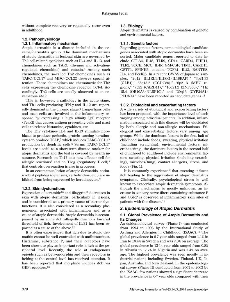
2.2. Epidemiological Survey in JapanA nationwide prevalence survey was conducted in Ja-pan from 2000 to 2008 using the medical examinationdata from public health centers, elementary schools,and universities. Figure 2 shows the prevalence byage groups. In addition, a prevalence survey on theadult atopic dermatitis was performed using themedical examination for 2,943 personnel of 2 univer-sities18 (Fig. 2). The data on occurrence and progres-sion of infantile atopic dermatitis is provided by a re-port based on a follow-up study of infants of 4 monthsto 3 years old performed in the Health and LabourSciences Research, from 2006 to 2008, in YokohamaCity, Chiba City, and Fukuoka City. The report showsthat 16.2% of ordinary infants, who received a medicalexamination at 4 months of age, developed atopic der-matitis. Atopic dermatitis remitted in 50% of the 4-month-old patients before the age of 18 months, indi-
cating an extremely dynamic change in progressionof atopic dermatitis in infancy.19 This survey showeda cumulative incidence rate before 3 years of age of alittle more than 30%, similar to reports from overseas(Fig. 3).
3. Diagnosis of Atopic Dermatitis
3.1. Diagnostics Criteria for Atopic Dermatitis(1) Diagnostic criteria proposed by Hanifin and Ra-
jka: The diagnostic criteria of Hanifin and Rajka areinternationally most popular.
(2) Diagnostic criteria proposed by the JapaneseDermatological Association: The Japanese Derma-tological Association developed diagnostic criteria in1994, which was partly revised in 2008 (Table 1). Us-ing these criteria, all diseases that meet the 3 require-ments of itch, characteristic rashes and distribution,and chronic�recurrent progression, will be diagnosedas atopic dermatitis irrespective of the severity ofsymptoms.1
3.2. Laboratory Data Used as a Reference for Di-agnosis
(1) Serum total IgE level: A high serum total IgE

Katayama I et al.
380 Allergology International Vol 63, No3, 2014 www.jsaweb.jp�
Fig. 2 Prevalence of atopic dermatitis by age groups in the fi scal year of 1996-2008. 4 months old from: Hok-
kaido, Kanto, Chubu, Kinki, Chugoku, Shikoku, and Kyushu (seven districts, n = 2744). Children with 1.5 years
old, 3 years old , 1st grade of elementary, 6th grade of elementary from: Hokkaido, Tohoku, Kanto, Chubu, Kinki,
Chugoku, Shikoku, and Kyushu (eight districts, n = 6424). University students from: University of Tokyo, Kinki
University, Hiroshima University (n = 8317). Adult (20s to 60s) from: Personnel of University of Tokyo and Kinki
University (n = 2943). Modifi ed from Ministry of Health and Welfare, Japan. [Guidelines for the Treatment of Atopic Dermatitis 2008] (in Japanese).
14
(%)
12
4 m
onth
s old
1.5
year
s old
3 ye
ars old
1st g
rade
of e
lem
enta
ry
6th g
rade
of e
lem
enta
ry
Unive
rsity
stu
dent
s
12.8
9.8
11.8
10.6
Ages
8.2
10
8
6
4
2
0
13.2
20s
30s
40s
50s +
60s
8.3
4.8
2.5
9.4
Pre
va
len
ce
level is observed in approximately 80% of patientswith atopic dermatitis. It is also reportedly signifi-cantly correlated with the severity (the later dis-cussed SCORing Atopic Dermatitis [SCORAD] sever-ity index).
(2) Blood eosinophil count: Eosinophilia is seen inblood and the rash tissue of many but not all patients.Because it changes more rapidly than IgE, it servesas an index to assess changes of the disease condi-tion.
(3) Specific IgE antibody titer: Patients with atopicdermatitis are apt to produce IgE antibodies in re-sponse to various allergens such as mites, foods, andpets and often show positive reactions to multiple al-lergens. Preventing exposure to the allergens thathave been proven positive is expected to improve orprevent exacerbation of rashes.
(4) Serum TARC level: The serum level of TARC, aTh2 chemokine, has been shown to sensitively reflectthe short-term condition of atopic dermatitis and is re-garded as a useful auxiliary marker to assess the se-verity of atopic dermatitis.
(5) Others: Laboratory data reportedly used as areference for disease conditions include lactate dehy-drogenase (LDH). The soluble IL-2R level, which ishigh in skin lymphoma, is a useful assessment tool.
3.3. Severity Criteria for Atopic Dermatitis(1) SCORAD: SCORAD is an international severity
criterion that has been most popularly adopted in theEnglish written literature at present. These criteriagrade the rash areas, severity of rash elements suchas erythema, edema�papule, exudation�crust, licheni-fication, scratch marks, xerosis cutis, and subjectivesymptoms such as itch, and insomnia, by weightingthem at a ratio of approximately 2 : 6 : 2.
(2) Atopic dermatitis severity classification of theJapanese Dermatological Association: This severitycriteria assesses severity based on the total scores of3 rash elements (erythema�acute papule, moisten-ing�crust, chronic papule�tubercle�lichenification)and rash areas of the body divided into the following5 sections: head and neck, anterior body trunk, poste-rior body trunk, upper limbs, and lower limbs.20
Atopic Dermatitis
Allergology International Vol 63, No3, 2014 www.jsaweb.jp� 381
Fig. 3 Individual follow-up analysis of symptom onset/progression of
atopic dermatitis in patients aged 4 months to 3 years (fi scal years sur-
veyed: FY2006-FY2008).
Nu
mb
er
of
pa
tie
nts
with
ato
pic
d
erm
atitis
56
5656
43
43
3030
144
74
4 months old
1 year 6 months
old
3 years old
5656
12.1%
16.2% 16.8%
100
200
300
0
159
Remission rate Between 4 months and 1 year 6 months of age: 70.1% (159+43 patients) Between 1 year 6 months and 3 years of age: 48.1% (74+30 patients) 144 children developed atopic dermatitis after 1 year 6 months.
Cumulative incidence rate up to 3 years of age: 31.6%
Physical examination of 4 month olds
Physical examination of 1 year 6 month olds
Physical examination of 3 year olds
Yokohama: 851 patients
Chiba: 523 patients
Fukuoka: 404 patients
Total: 1,778 patients
(3) Severity indicators: Although the above-ment-ioned severity criteria are more objective than theconventional criteria, they are difficult to use in dailymedical practice because of too many endpoints. Ac-cordingly, the “Guidelines for the Treatment ofAtopic Dermatitis 2008” of the Health and Labour Sci-ences Research proposes a simpler indicator of sever-ity (Table 2). This indicator measures severity by as-sessing both the severity and the spread of the rashand defines mild rashes and rashes with severe in-flammation based on photographic images (Fig. 4).
4. Clinical Symptoms of Atopic Dermatitis
Clinical symptoms: Clinical symptoms of atopic der-matitis are divided into 3 age groups: infancy(younger than 2 years old), childhood�school-age (2-12 years old), adolescence�adulthood (13 years andolder).
4.1. Cutaneous SymptomsThe most important cutaneous symptoms for atopicdermatitis are rashes and itches.
4.1.1. RashThere is a report on classification of rashes of atopicdermatitis from 3 perspectives: age groups, morphol-ogy and distribution, and sites.
(1) Rash morphology: Atopic dermatitis is a cutane-
ous disease that belongs to eczema�dermatitis. Pa-tients with atopic dermatitis under long-term treat-ment with a topical steroid develop further diversesymptoms with such side effects as skin atrophy andtelangiectasia. These symptoms will develop into fur-ther complicated cutaneous symptoms when an infec-tious disease occurs concurrently.
(2) Characteristics of rashes by age groupsa) Infancy (younger than 2 years old): A rash usu-
ally develops on the cheek, forehead, or the head,and causes flushing or papules. It will erode and ex-ude by being scratched and the exudate will form acrust when it is dried.
b) Childhood�school-age (2-12 years old): The skinis apt to develop an atopic condition by graduallytending to be dried due to degradation of the sebum-secretion capacity. In addition, the atopic skin may berepeatedly scratched to develop prurigo nodularis,erosions, blood crusts, and so on. Rashes most typi-cally observed in the childhood to the school-agegroup include a bend type.
c) Adolescence�adulthood (13 years and older):This age group is apt to suffer from seborrhea oracne due to the increase in sebum secretion withmodified rashes. The rash extends from the neck tothe upper chest region and the upper part of back andspreads like a clothes hanger. Together with therashes on the face and the neck, it is distributed like

Katayama I et al.
382 Allergology International Vol 63, No3, 2014 www.jsaweb.jp�
Table 1 Defi nition and diagnostic criteria for atopic dermatitis by the Japanese Dermatological Association
Defi nition
Atopic dermatitis is a pruritic, eczematous dermatitis; its symptoms chronically fl uctuate with remissions and relapses.
Most individuals with atopic dermatitis have atopic diathesis.
Atopic diathesis: (i) personal or family history (asthma, allergic rhinitis and/or conjunctivitis, and atopic dermatitis); and/or (ii) pre-disposition to overproduction of immunoglobulin E (IgE) antibodies.
Diagnostic criteria for atopic dermatitis
1. Pruritus
2. Typical morphology and distribution
1) Eczematous dermatitis
- Acute lesions: erythema, exudation, papules, vesiculopapules, scales, crusts
- Chronic lesions: infi ltrated erythema, lichenifi cation, prurigo, scales, crusts
2) Distribution
- Symmetrical
Predilection sites: forehead, periorbital area, perioral area, lips, periauricular area, neck, joint areas of limbs, trunk
- Age-related characteristics
Infantile phase: starts on the scalp and face, often spreads to the trunk and extremities
Childhood phase: neck, the fl exural surfaces of the arms and legs
Adolescent and adult phase: tendency to be severe on the upper half of body (face, neck, anterior chest and back)
3. Chronic or chronically relapsing course (usually coexistence of old and new lesions)
- More than 2 months in infancy
- More than 6 months in childhood, adolescence, and adulthood
Defi nitive diagnosis of atopic dermatitis requires the presence of all three features without any consideration of severity.
Other cases should be evaluated on the basis of the age and clinical course with the tentative diagnosis of acute or chronic, non-specifi c eczema.
Differential diagnosis (association may occur)
Contact dermatitis, seborrheic dermatitis, prurigo simplex, scabies, miliaria, ichthyosis, xerotic eczema, hand dermatitis (non-atopic), cutaneous lymphoma, psoriasis, immune defi ciency diseases, collagen diseases (systemic lupus erythematosus, derma-tomyositis), Netherton’s syndrome
Diagnostic aids
Family history (bronchial asthma, allergic rhinitis and/or conjunctivitis, atopic dermatitis), personal history (bronchial asthma, al-lergic rhinitis and/or conjunctivitis), follicular papules (goose-skin), elevated serum IgE level
Clinical types (not applicable to the infantile phase)
Flexural surface type, extensor surface type, dry form in childhood, head/face/neck/upper chest/back type, prurigo type, erythro-derma type, combinations of various types are common
Signifi cant complications
Ocular complication (cataract and/or retinal detachment): especially in patients with severe facial lesions, Kaposi’s varicelliformeruption, molluscum contagiosum, impetigo contagiosa
Adapted from reference 1.
a sculptural portrait (portrait type).(3) Secondary cutaneous changesa) White dermographismb) Goosebumps skinc) Pityriasis albad) Pigmentation: This includes postinflammatory
pigmentation, frictional pigmentation, dirty neck, etc.and is clinically known for orbital darkening andpunctate pigmentation in the lips.
e) Folds: Folds may be seen in from the inner an-
gle to the outer downward of the palpebra inferior(Dennie-Morgan infraorbital fold), in the neck (ante-rior neck folds), and in the abdomen. A finding calledpalmar hyperlinearity indicates a large number of in-born palmer folds.
f) Hair loss: Hair loss is seen in the occipital regionfor infant patients, in the temporal region for schoolage or older patients, and in the eyebrow for adult pa-tients. Eyebrow hair loss is seen mostly in the outerhalf (Hertoghe’s sign).

Atopic Dermatitis
Allergology International Vol 63, No3, 2014 www.jsaweb.jp� 383
Table 2 Severity index
There are several criteria proposed for severity assessment of atopic dermatitis at present that require profi ciency in assess-ment. Accordingly, the following severity levels are defi ned as indices for treatment.
Mild: Only mild rashes are observed irrespective of the area.
Moderate: Rashes with severe infl ammation are observed in less than 10% of the body surface area.
Severe: Rashes with severe infl ammation are observed in ≥10% to <30% of the body surface area.
Most severe: Rashes with severe infl ammation are observed in ≥30% of the body surface area.
Mild rash: Lesions are seen chiefl y with mild erythema, dry skin, or desquamation.
Rashes with severe infl ammation: Lesion with erythema, papule, erosion, infi ltration, lichenifi cation, etc.
Modifi ed from Ministry of Health and Welfare, Japan. [Guidelines for the Treatment of Atopic Dermatitis 2008] (in Japanese).
g) Nail luster: Hard scratching causes pearl-likeluster of nails (pearly nail).
h) Diffuse flushing of the facial surface: The facemay acutely flush over the entire surface. It is likelycaused by sustained inflammation and scratching.
4.1.2. ItchItch is caused by increase in skin temperature result-ing from bathing, exercise, sleep, ointment applica-tion, etc., sweating due to irritation and body warmth(itch when sweating), and perspiration stimulation. Italso occurs when woolen clothes contact the skin(wool intolerance).
(1) Control of itch: In early infancy, the negativeimpact of scratching can be weakened by coveringthe scratched site with bandages, clothing, etc., or bywearing gloves. It is not until adolescence that oneunderstands that scratching will aggravate the cuta-neous symptoms and comes to restrain oneself fromscratching.
(2) Sleep disorders: Sleep disorders include diffi-culty in falling asleep and awakening during thenight. The difficulty in falling asleep is caused by astrong sensitivity to itch due to body warmth whilesleeping.
(3) Scratch addiction: While scratching during thenighttime is an unconscious reflex movement,scratching during the daytime involves a consciouselement. It is reportedly a scratching phenomenonthat is performed unintentionally but preferably(scratch addiction).21
4.2. Symptoms Other than Cutaneous Symptoms4.2.1. Symptoms�findings incidental to rashesDermatopathic lymphadenopathy occurs when a cu-taneous symptom with certain severity and spreadmay cause swelling of regional superficial lymphnodes. It is indolent and not indicative of infection.
4.2.2. Symptoms in other organs(1) Cataract: Cortical cataract with an anterior sub-
capsular or posterior subcapsular opacity may pro-gress to cover all layers. With a gender ratio of ap-proximately 1:2, it occurs most frequently in individu-als 15-24 years of age, with a severe form of rash on
the facial surface in particular. The frequencies ofcomplications with cataracts are reported to be 2.0%of all patients and 5.5% of severe patients.
(2) Retinal detachment: The retinal detachment ac-companying atopic dermatitis is reported to occurwith a hiatus caused by continuous external forcesapplied to the eyeball. It occurs predominantly be-tween the ages of 16 to 25 and frequently among pa-tients who strongly rub their eyelids or who pat theirown faces. The frequencies of complications with reti-nal detachment are reported to be 0.5% of all patientsand 2% of severe patients.
(3) Airway hyperresponsiveness: A study on airwayhyperresponsiveness caused by histamine inhalationload in children with atopic dermatitis found that pa-tients with atopic dermatitis not complicated withasthma showed significantly worse airway hyperre-sponsiveness compared with healthy controls.
(4) Mental symptoms: Atopic dermatitis may causesymptoms such as social withdrawal resulting fromdisappointment with existing therapies, discontentwith responses of doctors, disappointment with theprobability of cure, and so on. Severe atopic dermati-tis cases may be accompanied by psychosomaticproblems.
(5) Neurological symptoms: Paresthesia may occurin the distal extremities with aggravation of dermati-tis symptoms and lesional atopic myelitis cervicalismay be observed in the cervical region in MRI.
(6) Intestinal tract symptoms: Severe cases may beaccompanied by such symptoms as diarrhea and con-stipation.
5. Investigation of Causes and Exacerbat-ing Factors for Atopic Dermatitis andCountermeasures
Given that causes and exacerbating factors may varyaccording to age, individual differences of patients,environment, and lifestyle, it is important to takecountermeasures in consideration of the conditions ofthe individual patients.
5.1. FoodsFood allergens are investigated by performing de-tailed history taking, allergen tests and then by com-

Katayama I et al.
384 Allergology International Vol 63, No3, 2014 www.jsaweb.jp�
Fig. 4 Examples of mild rashes and rashes with severe infl ammation of atopic dermatitis. Examples of
mild rashes includes (A) Face: Mild desquamation, erythema, (B) Body trunk: mild desquamation, dry
skin, (C) Lower limb: Mild desquamation, erythema, including partial mild lichenifi cation. Examples of
rashes with severe infl ammation includes (D) Face: Apparent erythema, desquamation, apparent infi ltra-
tion, (E) Body trunk: Apparent erythema and lichenifi cation, (F) Lower limb: Apparent erythema, papule,
scratch mark, lichenifi cation.
AA DD
BB EE
CC FF
bining elimination tests and provocation tests (notperformed for cases accompanied by anaphylaxis) forsuspicious allergens detected to remove the deter-
mined allergens. Countermeasures are taken by nu-tritional care introducing alternative foods introducedand providing guidance to the family without ran-
Atopic Dermatitis
Allergology International Vol 63, No3, 2014 www.jsaweb.jp� 385
domly removing allergens.
5.2. SweatingSweating is an important cause and exacerbating fac-tor for atopic dermatitis, hence washing away sweatby bathing and showering will lead to the improve-ment of symptoms. Bathing and showering are im-portant not only for washing away the components ofperspiration but also for washing away allergens,such as dust and pollens, and microbes on the skinsurface.
5.3. Physical IrritationCauses and exacerbating factors other than theabove-mentioned perspiration include clothes, dryair, hairs, and cosmetics for adult patients. Cosmet-ics, shampoo, and soaps need to be selected appropri-ately, exchanging products that cause symptoms.
5.4. Environmental FactorsAllergens such as mites and house dust, pollen aller-gens in specific seasons, and organic solvents such asformaldehyde and toluene can become problematic.Being sensitized to mites in infancy is reportedly amarker for the development of asthma.22 Periocularpathological changes are often observed during air-borne pollen seasons.23
5.5. Microbes�FungiIf no infectious symptoms are seen in the affectedpart, a topical steroid can be applied to encourage theimprovement of the cutaneous symptom, even if thesite is densely populated with Staphylococcus aureus(with bacterial counts of 1000 cfu�10 cm2 or more asdetected by the stamp method).
(1) An antimicrobial therapy should be performedif any infectious symptoms are observed.
(2) Care should be taken against microbial substi-tution with methicillin-resistant Staphylococcus aureus(MRSA).
(3) The skin should be kept clean by frequent bath-ing or showering, etc.
(4) Disinfectants such as povidone-iodine solutionshould not be applied.
5.6. Contact AntigenContact dermatitis is divided into allergic contact der-matitis, which is developed by a sensitized patient,and primary irritant contact dermatitis, which can bedeveloped by anyone depending on the antigen level.
5.7. StressAggravation by mental stress is often experienced indaily medical practice. The high rate of aggravation ofatopic dermatitis reported in areas affected by theGreat Hanshin-Awaji Earthquake definitely provesthe correlation between stress and atopic dermati-tis.24
5.8. ScratchingScratching will not only damage the cutaneous bar-rier functions by injuring the skin, but also worsenthe symptoms by causing the release of various phlo-gogenic agents.
5.9. Perinatal PreventionA randomized comparative study that assessedwhether consumption of an elimination diet free ofhighly sensitized food antigens such as eggs andcow’s milk by pregnant or lactating mothers can pre-vent newborns from developing sensitization to foodallergens or atopic dermatitis revealed that elimina-tion of eggs or cow’s milk has no preventive effect.25
A case-series study in high-risk children was con-ducted to determine whether moisturizer applicationduring the newborn period prevents the developmentof atopic dermatitis, in light of the importance of cuta-neous barrier functions in atopic dermatitis. The re-sults showed that the subject group, in which mois-turizer was applied during the newborn period, had alow incidence rate of 15%.26 However, the effect ofmoisturizer application during the newborn periodneeds to be demonstrated in a randomized compara-tive study in future.
5.10. OthersThe pathology of atopic dermatitis is a chronic inflam-mation of the skin that is aggravated by the deteriora-tion of compliance. It is necessary to educate patientsrepeatedly so they may understand the pathology andrealize that a long-term antiinflammatory therapy isrequired.
6. Summary of the Basic Therapy of AtopicDermatitis
6.1. Basics of TreatmentBecause the treatment of atopic dermatitis requiresappropriate diagnosis and evaluation of cutaneoussymptoms, the basics of treatment under this guide-line include 3 mainstays. These are investigation andcountermeasures of causes and exacerbating factors;second, correction of skin dysfunctions (skin care);and third, pharmacotherapy. These are based on theconcept that this disease is an inflammatory cutane-ous disease that forms an eczematous lesion against abackdrop of atopic diathesis, with numerous factorsinvolved in the occurrence and aggravation and aber-rant function in the skin (Fig. 5). These 3 points areequally important and thus need to be appropriatelycombined in accordance with the symptoms of eachpatient.
(1) Investigation and countermeasures of causesand exacerbating factors: Because the importance ofthe individual factors depends on the individual pa-tients, it is important to investigate fully the factorsthrough diagnosis, and to take reasonable and appro-priate countermeasures.

Katayama I et al.
386 Allergology International Vol 63, No3, 2014 www.jsaweb.jp�
Fig. 5 Important points of these guidelines. Modifi ed from Ministry of Health and Wel-
fare, Japan. [Guidelines for the Treatment of Atopic Dermatitis 2008] (in Japanese).
2. Assessment of cutaneous symptoms
In selecting a therapy, cutaneous symptoms need to be properly assessed.
3. Basics of treatment
Based on the above assessment, investigation/countermeasures against
causes and exacerbating factors, skin care, and pharmacotherapy should be
implemented by being optimally combined for each patient. Sufficient
information about the treatment should be transmitted to the patient to build a
favorable partnership.
1. Diagnosis
Appropriate diagnosis should be ensured by discriminating them from other
diseases with similar symptoms, such as eczema and dermatitis, in accordance
with the abovementioned concept.
Outline of the guidelines for therapy
Pharmacotherapy
Diagnosis
Severity assessment
Investigation/countermeas-
ures against causes and
exacerbating factors
Skin care
(correction of abnormal
cutaneous functions)
(2) Skin care (correction of abnormal cutaneousfunctions): The skin barrier functions of patients withatopic dermatitis are deteriorated due to abnormalityin function such as deteriorated water retentivity, low-ered threshold of itch, and susceptibility to infection.Correcting those abnormalities (skin care) is ex-tremely important in treatment, chiefly by good hy-giene and moisture retention of the skin.
(3) Basics of pharmacotherapy: If countermeasuresagainst the causes and exacerbating factors and theskin care have not resulted in improvement of derma-titis, pharmacotherapy will be needed.
a) Final objective: The final objective of treatmentis to achieve a level with no or mild symptoms, if any,which should not interfere with daily life and requireminimal pharmacotherapy.
b) Assessment of severity and activity level: At-tempts have been made to assess the severity and ac-tivity level by using the IgE level, eosinophilic leuko-cyte count, LDH level, TARC level, and VAS itchscore.
c) Remission-induction therapy: To maximize thetherapeutic effect, in particular the pharmacothera-peutic effect, and to minimize the side effects, the pa-
tient should be follow up every 1 or 2 weeks to evalu-ate and verify the effects and change the therapeuticagents and method as needed. If no improvement isobserved or if an abnormal change in symptoms isobserved after approximately 1 month of remission-induction therapy, referral of the patient to a morespecialized medical institution should be considered.
d) Remission maintenance therapy: If symptomscannot be controlled or if symptoms frequently re-lapse after completion of remission induction, therapyshould be changed to remission maintenance ther-apy. Remission maintenance therapy is mainly basedon the external application of tacrolimus. If remissioncannot be maintained after approximately 6 months,the patient should be referred to a specialized medi-cal institution to define the condition as severe, mostsevere, or intractable, in consideration of prolongeduse of topical tacrolimus.
e) Severe, most severe, or intractable conditions(cases where remission maintenance therapy is diffi-cult to execute): If remission cannot be maintained,external application of a higher ranked steroid, oraladministration of immunosuppressive agents (cy-closporine), oral administration of steroids, ultraviolet

Atopic Dermatitis
Allergology International Vol 63, No3, 2014 www.jsaweb.jp� 387
light therapy, or psychosomatic medical therapyshould be used.
Hospital treatment is effective for severe, most se-vere, and intractable cases. Education should be pro-vided to such patients on environmental factors, life-style, dietary habits, personal hygiene management,and correction of psychological problems, duringhospitalization.
Oral administration of steroids should not be exe-cuted in principle. However, if the patient developsextremely severe symptoms that cannot be controlledwith external medication, such oral administrationmay be used for a short period of time.
f) Precautions during treatment: Given that the du-ration of illness is often prolonged, its associationwith growth in children and with lifestyle related dis-eases, vitamin D deficiency, and ophthalmologicproblems in adults should be considered.
6.2. Precautions During TreatmentIf an abnormal change is observed in symptoms dur-ing treatment or if no improvement is observed aftertreatment based on a basic therapy for approximatelyone month, referral to a more specialized medical in-stitution should be considered. Hospital treatment iseffective for patients with severe or most severecases.
7. Skin Care Against Atopic Dermatitis
Skin care is highly important, as is shown by consid-eration of the fact that the skin tends to become moreprone to drying due to aging, and given the adverseeffects of settlement of Staphylococcus aureus on theskin on the aggravation of symptoms.
7.1. Dry SkinThe skin of patients with atopic dermatitis is gener-ally in a dried condition, not only in the lesion butalso in an apparently normal area, due to insensiblewater loss (transepidermal water loss; TWL). In suchconditions, with facilitation of transcutaneous inva-sion of allergens and irritants, likely resulting in aller-gic reactions and irritability, patients may come tosuffer from itch due to the lowered threshold of itch.Abnormality in the water barrier function and thewater retention capability of the horny cell layer areconsidered as causing�exacerbating factors of atopicdermatitis. Accordingly, the incidence rate of atopicdermatitis could be decreased by moisturizer applica-tion during the newborn period.
7.2. Staphylococcus Aureus FloraIf the bacterial count is small (in the case of a bacte-rial count of 1000 cfu�10 cm2 or less by the stampmethod), dry skin symptoms will be improved onlyby continuous use of a moisturizer. If the bacterialcount is larger, it is important to take a skin care regi-men with consideration of the bacterial flora on the
skin surface.
7.3. Important Points in Skin Care(1) Skin care against dry skin: Hydrophilic oint-
ments and water absorptive ointment with high mois-ture retention include urea preparations, heparinoidpreparations, water-soluble collagen preparations, so-dium lactate preparations, and elastin hydrolysispreparations.
(2) Skin care against injured skin: An oleaginousointment (ointment in a narrow sense) with skin pro-tective action should be externally applied. Given thata moisturizer is known to exert a higher moisturizingeffect when externally applied twice a day than whenapplied once a day, consideration should be given tothe number of external applications.27
(3) Good hygiene of the skin and skin care: To en-sure healthy skin, bathing and showering should bestrictly observed with appropriate moisturizer or skinprotectant applied externally as needed.
(4) Practical home skin carea) Bathing: Although moisturizer application is rec-
ommended immediately after bathing, reports haveshown that the moisturizing effect is not influencedby a time lapse after bathing. Therefore, physiciansshould instruct patients not to forget the external ap-plication of moisturizers.28
b) Shower: Sweating is considered as an exacerbat-ing factor for atopic dermatitis. In the sweaty summerseason, washing away sweat by showering improvesrashes.29
c) Shampoo: Given that shampoo and soap residuemay aggravate rashes, soap and shampoo should notbe left in such regions as the hairline, the side of thenose, and jaws, etc.
8. Pharmacotherapies for Atopic Dermati-tis
8.1. External Medicine8.1.1. Topical therapyTopical therapy refers to skin care chiefly with mois-turizers, and to inflammation control chiefly with topi-cal steroids and tacrolimus ointments (immunosup-pressive ointment, topical calcineurin inhibitor).
Although basic therapy for controlling inflamma-tion in the acute phase consists of topical steroids,the use of tacrolimus ointment or topical steroids in-termittently in combination with a moisturizer is use-ful in the remission maintenance period. This therapyis referred to as proactive therapy (Fig. 6) as opposedto reactive therapy (Fig. 7), in which topical therapyis administered only when the rash worsens. Proac-tive therapy has been shown to not only control therelapse of cutaneous symptoms, but also to be cost-effective.30 The use of a medical dressing or Tubifast(Allergy Health Care, Nara, Japan) with an externallyapplied medicine is effective for the prevention ofscratching.

Katayama I et al.
388 Allergology International Vol 63, No3, 2014 www.jsaweb.jp�
Fig. 6 Proactive therapy for atopic dermatitis.
† Topical steroid/topical tacrolimus.
Time course
Se
ve
rity
of
de
rma
titis
Firmly apply an external medicine at an optimum dose† + moisturizer.
Apply the medicine thinly over the entire site of rash development even if the symptoms are mild at least thrice, twice, or once per week.†
Thrice a week
Twice a week
Once a week
Twice a week
Once a week
Apply a moisturizer all over the body daily.
Fig. 7 Reactive therapy for atopic dermatitis.† Topical steroid/topical tacrolimus.
Apply a moisturizer all over the body daily.Apply an external medicine† repeatedly once a day to the site where an itch occurred during the day.
The remission period will increase progressively.
Time course
Seve
rity
of
de
rma
titis
Firmly apply an external medicine at an optimum dose† + moisturizer.
8.1.2. MoisturizerAlthough the use of an ointment-based drug productwith high moisture retaining properties is recom-mended for the treatment of mild cases with the aimof moistening and protecting the skin, recovering andmaintaining the barrier function, the drug productshould be selected in consideration of the outdoor airtemperature, humidity, and usability. For the treat-ment of weeping lesions in the acute phase, a surfacedressing with a zinc ointment may be effective. Ureapreparations should be used with caution, as they canstimulate an eroded surface or strongly inflammatoryskin. A moisturizer can also cause contact dermatitis.The use of a moisturizer is useful to prevent relapseof atopic dermatitis; however, studies have shownthat the continuous use of a moisturizer since thenewborn period may reduce the incidence rate or de-lay the development of atopic dermatitis.31,26
8.1.3. Pharmacology�action mechanism of thesteroid drugUpon entering a cell, steroids bind receptors thatform complexes with heat shock protein (HSP90),which is found in the cytoplasm, and migrate with thecomplex into the nucleus. There they activate steroid-responsive genes to exert their pharmacological ac-tions that include antiinflammatory action in a narrowsense, antiallergic action, and immunosuppressive ac-tion.32
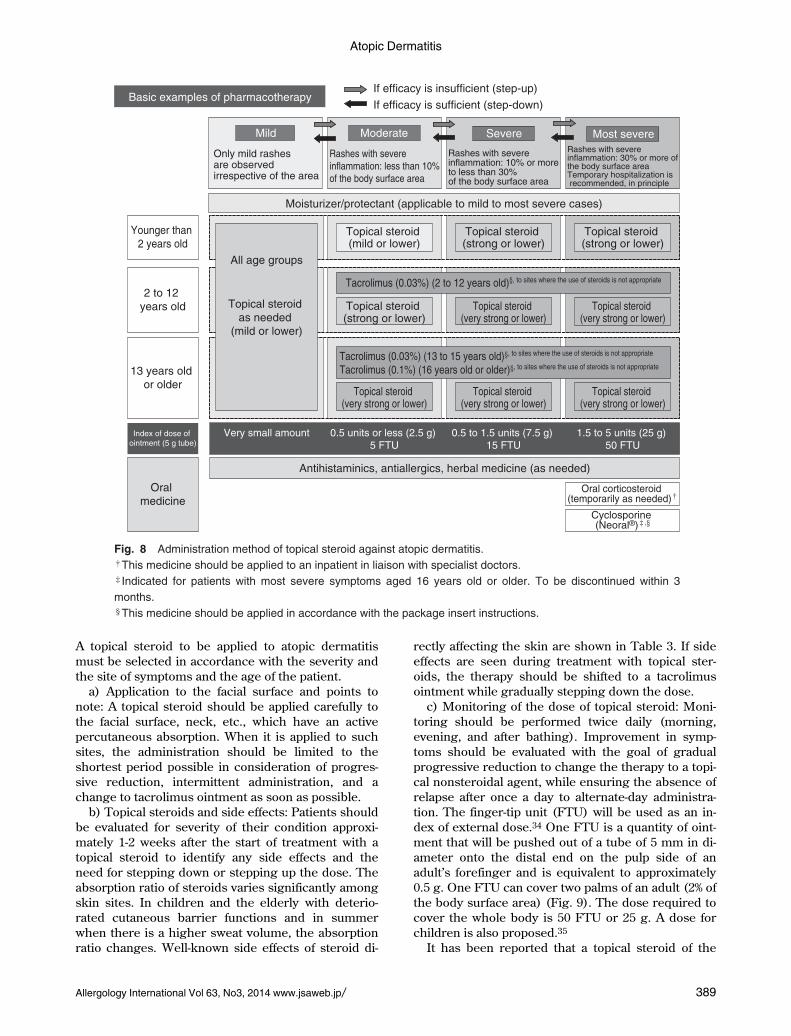
8.1.4. Administration method of topical steroids(1) Selection of topical steroid: Topical steroids are
classified into 5 ranks from weak to strongest. A ster-oid of an appropriate rank should be used in accor-dance with the severity of cutaneous symptom33 (Fig.8).
(2) Points to note in external application of steroid:
Atopic Dermatitis
Allergology International Vol 63, No3, 2014 www.jsaweb.jp� 389
Fig. 8 Administration method of topical steroid against atopic dermatitis.
†This medicine should be applied to an inpatient in liaison with specialist doctors.
‡Indicated for patients with most severe symptoms aged 16 years old or older. To be discontinued within 3
months.
§This medicine should be applied in accordance with the package insert instructions.
2 to 12 years old
Younger than 2 years old
13 years old or older
Only mild rashes are observed irrespective of the area
Rashes with severe
inflammation: less than 10%
of the body surface area
If efficacy is insufficient (step-up)
If efficacy is sufficient (step-down)
Topical steroid (mild or lower)
Oral corticosteroid (temporarily as needed)†
Cyclosporine (Neoral®)‡,§
Index of dose of
ointment (5 g tube)Very small amount 0.5 units or less (2.5 g)
5 FTU
0.5 to 1.5 units (7.5 g)
15 FTU
1.5 to 5 units (25 g)
50 FTU
Antihistaminics, antiallergics, herbal medicine (as needed)
Moisturizer/protectant (applicable to mild to most severe cases)
Oral medicine
Topical steroid (strong or lower)
Mild Moderate Most severeSevere
All age groups
Topical steroid as needed
(mild or lower)
Tacrolimus (0.03%) (13 to 15 years old)§, to sites where the use of steroids is not appropriate
Tacrolimus (0.1%) (16 years old or older)§, to sites where the use of steroids is not appropriate
Tacrolimus (0.03%) (2 to 12 years old)§, to sites where the use of steroids is not appropriate
Topical steroid (strong or lower)
Topical steroid (very strong or lower)
Topical steroid (very strong or lower)
Topical steroid (very strong or lower)
Topical steroid (very strong or lower)
Topical steroid (very strong or lower)
Topical steroid (strong or lower)
Basic examples of pharmacotherapy
Rashes with severe inflammation: 10% or more to less than 30% of the body surface area
Rashes with severe inflammation: 30% or more of the body surface areaTemporary hospitalization is recommended, in principle
A topical steroid to be applied to atopic dermatitismust be selected in accordance with the severity andthe site of symptoms and the age of the patient.
a) Application to the facial surface and points tonote: A topical steroid should be applied carefully tothe facial surface, neck, etc., which have an activepercutaneous absorption. When it is applied to suchsites, the administration should be limited to theshortest period possible in consideration of progres-sive reduction, intermittent administration, and achange to tacrolimus ointment as soon as possible.
b) Topical steroids and side effects: Patients shouldbe evaluated for severity of their condition approxi-mately 1-2 weeks after the start of treatment with atopical steroid to identify any side effects and theneed for stepping down or stepping up the dose. Theabsorption ratio of steroids varies significantly amongskin sites. In children and the elderly with deterio-rated cutaneous barrier functions and in summerwhen there is a higher sweat volume, the absorptionratio changes. Well-known side effects of steroid di-
rectly affecting the skin are shown in Table 3. If sideeffects are seen during treatment with topical ster-oids, the therapy should be shifted to a tacrolimusointment while gradually stepping down the dose.
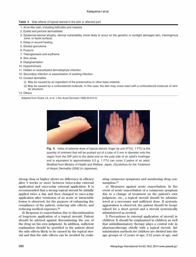
c) Monitoring of the dose of topical steroid: Moni-toring should be performed twice daily (morning,evening, and after bathing). Improvement in symp-toms should be evaluated with the goal of gradualprogressive reduction to change the therapy to a topi-cal nonsteroidal agent, while ensuring the absence ofrelapse after once a day to alternate-day administra-tion. The finger-tip unit (FTU) will be used as an in-dex of external dose.34 One FTU is a quantity of oint-ment that will be pushed out of a tube of 5 mm in di-ameter onto the distal end on the pulp side of anadult’s forefinger and is equivalent to approximately0.5 g. One FTU can cover two palms of an adult (2% ofthe body surface area) (Fig. 9). The dose required tocover the whole body is 50 FTU or 25 g. A dose forchildren is also proposed.35
It has been reported that a topical steroid of the
Katayama I et al.
390 Allergology International Vol 63, No3, 2014 www.jsaweb.jp�
Fig. 9 Index of external dose of topical steroid: fi nger tip unit (FTU). 1 FTU is the
quantity of ointment that will be pushed out of a tube of 5 mm in diameter onto the
region from the DIP joint to the distal end on the pulp side of an adult’s forefi nger
and is equivalent to approximately 0.5 g. 1 FTU can cover 2 palms of an adult.
Modifi ed from Ministry of Health and Welfare, Japan. [Guidelines for the Treatment of Atopic Dermatitis 2008] (in Japanese).
Table 3 Side effects of topical steroid in the skin or affected part
1. Acne-like rash, including folliculitis and rosacea
2. Eyelid and perioral dermatitides
3. Epidermal-dermal atrophy, dermal vulnerability (most likely to occur on the geriatric or sunlight damaged skin, intertriginous zone, or facial surface)
4. Delay in wound healing
5. Gluteal granuloma
6. Purpura
7. Telangiectasia and erythema
8. Skin striae
9. Depigmentation
10. Hypertrichosis
11. Hidden or exacerbated dermatophyte infection
12. Secondary infection or exacerbation of existing infection
13. Contact dermatitis
(i) May be caused by an ingredient of the preservative or other base material.
(ii) May be caused by a corticosteroid molecule. In this case, the skin may cross-react with a corticosteroid molecule of simi-lar structure.
14. Others
Adapted from Drake LA, et al. J Am Acad Dermatol 1996;35:615-9.
strong class or higher shows no difference in efficacyafter 3 weeks or more between twice-a-day externalapplication and once-a-day external application. It isrecommended that a strong topical steroid be initiallyapplied twice a day and then changed to once-a-dayapplication after remission of an acute or intractablelesion is observed, for the purpose of enhancing thecompliance of the patient, reducing side effects, andreducing medical expenses.36
d) Response to exacerbation due to discontinuationof long-term application of a topical steroid: Patientshould be advised against discontinuing the use ofthe drug on his own judgment. In addition, sufficientexplanation should be provided to the patient aboutthe side effects likely to be caused by the topical ster-oid and that the side effects can be avoided by evalu-
ating cutaneous symptoms and monitoring drug con-sumption.37
e) Measures against acute exacerbation: In theevent of acute exacerbation of a cutaneous symptomdue to a change of treatment on the patient’s ownjudgment, etc., a topical steroid should be adminis-tered at a necessary and sufficient dose. If systemicaggravation is observed, the patient should be hospi-talized for a short period and a steroid systemicallyadministered as needed.
f) Precautions in external application of steroid inchildren: It should be emphasized in children as wellthat antiinflammatory therapy plays a central role inpharmacotherapy chiefly with a topical steroid. Ad-ministration methods for children are divided into theage groups of <2 years of age, 2-12 years of age, and

Atopic Dermatitis
Allergology International Vol 63, No3, 2014 www.jsaweb.jp� 391
>13 years of age. Care should be taken so that insuffi-cient administration for fear of side effects likely gen-erated in the treatment of children may not result inprolonged symptoms.
(3) Selection of external medicine according tosymptoms: In principle, in the treatment of atopic der-matitis, a moisturizer or skin care product should beapplied to a mild eczematous lesion or dry skin onthe facial surface without applying any topical ster-oids. It is reported that twice-a-day external applica-tion of a moisturizer (preparation containing hepari-noid) significantly inhibits the relapse of inflamma-tion of atopic dermatitis compared with the untreatedgroup (no application group).
8.1.5. Topical immunosuppressant (tacrolimusointment [ProtopicⓇ])
(1) Pharmaceutical form and mechanism of actionof tacrolimus ointments: Tacrolimus ointments willbe used when existing therapies are not effectiveenough or not indicated because of side effects. Itshould be used in consideration of precautions and af-ter obtaining informed consent from the patients.
Tacrolimus ointments (trade name: protopic oint-ment) are available in 2 dose forms: ointment 0.1% ap-plicable to patients 16 years of age or older and oint-ment 0.03% applicable for children aged 2-15 years.The action mechanism of tacrolimus is the inhibitionof the function of T lymphocytes, which play a centralrole in the development of allergic inflammation, andimprovement in the barrier function. Althoughtacrolimus ointments have a similar beneficial effectas that of strong steroids, their absorption from nor-mal skin is lower than that of steroids because oftheir higher molecular weight. This property is con-sidered an advantage because they are more absorp-tive and effective for the treatment of lesions withacute dermatitis associated with compromised cuta-neous barrier function, whereas they become less ab-sorptive and less likely to show side effects as derma-titis subsides.
(2) Instructions for the use of tacrolimus oint-ments: The tacrolimus ointment is usually applied ex-ternally once a day after bathing. Excessive exposureto ultraviolet light should be avoided when the oint-ment is applied externally. A 0.1% tacrolimus oint-ment for adults should be administered at a dose of5 g or less. A 0.03% tacrolimus ointment for childrenaged 2 to 5 years (<20 kg in body weight) should beadministered at a dose of�1 g; for children aged 6 to12 years (20-50 kg in body weight), at a dose of 2-4 g;and for children 13 years or older (�50 kg in bodyweight), at a dose of �5 g. The tacrolimus ointmentshould be administered at a maximum of twice perday. When applied twice per day, an interval of ap-proximately 12 hours between applications is recom-mended. Occlusive dressing therapy should not beused because it may cause an increase in the blood
level. Continuous external application of a tacrolimusointment two to three times per week after remissioninduction can significantly inhibit the relapse ofsymptoms (proactive therapy).38-40
(3) Side effects of tacrolimus ointments: No severesystematic adverse events were observed in a studyof post-marketing prolonged administration; there-fore, tacrolimus ointments are considered safe. Pre-cautions for the use of tacrolimus ointment includethe possibility of skin effects such as a burning sensa-tion at the start of external application and the poten-tial aggravation of local infectious disease in the skin.Although the stimulatory effects of the ointment onthe skin will most likely disappear after several days,a tacrolimus ointment can become intolerable be-cause of the burning sensation. This problem can bemanaged by using a moisturizer before the applica-tion of the ointment or by using a tacrolimus oint-ment in combination with a topical steroid for a shortperiod of time. Combinations with other externalmedicines should be avoided because of potential ef-fects on the stability and absorptive properties of thetacrolimus ointment. Tacrolimus ointments shouldnot be used on rashes associated with infectious dis-eases of the skin. Tacrolimus ointments cannot be ap-plied to an eroded surface or the surface of an ulcerbecause of its stimulatory effects and absorption. Atacrolimus ointment cannot be applied to patientswith ichthyosiform erythroderma (such as Nether-ton’s syndrome), whose blood tacrolimus level can in-crease because of elevated percutaneous absorptionand because it can cause side effects such as nephro-pathy. In addition, tacrolimus ointments cannot be ap-plied for reasons of safety to patients with nephropa-thy, pregnant women, infants younger than 2 years ofage (at this point in time), and patients receiving pho-totherapy. Regarding the risk of lymphoma and skincancer, it has been reported that the external applica-tion of tacrolimus ointment will not lead to an in-crease in the incidence rate of these diseases.41-44
8.1.6. Topical non-steroidal antiinflammatory dr-ugsNo data definitely shows that nonsteroidal antiinflam-matory drugs (NSAIDs) are effective for eczematouslesions of atopic dermatitis. There is no description ofNSAIDs in the guidelines for the treatment of atopicdermatitis of the United States or Europe.45
8.2. Oral Medicine8.2.1. Antihistaminics�antiallergics
(1) Pharmacological actions�action mechanisma) Antihistamines: The main pharmacological ac-
tion of antihistamines is to antagonize histamine inthe histamine H1 receptor in tissues to inhibit its ac-tion. In recent years, a new concept of inverse ago-nism has been advocated for the H1 receptor.46 Theinverse agonist (H1 receptor antagonist) combines

Katayama I et al.
392 Allergology International Vol 63, No3, 2014 www.jsaweb.jp�
with the inactive receptor to shift the equilibrium ofthe receptor towards the inactive state in addition toacting as a competitive inhibitor of the agonist.
b) Antiallergics: Antiallergics are divided into me-diator antireleasers, which are antiallergics in a nar-row sense, thromboxane A2 inhibitors, leukotrienereceptor antagonists, and cytokine inhibitors, as wellas classic antihistamines.
(2) Administration method: Since differences in ef-ficacy are found amongst individual patients in dailymedical care, it is necessary to explore and replacedrugs that are not effective after 2 weeks of admini-stration with another drug appropriate for the individ-ual patient.
a) Treatment of itch: Since itch is normally causedby irritation of the end of C nerve fiber located at theepidermal-dermal junction. The histamine H1 recep-tor is located at the C fiber, and antihistaminics andantiallergics with antihistaminic action are expectedto inhibit itch.
b) Evidence of efficacy of antihistaminics: Hoare etal. reported that oral antihistaminics lack grounds forefficacy after their evidence-based analysis of ran-domized studies on the efficacy of treatments foratopic dermatitis extracted from various databases.47
Recently, however, a large-scale clinical study wasconducted with fexofenadine hydrochloride thatshowed its usefulness in a randomized, double-blind,parallel-group comparative study.48
(3) Side effectsa) Central nervous system effect: The drug gener-
ally generates side effects in the form of sleepiness,loss of concentration, or malaise, and it may cause ex-citement if administered in a large quantity. Precau-tion is necessary in use of antihistaminics in children,particularly against convulsion. A second-generationantihistaminics is characterized by reduced sleepi-ness and central nervous system effects because ithas more difficulty in passing through the blood-brain barrier. In evaluating sedation, sleepinessshould be differentiated from impaired performance(a condition with deteriorated operating efficiency).49
b) Anticholinergic action: The anticholinergic ac-tion may cause dry mouth, sense of mucosal dryness,urinary retention, and so on. This drug is contraindi-cated for patients with glaucoma or lower urinarytract obstructive disease (prostatomegaly, etc.).
c) Digestive symptoms: These include nausea,vomiting, diarrhea, and abdominal pain.
d) Teratogenicity: An antihistaminics generallypasses through the placenta and through the blood-brain barrier of the fetus. Antihistaminics known tobe safe to pregnant women are chlorpheniramine andclemastine, and these are first-line drugs for pregnantpatients. Most antiallergics are relatively new andthus their safety for pregnant women is not wellknown. Accordingly, the aforementioned antihista-minics should be selected as needed.
e) Application to patients with hepatic dysfunction:Antiallergics are mostly metabolized in the liver andexcreted into the urine. However, the frequency ofthe occurrence of drug induced hepatopathy due toantiallergics is low.
f) Application to patients with renal dysfunction:For drugs excreted through the kidney as the mainexcretion pathway (ketotifen fumarate, cetirizine hy-drochloride, epinastine hydrochloride, oxatomide, be-potastine besilate, tranilast, etc.), decreased renalfunction may inhibit the excretion of the drug, result-ing in a rise in blood levels of that drug. In that case,use of drugs that are decomposed chiefly in the liver(azelastine hydrochloride, ebastine, emedastine difu-marate, etc.) or excreted into feces (fexofenadine hy-drochloride) is recommended.
g) Drug interactions: A general drug interaction ofan antihistaminics is caused by concomitant use withother central depressants. Concomitant use of thedrug with alcohol, hypnotic, or psychotropic drugsand so on, may cause massive sedation, vertigo, mal-aise, weakness etc. Concomitant use with tricyclic an-tidepressants or anticholinergics may cause drymouth, intestinal obstruction, aggravated glaucoma,memory disorder, etc.. Concomitant use with amonoamine oxidase (MAO) inhibitor may causeheadache, arrhythmia, hypertension, etc. because ofthe strengthened action of catecholamine. Few antial-lergics have similar interactions as are seen in anti-histaminics.
8.2.2. Other oral medicines(1) Oral steroid: Because the oral administration of
a steroid has various severe side effects, long-termadministration of the agent should be avoided. Ingeneral, prednisolone should be administered to anadult patient at a dose of 10-15 mg�day in combina-tion with antihistaminics or antiallergics and discon-tinued within as few days as possible. The use ofthese drugs in children with atopic dermatitis is notrecommended in consideration of the side effects.
(2) Immunosuppressant: Cyclosporine (NeoralⓇ)was added to the list of treatments of atopic dermati-tis in October 2008, in Japan. Cyclosporine is newlyapplicable to patients aged 16 years or older with re-sistance to existing therapies, subject to a washoutwithin 3 months of administration, as required by theadministration guidelines. The drug will be usuallyadministered twice a day at a daily dose of 3-5 mg�kg�day. If a problematic side effect is reported or if aside effect does not improve by dose reduction, ad-ministration should be discontinued. The duration ofadministration should be minimized as much as pos-sible. If the side effect does not improve after 8 weeksof treatment, the administration of this drug shouldbe discontinued. Even if the drug shows efficacy, onetreatment period should be limited to 12 weeks orless. If the drug will be re-administered, a washout

Atopic Dermatitis
Allergology International Vol 63, No3, 2014 www.jsaweb.jp� 393
period of 2 weeks or longer is necessary. The pa-tients need to be periodically inspected for severalside effects, similar to cases of psoriasis vulgaris(e.g., renal function test, hypertension measurement,and blood cyclosporine level measurement [troughvalue]).50
9. Adjunctive Therapies Other than the Ba-sic Therapy of Atopic Dermatitis
This section introduces adjunctive therapies otherthan the basic therapy with relatively firm evidence.
9.1. Ultraviolet Light TherapyUltraviolet light therapy has been shown to be usefulin patients with severe, most severe, or intractableconditions who do not show significant improvementafter the administration of topical moisturizers, theprovision of appropriate patient education, remissioninduction therapy, and remission maintenance ther-apy based on a reliable diagnosis.51 Narrow-bandUVB (NB-UVB) therapy is used. UVA1 is also effec-tive in the acute exacerbation phase. In addition totopical PUVA therapy, bath-PUVA therapy and oralPUVA therapy are also available. Further, a 308-nmExcimer laser is also available. Ultraviolet light ther-apy and photochemotherapy are associated with arisk of skin cancer as a side effect.52 Therefore, theyshould not be applied to underage patients or patientsreceiving tacrolimus ointment therapy or oral CYAtherapy.
9.2. Herbal MedicinesThose herbal medicines covered by the health insur-ance system and medicines with safety proven in con-trolled studies, such as Hochuekkito are used.
Jumihaidokuto, Shofusan, Saikoseikanto, and Ho-chuekkito are Chinese medicine prescriptions thatcontain a licorice that may cause pseudoaldostero-nism or myopathy. Hochuekkito is reported to causeinterstitial pneumonitis, hepatic dysfunction, andjaundice and thus requires due caution in use.53
9.3. n-3 Polyunsaturated Fatty AcidThis therapy should be applied when a rise in the n-6�n-3 ratio is observed in measurements of blood un-saturated fatty acid levels. Adoption of this therapyshould be consideration when both poor intake of then-3 polyunsaturated fatty acids and excessive intakeof n-6 polyunsaturated fatty acids are confirmed witha rise in the n-6�n-3 ratio observed in a review of themenu of meals (food diary) conducted together witha nutritionist.
9.4. Psychosomatic ApproachCounseling will be given by the attending physicianor, as appropriate, by a psychosomatic physician, psy-chiatrist, or psychotherapist. For general physiciansnot specialized in psychosomatic medicine, treatment
by antianxiety agents, antidepressant, or hypnotic isrecommended instead of a psychotropic agent forpharmacotherapy.
9.5. Alternative TherapyMany reports are published on the efficacy of acu-puncture and moxibustion in journals of Easternmedicine. Balneotherapy, aromatherapy, herb ther-apy are also reported to be effective. However, sincemany of them lack scientific verification and suchtherapies may cause aggravation of cases, theyshould be performed under the supervision of a phy-sician. Probiotics are evaluated as having prophylac-tic effects when administered to the mother and childand reportedly expected to improve rashes withslight adjunctive therapeutic effects to children.
10. Points of Referral to a Specialist
In the treatment of atopic dermatitis, if the rash doesnot improve after approximately 1 month of treatmentin accordance with the guidelines, as we discusslater, referral of the patient to a specialist or specialinstitution should be considered. Caution should beexercised when complications are observed or whenthe patient is a child.
10.1. Complications and Countermeasures(1) Allergic diseases: Allergic diseases such as
asthma and allergic rhinitis�conjunctivitis are themost frequent complications.
(2) Cutaneous infectious diseases: The causativemicroorganisms for impetigo contagiosa are Staphy-lococcus aureus and Streptococcus hemolyticus. Le-sions should be kept clean by regular showering andthen covered with clean gauze so that it cannot bescratched. For expanding rashes, antimicrobialsshould be administered to the whole body. Kaposivaricelliform eruption indicates a condition with awide area of the body being percutaneously infectedwith herpes simplex virus. For treatment, an anti-herpesvirus agent will be systemically administered.In the case of a rash in the area surrounding the eye,consult an ophthalmologist in consideration of prob-able complication with herpes corneae.
(3) Ophthalmological diseases: Potential ophthal-mological diseases include cataract, retinal detach-ment, blepharitis, keratoconjunctivitis, and kerato-conus. It is important to treat rashes in the eye areaand allergic keratoconjunctivitis appropriately sinceinfancy or childhood in liaison with an ophthalmolo-gist.
10.2. Other Precautions1) Referral to a specialist: If the rash does not im-
prove after 1 month of treatment in accordance withthe “Guidelines for the Management of Atopic Der-matitis 2012 (ADGL 2012),” referral of the patient to aspecialist (e.g., a specialist in cutaneous allergy, a
Katayama I et al.
394 Allergology International Vol 63, No3, 2014 www.jsaweb.jp�
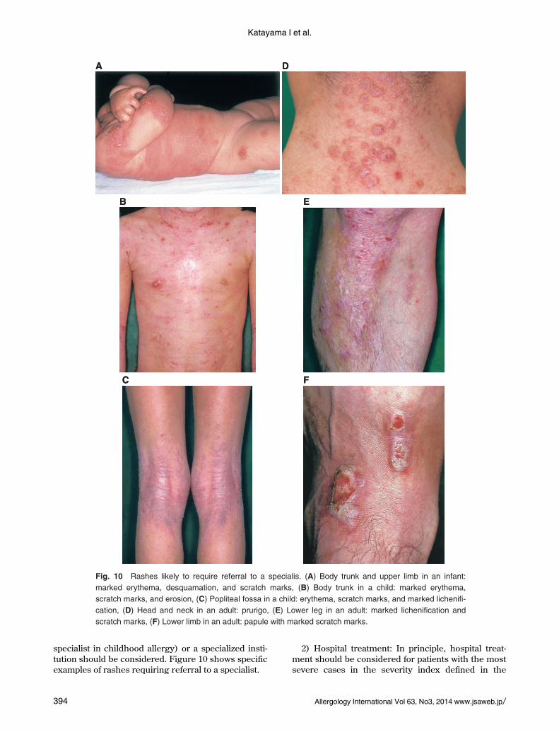
Fig. 10 Rashes likely to require referral to a specialis. (A) Body trunk and upper limb in an infant:
marked erythema, desquamation, and scratch marks, (B) Body trunk in a child: marked erythema,
scratch marks, and erosion, (C) Popliteal fossa in a child: erythema, scratch marks, and marked lichenifi -
cation, (D) Head and neck in an adult: prurigo, (E) Lower leg in an adult: marked lichenifi cation and
scratch marks, (F) Lower limb in an adult: papule with marked scratch marks.
AA DD
BB EE
CC FF
specialist in childhood allergy) or a specialized insti-tution should be considered. Figure 10 shows specificexamples of rashes requiring referral to a specialist.
2) Hospital treatment: In principle, hospital treat-ment should be considered for patients with the mostsevere cases in the severity index defined in the
Atopic Dermatitis
Allergology International Vol 63, No3, 2014 www.jsaweb.jp� 395
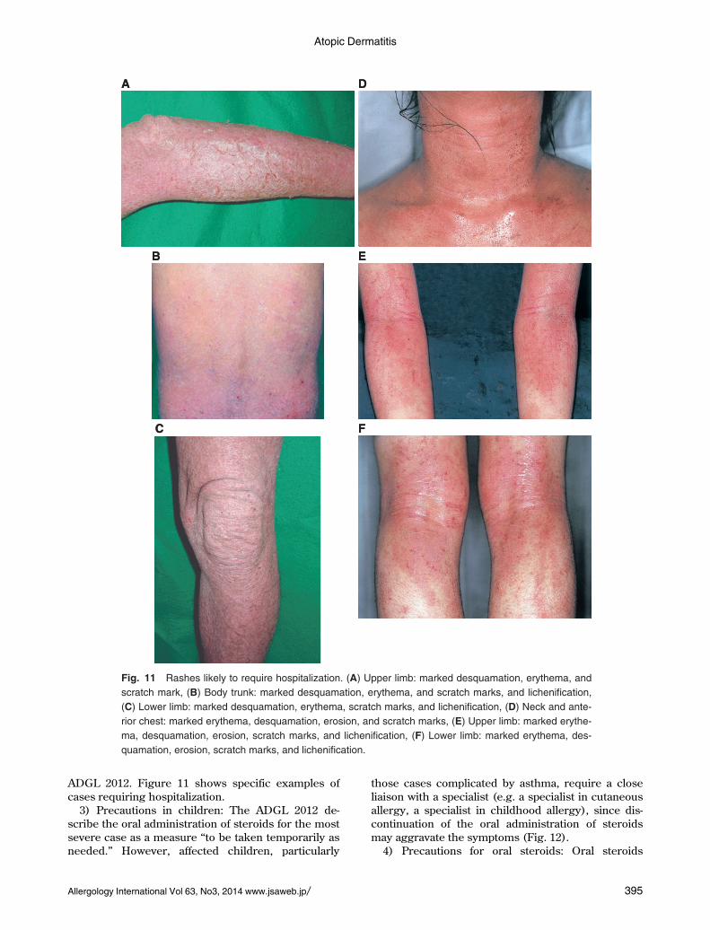
Fig. 11 Rashes likely to require hospitalization. (A) Upper limb: marked desquamation, erythema, and
scratch mark, (B) Body trunk: marked desquamation, erythema, and scratch marks, and lichenifi cation,
(C) Lower limb: marked desquamation, erythema, scratch marks, and lichenifi cation, (D) Neck and ante-
rior chest: marked erythema, desquamation, erosion, and scratch marks, (E) Upper limb: marked erythe-
ma, desquamation, erosion, scratch marks, and lichenifi cation, (F) Lower limb: marked erythema, des-
quamation, erosion, scratch marks, and lichenifi cation.
AA DD
BB EE
CC FF
ADGL 2012. Figure 11 shows specific examples ofcases requiring hospitalization.
3) Precautions in children: The ADGL 2012 de-scribe the oral administration of steroids for the mostsevere case as a measure “to be taken temporarily asneeded.” However, affected children, particularly
those cases complicated by asthma, require a closeliaison with a specialist (e.g. a specialist in cutaneousallergy, a specialist in childhood allergy), since dis-continuation of the oral administration of steroidsmay aggravate the symptoms (Fig. 12).
4) Precautions for oral steroids: Oral steroids

Katayama I et al.
396 Allergology International Vol 63, No3, 2014 www.jsaweb.jp�
Fig. 12 Cutaneous symptoms of children requiring referral to a specialized medical institu-
tion. (A) Face of an infant (9 months old): Aggravation by steroid withdrawal. Extreme fl ush
and erosion on the face, (B) Forearm of an infant (4 months old): Edematous and erosive
fl ush, (C) Scalp of an infant (4 months old): Red papule and crust spreading over the head,
(D) Cheek and ear of an infant (4 months old): Marked weeping fl ush and edema on the face
and neck, (E) Neck and shoulder of a child (9 years old): Severely scratching multiple num-
mular eczema, (F) Femurs of a child (9 years old): Lichenifi cation and pruritic tubercles due
to poor compliance with application of steroid.
AA DD
BB EE
CC FF

Atopic Dermatitis
Allergology International Vol 63, No3, 2014 www.jsaweb.jp� 397
should not be used at all or should be used with con-sideration of the minimum dose and the duration ofuse.
REFERENCES
1. Saeki H, Furue M, Furukawa F et al; Committee forGuidelines for the Management of Atopic Dermatitis ofJapanese Dermatological Association. Guidelines for man-agement of atopic dermatitis. J Dermatol 2009;36:563-77.
2. Bieber T. Atopic dermatitis. N Engl J Med 2008;358:1483-94.
3. Vestergaard C, Bang K, Gesser B, Yoneyama H, Mat-sushima K, Larsen CG. A Th2 chemokine, TARC, pro-duced by keratinocytes may recruit CLA+CCR4+lympho-cytes into lesional atopic dermatitis skin. J Invest Dermatol2000;115:640-6.
4. Grewe M, Bruijnzeel-Koomen CA, Schöpf E et al. A rolefor Th1 and Th2 cells in the immunopathogenesis ofatopic dermatitis. Immunol Today 1998;19:359-61.
5. Masuoka M, Shiraishi H, Ohta S et al. Periostin promoteschronic allergic inflammation in response to Th2 cytoki-nes. J Clin Invest 2012;122:2590-600.
6. Soumelis V, Reche PA, Kanizer H et al. Human epithelialcells trigger dendritic cell mediated allergic inflammationby producing TSLP. Nat Immunol 2002;3:673-80.
7. Koga C, Kabasima K, Shiraishi N, Kobayashi M, TokuraY. Possible pathogenic role of Th17 cells for atopic der-matitis. J Invest Dermatol 2008;128:2625-30.
8. Honda T, Miyachi Y, Kabashima K. Regulatory T cells incutaneous immune response. J Dermatol Sci 2011;63:75-82.
9. Ong PY, Ohtake T, Brandt C et al. Endogenous antimicro-bial peptides and skin infections in atopic dermatitis. NEngl J Med 2002;347:1151-60.
10. Imokawa G, Abe A, Jin K, Higaki Y, Kawashima M, Hi-dano A. Decreased level of ceramides in stratum corneumof atopic dermatitis: an etiologic factor in atopic dry skin?J Invest Dermatol 1991;96:523-6.
11. Palmer CN, Irvine AD, Terron-Kwiatkowski A et al. Com-mon loss-of-function variants of the epidermal barrier pro-tein filaggrin are a major predisposing factor for atopicdermatitis. Nat Genet 2006;38:441-6.
12. Snkoly E, Muller A, Lauerma A et al. IL-31: a new link be-tween T cells and pruritus in atopic skin inflammation. JAllerg Clin Immunol 2006;117:411-7.
13. Miyamoto T, Patapoutian A. Why does morphine makeyou itch? Cell 2011;147:261-2.
14. Hirota T, Takahashi A, Kubo M et al. Genome-wide asso-ciation study identifies eight new susceptibility loci foratopic dermatitis in the Japanese population. Nat Genet2012;44:1222-6.
15. Murota H, Izumi M, Abd EI-Latif MI et al. Artemin causeshypersensitivity to warm sensation, mimicking warmth-provoked pruritus in atopic dermatitis. J Allergy Clin Im-munol 2012;130:671-82.
16. Williams H, Robertson C, Stewart A et al. Worldwide vari-ations in the prevalence of symptoms of atopic eczema inthe International Study of Asthma and Allergies in Child-hood. J Allergy Clin Immunol 1999;103:125-38.
17. Williams H, Stewart A, von Mutius E, Cookson W, Ander-son HR; International Study of Asthma and Allergies inChildhood (ISAAC) Phase One and Three Study Groups.Is eczema really on the increase worldwide? J Allergy ClinImmunol 2008;121:947-54.e15.
18. Saeki H, Iizuka H, Mori Y et al. Community validation of
the U.K. diagnostic criteria for atopic dermatitis in Japa-nese elementary schoolchildren. J Dermatol Sci 2007;47:227-31.
19. Kohno Y. [Identification of Causative and ExacerbationFactors of Atopic Dermatitis and Studies for Improvementof Living Environment to Prevent the Development and Ex-acerbation of Symptoms. Reports of Research on AllergicDisease and Immunology by Ministry of Health, Labour andWelfare of Japan 2006-2007]. 2008;173-7(in Japanese).
20. Aoki T. [Review committee for severity classification ofatopic dermatitis. Second report]. [Jpn J Dermatol] 2001;111:2023-33(in Japanese).
21. Kobayashi M. [Curettage of patients with atopic dermati-tis]. [Jpn J Dermatol] 2000;110:275-82(in Japanese).
22. Sporik R, Holgate ST, Platts-Mills TA, Cogswell JJ. Expo-sure to house-dust mite allergen (Der p I) and the devel-opment of asthma in childhood. A prospective study. NEngl J Med 1990;323:502-7.
23. Yokozeki H, Satoh T, Katayama I, Nishioka K. Airbornecontact dermatitis due to Japanese cedar pollen. ContactDermatitis 2007;56:224-8.
24. Kodama A, Horikawa T, Suzuki T et al. Effect of stress onatopic dermatitis: investigation in patients after the greathanshin earthquake. J Allergy Clin Immunol 1999;104:173-6.
25. Kramer MS, Kakuma R. Maternal dietary antigen avoid-ance during pregnancy or lactation, or both, for prevent-ing or treating atopic disease in the child. Cochrane Data-base Syst Rev 2012;9:CD000133.
26. Simpson EL, Berry TM, Brown PA, Hanifin JM. A pilotstudy of emollient therapy for the primary prevention ofatopic dermatitis. J Am Acad Dermatol 2010;63:587-93.
27. Otani M, Otani Mi, Nozawa A et al. [A study of the influ-ence of the volume and frequency of application on the ef-ficacy of moisturizers]. [Jpn J Dermatol] 2012;122:39-43(in Japanese).
28. Serup J, Winther A, Blichmann CW. Effects of repeatedapplication of a moisturizer. Acta Derm Venereol 1989;69:457-9.
29. Murota H, Takahashi A, Nishioka M et al. Showering re-duces atopic dermatitis in elementary school students.Eur J Dermatol 2010;20:410-1.
30. Healy E, Bentley A, Fidler C, Chambers C. Cost-effectiveness of tacrolimus ointment in adults and chil-dren with moderate and severe atopic dermatitis: twice-weekly maintenance treatment vs. standard twice-daily re-active treatment of exacerbations from a third party payer(U.K. National Health Service) perspective. Br J Dermatol2011;164:387-95.
31. Szczepanowska J, Reich A, Szepietowski JC. Emollientsimprove treatment results with topical corticosteroids inchildhood atopic dermatitis: a randomized comparativestudy. Pediatr Allergy Immunol 2008;19:614-8.
32. Barnes PJ. Molecular mechanisms and cellular effects ofglucocorticosteroids. Immunol Allergy Clin North Am2005;25:451-68.
33. Katayama I, Kohno Y; Japanese Society of Allergology.[Guidelines for the Management of Atopic Dermatitis2009]. Tokyo: Kyowa Kikaku, 2009;55-65(in Japanese).
34. Long CC, Finlay AY. The finger-tip unit- a new practicalmeasure. Clin Exp Dermatol 1991;16:444-7.
35. Long CC, Mills CM, Finlay AY. A practical guide to topi-cal therapy in children. Br J Dermatol 1998;138:293-6.
36. Williams HC. Established corticosteroid creams shouldbe applied only once daily in patients with atopic eczema.BMJ 2007;334:1272.

Katayama I et al.
398 Allergology International Vol 63, No3, 2014 www.jsaweb.jp�
37. Furue M, Terao H, Rikihisa W et al. Clinical dose and ad-verse effects of topical steroids in daily management ofatopic dermatitis. Br J Dermatol 2003;148:128-33.
38. Wollenberg A, Reitamo S, Girolomoni G et al. Proactivetreatment of atopic dermatitis in adults with 0.1%tacrolimus ointment. Allergy 2008;63:742-50.
39. Breneman D, Fleischer AB Jr, Abramovits W et al. Inter-mittent therapy for flare prevention and long-term diseasecontrol in stabilized atopic dermatitis: a randomized com-parison of 3-times-weekly applications of tacrolimus ver-sus vehicle. J Am Acad Dermatol 2008;58:990-9.
40. Schmitt J, von Kobyletzki L, Svensson A, Apfelbacher C.Efficacy and tolerability of proactive treatment with topi-cal corticosteroids and calcineurin inhibitors for atopic ec-zema: systematic review and meta-analysis of randomizedcontrolled trials. Br J Dermatol 2011;164:415-28.
41. Fonacier L, Spergel J, Charlesworth EN et al. Report ofthe Topical Calcineurin Inhibitor Task Force of theAmerican College of Allergy, Athma and Immunologyand the American Academy of Allergy, Asthma and Im-munology. J Allergy Clin Immunol 2005;115:1249-53.
42. Berger TG, Duvic M, Van Voohees AS et al. The use oftopical calcineurin inhibitors indermatology: Safety con-cerns Report of the American Academy of DermatologyAssociation Task Force. J Am Acad Dermatol 2006;54:818-23.
43. Arellano FM, Wentworth CE, Arena A, Fernández C, PaulCF. Risk of lymphoma following exposure to calcineurininhibitors and topical steroids in patients with atopic der-matitis. J Invest Dermatol 2007;127:808-16.
44. Margolis DJ, Hoffstad O, Bilker W. Lack of associationbetween exposure to topical calcineurin inhibitors andskin cancer in adults. Dermatology 2007;214:289-95.
45. Ellis C, Luger T, Abeck D et al. International Consensus
Conference on Atopic Dermatitis II (ICCAD II): clinicalupdate and current treatment strategies. Br J Dermatol2003;148 (Suppl):3-10.
46. Leurs R, Church MK. Taglialatela M. H1-antihistamines:inverse agonism, anti-inflammatory actions and cardiac ef-fects. Clin Exp All 2002;32:489-98.
47. Hoare C, Li Wan Po A, Williams H. Systematic review oftreatments of atopic eczema. Health Technol Assess 2000;4:1-191.
48. Kawashima M, Tango T, Noguchi T, Inagi M, NakagawaH, Harada S. Addition of fexofenadine to a topical corti-costeroid reduces the pruritus associated with atopic der-matitis in a 1-week randomized, multicentre, double-blind,placebo-controlled, parallel-group study. Br J Dermatol2003;148:1212-21.
49. Yanai K, Tashiro M, Nobuyuki Okamura N. [Non-sedating second-generation antihistamines: The penetra-tion through blood-brain barrier measured by PET].[Nishinihon J Dermatol] 2009;71:3-6(in Japanese).
50. Igarashi A, Nakagawa H, Takigawa M et al. [Guidance forthe use of cyclosporine MEPC for atopic dermatitis]. [J.JOCD] 2009;63:1049-54(in Japanese).
51. Ring J, Alomar A, Bieber T et al. Guidelined for treatmentof atopic eczema (atopic dermatitis) Part II. J Eur AcadDermatol Venereol 2012;26:1176-93.
52. British Photodermatology Group. British Photodermatol-ogy Group guidelines for PUVA. Br J Dermatol 1994;130:246-55.
53. Kobayashi H, Ishi M, Takeuchi S et al. Efficacy and Safetyof a Traditional Herbal Medicine, Hochu-ekki-to in theLong-term Management of Kikyo (Delicate Constitution)Patients with Atopic Dermatitis: A 6-month, Multicenter,Double-blind, Randomized, Placebo-controlled Study.Evid Based Complement Alternat Med 2010;7:367-73.