Embed Size (px)
Citation preview

January 1989
EPIDEMIOLOGY AND CONTROL OF INFECTIOUS DISEASES OF SALMONIDS
IN THE COLUMBIA RIVER BASIN
THIS IS INVISIBLE TEXT TO KEEP VERTICAL ALIGNMENT THIS IS INVISIBLE TEXT TO KEEP VERTICAL ALIGNMENT THIS IS INVISIBLE TEXT TO KEEP VERTICAL ALIGNMENT THIS IS INVISIBLE TEXT TO KEEP VERTICAL ALIGNMENT THIS IS INVISIBLE TEXT TO KEEP VERTICAL ALIGNMENT
Annual Report 1987
DOE/BP-11987-3
This report was funded by the Bonneville Power Administration (BPA), U.S. Department of Energy, aspart of BPA’s program to protect, mitigate, and enhance fish and wildlife affected by the development andoperation of hydroelectric facilities on the Columbia River and its tributaries. The views of this report arethe author’s and do not necessarily represent the views of BPA.
This document should be cited as follows: Fryer, J. L., Department of Microbiology, Epidemiology and Control of Infectious Diseases of Salmonids in theColumbia River Basin, Annual Report 1987 to Bonneville Power Administration, Portland, OR, Contract 83-AI-11987,Project 83-312, 130 electronic pages (BPA Report DOE/BP-11987-3)
This report and other BPA Fish and Wildlife Publications are available on the Internet at:
http://www.efw.bpa.gov/cgi-bin/efw/FW/publications.cgi
For other information on electronic documents or other printed media, contact or write to:
Bonneville Power AdministrationEnvironment, Fish and Wildlife Division
P.O. Box 3621905 N.E. 11th Avenue
Portland, OR 97208-3621
Please include title, author, and DOE/BP number in the request.

EPIDEMIOLOGY AND CONTROL OF INFECTIOUSDISEASES OF SALMONIDS IN THE
COLUMBIA RIVER BASIN
Annual Report 1987
Prepared by:
J. L. Fryer
Department of Microbiology
Prepared for:
U.S. Department of EnergyBonneville Power AdministrationEnvironment, Fish and Wildlife
PO Box 3621Portland, Oregon 97208
Project No. 83-312Contract No. DE-AI79-83BP11987
January 1989

TABLE OF CONTENTS
PageAcknowledgements
Abst rac t
Chapter I. Development, Characterization, and Use ofMonoclonal and Polyclonal Antibodies against theMyxosporean, Ceratomyxa Shasta.
Abst rac tIntroductionMaterials and MethodsResultsDiscussionAcknowledgementsLiterature Cited
1235
112 32 62 7
Chapter II. Characterization of the Host Response to theMyxosporean parasite, Ceratomyxa Shasta, byHistology, Scanning Electron Microscopy, andImmunological Techniques.
AbstractIntroductionMaterials and MethodsResultsDiscussionAcknowledgementsLiterature Cited
2 93 0313 53 95 56 26 3
Chapter III. Characterization of a Monoclonal Antibody againstCeratomyxa Shasta Using Immunocytochemistry.
6 6Abstract 6 7Introduction 6 8Materials and Methods 6 9Results and Discussion 7 0Acknowledgements 7 7Literature Cited 7 8

TABLE OF CONTENTS (continued)
Page
Chapter IV. Artificial Transmission of Ceratomyxa Shasta ViaPotential Intermediates 7 9
Abstract 8 0Introduction 81Materials and Methods 83Results and Discussion 8 9Acknowledgements 9 5Literature Cited 9 6
Chapter V. In Vitro Inhibition of Renibacterium Salmoninarumby Experimental Antibiotics. 9 7
Abstract 9 8Introduction 9 9Materials and Methods 100Results 104Discussion 116Acknowledgements 120Literature Cited 121
Summary of Expenditures 1 2 2
ACKNOWLEDGEMENTS
Support for this research came from the region’s electrical rate
payers through the Bonneville Power Administration.
The cooperation of the Environmental Protection Agency -
Corvallis Environmental Research Laboratory is recognized for their
providing some of the fish holding facilities at the Western Fish
Toxicology Laboratory; The Oregon Department of Fish and Wildlife
provided experimental fish. Pfizer; Merck, Sharp and Dohme
Research Laboratories, Abbot Laboratories; and Lily Research
Laboratories have supplied antimicrobial agents.

ABSTRACT
The Department of Microbiology at Oregon State University
with funding from the Bonneville Power Administration has been
conducting a study concerning the epidemiology and control of three
fish pathogens which cause major disease problems in salmonids of
the Columbia River basin. The pathogens studied include Cera to m yxa
Shasta, the myxosporean parasite which causes ceratomyxosis;
Renibacterium salmoninarum, the bacterium which is the etiological
agent of bacterial kidney disease; and the rhabdovirus which causes
infectious hematopoietic necrosis (IHN). During this project, the host
and geographic range of C. Shasta have been more precisely
determined and the known geographic range has been significantly
expanded. The effects of the parasite on fish migrating through the
Columbia River and on their introduction into salt water have been
examined. Similar studies have been conducted with R.
salmoninarum and it has been shown that bacterial kidney disease
occurs at all life stages of salmonids and is responsible for mortality
in both fresh and salt water. It has also been demonstrated that
different isolates of R. salmoninarum have different antigenic
composition. Results of demonstration projects designed to control
IHN by using UV treated water for early rearing of salmonid fry
were equivocal.
The scope of the project was considerably narrowed and
focused during the past two years The project has concentrated on a
study concerning the biology of C. Shasta and the identification of
potential chemotherapeutants for control of bacterial kidney disease.
The emphasis of work on C. Shasta has been its pathogenesis. This
aspect of the parasite has been investigated using histopathologic
and immunologic methodology. Mode of transmission, the nature of
the infectious stage, and potential intermediate hosts of the parasite
have also been areas of active research.
Classes of chemotherapeutants with the highest potential for
efficacy against R. salmoninarum have been identified through
literature searches and consultation with pharmacologists.
Experimental drugs have been requested and received from several
pharmaceutical manufacturers. The in vitro sensitivity of R.
salmoninarum and other selected fish pathogens to more than 100
antimicrobial compounds has been tested.
The project is related to measure 704(h)(2)(d) of the Columbia
River Basin Fish and Wildlife Program. The results will contribute to
fish health which will directly contribute to the protection of fish.
CHAPTER I
DEVELOPMENT, CHARACTERIZATION, AND USE OF MONOCLONAL
AND POLYCLONAL ANTIBODIES AGAINST THE MYXOSPOREAN,
CERATOMYXA SHASTA
J.L. Bartholomew, J.S. Rohovec, and J.L. Fryer
Department of Microbiology
Oregon State University
Corvallis, Oregon 97331, USA
ABSTRACT
Both monoclonal- and polyclonal antisera were produced
against Ceratomyxa Shasta. Ascites containing trophozoites of the
parasite was collected from infected fish and used as antigen for
immunization of mice. The resulting monoclonal antibodies reacted
specifically with trophozoite and sporoblast stages but did not react
with C. Shasta spores by either indirect fluorescent antibody
techniques or in Western blots. This indicates that C. Shasta
contains unique antigens and that some of these antigens are
specific to certain life stages of the parasite. Polyclonal antiserum
was produced in a rabbit by injecting a spore protein electroeluted
from an SDS-polyacrylamide gel. This antiserum reacted with both
trophozoites and spores by indirect fluorescent antibody techniques
and in Western blots. All antisera were tested for cross-reactivity
to trout white blood cells, a contaminant of the ascites, and to other
myxosporeans. Two monoclonal antibodies reacted with white
blood cells and myxosporeans of the genera Sp haerospora and
Myxobilatus. One hybridoma produced antibodies of high
specificity for C. Shasta prespore stages. This is the first report of a
monoclonal antibody produced against a myxosporean parasite.
2

INTRODUCTION
The myxosporean parasite, Ceratomyxa Shasta (Noble, 1950)
causes an intestinal disease in susceptible salmonid fish in the
Pacific Northwest region of the United States and Canada. This
parasite has been responsible for epizootics in both wild and
hatchery populations of salmonids. At present there is no effective
means of control other than avoidance of the infectious stage of this
organism and stocking of resistant strains of salmonids in endemic
areas. Spores of C. Shasta are easily detected in moribund fish;
however, the infectious process is temperature dependent, with
mortality occurring about 56 days post-exposure among fish held at
12oC, (Udey et al., 1975). Development of mature spores occurs just
prior to death of the host, but before spore formation, trophozoite
stages of the parasite are abundant in the intestinal tract. While
these stages may be identified using histological techniques, they
often go undetected by microscopic examination of intestinal tract
scrapings, the standard diagnostic procedure. The similarity of the
multinuclear trophozoites to the same life stages of other
myxosporeans also complicates diagnosis.
The life cycle of C. Shasta, like that of most myxosporeans,
remains unknown. The morphology of the infective stage and the
initial site of infection in the host have not been determined,
Attempts to transmit the disease by feeding infected tissues,
cohabitation of infected and susceptible fish, and exposing
susceptible fish to a mixture of mud and infected tissues have
3
failed (Johnson et al., 1979). The inability to transmit the disease
between susceptible fish has led to speculation that an intermediate
host may be necessary. The life cycle of another myxosporean
parasite, Myxobolus cerebralis, was shown by Wolf and Markiw
(1984) to require Tubifex tubifex as an intermediate host. They
were also able to demonstrate, that within the oligochaete, the
myxosporean spore transforms into a form previously identified as
a triactinomyxon. This alternation of life stages and intermediate
host involvement has not yet been demonstrated for C. Shasta.
The usefulness of serological techniques for identifying
different life stages has been shown for several human parasites.
Results of those’ studies indicate that the presence of stage-specific
antigens is common to parasites such as Plasmodium vivax and P.
ovale (Andrysiak et al., 1986), and Trypanosoma cruzi (Wrightsman
et al., 1986). This paper presents immunological evidence of stage-
specific C. Shasta antigens which accompany the morphological
change from trophozoite to spore and is the first report of
monoclonal antibodies produced against a myxosporean parasite.
The use of specific antibodies for diagnostic purposes is also
described.
4

MATERIALS AND METHODS
Antigen
Ascites containing prespore (trophozoite and disporoblast)
stages of C . shasta was collected from naturally infected rainbow
trout. Cells were pelleted by centrifugation (1500 g) and washed
twice in 0.1 M phosphate-buffered saline , pH 7.6 (PBS). This
antigen was used both for injection of mice for hybridoma
production and for all screening procedures.
Spore stages of C . shasta were collected from the intestinal
tract of infected rainbow trout. After homogenization of the tissue
and low-speed centrifugation (1500 g) to remove fish tissue, the
spores were layered onto a 12-75% (v/v) gradient of modified
colloidal silica (Percoll, stock density 1.13 g/ml) (Sigma Chemical
Co., St Louis, MO). Gradients were centrifuged at 1500 g for 35 min
in a swinging bucket rotor. Spores were layered on top of the 75%
Percoll. This band was removed and the spores washed twice in
PBS. Spores were used for screening hybridoma supernatants and
for polyclonal antiserum production.
To obtain white blood cells, rainbow trout were bled from the
caudal vein. Blood was stored in heparin (15 IU/ml) for one hour,
cells were pelleted by centrifugation at 2500 g, washed twice and
resuspended in PBS. The buffy coat was collected after overnight
refrigeration.
5
Hybridoma Production
Monoclona l antibodies (Mabs) were produced by the method
of Campbell (1984) . Briefly, for the production of antibodies
specific t o C. shasta, freshly collected prespore stages (106
total/mouse) suspended in PBS were injected intraperitoneally into
Balb/c mice . Two booster injections were given at one month
intervals . Antibody production was evaluated by indirect
fluorescent antibody technique s (IFAT) on cells from th e ascites of
infected rainbow trout .Four days after the second booster, the
mice were primed with an injection of 106 parasites. Four days
later the spleen cells were removed and fused with a non-secreting
tumor line , SP2/0 , at a ratio o f 5:1, using polyethylene glycol (80%
PEG 1500, 20% PEG 4000). The cells were distributed in 96 well
microtiter plates (Costar ,Cambridge, Mass) in selective medium
(RPM1 with 10% (v/v) fetal calf serum plus hypoxanthine,
aminopterin , and thymidine), and incubated at 370C in 5% C02.
After 14 days, supernatants were screened for antibodies specific
for C. Shasta. A dot-immunobinding assay (Hawkes et al., 1982)
using prespore stages fro m ascites as antigen was the primary test
for reactivity . Supernatants eliciting a positive reaction were
screened again b y IFAT using cells from th e ascites of infected fish
and fish blood cells from uninfected fish as antigens .Those
hybridomas producing antibodies positive only for cells from
infected rainbow trout were cloned by limiting dilution. The
supernatants from wells with single clones were tested for antibody
as described; positive hybridomas were again cloned by limiting
6
dilution and reassayed afte r l0-14 days. Selected hybridomas
were expanded into 7 5cm2 flasks and maintained i n RPMI (Sigma
Chemical Co.) plus 10% (v/v) fetal bovine serum (Hyclone
Laboratories, Logan, Utah) .The immunoglobulin class and subclass
produced by each hybridoma was determined by an enzyme-linked
immunoassay (Mous e monoclona l subisotyping kit ; HyClone
Laboratories).
Western blot analysis was performed using antibodies from
the expanded clones . Following electrophoretic separation of C.
shasta spores, prespore stages, and fis h WBCs , on a 12% sodium
dodecyl sulfate-polyacrylamide gel (SDS-PAGE), the antigens were
transferred to nitrocellulose by electroblotting in a Trans-blot cell
(Bio-Rad Laboratories, Richmond, CA). The nitrocellulose was then
probed with hybridoma supernatants and peroxidase-labeled goat
anti-mouse serum (Hyclone Laboratories). Antibodies whic h cross-
reacted with fish WBC antigens were further characterized by
Western blot analysis, using immunized fish sera as a source of
antigen, and by enzyme-linked immunoassay (ELISA). In the
ELISA, the cross-reacting Mabs were used as a second antibody.
Plates were coated with trinitrophenyl-bovine serum albumin
(TNP-BSA) and incubated with trout anti-TNP antiserum as primary
antibody . In both Western blot and ELISA, a noncross-reacting
Mab was used as a negative control and a Mab produced against
fish immunoglobulin (cell line 1-14, a gift of Dr. G. Warr, Dept. of
Biochemistry, Univ .of South Carolina) served as a positive control.
Antibodies specific for carbohydrate epitopes were detected by
7

periodate oxidation of the antigens on nitrocellulose (Woodward et
al., 1985). Briefly , C. shasta prespore stages and fish sera were
separated by electrophoresis on a polyacrylamide gel and then
transferred onto nitrocellulose .After exposing to periodate
concentrations from 5-2 0 mM, blots were incubated with the
monoclonal antibodies and probed as described.
Antibodies were also screened for cross-reactivity with other
myxosporeans b y IFAT on histological sections from fish infected
with the following parasites : Henneguya exilis, Sphaerospora sp.,
PKX, Chloromyxum majori, Myxobilatus sp., an d Myxobolus
cerebralis, and also on purified spores o f Henneguya salmincola and
Myxobolus insidiosus.
Polyclonal Antiserum
To obtain the antigen used for the production of polyclonal
antisera, 1 x 10⁸ purified spores were diluted 1 :l with sample
buffer (Schleif and Wensink, 1981) and the proteins were
separated by SDS-PAGE . The gel was stained with Coomassie blue
(0.1% in 40% methanol and 10% acetic acid) and the polypeptide
profile examined for bands specific to the spore stage. A
preparative SDS-PAGE was used to purify a single protein band.
After electrophoresis of the spores, a narrow vertical strip of the
gel was excised and stained in order to identify the protein bands.
The portion of the gel corresponding to the band of interest was
excised and the protein electroeluted (Elutrap, Schleicher and
Schuell , Keene, NH). A sample of the eluent was diluted 1 :l in
8

sample buffer and run on a gel to assess the purity of the antigen.
Antigen was mixed 1 :l with Freund’s complete adjuvant and
injected into New Zealand white rabbits; 0.5 ml in each footpad and
0.5 ml subcutaneously between the scapula. After four weeks, a
booster was administered by injection of the antigen mixed 1 :l in
Freund’s incomplete adjuvant. Two weeks later the rabbit was bled
and the antiserum tested for specificity b y IFAT and Western blot
analysis.
IFAT
Monoclonal l or polyclonal antibodies were incubated for 15
minutes at room temperature with cells fixed in acetone-xylene
(1: 1). Specific antibodies were detected using biotinylated horse
anti-mous e IgG or anti-rabbit Ig and fluorescein isothiocyanate
conjugate d avidin D (Vector Laboratories, Burlingame, CA) .Methyl
green dye (1% in distilled water) was used as a counterstain. Cells
were examined using a Zeiss standard microscope with an I V Fl epi-
fluorescenc e condenser.
Histology
For serological diagnosis of infections in tissue sections,
viscera of fish exposed to the infectious stage o f C. shasta were
fixed in either Bouin’s or 10% (v/v) neutral buffered formalin,
processed routinely for histology to 6 pm and mounted on gelatin
coated slides . Sections to be examined b y IFAT were stained as
described. Sections for examination by bright light microscopy
9
were incubated with specific antibody, then biotinylate d anti-
mous e IgG, and finally with a n avidin DH - biotinylated alkaline
phosphatase H complex (Vectastain ABC-AP kit,; Vector
Laboratories) . The enzymatic activity was localized with an
insoluble substrate (Alkaline Phosphate Substrate Kit II; Vector
Laboratories).
10
RESULTS
Monoclonal Antibodies
One fusion resulted in four hybridomas which produced
antibodies that reacted positively by dot immunobinding an d IFAT
to fixed, whole trophozoites fro m ascites . Isotypes of the
immunoglobulins produced by each hybridoma were determined;
one Mab was a n IgM and three wer e IgGs , one eac h IgGl , IgG2a,
and IgG3 . Hybridoma supernatants were also tested for their
reactivity t o C. shasta spores and to fis h WBCs by IFAT (Table 1.1).
The Mabs did not react with spores. The pattern of fluorescence of
the parasite was similar after reacting with antibodies from each
hybridoma.
In Western blot analysis all Mabs recognized antigens from
prespore stages o f C. shasta and there were none that reacted with
spore antigens . Two Mabs reacted with fish WBC antigens,
recognizing a protein with a molecular weight (MW) of
approximately 80 kilodaltons (kdal) in both buffy coat preparations
and in fish sera (Fig. 1.1) . These Mabs also reacted specifically with
fish immunoglobulins in an ELISA. To determine if thes e cross-
reacting antibodies reacted with a protein or carbohydrate epitope,
Western transfers of antigens in prespore stages and in fish sera
were exposed to varying concentrations of periodate .Complete loss
of binding of Mabs 1 and 3 occurred at a concentration of 1 0 mM
periodate, indicating that these antibodies are directed against
11
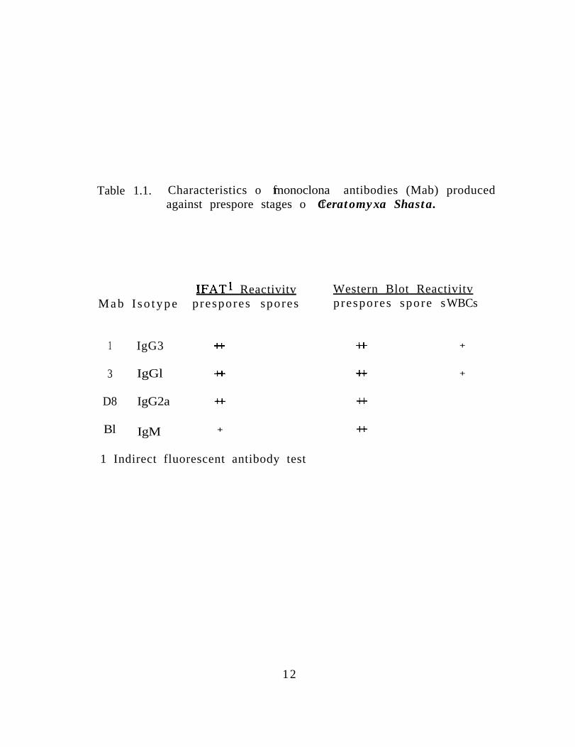
Table 1.1. Characteristics o f monoclona l antibodies (Mab) producedagainst prespore stages o f Ceratomyxa Shasta.
M a b I s o t y p e
1 IgG3
3 IgGl
D8 IgG2a
Bl IgM
IFATl Reactivitv Western Blot Reactivitvprespores spores prespores spore s WBCs
+t * +
+I- +t +
4-k -l-k
+ 4-k
1 Indirect fluorescent antibody test
12
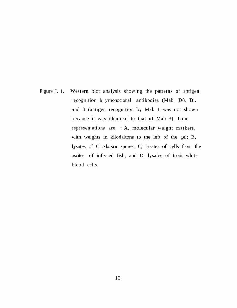
Figure I. 1. Western blot analysis showing the patterns of antigen
recognition b y monoclonal l antibodies (Mab ) D8, Bl,
and 3 (antigen recognition by Mab 1 was not shown
because it was identical to that of Mab 3). Lane
representations are : A, molecular weight markers,
with weights in kilodaltons to the left of the gel; B,
lysates of C . shasta spores, C, lysates of cells from the
ascites of infected fish, and D, lysates of trout white
blood cells.
13

18011684
2
36.5
26.6
A B C D D C BA
;..:IJ
‘; (f&2 *>-y .i.
i&w?t5iso;ir.
pf$* v.” I
z,.=,;-. .&I:. L I ;k-,
DCBA
1
1 4
carbohydrate antigens. Binding of Mab D8 to prespore antigens was
not inhibited even at the 20 mM periodate concentration.
The cross-reactivity of the monoclonal antibodies with other
myxosporeans was examined using IFAT on either histological
sections from infected fish or purified spores. Two Mabs, 1 and 3,
reacted with Sphaerospora sp. and Myxobilatus sp. from
sticklebacks (Gasterosteus sp), and their use resulted in high
background fluorescence of fish tissues. Although no specific cross-
reactivity was detected between Mabs Bl and any myxosporeans,
high background flourescence of fish tissues was again noted. Mab
D8 did not cross-react with other myxosporeans nor cause non-
specific fluorescence of fish tissues. In addition to reacting
specifically to C. shasta in IFATs, Mab D8 was also useful in
detecting the parasite when an alkaline phosphatase conjugated
second antibody was used (Fig. 1.2).
Polyclonal Antiserum
The protein chosen as antigen for production of polyclonal
antisera was shown by SDS-PAGE to be a major component of C.
shasta spores and present in lower concentration in prespore stages
(Fig. 1.3). The MW of the purified protein was approximately 84
kdal (Fig. 1.4). Antiserum against this antigen reacted by IFAT to
both prespore and spore stages of the parasite, but fluorescence of
later life stages was more pronounced (Fig. 1.5). In Western blot
analysis the antisera reacted with the 84 kdal protein of both
spores and prespores and also with a 180 kdal protein from spores.
15

No specific cross-reactivity between the polyclonal antiserum and
other myxosporeans was detected by IFAT.
16
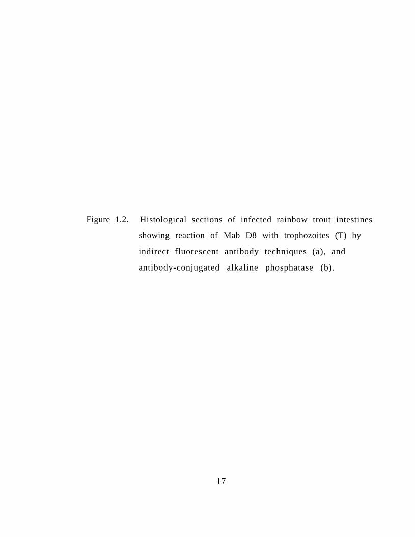
Figure 1.2. Histological sections of infected rainbow trout intestines
showing reaction of Mab D8 with trophozoites (T) by
indirect fluorescent antibody techniques (a), and
antibody-conjugated alkaline phosphatase (b).
17

2b
18
Figure 1.3. SDS-PAGE of lysates of C. shasta spore (B) and prespore
(C) stages, and trout white blood cells (D). Weights of
molecular weight standards (A) are indicated at left.
Figure 1.4, SDS-PAGE of the C. shasta antigen used for production of
polyclonal antiserum (C) and the lysate of spores from
which the antigen was purified (B). Weights of
molecular weight standards (A) are indicated.
19
11684
5848
F-m
36.5 k;Jlic
'. --*i .kc b -i_c I'
.
3
200
12.3 &&id.".>
& 1;
- r ‘*
4 A B C
20
Figure 1.5. Indirect fluorescent antibody reaction of whole spores
probed with polyclonal antiserum produced against an
antigen from Ceratomyxa shasta spores.
21

22

DISCUSSION
Four hybridomas synthesizing monoclonal antibodies which
reacted against trophozoite and disporoblast stages of C. shasta
were produced. Reactivity to C. shasta prespore stages was
determined by indirect immunofluorescence and by Western blot
analysis of electrophoresed trophozoite antigens. None of the
hybridomas produced antibodies that reacted with C. shasta spores
by dot immunobinding, IFAT, or in Western blots. These data
suggest the presence of stage-specific antigens associated with
different life stages of C. Shasta. SDS-PAGE analysis demonstrated
that some proteins present in high concentration in C. shasta spores
are either absent or in low concentration in earlier life stages. It is
likely that these noncross-reactive antigens are associated with the
formation of the spore coat. Because of difficulties in physically
separating C. shasta life stages, all prespore stages were grouped
together. This caused some problems in the analysis of
electrophoretic profiles because disporoblast stages might have
f o r m e d s o m e of the antigens found in mature spores.
Two of the hybridomas (Mabs 1 and 3) produced antibodies
which cross-reacted with the white blood cell population of
uninfected fish and with a high MW protein in fish sera. The cross-
reacting protein had a MW of approximately 80 kdal and was
determined to be fish immunoglobulin heavy chain by Western blot
analysis and ELISA. Because no attempt was made to purify C.
shasta prespores, it was not unexpected to find antibodies that
2 3

reacted with trout antigens; however, these antibodies were also
found to react specifically by IFAT with cells that could be
positively identified as C. Shasta. One possible explanation for this
cross-reactivity is that C. shasta had evoked an immune response
and was coated with fish antibody. However, when cells were
incubated with fluorescein-conjugated anti-trout Ig there was no
reactivity. It has been suggested (Pauley, 1974) that cross-
reactivity of a specific anti-parasite antibody with uninfected tissue
antigens may indicate that an antigenic component of the parasite
has been able to mimic normal host antigens and thus evade an
immune reaction. Profiles of antibody-antigen recognition seen in
Western blots (Fig. 1.1) suggested that the antibodies recognized a
common epitope which may indicate the presence of carbohydrate.antigens. Mild periodate oxidation, which destroys carbohydrate
determinants without affecting either proteins or lipids, caused a
complete loss of binding of both Mabs 1 and 3. There has been
evidence indicating that antisera produced against phylogenetically
distant immunogens may cross-react by virtue of their
carbohydrate moities (Bayne et al., 1987; Yamaga et al., 1978).
Whether or not this cross-reactivity represents a mechanism by
which the parasite avoids the host immune system, the presence of
common carbohydrates may be effective in facilitating parasitism
by C. Shasta.
In addition to Mabs which reacted with both C. shasta and
host antigens, we also produced one monoclonal antibody specific
for the prespore stages of the parasite. Hybridoma D8 secreted
2 4

antibodies which did not recognize any other myxosporeans and
which were effective in detecting low numbers of C. shasta both in
histological sections and in smears of intestinal material from
infected fish. Its specificity makes it suitable for use as a diagnostic
reagent, either in the IFAT or in enzyme immunohistochemistry. In
addition to its applications in diagnostics, this Mab will be useful in
studying the life history and pathogenesis of this parasite.
The polyclonal antiserum produced against an antigen
predominant in the spore stage of C. shasta was shown by IFAT to
react most specifically with these later life stages. This provides
additional evidence for the presence of stage-specific antigens
associated with the parasite. The abundance of this antigen in the
spore makes it a good candidate for production of either monoclonal
or polyclonal antibodies against a single defined antigen. The
production of these antibodies may be useful not only in
diagnostics, but in understanding the biology of C. shasta.
2 5
ACKNOWLEDGEMENTS
We thank Cindy Arakawa for her assistance in the production
and screening of the hybridomas and Mary Arkoosh for testing the
Mabs in the ELISA. This research was supported by Bonneville
Power Administration under contract No. DE-Al79-83 BP 11987; G.
R. Bouck, Contracting Officer’s Technical Representative. Oregon
Agricultural Station Technical Paper No. 8568.
2 6
LITERATURE CITED
Andrysiak, P. M., W. E. Collins, and G. H. Campbell. 1986. Stage-specific and species-specific antigens of Plasmodium vivaxand Plasmodium ovale defined by monoclonal antibodies.Infect. Immun. 54:609-6 12.
Bayne, C. J., C. A. Boswell, and M. A. Yui. 1987. Widespreadantigenic cross-reactivity between plasma proteins of agastropod, and its trematode parasite. Dev. Comp. Immunol.11:321-329.
Campbell, A.M. 1984. Monoclonal antibody technology, In:Laboratory Techniques in Biochemistry and Molecular Biology(ed. by Burton R. H. and P. H. van Kippenberg). Vol. 13 .Elsevier Science Publishers, Amsterdam.
Hawkes, R., E. Niday, and J. Gordon. 1982. A dot-immunobindingassay for monoclonal and other antibodies. Anal. Biochem.119:142.-147.
Johnson, K. A., J. E. Sanders, and J. L. Fryer. 1979. CeratomyxaShasta in salmonids. U.S. Dep. Int. Fish. Wild. Serv., Div. Fish.Res. Fish Disease Leafl. No. 58:llp.
Pauley, G. B. 1974. Fish sporozoa: extraction of antigens fromMyxosoma cerebralis spores which mimic tissue antigens ofrainbow trout (Salmo gairdneri). J. Fish. Res. Board Can.31:1481-1484.
Schleif, R. F., and P. C. Wensink. 1981. Practical methods inmolecular biology. Springer-Verlag. New York.
Udey, L. R., J. L. Fryer, and K. S. Pilcher. 1975. Relation of watertemperature to ceratomyxosis in rainbow trout (Salmogairdneri) and coho salmon (Oncorhynchus kisutch). J. Fish.Res. Board Can. 32:1545-1551.
Wolf, K., and M. E. Markiw. 1984. Biology contravenes taxonomy inthe myxozoa: new discoveries show alternation ofinvertebrate and vertebrate hosts. Science 2:1449-1452.
2 7
Woodward, M. P., W. W. Young, Jr., and R. A. Bloodgood. 1985.Detection of monoclonal antibodies specific for carbohydrateepitopes using periodate oxidation. J. Immunol. Methods78:143-153.
Wrightsman, R. A., W. Leon, and J. E. Manning. 1986. Variation inantigenic determinants specific to the infective stage ofTrypanosoma cruzi. Infect. Immun. 53:235-239.
Yamaga. K. M., R. T. Kubo, and H. M. Etlinger. 1978. Studies on thequestion of conventional immunoglobulin on thymocytes fromprimitive vertebrates: II. Delineation between Ig-specificand cross-reactive membrane components. J. Immunol.120:2074-2079.
2 8
CHAPTER II
CHARACTERIZATION OF THE HOST RESPONSE TO THE
MYXOSPOREAN PARASITE, CERATOMYXA SHASTA, BY
HISTOLOGY, SCANNING ELECTRON MICROSCOPY, AND
IMMUNOLOGICAL TECHNIQUES
J. L. Bartholomew I, C. E. Smith2, J. S. Rohovecl, and J. L.
Fryer 1
1 Department of Microbiology
Oregon State University
Corvallis, Oregon 9733 1
2Fish Technology Center
US Fish and Wildlife Service
Bozeman, Montana 597 15
2 9
ABSTRACT
The tissue response of Salmo gairdneri against the
myxosporean parasite, Ceratomyxa shasta, was investigated
using histological techniques, scanning electron microscopy,
and immunological methods. The progress of infection in C.
Shasta-susceptible and resistant steelhead and rainbow trout
was examined by standard histological techniques and by
indirect fluorescent antibody methods using monoclonal
antibodies directed against C. shasta antigens. Trophozoite
stages were first observed in the posterior intestine and there
was indication that resistance was due to the inability of the
parasite to penetrate this tissue rather than to an
inflammatory response. Examination of a severely infected
intestine by scanning electron microscopy showed extensive
destruction of the mucosal folds of the posterior intestine.
Western blotting and indirect fluorescent antibody techniques
were used to investigate the immunological component of the
host response. No antibodies specific for C. shasta were
detected by either method.
3 0

INTRODUCTlON
Ceratomyxa shasta is a histozoic myxosporean which
parasitizes the intestinal tissues of salmonids. Its geographic
range is limited to northern California and the Pacific
Northwest region of the United States and Canada (Hoffmaster
et al., 1988). The parasite is an important cause of mortality
among susceptible salmonids. Fish are infected while in fresh
water; however, anadromous salmonids may continue to die
of ceratomyxosis during the salt water phase of their life
cycle (Ching and Munday 1984). The life cycle of C. shasta
has not been defined, but its apparent discontinuous
distribution and the difficulty in achieving laboratory
transmission of the infectious agent suggests that one or more
unknown factor(s) is required. The involvement of an
intermediate host has been suggested; however, no life cycle
has been proposed.
Initial signs of infection by C. shasta may include
darkening, lethargy, and loss of appetite. As the disease
progresses, the descending intestine and anus become swollen
and hemorrhagic and ascites may collect in the coelom (Wales
and Wolf, 1955; Schafer, 1968; Johnson, 1975). Definitive
diagnosis is made by observation of the mature spore in wet
mounts or histological sections. However, before sporogenesis
is complete, trophozoites can be identified by their
multicellular ameboid morphology and characteristic nuclei
31
containing a large karyosome and peripheral chromatin
(Noble 1944). Multiplication of trophozoites is by nucleogony
followed by either budding or plasmotomy (cytoplasmic
division)(Noble 1941).
Many reports describe infections of fish by
myxosporeans, but the view that most species cause little host
response is commonly held. Coelozoic myxosporeans cause
little host reaction and have been considered the most
harmless (Lom 1970); however, Fantham (1912) described an
inflammatory reaction consisting of leucocyte infiltration,
desquamation and necrosis of epithelial cells, and increased
mucous secretion caused by myxosporeans parasitizing the
gall bladder. Lom (1969) also reported hyperemia and
hypertrophy associated with coelozoic myxosporeans.
A lack of tissue response has also been attributed to
cyst forming myxosporeans (Lom 1969). Dykova and Lom
(1978) reported histopathological changes in gills of fish
infected with two species of Henneguya and proposed that
the tissue response to myxosporeans invading soft tissue
occurred in two phases. First, as the plasmodium grew, there
occurred alterative changes in the tissue: displacement,
atrophy and hyperplasia. Although organ function may have
been impaired, there was no host defense reaction. In the
second stage, cysts were full of mature spores which evoked
an inflammatory reaction leading to replacement of the cyst
by granulomatous tissue. Amandi and Fryer (1985)
3 2
recognized a similar host reaction against Myxobolus
insidiosus which infects the muscle of salmonids. Contrary to
the lack of inflammatory response to immature plasmodia,
Duhamel (1986) reported that trophozoites of Henneguya
exilis were responsible for severe granulomatous branchitis in
channel catfish.
The tissue response against non-cyst forming histozoic
myxosporeans is also varied. Myxobolus cerebralis infects
young salmonids by invading and causing destruction of
cartilage cells. A granulomatous response with infiltration by
macrophages and mononuclear leucocytes has been reported
(Roberts and Elson 1970; Taylor and Haber 1974; Halliday
1976). Other myxosporeans cause complete degeneration of
invaded areas accompanied by hypertrophy of connective
tissue cells and invasion by fibroblasts. Kent and Hedrick
(1985) described the reaction of salmonids against the
etiological agent of proliferative kidney disease as a
granulomatous nephritis with an infiltration of macrophages
and mononuclear cells and suggested that the severity of the
inflammatory reaction is because salmonids are abnormal
hosts for this myxosporean.
Investigations of the immune component of the host
reaction against myxosporeans has produced varied results.
Some researchers (Lom 1969; Dykova and Lom 1978) suggest
that regression of Henneguya infections at high temperatures
is probably caused by an increase in antibody production and
3 3

enhanced cellular defenses. However, Siau (1980) found no
antibody response in Mugil immunized with Myxobolus
exiguus by gel precipitation or complement fixation assays.
Halliday (1974) and Pauley (1974) were unable to detect
antibodies to Myxobolus cerebralis spores in either naturally
infected or immunized fish. Halliday suggested that a lack of
antibody response indicates either a privileged site of
infection or non-pathogenicity of the spore stage. Pauley
presented evidence suggesting that parasite antigens mimic
host antigens. However, Griffin (1978) was able to detect
trout antibodies against M. cerebralis spores by an indirect
fluorescent antibody test.
The host range of C.. shasta includes a number of
salmonid species; however, different strains of the same
species vary in susceptibility to the disease. Development of
resistance appears to be a selective factor in waters where C.
shasta is present (Johnson 1975; Zinn et al., 1977; Buchanan et
al., 1983). Fish from watersheds endemic for the parasite are
resistant but those from areas free of C. shasta are susceptible
and may experience serious mortality when exposed to the
infective stage of the organism. In this study the
histopathology of infection was observed in resistant and
susceptible strains of salmonids, the immune reaction of the
host was investigated, and a scanning electron microscopic
examination was made of a heavily infected intestine.
3 4
MATERIALS AND METHODS
Infection of Fish.
Two strains of steelhead trout (Salmo gairdneri), C.
Shasta-susceptible Siletz River and C. Shasta-resistant North
Santiam, were exposed simultaneously to the infective stage
of C. shasta for three days in the Willamette River at Corvallis,
Oregon. One hundred-fifty fish (l-4 g in weight) of each
strain were exposed to the parasite. During the three days in
the river, 10 fish were removed from each group at each of
eight sampling periods which were 30 min, and 1, 2, 4, 8, 24,
48, and 72 h. After three days, the remaining fish were
returned to holding tanks and maintained at 120C in a
pathogen-free water supply. Fish were taken at 5, 8, 18, and
30 days post-exposure. From each exposure period, five
whole fish were fixed in Bouin’s fixative and five in 10%
neutral buffered formalin. Unexposed control fish from each
strain were obtained in the same manner.
Susceptible Shasta rainbow trout (Salmo gairdneri) were
exposed to the parasite as described; however, following the
three day exposure they were held at 210C. Ten fish were
removed at each of the following time intervals: 0, 2, and 8
hours, and 1, 2, 4, 7, 10, 14, 18, and 20 days. Visceral organs
and gills of the fish were removed and fixed in either Bouin’s
or 10% neutral buffered formalin. Additionally, two sets of
3 5

blood smears and kidney imprints were made from each fish;
one set was fixed in Schaudinn’s fixative and stained with
May-Gruenwald Giemsa and one set was air dried and
methanol fixed for examination by fluorescent antibody
techniques.
Preparation and Examination of Specimens for Histology.
Whole fish and organs were embedded in paraffin,
sectioned, and mounted on gelatin coated slides. Sections for
light microscopy were stained with either May-Gruenwald
Giemsa or haematoxylin-eosin (H&E). Sections were also
examined serologically using either indirect fluorescent
antibody techniques (IFAT) or alkaline phosphatase (AP)
immunoenzymatic staining techniques. For both
immunos taining procedures, sections were deparaffinized in
two, 15 sec changes of xylene and hydrated in three, 15 sec
changes of 95% ethanol followed by two, 5 min washes in
phosphate buffered saline (PBS). Sections and methanol fixed
smears and imprints were incubated with monoclonal
antibodies directed against antigens of C. shasta trophozoites
(Bartholomew et al., submitted), then with biotinylated horse
anti-mouse IgG (diluted 1:lOO in PBS)(Vector Laboratories,
Burlingame, CA). Slides to be examined by IFAT were
incubated with fluorescein isothiocyanate-conjugated avidin D
3 6
(diluted 1:200 in PBS)(Vector Laboratories), counterstained
with methyl green dye (1% in distilled water), and examined
using a Zeiss standard microscope with an IV Fl epi-
fluorescence condensor. Sections to be labeled
immunoenzymatically were incubated with avidin DH-
biotinylated AP H complex (Vector Laboratories). The
enzymatic activity was localized with an insoluble substrate
(Alkaline Phosphate Substrate Kit II; Vector Laboratories)
which was visualized by bright light microscopy.
Preparation and Examination of Specimens for Scanning
Electron Microscopy
Intestines from three heavily infected and one
uninfected Shasta rainbow trout were dissected and fixed in
3% glutaraldehyde in cacodylate buffer pH 7.0, 0.2 M for 3 h
then transferred through solutions of increasing
concentrations of acetone in water and trichlorofluroethane
(TF) in acetone, 30 min per change. From absolute TF, tissues
were critical point dried following the method of Cohen et al.
( 1968) in a Balzers CPD 020 CP dryer. Specimens were then
mounted on aluminum plancets using DUCO adhesive and
coated with 200 A of 60:40 wt % Au:Pd alloy in a Varian VE-
10 vacuum evaporator of 1 x 10 -5 Torr. Examination was
made using an AMRAY 1OOOA SEM, operated at 20 kv.
3 7

Detection of Antibody Response.
Shasta rainbow trout were exposed to the infective
stage of C. shasta for three days, returned to holding facilities,
and held at 150C. When signs of infection were evident, fish
were killed, bled, and examined for presence of the parasite.
Blood from positive individuals was pooled, allowed to clot at
room temperature, and the serum was collected. Antibodies
were detected by Western blotting, using electrophoresed
spore and prespore proteins as antigen. Following
electrophoretic separation of these antigens on a 12% sodium
dodecyl sulfate-polyacrylamide gel, they were transferred
onto nitrocellulose by electroblotting in a Trans-blot cell (Bio-
Rad Laboratories, Richmond, CA). The nitrocellulose was then
probed with the fish antiserum, mouse monoclonal antiserum
produced against trout immunoglobulin (cell line 1-14, a gift
of Dr. G. Warr, Dept. of Biochemistry, Univ. of South Carolina),
and finally with goat anti-mouse antibodies conjugated with
horseradish peroxidase (Hyclone Laboratories, Logan, Utah).
Attempts were also made to detect trout
immunoglobulin on the surface of trophozoites. Methanol
fixed smears of trophozoites in ascites were examined by
IFAT as described previously, using monoclonal antibodies
directed against trout immunoglobulin as the primary
antibody.
3 8

RESULTS
Histopathology in Salmonids Susceptible to Infection by
Ceratomyxa shasta.
Infection by C. shasta in Siletz River steelhead trout was
first detected 18 d post-exposure when fish were held at
12oc. Trophozoites were first seen in the descending
intestine and were located between or at the base of mucosal
epithelial cells. An inflammatory response, consisting of an
infiltration of lymphocytic cells, was usually seen in the
submucosa adjacent to the trophozoites (Fig. 11.1). These foci
were easily detected in histological sections stained by Giemsa
or H&E and the identity of the trophozoites was confirmed by
IFAT. The number of foci was limited and mucosal folds
adjacent to the infected site appeared normal. The epithelium
was usually intact, but cell nuclei showed signs of karyolysis
and cells were often necrotic (Fig. 11.2).
By 30 d, the entire intestinal tract was infected and
trophozoites were proliferating in all layers of the mucosa, the
submucosa, and in the muscularis (Fig. 11.3). Trophozoites
were diffusely scattered throughout the tissues and were
surrounded by necrotic host cells (Fig. 11.4). The lamina
propria was thickened due to the inflammatory response and
numerous parasites. The epithelium was no longer intact;
epithelial cells were rounded and many had pycnotic nuclei.
3 9
Many cells had sloughed so that trophozoites and necrotic
epithelial cells were present in the lumen of the intestine.
Trophozoites penetrated the muscularis and serosa and
invaded the adjacent adipose tissue in the coelomic cavity.
Trophozoites were not found in the stomach, pyloric caeca,
spleen, pancreas, or kidney but they were found in the blood
sinusoids of the liver. Trophozoites were present in the gill
capillaries and epithelial tissue of one fish (Fig. 11.5).
Fish began to die at 52 d post-exposure and it was at this
time that spores were first noted. Portions of the descending
intestine were severely necrotic and completely occluded.
Fibroblasts formed a network which enmeshed trophozoites,
lymphocytes, and necrotic host cells. Exudative material was found
throughout the intestine and the ascending intestine, pyloric caeca,
and stomach were heavily infected. Trophozoites were present in
all layers of the pyloric caeca; in some sections the epithelial cells
were intact and in others they were sloughed and caeca were
occluded (Fig. II.6 and 11.7). Adipose tissue surrounding the caeca
was destroyed and the pancreatic tissue heavily infected and
sometimes necrotic. Trophozoites were found in hematopoietic
tissue of the kidney; also, in the kidney tubules, between epithelial
cells and in their lumens (Fig. 11.8). Foci of infection were seen in
the liver, some were necrotic with exudative material and others
were characterized by a lymphocytic infiltration. Proliferating
trophozoites were observed throughout the organ and sporogenesis
was occurring (Fig. 11.9).
4 0

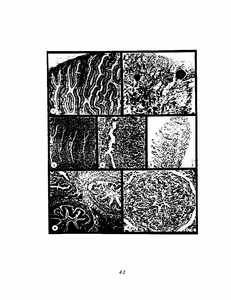
Figure II. 1. Section of intestinal tissue from a susceptible SiletzRiver steelhead trout showing an inflammatoryresponse (arrow) 18 d after exposure to the infectivestage of Ceratomyxa Shasta. H&E stain, 100X.
Figure 11.2. Ceratomyxa shasta trophozoites (arrows) in themucosal epithelium at 18 d post exposure. Giemsastain, 1000X.
Figure 11.3. Intestinal tract at 30 d post-exposure. Trophozoitesare proliferating in all intestinal layers and portionsof the mucosal epithelium have sloughed. Giemsastain, 100X.
Figure 11.4. Trophozoites (arrows) are diffusely scatteredthroughout the tissues and are surrounded bynecrotic host cells. Epithelial cells are rounded (whitearrow) and are being sloughed. Giemsa stain, 250X.
Figure 11.5. Trophozoites in the gill capillary and adjacent epithelialtissue (arrows). Giemsa stain, 40X.
Figure 11.6. Infected pyloric caeca and surrounding adipose andpancreatic tissue 52 d post-exposure. Muscularis ofcaeca is infected. Note necrosis and sloughing ofmucosal epithelium from underlying submucosa.Giemsa stain. 250X.
Figure 11.7. Severe necrosis and occlusion of the lumen of a pyloriccaeca. Numerous trophozoites are multiplying in alltissue layers. Giemsa stain, 400X.
41
4 2

Figure 11.8. Kidney of Siletz River steelhead trout 52 d post-exposure. Trophozoites (white arrows) are present inthe hematopoietic tissue and in necrotic kidneytubules (black arrows). Giemsa stain, 250X.
Figure 11.9. Liver tissue at 52 d showing area of focal necrosis.Trophozoites proliferated throughout the tissue andsporogenesis was occurring (arrows). Giemsa stain,250X.
43

P P
In Shasta rainbow trout held at 210C after exposure to the
infective stage, the parasite was first detected at 7 d post-exposure.
Trophozoites were most easily detected by IFAT because of limited
inflammation (Fig. II. 10). Multicellular trophozoites were detected
mainly at the base of epithelial cells in the posterior intestine and
some were also observed in hepatic blood sinusoids (Fig. II.1 1). By
10 d, focal inflammatory reactions had developed in some fish and
trophozoites were detected between epithelial cells or in the
submucosa of the inflamed area. Parasites were observed
throughout the posterior intestine (Fig. 11.12) and some appeared in
the anterior intestine and connective tissue surrounding blood
vessels in the pancreatic tissue. The livers of several fish were also
infected and hepatic cells were undergoing degeneration. Fish
sampled at 14 and 18 d post-exposure showed varying degrees of
infection and host response. Intestines were mildly to severely
infected and lumens were occluded in certain areas (Fig. II. 13). In
severely infected fish, trophozoites proliferated in the mucosa,
submucosa, muscularis, and serosa. Infected tissues were
hemorrhaged and infiltrated by eosinophilic granular leucocytes
and lymphocytes. The epithelium was necrotic and had sloughed in
certain areas so that necrotic host cells and trophozoites were
observed in the intestinal lumens. Parasites penetrated the
muscularis and serosa and were free in the peritoneal cavity.
Adipose tissue adjacent to the intestine was parasitized and
infiltrated by lymphocytes and there was a hyperplasia of
connective tissue (Fig. 11.14). At 18 d, sporogenesis was evident in
4 5
the intestine. The muscularis of the pyloric stomach was infected
and trophozoites were separating muscle fibers, leaving spaces
filled with necrotic material (Fig. 11.15). Nuclei of smooth muscle
fibers were pycnotic and the fibers were degenerating. Pyloric
caeca in close proximity to the infected stomach and intestine were
also infected (Fig. 11.16). Although trophozoites penetrated the
submucosa, muscularis, and serosa of these tissues, the epithelium
was still intact in certain areas. The liver in most fish had sites of
focal infection (Fig. II. 17). Hepatocytes were necrotic and
lymphocytes infiltrated the infected tissues. Blood smears and
kidney imprints were also positive for trophozoites (Fig. II.18 and
11.19). The spleen remained uninfected. At 20 d post-exposure, as
fish were dying of the disease, intestinal lumens were completely
occluded and most adjacent tissues and organs were infected to
varying degrees. The cardiac stomach and spleen remained
uninfected.
One Shasta rainbow, sampled at 18 d post-exposure, did
not show typical histopathology. In this fish, trophozoites
were observed in the lumen of the intestine and within the
epithelial layer (Fig. 11.20 and 11.21). Those in the lumen
were both free and attached to the epithelial surface. There
was no inflammatory inflammatory response. The identity of
these trophozoites as early stages of C. shasta was confirmed
by IFAT.
4 6

Figure II. 10. Fluorescent antibody stain of intestinal tissue fomShasta strain rainbow trout 7 d post-exposureshowing a multicellular trophozoite in the mucosalepithelium. 400x.
Figure II. 11. Trophozoite (arrow) in the blood sinusoids of the liverat 7 d. Giemsa stain, 1000X.
Figure II. 12. Intestine at 10 d with parasites (arrows) in or at thebase of the mucosal epithelium. Alkalinephosphatase immunostain, 100X.
Figure II. 13. Occluded lumen of posterior intestine from a Shastastrain rainbow trout sampled 18 d post-exposure.Epithelial cells are necrotic and sloughed andtrophozoites exist in all intestinal layers. Giemsastain, 100X.
Figure II. 14. Adipose tissue (A) adjacent to the intestinal serosa(S). The intestine has been penetrated, there ishemorraghing, and trophozoites (arrows) aremultiplying in the adipose tissue. Giemsa stain,250X.
Figure 11.15. Trophozoites (arrows) in the muscularis of thestomach were found between muscle fibers. Giemsastain, 400X.
Figure II. 16. Infected stomach and adjacent pyloric caeca.Trophozoites appear in all tissue layers but theepithelium is intact. Giemsa stain, 100X.
Figure II. 17. Liver at 14 d post-exposure showing numeroustrophozoites (arrows). Giemsa stain, 250X.
Figure II. 18. Blood smear with multicellular trophozoite (arrow)surrounded by blood cells. Giemsa stain, 400X.
Figure II. 19. Trophozoite (arrow) in kidney imprint. Giemsa stain,400x.
4 7

4 8

Figure 11.20. Trophozoites (arrows) in the lumen of the intestine ofa Shasta strain of rainbow trout 18 d post-exposure.Giemsa stain, 100X.
Figure II.2 1. Trophozoite (arrow) on the surface of the intestinalmucosal epithlium. Giemsa stain, 400X.
.Figure 11.22. Trophozoite in the lumen of the intestine of a North
Santiam strain steelhead trout 30 d post-exposure.Giemsa stain, 250X.
4 9

5 0

Histopathology of Infection in Salmonids Resistant to
Ceratomyxa Shasta.
Ceratomyxa shasta was detected in only two resistant fish,
both at 30 d post-exposure. In one fish, a single trophozoite was
observed in the lumen of the posterior intestine (Fig. 11.22). It was
not attached and there was no sign of a host response. The second
fish was severely infected. Trophozoites had penetrated the
intestinal epithelium and the infection appeared the same as in
severely infected susceptible fish.
Scanning Electron Microscopy.
Comparison of infected and uninfected posterior intestines by
scanning electron microscopy showed complete destruction of the
mucosal epithelium and different degrees of damage to the
structure of the mucosal folds in infected fish (Fig. II.23 - 11.28).
Lymphocytes, macrophages, and red blood cells were numerous in
infected areas. Sporogenesis was also observed. Desquamation of
the columnar epithelial cells caused loss of secondary structures of
the folds and in
51

Figure 11.23. Scanning electron micrograph of the posteriorintestine from an uninfected control fish showingparallel mucosal folds with secondary folds. Barindicates scale in micrometers.
Figure 11.24. Posterior intestine of an infected fish at the samemagnification. The secondary structure of themucosal folds has been destroyed in the lowerportion, and there is little left of any primarystructure in some areas (arrow). Bar indicates scalein micrometers.
Figure 11.25. Control intestine, showing smooth mucosal folds.Outlines of columnar epithelial cell surfaces arevisible. Bar indicates scale in micrometers.
Figure 11.26. Infected intestine at the same magnification.Destruction of the intestinal mucosa and penetrationinto the underlying layers. Little identifiablestructure remains except at the top of the folds,where trophozoites and inflammatory cells are notas numerous. Bar indicates scale in micrometers.
Figure 11.27. High magnification of the mucosal surface in anuninfected fish. Outlines of cell surfaces are visible.Depressions mark the pores of discharging mucouscells. Bar indicates scale in micrometers.
Figure 11.28. High magnification of an infected intestine showingextreme tissue damage. A variety of cell types arepresent, including lymphocytes, red blood cells, andC. shasta trophozoites and spores (arrow). Barindicates scale in micrometers.
5 2

5 3
certain areas the underlying connective tissues were also
destroyed.
Antibody Response.
The antibody response of naturally infected rainbow trout
was investigated by Western blot analysis and IFAT. Specific
antibody against C. shasta was not detected by either method.
5 4

DISCUSSION
Myxosporeans are generally known to parasitize fish without
damaging tissues; however, certain species cause extreme tissue
reactions and death of the host. In concurrence with this,
descriptions of host responses to these parasites range from a
complete lack of tissue reaction to a severe inflammatory response.
Ceratomyxa shasta belongs to this latter group, causing extensive
intestinal damage and death in susceptible salmonid fishes.
The first location that trophozoites were detected was
between epithelial cells of the descending intestine. This
observation was in agreement with that of Yamamoto and Sanders
(1979). Although Johnson (1975) suggested that the pyloric caeca
were the primary site of infection, we did not detect trophozoites in
this tissue by examination of Giemsa stained sections or by IFAT
until late in the infectious process. Furthermore, Johnson’s
descriptions of trophozoites in the pyloric caeca did not coincide
with those of trophozoites in the intestine, suggesting that they may
not have been the same organism. The inability to detect C. shasta
until 7 d post-exposure in fish held at 210C suggests that very few
organisms are required to initiate infection. An alternate
explanation is that the infective stage may enter the fish by a route
other than the intestine and was not observed using our detection
methods.
A vigorous host response, consisting mainly of a lymphocytic
infiltration, accompanied the proliferation of parasites in the
5 5

intestine. The observation of an inflammatory response against
these early stages differs from the observation of Dykova and Lom
(1978) that inflammatory reactions occur only with the
development of mature spores. However, their conclusions were
based on examining the pathology caused by cyst-forming histozoic
species. The trophozoite is the pathogenic stage of this parasite and
once established in the host is proteolytic and invasive.
Although C. shasta has a trophism for the intestine, it differs
from the tissue specificity shown by most myxosporeans by
effectively invading and multiplying in most other tissues and
organs. Noble (1950) noted the parasite in the entire alimentary
tract, gill capillaries, kidney, spleen, liver, gall bladder, and gonads;
Schafer (1968) reported infections of the eyes; Conrad and DeCew
(1966) found the parasite in muscle; and Margolis and Evelyn
(1975) reported lesions in the kidney, pyloric caeca, intestine and
on the flanks below the dorsal fin. Trophozoites multiply
extensively in the intestinal tract and several days after initial
detection, they are present in the mucosa and submucosa
throughout the intestine.
In this study, appearance of trophozoites in the liver closely
followed their presence in the intestine. The route of invasion into
the liver was probably venous blood which enters the hepatic
portal system directly from the alimentary tract. Blood transport
via the hepatic portal system was also observed by Jakowska
(1979) for Henneguya species. While necrotic degradation and
5 6

inflammatory responses were seen in the hepatic tissues, this organ
was not as extensively infected as the alimentary tract.
The infection proceeded anteriorly in the intestine, but at the
same time it penetrated the muscularis and serosa and became free
in the peritoneal cavity. This made it difficult to determine
whether the caeca became infected internally or by penetration of
the serosa from the outside. It is possible that both occur, as some
caeca were infected only in the muscularis and serosa and had an
intact epithelium, while other caeca were completely destroyed.
Adipose and pancreatic tissue adjacent to the pyloric caeca were
also infected. Penetration of the stomach did appear to be external
or via the bloodstream, as trophozoites were always found between
muscle layers while the mucosa was intact, with no parasites or
inflammation.
At the same time that trophozoites penetrated the intestinal
wall, they were also detected in blood smears and kidney imprints.
The presence of C. shasta in the bloodstream allowed its spread to
other tissues and organs which had not been penetrated by the
parasite from the body cavity.
Scanning electron microscopic studies of heavily infected
posterior intestines allowed assessment of the amount of structural
damage caused by this parasite. Secondary mucosal folds were
destroyed throughout the entire intestine and primary folds were
severly eroded in areas. Spores and trophozoites were numerous,
as were inflammatory and red blood cells.
5 7

Examination of the contents of the gall bladder has been
suggested in diagnosis of an infection (Amos, 1985). Johnson
(1975) noted that infection of the gall bladder occurred late in the
infectious process and in this study little or no infection of that
organ was observed. When the gall bladder and spleen were
infected, trophozoites appeared to have entered from the body
cavity. Because the posterior intestine is the primary site of
infection and because infection of this tissue always preceeds
invasion of other areas, and is more severe than infections in other
tissues, we believe examination of the intestine is sufficient for
diagnosis of the disease.
The progress of infection in resistant strains of fish was
difficult to determine because only two fish of the resistant, North
Santiam strain became infected. No evidence of the parasite or any
inflammatory response was observed in uninfected, North Santiam
steelhead. The appearance of the infection in one of the two fish
was similar to that in susceptible fish, indicating that the primary
site of infection is the same and once the parasite has penetrated
the intestine, the infection proceeds as in susceptible fish. Detection
of a trophozoite in the lumen of the intestine of the second fish
supports the hypothesis that resistance is at the site of entry. If
resistance was due to a cellular or humoral response it would be
expected that parasites would be in the tissue. One individual from
a susceptible strain also appeared refractory to infection. Many
trophozoites were detected in the lumen of its intestine, several
were attached to the epithelial surface, and some were present
58

between epithelial cells. Although this fish may have eventually
succumbed to the infection, it provided additional evidence that the
parasite first enters the fish from the lumen of the descending
intestine and that exclusion of the parasite from entering at this
site may be the mechanism of resistance.
The progress of the infection is temperature dependent, with
mean time to death inversely correlated to temperature (Udey et
a1.,1975). The histopathology of the infection paralleled this;
trophozoites were detected at an earlier time and the inflammatory
response developed earlier as temperature increased. Although
trophozoites multiplied faster, the progression of events was similar
to that in fish held at lower temperatures. These observations are
contrary to those of Lom (1969) and Dykova and Lom (1978) who
noted a regression of infection by Henneguya species as
temperatures increased. It was suggested that antibodies become
active at higher temperatures and help destroy the parasite.
No immune response was detected in rainbow trout naturally
infected with C. shasta by either Western blotting or IFAT, even
though the fish had harbored the parasite for over thirty days and
were dying from the infection. This supports the work of other
researchers (Siau, 1980; Halliday, 1974; Pauley, 1974) who were
unable to detect antibodies against myxosporeans by complement
fixation, gel precipitation, or immunoelectrophoresis. One criticism
of these studies is that they were trying to detect antibodies against
the spore stage of the parasite. Spores form later in the infectious
process, and spore antigens may differ from those of trophozoites
5 9
(Bartholomew et al., submitted). Therefore, fish antibodies against
these early stages may go undetected. Griffin and Davis (1978)
were able to demonstrate antibodies against M. cerebralis in
naturally infected trout by IFAT; however, their results did not
show absolute correlation between the presence of spores and the
presence of antibody, and their results required some
interpretation because of background fluoresence. Also, results of
monoclonal antibody studies (Bartholomew et al., submitted)
indicate that C. shasta trophozoites and trout immunoglobulin have
cross-reacting carbohydrate epitopes. If this is true for other
myxosporeans, polyclonal antibodies against trout immunoglobulin
may be reacting against the parasite itself rather than against
antibody on its surface. It appears that if an immune response is
directed against C. Shasta, it is ineffective in halting the infection.
Infection by C. shasta fits the description by Plehn (1932) of a
super-infection in which invasion leads to a generalized
myxosporidiasis and subsequently to death.
6 0
ACKNOWLEDGEMENTS
We thank Elizabeth MacConnell and Oliva Nunez for their
assistance in sectioning fish for the histological study and Al
Soeldner for preparation of samples for the scanning electron
microscope. This research was supported by Bonneville Power
Administration under contract No. DE-A179-83 BP 11987; G. R.
Bouck, Contracting Officer’s Technical Representative. Oregon
Agricultural Experiment Station Technical Paper No. 873 1
61

LITERATURE CITED
Amandi, A, and J. L. Fryer. 1985. Observations on Myxobolusinsidiosus (Myxozoa: Myxosporea) a parasite of salmonidfishes. Fish Path. 20:287-304.
Amos, K. H., ed. 1985. Procedures for the detection andidentification of certain fish pathogens, 3rd ed. Fish HealthSec., Amer. Fish. Soc., Corvallis, OR.
Bartholomew, J. L., J. S. Rohovec, and J. L. Fryer. Development,characterization, and use of monoclonal and polyclonalantibodies against the myxosporean, Ceratomyxa shasta.(manuscript submitted).
Buchanan, D. V. , J. E. Sanders, J..L. Zinn, and J.L. Fryer. 1983.Relative susceptibility of four strains of summer steelhead toinfection by Ceratomyxa shasta. Trans. Amer. Fish. Soc.112:541-543.
Ching, H. L., and D. R.. Munday. 1984. Susceptibility of six Fraserchinook. salmon stocks to Ceratomyxa shasta and the effects ofsalinity on ceratomyxosis. C a n . J . Z001. 62:1081-1083.
Cohen, A. L., D. P. Marlow, and G. E. Garner. 1968. A rapid criticalpoint method using fluorocarbons (Freons) as intermediateand transitional fluids. J . Micr. 7-3:33 l-342.
Conrad, J. F., and M. Decew. 1966. First report of Ceratomyxa injuvenile salmonids in Oregon. Prog. Fish-Cult. 28:238.
Duhamel, G. E., M. L. Kent, N. 0. Dybdal, and R. P. Hedrick. 1986.Henneguya exilis Kudo associated with granulomatousbranchitis of channel catfish Ictalurus punctatus (Rafinesque).Vet. Path. 23:354-361.
Dykova, I., and J. Lom 1978. Histopathological changes in fish gillsinfected with myxosporidian parasites of the genusHenneguya. J. Fish Biol. 12:197-202.
6 2
Fantham, H., and A. Porter. 1.912. Some effects of the occurrence ofmyxosporidia in the gall bladder of fishes. Ann. Trop. Med.Parasitol. 6:467-48 1.
Griffin, B., and E. Davis. 1978. Myxosoma cerebralis: Detection ofcirculating antibodies in infected rainbow trout (Salmogairdneri). J. Fish. Res. Board Can. 35:1186-l 190.
Halliday, M. 1974. Studies on Myxosoma cerebralis, a parasite ofsalmonids. IV. A preliminary immunofluorescen tinvestigation of the spores of Myxosoma cerebralis. NordicVet. Med. 26: 173-179.
Halliday, M. 1976. The biology of Myxosoma cerebralis: Thecausative organism of whirling disease of salmonids. J. FishBiol. 9:339-357.
Hoffmaster, J. L., J. E. Sanders, J. S. Rohovec, J. L. Fryer, and D. G.Stevens. 1988. Geographic distribution of the myxosporeanparasite, Ceratomyxa shasta Noble, 1950, in the ColumbiaRiver basin, USA. J. Fish Dis. 11:97-100.
Jakowska, S., and R. Nigrelli. 1953. The pathology ofmyxosporidiosis in the electric eel, Electrophorus electricus(Linnaeus), caused by Henneguya visceralis and H. electricaspp. nov. Zool. 38:183-191.
Johnson, K. A. 1975. Host susceptibility, histopathologic, andtransmission studies on Ceratomyxa shasta, a myxosporidanparasite of salmonid fish. PhD Thesis, Oregon State Univ.,Corvallis OR. 134 p.
Kent, M. L., and R. P. Hedrick. 1985. PKX, the causative agent ofproliferative kidney disease (PKD) in Pacific salmonid fishesand its affinities with the Myxozoa. J. Protozool. 32:254-260.
Lom, J. 1969. Cold-blooded immunity to protozoa. In: Immunity toParasitic Animals. Vol. 1 (ed. by G. J. Jackson, R, Herman, andI. Singer), p. 249-265. Appleton-Century-Crofts, New York.
6 3
Lom, J. 1970. Protoza causing diseases in marine fishes. In: ASymposium of Fishes and Shellfishes (ed. by S. Snieszko), p.101-123. Amer. Fish. Soc. Spec. Publ. No. 5. Washington, D. C.
Margolis, L., and T. P. Evelyn. 1975. Ceratomyxa shasta(Myxosporida) in chum salmon (Oncorhynchus keta) in BritishColumbia. J. Fish. Res. Board Can. 32:1640-1643.
Noble, E. 1941. Nuclear cycles in the life history of the protozoangenus Ceratomyxa. J. Morph.. 69:455-479.
Noble, E. R. 1944. Life cycles in the Myxosporidia. Quart. Rev. Biol.19:213-235.
Noble, E. R. 1950. On a myxosporidian (protozoan) parasite ofCalifornia trout. J. Parasitol. 36:457-460.
Pauley, G. 1974. Fish sporozoa: Extraction of antigens fromMyxosoma cerebralis which mimic tissue antigens of rainbowtrout (Salmo gairdneri, Rafinesque). J. Fish. Res. Board Can.31:1481-1484.
Plehn, M. 1932. Eine Schleienbrut-Krankfeit und ihr Erreger,Sphaerospora tincae n. sp. Inter. Rev. Ges. Hydrobiol.Hydrograph. 26:265-280.
Roberts, R. J., and K. G. R. Elson 1970. An outbreak of whirlingdisease in rainbow trout. Vet. Rec. 86:258-259.
Schafer, W. E. 1968. Studies on the epizootiology of themyxosporidian Ceratomyxa shasta Noble. Calif. Fish Game.54:90-99.
Siau. Y. 1980. Immunologic observations in fishes of the genusMugil parasitized by the myxosporidan Myxobolus exiguusThelohan, 1895. Zeitschrift Parasitenkd. 62: l-6.
Taylor, R. E., and M. H. Haber 1974. Opercular cyst formation introut infected with Myxosoma cerebralis. J. Wildl. Dis.10:347-35 1.
6 4
Udey, L. R., J. L. Fryer, and K. S. Pilcher. 1975. Relation of watertemperature to ceratomyxosis in rainbow trout (Salmogairdneri) and coho salmon (Oncorhynchus kisutch). J. Fish.Res. Board Can. 32:1545-1551.
Wales, J. H. and H. Wolf. 1955. Three protozoan diseases of troutin California. Calif. Fish Game. 41:183-187.
Yamamoto, T. and J. E. Sanders. 1979. Light and electronmicroscopic observations of sporogenesis in themyxosporidan, Ceratomyxa shasta (Noble, 1950). J. Fish Dis.2:41 l-428.
Zinn, J. L., K. A. Johnson, J. E. Sanders, and J. L. Fryer. 1977.Susceptibility of salmonid species and hatchery strains ofchinook salmon (Oncorhynchus tshawytscha) to infections byCeratomyxa shasta. J. Fish. Res. Board Can. 34:933-936.
6 5
CHAPTER III
CHARACTERIZATION OF A MONOCLONAL ANTIBODY AGAINST
CERATOMYXA SHASTA USING IMMUNOCYTOCHEMISTRY
J. L. Bartholomewl, T. Yamamoto2, J. S. Rohovecl, and J. L. Fryer1
IDepartment of Microbiology
Oregon State University
Corvallis, Oregon 9733 1
2Department of Microbiology
University of Alberta
Edmonton, Alberta, Canada T6G 2E9
66
ABSTRACT
The antigenic specificity of a monoclonal antibody (Mab)
against Ceratomyxa shasta was investigated using immunogold
labeling techniques and transmission electron microscopy and by
immunoenzymatic labeling. Specific binding by the Mab was
observed in the cytoplasm of the primary cell. Label was not
observed in the cytoplasm of the developing secondary cells or in
the nucleus of either the primary or secondary cells. A hypothesis
explaining the specificity of this Mab for prespore stages of the
parasite is presented.
6 7
INTRODUCTION
A monoclonal antibody (Mab) specific for the myxosporean
salmonid parasite, Ceratomyxa shasta, has been developed and
characterized (Bartholomew et al., submitted). This Mab reacts
specifically with the trophozoite and sporoblast stages of the
parasite in indirect fluorescent antibody tests (IFAT). In Western
blot analysis it recognizes many polypeptide bands from these
prespore stages, suggesting that it is formed against a common
epitope. The Mab did not react with C. shasta spores by
immunoblotting procedures, Western blot analysis, or IFAT. These
data indicate that the parasite has stage-specific antigens and that
Mabs may not react against all life stages.
Antibodies that recognize early life stages of C. shasta would
be helpful in studying the biology and life cycle of the parasite.
Presently, the only life stages identified are those that exist within
the salmonid host. A Mab recognizing trophozoites may also
recognize stages of the parasite prior to infection of the fish and
could be used to look for these stages in water and invertebrates
collected from endemic sites. In this study the specificity of the
Mab was investigated by using immunogold electron microscopic
techniques and immunoenzymatic labeling of histological sections.
6 8
MATERIALS AND METHODS
Immunogold Electron Microscopy
To determine the location of the antigens against which the
monoclonal antibodies are directed immunogold labeling
procedures were used. Prespore stages of the parasite from the
ascites of infected rainbow trout were fixed for 1 h in 2.5%
glutaraldehyde in 0.1 M phosphate buffered saline, pH 7.6 (PBS).
The suspension was centrifuged to pellet the cells which were
resuspended in PBS. Cells were embedded in Spurr’s resin (Spurr,
1969) without 0~04 staining. Sections were cut on an
ultramicrotome and mounted on nickel grids. The embedding
medium was etched with sodium metaperiodate (saturated solution
in distilled water) for 30 min, washed with distilled water, then
blocked with 100 mM glycine + 5% bovine serum albumin in PBS
for 15 min. Sections were incubated with undiluted monoclonal
antibody for 1 h at room temperature. After washing three times
with PBS, grids were incubated with biotinylated anti-mouse IgG
(1: 100 in PBS)(Hyclone Laboratories, Logan, Utah) for 1 h, washed,
and incubated for 30 min with Streptavidin-gold (1:200 in
PBS)(particle size, 20 nm)(Polysciences Inc., Warrington, PA). In
control experiments, the incubation conditions were identical
except that non-immune serum or PBS was substituted for specific
antibody in the first incubation step. All grids were washed
thoroughly, first with PBS then with distilled water, and examined
with a Philips EM-301 transmission electron microscope. Some
6 9
grids were post-stained with 2% 0~04 (5 min), uranyl acetate (2
min) and lead citrate (5 min).
Immunoenzymatic Staining
For examination of the parasite in tissue sections, viscera of
fish exposed to the infectious stage of C. shasta were fixed in 10%
neutral buffered formalin, processed routinely for histology to 6
pm, and mounted on gelatin coated slides. Sections were dewaxed
and hydrated in 2 changes each of xylol, absolute ethanol, 95%
ethanol, and were transferred to water. Sections were incubated
with undiluted anti-C. shasta monoclonal antibody for 1 l/2 to 3 h
at room temperature then rinsed with PBS. Sections were then
incubated with biotinylated goat anti-mouse Ig streptAvidin-
peroxidase conjugate (Biogenex, Dublin, CA). The enzymatic
activity was localized with the substrate hydrogen peroxide-AEC-
acetate buffer (Biogenex, Dublin, CA). Sections were counterstained
with a non-alcoholic solution of hematoxylin (Meyer’s) to provide a
contrasting background stain.
7 0
RESULTS AND DISCUSSION
Specific binding by the Mab was observed by electron
microscopy to occur in the cytoplasm of the prespore stages of C.
Shasta. Gold particles were randomly distributed in the cytoplasm
and on the cell membrane of the primary (mother) cell but did not
appear to bind to any specific organelle. Label was not observed in
the cytoplasm of the developing secondary (daughter) cells or in
the nucleus of either the primary or secondary cells. No gold
binding occurred in prespore stages incubated with either normal
mouse serum or PBS which served as negative controls (Fig. III. l-
3). Immunoenzymatic staining of trophozoites in histological
sections also showed that the antibody binding sites were on the
surface and in the cytoplasm of the primary cell (Fig. 111.4).
Secondary cells were clearly outlined and were not stained.
Chromogen was also observed outside the trophozoite, suggesting
that the antigen may be secreted.
Association of the gold particles with the cytoplasm of the
primary cell may explain why this Mab recognizes only prespore
stages of C. shasta and not mature spores. The first recognizable
stages of the parasite in the fish are unicellular. This cell
undergoes nucleogomy, the process of forming a multinuclear cell
from a uninuclear cell, and eventually consists of a primary cell
enveloping two sporoblasts. Each sporoblast contains two
sporogenic, two capsulogenic, and two gametogenic cells (Noble,
1941; Yamamoto and Sanders, 1979). The primary cell is present
71
until mature spores are released; therefore, antigens present only
in the cytoplasm of this enveloping cell may not be present in the
mature spore.
Observation of the antigen recognized by this Mab gives
additional support for its usefulness as a diagnostic reagent for
prespore stages of C. Shasta. The location of the antigen in the
cytoplasm of the early stages offers some hope that this antigen
would be present in life stages that exist outside of the salmonid.
7 2
Figure III. 1. Transmission electron micrograph of a Ceratomyxa
shasta trophozoite incubated with normal mouse
serum and immunogold reagents. 5700x.
Figure 111.2. Ceratomyxa shasta trophozoite incubated with
monoclonal antibody and immunogold reagents.
Arrows indicate gold label on cell surface and in the
cytoplasm. 7100x.
Figure 111.3. Ceratomyxa shasta trophozoite incubated with
monoclonal antibody and immunogold reagents.
Gold label diffusely scattered in primary cell
cytoplasm. Unstained, 11000X.
7 3

7 4
Figure 111.4. Immunoenzymatic stain of Ceratomyxa shasta
trophozoites in a histological section. Chromogen
stains only the cytoplasm of the primary cell,
secondary cells are unlabeled. xl000
7 5
a-&.?; .,PI. & rs” MS ;$&& .l-c%* ‘. i’$g
*a c.- --*.,--&J,.G ‘_-
\. .--..-!Pa ;:.-- II
P; g$ga
_ _ .-. . . _ .
--.
76

ACKNOWLEDGEMENTS.
The authors wish to thank Jim Winton for arranging use of
the facilities used in this study and Cindy Arakawa for her
assistance. This research was supported by Bonneville Power
Administration under contract No. DE-Al79433 BP 11987; G. R.
Bouck, Contracting Officer’s Technical Representative. Oregon
Agricultural Experiment Station Technical Paper No. 8732.
7 7

LITERATURE CITED
Bartholomew. J. L., J. S. Rohovec, and J. L. Fryer. Development,characterization, and use of monoclonal and polyclonalantibodies against the myxosporean, Ceratomyxa shasta.(manuscript submitted)
Noble, E. 1941. Nuclear cycles in the life history of the protozoangenus Ceratomyxa. J. Morph. 69:455-479.
Spurr, A. R. 1969. A low viscosity epoxy resin embedding mediumfor electron microscopy. J. Ultrastr. Res.. 26:31-34.
Yamamoto, T., and J. E. Sanders. 1979. Light and electronmicroscopic observations of sporogenesis in the myxosporida,Ceratomyxa shasta (Noble, 1950). J. Fish Dis. 2:411-428.
7 8
CHAPTERIV
ARTIFICIAL TRANSMISSION OF CERATOMYXA SHASTA VIA
POTENTIAL INTERMEDIATES
D. G. Stevens, J. L. Bartholomew, J. S. Rohovec and J. L. Fryer
Department of Microbiology
Oregon State University
Corvallis, Oregon 97331, USA
7 9
ABSTRACT
The life history of Ceratomyxa Shasta, a myxosporean parasite
of salmonid fish, is unknown. Transmission occurs naturally when
susceptible fish are exposed to waters containing the infectious unit
which remains undescribed. The inability to obtain transmission
from infected fish to susceptible animals and the geographic limits
within a river of the infectious zone suggest the involvement of an
intermediate host in the life cycle. Preliminary laboratory
experiments with the freshwater mussel (Margaritifera
margaritifera) have resulted in transmission. We have examined
several parameters necessary for reproducible transmission to
occur under laboratory conditions. Although the exact role of the
mussel has not been determined, experiments indicate a low
number of the animals are involved.
8 0

INTRODUCTION
Artificial transmission and description of the complete life
history of only one Myxosporean parasite has been accomplished
(Wolf and Markiw, 1984). The causative agent of whirling disease,
Myxobolus (Myxosoma) cerebralis, develops from trophozoites to
adult spores in the cartilage of juvenile salmonids. When the
spores are released they apparently develop into triactinomyxons
within the oligochaete worm Tubifex tubifex. The triactinomyxons
then complete the life cycle by infecting young fish. Although
many other Myxosporeans are parasites of fish, the life history of
none other than M. cerebralis has been described.
Unlike M. cerebralis, Ceratomyxa Shasta has a limited
geographic range and that range is unique because only certain
areas of the range where diseased fish are found have conditions
which allow infections to occur (Bartholomew et al. (in press),
Johnson et al., 1979). The geographic range in which infections can
be initiated include the Willamette River up to river mile 134 (near
Corvallis, Oregon). Conditions that are necessary for the
transmission of C. shasta have not been described and therefore the
life history of this organism remains an enigma.
Preliminary studies to determine if aquatic invertebrates are
required in some stage of the life cycle of C. shasta have been
performed (Fryer, 1988). It was noted that fresh water mussels
collected from a site where fish can be infected were involved in
the transmission of C. Shasta. Although experiments have been
81

repeated, the results are not consistantly reproducible. The
parameters necessary for C. shasta infections to occur were
investigated.
8 2
MATERIALS AND METHODS
Experimental Animals
Rainbow trout susceptible to C. shasta were obtained from
Oak Springs Hatchery and the OSU Food Toxicology and Nutrition
Laboratory. The fish were held at the OSU Fish Disease Laboratory
and Western Fish Toxicology Laboratory, United States
Environmental Protection Agency Laboratory in Corvallis, Oregon.
To infect fish with C. shasta, fish were held in live boxes in the
Willamette River near Corvallis (Udey et al., 1975). Fish were fed
Oregon Biodiet with 4% TM-50.
Invertebrate animals were collected by hand or dredged from
the Willamette River at mile 129.6 or 121.4. Invertebrate groups
studied included Oligochaete worms (Kincaidiana hexatheca), clams
(Corbicula fluminea), and freshwater mussels (Margaritifera
margaritifera). Freshwater mussels were also collected from the
Siletz River near Siletz, Oregon, a site where C. shasta is not
endemic. The mussels and clams were fed a mixture of 3 parts
instant yeast to 1 part brown rice flour.
Laboratory exposures were conducted in 1 and 20 gallon
tanks supplied with well water at a constant 15 C. Fish were fin-
clipped to differentiate among different groups.
83
Continued exposure of trout and Siletz River mussels to C. shasta
Susceptible rainbow trout and infected fish or viscera and
ascites fluid collected from infected trout were added to tanks
containing Siletz River mussels which had been collected six months
earlier. More than half of the mussels remained alive and this
second exposure was similar to the first where susceptible fish
were exposed to infected fish or viscera and ascites fluid with and
without mussels present. Five susceptible trout were added to each
of five aquaria containing five mussels. Three aquaria were
supplied with infected fish, one with viscera and ascites fluid, and
one served as a laboratory water control. Fish were kept in contact
with the mussels for 14 weeks and monitored for infection by C.
shasta. Following this exposure, the mussels and an additional lot
of susceptible fish were placed in the effluent from a tank of fish
which had been infected in the river and were dying from C. shasta.
The fish were exposed for four weeks.
Transmission of C. shasta using various substrates with rinsed and
brushed mussels
Mussels collected from river beds live in close association
with Oligochaete worms and generally have a significant algal
growth on the portion of the valves exposed to the river.
Therefore, tests are conducted with and without this algal covering
to determine if it is involved in transmission. Duplicate tanks were
8 4
prepared with the following substrates: 1) dirt, sand, and rock with
rinsed mussels; 2) dirt, sand, and rock with brushed mussels; 3)
sand with rinsed mussels; 4) sand with brushed mussels; 5) glass
beads with brushed mussels: 6) rinsed mussels with no substrate.
Control tanks included horizontal (fish to fish) transmission, and
laboratory water. Five susceptible uninfected and infected fish
were added to different experimental tanks immediately or after
the contents had been held overnight with aeration followed by
three days of water flow. Fish which died were removed, examined,
and returned to the tank to decay naturally. Replacement fish were
added to the tank where infections occurred at days 39 and 54 of
the test.
After the last replacement fish were exposed for seven days,
they were removed and placed in clean water for observation. The
mussels were removed from the large tank and placed in individual
small aquaria with three susceptible trout. This exposure was
continued for three months. Fish that died were examined for C.
shasta infections and replaced with naive fish. Six days after
initiation of the test, water samples were collected and stored for
examination pending the appearance of infections.
Examination of the possible role of Oligochaete worms in the
transmission of C. shasta
Previous preliminary experiments in this laboratory did not
show that Oligochaetes are involved in the transmission of C. Shasta.
8 5
We designed further tests to examine the role of worms in the life
cycle of C. Shasta. Four tanks were prepared with a substrate of
sterilized soil and sand. These tanks were supplied with worms
(Kincadiana hexatheca) only, worms and mussels, mussels only,
both brushed and rinsed. Controls for horizontal transmission and
laboratory water were included. Fifty worms and ten mussels were
added to the appropriate tanks with five infected and an equal
number of susceptible fish.
Effect of increased temperature on the release of infectious units
from mussels removed from the river during the winter
Experiments conducted, previously indicated that mussels
removed from the river during February were capable of infecting
susceptible fish with C. shasta for a period of at least 30 days. A
similar experiment was conducted, however, the susceptible fish
were removed after various exposure times. Mussels were hand
collected in February, brushed, and placed in a tank in which water
temperature could be controlled. The river temperature was 8 C at
the time of collection and the temperature of the laboratory tank
set at 10 C to match that of the transport water upon arrival at the
laboratory. Susceptible rainbow trout were added immediately and
the temperature was raised to 12.5 C at a rate of 0.7 C per day.
Separate lots of fish were exposed to the mussels for different time
intervals: two groups for periods of seven days, one for a period of
two weeks, and a fourth group for 30 days. Each group was kept in
8 6

clean water for a minimum of 42 days prior to examination for C.
shasta infections.
Exposure of rainbow trout to mussels and mussel pallial fluid
Exposures of fish to mussels and mussel pallial fluid were
conducted to evaluate the handling procedures and how they may
affect our ability to obtain transmission. Collecting pallial fluid
from the mussel immediately after removal from the river might
indicate if viable infectious units were present but were being
killed or diluted in the handling process.
Forty mussels were dredged from the river, blotted dry, and
the pallial fluid collected. Another group of mussels was collected
and handled similarly but without taking the pallial fluid. Both
groups were brushed and placed in clean well water for transport
to the laboratory. Susceptible fish were exposed to the pallial fluid,
the transport water, and to the effluent from four tanks containing
twenty mussels each. One liter water samples were collected on
day one and day two of the test. These were to be examined, if
infections occurred, for the presence of infectious unit using
monoclonal antibodies and IFAT.
8 7

Exposure of rainbow trout to mussels and clams from the
Willamette River near Albany
River exposures of susceptible fish have not always been
consistant in the Corvallis area (Johnson, 1975) and because
experiments conducted earlier this year with mussels collected near
Corvallis produced similar inconsistant infections, it was decided to
collect mussels further downstream where the concentration of
infectious units might be greater.
Mussels and clams were collected from a location, near the
city of Albany, eight miles downstream (river mile 121.4) from the
original sampling area. The mussels and clams were washed and
rinsed before placement in tanks with either sand or sand and mud.
Two tanks with 20 mussels and two tanks with 20 mussels and 50
clams were stocked with either ten susceptible or ten susceptible
and 5 infected fish. The fish were added to the tanks after
overnight aeration without water flow followed by three days of
water flow. Dead fish were removed for examination and those
positive for C. shasta were cut open and returned to the tank. The
fish were exposed for a minimum of eight weeks. Following the
initial exposure replacement fish were added to two of the tanks
and experiments in two tanks were terminated. If infections
continue to occur the mussels will be sacrificed and prepared for
histological examination.
8 8
RESULTS AND DISCUSSION
Continued exposure of trout and Siletz River mussels to C. shasta
None of the fish exposed to Siletz River mussels and C. shasta
organisms became infected. Only one fish died during the 14 week
exposure and it was not infected. Likewise, exposure to the effluent
from the tank of infected fish produced no infections. After more
than a year of intermittent exposure to infected fish and infected
viscera, no infections have occurred from the association of
susceptible rainbow trout with the mussels from the Siletz River, a
non-infectious stream. If the mussels from infectious areas are
truly associated with transmission of the disease, the requirements
necessary to initiate their involvement have not been met by the
conditions of these tests.
Transmission of C. shasta using various substrates with rinsed and
brushed mussels
Because we have been unable to produce consistant
infections, susceptible fish were introduced at selected intervals to
tanks containing three different substrates and different mussel
preparations to determine conditions necessary for transmission of
C. Shasta. Infections occurred in one tank only in which there were
mussels on a substrate of glass beads (Table 1). All of these fish
8 9

Table 1. Exposure of rainbow trout to the mussels (M.margaritifera) collected from a site infectious forCeratomyxa shasta and held on selected substrates.
Substrate typeMussel Fish Introductiona
T r e a t m e n t I m m e d i a t e D e l a y e d b
Mud, sand, rockSandMud, sand, rockSandGlass beadsGlass beads
(first replacement fish)(second replacement fish)
No substrate
Fish to fish controlLaboratory water control
Rinsed 0/5c 0/5Rinsed 0/5 0/5Brushed 0/5 0/5Brushed 0/5d 0/5Brushed 5/5
Rinsed
1/5e0/5
0/50/5
0/5
aFive infected fish were added to all tanks except where noted.bFish were introduced after overnight aeration without water flowfollowed by 3 days of flow at 1 gpm.
cNumber infected/number exposed.dOnly susceptible fish were added to this tank.eReplacement fish were added to this tank 39 and 54 days after
start of test.
9 0
were heavily infected with C. Shasta. Dead fish were examined and
those positive for C. shasta were cut open and returned to the tank.
Of the new fish added to the tank 39 days after initiation of the
test, only one of five became infected. None of the fish in the
second replacement group became infected. Placement of
individual mussels in small aquaria with susceptible fish failed to
produce additional infections.
These results and those of an earlier study (Fryer, 1988)
indicate that a complex substrate is not necessary to obtain
transmission. Therefore, the infectious unit must either be
harbored by the mussel or is released following developmental
changes of spores or trophozoites which are ingested. Previous
studies (Ratliff, 1983 ; Johnson, 1975) indicate that the infectious
unit does not survive longer than ten days in water alone. Our data
continue to indicate that not all mussels are involved because
although they are collected and transported to the laboratory
together, transmission of the disease does not occur in all aquaria to
which mussels are introduced.
Examination of the possible role of Oligochaete worms in the
transmission of C. shasta
Oligochaete worms (Tubifex) have been tested previously and
failed to facilitate transmission of ceratomyxosis. However, the
close association of the native species (Kincaidiana hexatheca) with
the mussels in the river beds prompted another study. No disease
91

transmission occurred in fish held with worms alone or worms in
association with mussels. In this experiment no transmission
occurred in any of the tanks with mussels. The river water
temperature was only 10.5 C when the organisms were collected
and fish exposed to the river water only suffered a 50% loss from C.
shasta. These conditions may have been responsible for the lack of
laboratory infections.
Effect of increased temperature on the release of infectious units
from mussels removed from the river during the winter
Mussels collected in February when the river temperature
was 8 C were gradually warmed to 12.5 C, and trout were exposed
to the mussels for up to one month. No transmission occurred in
the test; however, transmission had occurred in a similar test
conducted previously.
Exposure of rainbow trout to mussels and pallial fluid
No infections were obtained from either the mussels, pallial
fluid, or the water used to transport the animals. Prior to initiation
of this test, the river was infectious to nearly 50% of the fish
exposed. However, the positive controls from the river, exposed
during the first week of the test, only suffered 13% mortality from
C. Shasta. This may have been the result of heavy rains which
9 2
raised the river level and possibly diluted the concentration of C.
shasta organisms.
Exposure of rainbow trout to mussels and clams from the
Willamette River near Albany
Mussels and clams were collected from a site near Albany, OR
and examined for their involvement in disease transmission.
Infections of naive fish were obtained in one tank where infected
fish had been added (Table 2). Seven of ten were positive for C.
Shasta. Replacement fish were added to the tank in which
infections occurred and to date three of the fish have died and were
positive for C. Shasta. Water samples were taken from the aquarium
and slides are being examined for detection of the infectious unit.
In a replicate tank, in which no infections occurred, and additional
fish, both infected and naive, were added with viscera and ascites
fluid, transmission has failed to occur to this date.
As in previous experiments, infections occurred in only one
tank of fish exposed to replicate groups of mussels. This continues
to suggest that not all mussels are involved in the transmission of
ceratomyxosis. This study is being continued and others are
planned to identify the role of the mussel in the transmission of
ceratomyxosis.
9 3

Table 2. Exposure of rainbow trout to mussels and clams fromthe Willamette River near Albany.
Test Organisma Type of Fish Number of FishExposedc Infec ted
Mussels Susceptible 1 0 0Mussels Susceptible and
infectedb 1 0 7Mussels-clams Susceptible 1 0 0Mussels-clams Susceptible and
infected 1 0 0
aEach tank contained either 20 mussels or 20 mussels and 50clams.bFive fin-clipped infected fish were added where indicated.cStock fish served as laboratory water control. Trout exposed towater where the clams and mussels were collected suffered 100%mortality from C. Shasta.
9 4

ACKNOWLEDGEMENTS
The cooperation of the Environmental Protection Agency -
Corvallis Environmental Research Laboratory is recognized for
providing some of the fish holding facilities at the Western Fish
Toxicology Laboratory. The Oregon Department of Fish and Wildlife
and the OSU Food Toxicology and Nutrition Laboratory provided
experimental fish. This research was supported by the region’s
electrical rate payers through the Bonneville Power Administration.
9 5
LITERATURE CITED
Bartholomew, J. L., J. S. Rohovec and J. L .Fryer. 1989. CeratomyxaShasta, a Myxosporean parasite of salmonids. U.S. Fish andWildlife Service Fish Disease Leaflet (in press).
Fryer, J. L. 1988. Epidemiology and control of infectious diseases ofsalmonids in the Columbia River basin. Annual Report ofProject No. 83-312. U.S. Department of Energy, BonnevillePower Administration.
Johnson, K. A. 1975. Host susceptibility, histopathologic, andtransmission studies on Ceratomyxa Shasta, a myxosporidanparasite of salmonid fish. Ph.D. Thesis, Oregon StateUniversity, Corvallis, OR. 134 p.
Johnson, K. A., J E. Sanders, and J. L. Fryer. 1979. Ceratomyxashasta in salmonids. U.S. Fish and Wildlife Service FishDisease Leaflet 58. 11 p.
Ratliff, D. E. 1983. Ceratomyxa Shasta: longevity, distribution,timing, and abundance of the infective stage in centralOregon. Canadian Journal of Fisheries and Aquatic Sciences.40: 1622-1632.
Udey, L. R., J. L. Fryer, and K. S. Pilcher. 1975. Relation of watertemperature to ceratomyxosis in rainbow trout (Salmogairdneri) and coho salmon (Oncorhynchus kisutch). Journalof the Fisheries Research Board of Canada. 32:1545-1551.
Wolf, K. and M. E. Markiw. 1984. Biology contravenes taxonomy inthe Myxozoa: new discoveries show alternation ofinvertebrate and vertebrate hosts. Science. 255:1449-1452.
9 6
CHAPTER V
IN VITRO INHIBITION OF RENIBACTERIUM SALMONINARUM
BY EXPERIMENTAL ANTIBIOTICS.
S.K. Gutenberger, O.B. Dale, and J.S. Rohovec
Department of Microbiology
Oregon State University
Corvallis, Oregon USA
9 7
ABSTRACT
Over one hundred experimental antibiotics were incorporated
into KDM-2 agar and tested on four strains of Renibacterium
salmoninarum. R. salmoninarum was susceptible to 51 of the
compounds, with 19 of these effectively inhibiting at 1 mg/L or
less. Four other fish pathogens, Streptococcus sp., Aeromonas
salmonicida, Edwardsiella tarda, and Cytophaga psychrophila, were
also tested. Streptococcus was inhibited by 42 of the antibiotics, 33
of which also inhibited R. salmoninarum. Four antibiotics inhibited
all bac teria tested. Two drugs were effective against all bacteria
except R. salmoninarum. Seventeen of the drugs inhibited only R.
salmoninarum, Streptococcus, and C. psychrophila. A total of 32 of
the experimental compounds had no inhibitory effect on any of the
bacteria.
A microtiter plate assay for testing antimicrobial compounds
was developed for R. salmoninarum and compared to the agar plate
method. The results from the microtiter plate assay were
comparable to, and in some cases more sensitive than the agar plate
assay. The microtiter plate assay was more cost effective, saving
both time and media.
9 8
INTRODUCTION
Renibacterium salmoninarum, the causative agent of bacterial
kidney disease (BKD) is one of the most devastating bacterial
diseases of salmonid fish (Fryer and Sanders, 1981). The organism
is unique among bacteria. It is a very slow-growing organism
which has an absolute nutritional requirement for cysteine. The
bacterium causes a systemic infection which is usually chronic.
Currently there are no effective vaccines against BKD (Kaattari et al,
1988). The disease is difficult to treat, probably because of the
ability of the organism to survive phagocytosis by macrophages.
Currently, erythromycin is the drug of choice for therapy, but
treatments are not always successful. Others (Wolf and Dunbar,
1959; Austin, 1985) have evaluated antimicrobial compounds for
control of BKD and of the drugs that were tested, erythromycin was
the most effective. Unfortunately, this antibiotic is not yet
approved for use in food fish and laboratory studies have shown
that R. salmoninarum can become resistant to the effects of the
drugs (Bell et al, 1988). We initiated this study to test new
antimicrobial compounds for their in vitro effectiveness against R.
salmoninarum.
9 9

METHODS AND MATERIALS
Bacterial Strains: Four isolates of Renibacterium salmoninarum
(type strains ATCC 33209 and 33739; D-6, an USA isolate from coho
salmon (Oncorhynchus kisutch); and 932, a Norwegian isolate from
Atlantic salmon (Salmo salar)) were grown for 5-8 days in KDM-2
broth with 10% equine serum at 16oC, to an O.D.520 between 0.7-
1.1. Prior to use, the purity of the cultures was verified by Api-
Zym enzyme test strips (API, Plainville, New York) and by Gram
stain.
Four other fish pathogens were also tested, including the
gram-negative bacteria Aeromonas salmonicida, Cytophaga
psychrophila and Edwardsiella tarda. Streptococcus sp., a gram-
positive bacterium, was added as control for the R. salmoninarum.
A. salmonicida, E. tarda and Streptococcus were grown in tryptone
soy broth and C. psychrophila was grown in typtone yeast extract at
16oC for 24-48 hours. All bacterial cultures had greater than 107
colony forming units/ml (cfu) for antibiotic testing. Before actual
testing on the antibiotic-incorporated KDM-2 agar, the cfu for each
bacterium was compared on plates of KDM-2 and TYE agar. There
were no differences in growth on either media for A. salmonicida, E.
tarda and Streptococcus. Growth on KDM-2 agar by C psychrophila
was reduced (6.2 X 10⁶ cfu/ml) in comparison to that on TYE agar
(2.1 X 10⁷ cfu/ml) but was more than adequate for antibiotic
testing.
100

Preparation of Antibiotics: Over 100 antibiotics, untested on
fish pathogens, were obtained from Eli Lilly & Co. (104), Merck &
Co. (3), Abbot (1) and Pfizer (1) (Table 1). Thirty-two were
derivatives of the macrolide antibiotics erythromycin and tylosin.
Another 73 were fermentation products or derivatives. In addition,
a fluoroquinolone and a new drug related to
penicillin/cephalosporin were included. Immediately before
addition to agar, the drugs were weighed and diluted to 1 mg/ml in
either nanopure water, reagent grade methanol, or DMSO:methanol.
Those solubilized in water, methanol or methanol: DMSO (4% DMSO)
were filtered through 0.2 um polysulfone acrodisc filters (Gelman,
Ann Arbor, MI) into sterile vials. Those drugs solubilized in
DMSO:methanol (>4% DMSO) were not sterile-filtered; however,
there were no problems with contamination. Ampicillin (Sigma, St.
Louis, MO), rifampicin (Sigma, St. Louis, MO), penicillin-G (Grand
Island Biological Co.,Grand Island, NY), erythromycin phosphate
(Argent Chemical Laboratories, Inc., Redmond, WA) and
oxytetracycline (Sigma, St. Louis, MO) were used as control
antibiotics in each of the four trials required to complete the study.
Preparation of Antibiotic Media: Antibiotics were added into
sterile 50 ml tubes in amounts to make eight dilutions of each drug:
35, 10, 5, 1, 0.5, 0.1, 0.05 and 0.01 mg/L. Fifty milliliters of KDM-2
agar (10% equine serum), pH 6.8, was added to each tube, mixed,
and poured into two 100 mm quadrant petri dishes. As a positive
control for growth and to ensure solvents were not inhibitory to the
101

bacteria, each solvent used to solubilize the drugs was incorporated,
appropriately diluted, into additional agar plates.
Antibiotic Testing on Agar: Within 24 hours of antibiotic
incorporation, 0.1 ml of each bacteria was inoculated into a
quadrant. The plates containing R. salmoninarum were sealed with
parafilm and all plates were immediately incubated at 160C. The
plates of R. salmoninarum were examined at 7, 15 and 30 days
after inoculation. Those plates inoculated with the other bacteria
were examined at 3 and 5 days. For each bacterium, the minimum
inhibitory concentration (MIC) was determined to be the lowest
concentration of antibiotic that prevented growth.
Antibiotic Testing in Microtiter Plates: For each assay, at
least two strains of R salmoninarum (ATCC 33739 and the isolate
932) were used. In one assay, drugs 111, 112, 113, 114, and 115
were tested with four isolates of R. salmoninarum (ATCC 33209 and
33739, 932 and D-6) and the drug 116 was tested with eight
strains (including K-28 from France, 84-019 from open ocean
chinook, RC-88 from Oregon, and 325 from Norway). The
antibiotics were prepared as described above. To either round or
flat-bottomed 96-well microtiter plates (Falcon), KDM-2 broth (pH
6.8) with 10% equine serum was added to wells in amounts to
dilute antibiotics to 31.5, 10, 5, 1, 0.5, 0.1, 0.05, 0.01 and 0 mg/L or,
in the last assay, to 25, 5, 2.5, 0.5, 0.25, 0.05, 0.025, 0.005, 0.0025,
and 0 mg/L. Antibiotic (1 mg/ml) was added to the first two wells
1 0 2

of two rows, mixed, and serially diluted across the rows. Care was
taken to maintain the same solvent to media ratio as used in the
agar plate assay. Five to seven day old cultures of R. salmoninarum
were adjusted to an O.D.520 of 0.2 or 0.1 and then diluted again
with KDM-2 broth to give a final concentration of 20% cells in a
total of 180 ul, an amount determined previously to reproduce the
MICs of erythromycin obtained in the agar plate study. Ninety
microliters of the diluted bacteria was added to every other row in
the microtiter plate. In the other rows, 90 ul of KDM-2 was added
to serve as optical comparison. Antibiotic, media, and cells totaled
180 ul in each well. The microtiter plates were covered with
adherent transparent film and incubated at 160C on a shaker. The
results were read at 7, 15, and 30 days.
103
RESULTS
Agar plate assay of antibiotics: Renibacterium salmoninarum
was susceptible to 51 of the experimental antibiotics at day 7 after
inoculation of the agar plates (Table 2). At the higher
concentrations of drug, 13 antibiotics inhibited at 35 mg/L.
Another 19 drugs and one control drug, oxytetracycline, were found
to prevent growth at 5-10 mg/L. At lower concentrations, R.
salmoninarum was effectively inhibited by 19 of the antibiotics.
Seven antibiotics prevented growth at the same level as penicillin G
(1 mg/L), and six inhibited growth at 0.5 mg/L. Five of the
experimental antibiotics were inhibitory to R. salmoninarum at the
concentration of 0.1 mg/L, as were erythromycin and rifampicin.
One drug, 107, inhibited R. salmoninarum at 0.05 mg/L.
All four strains of R. salmoninarum, with few exceptions,
reacted similarly to all drugs (Table 3). Initially, the type strain,
ATCC 33209, was growing slowly in broth and was used at less than
an O.D. 520 of 0.1. Growth for this strain was not seen until 14 to
30 days after inoculation of the plates. Thus, in 24 out of 38 drugs
tested, this strain was more sensitive to the effects of the drug
although the sensitivity in all but one case increased by only one
dilution. In two cases, the weakly growing 33209 was actually less
resistant to the effects of the drugs than the other three strains.
Aside from this, antibiotic inhibition of strain 33209 was similar to
the other strains.
1 0 4

Table 1: List of Antibiotics
Drug # Name or Designation Class Drug # Name or Designation Class
123
:
7”a9101112131 4151 61718192 02 12 22 32 42 52 62 72 82 93 03 13 23 33 4353 63 73 83 94 0
A54177See 1.See 1.See 1.See 1.A5977OAAl50AA l 6 5A32357A2OlASee 1.A3 1323FA272A83586CA83580BCinoxacinA23444CaerulomyciPuromycinA38808A 4 2 2 1 9A45408A48662A82093A82243A82548A82548A83094A85761A258228A40104AA54556A60117A35512BA32256A33853A14187BA14787A31463AA31463B
tn
non-macrolide 4 1non-macrolide 4 2non-macrolide 4 3non-macrolide 4 4non-macrolide 4 5non-macrolide 4 6non-macrolide 47non-macrolide 4 8non-macrolide 4 9non-macrotide 5 0non-macrolide 51non-macrotide 5 2non-macrolide 5 3non-macrolide 5 4non-macrolide 5 5non-macrolide 56non-macrolide 5 7non-macrolide 5 8non-macrolide 59non-macrolide 6 0non-macrolide 6 1non-macrolide 6 2non-macrolide 6 3non-macrolide 6 4non-macrolide 6 5non-macrolide 6 6non-macrolide 6 7non-macrolide 6 8non-macrolide 6 9non-macrolide 70- 100.non-macrolide 107non-macrolide 108non-macrolide 109non-macrolide 1 1 0non-macrolide 111non-macrolide 112non-macrolide 113non-macrolide 1 1 4non-macrolide 115non-macrolide 116
A37812 non-macrolideA39079-S I non-macrotideA70195 non-macrolideA80889A&B non-macrolideA80889 Complex non-macrolideA54145A-1 non-macrolideA54145B non-macrolideA80915A non-macrolideA80915B non-macrolideA80915C non-macrolideA80915D non-macrolideA82516 non-macrolideA82775A non-macrolideA82775C non-macrolideA82775D non-macrolideA41980 non-macrolideA80407B non-macrolideA47934 non-macrolideMonensin non-macrolidePyrazofurin non-macrolideA16886B non-macrolideA4696.HCI non-macrolideHygromycin B non-macrolideA21978Cl non-macrolideA80509 non-macrolideA38533 non-macrolideA7413 non-macrolideA2315A non-macrolideA214 non-macrolideSee 1 non-macrolideAzitbromycin** MacrolidesFosfomycin* unknownImipenen* unknownNorfloxacin*A82846A
penicillim/cephalosporinfluoroquinolone
A83850A non-macrolideVancomycin HCI non-macrotideApramycin non-macrolideSee 1. non-macrolideA56620+ non-macrolide
1. Lilly has requested that the chemical structures not be published at this time.** - Pfizer* - Merck & Co.+ - Abbot
All other antibiotics are from Lilly.
Table 2: Effects of antibiotics on four strains of R. salmoniarum (Based on 7 day readings)
M I C mg/LD r u g # 3 3 2 0 9 D - 6 3 3 1 3 9 9 3 2 Mode D r u g # 3 3 2 0 9 D-6 3 3 1 3 9 9 3 2 Mode
167910111213141517181920212223242526272829303132333435363739414244454647
[35]350.535
5350.5
5<O.Ol
*35(-)
51035
53510
,335;[35]
10353535
53535[5]
55
35[35]
(5]5
*35*35I351[lO]
35
*0.5
‘10
0.5*100.1*35
1035
35
353535
10
555
5*5
*35‘35
*5
35
* 0 . 5
*10
0.5*100.1*35
1035
35
3535
35
10
555
5*5
*35
*5
35
*0.5
‘10
0.5*100.1*353510
35
35
3535
35
10
555
5‘5
*35
*5
35
0.50
100
0.510
0.1l 35
1035
3.5
353535
10
555
l 5-/*35
‘35
5
5657585962
6364677071
7273
78798287
8889
90919293949596979899
100107108110
AmpicillinRifampicin
Penicillin-GErthromycin
Oxytetracycline
*1[1]
(0.51*35
[ 5 ][-]
[ 1 0 ]*0.5
10*0.50.5*51
3535
5‘1
100.1
0.05*1
0.051
351
*50.05
‘0.050.5
0.05*35*35
[5]0.05
10.1
5
*151
*355
*l0*0.5
10*0.50.5
*51
3535
1*1
100.05
0.1*5
0.11
*1*l00.1
*0.51
0.05
*35
50.05
10.1
5 / 10
*151
‘355
*l0*0.5
10‘0.50.5‘5
13535
5l 1
100.10.1
*50.1
1
*1‘100.10.5
10.05
*35
50.1
10.1
5.0/10
*151
*355
35*10
*0.510
*0.50.5l 5
13535
5*1
100.05
0.1*5
0.11
‘1*100.10.5
10.05
‘35
50.1
10.1
5 / 1 0
‘151
+355
10*0.510
*0.50.5*5
13535
5*1
100.1/0.05
0.1*50.1
1
*1*100.10.5
10.05
‘35
50.1
10.1
5.0/10
(-) = no effect by drug; * = change over time[[ ]= value taken from 15 or 30 day reading (no growth at 7 day). These values are not included in final evaluation of drug.
Table 3: Effects of antimicrobial compounds on selected fish pathogens inoculated on agar plates. A (-) = no effect by drug.
Drug #
1
R. snlmoninarum
35
mg/L MICSlreprococcus sp. C. psychrophila A. salmonicida E. tarda
23479101112131415161718192123242529313334353637394041424445464756575859
1055
3510 105 55 5
0.5
101010355
0.1
0.5 1
1
1035
100.135
135 3535
103535353535
10
35
3535
35
35135
535
351035
3535 35
355355
1535
103535 5 5
1055-/3535
1110
5151
35
10
3510
510
Table 3: (continued)mg/L MIC
Drug # R. salmoninarum Streptococcus sp. C. psychrophila A. salmonicida E. tarda
61 35 562 5 563 35 35 3564 10 165 5
0.5 1 1068 3570 10 35 3571 0.5 5 572 0.5 5 573 5 35 1075 3576 3578 1 5 179 35 3581 3582 35 1084 1087 5 588 1 1 3589 10 3590 0.1 191 0.1 0.5 1092 5 593 0.1 1 35 35 3594 1 5 595 3596 1 10 35 3597 10 35 3598 0.1 1 35 1099 0.5 5 35 35100 1 5 35 35107 0.05 0.5 35 5 10110 35 35 0.5Ampicillin 5 1 1.15 5 35Rifampicin 0.1/0.05 1 5 35Penicillin G 1 l./0.5 5 10Erythromycin 0.1 0.5 5 10Oxytetracycline 10/5. 5 35 5
The sensitivity of R. salmoninarum to twenty antibiotics and
one control drug, rifampicin, decreased over 30 days (Table 4). A
fivefold decrease in sensitivity occurred with four experimental
drugs (7, 42, 47 and 73). The change was even more extreme for
rifampicin, whose MIC increased from 0.5 to35 mg/L at 14 days.
Five other drugs (15, 24, 25, 45, 59, and 110), that initially
inhibited growth of R. salmoninarum at 35 mg/L, did not prevent
growth after several more weeks of incubation. The other ten
antibiotics permitted further growth of the R. salmoninarum, but
still had measurable MICs.
The gram positive bacterium, Streptococcus sp., was inhibited
by 33 of the 51 drugs which inhibited R. salmoninarum (Table 2).
Streptococcus was also inhibited by nine other antibiotics, with
significant inhibition by drugs 3, 4, and 46, that did not affect R.
saimoninarum. Twenty-two of the drugs, inhibited R.
salmoninarum to a greater extent than Streptococcus; thirteen (14,
36, 41, 56, 71, 72, 73, 90, 93, 96, 98, 99, 107) were significantly
more inhibitive (greater than or equal to two dilution decrease in
MIC). Only five antibiotics (34, 35, 47, 59, 64) inhibited
Streptococcus more than R. salmoninarum. All five of the control
drugs prevented growth of both R. salmoninarum and
Streptococcus, but rifampicin and ampicillin produced significantly
more or less inhibition, respectively, in R. salmoninarum.
The three gram negative bacteria, A. salmonicida, C.
psychrophila, and E. tarda, showed differential sensitivity to the
antibiotics (Table 2). Of the three, C. psychrophila was inhibited by
109

Table 4: Changes in MIC over time.
Antibiotic #MIC mg/L
Day 7 Day 14 Day 30
7 0.513 1015 3 52 4 3 52 5 3 53 5 54 2 54 5 3 54 7 55 6 55 9 3 56 4 106 7 0.571 0.57 2 0.57 3 59 2 59 6 19 7 101 1 0 3 5rifampicin 0.1/.05
13 53 5
3 5553 51053 53 5110.5101053 5
0.5/35
3 51113 51053 5
3 5
( - ) = no effect by drug
110

more antibiotics (47) than the other two. Twenty-seven antibiotics
and oxytetracycline prevented growth at 35 mg/L;whereas, another
18 drugs and erythromycin inhibited growth at 5-10 mg/L. Those
drugs most effectively inhibiting C. psychrophila were 3 1, 78,
erythromycin, and rifampicin (1 .O mg/L) and 14 (0.1 mg/L).
In contrast, A. salmonicida was sensitive to only 15 of the
experimental antibiotics. Nine drugs produced MICs of 35 mg/L;
whereas, three others and the control drugs penicillin G and
erythromycin inhibited growth at 10 mg/L. Ampicillin, rifampicin,
oxytetracycline and five experimental antibiotics prohibited growth
at 5 mg/L. One drug (110) killed A. salmonicida at the
concentration of 0.5 mg/L.
Of the three gram negative bacteria, E. tarda proved the most
resistant, being inhibited by only 11 experimental and two of the
control antibiotics. Rifampicin, penicillin G, and five other drugs,
prevented bacterial growth at the highest concentration, 35 mg/L.
Better inhibition was obtained by two antibiotics at 10 mg/L and
three at 5 mg/L; however, the best inhibition (1 mg/L) was induced
by 16, a drug which was effective against only one other bacterium,
Streptococcus.
Although the antibiotics provided were intended for testing
only gram positive bacteria, many also inhibited the gram negative
organisms. Three drugs (44, 93, 107) plus ampicillin, inhibited all
bac teria tested. Five other experimental drugs (35, 96, 98, 99, 100)
and penicillin G, erythromycin and oxytetracycline also had a broad
inhibitory reaction against all bacteria except E. tarda. In addition,
111

another two drugs, 3 and 17, were effective against all bacteria but
R. salmoninarum. Two antibiotics (33, 63) inhibited just the gram
negative and eight drugs (21, 24, 25, 34, 47, 62, 87, 89) inhibited
the gram positive only. Seventeen of the drugs inhibited only the
two gram positive bacteria and the gram negative C. psychrophila.
A total of 32 of the experimental compounds (5, 6, 8, 20, 22, 26-28,
30, 32, 37, 38, 43, 48-55, 60, 66, 69, 74, 77, 80, 83, 85, 86, 108,
109) had no inhibitory effect on any of the bacteria.
Microtiter plate assay of antibiotics: Five control and 18
experimental antibiotics were tested in 96-well microtiter plates
and the MIC of each for R. salmoninurum were compared to the
results from the agar plate method (Table 5). Of the 23 antibiotics,
the MIC of 18 drugs were essentially the same for both methods.
Eight drugs (5, 25, 34, 42, 84, 99, penicillin and erythromycin) had
the same MIC in both procedures. For six drugs (18, 64, 92, 96, 98,
and 107), the microtiter plate assay was less sensitive; however,
this was only a difference of one dilution. Eight antibiotics (12, 14,
17, 58, 62, 67, ampicillin, and rifampicin) inhibited R.
salmoninarum more in the microtiter plate assay, four of these
without a significant difference (17, 58, 62, and ampicillin). The
other four, (12, 14, 67, and rifampicin), significantly inhibited the
growth of R. salmoninarum in comparison to the agar plate assay.
One control drug, oxytetracycline, when dissolved in water, failed to
inhibit growth in the microtiter assay; however, when it was
dissolved in DMSO:methanol, its effective MIC was 0.5 mg/L.
112
Table 5: Comparison of microtiter plate and agar plate assay
Drug #
51 21 417182 53 44 25 86 26 46 78 49 29 69 89 9107AmpicillinRif ampicinPenicillinErythromycinOxytetracycline
MIC (mg/L)Microtiter Agar plate
*<0.01 *0.5*<0.01 *o. 131.531.5 1031.5 3 55 55 50.5 10.5 5
1 0*<0.01 *0.5
1 0 5*5 *10.5 0.10.5 0.50.1 0.051 5<O.Ol 0.1/0.050.5/l 10.1 0.1-/*0.5 10/*5
(-) = no effect by drug*compound dissolved in DMSO:methanol
113
Six antibiotics (111, 112, 113, 114, 115, and 116) were tested
by the microtiter plate assay alone (Table 6). Drug 115 was very
effective (0.5 mg/L) in inhibiting growth of R. salmoninarum;
whereas, three others (111, 112, and 113) prevented growth at 5
mg/L. The antibiotics, 114 and 116 were the least inhibitory at 25
mg/L. There were inconsistencies in the readings for the different
strains of R. salmoninarum; however, what contributed to this is
unknown because this did not occur in the agar plate assay or in
the two previous microtiter plate assays using the two strains 932
and ATCC 33739.
1 1 4
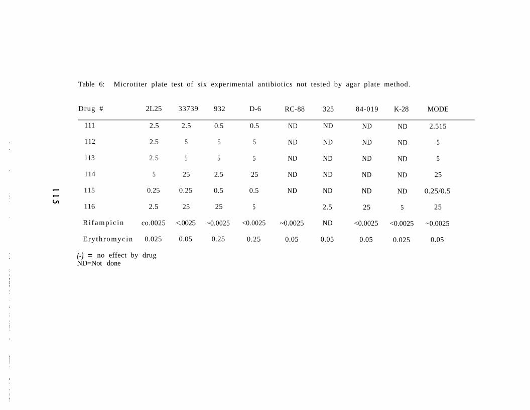
Table 6: Microtiter plate test of six experimental antibiotics not tested by agar plate method.
Drug # 2L25 33739 932 D-6 RC-88 325 84-019 K-28 MODE
111 2.5
112 2.5
113 2.5
114 5
c 115 0.25
tf116 2.5
R i f a m p i c i n co.0025
Ery th romyc in 0.025
2.5 0.5 0.5
5 5 5
5 5 5
25 2.5 25
0.25 0.5 0.5
25 25 5
<.0025 ~0.0025 <0.0025
0.05 0.25 0.25
ND
ND
ND
ND
ND
~0.0025
0.05
ND ND ND 2.515
ND ND ND 5
ND ND ND 5
ND ND ND 25
ND ND ND 0.25/0.5
2.5 25 5 25
ND <0.0025 <0.0025 ~0.0025
0.05 0.05 0.025 0.05
(-) = no effect by drugND=Not done
DISCUSSION
In a study by Austin (1985) over 70 commercially available
antimicrobial compounds were incorporated into KDM-2 agar and
tested on isolates of R. salmoninarum. Five of the antibiotics tested
(ampicillin, rifampicin, penicillin G, erthromycin phosphate, and
oxytetracycline) were incorporated as controls into our study of
over 100 new, untested drugs. A majority of these are unnamed,
designated by the company with a number only; however, some are
on the market for chemotherapeutic use. All but six of the new,
experimental drugs were incorporated into KDM-2 agar and their
minimum inhibitory concentration (MIC) evaluated. In addition,
twenty-three antibiotics, including 18 experimental and five control
drugs, were tested in the newly developed microtiter plate assay
and compared to the agar plate method for determination of MIC.
Because the microtiter plate results were equivalent to the agar
plate results, the last six antibiotics were tested by the microtiter
plate assay only.
Although the results were essentially the same, there were
differences in the two assays, based on the media. In the microtiter
method, in which the R, salmoninarum were suspended in KDM-2
broth, the early seven day reading was 25% more likely to produce
a false negative result than the readings at 15 and 30 days. On the
solid KDM-2 agar, the colonies of R. salmoninarum were distinct and
easily read at this time. Another difference between the two
methods was that the development of resistance by R.
116

salmoninarum to some antibiotics. was apparent on the agar media
at the end of 30 days, unlike the microtiter plates where there was
no change from the initial MIC. This was particularly obvious in the
agar plate assay with the control drug, rifampicin, whose MIC
changed 350 fold (0.l /0.05 to 35 mg/L) over the 30 day period.
Rifampicin inhibits bacterial growth by binding the RNA
polymerase enzyme and blocking nucleic acid synthesis, but many
bacteria, including R. salmoninarum, can develop a resistant RNA
polymerase within two cell divisions (Williams and Kruk, 1981). In
the microtiter plate assay, however, rifampicin appeared to either
kill or inhibit R. salmoninarum so effectively that resistant growth
was not seen by 30 days.
In addition, R. salmoninarum was markedly more sensitive to
some drugs (12, 14, 67, oxytetracycline, and rifampicin) in the
microtiter plate assay. Four of these drugs, excluding rifampicin,
were solubilised in a small amount (1:25) of DMSO, which in
solution may have permitted better penetration of the drug into the
bacterial cells. These same antibiotics were also very effective
inhibitors in the agar plate assay, however, not to the extent seen in
the microtiter plates. Alone, DMSO at any concentration used did
not inhibit growth of R. salmoninarum in either the microtiter or
agar plate assays. The effectiveness of DMSO as a solvent was
demonstrated by oxytetracycline. When this drug was diluted in
water, it inhibited R. salmoninarum at 10 mg/L in the agar plate
assay and failed to inhibit the bacterium in the microtiter plate
assay. The efficacy of oxytetracycline, when solubilized with DMSO,
117
increased to 5 mg/L in the agar plate assay and to 0.5 mg/L in
microtiter plate assay.
We were successful in finding twenty drugs which would
effectively inhibit R.salmoninarum, six of which were at least as
effective as erythromycin (0.1 mg/L). Four of these were
macrolides of known chemical structure (91, 92, 93, and 98; Lilly)
and one was a non-macrolide (14; Lilly) with no structure given.
Azithromycin was more effective than rifampicin, inhibiting R.
salmoninarum bactericidally at concentrations of 0.05 mg/L.
Another 14 prevented the growth of R. salmoninarum at least
as well or better than penicillin (1 mg/L). Those inhibiting growth
at 0.5 mg/L were 7, 8, and 12 (non-macrolides; Lilly) and 71, 72, 99
and 115 (macrolides; Lilly). The other seven inhibiting at 1 mg/L
were two non-macrolides (56 and 58; Lilly) and five macrolides
(78, 99, 94, 96, and 100; Lilly).
Although many of the compounds tested were macrolides or
macrolide derivatives which likely inhibit protein synthesis by
binding the 50S ribosomal subunit (Williams and Kruk, 1981), at
least two were fluoroquinolones and another was of the penicillin
or cephalosporin class. The fluoroquinolones are able to penetrate
intracellularly into phagocytic cells, unlike most other antibiotics,
and are active against other gram positive intracellular pathogens
(Neer, 1988). However, R. salmoninarum was relatively insensitive
(35 mg/L) to one of the fluoroquinolones, 110 (norfloxacin), and
differentially sensitive to the other, 116, with the MIC ranging from
118

0.25 mg/L to no effect. The penicillin-type antibiotic, imipenim
(109) had no effect on R. salmoninarum.
This study has demonstrated that a number of antibiotics
inhibit R. salmoninarum in vitro, some more effectively than those
currently in use. The true efficacy and/or toxicity of these
antimicrobial agents in vivo remains to be determined.
119
ACKNOWLEDGEMENTS
The authors thank the pharmaceutical companies which willingly
supplied antimicrobial compounds for use in this study which was
supported by Bonneville Power Administration under contract No.
AE-A179-83 BP 11987; Alan Ruger, contracting officer’s technical
representative.
120

REFERENCES
Austin, B. 1985. Evaluation of antimicrobial compounds for thecontrol of bacterial kidney disease in rainbow trout, Salmogairdneri Richardson. Journal of Fish Diseases. 8:209-220.
Bell, G.R., G.S. Traxler, and C. Dworschak. 1988. Development invitro and pathogenicity of an erythromycin-resistant strain ofRenibacterium salmoninarum, the causative agent of bacterialkidney disease in salmonids.4:19-25.
Diseases of Aquatic Organisms.
Fryer, J.L. and J.E. Sanders. 1981. Bacterial kidney disease ofsalmonid fish. Annual Review of Microbiology. 35:273-298.
Kaattari, S.L., D.D. Rockey, G. Weins, P. Turaga, and J.S. Rohovec.1988. Bacterial kidney disease and furunculosis vaccines.Pages 451- 458 in Proceedings of the Congress of AquacultureInternational. September 6, 1988. Vancouver, B.C., Canada.
Neer, T.M. 1988. Clinical pharmacological features offluoroquinolone antimicrobial drugs. Journal of AmericanVeterinary Medicine Association. 193:577-580.
Williams, R.A.D. and Z.L. Kruk. 1981. The Biochemistry andPharmacology of Antibacterial .C r o o m H e l m L t d ,London. Pages 61-63 and 74.
Wolf, K. and C.E. Dunbar. 1959. Tests of 34 therapeutic agents forcontrol of kidney disease in trout. Transactions of theAmerican Fisheries Society. 88:117-124.
121
SUMMARY OF EXPENDITURES
Salary 41,130.73
Fringe 10,640.16
Utilities 3,429.47
Supplies 14,923.14
Travel 2,743.91
Indirect Costs 25,303.90
Sub-Contract 23,950.15
Total 122,121.46
1 2 2