Embed Size (px)
Citation preview
STEM CELLS AND REGENERATION RESEARCH ARTICLE
JAK/STAT signalling mediates cell survival in response to tissuestressMarco La Fortezza, Madlin Schenk, Andrea Cosolo, Addie Kolybaba, Isabelle Grass and Anne-Kathrin Classen*
ABSTRACTTissue homeostasis relies on the ability of tissues to respond tostress. Tissue regeneration and tumour models in Drosophila haveshown that c-Jun amino-terminal kinase (JNK) acts as a prominentstress-response pathway promoting injury-induced apoptosisand compensatory proliferation. A central question remainingunanswered is how both responses are balanced by activation of asingle pathway. Signalling through the Janus kinase/Signaltransducers and activators of transcription (JAK/STAT) pathway,which is a potential JNK target, is implicated in promotingcompensatory proliferation. While we observe JAK/STAT activationin imaginal discs upon damage, our data demonstrate that JAK/STATand its downstream effector Zfh2 promote the survival of JNKsignalling cells. The JNK component fos and the pro-apoptotic genehid are regulated in a JAK/STAT-dependent manner. This molecularpathway restrains JNK-induced apoptosis and spatial propagation ofJNK signalling, thereby limiting the extent of tissue damage, as wellas facilitating systemic and proliferative responses to injury. We findthat the pro-survival function of JAK/STAT also drives tumour growthunder conditions of chronic stress. Our study defines the function ofJAK/STAT in tissue stress and illustrates how crosstalk betweenconserved signalling pathways establishes an intricate equilibriumbetween proliferation, apoptosis and survival to restore tissuehomeostasis.
KEY WORDS: JAK/STAT, JNK, Cell survival, Compensatoryproliferation, Cancer, Injury-induced apoptosis, Eiger, Cell ablation
INTRODUCTIONTissue homeostasis relies on the ability of tissues to respond to stresscaused by damaging environmental insults. Physical wounding,toxins, reactive oxygen species and UV irradiation all inducecellular damage and thus disrupt tissue integrity. Mounting anappropriate response to these insults is essential for tissue repair andto prevent chronic cellular stress, which can lead to disease (Fuldaet al., 2010). While much progress has been made to elucidatesignalling pathways that seal wounds, remove damaged cells,promote regenerative proliferation or mediate patterning ofregenerated tissue, little is known about how crosstalk betweenthese pathways coordinates repair processes to restore homeostasis.Studies on Drosophila progenitor organs called imaginal discs
have provided deep insights into cellular adaptations to tissue stress.Surgical excision (Bryant, 1975; Haynie and Bryant, 1976;
Katsuyama et al., 2015) or cell ablation induced by pro-apoptotictransgenes (Grusche et al., 2011; Herrera et al., 2013; Smith-Boltonet al., 2009) has revealed cellular responses that promote woundhealing (Sun and Irvine, 2014; Kashio et al., 2014; Razzell et al.,2011). Disc size is restored by accelerated proliferation of cellsproximal (Sustar et al., 2011; Bosch et al., 2008) and distal (Herreraet al., 2013) to the wound site. Lacking tissue-resident stem cells,fate plasticity displayed by parenchymal disc cells facilitatesrepatterning of replaced tissues (Herrera et al., 2013; Herrera andMorata, 2014; Repiso et al., 2013; Schuster and Smith-Bolton,2015). In contrast, regeneration of the Drosophila adult midgut isdriven by tissue-resident stem cells (Ohlstein and Spradling, 2006;Jiang et al., 2009; Osman et al., 2013; Staley and Irvine, 2010).Strikingly, while midgut and imaginal discs utilize stem cell-dependent and -independent repair processes, both tissues activatesimilar signalling pathways that underlie potentially highlyconserved stress responses.
The c-Jun amino-terminal kinase (JNK)/mitogen-activatedprotein kinase (MAPK) cascade is one of the earliest pathwaysactivated in damaged tissues and it is triggered by loss of epithelialpolarity (Brumby and Richardson, 2003; Uhlirova et al., 2005;Igaki, 2009; Wu et al., 2010; Zhu et al., 2010), apoptosis (Ryooet al., 2004; Shlevkov and Morata, 2012) or physical wounding(Bosch et al., 2005; Rämet et al., 2002; Lee et al., 2005). JNKregulates stress responses via activation of the transcription factorAP-1 (Eferl and Wagner, 2003), which comprises Jun and Foshomo- and heterodimers. JNK activation is required for cytoskeletalrearrangement during wound closure (Ríos-Barrera and Riesgo-Escovar, 2013; Bosch et al., 2005) and promotes elimination ofdamaged cells by injury-induced apoptosis (Bogoyevitch et al.,2010; Chen, 2012; Shlevkov and Morata, 2012; Luo et al., 2007;Moreno et al., 2002). Importantly, JNK drives compensatoryproliferation to replace damaged tissues, through cell-autonomous(Bosch et al., 2008; Sun and Irvine, 2011; Grusche et al., 2011) andnon-autonomous (Bergantinos et al., 2010; Mattila et al., 2005;Ryoo et al., 2004) mechanisms. A central question remainingunanswered is how these two different responses – proliferation andapoptosis – are brought into equilibrium downstream of JNK torestore tissue homeostasis.
Apoptotic JNK targets that facilitate clearing of damaged cellsinclude the Diap1 inhibitors reaper (rpr), head involution defective(hid) and p53 (Shlevkov and Morata, 2012; Luo et al., 2007;Moreno et al., 2002). Activation of rpr, hid and p53 drives furtheractivation of JNK via the initiator caspase Dronc (Shlevkov andMorata, 2012). JNK also activates the TNFα homologue Eiger(Pérez-Garijo et al., 2013), leading to activation of JNK in nearbycells (Pérez-Garijo et al., 2013; Bergantinos et al., 2010; Wu et al.,2010). This positive feedback could drive excessive cell eliminationand therefore, needs to be restrained to prevent unlimited spatialpropagation of pro-apoptotic JNK activity. It is not known howspatial constraints on JNK propagation are established.Received 1 November 2015; Accepted 23 June 2016
Ludwig-Maximilians-University Munich, Faculty of Biology, Grosshaderner Strasse2-4, Planegg-Martinsried 82152, Germany.
*Author for correspondence ([email protected])
A.-K.C., 0000-0001-5157-0749
2907
© 2016. Published by The Company of Biologists Ltd | Development (2016) 143, 2907-2919 doi:10.1242/dev.132340
DEVELO
PM
ENT

Importantly, apoptosis of damaged cells stimulates compensatoryproliferation by JNK-dependent activation of growth pathwaysincluding Wnt/β-catenin, TFGβ/SMAD, Janus kinase/Signaltransducer and activator of transcription (JAK/STAT), Hippo/Yorkie and EGF (Sun and Irvine, 2011, 2014; Wells et al., 2006;Kondo et al., 2006; Huh et al., 2004; Morata et al., 2011; Pastor-Pareja and Xu, 2013). While the requirement for these pathwaysdiffers depending on context (Smith-Bolton et al., 2009; Herreraet al., 2013; Fan and Bergmann, 2008), many have been implicatedin metazoan responses to stress (Sun and Irvine, 2014; Pahlavanet al., 2006; Chen, 2012). Importantly, prevention of apoptosis indamaged cells causes sustained proliferative signalling and eventualtumour formation in Drosophila (Kondo et al., 2006; Perez-Garijoet al., 2004, 2009; Ryoo et al., 2004; Huh et al., 2004; Martin et al.,2009), emphasizing how strongly apoptotic, survival andproliferative signals need to be balanced to re-establish normaltissue size.Current models propose that JAK/STAT signalling is a major
mediator of compensatory, cancerous and developmentalproliferation. Cytokine-like ligands in Drosophila are encoded byupd1 (unpaired 1), upd2 and upd3 and signal through conservedpathway components encoded by single genes, specifically thereceptor Domeless (Dome), the Janus kinase Hopscotch (Hop) andthe transcription factor Stat92E (Arbouzova and Zeidler, 2006). Asin mammals, JAK/STAT plays diverse roles in fly development byregulating haemocyte activation (Myllymäki and Rämet, 2014),appendage patterning (Ayala-Camargo et al., 2013, 2007;Johnstone et al., 2013) and stem cell maintenance (Gregory et al.,2008). A proliferative function for JAK/STAT was suggested byearly studies of tissue growth during disc development (Bach et al.,2003; Tsai and Sun, 2004; Mukherjee et al., 2005). Subsequently,JAK/STAT has been widely implicated in promoting proliferationof neoplastic cells (Classen et al., 2009;Wu et al., 2010; Davie et al.,2015; Bunker et al., 2015; Amoyel et al., 2014) and aberrant non-autonomous proliferation of wild-type cells in fly models oftumorigenesis (Vaccari and Bilder, 2005; Herz et al., 2006; Moberget al., 2005). Activation of Upd gene transcription upon tissuedamage has been linked to compensatory proliferation in imaginaldiscs and adult guts (Jiang et al., 2009; Pastor-Pareja et al., 2008;Katsuyama et al., 2015; Wu et al., 2010; Lin et al., 2009; Bunkeret al., 2015; Santabárbara-Ruiz et al., 2015). Other studies suggestthat JAK/STAT is involved in cell competition, where signallingeither promotes ‘winner’ cell state (Schroeder et al., 2013;Rodrigues et al., 2012) or compensatory proliferation in responseto ‘loser’ cell elimination (Kolahgar et al., 2015).Despite this wealth of work, little is known about target genes
mediating proliferative JAK/STAT function. In fly tumours, STAT-responsive enhancers have been mapped (Davie et al., 2015), butfew target genes regulating tissue size have been described (Tsai andSun, 2004; Betz et al., 2008; Hasan et al., 2015). However, severalstudies suggest that developmental functions of JAK/STAT aremediated by the transcriptional repressors Chinmo (Flaherty et al.,2010), Zfh1 (Ohayon et al., 2009; Leatherman and Dinardo, 2008)and Zfh2 (Perea et al., 2013; Guarner et al., 2014; Ayala-Camargoet al., 2013).We wanted to better understand the precise role of JAK/STAT in
cellular adaptations to stress and to gain further insight into howJAK/STAT might regulate compensatory proliferation after tissuedamage. Because JAK/STAT is required for regeneration in tissuesas diverse as the grasshopper leg to the mouse liver (Yamada et al.,1997; Bando et al., 2013; Cressman et al., 1996; Li et al., 2002;Wuestefeld et al., 2003; Zhao et al., 2014), dissecting the function of
this highly conserved pathway is essential for our understanding ofregenerative processes. To achieve this, we employed an imaginaldisc model of tissue stress, which utilizes ectopic expression of theTNFα homologue Eiger (Smith-Bolton et al., 2009) (Fig. S1),triggering JNK activation (Igaki and Miura, 2014; Andersen et al.,2015) and apoptosis (Fig.S1D-D‴). This model has been previouslyused to study tissue regeneration in response to local cell ablation(Herrera et al., 2013; Smith-Bolton et al., 2009). Expression ofEiger induces multiple hallmarks of local and systemic stressresponses, such as compensatory proliferation and dILP8-induceddevelopmental delay (Fig. S1E,G) (Smith-Bolton et al., 2009).Because Eiger induces tissue stress in wild-type discs, it representsan ideal model to distinguish JAK/STAT functions in physiologicalstress from secondary effects incurred by developmental patterningor tumorigenic models. Instead of promoting compensatoryproliferation, we found that JAK/STAT promotes survival ofJNK-signalling cells, thereby acting as a central regulator ofinjury-induced apoptosis to restrain excessive tissue damage andfacilitate initiation of compensatory responses.
RESULTSEiger-mediated JNK activation enables functional studies ofJNK-dependent stress responsesWe expressed Eiger under the control of rotund (rn)-GAL4, whichdrives expression in the wing disc pouch, fated to give rise to thefuture wing blade. We induced eiger expression on developmentalday 7 and temporally limitedUAS-eiger expression to 40 h by usinga temperature-sensitive GAL80 repressor (Fig. S1A-C). Inagreement with previous studies (Herrera et al., 2013; Smith-Bolton et al., 2009), we observed extensive cell ablation in discs(Fig. S1E-E′), which gave rise to a 50% reduction in adult wing size(Fig. S1H). G-trace lineage labelling (Evans et al., 2009) of rn-GAL4-expressing cells followed by FACS analysis revealed thatabout 85% of rn-GAL4 lineage cells were eliminated after 40 h ofEiger stimulation (Fig. 1A,A′, Fig. S2A-A″). This correlated withan induction of a 2 day developmental delay at the larval-pupaltransition (Fig. S1G).
In agreement with Eiger triggering JNK activation (Igaki andMiura, 2014), we found that Eiger-stimulated discs broadly activatethe JNK-responsive TRE reporter (Fig. 1C) (Chatterjee andBohmann, 2012). The TRE reporter was specifically activated atthe wound site, whereas reporter activity is completely absent incontrol discs (Fig. 1B,C). Consistent with non-autonomousactivation of JNK by Eiger (Pérez-Garijo et al., 2013) and tissuedamage (Bosch et al., 2005; Herrera et al., 2013; Wu et al., 2010),TRE activation extended beyond a G-trace-labelled domain, inwhich eiger expression was induced (Fig. 1C and Fig. 2F). FACSanalysis demonstrated that while 4.5% of cells in the disc belongedto the surviving rn-GAL4 lineage, 29% of cells in the disc activatedTRE-GFP (Fig. S2A-A′,B-B′).
We first wanted to quantify cell proliferation in Eiger-stimulateddiscs near the wound site, as well as in the whole disc between 0 h(R0) and 48 h (R48) into the recovery period after the end of cellablation (Fig. S1A,E-E″). We used an image segmentationalgorithm (Fiji) to specifically measure total disc size, the size ofthe surviving rn-GAL4 lineage labelled by G-trace, the number ofmitotic cells marked by phospho-Histone 3 (pH3) or the number ofcells in S-phase marked by BrdU incorporation (Fig. 1D-G,Fig. S1F). We found that at R0, G-trace-labelled cells in thepouch account for about 3% of the total disc volume (Fig. 1D). Thisis in close agreement with FACS measurements (4.5%, Fig. S2A′),which also detect an additional subset of G-trace-labelled cells in
2908
STEM CELLS AND REGENERATION Development (2016) 143, 2907-2919 doi:10.1242/dev.132340
DEVELO
PM
ENT

the notum. This analysis verified that volume quantifications of cellpopulations in Fiji accurately approximate cell counts. Our analysisrevealed a marked increase in cell proliferation between R0 andR24, both near the wound site (Fig. 1F), as well as in the entire disc(Fig. 1G, Fig. S2E). Mitotic rates do not increase further betweenR24 and R48 (Fig. 1F,G). When we quantified the volume of thetotal disc and that of the G-trace-labelled population, we found thatthey increased in size by 36% and 126%, respectively (Fig. 1D,E).While cell ablation can be induced more efficiently by expression
of pro-apoptotic transgenes, such as UAS-reaper (Herrera et al.,2013), we did not observe broad activation of JNK in this context(Fig. S2C,D). Because Eiger-stimulated discs displayed broadactivation and provided genetic access to JNK-signalling cells, weutilized expression of Eiger to investigate the role of JAK/STAT inJNK-mediated responses to tissue stress.
JAK/STAT is activated in response to Eiger-induced tissuedamageTo understand if JAK/STAT plays a role in imaginal discregeneration, we analysed JAK/STAT activity in response to eigerexpression. JAK/STAT activation can be visualized using the10xSTAT-dGFP reporter (Bach et al., 2007), which is almostundetectable in wild-type discs (Fig. 2A). Analysis of the 10xSTAT-dGFP reporter revealed that JAK/STAT is upregulated in responseto eiger expression induced at day 5 or day 7 of development
(Fig. 2B,C). Reporter activation can also be observed upon reaperexpression, suggesting that stress induced by ectopic cell death issufficient to cause JAK/STAT activation (Fig. S3E). Importantly, inEiger-stimulated discs, JAK/STAT activation was detected beyondthe pouch periphery and thus extended beyond JNK domainsmarked by TRE activity (Fig. 2F). JAK/STAT signalling declinedwithin the following 24 h suggesting that it is downregulated aftereiger expression ceased (compare Fig. S3A-C with Fig. S3B′-C′).We furthermore examined JAK/STAT activation when eiger wasinduced at later stages of development. Surprisingly, JAK/STATactivity was almost undetectable when eiger was induced at day 8(Fig. 2D, Fig. S3D-D′).
Unpaired ligands are upregulated upon Eiger-induced tissuedamagePrevious reports and our studies demonstrate that the JAK/STATligands upd1, upd2 and upd3 are transcriptionally upregulated inresponse to stress from physical injury (Katsuyama et al., 2015;Pastor-Pareja et al., 2008) or tumorous growth (Wu et al., 2010;Bunker et al., 2015), indicating that pathway upregulation may bedriven by JNK-dependent Upd gene transcription. We found thattranscription of Upd genes is highly elevated in Eiger-stimulateddiscs, whereas transcription of dome, hop and Stat92E is not(Fig. 2E). Recapitulating the decline in 10xSTAT-dGFP activity inresponse to eiger induction at day 8, Upd gene transcription was
Fig. 1. Eiger expression enables functional studies of JNK stress responses. (A) G-trace labelling (cyan) visualizes the rotund (rn)GAL4 cell lineage inwing discs. Actin labelling is red. (A′) Progeny of rn-GAL4G-trace-labelled cells (cyan) after 40 h of Eiger-mediated cell ablation (CA) at recovery points R0, R24,R48 h. (B) Wing pouch expressing the JNK reporter TRE>GFP (red in overlay), stained for DAPI and Nubbin (cyan in overlay) visualizing a lineage similarto rn-GAL4-derived cells. (C)Wing pouch after cell ablation (CA) expressing TRE>RFP (red in overlay) containing rn-GAL4G-trace-labelled cells (cyan in overlay)stained for DAPI. (D) Volume occupied by rn-GAL4 G-trace-labelled cells and (E) total wing imaginal disc (WID) volumes at recovery time points R0, R24 andR48. (F) Normalized pH3-positive mitotic events within rn-GAL4 G-trace-labelled volume and (G) within total disc volumes at R0, R24, R48. Graphs displaymean±s.e.m. for R0, n=16; R24, n=12; R48, n=13 discs. U-tests were performed to test for statistical significance (*P<0.05, **P<0.01, ***P<0.001). Scale bars:100 µm.
2909
STEM CELLS AND REGENERATION Development (2016) 143, 2907-2919 doi:10.1242/dev.132340
DEVELO
PM
ENT

strongest at day 5 and day 7 but decreased by more than half wheneiger was induced at day 8 (Fig. 2E).Loss of JAK/STAT activation at day 8 was not due to a decline in
Eiger-mediated JNK activation, because TRE activity was as strongon day 8 as on day 7 (Fig. S3F,G). This suggests that even thoughJAK/STAT activation coincides with JNK activity (Katsuyamaet al., 2015; Pastor-Pareja et al., 2008), JNK alone may not besufficient to stimulate this pathway. This is supported by broaderactivation of JAK/STAT compared with JNK (Fig. 2F). Strikingly,the failure to activate JAK/STAT at late developmental stagescorrelated with a pronounced decline in the ability of larvae toinduce a developmental delay at the larval-pupal transition(Fig. S3H) and with a strong reduction in recovered adult wingsize (Fig. S3I). These correlations suggest that JAK/STAT may be acrucial mediator of JNK-induced compensatory responses inimaginal discs.
JAK/STAT activity is not required for compensatoryproliferationJAK/STAT has been reported to play a role in promoting cellproliferation in wild-type (Bach et al., 2003; Tsai and Sun, 2004;Mukherjee et al., 2005), tumorous (Classen et al., 2009; Wu et al.,2010; Davie et al., 2015; Bunker et al., 2015; Amoyel et al., 2014)and surgically injured discs (Katsuyama et al., 2015). To testwhether JAK/STAT is generally required to drive compensatoryproliferation, we quantified mitotic events after Eiger stimulation indiscs with impaired JAK/STAT signalling. We genetically reducedJAK/STAT activity by two approaches: (1) we reduced gene dosageof JAK/STAT components in the entire animal by heterozygosity
for domeG0441, hop34 and Stat92E85C9 alleles or (2) we interferedwith JAK/STAT signalling exclusively in eiger-expressing cellsthrough expression of RNAi constructs targeting JAK/STATcomponents (Fig. S5A), a dominant-negative dome (domeΔcyt) orthe inhibitor Socs36E under the control of rn-GAL4-tubGAL80ts.
Strikingly, even though 50% of the tissue in Eiger-stimulateddiscs activated JAK/STAT (Fig. 2B,C), discs heterozygous fordomeG0441, hop34 and Stat92E85C9 alleles did not show anyevidence of a reduction in mitotic events or total disc size(Fig. 3A-F). Instead, we observed a mild increase in Eiger-stimulated discs heterozygous for hop34. These results are theopposite to what we would expect if JAK/STAT regulatedcompensatory proliferation. We thus wanted to confirm thesefindings by interfering with JAK/STAT specifically in Eiger-stimulated cells. rn-GAL4-driven co-expression of dome-RNAi,domeΔcyt or Socs36E did not reduce the number of mitotic eventswithin surviving eiger-expressing cells (Fig. 3G-K). Instead,mitotic rates slightly increased, whereas they remained unchangedin the rest of the disc (Fig. 3K, Fig. S4A,C). Importantly, effects oneiger-expressing cells co-expressing transgenic constructs arestrongest at R0 but decline as GAL4 activity decreased (Fig. 3K).Together, our results strongly argue against the previously assignedrole of JAK/STAT in directly promoting compensatory proliferationin response to stress.
JAK/STAT is required for survival of JNK-signalling cellsWhile the total disc size of Eiger-stimulated discs was similar to thatof discs with genetically impaired JAK/STAT activity (Fig. 3F,Fig. S4D), we were surprised to notice that the number of
Fig. 2. JAK/STAT is activated in response to tissue damage. (A) Wing disc expressing the JAK/STAT-reporter 10XSTAT>dGFP (cyan in overlay) stained forDAPI and Actin (red in overlay). (B-D) Wing pouch after cell ablation (CA) induced at day 5 (B), day 7 (C) or day 8 (D). Discs were stained for DAPI, Actin (red inoverlay) and express 10XSTAT>dGFP (cyan in overlay). (E) qRT-PCR analysis of upd1, upd2, upd3, dome, hop and Stat92E transcripts after cell ablation (CA)induced at day 5, day 7 or day 8. Fold induction relative to unablated controls is reported. Graphs display mean±s.e.m. for n≥3 biological replicates.(F) Fluorescence intensity of rn-GAL4 G-trace (n=5 discs), TRE>RFP (n=6 discs) and 10XSTAT>dGFP (n=12 discs) reporter signals at R0. Intensity plots arescaled to maximummeasured values; interpolated averages are reported as a function of relative positions between pouch centre and disc edge. Graphs displayinterpolated mean±s.e.m. Scale bars: 100 µm.
2910
STEM CELLS AND REGENERATION Development (2016) 143, 2907-2919 doi:10.1242/dev.132340
DEVELO
PM
ENT

G-trace-labelled cells that survived eiger expression wasdramatically reduced when JAK/STAT signalling was inhibited.Specifically, we found that expression of dome-RNAi, domeΔcyt orSOCS36E caused a 3- to 10-fold reduction in the volume of G-trace-labelled populations (Fig. 3L, Fig. S4B).These observations suggested that more eiger-expressing cells
die when JAK/STAT signalling is impaired. Indeed, we observed a2-fold increase in the volume positive for activated Caspase-3 ifJAK/STAT activity was exclusively reduced in eiger-expressingcells (Fig. 4A-E) and up to a 10-fold increase in the volume ofEiger-stimulated discs heterozygous for domeG0441, hop34 orStat92E85C9 (Fig. 4F-I). The more pronounced effect observed forheterozygous tissues likely arises as a result of the tissue-widereduction of JAK/STAT activity in this background. Reduction wasalso achieved in domains that display JAK/STAT and JNKactivation but are located outside of the rnGAL4 lineage (Fig. 1C,Fig. 2B,C,F). Importantly, genetic reduction of JAK/STAT indeveloping wild-type discs under the same conditions does not
cause any elevation of apoptosis (Fig. S5B-E). Combined, thesedata highlight a specific role for JAK/STAT as an importantmediator of cell survival, specifically in JNK-signalling cells.
JAK/STAT activity suppresses activation of JNK signallingTo understand how JAK/STAT promotes cell survival, we tested ifgenetically reducing JAK/STAT activity causes further elevation ofJNK, which could divert JNK-dependent compensatory responsestowards apoptosis. To this end, we monitored JNK activity usingTRE reporters in eiger-expressing discs heterozygous fordomeG0441, hop34 and Stat92E85C9 alleles. Our results suggestedthat after cell ablation, 14% of the wing pouch area activated theTRE reporter (Fig. 4J,N). In Eiger-stimulated discs heterozygous fordomeG0441, hop34 and Stat92E85C9, we observed a 2- to 3-foldincrease in the area positive for TRE activation (Fig. 4K-N). Theseresults indicate that reduction of JAK/STAT signalling promotesnon-autonomous expansion of JNK signalling beyond eiger-expressing cells and that this may underlie the observed increase
Fig. 3. JAK/STAT is not required for compensatory proliferation. (A) Wing disc after cell ablation (CA) stained for pH3 (A, red in A′, Actin in grey). (B-D) Wingdisc heterozygous for domeG0441 (B,B′), hop34 (C,C′) orStat92E85C9 (D,D′) after cell ablation (CA) stained for pH3 (B-D, red in B′-D′; Actin in grey). (E) pH3 eventsper disc or (F) total wild-type disc volume (n=12) and discs heterozygous for domeG0441 (n=5), hop34 (n=9), or Stat92E85C9 (n=10) after cell ablation (CA).(G-J)Wing pouch containing surviving rn-GAL4G-trace-labelled cells (G-J, cyan in G″-J″), stained for pH3 (G′-J′, red in G″-J″). Awild-type disc (G) and discs withrn-GAL4-mediated co-expression of domeΔcyt (H), dome-RNAi (I) or Socs36E (J) in eiger-expressing cells are shown. (K) Normalized pH3 events within rn-GAL4G-trace-labelled volume and (L) relative rn-GAL4 G-trace-labelled volume per disc at R0 and R24 in ablated discs (CA) (R0, n=16; R24, n=12 discs) or withrn-GAL4-mediated co-expression of domeΔcyt (R0, n=8; R24, n=12 discs), dome-RNAi (R0, n=8; R24, n=9 discs), Socs36E (R0, n=6; R24, n=6 discs) in eiger-expressing cells. Graphs display mean±s.e.m. U-tests were performed to test for statistical significance (*P<0.05, **P<0.01, ***P<0.001). Scale bars: 100 µm.
2911
STEM CELLS AND REGENERATION Development (2016) 143, 2907-2919 doi:10.1242/dev.132340
DEVELO
PM
ENT

in the apoptotic index in JAK/STAT-impaired Eiger-stimulateddiscs (Fig. 4A-I).Our observations prompted two important predictions. First,
broad activation of JNK in JAK/STAT-impaired discs suggestedthat JAK/STAT acts as a suppressor of JNK signalling. Repressionof JNK by JAK/STAT could either be mediated by directtranscriptional effects on JNK core components, or indirectly, bysuppression of apoptosis and prevention of Dronc-driven positivefeedback activation of JNK. This mechanism would restrainnon-autonomous activation of JNK, excessive apoptosis andtissue damage. A second prediction implies that interferingwith JAK/STAT increases the extent of tissue damage due toelevation of cell death. Thereby, the ability of discs to mountan appropriate regenerative response is reduced as cells requiredto drive regenerative responses are eliminated by excessive celldeath.
A survival-promoting function of JAK/STAT is mediated byZfh2We did not find any evidence that negative JNK regulators aretranscriptionally activated by Stat92E. We therefore wanted tounderstand if JAK/STAT regulates cell survival by impinging onpro-apoptotic JNK target genes rpr and hid, which act incombination with grim as inhibitors of IAP (inhibitor of
apoptosis) proteins. To this end, we first performed abioinformatic Clover analysis of the promoter regions of rpr, hidand grim using highly stringent parameter selections (Frith et al.,2004). As previously suggested (Shlevkov and Morata, 2012; Luoet al., 2007; Moreno et al., 2002), we identified multiple AP-1binding motifs associated with these loci (Fig. S7A, data notshown). In agreement with studies on JNK-induced apoptosis(Shlevkov and Morata, 2012; Luo et al., 2007; Moreno et al., 2002),we specifically observed induction of hid expression in Eiger-stimulated discs (Fig. 5A). The mild increase in hid levels is likely tobe an underestimate because eiger-expressing cells make up only4.5% of the entire disc used for qRT-PCR analysis (Fig. S2A,A′).Eiger-expressing discs heterozygous for Df(3L)H99, a deficiencyremoving rpr, hid and grim loci, displayed a pronounced ‘undeadcell’ phenotype (Fig. S6A,B) (Perez-Garijo et al., 2009; Martinet al., 2009; Kondo et al., 2006), suggesting that upregulation of hidcontributes to Eiger-mediated cell death. While one previous studyreports that Eiger-mediated induction of apoptosis is independent ofhid activation (Igaki et al., 2002), we suggest that the small adult eyephenotype observed upon eiger co-expression with the strongapoptosis inhibitor p35 is a consequence of epithelial tissuearchitecture disruption rather than a failure to prevent Eiger-induced cell death (Fig. S6L). Together, previous reports and ourresults support the notion that induction of cell death in eiger-
Fig. 4. JAK/STAT is required for survival of Eiger-stimulated cells. (A-D) Wing disc after cell ablation (CA) (A) and with rn-GAL4-mediated co-expression ofdomeΔcyt (B), dome-RNAi (C) or Socs36E (D) in eiger-expressing cells stained for cleaved Caspase-3 to visualize apoptotic cells (cyan) and Actin (red).(E) Cleaved Caspase-3 volume in ablated discs (CA; R0, n=15; R24, n=4 discs) or with rn-GAL4-mediated co-expression of domeΔcyt (R0, n=12; R24, n=3 discs),dome-RNAi (R0, n=10; R24, n=4 discs) or Socs36E (R0, n=6; R24, n=6 discs) in eiger-expressing cells at R0 and R24. (F-H) Ablated discs heterozygous fordomeG0441 (F), hop34 (G) or Stat92E85C9 (H) stained for cleaved Caspase-3 (cyan) and Actin (red) at R0. (I) Quantification of cleaved Caspase-3-positive volumeper disc after cell ablation (CA) (n=12 discs) or in discs heterozygous for domeG0441 (n=4 discs), hop34 (n=9 discs), or Stat92E85C9 (n=8 discs) at R0.(J-M) Disc expressing the JNK reporter TRE>RFP (cyan) after cell ablation (CA) (J) or if heterozygous for domeG0441 (K), hop34 (L) or Stat92E85C9 (M) at R0.(N) Quantification of TRE>RFP-positive area per disc after cell ablation (CA) (n=14 discs) and in discs heterozygous for domeG0441 (n=8 discs), hop34 (n=4 discs),or Stat92E85C9 (n=8 discs). Graphs display mean±s.e.m. U-tests were performed to test for statistical significance (*P<0.05, **P<0.01, ***P<0.001). Scale bars:100 µm.
2912
STEM CELLS AND REGENERATION Development (2016) 143, 2907-2919 doi:10.1242/dev.132340
DEVELO
PM
ENT

expressing discs is specifically mediated by AP-1/JNK-dependentactivation of hid.Strikingly, our Clover analysis also revealed multiple, highly
clustered mammalian ZEB1-binding motifs the hid promoter(Fig. S7A) as well as in the promoter of the kay gene coding forthe AP-1 component dFos (Fig. S7B). ZEB1 binding motifslocalized to highly conserved DNA sequences as shown bymVISTA analysis (Frazer et al., 2004; Bray et al., 2003) and toopen chromatin regions, likely representing regulatory elements, asdemonstrated by overlays with previously published ATAC-seq datasets (Fig. S7) (Davie et al., 2015). ZEB proteins are homologous toDrosophila Zfh1 and Zfh2 (Zinc-finger homeobox) proteins, whichact as transcriptional repressors (Postigo et al., 1999; Postigo andDean, 1999). Both Zfh1 and Zfh2 were previously identified to bedownstream effectors of JAK/STAT (Leatherman and Dinardo,
2008; Ayala-Camargo et al., 2013). This is reflected by Zfh2expression mirroring JAK/STAT activation patterns in developingwing discs (Fig. 5E) (Ayala-Camargo et al., 2013) and the ability ofJAK/STAT to induce Zfh2 expression de novo (Fig. S6C,D). Wehypothesized that Drosophila ZEB proteins could directly mediaterepression of kay in Eiger-stimulated discs (Fig. 5A), therebyrestraining JNK activation to promote cell survival. At the sametime, ZEB proteins could compete with AP-1 for transcriptionalrepression of hid, thereby limiting AP-1/Hid-induced apoptosis topromote cell survival. Indeed, in eiger-expressing discsheterozygous for Stat92E85C9, the upstream regulator of Zfhproteins, we observed strong upregulation of hid and kayexpression compared with discs expressing eiger alone (Fig. 5B).
To first understand if Zfh1 or Zfh2 expression was altered intissue damage, we performed immunofluorescence and qRT-PCR
Fig. 5. A survival-promoting function of JAK/STAT is mediated by Zfh2. (A) qRT-PCR analysis of rpr, hid, grim and fos (kay) transcripts at R0. Fold inductionrelative to unablated discs is reported. Each graph shows mean±s.e.m. for n≥3 biological replicates. (B) qRT-PCT analysis of hid and kay at R0 in Eiger-expressing discs heterozygous mutant for Stat92E85C9. Fold induction relative to ablated discs. Each graph shows mean±s.e.m. for n=2 biological replicates.(C-F) Unablated discs (C,E) and ablated discs (D,F) stained for Zfh1 (C,D) or Zfh2 (E,F) at R0. (G) qRT-PCR analysis of zfh1 and zfh2 transcripts at R0. Foldinduction relative to unablated discs is reported. Each graph shows mean±s.e.m. for n=3 biological replicates. (H,I) Disc after cell ablation (H) or with rn-GAL4-mediated co-expression of zfh2-RNAi (I) in eiger-expressing cells stained for Dcp-1 to visualize apoptotic cells (cyan) and Actin (red) at R0. (J) Dcp1-positivevolume per disc after cell ablation (CA) (n=4 discs) or in discs with rn-GAL4-mediated co-expression of zfh2-RNAi (n=3 discs) in eiger-expressing cells at R0.(K,L) Disc after cell ablation (K) or discs with rn-GAL4-mediated co-expression ofUAS-zfh2 (L) in eiger-expressing cells stained for Dcp-1 (cyan) and Actin (red) atR0. (M) rn-GAL4 G-trace-labelled volume in ablated discs (n=16 discs) and RFP-labelled volume in discs with rn-GAL4-mediated co-expression UAS-Zfh2[EAB]
(n=7 discs) in eiger-expressing cells at R0. Graphs display mean±s.e.m. U-tests were performed to test for statistical significance (*P<0.05, **P<0.01,***P<0.001). Scale bars: 100 µm.
2913
STEM CELLS AND REGENERATION Development (2016) 143, 2907-2919 doi:10.1242/dev.132340
DEVELO
PM
ENT

analysis on Eiger-stimulated discs.We found that levels of Zfh2, butnot Zfh1, are elevated in response to eiger expression (Fig. 5C-F).Zfh2 upregulation occurred specifically in regions with high levelsof JAK/STAT activation (compare Fig. 2B,C with Fig. 5F). Inaddition, we found that transcripts of zfh2 but not of zfh1 wereelevated (Fig. 5G). The observed mild increase in zfh2 transcriptionrepresents a strong underestimate, as cells expressing zfh-2 de novomake up only a small portion of entire imaginal discs used for qRT-PCR analysis. In agreement with Zfh2 being a JAK/STAT effector,heterozygosity for STAT92E85C9 reduces Zfh2 expression in Eiger-stimulated discs (Fig. S6E-G).To test if Zfh2 in Eiger-stimulated discs is required to promote
JAK/STAT-dependent survival by repressing hid and kaytranscription, we performed genetic experiments to reduce orincrease Zfh2 function. We found that expression of an RNAiconstruct targeting zfh2 (Fig. S6H) increased apoptosis in eiger-expressing discs almost 4-fold (Fig. 5H-J). In contrast,overexpression of UAS-zfh2 strongly promoted survival of eiger-expressing cells and resulted in a 10-fold increase in the size ofsurviving rn-GAL4-derived cell populations (Fig. 5K-M). Whilewe found that Zfh1 levels were not upregulated in responseto stimulation with Eiger, overexpression of zfh1 also promotedsurvival of eiger-expressing cells, even phenocopyingoverexpression of the strong apoptosis inhibitor p35 (Fig. S6I-L).The survival-promoting function of either Zfh1 or Zfh2 suggeststhat both proteins can induce potent survival signals in stressedtissues, similar to developmental contexts (Ohayon et al., 2009;Guarner et al., 2014).
JAK/STAT activity prevents excessive tissue damage inresponse to tissue stressWe predicted that interfering with JAK/STAT signalling andtherefore with Zfh2 function, increases the extent of tissuedamage incurred by Eiger due to elevation of cell death.Consistent with these predictions, we found that Eiger-stimulateddiscs with genetically reduced JAK/STAT activity developed intosignificantly smaller adult wings (Fig. 6A,B). Expression ofdomeΔcyt, Socs36E or RNAi constructs targeting multiple pathwaycomponents, including zfh2, as well as heterozygosity fordomeG0441, hop34 and Stat92E85C9, caused a significant drop inadult wing size index by 30-90% (Fig. 6B). Importantly, geneticdownregulation of JAK/STAT in wild-type discs at day 7 does notcause a comparable reduction in wing size (Fig. S8A), emphasizingthat the survival-promoting function of JAK/STAT is specificallyrequired during tissue stress responses.Notably, rn-GAL4-driven overexpression of upd1, upd2 or zfh2
in eiger-expressing cells did not increase adult wing size (Fig. 6B).Extra Upd may not translate into JAK/STAT hyperactivation,because of pathway saturation in eiger-expressing cells. While zfh2overexpression promoted cell survival, it did not rescue otherdefects such as loss of epithelial polarity (Fig. 5L), which interfereswith wing morphogenesis.To test whether JAK/STATwas also required for stress responses
induced by physical wounding, we analysed adult wings thatdeveloped from discs of surgically pinched larvae (Pastor-Parejaet al., 2008; Kashio et al., 2014). Reducing JAK/STAT function inthe posterior compartment by expressing an RNAi constructtargeting hop caused a pronounced reduction in adult wing sizesdeveloping from discs, in which pinching had been targeted to theposterior compartment as visualized by co-expression of GFP(Fig. S8B). In contrast, no reduction in size of the posteriorcompartment was observed for adult wings derived from
undamaged control discs raised under the same conditions (notshown).
These data suggest that cellular responses to genetically orsurgically induced damage critically rely on JAK/STAT activationto facilitate restoration of normal tissue homeostasis. Combined, ourresults strongly imply that a reduction in final tissue size upon JAK/STAT inhibition reflects an excessive loss of tissue due to cell deathrather than a failure of the tissue to undergo compensatoryproliferation.
JAK/STAT activity promotes efficient induction ofcompensatory responsesWe wanted to investigate further if the observed reduction in adultwing sizes upon JAK/STAT inhibition is exclusively caused by aloss of tissue to cell death or if other regenerative processes may bedisturbed. A process that contributes to successful tissue restorationis the induction of a dILP8-dependent developmental delay at thelarval-pupal transition, which extends the time available for repairbefore metamorphosis (Colombani et al., 2012; Garelli et al., 2012).We found that interfering with JAK/STAT in eiger-expressing cellsalso caused a profound reduction in developmental delays inducedby Eiger stimulation (Fig. 6C).
To understand if altered Ilp8 expression caused this observation,we quantified expression of a GFP-reporter driven from theendogenous Ilp8 locus (Garelli et al., 2012). We found that thereporter was strongly expressed in the pouch of Eiger-stimulateddiscs (Fig. S8C). In contrast, interfering with JAK/STAT byexpression of dome-RNAi or domeΔcyt in eiger-expressingcells significantly reduced the area of GFP expression (Fig. 6D,Fig. S8D,E). This suggests that loss of Ilp8 expression, caused byimpaired JAK/STAT in JNK-signalling cells, underlies the observedfailure to efficiently induce a developmental delay.
To test whether JAK/STAT signalling is sufficient to inducedevelopmental delays, we expressed the JAK/STAT-ligands Upd1or Upd2 in wild-type discs using MS1096-GAL4. However, nodifference in pupariation timing between Upd-expressing and stage-matched wild-type larvae was observed (Fig. S8F). While a recentstudy links Ilp8-expression to JAK/STAT signalling (Katsuyamaet al., 2015), our data implies that Ilp8 is not a direct target gene ofSTAT92E. Instead, we suggest that cells that normally express Ilp8in response to JNK activation are more likely to die when JAK/STAT signalling is reduced, thereby preventing efficient expressionof Ilp8 and induction of a developmental delay. The failure toinduce this important systemic response reduces the time availablefor repair and probably contributes to the decrease in recovered adultwing sizes that we observed upon genetic reduction of JAK/STATsignalling (Fig. 6B).
JAK/STAT regulates survival in a RasV12; scrib2 tumourmodelTo understand if JAK/STAT generally acts as a survival-promotingpathway in the context of tissue stress, we revisited the role of JAK/STAT in established fly tumour models. Previous studies suggestthat activation of JAK/STAT drives tumorous overgrowth in discsmutant for tumour suppressor genes, such as scribbled (scrib) orPsc-Su(z)2 (Wu et al., 2010; Classen et al., 2009). scrib cells,similar to eiger-expressing cells, exhibit strong JNK activation,correlating with elevated transcription of Upd cytokines (Wu et al.,2010; Bunker et al., 2015; Leong et al., 2009; Brumby andRichardson, 2003). While scrib cells have a growth disadvantage ifsurrounded by wild-type cells, they efficiently cooperate withoncogenic RasV12 to create invasive tumours in clonal assays
2914
STEM CELLS AND REGENERATION Development (2016) 143, 2907-2919 doi:10.1242/dev.132340
DEVELO
PM
ENT

(Brumby and Richardson, 2003; Wu et al., 2010). Larvae carryingMARCM-induced RasV12; scrib2 clones fail to pupariate (Fig. 7M),suggesting that Ilp8 activation correlates with tumour load (Garelliet al., 2012). We found that RasV12; scrib2 clones covered about47% of eye antennal discs, compared with 19% for wild-type clones(Fig. 7A,B,D). When we probed RasV12; scrib2 clones for activatedDcp-1, we did not observe any difference in apoptotic patternscompared with wild-type tissue (Fig. 7F,G).When we completely removed JAK/STAT function in RasV12;
scrib2 clones by homozygosity for a Stat92E85C9 allele, RasV12;scrib2 clone size was reduced from 47% to 28% of the eyedisc area (Fig. 7B-D). Comparison of cell division rates withinRasV12; scrib2 and RasV12; scrib2, Stat92E85C9 clones did notreveal any significant changes upon loss of JAK/STAT function(Fig. 7B′,C′,E). However, in contrast to RasV12; scrib2 clones,RasV12; scrib2, Stat92E85C9 clones displayed a 4.3-fold increase inareas of Dcp-1 activation (Fig. 7G,H,I), suggesting that increased
cell death of Stat92E mutant cells underlies the smaller sizes ofRasV12; scrib2, Stat92E85C9 clones (Fig. 7A-D). The reduction inRasV12; scrib2, Stat92E85C9 clone size reduced total tumour loadand allowed a significant proportion of host larvae to progress topupal stages (Fig. 7M).
Strikingly, RasV12; scrib2 clones displayed ectopic activationof Zfh2 (Fig. 7J,K) but not of Zfh1 (not shown), indicatingactivation of a JNK-JAK/STAT-Zfh2 stress module by neoplastictransformation. Importantly, ectopic expression of Zfh2 wascompletely abolished within RasV12; scrib2, Stat92E85C9 clones(Fig. 7L). These results support the notion that Zfh2 expression isregulated by JAK/STAT in multiple contexts of cellular stress andthat stress-dependent Zfh2 activation in imaginal discs directlycorrelates with cell survival.
Altogether, our results support a model in which cellular stresscaused by genetic cell ablation, physical wounding or tumorousgrowth drives activation of JAK/STAT to promote survival of
Fig. 6. JAK/STAT prevents excessive damage upon tissue stress. (A) Five adult wing size phenotypes were classified to score tissue damage after eigerexpression at D7. (B) Average wing sizes developing from Eiger-stimulated disc (CA) or combined with rn-GAL4-mediated co-expression of UAS transgenes orin genetic backgrounds heterozygous for domeG0441, hop34 or Stat92E85C9. Graphs display mean±s.d. of average scores derived from ≥3 experiments. t-testswere performed to test for statistical significance (*P<0.05). n=number of wings scored. (C) Cumulative fraction of larvae undergoing larval-pupal transitions,which carry Eiger-stimulated discs (CA) (n=210) or were combined with rn-GAL4-mediated co-expression of upd1 (n=213), domeΔcyt (n=201), dome-RNAi(n=196), or hop-RNAi (n=230). Graphs display mean±s.e.m. of average scores from ≥3 experiments. (D) Relative disc area expressing Ilp8>GFP after cellablation (CA) (R0, n=12; R24, n=4 discs) and in discs combined with rn-GAL4-mediated co-expression of domeΔcyt (R0, n=5; R24, n=3 discs) or dome-RNAi(R0, n=5; R24, n=7 discs) in eiger-expressing cells. Graphs display mean±s.e.m. U-tests were performed to test for statistical significance (*P<0.05, **P<0.01,***P<0.001).
2915
STEM CELLS AND REGENERATION Development (2016) 143, 2907-2919 doi:10.1242/dev.132340
DEVELO
PM
ENT
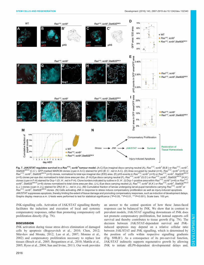
JNK-signalling cells. Activation of JAK/STAT signalling therebyfacilitates the induction and execution of local and systemiccompensatory responses, rather than promoting compensatory cellproliferation directly (Fig. 7N).
DISCUSSIONJNK activation during tissue stress drives elimination of damagedcells by apoptosis (Bogoyevitch et al., 2010; Chen, 2012;Shlevkov and Morata, 2012; Luo et al., 2007; Moreno et al.,2002) and compensatory proliferation necessary to replace losttissues (Bosch et al., 2005; Bergantinos et al., 2010; Mattila et al.,2005; Ryoo et al., 2004; Sun and Irvine, 2011). Our work provides
an answer to the central question of how these Janus-facedresponses can be balanced by JNK. We show that in contrast toprevalent models, JAK/STAT signalling downstream of JNK doesnot promote compensatory proliferation, but instead supports cellsurvival and thereby contributes to tissue growth (Fig. 7N). Thedecision between JAK/STAT-dependent survival and JNK-induced apoptosis may depend on a relative cellular ratiobetween JAK/STAT and JNK signalling, which is determined bythe position of cells within respective signalling gradients(Fig. S9B,B′). As a consequence of its pro-survival function,JAK/STAT indirectly supports regenerative growth by allowingJNK to initiate dILP8-dependent developmental delays and,
Fig. 7. JAK/STAT regulates survival in a RasV12; scrib2 tumour model. (A-C) Eye imaginal discs carrying neutral (A), RasV12; scrib2 (B,B′) or RasV12; scrib2,Stat92E85C9 (C,C′). GFP-marked MARCM clones (cyan in A-C) stained for pH3 (B′,C′, red in A-C). (D) Area occupied by neutral (n=4), RasV12; scrib2 (n=5) orRasV12; scrib2, Stat92E85C9 (n=5) clones, normalized to total eye imaginal disc (EID) area. (E) pH3-events in RasV12; scrib2 (n=5) or RasV12; scrib2, Stat92E85C9
(n=5) clones per eye disc normalized to total clone area per disc. (F-H) Eye discs carrying neutral (F), RasV12; scrib2 (G,G′) or RasV12; scrib2, Stat92E85C9 (H,H′)clones (cyan in F-H) stained for Dcp-1 (G′,H′, red in F-H). Clone borders indicated by outline in G′,H′. (I) Dcp-1-positive areawithinRasV12; scrib2 (n=6) orRasV12;scrib2, Stat92E85C9 (n=8) clones normalized to total clone area per disc. (J-L) Eye discs carrying neutral (J), RasV12; scrib2 (K,K′) or RasV12; scrib2, Stat92E85C9
(L,L′) clones (cyan in J-L) stained for Zfh2 (K′,L′, red in J-L). (M) Cumulative fraction of larvae undergoing larval-pupal transitions carrying RasV12; scrib2 orRasV12; scrib2, Stat92E85C9 clones. (N) Cells activating JNK in response to stress induce compensatory proliferation as well as injury-induced apoptosis.JAK/STAT suppresses apoptosis, thereby limiting the extent of tissue damage and promoting compensatory responses, such as induction of development delays.Graphs display mean±s.e.m. U-tests were performed to test for statistical significance (*P<0.05, **P<0.01, ***P<0.001). Scale bars: 100 µm.
2916
STEM CELLS AND REGENERATION Development (2016) 143, 2907-2919 doi:10.1242/dev.132340
DEVELO
PM
ENT

possibly, by facilitating Hippo/Yorkie-driven proliferation (Sunand Irvine, 2014). In fact, JNK-dependent Yorkie activation (Sunand Irvine, 2011) may occur independent of Jun/Fos-mediatedtranscription (Sun and Irvine, 2013), therefore JAK/STAT andZfh2-dependent repression of kay/fos could suppress inductionof JNK-dependent apoptosis without suppressing activation ofYorkie and thus of Hippo/Yorkie-driven compensatoryproliferation. Hippo/Yorkie signalling, in turn, may sustainactivation of unpaired and JAK/STAT signalling (Staley andIrvine, 2010; Bunker et al., 2015; Sarikaya and Extavour, 2015)even if JNK signalling is low. This signalling crosstalk wouldfacilitate the stabilisation of a wound-proximal tissue domainsupporting compensatory survival and proliferation.Our new interpretation of JAK/STAT in promoting cell survival
in response to tissue damage and in tumours can be reconciledwith previous studies: JAK/STAT mutations frequently reducedtissue size. This was interpreted as a reduction in growth (Classenet al., 2009; Wu et al., 2010; Mukherjee et al., 2005; Amoyelet al., 2014), but we suggest that it is a consequence of excessivecell death. Similarly, Upd overexpression has been previouslyreported to drive cell proliferation (Bach et al., 2003; Classenet al., 2009; Tsai and Sun, 2004). However, continuousoverexpression of Upd can also induce apoptosis (not shown)and, thus potentially also sustained JNK-dependent compensatoryproliferation, driving tissue growth. So far, only isolated studieshave implicated JAK/STAT in cell survival (Betz et al., 2008;Hasan et al., 2015; Guarner et al., 2014; Ohayon et al., 2009). Forexample, the apoptosis inhibitor IAP has been suggested to be apositively regulated target of Stat92E, protecting cells fromapoptosis (Betz et al., 2008; Hasan et al., 2015). We identifythe JAK/STAT effector Zfh2 as a potential repressor of kay andhid activity – a molecular pathway expected to restrain excessiveJNK-activity and induction of apoptosis (Fig. S9A). Similarly,Dpp/TGFβ-dependent repression of rpr by Schnurri has beenreported to prevent JNK-mediated apoptosis. Curiously, thisoccurs in contexts where JNK function is required to mediatecell shape remodelling during development (Beira et al., 2014)and therefore bears conceptual similarities to a model where JAK/STAT-dependent repression of kay and hid prevents JNK-mediatedapoptosis but not Hippo/Yorkie-dependent compensatoryproliferation. Our work does not address whether JAK/STATonly promotes survival during stress or if this also occurs duringdevelopment. Cells carrying JAK/STAT mutations are eliminatedfrom developing wing discs (Rodrigues et al., 2012) by cellcompetition. However, in the light of recent studies, whichimplicate JNK signalling in competitive cell elimination (Kolahgaret al., 2015), more studies are clearly needed to dissect thefunctional contribution of stress signals in this context. Combined,these studies and our work suggest that JNK-dependent apoptosiscan be counteracted by multiple molecular pathways impinging onanti- and pro-apoptotic genes.While the role of mammalian IL-6/STAT3 in regeneration needs
to be further investigated, much evidence points to JNK and JAK/STAT pathways as crucial mediators of compensatory responsesand tumorigenesis in mammalian tissues (Chen, 2012). A previousreport suggests that JAK/STAT activation during mouse liverregeneration potentially confers a cell-protective function,similarly facilitating initiation of compensatory responses, ratherthen directly promoting cell proliferation (Wuestefeld et al., 2003).Therefore, the dominant pro-survival function of JAK/STAT inresponse to tissue stress, which we find to be essential forsuccessful restoration of tissue homeostasis, appears highly
relevant to human contexts of cellular stress in physiological orpathological conditions.
MATERIALS AND METHODSDrosophila stocksFor a detailed list of genotypes used in all experiments and shown in figures,refer to Table S1.
Temporal and spatial control of eiger expressionFly crosses were carried out as described in supplementary Materials andMethods and eiger expression was induced as described in Smith-Boltonet al. (2009) and Fig. S1.
ImmunohistochemistryLarval cuticles were fixed in 4% PFA/PBS for 15 min at room temperature.Washing steps were performed in 0.1%Triton X-100/PBS (PBT), blocking in5% NGS/PBT. Tissue samples were incubated in the following primaryantibodies overnight at 4°C: mouse anti-Nub (1:100, S. Cohen, University ofCopenhagen), rabbit anti-GFP (1:1000, Immunokontakt, cat. no. 210-PS-1GFP), mouse anti-H3S10p (1:2000, Abcam, cat. no. ab14955), rabbit anti-cCasp-3 (1:500, Cell Signaling, cat. no. 9661), rabbit anti-Dcp1 (1:500, CellSignaling, cat. no. 9578), rabbit anti-β-Gal (1:500, Cappel, cat. no. 559762 ),rat anti-Zfh1 (1:500, R. Lehmann, NYU School of Medicine), rat anti-Zfh2(1:300, C. Doe, HHMI/Institute ofMolecular Biology). Secondary antibodies(Molecular Probes), DAPI and phalloidin-TRITC (Sigma) were applied andsamples were incubated for 2 h at room temperature.
Flow cytometry and BrdU labellingFlow cytometry of wing imaginal disc cells and labelling of fixed cuticletissue with BrdU was carried out using standard techniques as described insupplementary Materials and Methods.
Real-time qPCRRNA was extracted from ∼80 wing discs using Qiagen RNAlater andRNeasy protocols. cDNA libraries were prepared using standard protocols,including Ambion TurboDNase and Invitrogen Superscript III kits. qPCRwas performed using Fast SYBR Green (Applied Biosystems) on a CFX-96Real-Time machine (Bio-Rad). Data were analysed using the ΔΔCt methodand normalized to at least two housekeeping genes (Table S2).
Transcription factor binding site predictionBioinformatic analysis for potential transcription factor binding sites wasperformed using the program Clover (cis-element over-representation)(Frith et al., 2004) in combination with position-weighted matrices (PWM)obtained from the JASPAR collection (Mathelier et al., 2014) as detailed insupplementary Materials and Methods.
Adult wing size analysisWing size index (Ws) was calculated as the mean of five different wingphenotypes (w) (Fig. 5A) weighted with the frequencies at which theyoccurred ( f ) (Ws=Σwi×fi/Σfi). Samples were compared in a paired mannerto control wings from the same experimental replicate (Wilcoxon signedrank test, α=0.05, n≥3 sample populations).
AcknowledgementsWe thank R. Smith-Bolton, D. Bohmann, E. Bach, G. Halder, S. Cohen, R. Lehmann,C. Doe, E. Sanchez-Herrero, F. Diaz-Benjumea, N. Gompel and H. Leonhardt forsharing reagents and resources. We thank Bloomington and VDRC for fly stocks,and DSHB for antibodies. We thank the LSM, IMPRS-LS and IRTG-SFB1064graduate schools for supporting our students.
Competing interestsThe authors declare no competing or financial interests.
Author contributionsDesigned the experiments: M.L.F., A.-K.C. Performed the experiments: M.L.F.,M.S., A.C., A.K., I.G., A.-K.C. Data analysis: M.L.F., M.S., A.C., A.-K.C. Wrote thepaper: M.L.F., A.-K.C.
2917
STEM CELLS AND REGENERATION Development (2016) 143, 2907-2919 doi:10.1242/dev.132340
DEVELO
PM
ENT

FundingThis research was funded by the Deutsche Forschungsgemeinschaft [CL490-1to A.-K.C.]
Supplementary informationSupplementary information available online athttp://dev.biologists.org/lookup/doi/10.1242/dev.132340.supplemental
ReferencesAmoyel, M., Anderson, A. M. and Bach, E. A. (2014). JAK/STAT pathwaydysregulation in tumors:aDrosophilaperspective.Semin.CellDev.Biol.28, 96-103.
Andersen, D. S., Colombani, J., Palmerini, V., Chakrabandhu, K., Boone, E.,Rothlisberger, M., Toggweiler, J., Basler, K., Mapelli, M., Hueber, A.-O. et al.(2015). The Drosophila TNF receptor Grindelwald couples loss of cell polarity andneoplastic growth. Nature 522, 482-486.
Arbouzova, N. I. and Zeidler, M. P. (2006). JAK/STAT signalling in Drosophila:insights into conserved regulatory and cellular functions. Development 133,2605-2616.
Ayala-Camargo, A., Ekas, L. A., Flaherty, M. S., Baeg, G. -H. and Bach, E. A.(2007). The JAK/STAT pathway regulates proximo-distal patterning in Drosophila.Dev. Dyn. 236, 2721-2730.
Ayala-Camargo, A., Anderson, A. M., Amoyel, M., Rodrigues, A. B., Flaherty,M. S. and Bach, E. A. (2013). JAK/STAT signaling is required for hinge growth andpatterning in the Drosophila wing disc. Dev. Biol. 382, 413-426.
Bach, E. A., Vincent, S., Zeidler, M. P. and Perrimon, N. (2003). A sensitizedgenetic screen to identify novel regulators and components of the Drosophilajanus kinase/signal transducer and activator of transcription pathway. Genetics165, 1149-1166.
Bach, E. A., Ekas, L. A., Ayala-Camargo, A., Flaherty, M. S., Lee, H., Perrimon,N. and Baeg, G.-H. (2007). GFP reporters detect the activation of the DrosophilaJAK/STAT pathway in vivo. Gene Expr. Patterns 7, 323-331.
Bando, T., Ishimaru, Y., Kida, T., Hamada, Y., Matsuoka, Y., Nakamura, T.,Ohuchi, H., Noji, S. and Mito, T. (2013). Analysis of RNA-Seq data revealsinvolvement of JAK/STAT signalling during leg regeneration in the cricket Gryllusbimaculatus. Development 140, 959-964.
Beira, J. V., Springhorn, A., Gunther, S., Hufnagel, L., Pyrowolakis, G. andVincent, J. -P. (2014). The Dpp/TGFbeta-dependent corepressor Schnurriprotects epithelial cells from JNK-induced apoptosis in drosophila embryos.Dev. Cell 31, 240-247.
Bergantinos, C., Corominas, M. and Serras, F. (2010). Cell death-inducedregeneration in wing imaginal discs requires JNK signalling. Development 137,1169-1179.
Betz, A., Ryoo, H. D., Steller, H. and Darnell, J. E., Jr. (2008). STAT92E is apositive regulator of Drosophila inhibitor of apoptosis 1 (DIAP/1) and protectsagainst radiation-induced apoptosis. Proc. Natl. Acad. Sci. USA 105,13805-13810.
Bogoyevitch, M. A., Ngoei, K. R. W., Zhao, T. T., Yeap, Y. Y. C. and Ng, D. C. H.(2010). c-Jun N-terminal kinase (JNK) signaling: recent advances andchallenges. Biochim. Biophys. Acta 1804, 463-475.
Bosch, M., Serras, F., Martın-Blanco, E. and Bagun a, J. (2005). JNK signalingpathway required for wound healing in regenerating Drosophila wing imaginaldiscs. Dev. Biol. 280, 73-86.
Bosch, M., Bagun, J. and Serras, F. (2008). Origin and proliferation of blastemacells during regeneration of Drosophila wing imaginal discs. Int. J. Dev. Biol. 52,1043-1050.
Bray, N., Dubchak, I. and Pachter, L. (2003). AVID: a global alignment program.Genome Res. 13, 97-102.
Brumby, A. M. and Richardson, H. E. (2003). scribble mutants cooperate withoncogenic Ras or Notch to cause neoplastic overgrowth in Drosophila. EMBO J.22, 5769-5779.
Bryant, P. J. (1975). Regeneration and duplication in imaginal discs. Ciba Found.Symp. 0, 71-93.
Bunker, B. D., Nellimoottil, T. T., Boileau, R. M., Classen, A. K. and Bilder, D.(2015). The transcriptional response to tumorigenic polarity loss in Drosophila.eLife 4, e03189.
Chatterjee, N. and Bohmann, D. (2012). A versatile PhiC31 based reporter systemfor measuring AP-1 and Nrf2 signaling in Drosophila and in tissue culture. PLoSONE 7, e34063.
Chen, F. (2012). JNK-induced apoptosis, compensatory growth, and cancer stemcells. Cancer Res. 72, 379-386.
Classen, A.-K., Bunker, B. D., Harvey, K. F., Vaccari, T. and Bilder, D. (2009). Atumor suppressor activity of Drosophila Polycomb genes mediated by JAK-STATsignaling. Nat. Genet. 41, 1150-1155.
Colombani, J., Andersen, D. S. and Leopold, P. (2012). Secreted peptide Dilp8coordinates Drosophila tissue growth with developmental timing. Science 336,582-585.
Cressman, D. E., Greenbaum, L. E., Deangelis, R. A., Ciliberto, G., Furth, E. E.,Poli, V. and Taub, R. (1996). Liver failure and defective hepatocyte regenerationin interleukin-6-deficient mice. Science 274, 1379-1383.
Davie, K., Jacobs, J., Atkins, M., Potier, D., Christiaens, V., Halder, G. andAerts, S. (2015). Discovery of transcription factors and regulatory regions drivingin vivo tumor development by ATAC-seq and FAIRE-seq open chromatin profiling.PLoS Genet. 11, e1004994.
Eferl, R. and Wagner, E. F. (2003). AP-1: a double-edged sword in tumorigenesis.Nat. Rev. Cancer 3, 859-868.
Evans, C. J., Olson, J. M., Ngo, K. T., Kim, E., Lee, N. E., Kuoy, E., Patananan,A. N., Sitz, D., Tran, P., Do, M.-T. et al. (2009). G-TRACE: rapid Gal4-based celllineage analysis in Drosophila. Nat. Methods 6, 603-605.
Fan, Y. and Bergmann, A. (2008). Distinct mechanisms of apoptosis-inducedcompensatory proliferation in proliferating and differentiating tissues in theDrosophila eye. Dev. Cell 14, 399-410.
Flaherty, M. S., Salis, P., Evans, C. J., Ekas, L. A., Marouf, A., Zavadil, J.,Banerjee, U. and Bach, E. A. (2010). chinmo is a functional effector of the JAK/STAT pathway that regulates eye development, tumor formation, and stem cellself-renewal in Drosophila. Dev. Cell 18, 556-568.
Frazer, K. A., Pachter, L., Poliakov, A., Rubin, E. M. and Dubchak, I. (2004).VISTA: computational tools for comparative genomics. Nucleic Acids Res. 32,W273-W279.
Frith, M. C., Fu, Y., Yu, L., Chen, J.-F., Hansen, U. andWeng, Z. (2004). Detectionof functional DNAmotifs via statistical over-representation.Nucleic Acids Res. 32,1372-1381.
Fulda, S., Gorman, A. M., Hori, O. and Samali, A. (2010). Cellular stressresponses: cell survival and cell death. Int. J. Cell Biol. 2010, 214074.
Garelli, A., Gontijo, A. M., Miguela, V., Caparros, E. and Dominguez, M. (2012).Imaginal discs secrete insulin-like peptide 8 to mediate plasticity of growth andmaturation. Science 336, 579-582.
Gregory, L., Came, P. J. and Brown, S. (2008). Stem cell regulation by JAK/STATsignaling in Drosophila. Semin. Cell Dev. Biol. 19, 407-413.
Grusche, F. A., Degoutin, J. L., Richardson, H. E. and Harvey, K. F. (2011). TheSalvador/Warts/Hippo pathway controls regenerative tissue growth in Drosophilamelanogaster. Dev. Biol. 350, 255-266.
Guarner, A., Manjon, C., Edwards, K., Steller, H., Suzanne, M. and Sanchez-Herrero, E. (2014). The zinc finger homeodomain-2 gene of Drosophila controlsNotch targets and regulates apoptosis in the tarsal segments. Dev. Biol. 385,350-365.
Hasan, S., Hetie, P. and Matunis, E. L. (2015). Niche signaling promotes stem cellsurvival in the Drosophila testis via the JAK–STAT target DIAP1. Dev. Biol. 404,27-39.
Haynie, J. L. andBryant, P. J. (1976). Intercalary regeneration in imaginal wing diskof Drosophila melanogaster. Nature 259, 659-662.
Herrera, S. C. and Morata, G. (2014). Transgressions of compartment boundariesand cell reprogramming during regeneration in Drosophila. eLife 3, e01831.
Herrera, S. C., Martın, R. and Morata, G. (2013). Tissue homeostasis in the wingdisc of Drosophila melanogaster: immediate response to massive damage duringdevelopment. PLoS Genet. 9, e1003446.
Herz, H. -M., Chen, Z., Scherr, H., Lackey, M., Bolduc, C. and Bergmann, A.(2006). vps25mosaics display non-autonomous cell survival and overgrowth, andautonomous apoptosis. Development 133, 1871-1880.
Huh, J. R., Guo, M. and Hay, B. A. (2004). Compensatory proliferationinduced by cell death in the Drosophila wing disc requires activity of theapical cell death caspase Dronc in a nonapoptotic role. Curr. Biol. 14,1262-1266.
Igaki, T. (2009). Correcting developmental errors by apoptosis: lessons fromDrosophila JNK signaling. Apoptosis 14, 1021-1028.
Igaki, T. and Miura, M. (2014). The Drosophila TNF ortholog Eiger: emergingphysiological roles and evolution of the TNF system. Semin. Immunol. 26,267-274.
Igaki, T., Kanda, H., Yamamoto-Goto, Y., Kanuka, H., Kuranaga, E., Aigaki, T.andMiura, M. (2002). Eiger, a TNF superfamily ligand that triggers the DrosophilaJNK pathway. EMBO J. 21, 3009-3018.
Jiang, H., Patel, P. H., Kohlmaier, A., Grenley, M. O., Mcewen, D. G. and Edgar,B. A. (2009). Cytokine/Jak/Stat signalingmediates regeneration and homeostasisin the Drosophila midgut. Cell 137, 1343-1355.
Johnstone, K., Wells, R. E., Strutt, D. and Zeidler, M. P. (2013). Localised JAK/STAT pathway activation is required for Drosophila wing hinge development.PLoS ONE 8, e65076.
Kashio, S., Obata, F. and Miura, M. (2014). Interplay of cell proliferation and celldeath in Drosophila tissue regeneration. Dev. Growth Differ. 56, 368-375.
Katsuyama, T., Comoglio, F., Seimiya, M., Cabuy, E. and Paro, R. (2015).During Drosophila disc regeneration, JAK/STAT coordinates cell proliferationwith Dilp8-mediated developmental delay. Proc. Natl. Acad. Sci. USA 112,E2327-E2336.
Kolahgar, G., Suijkerbuijk, S. J. E., Kucinski, I., Poirier, E. Z., Mansour, S.,Simons, B. D. and Piddini, E. (2015). Cell competition modifies adult stem celland tissue population dynamics in a JAK-STAT-dependent manner. Dev. Cell 34,297-309.
Kondo, S., Senoo-Matsuda, N., Hiromi, Y. and Miura, M. (2006). DRONCcoordinates cell death and compensatory proliferation. Mol. Cell. Biol. 26,7258-7268.
2918
STEM CELLS AND REGENERATION Development (2016) 143, 2907-2919 doi:10.1242/dev.132340
DEVELO
PM
ENT
Leatherman, J. L. and Dinardo, S. (2008). Zfh-1 controls somatic stem cell self-renewal in the Drosophila testis and nonautonomously influences germline stemcell self-renewal. Cell Stem Cell 3, 44-54.
Lee, N., Maurange, C., Ringrose, L. and Paro, R. (2005). Suppression ofPolycomb group proteins by JNK signalling induces transdetermination inDrosophila imaginal discs. Nature 438, 234-237.
Leong, G. R., Goulding, K. R., Amin, N., Richardson, H. E. and Brumby, A. M.(2009). Scribble mutants promote aPKC and JNK-dependent epithelial neoplasiaindependently of Crumbs. BMC Biol. 7, 62.
Li, W., Liang, X., Kellendonk, C., Poli, V. and Taub, R. (2002). STAT3 contributesto the mitogenic response of hepatocytes during liver regeneration. J. Biol. Chem.277, 28411-28417.
Lin, G., Xu, N. and Xi, R. (2009). Paracrine unpaired signaling through the JAK/STAT pathway controls self-renewal and lineage differentiation of drosophilaintestinal stem cells. J. Mol. Cell Biol 2, 37-49.
Luo, X., Puig, O., Hyun, J., Bohmann, D. and Jasper, H. (2007). Foxo and Fosregulate the decision between cell death and survival in response to UVirradiation. EMBO J. 26, 380-390.
Martin, F. A., Perez-Garijo, A. and Morata, G. (2009). Apoptosis inDrosophila: compensatory proliferation and undead cells. Int. J. Dev. Biol.53, 1341-1347.
Mathelier, A., Zhao, X., Zhang, A. W., Parcy, F., Worsley-Hunt, R.,Arenillas, D. J., Buchman, S., Chen, C. Y., Chou, A., Ienasescu, H.et al. (2014). JASPAR 2014: An extensively expanded and updated open-access database of transcription factor binding profiles. Nucleic Acids Res.42, D142-D147.
Mattila, J., Omelyanchuk, L., Kyttala, S., Turunen, H. and Nokkala, S. (2005).Role of Jun N-terminal Kinase (JNK) signaling in the wound healing andregeneration of a Drosophila melanogaster wing imaginal disc. Int. J. Dev. Biol.49, 391-399.
Moberg, K. H., Schelble, S., Burdick, S. K. and Hariharan, I. K. (2005). Mutationsin erupted, the Drosophila ortholog of mammalian tumor susceptibility gene 101,elicit non-cell-autonomous overgrowth. Dev. Cell 9, 699-710.
Morata, G., Shlevkov, E. and Perez-Garijo, A. (2011). Mitogenic signaling fromapoptotic cells in Drosophila. Dev. Growth Differ. 53, 168-176.
Moreno, E., Yan, M. and Basler, K. (2002). Evolution of TNF signalingmechanisms: JNK-dependent apoptosis triggered by Eiger, the Drosophilahomolog of the TNF superfamily. Curr. Biol. 12, 1263-1268.
Mukherjee, T., Hombrıa, J. C.-G. and Zeidler, M. P. (2005). Opposing roles forDrosophila JAK/STAT signalling during cellular proliferation. Oncogene 24,2503-2511.
Myllymaki, H. and Ramet, M. (2014). JAK/STAT pathway in Drosophila immunity.Scand. J. Immunol. 79, 377-385.
Ohayon, D., Pattyn, A., Venteo, S., Valmier, J., Carroll, P. and Garces, A.(2009). Zfh1 promotes survival of a peripheral glia subtype by antagonizing aJun N-terminal kinase-dependent apoptotic pathway. EMBO J. 28,3228-3243.
Ohlstein, B. and Spradling, A. (2006). The adult Drosophila posterior midgut ismaintained by pluripotent stem cells. Nature 439, 470-474.
Osman, D., Buchon, N., Chakrabarti, S., Huang, Y. -T., Su, W.-C., Poidevin, M.,Tsai, Y.-C. and Lemaitre, B. (2013). Autocrine and paracrine unpaired signalingregulate intestinal stem cell maintenance and division. J. Cell Sci. 125,5944-5949.
Pahlavan, P. S., Feldmann, R. E., Jr., Zavos, C. and Kountouras, J. (2006).Prometheus’ challenge: molecular, cellular and systemic aspects of liverregeneration. J. Surg. Res. 134, 238-251.
Pastor-Pareja, J. C. and Xu, T. (2013). Dissecting social cell biology and tumorsusing Drosophila genetics. Annu. Rev. Genet. 47, 51-74.
Pastor-Pareja, J. C., Wu, M. and Xu, T. (2008). An innate immune response ofblood cells to tumors and tissue damage in Drosophila. Dis. Model. Mech. 1,144-154; discussion 153.
Perea, D., Molohon, K., Edwards, K. and Dıaz-Benjumea, F. J. (2013). Multipleroles of the gene zinc finger homeodomain-2 in the development of the Drosophilawing. Mech. Dev. 130, 467-481.
Perez-Garijo, A., Martın, F. A. and Morata, G. (2004). Caspase inhibition duringapoptosis causes abnormal signalling and developmental aberrations inDrosophila. Development 131, 5591-5598.
Perez-Garijo, A., Shlevkov, E. and Morata, G. (2009). The role of Dpp and Wg incompensatory proliferation and in the formation of hyperplastic overgrowthscaused by apoptotic cells in the Drosophila wing disc. Development 136,1169-1177.
Perez-Garijo, A., Fuchs, Y. and Steller, H. (2013). Apoptotic cells can induce non-autonomous apoptosis through the TNF pathway. eLife 2, e01004.
Postigo, A. A. and Dean, D. C. (1999). Independent repressor domains in ZEBregulate muscle and T-cell differentiation. Mol. Cell. Biol. 19, 7961-7971.
Postigo, A. A., Ward, E., Skeath, J. B. and Dean, D. C. (1999). zfh-1, theDrosophila homologue of ZEB, is a transcriptional repressor that regulatessomatic myogenesis. Mol. Cell. Biol. 19, 7255-7263.
Ramet, M., Lanot, R., Zachary, D. and Manfruelli, P. (2002). JNK signalingpathway is required for efficient wound healing in Drosophila. Dev. Biol. 241,145-156.
Razzell, W., Wood, W. and Martin, P. (2011). Swatting flies: modelling woundhealing and inflammation in Drosophila. Dis. Model. Mech. 4, 569-574.
Repiso, A., Bergantinos, C. and Serras, F. (2013). Cell fate respecification and celldivision orientation drive intercalary regeneration in Drosophila wing discs.Development 140, 3541-3551.
Rıos-Barrera, L. D. and Riesgo-Escovar, J. R. (2013). Regulating cellmorphogenesis: the Drosophila Jun N-terminal kinase pathway. Genesis 51,147-162.
Rodrigues, A. B., Zoranovic, T., Ayala-Camargo, A., Grewal, S., Reyes-Robles, T., Krasny, M., Wu, D. C., Johnston, L. A. and Bach, E. A. (2012).Activated STAT regulates growth and induces competitive interactionsindependently of Myc, Yorkie, Wingless and ribosome biogenesis.Development 139, 4051-4061.
Ryoo, H. D., Gorenc, T. and Steller, H. (2004). Apoptotic cells can inducecompensatory cell proliferation through the JNK and the Wingless signalingpathways. Dev. Cell 7, 491-501.
Santabarbara-Ruiz, P., Lopez-Santillan, M., Martınez-Rodrıguez, I., Binagui-Casas, A., Perez, L., Milan, M., Corominas, M. and Serras, F. (2015). ROS-Induced JNK and p38 signaling is required for unpaired cytokine activation duringDrosophila regeneration. PLoS Genet. 11, e1005595.
Sarikaya, D. P. and Extavour, C. G. (2015). The Hippo pathway regulateshomeostatic growth of stem cell niche precursors in the Drosophila ovary. PLoSGenet. 11, e1004962.
Schroeder, M. C., Chen, C.-L., Gajewski, K. and Halder, G. (2013). A non-cell-autonomous tumor suppressor role for Stat in eliminating oncogenic scribble cells.Oncogene 32, 4471-4479.
Schuster, K. J. and Smith-Bolton, R. K. (2015). Taranis protects regeneratingtissue from fate changes induced by the wound response in Drosophila. Dev. Cell34, 119-128.
Shlevkov, E. and Morata, G. (2012). A dp53/JNK-dependant feedbackamplification loop is essential for the apoptotic response to stress in Drosophila.Cell Death Differ. 19, 451-460.
Smith-Bolton, R. K., Worley, M. I., Kanda, H. and Hariharan, I. K. (2009).Regenerative growth in Drosophila imaginal discs is regulated by Wingless andMyc. Dev. Cell 16, 797-809.
Staley, B. K. and Irvine, K. D. (2010). Warts and Yorkie mediate intestinalregeneration by influencing stem cell proliferation. Curr. Biol. 20, 1580-1587.
Sun, G. and Irvine, K. D. (2011). Regulation of Hippo signaling by Jun kinasesignaling during compensatory cell proliferation and regeneration, and inneoplastic tumors. Dev. Biol. 350, 139-151.
Sun, G. and Irvine, K. D. (2013). Ajuba family proteins link JNK to Hippo signaling.Sci. Signal. 6, ra81.
Sun, G. and Irvine, K. D. (2014). Control of growth during regeneration. Curr. Top.Dev. Biol. 108, 95-120.
Sustar, A., Bonvin, M., Schubiger, M. and Schubiger, G. (2011). Drosophila twinspot clones reveal cell division dynamics in regenerating imaginal discs.Dev. Biol.356, 576-587.
Tsai, Y.-C. and Sun, Y. H. (2004). Long-range effect of upd, a ligand for Jak/STAT pathway, on cell cycle in Drosophila eye development. Genesis 39,141-153.
Uhlirova, M., Jasper, H. and Bohmann, D. (2005). Non-cell-autonomous inductionof tissue overgrowth by JNK/Ras cooperation in a Drosophila tumor model. Proc.Natl. Acad. Sci. USA 102, 13123-13128.
Vaccari, T. and Bilder, D. (2005). The Drosophila tumor suppressor vps25 preventsnonautonomous overproliferation by regulating notch trafficking. Dev. Cell 9,687-698.
Wells, B. S., Yoshida, E. and Johnston, L. A. (2006). Compensatory proliferation inDrosophila imaginal discs requires Dronc-dependent p53 activity. Curr. Biol. 16,1606-1615.
Wu, M., Pastor-Pareja, J. C. and Xu, T. (2010). Interaction between Ras(V12)and scribbled clones induces tumour growth and invasion. Nature 463,545-548.
Wuestefeld, T., Klein, C., Streetz, K. L., Betz, U., Lauber, J., Buer, J., Manns,M. P., Muller, W. and Trautwein, C. (2003). Interleukin-6/glycoprotein 130-dependent pathways are protective during liver regeneration. J. Biol. Chem. 278,11281-11288.
Yamada, Y., Kirillova, I., Peschon, J. J. and Fausto, N. (1997). Initiation of livergrowth by tumor necrosis factor: deficient liver regeneration in mice lacking type Itumor necrosis factor receptor. Proc. Natl. Acad. Sci. USA 94, 1441-1446.
Zhao, X. -F., Wan, J., Powell, C., Ramachandran, R., Myers, M. G., Jr. andGoldman, D. (2014). Leptin and IL-6 family cytokines synergize tostimulate Muller glia reprogramming and retina regeneration. Cell Rep. 9,272-284.
Zhu, M., Xin, T., Weng, S., Gao, Y., Zhang, Y., Li, Q. and Li, M. (2010). Activation ofJNK signaling links lgl mutations to disruption of the cell polarity and epithelialorganization in Drosophila imaginal discs. Cell Res. 20, 242-245.
2919
STEM CELLS AND REGENERATION Development (2016) 143, 2907-2919 doi:10.1242/dev.132340
DEVELO
PM
ENT