Embed Size (px)
Citation preview

7/31/2019 J. Biol. Chem.-2007-Mellgren-35868-77
http://slidepdf.com/reader/full/j-biol-chem-2007-mellgren-35868-77 1/10
Fetuin A Stabilizesm-Calpain and Facilitates PlasmaMembrane Repair*Received forpublication,August 20, 2007, andin revised form, October 5, 2007 Published, JBC Papers in Press, October 17, 2007, DOI 10.1074/jbc.M706929200
RonaldL.Mellgren1 andXinhuaHuang2
From the Department of Physiology and Pharmacology, University of Toledo College of Medicine, Toledo, Ohio 43614
Yeast two-hybrid experiments identified 2-Heremans-
Schmid glycoprotein (human fetuin A) as a binding partner for
calpain domain III (DIII). The tandem DIIIs of calpain-10 inter-
acted under the most selective culture conditions, but DIIIs of
m-calpain, calpain-3, and calpain-5 also interacted under less
stringent selection. DIIIs of -calpain, calpain-6, and the tan-
dem DIII-like domains of the DictyosteliumCpl protein did not
interact with 2-Heremans-Schmid glycoprotein in the yeast
two-hybrid system. Bovine fetuin A stabilized proteolytic activ-
ity of purified m-calpain incubated in the presence of mM cal-
cium chloride and prevented calcium-dependent m-calpainaggregation. Consistent with the yeast two-hybrid studies,
fetuin A neither stabilized -calpain nor prevented its aggrega-
tion. Confocal immunofluorescence microscopy of scratch-
damaged L6 myotubes demonstrated accumulation of m-cal-
pain at the wound site in association with the membrane repair
protein, dysferlin. m-Calpain also co-localized with fluorescein-
labeled fetuin A at the wound site. The effect of fetuin A on
calpain-mediated plasma membrane resealing was investigated
using fibroblasts from Capns1/ and Capns1/ mouse
embryos. Capns1 encodes the small noncatalytic subunit that is
required for the proteolytic function of m- and -calpains.
Thus, Capns1/ fibroblasts do not express these calpains in
active form. Fetuin A increased resealing of scrape-damaged
wild-type fibroblasts but not Capns1/ fibroblasts. These stud-
ies identify fetuin A as a potential extracellular regulator of
m-calpain at nascent sites of plasma membrane wounding.
Calpains, cytoplasmic cysteine proteinases found in all mam-malian cells, appear to contribute to a variety of cell functionsby producing limited, calcium-dependent proteolysis of key regulatory proteins (1, 2). The ubiquitous, typical calpains, -
(micro) and m- (milli) calpains (calpains 1 and 2, respectively),are heterodimers comprised of unique large, catalytic subunits
(Capn1 and Capn2, respectively) combined with a small sub-unit (the Capns1 gene product). Although the role of the smallsubunit has not been clearly established, it is thought to func-
tion as a regulatory, targeting, or stabilizing component for thecatalytic subunit (1). Twelve other calpain genes are expressedin the human and mouse genomes. Some of these genes areubiquitously expressed, but many have a tissue-selectiveexpression pattern (1, 3). For example, Capn3 is expressed pre-
dominantly in skeletal muscle, and inactivating mutations inhuman Capn3 result in limb girdle muscular dystrophy type 2A(4, 5). Very little is currently known about the protein proper-ties of most of the calpain gene products. Because the currentwork deals predominantly with - and m-calpain, we will refer
to these generically as calpain hereafter, with theunderstandingthat properties of the other calpain family members, when they come to be known, may differ substantially.
The calpain large subunit domain structure is well known
from crystallographic studies (6– 8). The amino-terminaldomains I and II (DI and DII),3 contain the catalytic triad struc-ture that assembles into an active site in the presence of calciumion. DIII is similar in three-dimensional structure to C
2cal-
cium, phospholipid, and protein-binding domains previously
identified in conventional protein kinase C isozymes, phospho-lipases, and several other proteins (9–11). DIV is a penta-EFhand domain that binds calcium and undergoes conforma-tional alterations that facilitate calpain activation and its inter-action with calpastatin, an endogenous calpain-specific inhibi-
tor protein. Calpastatin, is a highly efficient regulator of theubiquitous, typical calpains, producing complete inhibition at1:1 ratios of calpain to calpastatin inhibitory domain (12, 13).Depending on the splice variant produced in a particular cell,
there are three or four functionally independent calpain inhib-itory domains/calpastatin molecule (1, 14).
In addition to calpastatin, other factors are thought to tightly control calpain activity in healthy cells, consistent with theobservation that uncontrolled proteolysis by calpains leads to
cell death (15, 16). Of paramount importance is the nearabsence of calpain activity at basal free calcium concentrations
in cells. Supraphysiologic intracellular calcium concentrationswould be required for half-maximum proteolytic activity (5–50 M for -calpain, and 200–1000 M for m-calpain).
Moreover, the activation of calpains by calcium is a highly cooperative process (17), and neither isozyme would appear tohave significant activity at cell volume-averaged levels of freecalcium ion in healthy cells. Recent studies provide compelling
* This work was supported in part by a grant from the American Heart Asso-ciation. Thecostsof publication of this article were defrayedin part by thepayment of page charges. This article must therefore be hereby marked“advertisement ” in accordance with 18 U.S.C. Section 1734 solely to indi-cate this fact.
1 To whom correspondence should be addressed: Dept. of Physiology andPharmacology, University of Toledo College of Medicine, 3000 ArlingtonAve., Toledo, OH 43614-2598. Tel.: 419-383-5307; Fax: 419-383-2871;E-mail: [email protected].
2 Present address: Dept. of Biological Chemistry,David Geffen School of Med-icine, UCLA, Los Angeles, CA 90095.
3 The abbreviations used are: DI–DIV, calpain domains 1– 4; AHSG, 2-Her-emans-Schmid glycoprotein; FBS, fetal bovine serum; BSA, bovine serumalbumin; FITC, fluorescein isothiocyanate; MOPS, 4-morpholinepropane-sulfonic acid; DMEM, Dulbecco’s modified Eagle’s medium; TRITC, tetram-ethylrhodamine isothiocyanate; PBS, phosphate-buffered saline.
THE JOURNAL OF BIOLOGICAL CHEMISTRY VOL. 282, NO. 49, pp. 3 5868 –35877, December 7, 2007© 2007 by The American Society for Biochemistry and Molecular Biology, Inc. Printed in the U.S.A.
35868 JOURNAL OF BIOLOGICAL CHEMISTRY VOLUME 282•NUMBER 49• DECEMBER 7, 2007

7/31/2019 J. Biol. Chem.-2007-Mellgren-35868-77
http://slidepdf.com/reader/full/j-biol-chem-2007-mellgren-35868-77 2/10
evidence that -calpain is, however, active in cells (18–20).This is probably because transient free calcium gradients reach
concentrations necessary for activation of this calpain isozymeat discrete subcellular locations.In otherstudies, m-calpain wasfound to be activated in cells following its growth factor-medi-ated phosphorylation (21), which may substantially decrease orabolish its requirement for calcium ion.
An obvious mechanismfor calpain activation would be expo-sure to the extracellular space, where mM free calcium ion con-centrations would allow nearly maximal activity of either - orm-calpain. Indeed, over the years there have been reports of extracellular calpain, for example in arthritic bone joints (22)
and in the serum of thrombotic thrombocytopenic purpurapatients (23, 24). There is evidence that calpain released frominjured hepatocytes can produce extended damage of sur-rounding liver cells (25, 26). Recently, calpains have been asso-
ciated with repair of mechanically damaged plasma membrane,functioning to remodel cortical cytoskeleton at the interfacebetween the intracellular and extracellular environment (27). Acatalytic function for extracellular calpains assumes that they
will remain active for a significant period of time after theirrelease from cells. In fact, in vitro studies with purified calpainsraise doubts about this assumption. Calpains undergo rapidautoproteolytic inactivation (28) and also aggregate and precip-itate within a few minutes (29, 30) at 37 °C in the presence of mM calcium ion. Neither autoproteolysis nor aggregation is
influenced by the addition of large stoichiometric excesses of various other proteins, implying a marked tendency of calpainsto self-associate in thepresence of mM calcium ion. These prop-erties would tend to rapidly limit the proteolytic activity of cal-
pains that escape the intracellular environment.Here we show that a major blood plasma protein, fetuin A,
markedly stabilizes m-calpain while having no apparent effecton the stability or solubility of -calpain, in millomolar concen-trations of calcium-containing buffer. Moreover, fetuin A facil-
itated calpain-mediated membrane repair of scrape-damagedfibroblasts. These findings represent to our knowledge the firstexample of a blood-borne factor enhancing plasma membranerepair. Extracellular m-calpain, stabilized by fetuin A, wouldhave the potential to affect extracellular matrix and neighbor-
ing cells.
EXPERIMENTAL PROCEDURES
Materials—Fetuin A isolated from bovine serum was
obtained from Sigma (product number F-3385). For immuno-
fluorescence localization studies, it was labeled with FITC,using an established procedure (31), and purified by SephadexG25 chromatography. BSA (product number A-6003) andPoloxamer 188 (Pluronic F-68, a product of BASF), were
obtained from Sigma. Vitamin-free bovine casein, chicken egglysozyme, and whale muscle myoglobin were purchased fromSigma. Human erythrocyte -calpain and bovine myocardialm-calpain were purified to near homogeneity by establishedprocedures (32). They were stored at 20 °C as 50% glycerol
solutions in 50 mM imidazole-HCl, 5 mM EGTA, 1 mM dithio-threitol, pH 7.4. Prior to use in experiments, they were diluted5-fold in buffer A (20 mM MOPS, 150 mM NaCl, 0.2 mM EGTA,1 mM dithiothreitol, pH 7.2), incubated at 37 °C for 10 min, and
then kept overnight on ice. Unless this procedure was followed,full recovery of calpain activity was not achieved.
Capns1-null mouse embryonic fibroblasts and wild-typefibroblasts from littermates, both transformed with SV40 largeT-antigen, were kindly provided by Dr. Peter Greer (Queen’sUniversity, Kingston, Canada). HFL-1 normal human lungfibroblasts and L6 rat myoblasts were obtained from American
Type Culture Collection. The cells were maintained at 37 °C ina humidified 5% CO2
atmosphere, in DMEM (Invitrogen) sup-plemented with 10% FBS (Atlanta Biologicals). ECL extracellu-lar matrix preparation was purchased from Upstate Biotech-nology, Inc., Insulin-transferrin-selenium 100 stock solution
was obtained from Invitrogen, and FM1– 43FX, a fixable fluo-rescent membrane probe, was obtained from MolecularProbes.
The following first antibodies were used in confocal micro-
scopic immunolocalization studies. Mouse anti-talin clone 8d4(Sigma) at 40-fold dilution; mouse anti-dysferlin clone Ham1/7B6 (Vector) at 20-fold dilution; goat anti-annexin-A2 (SantaCruz SC-1924) at 20-fold dilution; and a chicken IgY developed
against rat m-calpain at 50-fold dilution. Second antibodieswere: TRITC goat anti-mouse IgG (Sigma T-7782) at 200-folddilution; Cy5 rabbit anti-mouse IgG (Zymed Laboratories Inc.81-6716) at 100-fold dilution; TRITC rabbit anti-goat (SigmaT-7028) at 100-fold dilution; and Alexa 633 goat anti-chickenIgY (Molecular Probes A21103) at 200-fold dilution.
Yeast Two-hybrid Studies—Matchmaker Two-Hybrid Sys-tem 3 was utilized for our studies. In general, the methods rec-ommended by the manufacturer (Clontech) were followed. Foridentification of calpain-10-interacting proteins, DIIIX2 was
cloned into the pGBKT7 bait vector. AH109 strain yeast bear-ingthe bait vectorwas mated with yeast bearingthe Matchmak-er fetal human liver cDNA library in the pGADT7 vector, andblue colonies were selected following plating on the higheststringency medium (SD/Ade/His/Leu/Trp/5-bromo-
4-chloro-3-indolyl--D-galactopyranoside). Plasmids from ini-tially positive clones were rescued in Escherichia coli, and thoseshown to recapitulate interaction with DIIIX2 in yeast werestudied further. The following criteria were used to select pos-itive clones from within this population: 1) failure to grow in
highest stringency medium in the absence of the bait constructor when mated with yeast bearing negative control lamin C orp53 bait constructs; 2) the presence of multiple identical clonesin the initial screen, as determined by Alu I digestion of plas-
mids; and 3) verification that the inserts were in-frame and
represented at least part of the cDNA coding sequence. Fourunique clones satisfied these criteria, out of the 3 106 copy library screen. One of them was the 80% full-length AHSGclone that is the subject of this report.
To investigate the specificity of DIIIX2 association withAHSG, the DIIIs of human-calpain (calpain-1), rat m-calpain(calpain-2), mouse calpain-3, mouse calpain-5, human cal-pain-6, and Dictyostelium discoideum Cpl were amplified fromthe corresponding cDNAs by PCR and inserted into the
pGADT7 vector in-frame. Vectors bearing cDNAs for the indi- vidual DIIIa and DIIIb of mouse calpain-10 were also con-structed.Yeasttransformed with these vectors were mated with yeast bearing the prey vector containing the AHSG insert, and
FetuinA Stabilizesm-Calpain
DECEMBER 7, 2007 • VOLUME 282 • NUMBER 49 JOURNAL OF BIOLOGICAL CHEMISTRY 35869

7/31/2019 J. Biol. Chem.-2007-Mellgren-35868-77
http://slidepdf.com/reader/full/j-biol-chem-2007-mellgren-35868-77 3/10
growth was assessed on SD/His/Leu/Trp (low strin-gency) or SD/Ade/His/Leu/Trp (moderate stringency)
plates. Mating efficiency was confirmed for all experiments by demonstrating vigorous growth on SD/Leu/Trp plates.
Calpain Stability and Aggregation in the Presence of Calci-um—Tostudy theeffect of fetuin A on calpain stability, purified
- or m-calpain were incubated with or without added fetuin A
at 37 °C in 20 mM MOPS, 150 mM NaCl, 1.5 mM CaCl2, pH 7.2.In different experiments, as indicated in the figure legends, cal-pain concentration varied between 25 and 100 g/ml. At vari-ous times, aliquots of reaction mixture were removed to ice anddiluted 2-fold in buffer A containing 0.2 mg BSA/ml and 5 m M
EGTA. When all of the samples were collected, they wereassayed for calpain activity in the presence of 5 mM CaCl
2, using
the standard caseinolysis assay and [14C]methylcasein as sub-strate (28). Maximum carry-over of fetuin A in the assay was 50
g/ml. At this concentration, fetuin A did not significantly affect the assay results.
Aggregation and precipitation of calpains was carried out in20 mM MOPS, 1.5 mM CaCl
2, 1 mM dithiothreitol, 100 M leu-
peptin, pH 7.2, at 37 °C, with or without the addition of 1 mg/mlfetuin A. After 10 min, the samples were placed on ice andimmediately centrifuged at 20,000 g for 5 min. The superna-tants were removed, and the pellets were resuspended in anequivalent volume of MOPS buffer with 5 mM EGTA in place of CaCl
2. The samples were heated in SDS sample preparationbuffer, and equivalent volumes were subjected to SDS-PAGEand Coomassie Blue staining. The same procedure wasemployed to investigate fetuin cleavage by calpains, except thatleupeptin wasomitted, and samples of incubation mixture were
directly subjected to SDS-PAGE.Calpain Assay and Kinetic Analysis—Calpain proteolytic
activity was assayed using [14C]methylcasein as substrate. Inhi-bition by fetuin A was analyzed using the Woolf-Hanes kineticplot (33). In this graphical representation, competitive inhibi-
tion is reflected by intersection of plots at a positive Y valuewhen substrate (the x axis) is zero. In noncompetitive inhibi-tion, plots intersect at the negative x axis ( K
m) whenthe y axis
(substrate/velocity) is zero.Confocal Immunofluoresence Microscopy—Confocal images
were obtained using a Leica TCS SP5 multi-photon laser scan-ning confocal microscope housed in the Advanced Microscopy and Imaging Center, University of Toledo Health Science Cam-pus. Except where noted, the microscope was focused through
the middle of the cells. Emission and excitation wavelengths
were chosen from the provided software for each fluorophoreutilized in the studies, and we confirmed that settings used inco-localization studies did not produce overlapping fluorescentsignals. Identical instrument sensitivity settings were used for
all samples within a given experiment. Image manipulationusing Corel PhotoPaint was limited to alteration of intensity,contrast, and brightness settings, all of which were identical fordata sets to be compared with each other.
L6 myoblasts were grown on acid-washed, ECL-coated
microscope coverslips to confluence in DMEM containing 10%FBS and then switched to differentiation medium: DMEMwithout serum, supplemented with insulin, transferrin, andselenium from a commercial 100 stock solution (Invitrogen).
After 4 days, most of the myoblasts had differentiated, as deter-mined by the presence of large, multinucleated myotubes that
stained positive for desmin (not shown). Four-day-old myo-tubes were washed once with PBS and incubated in Dulbecco’sPBS containing 1.5 mM CaCl
2, 2.8 mM D-glucose, and 0.33 mM
sodium pyruvate at 37 °C. In some experiments 2 M FM1–43FX or 1 mg/ml FITC-fetuin A was included in the scraping
buffer. The coverslips were scratched twice in a cross-patternwith a 23-gauge needle and aspirated after 1 min to removebuffer. The myotubes were then immediately fixed for 10 minwith 0.5 ml of cold 4% paraformaldehyde in PBS. The sampleswere processed for immunofluorescence microscopy by a
standard protocol (31) without detergent permeabilization.First antibodies were diluted in PBS containing 2% BSA, andsecond antibodies were diluted in PBS containing 10% nonim-mune serum derived from the same species used to develop the
antibody. Control immunostains using nonrelevant mono-clonal first antibodies or without first antibody treatment wereroutinely employed and did not produce significant signals(data not shown).
In some experiments, relative fluorescence intensities of FM1–43FXin damaged myotubes were calculated using imagejsoftware. The total fluorescence intensity was measured for allmyotubes within the field of a 60 objective micrograph span-ning both sides surrounding a needle-denuded zone of myo-tubes. All of the readings were corrected for background fluo-
rescence determined from an area within the cell-freescratched zone.
Cell Damage Protocols—HFL-1 human lung fibroblasts orSV-40 large T-antigen transformed mouse embryonic fibro-blasts were cultured in DMEM containing 10% FBS in 12-well
plates until 50% confluent. The cells were subjected to cellscrape damage as previously described (27). Briefly the cellswere transferred to 37 °C Dulbecco’s PBS containing 1.5 mM
CaCl2
and other additions where indicated. A cell scraper was
used to produce membrane damage as previously described(27), with a minor modification when HFL-1 fibroblasts wereemployed. To minimize cell clumping after the scrape protocol,the HFL-1 cellswereallowed to recover for 10 min, collected by centrifugation, and then trypsinized (Sigma #T-4174 trypsin-
EDTA solution, diluted 2-fold with PBS). After 10 min, 10 vol-umes of growthmediumwas added, thecells were replated, andthe 3-(4,5-dimethylthiazol-2-yl)-2,5-diphenyltetrazolium bro-mide reductase cell viability assay (34) was performed after 12 h
of culture. The percentage of survival was determined by com-
parison with cellsin wellsof the same culturedishthat were notscraped but carried through all other steps of the procedure.
Plasma membrane resealing was determined by assessingcellular uptake of trypan blue dye added 10 s after scrape-in-
duced damage. Capns1/ or Capns1/ fibroblasts werescraped in thepresence of 150l Dulbecco’s PBS containing 1.5mM CaCl
2and other additions where indicated. Ten seconds
later, 150l of 0.4% trypan blue dye in 0.85% saline (Invitrogen)was added. The cells were introduced into a hemacytometer
and scored as trypan blue-positive or -negative within 2 minafter the addition of dye. For zero time samples, trypan blue wasincluded in the buffer before cell scraping. The percent of recovery of membrane impermeability 10 s after scraping was
FetuinA Stabilizesm-Calpain
35870 JOURNAL OF BIOLOGICAL CHEMISTRY VOLUME 282•NUMBER 49• DECEMBER 7, 2007

7/31/2019 J. Biol. Chem.-2007-Mellgren-35868-77
http://slidepdf.com/reader/full/j-biol-chem-2007-mellgren-35868-77 4/10
determined by subtracting the percentage of zero time imper-meant (colorless) cells from the percentage of impermeant cellsin the 10-s samples.
Statistical Analysis—Except where otherwise indicated, sta-tistical analyses were performed using Student’s unpaired t test.
A p value of 0.05 was considered to reflect a statistically sig-nificant difference. All of the error bars in the graphs representstandard deviation.
RESULTS
Yeast Two-hybrid Studies Identify AHSG as a DIII-interact-ing Protein—Yeast two-hybrid screening of a human fetal liverlibrary, as described under “Experimental Procedures,” identi-
fied a clone expressing an insert comprising amino acids69–366 of AHSG (human fetuin A) as a binding partner for thetandem DIIIs of calpain-10 (DIIIX2) (Fig. 1 A). Interaction of AHSG with various calpain DIIIs was assessed using moderatestringency (Fig. 1 B) or low stringency (Fig. 1C ) plates. The ami-
no-terminal DIIIa appeared to interact more strongly withAHSG than the carboxyl-terminal DIIIb (Fig. 1 B). At less strin-gent selection, several other calpain DIIIs appeared to interactwith AHSG. In addition to the DIIIs of calpain-10, DIIIs of
m-calpain (C2 in Fig. 1C ), calpain-3 (C3), and calpain-5 (C5)interacted with AHSG. The DIIIs of -calpain (C1), and cal-
pain-6 (C6 ) did not interact; neither did the tandem DIII-likedomains of the Dictyostelium Cpl protein (35).
Fetuin A Stabilizes m-Calpain—Consistent with the weakinteraction suggested by the yeast two-hybrid studies, co-precipi-tation studies, using either immobilized fetuin A or m-calpain as
bait, failed to reproducibly demonstrate association of the twopurified proteins (data not shown). However, because fetuin A is amajor serum protein (36–38), it seemed likely that a relatively weak interaction with m-calpain could still have important physi-ologic consequences. Therefore, other studies were initiated to
assess weak interactions between m-calpain and fetuin A.We exploited the rapid autoproteolysis of calpains to inves-
tigate their potential interaction with fetuin A. The caseinolyticactivity of m-calpain decays with a half-life of only a few min-
utes when incubated at 37 °C in the presence of mM calcium ion(Ref. 28andFig. 2 A, left panel , open circles). However, in at leastthree independent experiments, m-calpain was remarkably sta-ble when incubated in the presence of 1.5 m M calcium chlorideand 1 mg of fetuin A/ml (Fig. 2 A, left panel , filled circles),
whereas the stability of -calpain was not affected by fetuin A(Fig. 2 A, right panel ). Exposure to fetuin A also decreased cal-cium-mediated m-calpain aggregation and precipitation (Fig.2 B), whereas-calpain precipitation was not altered. Preserva-
FIGURE 1. Yeast two-hybrid screening suggests an interaction betweenAHSG and DIIIspresent in several calpains. A shows the domain structureof AHSG and the partial clone identified in the yeast two-hybrid interactionstudies. Cys, thetandem cystatin domains of AHSG. Yeast two-hybrid studieswere carried out as described under “Experimental Procedures.” Moderatelyselective culture conditions (SD/Ade/His/Leu/ Trp) are depicted in B,whereas low stringencyconditions (SD/His/Leu/ Trp) are representedinC . C1,-calpain DIII; C2, m-calpainDIII; C3, calpain-3DIII; C5, calpain-5 DIII; C6,calpain-6 DIII; C10DIIIa, first DIII of calpain-10; C10DIIIb, second (carboxyl-ter-minal) DIII of calpain-10;C10DIIIX2, tandemDIIIs of calpain-10;Cpl DIIIX2, tan-dem DIII-like domains of D. discoideum Cpl protein; LamC , lamin C. Labels C1and C2 are enlarged for ease of identification.
FIGURE 2. Fetuin A selectively stabilizes and solubilizes m-calpain in thepresence of calciumion. Calpainswereincubatedat 37 °Cin thepresence of 1.5mM CaCl2 plus or minus 1 mg/ml fetuinA andassessedfor catalyticstabil-ity( A) or aggregation (B), as describedunder “Experimental Procedures.” Cal-pain concentrations in both panels are 100g/ml. B: Ten microliters of eachfraction were loaded on SDS-PAGE gels. S, supernatant fraction; P , resus-pended pellets.
FetuinA Stabilizesm-Calpain
DECEMBER 7, 2007 • VOLUME 282 • NUMBER 49 JOURNAL OF BIOLOGICAL CHEMISTRY 35871
7/31/2019 J. Biol. Chem.-2007-Mellgren-35868-77
http://slidepdf.com/reader/full/j-biol-chem-2007-mellgren-35868-77 5/10
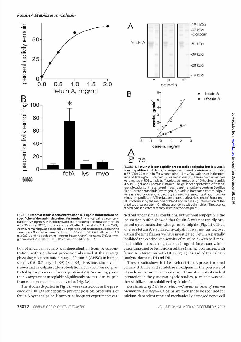
tion of m-calpain activity was dependent on fetuin A concen-tration, with significant protection observed at the average
physiologic concentration range of fetuin A (AHSG) in humanserum, 0.5–0.7 mg/ml (39) (Fig. 3 A). Previous studies hadshownthat m-calpain autoproteolytic inactivation was not pro-tected by the presence of added proteins (28). Accordingly, nei-ther lysozyme nor myoglobin significantly protected m-calpain
from calcium-mediated inactivation (Fig. 3 B).The studies depicted in Fig. 2 B were carried out in the pres-
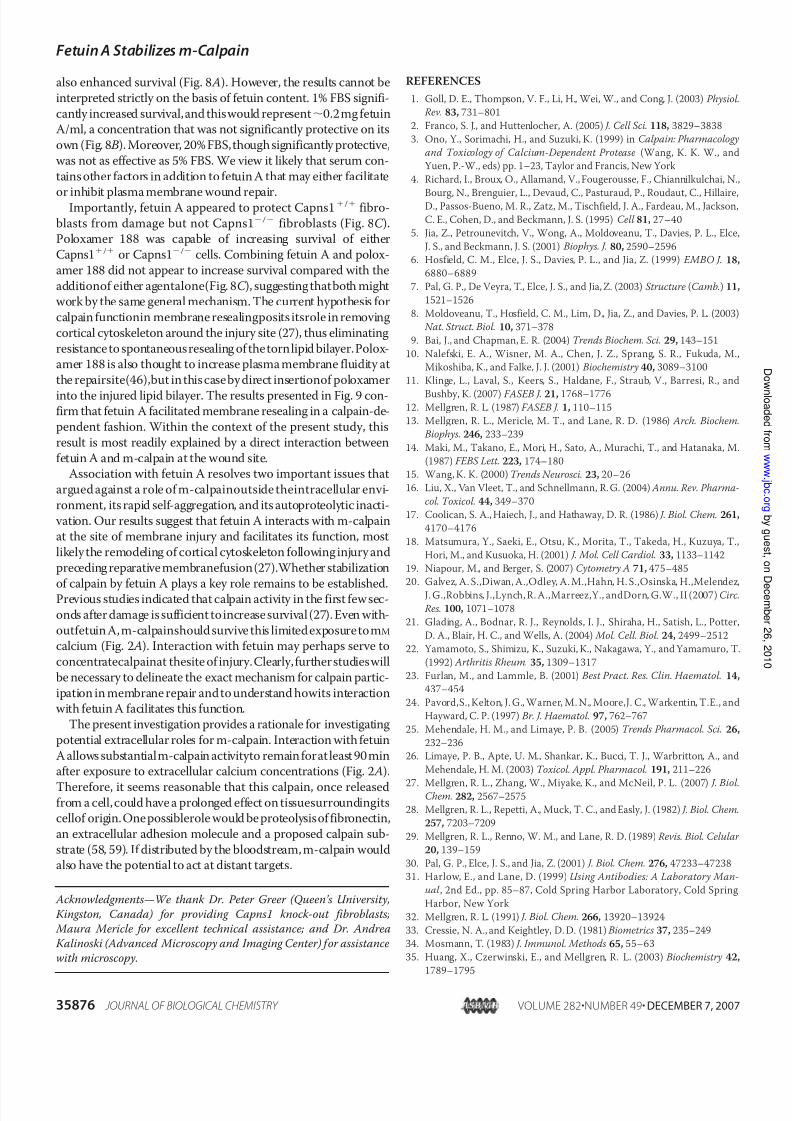
ence of 100 M leupeptin to prevent possible proteolysis of fetuin A by thecalpains. However, subsequent experiments car-
ried out under similar conditions, but without leupeptin in theincubation buffer, showed that fetuin A was not rapidly pro-cessed upon incubation with - or m-calpain (Fig. 4 A). Thus,whereas fetuin A stabilized m-calpain, it was not turned over
within the time frames we have investigated. Fetuin A partially
inhibited the caseinolytic activity of m-calpain, with half-max-imal inhibition occurring at about 1 mg/ml. Importantly, inhi-bition appeared to be noncompetitive (Fig. 4 B), consistent withfetuin A interaction with DIII (Fig. 1) instead of the calpain
catalytic domains DI and DII.These results show that the levels of fetuin A present in blood
plasma stabilize and solubilize m-calpain in the presence of physiologic extracellular calcium ion. Consistent with itslack of interaction in the yeast two-hybrid studies, -calpain was nei-
ther stabilized nor solubilized by fetuin A. Localization of Fetuin A with m-Calpain at Sites of Plasma
Membrane Damage—Calpains are thought to be required forcalcium-dependent repair of mechanically damaged nerve cell
FIGURE 3. Effect of fetuin A concentration on m-calpainstabilizationandspecificity of the stabilizing effect for fetuin A. A, m-calpain at a concen-tration of 25g/ml was incubatedwith the indicated concentration of fetuinA for 30 min at 37 °C, in the presence of buffer A containing 1.5 mM CaCl2.Activity remainingwas assessedby comparison with untreatedcalpainin thesameassay. B, m-calpainwas incubatedfor 30 minat 37 °Cin bufferA plus 1.5mM CaCl2, and noaddition,or 1 mg/ml fetuin A (fetA), lysozyme (lys), ormyo-
globin (myo). Asterisk , p 0.0006 versus no addition (n 4).
FIGURE 4. Fetuin A is not rapidly processed by calpains but is a weak noncompetitive inhibitor. A, onemg/mlsamplesof fetuinA were incubatedat 37 °C for 20 min in buffer A containing 1.5 mM CaCl2 alone, or in the pres-ence of 100 g/ml -calpain () or m-calpain (m). Ten-microliter samples
wereheated in SDS sample buffer, electrophoresed on a 10% polyacrylamideSDS-PAGE gel, and Coomassie-stained.The gel lanes depicted were from dif-ferent locationsof the same gel. In each case the right lane contains See BluePlus2protein standards (Invitrogen). B, quadruplicate samples of m-calpainwereassayed for caseinolytic activity at various casein concentrationsplus orminus1 mg/mlfetuin A.The dataare plotted asdescribed under“Experimen-tal Procedures” by the method of Woolf and Hanes (33). Intersection of thegraphsat the x axis at y 0 indicatesnoncompetitiveinhibition. The absenceof error bars indicates that they lie within the data point.
FetuinA Stabilizesm-Calpain
35872 JOURNAL OF BIOLOGICAL CHEMISTRY VOLUME 282•NUMBER 49• DECEMBER 7, 2007

7/31/2019 J. Biol. Chem.-2007-Mellgren-35868-77
http://slidepdf.com/reader/full/j-biol-chem-2007-mellgren-35868-77 6/10
axons and dendrites (40, 41), as well as mechanically damagedfibroblast plasma membrane (27). Because cytoplasmic pro-
teins, including calpains, would be exposed to extracellular fac-tors following plasma membrane rupture, we investigatedwhether fetuin A localizes at sites of membrane damage whereit could potentially influence calpain-dependent repair. Forthese studies we utilized myotubes, which express dysferlin, a
protein that has been shown to accumulate at sites of mem-brane injury and is a putative membrane repair protein (11, 42).Previous studies demonstrated that calpain-null fibroblasts
failed to reseal laser damage to the plasma membrane. Theselatter experiments measured intracellular uptake of the phos-
pholipid-staining green fluorescent dye, FM1–43 (43), which isnormally impermeant but accumulates in cells through dam-aged plasma membrane. We carried out preliminary studieswith injured myotubes to confirm that they also resealed in a
calpain-dependent fashion. Needle scratch damage of L6 myo-tubes, as described under “Experimental Procedures,” alloweduptake of FM1–43FX, a fixable analog of FM1–43 (Fig. 5 A).Preincubation with calpeptin to inactivate endogenous calpains
resulted in increased accumulation of FM1–43FX (Fig. 5 B, andmiddle rows in Fig. 5 A), indicating failure to repair membranedamage in calpain-depleted myotubes.
Cell surface biotinylation studies indicated that talin andother cortical cytoskeletal proteins become associated withdamaged plasma membrane of scraped fibroblasts (27).
Because these proteins are cleaved by calpains after membranedamage, it has been proposed that their processing may facili-tate cytoskeletal remodeling as part of the repair mechanism(27). Talin and dysferlin were exposed at the wound site of needle-scratched myotubes (Fig. 5 A, top row and enlarged
images in the bottom row), and calpeptin treatment decreasedaccumulation of talin at the injury site. Dysferlin still accumu-lated at the edge of the wound site in calpeptin-treated myo-tubes but was associated with regions that were highly labeled
with FM1–43FX, indicating a failure to reseal (Fig. 5 A, middlerow). In contrast, dysferlin was present as discrete patches onFM1–43FX-labeled membranes in myotubes that were notpretreated with calpeptin (Fig. 5 A, bottom row), similar to pre- viously observed immunolocalization patterns at sarcolemma
damage sites (11, 42). Our studies show that talin, like dysferlin,accumulates at sites of mechanical damage to myotubes. Thisobservation is consistent with biotinylation studies showingtalin accumulation at the cell surface after plasma membranedamage of fibroblasts (27).
Further experiments demonstrated accumulation of m-cal-pain in a pattern that overlapped dysferlin immunofluores-cence (Fig. 6 A). In contrast, annexin-A2, another proteinthought to be involved in membrane repair (44), did not appearto co-localize with dysferlin (Fig. 6 B). Previous studies had
localized fetuin A labeling to cells adjacent to scrape zones of wounded monolayer cultures (45). However, its co-localizationwith membrane repair proteins was not investigated. We foundthat m-calpain co-localized with FITC-labeled fetuin A at
wound sites in myotubes (Fig. 7 A). In contrast,labeling of fetuinA and annexin-A2 was only partially overlapping. Fetuin wasconcentrated at the damaged ends of myotubes (asterisk in Fig.7 B), whereas thestrongest signals for annexin-A2 were found at
small punctate structures, sometimes coinciding with readily identifiable particles in phase contrast images (arrows in Fig.7 B, and upper arrowheads in Fig. 6 B). These experiments dem-onstrate that fetuin A accumulates at damaged plasma mem-brane where it has the potential to interact with m-calpain.
Fetuin A Facilitates Plasma Membrane Resealing —To inves-tigate the potential effect of fetuin A on plasma membranerepair, we subjected cell monolayers to scrape damage as pre- viously described (27), with or without the addition of fetuin Ain the scrape medium. About 30% of HFL1 human fibroblasts
survived following scraping in standard calcium-containingbuffer, under the conditions described under “ExperimentalProcedures” (Fig. 8, A and B). However, the addition of FBS,which contains high levels of fetuin A (37), significantly
increased survival, as did 1 mg/ml or 5 mg/ml purified fetuin A.To determine whether the ubiquitous, typical calpains wererequired for enhanced survival imparted by fetuin A,Capns1/ and Capns1/ fibroblasts were scraped with or
FIGURE 5. L6 myotubes undergo calpain-dependent resealing after nee-dlescratch injuryand express talin anddysferlin at thesitesof damage.
A, 4-day differentiated rat L6 myotubes were injured by a needle scratch asdescribedunder “Experimental Procedures.” Where indicated, the myotubeswere preincubated for one h with 20 M calpeptin (CP ) to inactivate endog-enous calpains. White dashed lines in the micrographs indicate the scratchsite. The myotubes were scratched in the presence of 2 M FM1–43FX dyeandfixed in cold 4% paraformaldehyde 1 min after injury. Thefixed, nonper-meabilizedcells wereimmunostainedfor talinor dysferlin,as indicatedin thefigure, using TRITC-goat anti-mouse IgGas thesecond antibody.The bottomrows showmagnifiedimagesofthe boxedareasdepictedinthe toprows.Allof the micrographs represent averaged fluorescent signals throughout all 10 z
planes scannedthroughthe cells. Asterisks indicatelocationof myotubes,andarrows show the presence of talin or dysferlin at the surface of damagedmyotubes. Note that dysferlin appears as discrete patches continuous withgreen FM1–43FX fluorescence on cell membranes (arrows). Bars, 50 m.B, mean fluorescence intensities of FM1–43FX in damaged myotubes thatwere preincubated with 20 M calpeptin or vehicle (Me2SO). Details of theanalysis are provided under “Experimental Procedures.” *, p 0.015 versusminus calpeptin samples (n 5), Student’s paired t test.
FetuinA Stabilizesm-Calpain
DECEMBER 7, 2007 • VOLUME 282 • NUMBER 49 JOURNAL OF BIOLOGICAL CHEMISTRY 35873

7/31/2019 J. Biol. Chem.-2007-Mellgren-35868-77
http://slidepdf.com/reader/full/j-biol-chem-2007-mellgren-35868-77 7/10
without fetuin A in the scrape medium (Fig. 8C ). For compari-son, poloxamer 188, which protects skeletal myocytes andother cells from mechanical damage (reviewed in Ref. 46), was
also included in this study. Poloxamer significantly increasedsurvival of both Capns1/ and Capns1/ fibroblasts. How-ever, only Capns1/ fibroblasts appeared to be protected by fetuin A. When cells were scraped in thepresenceof both fetuin
A and poloxamer 188, there was no additional increase in sur- vival relative to treatment with either agent alone.
In previous studies, Capns1/ and Capns1/ fibroblastswere shown to take up trypan blue dye added at various times
after scrape damage. The loss of trypan blue uptake with timeafter scraping correlated well with the time course of mem-brane resealing observed in laser-damaged Capns1/ fibro-blasts (27). To determine whether fetuin A enhanced plasmamembrane resealing in a calpain-dependent fashion, we added
trypan blue to Capns1/ or Capns1/ fibroblasts 10 s afterscraping, using the protocol described under “ExperimentalProcedures.” At this time point, the fibroblasts were still under-going resealing (27). Accordingly, both cell types incorporatedtrypan blue through damaged plasma membrane, indicating a
failure to recover membrane impermeability (Fig. 9 A).Capns1/ fibroblasts, which fail to reseal for at least 2 minafter laser damage(27), were only10 –15% trypanblue-negative10 s after scraping. Capns1/ fibroblasts were 38% trypan blue
negative, consistent with their rapid recovery after laser injury.When Capns1/ fibroblasts were scraped in the presence of 1or 5 mg fetuin A/ml, the number of trypan blue negative cells10 s after scrape damage was significantly increased, indicatingenhanced resealing of damaged plasma membrane. In contrast,
5 mg/ml BSA, the major protein constituent of adult bovineserum, did not significantly increase the rate of Capns1/
membrane resealing (Fig. 9 B). The addition of 5 mg/ml fetuin Ato Capns1/ scraping medium did not significantly affect
FIGURE 6. m-Calpain co-localizes with dysferlin at the sarcolemma of injured L6 myotubes. A, m-calpain (m-calp), stained with Alexa 633 goatanti-chicken, co-localized with dysferlin at membrane damage sites in nee-dle-scratched L6 myotubes. Dysferlin was detected with TRITC-labeled anti-goatsecond antibody,false-coloredgreen in the micrographto distinguishitfrom theAlexa 633signal.Toprow , bar , 50m. Bottom row , enlargedimage of boxed areas of the top row . Bar , 10 m. The arrows show two signals for dys-ferlin localization, consistent with its presence in membrane repair patchesderived from internal cell membranes (see Fig. 5). B, another putative mem-brane repair protein, annexin-A2, did not co-localize with dysferlin, butappeared to be associated with large cell fragments, perhaps dead cells(asterisk ), andwithsmaller cell fragments(arrowheads). Thearrows showdys-ferlin localization at the edge of the scratch zone. Bars, 10 m.
FIGURE 7. Fetuin A and m-calpain co-localize at the surface of injuredmyotubes. A, FITC-fetuin A andm-calpain werepresentat theinjuredsurfaceof L6 myotubes adjacent to the scratch zone. Alexa 633 goat anti-chicken
second antibodywas usedto detect m-calpainlocalization.Note the localiza-tion of m-calpain at the edge of the fetuin-labeled damage site (arrow ), indi-cated by yellow coloration in the merged image. B, fetuin A and annexin-A2didnot extensively co-localize. Annexin wasdetectedwith TRITC rabbit anti-goat IgG second antibody. Asterisk , damaged end of a single large myotube.
The arrows show annexin labeling of small cell fragments. Bars, 25 m.
FIGURE 8. Fetuin A protectsHFL-1 human lungfibroblastsandCapns1/
mouse fibroblasts from scrape damage-induced cell death. Quadrupli-cate wells were used for each condition. HFL-1 fibroblasts were scraped asdescribedunder“ExperimentalProcedures”in thepresenceof FBS( A)orpuri-fied fetuin A (B). 3-(4,5-Dimethylthiazol-2-yl)-2,5-diphenyltetrazolium bro-mide reductase cell survival assays were carried out 12 h later. Asterisk , p 0.01 versus no fetuin; double asterisk , p 0.001 versus no additions.C , Capns1 / and Capns1 / fibroblasts were scraped in the presence of fetuinA(5mg/ml)orpoloxamer188(100or500M) where indicated. Asterisk ,
p 0.007 versus no additions; double asterisk , p 0.03 versus no additions.
FetuinA Stabilizesm-Calpain
35874 JOURNAL OF BIOLOGICAL CHEMISTRY VOLUME 282•NUMBER 49• DECEMBER 7, 2007

7/31/2019 J. Biol. Chem.-2007-Mellgren-35868-77
http://slidepdf.com/reader/full/j-biol-chem-2007-mellgren-35868-77 8/10
trypan blue uptake 10 s after scrape damage (Fig. 9 A). Similarresultswere obtained in two other independent experiments.Inzero time control experiments, both Capns1/ andCapns1/ fibroblasts were 90% trypan blue-positive whenscraped in the presence of the dye, in the absence or presence of
5 mg/ml fetuin A (not shown). Thus, fetuin A did not appear todecrease the number of cells damaged during the scrapingprocess.
DISCUSSION
AHSG, human fetuin A, is most often associated with regu-lation of bone mineralization (47). By stabilizing hydroxyapa-tite constituents in the blood, fetuin A has been shown to pre-
vent their deposition in soft tissues. Consistent with this role,fetuin A knock-out mice exhibit a modest degree of soft tissuecalcification (48). AHSG may also have other functions, includ-ing acting as a negative regulator of insulin receptor signaling(49). It was this latter proposed role that was of interest when
we initially identified AHSG as a binding partner for the tan-dem DIIIs of calpain-10, another diabetes-related protein, in yeast two-hybrid studies (Fig. 1). Because calpains are intracel-lular proteinases and AHSG/fetuin A is an extracellular proteinsecreted predominantly by the liver, their interaction under
physiologic conditions was problematic, and this led us toexplore hypotheses that would account for fetuin A and calpaininteractions. For these studies, we chose - and m-calpainsbecause they were readily available in highly purified form from
natural sources and because much more is known about theirenzymatic and physiologic properties compared with cal-pain-10 and other more recently described members of the cal-pain gene family. The apparent lack of interaction of -calpainDIII with AHSG in the yeast two-hybrid system (Fig. 1 C ) made
this calpain isozyme a convenient negative control for explor-ing fetuin A interactions with m-calpain.
The studies presented in Figs. 2 and 3 show that fetuin A hadspecific stabilizing effects on m-calpain exposed to calcium
concentrations that would be encountered in the extracellularenvironment or at the interface between cytoplasm and extra-
cellular space in damaged plasma membrane. Moreover, fetuinA did not appear to be a good calpain substrate (Fig. 4). Thus, itmay function by interacting with calpain DIII domains (Fig. 1)without substantially blocking catalytic activity. The stabiliza-tion of m-calpain by fetuin A is reminiscent of recent studies
demonstrating that fetuin A and other cystatin superfamily members can stabilize the gelatinolytic activities of the matrixmetalloproteinase, MMP-9 (50), an important modifier of extracellular matrix architecture. The present investigationsuggests that fetuin A could play a similar role for externalized
m-calpain. Whether other cystatin family members influencecalpain stability or activity remains to be established; however,it is intriguing that low and high molecular weight kininogenspossess a potent calpain inhibitory domain (51).
The recent observation that calpains are required for repairof mechanically damaged plasma membrane (27), an event thatnecessarily exposes the intracellular environment to extracel-lular factors, made it possible to directly test the potential for
fetuin A to facilitate repair. We first showed that m-calpainco-localized with dysferlin, a proposed plasma membranerepair protein (42), at the injured surface of damaged myotubes(Fig. 6). This placed calpain at the exterior surface of damagedsarcolemma, where it had the potential to interact with fetuinA. Further experiments utilizing FITC-labeled bovine fetuin A
confirmed its accumulation at membrane damage sites. Impor-tantly, m-calpain immunoreactivity co-localized with theFITC-fetuin A signal (Fig. 7 A), confirming its potential to beinfluenced by interaction with fetuin A during membranerepair. In contrast, annexin-A2, a protein that is thought to
participate in repair of damaged plasma membrane (44), wasnot closely associated with fetuin A at sites of membrane dam-age (Fig. 7 B). Moreover, it did not co-localize with dysferlin(Fig. 6 B). In contrast, earlier studies had demonstrated co-lo-
calization of dysferlin and annexin-A2 in damaged myotubes(44). The latter immunofluorescence experiments employeddetergent-containing buffers in the first antibody solutions andwould therefore have detected intracellular antigens. It is worthemphasizing that our cell studies employed fixed,but unperme-
abilized, myotubes. Therefore, we believe that our localizationstudies identify cell surface antigens that remain externalizedduring membrane repair. Taken within this context, our obser- vations suggest that externalized annexin-A2 is mainly local-ized at sites other than the patch complex containing dysferlin,
m-calpain, and fetuin A. These results agree with the generalconcept that annexins and integrins are released from cells asmajor protein components of “matrix vesicles” (also calledmicroparticles (52)) small plasma membrane-derived frag-ments released by blood platelets (53), migrating fibroblasts
(54, 55), osteoblasts (56), and metastatic cancer cells (57). Inprevious studies similar fragments appeared to be releasedfrom scrape-damaged fibroblasts (27).
Fetuin A not only associated with m-calpain at membrane
damage sites but also facilitated recovery after damage (Figs. 8and 9). HFL-1 fibroblasts were more likely to remain viablewhen scraped inthepresence of 1 or 5 mg/ml fetuin A (Fig. 8 B).Not surprisingly,FBS, whichcontains20 mg fetuin A/ml (37),
FIGURE 9. Fetuin A increased resealing of wounded Capns1/ fibro-blasts. A, Capns1 / andCapns1 / fibroblastswere scrape-damaged in thepresenceof 1 mg or5 mg fetuinA/ml, and membraneresealing wasassessedby uptake of trypan blue 10 s later, as described under “Experimental Proce-dures.” Asterisk , p 0.03 versus no fetuin (n 4); double asterisk , p 0.001versus nofetuin (n 4). The results arerepresentative of two other independ-ent experiments. B, Capns1 / fibroblasts werescrapedin the presenceof noadditions, 5 mg fetuinA/ml(fet A), or 5 mg BSA/ml. Uptakeof trypanbluewasassessed as in A. Asterisk , p 0.01 versus either no additions or the plus BSA
condition (n 5 scraped wells for each condition).
FetuinA Stabilizesm-Calpain
DECEMBER 7, 2007 • VOLUME 282 • NUMBER 49 JOURNAL OF BIOLOGICAL CHEMISTRY 35875
7/31/2019 J. Biol. Chem.-2007-Mellgren-35868-77
http://slidepdf.com/reader/full/j-biol-chem-2007-mellgren-35868-77 9/10
also enhanced survival (Fig. 8 A). However, the results cannot be
interpreted strictly on the basis of fetuin content. 1% FBS signifi-
cantly increased survival,and thiswould represent0.2mg fetuinA/ml, a concentration that was not significantly protective on its
own (Fig. 8 B). Moreover, 20% FBS,though significantly protective,
was not as effective as 5% FBS. We view it likely that serum con-
tains other factors in addition to fetuin A that may either facilitate
or inhibit plasma membrane wound repair.Importantly, fetuin A appeared to protect Capns1/ fibro-
blasts from damage but not Capns1/ fibroblasts (Fig. 8C ).Poloxamer 188 was capable of increasing survival of either
Capns1/ or Capns1/ cells. Combining fetuin A and polox-
amer 188 did not appear to increase survival compared with the
additionof either agentalone(Fig. 8C ), suggesting thatboth mightwork by the same general mechanism. The current hypothesis for
calpain functionin membrane resealingposits itsrole in removing
cortical cytoskeleton around the injury site (27), thus eliminating
resistance to spontaneous resealing of the tornlipid bilayer.Polox-amer 188 is also thought to increase plasma membrane fluidity at
the repairsite(46),but in this case by direct insertionof poloxamer
into the injured lipid bilayer. The results presented in Fig. 9 con-firm that fetuin A facilitated membrane resealing in a calpain-de-
pendent fashion. Within the context of the present study, this
result is most readily explained by a direct interaction between
fetuin A and m-calpain at the wound site.Association with fetuin A resolves two important issues that
argued against a role of m-calpainoutside theintracellular envi-
ronment, its rapid self-aggregation, and its autoproteolytic inacti-
vation. Our results suggest that fetuin A interacts with m-calpainat the site of membrane injury and facilitates its function, most
likely the remodeling of cortical cytoskeleton following injury and
preceding reparative membranefusion(27).Whether stabilization
of calpain by fetuin A plays a key role remains to be established.Previous studies indicated that calpain activity in the first few sec-
onds after damage is sufficient toincrease survival (27). Even with-
outfetuin A,m-calpainshould survive this limitedexposure tomM
calcium (Fig. 2 A). Interaction with fetuin may perhaps serve to
concentratecalpainat thesite of injury.Clearly,furtherstudieswill
be necessary to delineate the exact mechanism for calpain partic-
ipation in membrane repair andto understand howits interactionwith fetuin A facilitates this function.
The present investigation provides a rationale for investigating
potential extracellular roles for m-calpain. Interaction with fetuin
A allows substantialm-calpain activityto remain forat least 90 minafter exposure to extracellular calcium concentrations (Fig. 2 A).
Therefore, it seems reasonable that this calpain, once releasedfrom a cell,could have a prolonged effect on tissuesurroundingitscellof origin.One possiblerole would be proteolysisof fibronectin,
an extracellular adhesion molecule and a proposed calpain sub-
strate (58, 59). If distributed by the bloodstream, m-calpain wouldalso have the potential to act at distant targets.
Acknowledgments—We thank Dr. Peter Greer (Queen’s University,
Kingston, Canada) for providing Capns1 knock-out fibroblasts;
Maura Mericle for excellent technical assistance; and Dr. Andrea
Kalinoski (Advanced Microscopy and Imaging Center) for assistance
with microscopy.
REFERENCES
1. Goll, D. E., Thompson, V. F., Li, H., Wei, W., and Cong, J. (2003) Physiol.
Rev. 83, 731–801
2. Franco, S. J., and Huttenlocher, A. (2005) J. Cell Sci. 118, 3829–3838
3. Ono, Y., Sorimachi, H., and Suzuki, K. (1999) in Calpain: Pharmacology
and Toxicology of Calcium-Dependent Protease (Wang, K. K. W., and
Yuen, P.-W., eds) pp. 1–23, Taylor and Francis, New York
4. Richard, I., Broux, O., Allamand, V., Fougerousse, F., Chiannilkulchai, N.,
Bourg, N., Brenguier, L., Devaud, C., Pasturaud, P., Roudaut, C., Hillaire,D., Passos-Bueno, M. R., Zatz, M., Tischfield, J. A., Fardeau, M., Jackson,
C. E., Cohen, D., and Beckmann, J. S. (1995) Cell 81, 27–40
5. Jia, Z., Petrounevitch, V., Wong, A., Moldoveanu, T., Davies, P. L., Elce,
J. S., and Beckmann, J. S. (2001) Biophys. J. 80, 2590–2596
6. Hosfield, C. M., Elce, J. S., Davies, P. L., and Jia, Z. (1999) EMBO J. 18,
6880–6889
7. Pal, G. P., De Veyra, T., Elce, J. S., and Jia, Z. (2003) Structure (Camb.) 11,
1521–1526
8. Moldoveanu, T., Hosfield, C. M., Lim, D., Jia, Z., and Davies, P. L. (2003)
Nat. Struct. Biol. 10, 371–378
9. Bai, J., and Chapman, E. R. (2004) Trends Biochem. Sci. 29, 143–151
10. Nalefski, E. A., Wisner, M. A., Chen, J. Z., Sprang, S. R., Fukuda, M.,
Mikoshiba, K., and Falke, J. J. (2001) Biochemistry 40, 3089–3100
11. Klinge, L., Laval, S., Keers, S., Haldane, F., Straub, V., Barresi, R., and
Bushby, K. (2007) FASEB J. 21, 1768–177612. Mellgren, R. L. (1987) FASEB J. 1, 110–115
13. Mellgren, R. L., Mericle, M. T., and Lane, R. D. (1986) Arch. Biochem.
Biophys. 246, 233–239
14. Maki, M., Takano, E., Mori, H., Sato, A., Murachi, T., and Hatanaka, M.
(1987) FEBS Lett. 223, 174–180
15. Wang, K. K. (2000) Trends Neurosci. 23, 20–26
16. Liu, X., Van Vleet, T., and Schnellmann, R. G. (2004) Annu. Rev. Pharma-
col. Toxicol. 44, 349–370
17. Coolican, S. A., Haiech, J., and Hathaway, D. R. (1986) J. Biol. Chem. 261,
4170–4176
18. Matsumura, Y., Saeki, E., Otsu, K., Morita, T., Takeda, H., Kuzuya, T.,
Hori, M., and Kusuoka, H. (2001) J. Mol. Cell Cardiol. 33, 1133–1142
19. Niapour, M., and Berger, S. (2007) Cytometry A 71, 475–485
20. Galvez, A. S.,Diwan, A.,Odley, A. M.,Hahn, H. S.,Osinska, H.,Melendez,
J. G.,Robbins, J.,Lynch, R. A.,Marreez,Y., andDorn, G.W., II (2007) Circ.
Res. 100, 1071–1078
21. Glading, A., Bodnar, R. J., Reynolds, I. J., Shiraha, H., Satish, L., Potter,
D. A., Blair, H. C., and Wells, A. (2004) Mol. Cell. Biol. 24, 2499–2512
22. Yamamoto, S., Shimizu, K., Suzuki, K., Nakagawa, Y., and Yamamuro, T.
(1992) Arthritis Rheum. 35, 1309–1317
23. Furlan, M., and Lammle, B. (2001) Best Pract. Res. Clin. Haematol. 14,
437–454
24. Pavord,S., Kelton, J. G., Warner, M. N., Moore,J. C., Warkentin, T.E., and
Hayward, C. P. (1997) Br. J. Haematol. 97, 762–767
25. Mehendale, H. M., and Limaye, P. B. (2005) Trends Pharmacol. Sci. 26,
232–236
26. Limaye, P. B., Apte, U. M., Shankar, K., Bucci, T. J., Warbritton, A., and
Mehendale, H. M. (2003) Toxicol. Appl. Pharmacol. 191, 211–226
27. Mellgren, R. L., Zhang, W., Miyake, K., and McNeil, P. L. (2007) J. Biol.Chem. 282, 2567–2575
28. Mellgren, R. L., Repetti, A., Muck, T. C., and Easly, J. (1982) J. Biol. Chem.
257, 7203–7209
29. Mellgren, R. L., Renno, W. M., and Lane, R. D. (1989) Revis. Biol. Celular
20, 139–159
30. Pal, G. P., Elce, J. S., and Jia, Z. (2001) J. Biol. Chem. 276, 47233–47238
31. Harlow, E., and Lane, D. (1999) Using Antibodies: A Laboratory Man-
ual , 2nd Ed., pp. 85–87, Cold Spring Harbor Laboratory, Cold Spring
Harbor, New York
32. Mellgren, R. L. (1991) J. Biol. Chem. 266, 13920–13924
33. Cressie, N. A., and Keightley, D. D. (1981) Biometrics 37, 235–249
34. Mosmann, T. (1983) J. Immunol. Methods 65, 55–63
35. Huang, X., Czerwinski, E., and Mellgren, R. L. (2003) Biochemistry 42,
1789–1795
FetuinA Stabilizesm-Calpain
35876 JOURNAL OF BIOLOGICAL CHEMISTRY VOLUME 282•NUMBER 49• DECEMBER 7, 2007
7/31/2019 J. Biol. Chem.-2007-Mellgren-35868-77
http://slidepdf.com/reader/full/j-biol-chem-2007-mellgren-35868-77 10/10
36. Kazi,J. A.,Nakamura,O., Ohnishi, T.,Arakaki,N., Kajihara,T., Nakagawa,
S., and Daikuhara, Y. (1998) J. Biochem. (Tokyo) 124, 179–186
37. Cartellieri, S., Hamer, O., Helmholz, H., and Niemeyer, B. (2002) Biotech-
nol. Appl. Biochem. 35, 83–89
38. Osawa, M., Tian, W., Horiuchi, H., Kaneko, M., and Umetsu, K. (2005)
Hum. Genet. 116, 146–151
39. Brown, W. M., Saunders, N. R., Mollgard, K., and Dziegielewska, K. M.
(1992) Bioessays 14, 749–755
40. Xie, X. Y., and Barrett, J. N. (1991) J. Neurosci. 11, 3257–3267
41. Godell, C. M., Smyers, M. E., Eddleman, C. S., Ballinger, M. L., Fishman,
H. M., and Bittner, G. D. (1997) Proc. Natl. Acad. Sci. U. S. A. 94,
4751–4756
42. Bansal, D., Miyake, K., Vogel, S. S., Groh, S., Chen, C. C., Williamson, R.,
McNeil, P. L., and Campbell, K. P. (2003) Nature 423, 168–172
43. Schote, U., and Seelig, J. (1998) Biochim. Biophys. Acta 1415, 135–146
44. Lennon, N. J., Kho, A., Bacskai, B. J., Perlmutter, S. L., Hyman, B. T., and
Brown, R. H., Jr. (2003) J. Biol. Chem. 278, 50466–50473
45. Patchell, B. J., and Dorscheid, D. R. (2006) Clin. Exp. Allergy 36, 585–593
46. Townsend, D., Yasuda, S., and Metzger, J. (2007) Expert Rev. Cardiovasc.
5, 99–109
47. Schinke, T., Amendt, C., Trindl, A., Poschke, O., Muller-Esterl, W., and
Jahnen-Dechent, W. (1996) J. Biol. Chem. 271, 20789–20796
48. Schafer, C., Heiss, A., Schwarz, A., Westenfeld, R., Ketteler, M., Floege, J.,
Muller-Esterl, W., Schinke, T., and Jahnen-Dechent, W. (2003) J. Clin.
Investig. 112, 357–366
49. Mathews,S. T.,Srinivas, P. R.,Leon, M. A.,and Grunberger, G.(1997) Life
Sci. 61, 1583–1592
50. Ray, S., Lukyanov, P., and Ochieng, J. (2003) Biochim. Biophys. Acta 1652,
91–102
51. Bradford, H. N., Jameson, B. A., Adam, A. A., Wassell, R. P., and Colman,
R. W. (1993) J. Biol. Chem. 268, 26546–26551
52. Ardoin, S. P., Shanahan, J. C., and Pisetsky, D. S. (2007) Scand J. Immunol.
66, 159–165
53. Sandberg, H., Bode, A. P., Dombrose, F. A., Hoechli, M., and Lentz, B. R.
(1985) Thromb. Res. 39, 63–79
54. Lee, T. L., Lin, Y. C., Mochitate, K., and Grinnell, F. (1993) J. Cell Sci. 105,
167–177
55. Taverna, S., Ghersi, G., Ginestra, A., Rigogliuso, S., Pecorella, S.,
Alaimo, G., Saladino, F., Dolo, V., Dell’Era, P., Pavan, A., Pizzolanti, G.,
Mignatti, P., Presta, M., and Vittorelli, M. L. (2003) J. Biol. Chem. 278,
51911–51919
56. Xiao, Z., Camalier, C. E., Nagashima, K., Chan, K. C., Lucas, D. A., de la
Cruz, M. J., Gignac, M., Lockett, S., Issaq, H. J., Veenstra, T. D., Conrads,
T. P., and Beck, G. R., Jr. (2007) J. Cell. Physiol. 210, 325–335
57. Ginestra, A., La Placa, M. D., Saladino, F., Cassara, D., Nagase, H., and
Vittorelli, M. L. (1998) Anticancer Res. 18, 3433–3437
58. Dourdin, N., Balcerzak, D., Brustis, J. J., Poussard, S., Cottin, P., and Du-
castaing, A. (1999) Exp. Cell Res. 246, 433–442
59. Frangie, C., Zhang, W., Perez, J., Dubois, Y.-C. X., Haymann, J.-P., and
Baud, L. (2006) J. Biol. Chem. 281, 26624–26632
FetuinA Stabilizesm-Calpain
DECEMBER 7, 2007 • VOLUME 282 • NUMBER 49 JOURNAL OF BIOLOGICAL CHEMISTRY 35877


![[ITDG] BIOL](https://img.dokumen.tips/doc/110x75/5571fcdd49795991699815b6/itdg-biol.jpg)