Embed Size (px)
Citation preview

International Research Journal of Medicine and Biomedical Sciences Vol.3 (2),pp. 15-29, November 2018 Available online at http://www.journalissues.org/IRJMBS/ https://doi.org/10.15739/irjmbs.18.004 Copyright © 2018 Author(s) retain the copyright of this article ISSN 2488-9032
Case Study
ISW for the treatment of adult anterior crossbite with severe crowding combined facial asymmetry case
Received 18 September, 2018 Revised 1 November, 2018 Accepted 5 November, 2018 Published 16 November, 2018
Chun-shuo HUANG1,2, Jian-hong YU*1,2
and Cheng-yen HSIEH1,2
1Department of Orthodontics,
China Medical University Hospital, Taichung, Taiwan.
2School of Dentistry, College of Dentistry, China Medical
University, Taichung, Taiwan.
*Corresponding Author: Email: [email protected]
The objective of this case report was to discuss ISW (Improved Super-elastic Ti-Ni alloy wire, developed by Tokyo Medical and Dental University) for the treatment of adult anterior crossbite with facial asymmetry and severe crowding. An adult female (29 years old) came to our clinic with a chief complaint of not able to chew food well and irregularity of her teeth. Clinical examination revealed right side Class III, left side Class I molar relationship, anterior crossbite and severe crowding combined with full mouth periodontitis and facial asymmetry with a mandible shift to left side. We decided to adopt orthodontic non-extraction treatment because the patient strongly refused to receive orthognathic surgery. Before orthodontic active treatment, the periodontal phase I therapy was performed and the results were controlled well. ISW and open coil spring for #26, 27 distalization were performed to facilitate the correction of anterior crossbite and relief of blocked-in #25. With the use of differential ISW MEAW technique and intermaxillary elastics(IME), severe dental crowding was corrected. The treatment was completed within 18 months and a desirable occlusion with adequate overbite and overjet was achieved. Key words : ISW, skeletal Class III, non-surgical orthodontics, 3 incisor finish, ISW expansion arch and MEAW technique.
INTRODUCTION The treatment of adult skeletal Class III with facial asymmetry is usually a challenge to orthodontists. It’s important to differentially diagnose a facial asymmetry case by dental, functional or skeletal problems(McLaughlin, 1988; Stellzig-Eisenhauer et al., 2002; Baccetti et al., 2007 ;Tollaro et al., 1995).
The patient had skeletal Class III malocclusion and facial asymmetry, orthognathic surgery combined with orthodontic treatment may be a better choice. But the patient refused the surgery. Before the orthodontic treatment was performed, differentially diagnosing a facial asymmetry case by dental/functional/skeletal is very crucial. Sometimes a case may combine two or more. For instance, this case showed both functional and skeletal problems. It was important to locate and early eliminate functional interference in a facial asymmetry case. Sometimes a
functional wax bite was helpful. This case showed possibility of functional interference around the right upper canine. With ISW (developed by the Tokyo Medical and Dental University) Expansion Arch around the canine area and trans-midline elastic traction, functional interference was relieved. ISW MEAW was used to correct canted occlusion. ISW wire with tip-back bends were added from canine to 2nd molar at upper left and lower right quadrants to correct canted occlusion. When we used ISW wire with tip-back bends, we could expect an upright and intrusion effect over the posterior teeth and to correct uneven occlusion.
Concerning retention strategy for a facial asymmetry case, in order to maintain a fixed intermaxillary relationship and to successfully relieve musculature tension, we adopted a functional appliance to keep a rigid and unchanged upper and lower jaw position. Although camouflage treatment may

Int. Res. J. Med. Biomed. Sci. 16
Figure 1:Facial photos before active treatment
be available for some patients, we have to pay attention to their musculature tension to prevent the relapse (Delaire, 1997;Troy et al., 2009; Guyer et al., 1986; Giancotti et al., 2003; Hägg et al., 2004; Rabie and Gu, 1999; Rabie and Gu, 2000; Hisano et al., 2006; Kanno et al., 2007; Miyajima et al., 1997). History and Diagnosis An adult female aged 29 came to our clinic with a chief complaint of irregular dentition and could not bite food well. Her lateral profile was concave, and the frontal view showed facial asymmetry phenomenon (Figure 1). Clinical examination revealed right Class III (left Class I) molar relationship, bilateral canine Class III relationship, anterior crossbite, and severe crowding combined with full mouth periodontitis and a mandibular shift to left side resulting in facial asymmetry (Figure 2). Panoramic film showed existence of #18 ,#28 ,#38 ,#48 (Figure 3).
This patient refused to receive orthognathic surgery, so we decided to adopt non-surgical orthodontic treatment. Before orthodontic treatment, she accepted the periodontal
phase I therapy and the situation had been under control. ISW (Low Hysteresis Improved Super-elastic Ti-Ni alloy wire, developed by Tokyo Medical and Dental University) technique and newly developed open coil spring for #26, 27 distalization were performed to facilitate the correction of anterior crossbite and #25 blocked-in. With the use of differential MEAW technique and intermaxillary elastics(IME), the severe dental crowding was corrected. The treatment was completed within 18 months and a desirable occlusion with adequate overbite and overjet was achieved.
The cephalometric analysis showed a skeletal Class III jaw relationships (SNA:84.3o、SNB:88.1o、ANB:-3.8o ) and dental
compensation (U-1 to FH plane : 104.3o, L-1 to mandibular plane : 71.2o) (Figure 4). P-A (postero-anterior)
cephalometric radiograph showed mandibular discrepancy between the right and the left ramus heights (Right side:
49.0mm ; Left side : 47.5mm, ramus discrepancy :
1.5mm)(Figure 5 and 6). Therefore, a summary of diagnosis includes:
1. Functional(-): 2. Skeletal(+) : skeletal Class III

Huang et al. 17
Figure 2:Intraoral photos before active treatment
Figure 3:Panoramic film before active treatment
Int. Res. J. Med. Biomed. Sci. 18
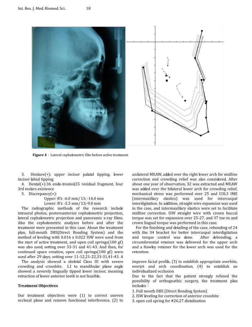
Figure 4:Lateral cephalometric film before active treatment
3. Denture(+): upper incisor palatal tipping, lower incisor labial tipping
4. Dental(+):36 endo-treated,55 residual fragment, four 3rd molars existence
5. Discrepancy(+): Upper: R’t: -6.0 mm/ L’t: -14.0 mm Lower: R’t: -5.5 mm/ L’t:-9.0 mm The radiographic methods of the research include
intraoral photos, posteroanterior cephalometric projection, lateral cephalometric projection and panoramic x-ray films. Also the cephalometric analyses before and after the treatment were presented in this case. About the treatment plan, full-mouth DBS(Direct Bonding System) and the method of leveling with 0.016 x 0.022 ISW were used from the start of active treatment, and open coil springs(100 gf) was also used, setting over 33-31 and 41-43. And then, for continued space creation, open coil springs(100 gf) were used after 29 days, setting over 11-12,21-22,33-31,41-43. A
unilateral MEAW, added over the right lower arch for midline correction and crowding relief was also considered. After about one year of observation, 32 was extracted and MEAW was added over the bilateral lower arch for crowding relief, mechanical stress was performed over 25 and U3L3 IME (intermaxillary elastics) was used for intercuspal interdigitation. In addition, straight wire expansion was used in the case, and intermaxillary elastics were set to facilitate midline correction. ISW straight wire with crown buccal torque was set for expansion over 25-27, and 37 toe-in and crown lingual torque was performed in this case.
For the finishing and detailing of the case, rebonding of 24 with the 34 bracket for better intercuspal interdigitation and torque control was done. After debonding, a circumferential retainer was delivered for the upper arch and a Hawley retainer for the lower arch was used for the retention.
The analysis showed a skeletal Class III with severe crowding and crossbite. L1 to mandibular plane angle showed a severely lingually tipped lower incisor, meaning retraction of lower anterior teeth is not feasible. Treatment Objectives Our treatment objectives were (1) to correct uneven occlusal plane and remove functional interference, (2) to
improve facial profile, (3) to establish appropriate overbite, overjet and arch coordination, (4) to establish an individualized occlusion Due to the fact that the patient strongly refused the possibility of orthognathic surgery, the treatment plan includes:
1. Full mouth DBS (Direct Bonding System) 2. ISW leveling for correction of anterior crossbite 3. open coil spring for #26,27 distalization

Huang et al. 19
Figure 5:Postero-anterior(PA) film before active treatment
Figure 6:Polygon before active treatment
Int. Res. J. Med. Biomed. Sci. 20
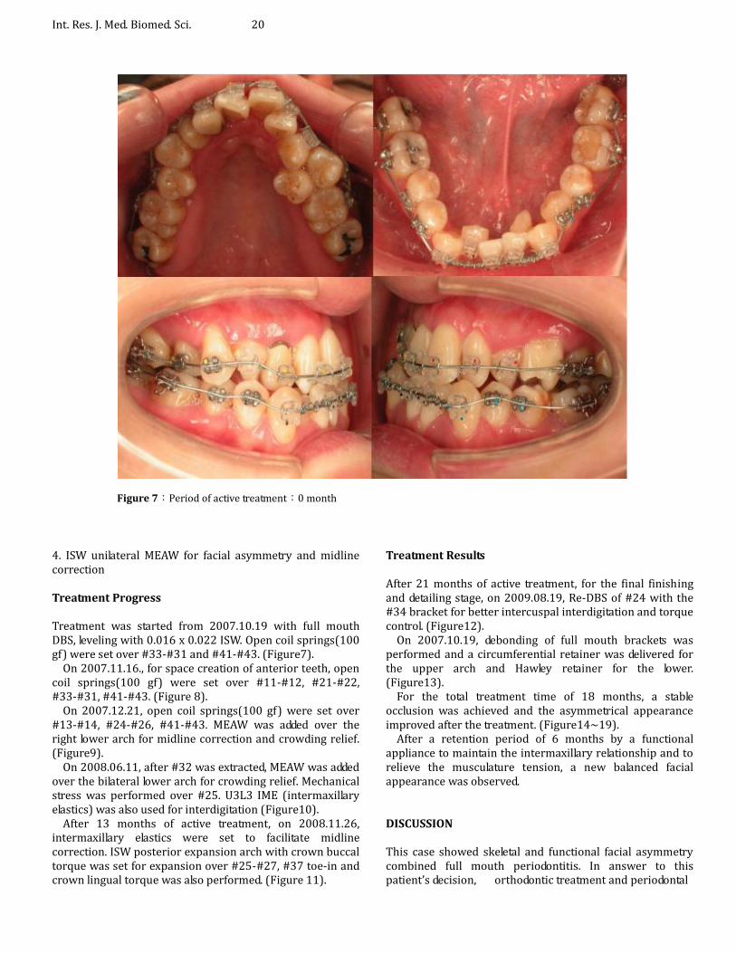
Figure 7:Period of active treatment:0 month
4. ISW unilateral MEAW for facial asymmetry and midline correction Treatment Progress Treatment was started from 2007.10.19 with full mouth DBS, leveling with 0.016 x 0.022 ISW. Open coil springs(100 gf) were set over #33-#31 and #41-#43. (Figure7).
On 2007.11.16., for space creation of anterior teeth, open coil springs(100 gf) were set over #11-#12, #21-#22, #33-#31, #41-#43. (Figure 8).
On 2007.12.21, open coil springs(100 gf) were set over #13-#14, #24-#26, #41-#43. MEAW was added over the right lower arch for midline correction and crowding relief. (Figure9).
On 2008.06.11, after #32 was extracted, MEAW was added over the bilateral lower arch for crowding relief. Mechanical stress was performed over #25. U3L3 IME (intermaxillary elastics) was also used for interdigitation (Figure10).
After 13 months of active treatment, on 2008.11.26, intermaxillary elastics were set to facilitate midline correction. ISW posterior expansion arch with crown buccal torque was set for expansion over #25-#27, #37 toe-in and crown lingual torque was also performed. (Figure 11).
Treatment Results After 21 months of active treatment, for the final finishing and detailing stage, on 2009.08.19, Re-DBS of #24 with the #34 bracket for better intercuspal interdigitation and torque control. (Figure12).
On 2007.10.19, debonding of full mouth brackets was performed and a circumferential retainer was delivered for the upper arch and Hawley retainer for the lower. (Figure13).
For the total treatment time of 18 months, a stable occlusion was achieved and the asymmetrical appearance improved after the treatment. (Figure14~19).
After a retention period of 6 months by a functional appliance to maintain the intermaxillary relationship and to relieve the musculature tension, a new balanced facial appearance was observed. DISCUSSION This case showed skeletal and functional facial asymmetry combined full mouth periodontitis. In answer to this patient’s decision, orthodontic treatment and periodontal

Huang et al. 21
Figure 8:Period of active treatment:1 month
Figure 9:Period of active treatment:2 months

Int. Res. J. Med. Biomed. Sci. 22
Figure 10:Period of active treatment:8 months
Figure 11:Period of active treatment:13 months

Huang et al. 23
Figure 12:Period of active treatment:21 months
Figure 13:Period of active treatment:18 months
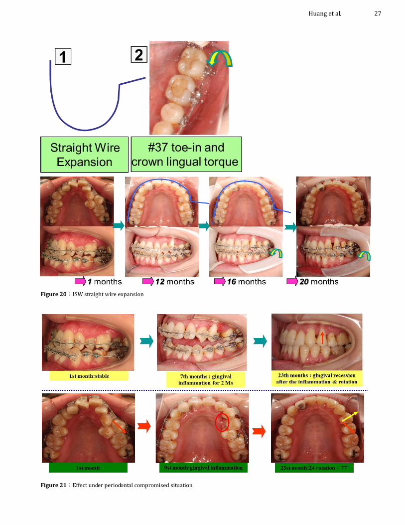
phase I therapy were performed. After 18 months of active treatment, a normal occlusion and a desirable cusp interdigitation were achieved. Therefore, adult anterior crossbite with severe crowding combined facial asymmetry and full mouth periodontitis can be well treated with differential ISW MEAW technique. ISW straight wire expansion In the past, it’s very difficult to “labially” expand one specified section of the dental arch. But with ISW straight
wire expansion, we can specifically expand the section where we want to. In this case, ISW straight wire expansion corrected the unilateral posterior crossbite from the second premolar to the second molar progressively. (Figure 20). Effect under periodontal compromised situation We can still observe the effect under periodontal compromised situation. Gingival response and tooth migration(rotation) of #24 tooth was noticed. After 7th months of active treatment, gingival inflammation over #24

Int. Res. J. Med. Biomed. Sci. 24
Figure 14:Facial photos after active treatment
Figure 15:Intraoral photos after active treatment

Huang et al. 25
Figure 16:Lateral Cephalometric and postero-anterior after active treatment
Figure 17:Lateral Cephalometric after active treatment
region occured for 2 months. After 18th months of active treatment, gingival recession after the inflammation & rotation was noticed and the angle of rotation #24 was measured approximately 77 o. (Figure 21).
Unilateral MEAW effect Furthermore, by using ISW MEAW combined with intermaxillary elastics to tip the right lower posterior teeth

Int. Res. J. Med. Biomed. Sci. 26
Figure 18:Polygon after active treatment
Figure 19:Superimposition after active treatment
back, space can be created and the R’t canine Class I can be achieved. (Figure 22).
Finally, when using unilateral ISW MEAW technique, there are intrusion effect and toe-in effect. The two effects aided in compromising the facial asymmetry by the rotation of mandible. (Figure 23). Conclusion This case showed skeletal facial asymmetry combined with
full mouth periodontitis. In answer to this patient’s decision, orthodontic treatment and periodontal phase I therapy were performed. Clinical examination by functional wax bite reveals a possibility of functional interference around the right upper canine. P-A view check finds slight discrepancy between the right and the left ramus heights. Orthognathic surgery was suggested to the patient but was refused. After orthodontic treatment, the patient was pleased with the outcome and thus orthodontic treatment was performed. After 18 months of active treatment, a normal occlusion and a desirable intercuspal interdigitation were achieved.
Huang et al. 27
Figure 20:ISW straight wire expansion
Figure 21:Effect under periodontal compromised situation

Int. Res. J. Med. Biomed. Sci. 28
Discussion (3)- unilateral MEAW effect I
By using ISW MEAW
combined with
intermaxillary elastics to tip
the right lower posterior
teeth back, space was
created and the R’t canine
Class I was achieved. ISW unilateral MEAW
Canine class III relationship Canine class I relationship
Initial : 78°
After Tx : 71°
Figure 22:Unilateral MEAW effect(1)
Discussion (4)- unilateral MEAW effect IIWhen using unilateral ISW MEAW, there are intrusion effect and toe in
effect. These two effects aided in compromising the facial asymmetry by
rotating the mandible.
1.9°
Mandible shift for facial
asymmetry compensation
Intrusion effect Toe in effect
L’t crossbite
R’t
R’t
R’t
R’t
Figure 23:Unilateral MEAW effect(2)

Therefore, adult anterior skeletal crossbite with severe crowding combined with facial asymmetry and full mouth periodontitis can be treated with ISW treatment. Conflict of Interests The authors declare that there is no conflict of interests regarding the publication of the paper. REFERENCE Baccetti T, Reyes BC, McNamara JA Jr. (2007). "Craniofacial
changes in Class III malocclusion as related to skeletal and dental maturation." Ame. J. Orthod. Dentofacial Orthop. 132(2): 171. e171-171. e112.
Delaire J (1997). "Maxillary development revisited: relevance to the orthopaedic treatment of Class III malocclusions." Eur. J. orthod. 19(3): 289-311.
Giancotti A, Maselli A, Mampieri G, Spanò E (2003). "Pseudo-Class III malocclusion treatment with Balters’ Bionator." J. orthod.
Guyer EC, Ellis, EE, McNamara JA, Behrents RG (1986). "Components of Class III malocclusion in juveniles and adolescents." The Angle Orthod. 56(1): 7-30.
Hägg U, Tse A, Bendeus M, Rabie AB (2004). "A follow-up study of early treatment of pseudo Class III malocclusion." The Angle Orthod. 4(4): 465-472.
Hisano M, Ohtsubo K, Chung CJ, Nastion F, Soma K (2006). "Vertical control by combining a monoblock appliance in adult Class III overclosure treatment." The Angle Orthod. 76(2): 226-235.
Kanno Z, Kim Y, Soma K (2007). "Early correction of a developing skeletal Class III malocclusion." The Angle Orthod. 77(3): 549-556.
McLaughlin RP (1988). "Malocclusion and the Temporomandibular Joint: —An Historical Perspective." The Angle Orthodontist 58(2): 185-190.
Miyajima K, McNamara JA Jr, Sana M, Murata S (1997). "An estimation of craniofacial growth in the untreated Class III female with anterior crossbite." Ame. J. Orthod. Dentofacial Orthop. 112(4): 425-434.
Ngan P, Moon W (2015). "Evolution of Class III treatment in orthodontics." Ame. J. Orthod. Dentofacial Orthop. 148(1): 22-36.
Rabie A, Gu Y (1999). "Orthodontics: management of pseudo Class III malocclusion in southern Chinese children." British dental J. 186(4): 183-187.
Rabie A, Gu Y (2000). "Diagnostic criteria for pseudo–Class III malocclusion." Ame. J. Orthod. Dentofacial Orthop. 117(1): 1-9.
Stellzig-Eisenhauer A, Lux CJ, Schuster G (2002). "Treatment decision in adult patients with Class III malocclusion: orthodontic therapy or orthognathic surgery?" Ame. J. Orthod. Dentofacial Orthop. 122(1): 27-37.
Tollaro I, Baccetti T, Franchi L (1995). "Mandibular skeletal changes induced by early functional treatment of Class III maloccsion: A superimposition study." Ame. J. Orthod.
Huang et al. 29
Dentofacial Orthop. 108(5): 525-532. Troy BA, Shanker S, Fields HW, Vig K, Johnston W (2009).
"Comparison of incisor inclination in patients with Class III malocclusion treated with orthognathic surgery or orthodontic camouflage." Ame. J. Orthod. Dentofacial Orthop. 135(2): 146. e141-146. e149.


![Myofunctional Treatment of Anterior Crossbite in a Growing ... · anterior crossbite malocclusion with EGA [12, 13]. The present case report was carried out to investigate the effectiveness](https://img.dokumen.tips/doc/110x75/60b795e2459fae307d78d20c/myofunctional-treatment-of-anterior-crossbite-in-a-growing-anterior-crossbite.jpg)