Embed Size (px)
Citation preview

Published by Baishideng Publishing Group Inc
World Journal of Respirology World J Respirol 2017 July 28; 7(2): 35-47
ISSN 2218-6255 (online)

World Journal of RespirologyW J R
EDITOR-IN-CHIEFRafael Rosell, Barcelona
ASSOCIATE EDITORSGiovanna Elisiana Carpagnano, BariWCS Cho, KowloonAndrea Imperatori, VaresePaul Zarogoulidis, Thessaloniki
GUEST EDITORIAL BOARD MEMBERSShi-Chuan Chang, TaipeiYuh-Min Chen, TaipeiJing-Hsien Chen, TaichungTai-An Chiang, TainanHan Shui Hsu, TaipeiWu-Huei Hsu, TaichungKun-Lun Huang, TaipeiChun-Jen Huang, New Taipei CityChung-Jen Huang, TaipeiJiunn-Liang Ko, TaichungYu-Hsiang Kuan, TaichungTzong-Shyuan Lee, TaipeiYun-Wei Lin, ChiayiMeng-Chin Lin, KaohsiungJiu-Yao Wang, TainanJeng-Shing Wang, KaohsiungRong-Sen Yang, TaipeiChuen-Mao Yang, Kwei-San
MEMBERS OF THE EDITORIAL BOARD
Argentina
Nidia Noemí Gomez, San Luis
Australia
Moyez Jiwa, Bentley
Robert James Boots, QueenslandZoe Jane McKeough, SydneyJean-Pierre Scheerlinck, MelbourneKrishna Bajee Bajee Sriram, Gold Coast
Austria
Clemens Aigner, ViennaBernd Lamprecht, SalzburgDaniel Zimpfer, Vienna
Bangladesh
Dilruba Ahmed, Dhaka
Belgium
Lindsey Van Haute, JetteTeresinha Leal, BrusselsDaniel O Rodenstein, LouvainSteven Raphael Simoens, Leuven
BrazilFlavio Aimbire, Sao Jose dos CamposCristiano Feijó Andrade, Porto AlegreValquiria Bueno, Sao PauloRamon Andrade de Mello, FortalezaLuis Cristovao Porto, Rio de JaneiroMarcus Vinícius Nora De Souza, Rio de JaneiroDaniela Zanini, Santa Maria
Bulgaria
Papantchev Vassil Gueorguiev, Sofia
CanadaKenneth R Chapman, TorontoAbdelilah Soussi Gounni, ManitobaGrzegorz Janusz Korpanty, TorontoAndrew Leask, LondonGeorg Marcus Schmolzer, EdmontonSat Sharma, ManitobaChangqi Zhu, Toronto
Chile
Dante Daniel Cáceres, Santiago
China
Yi-Ran Cai, BeijingHai-Quan Chen, ShanghaiGeorge G Chen, Hong KongYa-Hong Chen, BeijingBo Deng, ChongqingLiang Dong, JinanDavid Harold Garfield, ShanghaiXiao-Ye He, ShanghaiJian-An Huang, SuzhouCheng-Yu Jiang, BeijingWen-Jie Jiao, QingdaoPing-Ping Li, BeijingHui Li, BeijingJia-Chun Lu, GuangzhouShao-Hua Lu, ShanghaiShen Lu, DalianWan-Li Ma, WuhanXiao-Qun Qin, ChangshaXiao Su, ShanghaiAi-Lin Tao, GuangzhouLin-Wei Tian, Hong Kong
I
Editorial Board2016-2019
The World Journal of Respirology Editorial Board consists of 375 members, representing a team of worldwide experts in respirology. They are from 48 countries, including Argentina (1), Australia (5), Austria (3),Bangladesh (1), Belgium (4), Brazil (7), Bulgaria (1), Canada (7), Chile (1), China (57), Colombia (2), Cyprus (1), Czech Republic (2), Denmark (2), Egypt (8), Ethiopia (1), France (7), Germany (11), Greece (11), Hungary (1), India (10), Iran (2), Iraq (1), Ireland (1), Israel (4), Italy (32), Japan (34), Jordan (1), Kuwait (1), Netherlands (1), New Zealand (1), Poland (3), Portugal (1), Romania (2), Russian Federation (1), Serbia (2), Singapore (3), South Africa (2), South Korea (13), Spain (16), Sri Lanka (1), Sweden (1), Switzerland (3), Thailand (2), Tunisia (1), Turkey (18), United Kingdom (11), and United States (74).
February 25, 2016WJR|www.wjgnet.com

Gang Wang, Chengdu Hui-Ying Wang, HangzhouLu-Hua Wang, BeijingXiong-Biao Wang, ShanghaiHuai-Liang Wang, ShenyangYa-Ping Xu, HangzhouShu-Juan Yang, ChengduXue-Qin Yang, ChongqingYong-Qing Yang, ShanghaiHao-Xian Yang, GuangzhouMian Zeng, GuangzhouChun-Fu Zhang, ShanghaiYong-Jun Zhang, HangzhouWang-Meng Zhao, BeijingKe-Seng Zhao, GuangzhouZhao-Qian Liu, ChangshaCai-Cun Zhou, Shanghai
Colombia
Aureliano Hernandez, BogotáAngel Gonzalez Marin, Medellin
Cyprus
Ilias Porfyridis, Nicosia
Czech Republic
Norbert Patel, PragueMartina Vasakova, Prague
Denmark
Morten Dahl, CopenhagenNils Milman, Copenhagen
Egypt
Mohamed EA Abdelrahim, Beni SuefYasser Mohamed Amr, TantaDina S El-Agamy, MansouraAhmed M Malki, AlexandriaAliae AR Mohamed-Hussein, AssiutHeba F Pasha, ZagazigNirmeen A Sabry, CairoHatem Yazid El-Bawab, Cairo
Ethiopia
Abraham Aseffa, Addis Ababa
France
Nawwar Al-Attar, ParisAvi Assouline, ParisYves Dominique Durandy, MassySylvie Gazzeri, GrenobleHervé JP Guenard, BordeauxVinit Stéphane, Montigny-le-BretonneuxMichael Soussan, Bobigny
GermanyServet Bolukbas, WiesbadenFlorian S Fuchs, ErlangenTorsten Goldmann, BorstelSabina Janciauskiene, HannoverMartin Kolditz, DresdenPeter Matt, SwitzerlandMarc-Ulrich Regier, HamburgJonas Roller, HomburgChristian Schumann, ImmenstadtWilliam Sterlacci, BayreuthGernot Zissel, Freiburg
Greece
Vassilis Aidinis, AthensDemosthenes Bouros, AlexandroupolisStavros Dimopoulos, AthensChryssi Athanasios Hatzoglou, LarissaNikolaos G Koulouris, AthensArgyris S Michalopoulos, AthensKonstantinos Theodoros Pappas, AthensEmmanouil Paraskakis, AlexandroupolisNikoletta Rovina, AthensSpyros G Zakynthinos, Athens
Hungary
Balazs Antus, Budapest
India
Deepak Aggarwal, ChandigarhRinti Banerjee, MumbaiDigambar Behera, New DelhiAlok Kumar Chakrabarti, PuneDheeraj Gupta, ChandigarhAnant Mohan, New DelhiBhola Nath Paul, LucknowC S Pramesh, MumbaiNavneet Singh, ChandigarhS Sundarraj, Coimbatore
Iran
Mohhammad Hossein Boskabady, MashhadMasoud Neghab, Shiraz
Iraq
Fadhil Al-Amran, Najaf
Ireland
Terence O' Connor, Cork
Israel
Hossam Haick, HaifaMoshe Nimrod Maimon, Beer-Sheva
Boris A Portnov, HaifaAlex Starr, Tel-Aviv
Italy
Maurizio Amichetti, TrentoGiuseppe Luigi Banna, CataniaMirko Belliato, PaviaGiovanni Di Bonaventura, ChietiLuigi Bonavina, MilanoGiovanni Luca Ceresoli, BergamoMarco Ciotti, RomeFrancesco Dentali, Gornate OlonaDiego Dongiovanni, TorinoEugenio Pompeo, RomeCarla Fanizza, RomeMatteo Fassan, VeronaVittorio Gebbia, Palermo Giovenzio Genestreti, CattolicaCesare Gridelli, AvellinoSalvatore Lentini, Messina Carlotta Marianecci, RomeOreste Marrone, PalermoMaria Antonietta Mazzei, SienaGiulio Melloni, MilanGiulio Metro, PerugiaGiorgio Pennazza, RomeAndrea Rossi, VeronaGIulio Sancini, MonzaLuciano Solaini, RavennaGino Soldati, LuccaGiulio F Tarro, NapoliGiovanni Vicidomini, NaplesGiuseppe Viglietto, CatanzaroGiovanni Volpicelli, Torino
Japan
Vishwajeet J Amatya, HiroshimaJun Araya, Minato-kuYoh Dobashi, SaitamaJiro Fujita, OkinawaSatoshi Hagiwara, OitaAkito Hata, KobeNoboru Hattori, HiroshimaToyoaki Hida, NagoyaGenichiro Ishii, ChibaKyoichi Kaira, GunmaNobuhiro Kanaji, KagawaTakatoshi Kasai, TokyoHideki Kawai, AkitaYoung Hak Kim, KyotoTatsuo Kimura, OsakaTakaomi Koga, FukuokaNobuyuki Koyama, TokyoTomoshige Matsumoto, Habikino-CityIchiro Miki, ShizuokaToru Mori, TokyoYoichi Naito, ChibaToru Oga, KyotoKouki Ohtsuka, TokyoYusuke Okuma, TokyoMitsuo Sato, NagoyaHaruhiko Sugimura, HamamatsuNoriaki Sunaga, MaebashiToshinori Takada, NiigataMinoru Takeuchi, KyotoMotohirp Tamiya, Habikino
II February 25, 2016WJR|www.wjgnet.com

Hidetaka Uramoto, KitakyushuKazuhiro Yamaguchi, TokyoNaruo Yoshimura, Osaka
Jordan
Ahmad Khaled Taleb Darwazah, Darwazah
Kuwait
Mohamed Osama Hegazi, Hadeya
Netherlands
E Mortaz, Utrecht
New Zealand
M Bhatia, Christchurch
Poland
Piotr Wiktor Boros, WarsawDariusz Sagan, LublinJacek Tabarkiewicz, Lublin
Portugal
Antonio MF Araujo, Porto
Romania
Anca Maria Cimpean, TimisoaraSorin Hostiuc, Bucharest
Russian Federation
Anastasiia A Ponomaryova, Tomsk
Serbia
Nebojsa Arsenijevic, KragujevacBranislav S Gvozdenovic, Belgrade
Singapore
W.S. Fred Wong, SingaporeSanjay H Chotirmall, SingaporeDevanand Anantham, Singapore
South Africa
John Ndegwa Maina, JohannesburgPoovendhree Reddy, Durban
South Korea
Myung-Ju Ahn, SeoulMyung-Haing Cho, SeoulYoon-La Choi, SeoulSung Chul Hwang, SuwonHyo Sung Jeon, SeoulJiun Kang, Cheoan Euikyung Kim, JinjuKyuseok Kim, Seongnam-siWoon Yong Kwon, SeoulChoon-Sik Park, Gyeonggi-DoKwang Joo Park, SuwonSoo Hyun Park, GwangjuMee Sook Roh, Busam
Spain
Cristobal Belda-Iniesta, MadridMiriam Echevarria, SevilleSergio Vázquez Estévez, LugoRamon Fernandez, GijonJorge Freixinet, Las Palmas de Gran CanariaPilar Garrido, MadridD Gomez-de-Antonio, MadridJosé Angel Lorente, MadridLuis Miguel Seijo Maceiras, NavarraAntonio T Martí, BarcelonaMarc Miravitiles, BarcelonaAntonio Pereira-Vega, HuelvaJosé M Porcel, LleidaMariano Provencio, MadridJose N Sancho-Chust, Sant Joan d’Alacant
Sri Lanka
Dhammika N Magana-Arachchi, Kandy
Sweden
Luigi De Petris, Stockholm
Switzerland
Alexandre Arcaro, BernOliver Gautschi, LuzernMichel Gonzalez, Lausanne
Thailand
Viboon Boonsarngsuk, BangkokPhunsup Wongsurakiat, Bangkok
Tunisia
Fekri Abroug, Monastir
Turkey
Sedat Altug, Istanbul
Ayten Kayi Kayi Cangir, AnkaraIrfan Cicin, EdirneNecati Citak, KarsAzize Yasemin Goksu Erol, IspartaHasan Veysi Gunes, EskisehirVolkan Hanci, Izmirkin Kaya, AnkaraCenk Kirakli, Karsiyaka IzmirMuradiye Nacak, GaziantepSevket Ozkaya, SamsunKemal Polat, BoluOzgur Samancilar, IzmirEmine Yilmaz Sipahi, ZonguldakZeliha Selamoglu Talas, NigdeMurat Ugurlucan, IstanbulEngin Ulukaya, BursaAdnan Yilmaz, Istanbul
United Kingdom
Jack A Kastelik, HullStephen C Land, DundeeRavi Mahadeva, CambridgeStephanie Carline Manson, UxbridgeEmmet E McGrath, BirminghamJohn T Murchison, EdinburghSamuel Murray, LondonAli Nokhodchi, ChathamMihalis Iraklis Panayiotidis, EdinburghStuart Schembri, DundeeAlice Margaret Turner, Birmingham
United States
Mohamed N Ahmed, New YorKThaddeus David Allen, San FranciscoVinicius C Antao, AtlantaShitij Arora, ManhassetUlas Bagci, BethesdaSanjay Batra, Baton RougeVijay Boggaram, TylerHosein Borghaei, PhiladelphiaJohann C Brandes, AtlantaTodd Carpenter, AuroraArjun Bijoy Chatterjee, Winston-SalemLei-Shih Chen, College Station Danny Chu, PittsburghLouis Anthony Cox, DenverSteven M Donn, Ann ArborLadislav Dory, Fort WorthMichael Eberlein, Iowa CityAhmed HK El-Hashash, Los AngelesRaja M Flores, New YorkWeimin Gao, LubbockNicos P Hadjiangelis, New YorkRyuji Hamamoto, ChicagoShou-Wei Han, AtlantaDon Hayes, ColumbusJia-Peng Huang, LouisvilleIsham A Huizar, LubbockFarzan Irani, CincinnatiDawn E Jaroszewski, PhoenixLewis J Kaplan, New HavenRoop K Kaw, ClevelandTheodoros Kelesidis, Los AngelesImran Khalid, JeddahAmir Maqbul Khan, MarshallJulie G Ledford, Durham
III February 25, 2016WJR|www.wjgnet.com

Zhongmin Li, SacramentoDong-Pei Li, AugustaRui-Ming Liu, BirminghamLan H Ly, College StationMichael Steven Niederman, MineolaGary F Nieman, SyracuseLeo E Otterbein, BostonGovindarajan T Ramesh, NorfolkTirumalai Rangasamy, RochesterDabin Ren, RochesterJuan Sanchez-Esteban, ProvidenceDavid Schreiber, BrooklynJoseph Ben Shrager, Stanford
Akshay Sood, AlbuquerqueMelody Stallings-Mann, JacksonvilleZhifu Sun, RochesterGerald S Supinski, LexingtonMasaaki Tamura, ManhattanMaged A Tanios, Long BeachYohannes Tesfaigzi, AlbuquerqueYoshiya Toyoda, PittsburghTerence Trow, New HavenAlexander D Verin, AugustaVaclav Vetvicka, LouisvilleNeeraj Vij, BaltimoreLiying Wang, Morgantown
Hong Wang, PiscatawayHe Wang, New OrleansZhen-Ke Wen, StanfordMin Wu, Grand ForksDong-Feng Wu, LouisvilleYaguang Xi, MobileFadi Xu, AlbuquerqueYi Xu, HoustonCong Yan, IndianapolisLiang You, San FranciscoYutong Zhao, PittsburghGuofei Zhou, ChicagoYong Zhou, BirminghamJoseph Zwischenberger, Lexington
IV February 25, 2016WJR|www.wjgnet.com

World Journal of RespirologyW J R
Contents
IWJR|www.wjgnet.com July 28, 2017|Volume 7|Issue 2|
Four-monthly Volume 7 Number 2 July 28, 2017
FRONTIER35 IsthedeterminationofctDNAascientific“spy”thatforeseescancer?
de Macedo JE, Machado M
MINIREVIEWS39 Lungmicrobiomeinhealthyanddiseasedindividuals
Evsyutina Y, Komkova I, Zolnikova O, Tkachenko P, Ivashkin V

ContentsWorld Journal of Respirology
Volume 7 Number 2 July 28, 2017
EDITORS FOR THIS ISSUE
Responsible Assistant Editor: Xiang Li Responsible Science Editor: Fang-Fang JiResponsible Electronic Editor: Ya-Jing Lu Proofing Editorial Office Director: Yuan Qi Proofing Editor-in-Chief: Lian-Sheng Ma
World Journal of RespirologyBaishideng Publishing Group Inc7901 Stoneridge Drive, Suite 501, Pleasanton, CA 94588, USATelephone: +1-925-2238242Fax: +1-925-2238243E-mail: [email protected] Desk: http://www.f6publishing.com/helpdeskhttp://www.wjgnet.com
PUBLISHERBaishideng Publishing Group Inc7901 Stoneridge Drive, Suite 501, Pleasanton, CA 94588, USATelephone: +1-925-2238242Fax: +1-925-2238243E-mail: [email protected] Desk: http://www.f6publishing.com/helpdeskhttp://www.wjgnet.com
PUBLICATION DATEJuly 28, 2017
COPYRIGHT© 2017 Baishideng Publishing Group Inc. Articles published by this Open-Access journal are distributed under the terms of the Creative Commons Attribution Non-commercial License, which permits use, distribu-tion, and reproduction in any medium, provided the original work is properly cited, the use is non commer-cial and is otherwise in compliance with the license.
SPECIAL STATEMENT All articles published in journals owned by the Baishideng Publishing Group (BPG) represent the views and opinions of their authors, and not the views, opinions or policies of the BPG, except where other-wise explicitly indicated.
INSTRUCTIONS TO AUTHORShttp://www.wjgnet.com/bpg/gerinfo/204
ONLINE SUBMISSION http://www.f6publishing.com
IIWJR|www.wjgnet.com
ABOUT COVER
AIM AND SCOPE
FLYLEAF
July 28, 2017|Volume 7|Issue 2|
NAME OF JOURNAL World Journal of Respirology
ISSNISSN 2218-6255 (online)
LAUNCH DATEDecember 30, 2011
FREQUENCYFour-monthly
EDITOR-IN-CHIEFRafael Rosell, MD, PhD, Professor, Chief, Medical Oncology Service, Catalan Institute of Oncology, Hospital Germans Trias i Pujol, Ctra Canyet, s/n, 08916 Badalona (Barcelona), Spain
EDITORIAL BOARD MEMBERSAll editorial board members resources online at http://www.wjgnet.com/2218-6255/editorialboard.htm
EDITORIAL OFFICEFang-Fang Ji, Director
Editorial Board Member of WorldJournalofRespirology , Rong-Sen Yang, MD, PhD,Director,Professor,DepartmentofOrthopaedics,CollegeofMedicine,Na-tionalTaiwanUniversity,Taipei100,Taiwan
World Journal of Respirology (World J Respirol, WJR, online ISSN 2218-6255, DOI: 10.5320) is a peer-reviewed open access (OA) academic journal that aims to guide clinical practice and improve diagnostic and therapeutic skills of clinicians.
WJR covers topics concerning respiratory physiology, respiratory endoscopy, respiratory system tumors, chronic obstructive pulmonary disease, bronchial asthma, respiratory infections, critical respiratory illness, sleep-related respiratory disorders, interstitial lung disease, pulmonary vascular diseases, pulmonary embolism, diagnostic imaging, evidence-based medicine, epidemiology and nursing. The following aspects are covered: Clinical diagnosis, laboratory diagnosis, differential diagnosis, imaging tests, pathological diagnosis, molecular biological diagnosis, immunological diagnosis, genetic diagnosis, functional diagnostics, and physical diagnosis; and comprehensive therapy, drug therapy, surgical therapy, interventional treatment, minimally invasive therapy, and robot-assisted therapy.
We encourage authors to submit their manuscripts to WJR. We will give priority to manuscripts that are supported by major national and international foundations and those that are of great basic and clinical significance.
World Journal of Respirology is now indexed in China National Knowledge Infrastructure(CNKI).
I-IV Editorial Board
INDEXING/ABSTRACTING

Joana Espiga de Macedo, Manuela Machado
Joana Espiga de Macedo, Department of Medical Oncology, Centro Hospitalar de Entre Douro e Vouga, 4520-211 Santa Maria da Feira, Portugal
Manuela Machado, Department of Medical Oncology, Portuguese Institute of Oncology, 4200-072 Oporto, Portugal
Author contributions: de Macedo JE and Machado M contributed to article conception, writing, editing and reviewing the final ap-proval of the article.
Conflict-of-interest statement: Joana Espiga de Macedo and Manuela Machado have received fees for serving as a speaker, such as consultant and/or an advisory board member for Celgene, Merck, BMS, Amgen and Roche.
Open-Access: This article is an open-access article which was selected by an in-house editor and fully peer-reviewed by external reviewers. It is distributed in accordance with the Creative Commons Attribution Non Commercial (CC BY-NC 4.0) license, which permits others to distribute, remix, adapt, build upon this work non-commercially, and license their derivative works on different terms, provided the original work is properly cited and the use is non-commercial. See: http://creativecommons.org/licenses/by-nc/4.0/
Manuscript source: Invited manuscript
Correspondence to: Joana Espiga de Macedo, MD, Consultant of Medical Oncology, Department of Medical Oncology, Centro Hospitalar de Entre Douro e Vouga, Rua Dr. Cândido de Pinho, 4520-211 Santa Maria Da Feira, Portugal. [email protected]: +351-93-6050138 Fax: +351-25-6373867
Received: April 12, 2017 Peer-review started: May 3, 2017First decision: May 23, 2017Revised: June 5, 2017 Accepted: June 30, 2017Article in press: July 1, 2017Published online: July 28, 2017
AbstractSince 1948, circulating tumour DNA (ctDNA) was first identified in human blood. ctDNA is in fact DNA shed by tumour cells from all metastatic tumour locations throughout the whole body, and is thrown into the bloodstream and can then be isolated by a standard blood draw. Using this technique scientists can obtain a wide view of tumour heterogeneity, identify different mechanisms of drug resistance, what is its predominance and the clinical rational of precision cancer medicine become a part of our daily practice. Secondly, early detection of cancer may also contribute to global decrease in cancer mortality.
Key words: Tumour biopsies; Liquid biopsies; Circulating tumour DNA; Precision cancer medicine
© The Author(s) 2017. Published by Baishideng Publishing Group Inc. All rights reserved.
Core tip: With the increase development of molecular medicine we may further change our clinical rational to a precise cancer medicine rational way. Consequently, we may improve the quality of life of our patients, with less toxicity, more cost-effectiveness decisions and above all improve response rate and survival. Defining the complete genomic “picture” of all cancerous lesions, in the near future as a standard of care, will require all genetic information concerning each individual cancer.
de Macedo JE, Machado M. Is the determination of ctDNA a scientific “spy” that foresees cancer? World J Respirol 2017; 7(2): 35-38 Available from: URL: http://www.wjgnet.com/2218-6255/full/v7/i2/35.htm DOI: http://dx.doi.org/10.5320/wjr.v7.i2.35
INTRODUCTIONCancer mortality has decreased globally in the United
FRONTIER
35 July 28, 2017|Volume 7|Issue 2|WJR|www.wjgnet.com
Submit a Manuscript: http://www.f6publishing.com
DOI: 10.5320/wjr.v7.i2.35
World J Respirol 2017 July 28; 7(2): 35-38
ISSN 2218-6255 (online)
World Journal of Respirology W J R
Is the determination of ctDNA a scientific “spy” that foresees cancer?
States and Europe, as well as the individual risk of dying from cancer, due to recent reliable data[1]. Although analysis confirms that decrease in cancer mortality is across all income levels, a difference has also been reported in low and middle income countries in which this decline still needs to be clarified. Preventive strategies in cancer control, specifically addressing risk factors, have been one of the measures that lead to these results. Nevertheless, early detection and intervention in high risk groups, with a non-invasive, accurate, sensitive and specific method such as analysis of circulating tumour DNA (ctDNA), is still the most effective measure to reduce cancer mortality[2].
Since 1948, ctDNA was first identified in human blood[3]. At that time scientist were innocent of the real meaning and consequences of this procedure. The discovery of tumour DNA in blood samples, also called “liquid biopsy”, may have bypassed the need for traditional invasive measures. The first challenge was how to discriminate ctDNA from normal cell free DNA. The sensitivity of polymerase chain reaction (PCR) based on digital techniques improved along time with the addition of next generation sequencing (NGS). ctDNA have a median half-life round about two hours, and changes in ctDNA levels can be detected for weeks, before changes in imaging or in protein biomarkers. The blood stream is in fact the reservoir of ctDNA of all sites of metastases[4,5].
ctDNA is in fact DNA shed by tumour cells from all metastatic tumour locations throughout the whole body, and is thrown into the bloodstream and can then be isolated by a standard blood draw. Using this technique scientist can obtain a wide view of tumour heterogeneity and identify different mechanisms of drug resistance and what is it’s predominance in a global view. This open view may be missed by a single lesion tumour biopsy, and only a small perspective of the whole disease may be captured. In other words, additional resistance alterations may not be found[5].
DISCUSSIONWhat evidence do we have that justify the benefit of identifying early ctDNA? The answer is just because the test exists doesn’t mean we all have to do it. There is evidence that cancers that may rapidly lead to resistance such as lung, melanoma and colorectal cancer, should be mandatory to be mo-nitored by genetic analysis of ctDNA[6]. Secondly, is there a prognostic and predictive factor in early stage surgical lung cancers patients? As Dr. Karachaliou mentioned at ASCO 2016, in 25% of lung cancer cases, the tissue from small biopsies is insufficient to execute the genotyping sequencing in order to offer a personalizing cancer therapy[7].
The idea that liquid biopsy matches tissue biopsy has been around for several years now. In ASCO 2016, a study funded by Guardian Health Inc., obtained liquid biopsies
form 15000 cancer patients (lung cancer 37%, breast cancer 14%, colorectal cancer 10% and other cancers 38%). From 386 of these patients, tumour biopsies were also available. When compared tissue samples with ctDNA, by sequencing method, the results reported showed, an overall accuracy of 87% (336/386 patients)[6,7].
There has also been established a correlation between quantification of ctDNA with stage and tumour burden in colon, breast and lung cancer. Some investigation has also proven a statistical significance between detection also of ctDNA tumour relapse and resistance to target therapies[8].
Considering surgical stages, the presence of ctDNA in localized disease at the time of sample collection among different types of cancer, levels of ctDNA were detectable in 55%[8]. This percentage was lower than in metastatic stages.
In patients submitted to surgical resection of their localized tumour, but before chemotherapy, identification of ctDNA may indicate residual disease. Its absence may identify subgroup of patients at low risk of recurrence, who could be spared of adjuvant treatment and all its consequences: Risk, cost and discomfort. Nevertheless, regular measurements of ctDNA could monitor total systemic tumour burden, as it should decrease after complete surgical resection. As a monitoring tool, it should increase before new radiological lesions become apparent. It has also been reported that micrometastatic lesions, smaller than a few millimetres may also be detected by increase in ctDNA (86% to 100%) and not yet detected by imaging[8].
ctDNA levels can also predict early relapse and early identification of resistance to treatment, namely detect sensitive and resistant EGFR mutations in lung cancer, such as T790M. Concerning EGFR mutation several studies have reported a wide range of concordance rates between ctDNA and tissue samples[9-12]. T790M genetic aberrations in EGFR have also been referenced as acquired resistance to target therapies[13,14]. For example, as stated by Naygaard et al[15] in plasma using the ARMS-qPCR technique KRAS mutations have also been reported in NSCLC. Concerning ALK mutations such as, C1156Y and L1196M have also been identified as acquired resistance to target therapy with crizotinib[16]. Understanding the mechanisms of acquired resistance to target agents at molecular levels can allow science to use selective targeted treatments focusing in a modern precision medical care[17].
How should then ctDNA sequences be checked for early detection, evaluate relapse and how often? First it requires detection of a specific mutation or mutations in the tumour tissue to then look for same ones in the ctDNA after surgery and during follow-up. Concerning detection of residual disease after curative surgery, 6 to 8 wk ctDNA should be measured. They were measured in this study during two to five years after surgery. ctDNA can in fact detect upfront specific genetic alterations months before clinical biomarkers or imaging studies[18].
36WJR|www.wjgnet.com
de Macedo JE et al . New era of precision medicine
July 28, 2017|Volume 7|Issue 2|

What are the pros and cons in performing liquid biopsies?The advantages in performing liquid biopsies are many. First of all, it’s a harmless, non-invasive and easily executed technique. Blood samples are easily obtained and patients tolerate well several blood draws and when explained, there is a greater compliance. Secondly, we become less dependent on the original tumour site, since either primary tumour or metastatic lesions, both release DNA into the bloodstream. ctDNA is representative for all sites of metastases. Concerning cost-effectiveness it’s a cheap method with a good sensitivity and sp-ecificity as demonstrated in several studies[19]. As scientists, we may bypass clinical signs and symptoms and various diagnostic invasive and more expensive diagnostic methods by introducing liquid biopsies in our daily clinical practice. Early detection by sequential analysis, in absence of detectable primary tumours or metastases, response or failure to therapy, detection of residual disease after primary surgery and development of resistance during therapy are the fundamental ad-vantages of detection of ctDNA.
The main disadvantages of tumour biopsies, besides being the gold standard for diagnosis, are the facts that it’s an invasive procedure, requires an experience technician to target the specific tumour site. On the other hand, being cancer a heterogeneous disease, the biopsy may be limited to a section of the tumour biopsied and analysed. Different sites of the disease have distinct mutational profiles. Some lung locations are difficult to target, even if well localised via imaging exams. If the tumour was resected it can no longer be biopsied, only in metastatic locations (stage Ⅳ). Serial lung or mediastinal biopsies are not very friendly to be performed. Obviously, we have a problem with surgically treated patients, non-metastatic if further genetically analysis or disease monitoring wants to be performed, unless liquid biopsies can be done as a monitoring tool (Table 1)[20,21].
FUTURE PERSPECTIVE OF LIQUID BIOPSIESThe possibility of identifying neoplastic ctDNA is a silent
inoffensive tool, that acts as a cancer spy and can trigger appropriate reactions even before the disease (the enemy) or the doctors (the fighters), could have the minimal idea of its manifestation, besides all experience and scientific knowledge of time to progression or acquired resistance to target therapies[22].
A good spy (ctDNA) is the one which triggers the alarm discretely but with high sensitivity and specificity, to produce a good defence and an unexpected combat. It should be performed at the appropriate time (fit patient) permitting to use the best armament (target therapies). This is what all clinicians, molecular scientists and patients should wish for: An excellent precision cancer medicine in a silent mode (minimal side effects), in order to preserve and recover benign territory, before further one is damaged or definitely lost.
The turnaround timer is quicker on liquid biopsies than tissue biopsy, which means that the molecular personalized information of this type of cancer is available more rapidly. In other words, liquid biopsies may be useful at early diagnosis, real-time monitoring disease and at estimating risk of relapse (prognostic information) in order to define therapy selection, therapy resistance (predictive information) and secondary therapy se-lection[23-25].
Is the cost-benefit increment of liquid biopsies as a routine practice, an affordable procedure? If we think what a normal tissue sample biopsy implies we can enumerate. First of all, the interventional radiologist and all occupational time of the computed tomography (CT) scan or other technique chosen; secondly the cost of the pathologist and all that it implies in time and material to extract DNA, and finally it’s analysis. Whilst ctDNA relies on a blood sample withdrawal, happening every day, and the final procedure is the same. Indeed we are removing steps and costs, in favour of a more modern, comfortable and updated technique and also, a more accurate and precise diagnostic method. Based on two articles, both sate that tissue biopsies increase cost for patient care and an uncomfortable invasive procedure[20,21].
Unfortunately, it is not yet a standard of care because only, this May 2017, were the guidelines for NGS sug-gestions for liquid biopsies validated by the College of American Pathologists, by the Association of Molecular Pathology as well as the European Society of Pathology. For these reasons, although they have been added to the testing palette for NSCLC by NCCN, only now has validation occurred and therefore, it is not yet a standard of care. Nevertheless many clinical studies are attempting to empower the utility and benefit of liquid biopsies as a whole, and not only ctDNA[26].
CONCLUSIONWith the increase development of molecular medicine and expanding fields in translational cancer research, we may further change our clinical rational to a new era of precision cancer medicine[20]. Consequently, we
37WJR|www.wjgnet.com July 28, 2017|Volume 7|Issue 2|
Table 1 Pros and Cons: Liquid vs tumour biopsy[20]
Liquid biopsy Tumour biopsy
Non-invasive InvasiveBetter compliance Difficult to tolerateSeveral withdrawals Serial biopsiesEasily performed Difficult to biopsyIndependent DependentLess expensive ExpensiveEarly detection of cancerClonal heterogeneity Minor sub-cloneEvaluate response to treatmentEvaluate residual diseaseEvaluate relapse Non-prognosticEvaluate therapy resistance Non-predictive
de Macedo JE et al . New era of precision medicine

38WJR|www.wjgnet.com
clamping. J Exp Clin Cancer Res 2013; 32: 50 [PMID: 23927790 DOI: 10.1186/1756-9966-32-50]
13 Yun CH, Mengwasser KE, Toms AV, Woo MS, Greulich H, Wong KK, Meyerson M, Eck MJ. The T790M mutation in EGFR kinase causes drug resistance by increasing the affinity for ATP. Proc Natl Acad Sci USA 2008; 105: 2070-2075 [PMID: 18227510 DOI: 10.1073/pnas.0709662105]
14 Murtaza M, Dawson SJ, Tsui DW, Gale D, Forshew T, Piskorz AM, Parkinson C, Chin SF, Kingsbury Z, Wong AS, Marass F, Humphray S, Hadfield J, Bentley D, Chin TM, Brenton JD, Caldas C, Rosenfeld N. Non-invasive analysis of acquired resistance to cancer therapy by sequencing of plasma DNA. Nature 2013; 497: 108-112 [PMID: 23563269 DOI: 10.1038/nature12065]
15 Nygaard AD, Garm Spindler KL, Pallisgaard N, Andersen RF, Jakobsen A. The prognostic value of KRAS mutated plasma DNA in advanced non-small cell lung cancer. Lung Cancer 2013; 79: 312-317 [PMID: 23238036 DOI: 10.1016/j.lungcan.2012.11.016]
16 Choi YL, Soda M, Yamashita Y, Ueno T, Takashima J, Nakajima T, Yatabe Y, Takeuchi K, Hamada T, Haruta H, Ishikawa Y, Kimura H, Mitsudomi T, Tanio Y, Mano H; ALK Lung Cancer Study Group. EML4-ALK mutations in lung cancer that confer resistance to ALK inhibitors. N Engl J Med 2010; 363: 1734-1739 [PMID: 20979473 DOI: 10.1056/NEJMoa1007478]
17 Karlovich C, Goldman JW, Sun JM, Mann E, Sequist LV, Konopa K, Wen W, Angenendt P, Horn L, Spigel D, Soria JC, Solomon B, Camidge DR, Gadgeel S, Paweletz C, Wu L, Chien S, O’Donnell P, Matheny S, Despain D, Rolfe L, Raponi M, Allen AR, Park K, Wakelee H. Assessment of EGFR Mutation Status in Matched Plasma and Tumor Tissue of NSCLC Patients from a Phase I Study of Rociletinib (CO-1686). Clin Cancer Res 2016; 22: 2386-2395 [PMID: 26747242 DOI: 10.1158/1078-0432.CCR-15-1260]
18 Diehl F, Schmidt K, Choti MA, Romans K, Goodman S, Li M, Thornton K, Agrawal N, Sokoll L, Szabo SA, Kinzler KW, Vogelstein B, Diaz LA Jr. Circulating mutant DNA to assess tumor dynamics. Nat Med 2008; 14: 985-990 [PMID: 18670422 DOI: 10.1038/nm.1789]
19 Luo J, Shen L, Zheng D. Diagnostic value of circulating free DNA for the detection of EGFR mutation status in NSCLC: a systematic review and meta-analysis. Sci Rep 2014; 4: 6269 [PMID: 25201768 DOI: 10.1038/srep06269]
20 Diaz LA Jr, Bardelli A. Liquid biopsies: genotyping circulating tumor DNA. J Clin Oncol 2014; 32: 579-586 [PMID: 24449238 DOI: 10.1200/JCO.2012.45.2011]
21 Overman MJ, Modak J, Kopetz S, Murthy R, Yao JC, Hicks ME, Abbruzzese JL, Tam AL. Use of research biopsies in clinical trials: are risks and benefits adequately discussed? J Clin Oncol 2013; 31: 17-22 [PMID: 23129736 DOI: 10.1200/JCO.2012.43.1718]
22 Chi KR. The tumour trail left in blood. Nature 2016; 532: 269-271 [PMID: 27075102 DOI: 10.1038/532269a]
23 Gremel G, Lee RJ, Girotti MR, Mandal AK, Valpione S, Garner G, Ayub M, Wood S, Rothwell DG, Fusi A, Wallace A, Brady G, Dive C, Dhomen N, Lorigan P, Marais R. Distinct subclonal tumour responses to therapy revealed by circulating cell-free DNA. Ann Oncol 2016; 27: 1959-1965 [PMID: 27502704 DOI: 10.1093/annonc/mdw278]
24 Pérez-Callejo D, Romero A, Provencio M, Torrente M. Liquid biopsy based biomarkers in non-small cell lung cancer for diagnosis and treatment monitoring. Transl Lung Cancer Res 2016; 5: 455-465 [PMID: 27826527 DOI: 10.21037/tlcr.2016.10.07]
25 Garzón M, Villatoro S, Teixidó C, Mayo C, Martínez A, de Los Llanos Gil M, Viteri S, Morales-Espinosa D, Rosell R. KRAS mutations in the circulating free DNA (cfDNA) of non-small cell lung cancer (NSCLC) patients. Transl Lung Cancer Res 2016; 5: 511-516 [PMID: 27826532 DOI: 10.21037/tlcr.2016.10.14]
26 Batth IS, Mitra A, Manier S, Ghobrial IM, Menter D, Kopetz S, Li S. Circulating tumor markers: harmonizing the yin and yang of CTCs and ctDNA for precision medicine. Ann Oncol 2017; 28: 468-477 [PMID: 27998963]
P- Reviewer: Araujo AMF, Boonsarngsuk V, Nacak M, Pereira-Vega A S- Editor: Ji FF L- Editor: A E- Editor: Lu YJ
may improve the quality of life of our patients, with less toxicity, more cost-effectiveness decisions and above all, improve response rate and survival. Defining the whole genetic “picture” or genomic landscape of each patient, in the near future as a standard of care, will require all genetic information concerning each individual cancer, in order to offer personalized medicine by customizing healthcare by molecular analyses.
REFERENCES1 Hashim D, Boffetta P, La Vecchia C, Rota M, Bertuccio P, Malvezzi
M, Negri E. The global decrease in cancer mortality: trends and disparities. Ann Oncol 2016; 27: 926-933 [PMID: 26802157 DOI: 10.1093/annonc/mdw027]
2 Etzioni R, Urban N, Ramsey S, McIntosh M, Schwartz S, Reid B, Radich J, Anderson G, Hartwell L. The case for early detection. Nat Rev Cancer 2003; 3: 243-252 [PMID: 12671663 DOI: 10.1038/nrc1041]
3 Mandel P, Metais P. [Not Available]. C R Seances Soc Biol Fil 1948; 142: 241-243 [PMID: 18875018]
4 Crowley E, Di Nicolantonio F, Loupakis F, Bardelli A. Liquid biopsy: monitoring cancer-genetics in the blood. Nat Rev Clin Oncol 2013; 10: 472-484 [PMID: 23836314 DOI: 10.1038/nrclinonc.2013.110]
5 Brock G, Castellanos-Rizaldos E, Hu L, Coticchia C, Skog J. Liquid biopsy for cancer screening, patient stratification and monitoring. Transl Cancer Res 2015; 4: 280-290
6 Rosell R, Karachaliou N. Lung cancer: Using ctDNA to track EGFR and KRAS mutations in advanced-stage disease. Nat Rev Clin Oncol 2016; 13: 401-402 [PMID: 27245284 DOI: 10.1038/nrclinonc.2016.83]
7 ASCO. Abstract: Karachaliou N. LBA 11501, Jun 07, 20168 Bettegowda C, Sausen M, Leary RJ, Kinde I, Wang Y, Agrawal N,
Bartlett BR, Wang H, Luber B, Alani RM, Antonarakis ES, Azad NS, Bardelli A, Brem H, Cameron JL, Lee CC, Fecher LA, Gallia GL, Gibbs P, Le D, Giuntoli RL, Goggins M, Hogarty MD, Holdhoff M, Hong SM, Jiao Y, Juhl HH, Kim JJ, Siravegna G, Laheru DA, Lauricella C, Lim M, Lipson EJ, Marie SK, Netto GJ, Oliner KS, Olivi A, Olsson L, Riggins GJ, Sartore-Bianchi A, Schmidt K, Shih lM, Oba-Shinjo SM, Siena S, Theodorescu D, Tie J, Harkins TT, Veronese S, Wang TL, Weingart JD, Wolfgang CL, Wood LD, Xing D, Hruban RH, Wu J, Allen PJ, Schmidt CM, Choti MA, Velculescu VE, Kinzler KW, Vogelstein B, Papadopoulos N, Diaz LA Jr. Detection of circulating tumor DNA in early- and late-stage human malignancies. Sci Transl Med 2014; 6: 224ra24 [PMID: 24553385 DOI: 10.1126/scitranslmed.3007094]
9 Wang X, Wang X, Wang X, Chen F, Zhu K, Xu Q, Tang M. Novel electrochemical biosensor based on functional composite nanofibers for sensitive detection of p53 tumor suppressor gene. Anal Chim Acta 2013; 765: 63-69 [PMID: 23410627 DOI: 10.1016/j.aca.2012.12.037]
10 Jing CW, Wang Z, Cao HX, Ma R, Wu JZ. High resolution melting analysis for epidermal growth factor receptor mutations in formalin-fixed paraffin-embedded tissue and plasma free DNA from non-small cell lung cancer patients. Asian Pac J Cancer Prev 2014; 14: 6619-6623 [PMID: 24377577 DOI: 10.7314/APJCP.2013.14.11.6619]
11 Zhang H, Liu D, Li S, Zheng Y, Yang X, Li X, Zhang Q, Qin N, Lu J, Ren-Heidenreich L, Yang H, Wu Y, Zhang X, Nong J, Sun Y, Zhang S. Comparison of EGFR signaling pathway somatic DNA mutations derived from peripheral blood and corresponding tumor tissue of patients with advanced non-small-cell lung cancer using liquidchip technology. J Mol Diagn 2013; 15: 819-826 [PMID: 23988622]
12 Kim HR, Lee SY, Hyun DS, Lee MK, Lee HK, Choi CM, Yang SH, Kim YC, Lee YC, Kim SY, Jang SH, Lee JC, Lee KY. Detection of EGFR mutations in circulating free DNA by PNA-mediated PCR
July 28, 2017|Volume 7|Issue 2|
de Macedo JE et al . New era of precision medicine

Yulia Evsyutina, Inna Komkova, Oksana Zolnikova, Petr Tkachenko, Vladimir Ivashkin
Yulia Evsyutina, Department of Gastroenterology, First Moscow State Medical University, Moscow 119991, Russia
Inna Komkova, Oksana Zolnikova, Department of Pulmonology, First Moscow State Medical University, Moscow 119991, Russia
Petr Tkachenko, Department of Hepatology, First Moscow State Medical University, Moscow 119991, Russia
Vladimir Ivashkin, Director of University Clinical Hospital 2, First Moscow State Medical University, Moscow 119991, Russia
Author contributions: All authors contributed to this paper with the conception, literature review and analysis; Evsyutina Y, Komkova I and Zolnikova O wrote the manuscript; Tkachenko P and Ivashkin V reviewed and edited the manuscript critically; all authors approved the final version.
Conflict-of-interest statement: All the authors have no conflicts of interests to declare.
Open-Access: This article is an open-access article which was selected by an in-house editor and fully peer-reviewed by external reviewers. It is distributed in accordance with the Creative Commons Attribution Non Commercial (CC BY-NC 4.0) license, which permits others to distribute, remix, adapt, build upon this work non-commercially, and license their derivative works on different terms, provided the original work is properly cited and the use is non-commercial. See: http://creativecommons.org/licenses/by-nc/4.0/
Manuscript source: Unsolicited manuscript
Correspondence to: Inna Komkova, PhD, Department of Pulmonology, First Moscow State Medical University, Trubetskaya Str., Bld. 8/2, Moscow 119991, Russia. [email protected]: +7-926-7166875
Received: October 30, 2016 Peer-review started: November 3, 2016First decision: February 16, 2017Revised: March 15, 2017 Accepted: April 16, 2017Article in press: April 17, 2017Published online: July 28, 2017
AbstractThe data on quantitative and qualitative microbial com-position of the respiratory tract of healthy individuals revealed significant differences when compared with the microbiota of patients suffering from respiratory diseases. Possible etiological role of microbiota in pulmonary diseases as well as drug resistance development is of profound interest nowadays. Numerous studies have provided evidence confirming the relationship between gut microbiome and those of lungs. This relationship could explain how changes in the microbial communities in one organ may lead to pathological changes in the other. Till date, some progress has been made in the study of the biological properties of probiotic bacteria, considering their modulating effect on inflammatory immune response. The use of probiotics which exhibits an immunomodulatory potential looks promising.
Key words: Microbiome; Respiratory diseases; Probiotics; Prebiotics; Gut-lung axis; Synbiotics
© The Author(s) 2017. Published by Baishideng Publishing Group Inc. All rights reserved.
Core tip: The role of the respiratory tract microbiota in a healthy state and in respiratory diseases is broadly discussed nowadays. There is also a big amount of data regarding contribution of gastrointestinal microbiota changes in respiratory diseases development. A gut-lung axis conception is of great interest. Perspective of prebiotics and probiotics application in lung diseases treatment looks very promising. Huge number of re-searches has been done on topics mentioned above. Our objective is to consolidate the current literature to summarize the most recent and most important data concerning this subject.
Evsyutina Y, Komkova I, Zolnikova O, Tkachenko P, Ivashkin V. Lung microbiome in healthy and diseased individuals. World J Respirol 2017; 7(2): 39-47 Available from: URL: http://www.
MINIREVIEWS
39 July 28, 2017|Volume 7|Issue 2|WJR|www.wjgnet.com
Submit a Manuscript: http://www.f6publishing.com
DOI: 10.5320/wjr.v7.i2.39
World J Respirol 2017 July 28; 7(2): 39-47
ISSN 2218-6255 (online)
World Journal of Respirology W J R
Lung microbiome in healthy and diseased individuals

wjgnet.com/2218-6255/full/v7/i2/39.htm DOI: http://dx.doi.org/10.5320/wjr.v7.i2.39
INTRODUCTIONUntil recently, microbial structure of a human body remained poorly understood. Nowadays large-scale research conducted within the framework of the “Human Microbiome Project” (HMP 2007) provide us with novel knowledge on the diversity of human microflora. Previously lower respiratory tract supposed to be sterile, except when infection affects it. This concept existed due to limited experimental access to the respiratory tract of healthy individuals, and limitations of classical methods of culturing. Therefore, the study of lungs was initially not included in the original HMP. However later, molecular-genetic identification methods showed that native microbiome exists in lungs as well. Significant progress in the study of microbial ecosystems was associated with genomic analysis of 16S ribosomal RNA (16S rRNA). Currently more than 16000 sequences of bacterial 16S rRNA gene have been described. Preliminary data showed differences in the composition of respiratory tract microbiome in patients suffering from various respiratory diseases and relatively healthy volunteers. In comparison with the gastrointestinal tract (GIT) maintenance of the natural microbiome composition in the respiratory tract appears to be an important factor for protection against bronchopulmonary diseases.
Pulmonary microbiomeOne of the earliest researches was focused on qu-antitative and qualitative analysis of the upper and lower respiratory tracts microbiome in relatively healthy volunteers, refuting the hypothesis of lung sterility[1-3]. The lower respiratory tract was found to contain bacterial 16S rRNA sequences. Thus, constant presence of unique symbiotic microbiota in a healthy lung was confirmed. The composition of the microorganisms in the lower respiratory tract was generally indistinguishable from those in the upper respiratory tract which explained their origin[2-4]. The respiratory tract was shown to contain 2000 bacterial genomes per sm[2]. Bacteroidetes (Pseudomonas, Streptococcus, Prevotella, Fusobacteria and Veillonella), particularly Prevotella spp. predominate in healthy individuals[5]. Despite the whole genome sequencing techniques do not provide firm conclusion of live microorganisms presence in the lower respiratory tract, there are some indirect evidences supporting the existence of an active, viable lung microbiome. For instance, a considerable variation was shown in both quantitative and qualitative composition of the microorganisms in the different regions of the respiratory tract of the same individual, suggesting growth inhibition of one type and active reproduction of another type of bacteria to be possible mechanism, depending on various local environmental conditions (temperature, pH, oxygen
saturation, etc.)[6]. The composition of lung microbiome is determined
by the balance of three factors: Microbial immigration, microbial elimination and local growth conditions (Figure 1)[4]. The respiratory microbiome is determined by three factors: Microbial immigration, microbial elimination (mainly in healthy individuals) and local growth conditions (mainly in advanced lung disease) (adapted from Dickson RP 2015). Therefore, any modifications in microbiome in pathogenic conditions occur due to changes in these factors. Potential sources of microbial immigration are: Air inhalation (contains 104-106 bacterial cells/mm3), microaspiration (found in healthy individuals) and direct dispersion through the respiratory tract mucosa[4]. Thus high affinity of lung and oral cavity microbiome compared to air microbiome supports microaspiration and direct dispersion to be major microbial sources[7,8]. Clinical studies confirmed that the microbiome of lungs and oral cavity resemble each other more than the microbiome of the nasal mucosa[4,9]. Microbial elimination is determined by mucociliary clearance, cough and antimicrobial mechanisms - innate and adaptive immunity[4]. pO2, pH, blood perfusion, alveolar ventilation, temperature, lung epithelium, mucociliary clearance and activity of inflammatory cells are major components of microbial local growth conditions[6]. Surfactant that covers di-stal alveoli has bacteriostatic activity affecting the reproduction of bacterial communities[10].
Lung pathology leads to both structural and microbial changes. For example, in destructive pulmonary dis-eases (emphysema, idiopathic pulmonary fibrosis) inner surface area of lungs is significantly reduced (up to 90%)[11]. Gastroesophageal reflux disease (GERD), which is often found in patients with progressive pulmonary diseases, increases microbial immigration and acts as an additional source of bacteria[12]. Chronic pulmonary diseases (cystic fibrosis, bronchiectasis, chronic bronchitis) lead to impairment of the mucociliary
40WJR|www.wjgnet.com
Evsyutina Y et al . Lung microbiome: Health and disease
July 28, 2017|Volume 7|Issue 2|
Mic
robi
al im
mig
ratio
n
Mic
robi
al e
limin
atio
n
AllergenCigarette smokeInfection
Local growth conditions
Figure 1 Ecological determinants of the lung microbiome. The respiratory microbiome is determined by three factors: Microbial immigration, microbial elimination (mainly in health individuals) and regional growth conditions (mainly in advanced lung disease) (adapted from Dickson RP 2015).
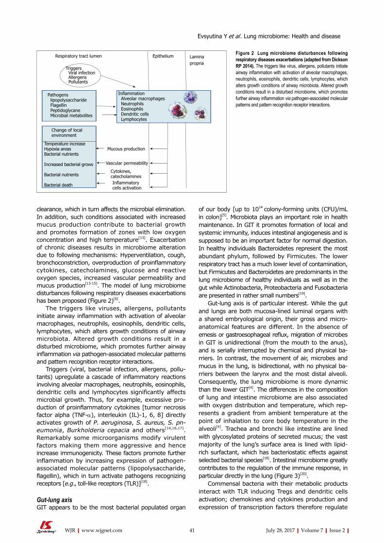
clearance, which in turn affects the microbial elimination. In addition, such conditions associated with increased mucus production contribute to bacterial growth and promotes formation of zones with low oxygen concentration and high temperature[13]. Exacerbation of chronic diseases results in microbiome alteration due to following mechanisms: Hyperventilation, cough, bronchoconstriction, overproduction of proinflammatory cytokines, catecholamines, glucose and reactive oxygen species, increased vascular permeability and mucus production[13-15]. The model of lung microbiome disturbances following respiratory diseases exacerbations has been proposed (Figure 2)[6].
The triggers like viruses, allergens, pollutants initiate airway inflammation with activation of alveolar macrophages, neutrophils, eosinophils, dendritic cells, lymphocytes, which alters growth conditions of airway microbiota. Altered growth conditions result in a disturbed microbiome, which promotes further airway inflammation via pathogen-associated molecular patterns and pattern recognition receptor interactions.
Triggers (viral, bacterial infection, allergens, pollu-tants) upregulate a cascade of inflammatory reactions involving alveolar macrophages, neutrophils, eosinophils, dendritic cells and lymphocytes significantly affects microbial growth. Thus, for example, excessive pro-duction of proinflammatory cytokines [tumor necrosis factor alpha (TNF-α), interleukin (IL)-1, 6, 8] directly activates growth of P. aeruginosa, S. aureus, S. pneumonia, Burkholderia cepacia and others[14,16, 17]. Remarkably some microorganisms modify virulent factors making them more aggressive and hence increase immunogenicity. These factors promote further inflammation by increasing expression of pathogen-associated molecular patterns (lipopolysaccharide, flagellin), which in turn activate pathogens recognizing receptors [e.g., toll-like receptors (TLR)][18].
Gut-lung axisGIT appears to be the most bacterial populated organ
of our body [up to 1014 colony-forming units (CFU)/mL in colon][5]. Microbiota plays an important role in health maintenance. In GIT it promotes formation of local and systemic immunity, induces intestinal angiogenesis and is supposed to be an important factor for normal digestion. In healthy individuals Bacteroidetes represent the most abundant phylum, followed by Firmicutes. The lower respiratory tract has a much lower level of contamination, but Firmicutes and Bacteroidetes are predominants in the lung microbiome of healthy individuals as well as in the gut while Actinobacteria, Proteobacteria and Fusobacteria are presented in rather small numbers[19].
Gut-lung axis is of particular interest. While the gut and lungs are both mucosa-lined luminal organs with a shared embryological origin, their gross and micro-anatomical features are different. In the absence of emesis or gastroesophageal reflux, migration of microbes in GIT is unidirectional (from the mouth to the anus), and is serially interrupted by chemical and physical ba-rriers. In contrast, the movement of air, microbes and mucus in the lung, is bidirectional, with no physical ba-rriers between the larynx and the most distal alveoli. Consequently, the lung microbiome is more dynamic than the lower GIT[4]. The differences in the composition of lung and intestine microbiome are also associated with oxygen distribution and temperature, which rep-resents a gradient from ambient temperature at the point of inhalation to core body temperature in the alveoli[4]. Trachea and bronchi like intestine are lined with glycosylated proteins of secreted mucus; the vast majority of the lung’s surface area is lined with lipid-rich surfactant, which has bacteriostatic effects against selected bacterial species[18]. Intestinal microbiome greatly contributes to the regulation of the immune response, in particular directly in the lung (Figure 3)[20].
Commensal bacteria with their metabolic products interact with TLR inducing Tregs and dendritic cells activation; chemokines and cytokines production and expression of transcription factors therefore regulate
41WJR|www.wjgnet.com July 28, 2017|Volume 7|Issue 2|
Respiratory tract lumen Epithelium Lamina propria
Triggers Viral infection Allergens Pollutants
Inflammation Alveolar macrophages Neutrophils Eosinophils Dendritic cells Lymphocytes
Mucous production
Vascular permeability
Cytokines, catecholaminesInflammatory cells activation
Pathogens lipopolysaccharide Flagellin Peptidoglycane Microbial metabolites
Change of local environment
Temperature increaseHypoxia areasBacterial nutrients
Increased bacterial grows
Bacterial nutrients
Bacterial death
Figure 2 Lung microbiome disturbances following respiratory diseases exacerbations (adapted from Dickson RP 2014). The triggers like virus, allergens, pollutants initiate airway inflammation with activation of alveolar macrophages, neutrophils, eosinophils, dendritic cells, lymphocytes, which alters growth conditions of airway microbiota. Altered growth conditions result in a disturbed microbiome, which promotes further airway inflammation via pathogen-associated molecular patterns and pattern recognition receptor interactions.
Evsyutina Y et al . Lung microbiome: Health and disease

42WJR|www.wjgnet.com
It was found that mice fed with low-fiber diet had decreased levels of SCFAs and higher prevalence of allergic reactions in the respiratory tract[27]. The administration of probiotics was associated with IL-10 secretion by DCs, which promoted T-regs differentiation, causing shift to the Th1 response[26]. Bacterial colonization in sterile mice lead to stimulation of the secretory IgA and CD4+ T-cells, reducing the IgE levels[22].
There is a strong correlation between the bacterial composition of the GIT in infancy and asthma phenotype in childhood[27,28]. Low total microbiome diversity of the colon during the first month of life was shown to be linked with bronchial asthma development at the age of 7 years. Also decrease in Bifidobacteria and an increase in the number of Clostridia in the colon at the early age were associated with the subsequent development of bronchial asthma[28]. In mice models it was found that the use of antibacterial drugs in the first 3 wk of life worsens the course of allergic respiratory inflammations in adulthood[29].
Microbiome and respiratory diseasesCurrently, the role of lung microbiome in respiratory pathology is being discussed. Lung microbiome tr-ansformation, particularly reduction of probiotic sp-ecies and potential increase of pathogenic bacteria appears to be the fundamental factor for susceptibility, chronization and progression of respiratory diseases. In recent study Bacteroidetes, mostly Prevotella spp., was significantly more common in healthy individuals, whereas Pseudomonas was frequently found in the lower respiratory tract of COPD patients[30]. A smaller diversity of bacteria in patients with COPD was also observed. Another study showed that Streptococcus, Prevotella, Fusobacterium and Veillonella were prevalent in individuals without COPD, while Pseudomonas and Haemophilus were dominated in COPD patients
immune response[20-22].It is a well-known that asthma, chronic cough, COPD,
and idiopathic pulmonary fibrosis can be associated with GERD. Acid-suppression medications, including proton pump inhibitors (PPIs), are some of the most prescribed medications in patients with GERD. Rosen et al[23] investigated the impact of acid-suppression medication in children ages 1 to 18 years with chronic cough on gastric and lung microbiome. No significant differences in the prevalence of various bacterial genera or the median concentration of total bacteria in the lungs between treated and untreated patients were shown. There were positive correlations between proximal nonacid reflux burden and lung concentrations of Bacillus, Dermabacter, Lactobacillus, Peptostreptococcus, and Capnocytophagia. These results could be evidence of reflux influence on lung microbiome, but further studies are needed.
The effect of the bacterial metabolites, in particular short chain fatty acids (SCFAs) on modulation of the immune response is one of the most discussed topics. SCFAs act directly on the epithelial and immune cells, contributing to powerful anti-inflammatory effects[20-22,24]. SCFAs were shown to modulate the activity of NFkB, reduce TNF-α production and downregulate the PRRs stimulation (pattern recognition receptors)[21,22]. Po-stulated that the ability of SCFAs to interact with certain G-binding receptors of neutrophils depend on their profile which defined by bacterial composition. Stimulation of Ffar2 (GPR43) receptor was associated with decreased level of eosinophils and reduced bronchoconstriction compared with the Ffar3 (or GPR41) stimulation, wh-ich was associated with increased production of pro-inflammatory mediators[25]. SCFAs were also shown to downregulate expression of CD-markers on the surface of tissue specific DCs[26]. Depressed expression of costimulatory molecules CD80, CD86 and CD40 modify the DCs ability to interact with regulatory T-cells (T-regs).
July 28, 2017|Volume 7|Issue 2|
LungCD4+ Th17
Intestine
Healthy Microbiota LPS
TLR
NF-kB
DC migration to the mesenteric lymph node
IL-6, TNF-α
CD4+
Treg Th17 Th1
AllergenCigarette smokeInfection
Bacterial Methabolites (SCFAs)
Lympha
tic and
Circulato
ry Sys
tems
IL-6, INF-γ, TNF-α
SCFAs
CD4+
Treg
Th17
Th1
Figure 3 Model of intestinal microbiome effects on lung immunology (adapted from Samuelson DR 2015). Microbes in the intestine is sampled by dendritic cells (DCs) either directly from the lumen or following translocation through M-cells to the gut-associated lymphoid tissue. A combination of signals from the microbes results in phenotypic changes in the DCs. DCs promote activation of various T-cell subsets within the mesenteric lymph nodes (MLN) and production of regulatory cytokines. Following the immune challenge in the airways T-cells activated in the gastrointestinal associated lymphoid tissue (GALT) and MLN move to the respiratory mucosa where they promote protective and anti-inflammatory responses. Production of various bacterial metabolites (e.g., SCFAs) also affects the gut-lung axis, as these products get to the lung, where they can alter the levels of inflammation. SCFA: Short chain fatty acid; IL: Interleukin; TNF: Tumor necrosis factor.
Evsyutina Y et al . Lung microbiome: Health and disease

43WJR|www.wjgnet.com
microbiome[31]. International research revealed that in COPD patients Streptococcus sp. and Haemophilus sp. were associated with decreased pulmonary fun-ction, while low level of FEV1 was a predictor of bacterial diversity reduction[32]. COPD exacerbation increases the number of Proteobacteria (Moraxellaceae, Pas-teurellaceae, Pseudomonadaceae, Enterobacteriaceae) and reduces the amount of Actinobacteria, Clostridia and Bacteroidia[33]. It is interesting that Actinobacteria produces metabolites with antimicrobial activity and classes IV and XIVa Clostridia known to be inducers of anti-inflammatory T-regs[34]. Treatment strategy was shown to modify lung microbiome in respiratory diseases. Thus antibacterial therapy in patients with COPD exacerbation results in reducing number of Proteobacteria. Administration of corticosteroids increases their number, as well as the number of Bacteroidetes and Firmicutes, especially Enterobacteriaceae (more than 16 times), Lachnospiraceae, Burkholderiaceae and Neisseriaceae. Combination therapy with corticosteroids and antibiotics leads to increase of Proteobacteria[33].
An attempt to prove the etiological role of the microbial composition of the respiratory tract in COPD development was made. In mice models, reduction in the microbial diversity significantly increases the number of Pseudomonas genera, Lactobacillus, Chryseobacterium and reduction Prevotella. Also there was a marked enhancement of the inflammatory response which included the formation of lymphoid follicles in the lung tissue, increased production of IL-17A, which level was positively correlated with limited airflow and COPD progress[35]. Finally broncho-alveolar lavage fluid (BALF) of such animals was intranasally translocated to sterile and antibiotic-treated mice, as a result an increase in the number of cells producing IL-17A in the lung tissue, particularly CD4+ T cells in the recipients were noted[36].
One of the early studies confirmed the role of microbiome in the bronchial asthma development was done in Denmark. The presence of Moraxella cata-rrhalis, Haemophilus influenzae and Streptococcus pneumoniae in the oropharynx of children of 1-mo age significantly increased the risk of bronchial asthma development[37]. Pathologic role of mentioned bacteria in asthma development had been confirmed in more recent studies[38]. Asthmatic patients were found to have higher number of pathogenic proteobacteria (e.g., Haemophilus) and significantly lesser Bacteroidetes, especially of genus Prevotella compared to healthy individuals[5]. The prevalence of families Comamonadaceae, Sphin-gomonadaceae, Oxatobacteraceae was shown to correlate with bronchial hyperresponsiveness. Inter-estingly, colonization with certain pathogenic bacteria is strictly associated with an immune response in newborns. Thus at high amounts of M. catarrhalis and H. influenzae production of IL-1, IL-17 increases. Also prevalence of S. aureus leads to overproduction of IL-17[39].
The microbiome composition varies in patients depending on the disease severity. Thus in patients with severe bronchial asthma when compared with those
with mild to moderate severity, there is a significantly higher (7-8 times) number of Klebsiella[40]. In a recently published study healthy individuals when compared to patients with mild bronchial asthma were shown to have a decreased number of Bacteroidetes such as Prevotella spp.[41]. At the same time the number of pathogenic Proteobacteria, including Neisseria and Moraxella spp., were 2-times higher in patients with mild bronchial asthma. Bacteroidetes (OR = 0.62) and Fusobacteria (OR = 0.38) were decreased in patients with severe bronchial asthma, compared to the control group. Significant increase in Firmicutes, consisting mainly of streptococci in comparison with healthy individuals and patients with mild bronchial asthma (OR = 2.15 for both comparisons) was observed. Also there was a positive association between the severity of bronchial asthma and the level of Streptococcus (Streptococcus spp., Streptococcus_23 and Streptococcus_155) and negative with the level of Prevotella spp.
Imbalance in the oropharyngeal flora was found to decrease resistance, increase bacterial colonization and dissemination of the potential pathogen in the airway and pneumonia development. Oropharyngeal microbiome of healthy individuals and patients with pneumonia in two age groups: 18-59 years old and group 60 years and older were compared. Three microbial profiles associated with pneumonia in both age groups were revealed: prevalence of bacteria genus Streptococcus, Rothia and Lactobacillus. At the same time in healthy individuals, the microbiome was dominated by Veilonella, Prevotella, Leptotrichia and Gemellales. Moreover, the overall number of viruses in the microbiome of patients with pneumonia significantly increased. The composition of the microbiome was less diverse, while bacterial load was significantly higher which was also correlated with the disease severity. Furthermore the number of Anaerobes, Bacteroides decreases with age, while overgrowth of lactobacilli was noted[42].
Several studies have shown protective role of the intestinal microbiota in the course of pneumonia. The role of normal gut microbiota in mice, particularly segmented filamentous bacteria (SFB) in the course of pneumonia caused by S. aureus was studied. It was shown that the number of CFU of S. aureus in the lungs and spleen were significantly higher in SFB-negative mice in comparison with SFB-positive mice and the clearance of pathogenic bacteria in SFB-negative mice was reduced. In addition, the bacterial load decreased in SFB-negative mice when they were co-housed with healthy mice and similarly after fecal transplantation from healthy mice. All SFB-negative infected mice died within 36 h, whereas the survival rate in mice with normal gut microbiota was 70%[43]. In another research the role of microbiota during the course of pneumococcal pneumonia was studied[44]. Microbiota-depleted mice were shown to have an increase in bacterial dissemination, inflammatory response, organ damage, higher mortality due to pneumonia, impaired phagocytic activity of alveolar macrophages, whereas after subsequent fecal transplantation from healthy mice, there
July 28, 2017|Volume 7|Issue 2|
Evsyutina Y et al . Lung microbiome: Health and disease

44WJR|www.wjgnet.com
were cytokines normalization (TNF-α, IL-6 and IL-10) and an accelerated elimination of Str. pneumoniae.
Place of probiotics, prebiotics, and synbiotics in respiratory pathology treatmentSeveral studies confirm that antibiotic administration can result in gut microbiota dysbiosis. Broad-spectrum antibiotics can affect the bacterial abundance in the gut causing rapid and significant decrease in taxonomic richness and diversity. Thus Jernberg et al[45] documented a decline in the clonal diversity of Bacteroides isolates, insurgence of antibiotic-resistant strains, and upregulation of antibiotic resistance genes in healthy volunteers treated for 1 wk with clindamycin. These effects persisted up to 2 years after treatment[45]. In another study vancomycin has been shown to cause long-lasting susceptibility to secondary infections in humans and mice. Vancomycin markedly disrupted the microbiota, leading to prolonged loss of resistance to C. difficile infection and dense colonization by vancomycin-resistant Enterococcus, K. pneumoniae, and E. coli[46].
In mouse models antibiotic administration during the perinatal period changes the lung microbial composition towards Th2 (vancomycin) or Th17 immune responses (streptomycin)[47].
However, some antibiotics like azithromycin could reduce pulmonary inflammation and exacerbations in patients with COPD. In the recent randomized, double-blind, placebo-controlled trial of 20 smokers (current or ex-smokers) with emphysema and CORD, administration of azithromycin 250 mg daily for 8 wk compared with placebo led to reduce invivo levels of chemokine (C-X-C) ligand 1 (CXCL1), TNF-α, IL-13 and IL-12 p40 in BAL, but increase levels of bacterial metabolites such as glycolic acid, indol-3-acetate and linoleic acid. Azithromycin treatment altered both lung microbiota and metabolome, affecting anti-inflammatory bacterial metabolites that
may contribute to its therapeutic effects[48]. The relationship between respiratory pathology
and the changes in the microbiome composition pre-disposed the use of probiotics. Anti-inflammatory ef-fects of Lactobacillus rhamnosus and Bifidobacterium breve in smokers were evaluated. Both probiotic strains significantly inhibited nicotine-mediated production of IL-1B, IL-6, IL-10, TNF-α, activation of the NF-kB as well as TLR4 and TLR9-induced expression of IL-8[49]. Use of Lactobacillus rhamnosus, Bifidobacterium lactis and Bifidobacterium breve in bronchial asthma resulted in reduction of allergic reactions[50,51]. Lactobacillus reuteri ATCC 23272 and Lactobacillus rhamnosus GG (LGG) administration leads to a significant reduction in the inflammatory cells of BALF[52]. The use of probiotic bacteria LGG and Lactobacillus casei (Sirota and DN 114 001 strain) showed high-potency for the prevention and treatment of both bacterial and viral infections of the respiratory tract[53]. The introduction of Enterococcus faecalis FK-23 in mice reduced the frequency of bronchial asthma exacerbations because of its ability to suppress T-lymphocytes and cytokine production[54].
Treatment of Klebsiella pneumoniae infected mice with Bifidobacterium longum, leads to more rapid resolution of inflammation, decrease in mortality, which has been associated with increased production of IL-10, lower levels of TNF-α and IL-6. Also in the group of mice, treated with probiotics, the ability of alveolar macrophages to produce reactive oxygen species was significantly higher when compared with the control group[55].
Probiotics are now used to treat and control a variety of gastrointestinal diseases including diarrhea, inflammatory bowel disease, irritable bowel syndrome, liver diseases. In rodent models, administration of probiotics prevents chronic stress-induced bacterial translocation[56], colorectal hypersensitivity[57], and
July 28, 2017|Volume 7|Issue 2|
Table 1 Probiotics and synbiotics in respiratory diseases
Probiotics/synbiotics Medical condition Results Source
Lactobacillus rhamnosus, Bifidobacterium breve
Smokers Inhibition of nicotine -mediated IL-1β, IL-6, IL-10, TNF-α production, NF-KB, TLR4 and TLR9-
induced expression of IL-8 activation
Mortaz et al[49], 2015
Lactobacillus rhamnosus, Bifidobacterium lactis, Bifidobacterium breve
Allergic asthma Antigen-specific Tregs activation Sagar et al[50], 2014Jang et al[51], 2012
Lactobacillus reuteri АТСС 23272Lactobacillus rhamnosus GG
Allergy Significant reduction of inflammatory cells in BALF, increasing CD4+CD25+Foxp3+ Treg in
spleen and mediastinal lymph nodes
Forsythe et al[52], 2007
Lactobacillus rhamnosus GGLactobacillus casei (Sirota and DN 114001)
Acute infectious respiratory diseases
Increasing of IgА- secreting cells in bronchial mucosa
Tapiovaara et al[53], 2016
Enterococcus faecalisFK-23
Asthma Suppression of T-cells and cytokines production Zhang et al[54], 2012
Bifidobacterium longum Klebsiella-induced pneumoniae
Increased production of IL-10, decrease of TNF-α and IL-6 levels
Viera et al[55], 2016
Acidic oligosaccharides P. aeruginosa-induced infection
Increase in IL-10 production, decrease in cytotoxic T lymphocyte production
Bernard et al[62], 2015
Bifidobacterium breve M-16V galacto- oligosaccharides fructo- oligosaccharides
Allergic asthma Significant increase in peak expiratory flow rate and reduction of IL-5 production
van de Pol et al[63], 2011
IL: Interleukin; TNF: Tumor necrosis factor.
Evsyutina Y et al . Lung microbiome: Health and disease

45WJR|www.wjgnet.com
restored intestinal barrier dysfunction[58].The overall effects of prebiotics are similar to those of
probiotics. It was shown that prebiotics provide optimal facilities for functional capacity of resident microbiota, stimulate different biochemical reactions within intestinal microbiome, promoting proliferation and renewal of intestinal cells and therefore prebiotics appear to be an active and important part of gut-lung axis.
Prebiotics can ameliorate gut microbiota. Most prebiotics, including inulin and fructo-oligosaccharides are digested by Bifidobacteria and stimulate growth of their colonies[59]. These bacteria influence homeostasis of intestinal cells and inhibit the growth of pathogenic bacteria[60]. Moreover, SCFAs such as propionic acid, acetic acid, and butyric acid reduce the development of gastrointestinal disorders by inducing apoptosis[61]. Additionally SCFAs are important participants in ma-croorgsnism’s immune system modulation as it was mentioned above[20-27].
As can be seen from the above prebiotics and probiotics are essentially different biological structures but their effects are mutually reinforcing.
The use of pre- and synbiotics in the treatment of the respiratory diseases was also studied. Effect of acidic oligosaccharides in the treatment of mice infected with P.aeruginosa was investigated. A significant reduction in mortality, an increase in IL-10 production was achieved. Diminished production of cytotoxic T-lymphocytes was revealed and as a result, reduction in the severity of inflammation and limitation of tissue damage was observed. In re-infected with Pseudomonas aeruginosa mice, the bacterial load in the lung tissue was lower when compared with the control group[62]. In the study on patients with allergic bronchial asthma treatment with synbiotic, containing Bifidobacterium breve M-16V showed a significant increase in peak expiratory flow rate and reduction in the production of IL-5 when compared with the placebo group[63]. Data summarized in Table 1.
CONCLUSIONThe status of the lung microbiome is normally de-termined by the relationship between microbial immi-gration, elimination and local conditions of bacterial growth. The results of studies indicate changes both in the lung and the intestinal microbiome in patients with respiratory diseases, occurring due to imbalance between the factors mentioned above. The studies on human microbiome have aroused great interest in application of probiotics for the prevention and treatment of somatic diseases. However, there is a necessity of further studies to determine the appropriate dose, selecting an optimal bacterial strain, duration of treatment, as well as groups of patients that will provide desirable effect in the prevention and/or treatment of a particular disease. Studies on mice models have shown a positive effect of probiotics on the course of pneumonia, acute exacerbation of bronchial asthma and COPD, which dictates the need for its research on human population.
It gives hope that the treatment of these diseases might be improved in the nearest future.
REFERENCES1 Charlson ES, Bittinger K, Haas AR, Fitzgerald AS, Frank I, Yadav
A, Bushman FD, Collman RG. Topographical continuity of bacterial populations in the healthy human respiratory tract. Am J Respir Crit Care Med 2011; 184: 957-963 [PMID: 21680950 DOI: 10.1164/rccm.201104-0655OC]
2 Charlson ES, Bittinger K, Chen J, Diamond JM, Li H, Collman RG, Bushman FD. Assessing bacterial populations in the lung by replicate analysis of samples from the upper and lower respiratory tracts. PLoS One 2012; 7: e42786 [PMID: 22970118 DOI: 10.1371/journal.pone.0042786]
3 Huang YJ, Charlson ES, Collman RG, Colombini-Hatch S, Martinez FD, Senior RM. The role of the lung microbiome in health and disease. A National Heart, Lung, and Blood Institute workshop report. Am J Respir Crit Care Med 2013; 187: 1382-1387 [PMID: 23614695 DOI: 10.1164/rccm.201303-0488WS]
4 Dickson RP, Huffnagle GB. The Lung Microbiome: New Principles for Respiratory Bacteriology in Health and Disease. PLoS Pathog 2015; 11: e1004923 [PMID: 26158874 DOI: 10.1371/journal.ppat.1004923]
5 Hilty M, Burke C, Pedro H, Cardenas P, Bush A, Bossley C, Davies J, Ervine A, Poulter L, Pachter L, Moffatt MF, Cookson WO. Disordered microbial communities in asthmatic airways. PLoS One 2010; 5: e8578 [PMID: 20052417 DOI: 10.1371/journal.pone.0008578]
6 Dickson RP, Erb-Downward JR, Huffnagle GB. Towards an ecology of the lung: new conceptual models of pulmonary microbiology and pneumonia pathogenesis. Lancet Respir Med 2014; 2: 238-246 [PMID: 24621685 DOI: 10.1016/S2213-2600(14)70028-1]
7 Bowers RM, Sullivan AP, Costello EK, Collett JL Jr, Knight R, Fierer N. Sources of bacteria in outdoor air across cities in the midwestern United States. Appl Environ Microbiol 2011; 77: 6350-6356 [PMID: 21803902 DOI: 10.1128/AEM.05498-11]
8 Bertolini V, Gandolfi I, Ambrosini R, Bestetti G, Innocente E, Rampazzo G, Franzetti A. Temporal variability and effect of environmental variables on airborne bacterial communities in an urban area of Northern Italy. Appl Microbiol Biotechnol 2013; 97: 6561-6570 [PMID: 23053100 DOI: 10.1007/s00253-012-4450-0]
9 Dickson RP, Erb-Downward JR, Freeman CM, Walker N, Scales BS, Beck JM, Martinez FJ, Curtis JL, Lama VN, Huffnagle GB. Changes in the lung microbiome following lung transplantation include the emergence of two distinct Pseudomonas species with distinct clinical associations. PLoS One 2014; 9: e97214 [PMID: 24831685 DOI: 10.1371/journal.pone.0097214]
10 Wu H, Kuzmenko A, Wan S, Schaffer L, Weiss A, Fisher JH, Kim KS, McCormack FX. Surfactant proteins A and D inhibit the growth of Gram-negative bacteria by increasing membrane permeability. J Clin Invest 2003; 111: 1589-1602 [PMID: 12750409 DOI: 10.1172/JCI16889]
11 Coxson HO, Rogers RM, Whittall KP, D’yachkova Y, Paré PD, Sciurba FC, Hogg JC. A quantification of the lung surface area in emphysema using computed tomography. Am J Respir Crit Care Med 1999; 159: 851-856 [PMID: 10051262 DOI: 10.1164/ajrccm.159.3.9805067]
12 Raghu G, Freudenberger TD, Yang S, Curtis JR, Spada C, Hayes J, Sillery JK, Pope CE 2nd, Pellegrini CA. High prevalence of abnormal acid gastro-oesophageal reflux in idiopathic pulmonary fibrosis. Eur Respir J 2006; 27: 136-142 [PMID: 16387946 DOI: 10.1183/09031936.06.00037005]
13 Schmidt A, Belaaouaj A, Bissinger R, Koller G, Malleret L, D’Orazio C, Facchinelli M, Schulte-Hubbert B, Molinaro A, Holst O, Hammermann J, Schniederjans M, Meyer KC, Damkiaer S, Piacentini G, Assael B, Bruce K, Häußler S, LiPuma JJ, Seelig J, Worlitzsch D, Döring G. Neutrophil elastase-mediated increase in airway temperature during inflammation. J Cyst Fibros 2014; 13: 623-631 [PMID: 24713593 DOI: 10.1016/j.jcf.2014.03.004]
July 28, 2017|Volume 7|Issue 2|
Evsyutina Y et al . Lung microbiome: Health and disease

46WJR|www.wjgnet.com
14 Marks LR, Davidson BA, Knight PR, Hakansson AP. Interkingdom signaling induces Streptococcus pneumoniae biofilm dispersion and transition from asymptomatic colonization to disease. MBio 2013; 4: [PMID: 23882016 DOI: 10.1128/mBio.00438-13]
15 Sass AM, Schmerk C, Agnoli K, Norville PJ, Eberl L, Valvano MA, Mahenthiralingam E. The unexpected discovery of a novel low-oxygen-activated locus for the anoxic persistence of Burkholderia cenocepacia. ISME J 2013; 7: 1568-1581 [PMID: 23486248 DOI: 10.1038/ismej.2013.36]
16 Freestone PP, Hirst RA, Sandrini SM, Sharaff F, Fry H, Hyman S, O’Callaghan C. Pseudomonas aeruginosa-catecholamine inotrope interactions: a contributory factor in the development of ventilator-associated pneumonia? Chest 2012; 142: 1200-1210 [PMID: 22556319 DOI: 10.1378/chest.11-2614]
17 Kaza SK, McClean S, Callaghan M. IL-8 released from human lung epithelial cells induced by cystic fibrosis pathogens Burkholderia cepacia complex affects the growth and intracellular survival of bacteria. Int J Med Microbiol 2011; 301: 26-33 [PMID: 20829108 DOI: 10.1016/j.ijmm.2010.06.005]
18 Dickson RP, Martinez FJ, Huffnagle GB. The role of the microbiome in exacerbations of chronic lung diseases. Lancet 2014; 384: 691-702 [PMID: 25152271 DOI: 10.1016/S0140-6736(14)61136-3]
19 Marsland BJ, Trompette A, Gollwitzer ES. The Gut-Lung Axis in Respiratory Disease. Ann Am Thorac Soc2015; 12 Suppl 2: S150-S156 [PMID: 26595731 DOI: 10.1513/AnnalsATS.201503-133AW]
20 Samuelson DR, Welsh DA, Shellito JE. Regulation of lung immunity and host defense by the intestinal microbiota. Front Microbiol 2015; 6: 1085 [PMID: 26500629 DOI: 10.3389/fmicb. 2015.01085]
21 Rogers GB, Wesselingh S. Precision respiratory medicine and the microbiome. Lancet Respir Med 2016; 4: 73-82 [PMID: 26700443 DOI: 10.1016/S2213-2600(15)00476-2]
22 Gallacher DJ, Kotecha S. Respiratory Microbiome of New-Born Infants. Front Pediatr 2016; 4: 10 [PMID: 26942168 DOI: 10.3389/fped.2016.00010]
23 Rosen R, Amirault J, Liu H, Mitchell P, Hu L, Khatwa U, Onderdonk A. Changes in gastric and lung microflora with acid suppression: acid suppression and bacterial growth. JAMA Pediatr 2014; 168: 932-937 [PMID: 25133779 DOI: 10.1001/jamapediatrics.2014.696]
24 Venkataraman A, Bassis CM, Beck JM, Young VB, Curtis JL, Huffnagle GB, Schmidt TM. Application of a neutral community model to assess structuring of the human lung microbiome. MBio 2015; 6: e02284-14 [PMID: 25604788 DOI: 10.1128/mBio.02 284-14]
25 Ganesh BP, Versalovic J. Luminal Conversion and Immun-oregulation by Probiotics. Front Pharmacol 2015; 6: 269 [PMID: 26617521 DOI: 10.3389/fphar.2015.00269]
26 Larsen JM, Steen-Jensen DB, Laursen JM, Søndergaard JN, Musavian HS, Butt TM, Brix S. Divergent pro-inflammatory profile of human dendritic cells in response to commensal and pathogenic bacteria associated with the airway microbiota. PLoS One 2012; 7: e31976 [PMID: 22363778 DOI: 10.1371/journal.pone.0031976]
27 Trompette A, Gollwitzer ES, Yadava K, Sichelstiel AK, Sprenger N, Ngom-Bru C, Blanchard C, Junt T, Nicod LP, Harris NL, Marsland BJ. Gut microbiota metabolism of dietary fiber influences allergic airway disease and hematopoiesis. Nat Med 2014; 20: 159-166 [PMID: 24390308 DOI: 10.1038/nm.3444]
28 Abrahamsson TR, Jakobsson HE, Andersson AF, Björkstén B, Engstrand L, Jenmalm MC. Low gut microbiota diversity in early infancy precedes asthma at school age. Clin Exp Allergy 2014; 44: 842-850 [PMID: 24330256 DOI: 10.1111/cea.12253]
29 Russell SL, Gold MJ, Willing BP, Thorson L, McNagny KM, Finlay BB. Perinatal antibiotic treatment affects murine microbiota, immune responses and allergic asthma. Gut Microbes 2013; 4: 158-164 [PMID: 23333861 DOI: 10.4161/gmic.23567]
30 Einarsson GG, Comer DM, McIlreavey L, Parkhill J, Ennis M, Tunney MM, Elborn JS. Community dynamics and the lower airway microbiota in stable chronic obstructive pulmonary disease, smokers and healthy non-smokers. Thorax 2016; 71: 795-803 [PMID:
27146202 DOI: 10.1136/thoraxjnl-2015-207235]31 Erb-Downward JR, Thompson DL, Han MK, Freeman CM,
McCloskey L, Schmidt LA, Young VB, Toews GB, Curtis JL, Sundaram B, Martinez FJ, Huffnagle GB. Analysis of the lung microbiome in the “healthy” smoker and in COPD. PLoS One 2011; 6: e16384 [PMID: 21364979 DOI: 10.1371/journal.pone.0016384]
32 Han MK, Erb-Downward JR, Wang X. An International Study of the Lung Microbiome in Chronic Obstructive Pulmonary Disease (COPD). American Thoracic Society International Conference Abstracts. C93. It won’t be long: emerging concepts in copd pathobiology. May 18-23, 2007, San Francisco, California, USA
33 Huang YJ, Sethi S, Murphy T, Nariya S, Boushey HA, Lynch SV. Airway microbiome dynamics in exacerbations of chronic obstructive pulmonary disease. J Clin Microbiol 2014; 52: 2813-2823 [PMID: 24850358 DOI: 10.1128/JCM.00035-14]
34 Atarashi K, Tanoue T, Shima T, Imaoka A, Kuwahara T, Momose Y, Cheng G, Yamasaki S, Saito T, Ohba Y, Taniguchi T, Takeda K, Hori S, Ivanov II, Umesaki Y, Itoh K, Honda K. Induction of colonic regulatory T cells by indigenous Clostridium species. Science 2011; 331: 337-341 [PMID: 21205640 DOI: 10.1126/science.1198469]
35 Zhang J, Chu S, Zhong X, Lao Q, He Z, Liang Y. Increased expression of CD4+IL-17+ cells in the lung tissue of patients with stable chronic obstructive pulmonary disease (COPD) and smokers. Int Immunopharmacol 2013; 15: 58-66 [PMID: 23127823 DOI: 10.1016/j.intimp.2012.10.018]
36 Yadava K, Pattaroni C, Sichelstiel AK, Trompette A, Gollwitzer ES, Salami O, von Garnier C, Nicod LP, Marsland BJ. Microbiota Promotes Chronic Pulmonary Inflammation by Enhancing IL-17A and Autoantibodies. Am J Respir Crit Care Med 2016; 193: 975-987 [PMID: 26630356 DOI: 10.1164/rccm.201504-0779OC]
37 Bisgaard H, Hermansen MN, Buchvald F, Loland L, Halkjaer LB, Bønnelykke K, Brasholt M, Heltberg A, Vissing NH, Thorsen SV, Stage M, Pipper CB. Childhood asthma after bacterial colonization of the airway in neonates. N Engl J Med 2007; 357: 1487-1495 [PMID: 17928596 DOI: 10.1056/NEJMoa052632]
38 Bisgaard H, Hermansen MN, Bønnelykke K, Stokholm J, Baty F, Skytt NL, Aniscenko J, Kebadze T, Johnston SL. Association of bacteria and viruses with wheezy episodes in young children: prospective birth cohort study. BMJ 2010; 341: c4978 [PMID: 20921080 DOI: 10.1136/bmj.c4978]
39 Følsgaard NV, Schjørring S, Chawes BL, Rasmussen MA, Krogfelt KA, Brix S, Bisgaard H. Pathogenic bacteria colonizing the airways in asymptomatic neonates stimulates topical inflammatory mediator release. Am J Respir Crit Care Med 2013; 187: 589-595 [PMID: 23370914 DOI: 10.1164/rccm.201207-1297OC]
40 Huang YJ, Nariya S, Harris JM, Lynch SV, Choy DF, Arron JR, Boushey H. The airway microbiome in patients with severe asthma: Associations with disease features and severity. J Allergy Clin Immunol 2015; 136: 874-884 [PMID: 26220531 DOI: 10.1016/j.jaci.2015.05.044]
41 Zhang Q, Cox M, Liang Z, Brinkmann F, Cardenas PA, Duff R, Bhavsar P, Cookson W, Moffatt M, Chung KF. Airway Microbiota in Severe Asthma and Relationship to Asthma Severity and Phenotypes. PLoS One 2016; 11: e0152724 [PMID: 27078029 DOI: 10.1371/journal.pone.0152724]
42 de Steenhuijsen Piters WA, Huijskens EG, Wyllie AL, Biesbroek G, van den Bergh MR, Veenhoven RH, Wang X, Trzciński K, Bonten MJ, Rossen JW, Sanders EA, Bogaert D. Dysbiosis of upper respiratory tract microbiota in elderly pneumonia patients. ISME J 2016; 10: 97-108 [PMID: 26151645 DOI: 10.1038/ismej.2015.99]
43 Gauguet S, D’Ortona S, Ahnger-Pier K, Duan B, Surana NK, Lu R, Cywes-Bentley C, Gadjeva M, Shan Q, Priebe GP, Pier GB. Intestinal Microbiota of Mice Influences Resistance to Staphylococcus aureus Pneumonia. Infect Immun 2015; 83: 4003-4014 [PMID: 26216419 DOI: 10.1128/IAI.00037-15]
44 Schuijt TJ, Lankelma JM, Scicluna BP, de Sousa e Melo F, Roelofs JJ, de Boer JD, Hoogendijk AJ, de Beer R, de Vos A, Belzer C, de Vos WM, van der Poll T, Wiersinga WJ. The gut microbiota plays a protective role in the host defence against pneumococcal pneumonia. Gut 2016; 65: 575-583 [PMID: 26511795 DOI: 10.1136/gutjnl-
July 28, 2017|Volume 7|Issue 2|
Evsyutina Y et al . Lung microbiome: Health and disease

47WJR|www.wjgnet.com
2015-309728]45 Jernberg C, Löfmark S, Edlund C, Jansson JK. Long-term
ecological impacts of antibiotic administration on the human intestinal microbiota. ISME J 2007; 1: 56-66 [PMID: 18043614 DOI: 10.1038/ismej.2007.3]
46 Lewis BB, Buffie CG, Carter RA, Leiner I, Toussaint NC, Miller LC, Gobourne A, Ling L, Pamer EG. Loss of Microbiota-Mediated Colonization Resistance to Clostridium difficile Infection With Oral Vancomycin Compared With Metronidazole. J Infect Dis 2015; 212: 1656-1665 [PMID: 25920320 DOI: 10.1093/infdis/jiv256]
47 Russell SL, Gold MJ, Reynolds LA, Willing BP, Dimitriu P, Thorson L, Redpath SA, Perona-Wright G, Blanchet MR, Mohn WW, Finlay BB, McNagny KM. Perinatal antibiotic-induced shifts in gut microbiota have differential effects on inflammatory lung diseases. J Allergy Clin Immunol 2015; 135: 100-109 [PMID: 25145536 DOI: 10.1016/j.jaci.2014.06.027]
48 Segal LN, Clemente JC, Wu BG, Wikoff WR, Gao Z, Li Y, Ko JP, Rom WN, Blaser MJ, Weiden MD. Randomised, double-blind, placebo-controlled trial with azithromycin selects for anti-inflammatory microbial metabolites in the emphysematous lung. Thorax 2017; 72: 13-22 [PMID: 27486204 DOI: 10.1136/thoraxjnl-2016-208599]
49 Mortaz E, Adcock IM, Ricciardolo FL, Varahram M, Jamaati H, Velayati AA, Folkerts G, Garssen J. Anti-Inflammatory Effects of Lactobacillus Rahmnosus and Bifidobacterium Breve on Cigarette Smoke Activated Human Macrophages. PLoS One 2015; 10: e0136455 [PMID: 26317628 DOI: 10.1371/journal.pone.0136455]
50 Sagar S, Morgan ME, Chen S, Vos AP, Garssen J, van Bergen-henegouwen J, Boon L, Georgiou NA, Kraneveld AD, Folkerts G. Bifidobacterium breve and Lactobacillus rhamnosus treatment is as effective as budesonide at reducing inflammation in a murine model for chronic asthma. Respir Res 2014; 15: 46 [PMID: 24735374 DOI: 10.1186/1465-9921-15-46]
51 Jang SO, Kim HJ, Kim YJ, Kang MJ, Kwon JW, Seo JH, Kim HY, Kim BJ, Yu J, Hong SJ. Asthma Prevention by Lactobacillus Rhamnosus in a Mouse Model is Associated With CD4(+)CD25(+)Foxp3(+) T Cells. Allergy Asthma Immunol Res 2012; 4: 150-156 [PMID: 22548208 DOI: 10.4168/aair.2012.4.3.150]
52 Forsythe P, Inman MD, Bienenstock J. Oral treatment with live Lactobacillus reuteri inhibits the allergic airway response in mice. Am J Respir Crit Care Med 2007; 175: 561-569 [PMID: 17204726]
53 Tapiovaara L, Pitkaranta A, Korpela R. Probiotics and the Upper Respiratory Tract - A Review. Pediatric Infect Dis 2016; 1: 19 [DOI:
10.4172/PIDO.100019]54 Zhang B, An J, Shimada T, Liu S, Maeyama K. Oral administration
of Enterococcus faecalis FK-23 suppresses Th17 cell development and attenuates allergic airway responses in mice. Int J Mol Med 2012; 30: 248-254 [PMID: 22641478 DOI: 10.3892/ijmm.2012.1010]
55 Vieira AT, Rocha VM, Tavares L, Garcia CC, Teixeira MM, Oliveira SC, Cassali GD, Gamba C, Martins FS, Nicoli JR. Control of Klebsiella pneumoniae pulmonary infection and immunomodulation by oral treatment with the commensal probiotic Bifidobacterium longum 5(1A). Microbes Infect 2016; 18: 180-189 [PMID: 26548605 DOI: 10.1016/j.micinf.2015.10.008]
56 Zareie M, Johnson-Henry K, Jury J, Yang PC, Ngan BY, McKay DM, Soderholm JD, Perdue MH, Sherman PM. Probiotics prevent bacterial translocation and improve intestinal barrier function in rats following chronic psychological stress. Gut 2006; 55: 1553-1560 [PMID: 16638791 DOI: 10.1136/gut.2005.080739]
57 Eutamene H, Lamine F, Chabo C, Theodorou V, Rochat F, Bergonzelli GE, Corthésy-Theulaz I, Fioramonti J, Bueno L. Synergy between Lactobacillus paracasei and its bacterial products to counteract stress-induced gut permeability and sensitivity increase in rats. J Nutr 2007; 137: 1901-1907 [PMID: 17634262]
58 Gareau MG, Jury J, MacQueen G, Sherman PM, Perdue MH. Probiotic treatment of rat pups normalises corticosterone release and ameliorates colonic dysfunction induced by maternal separation. Gut 2007; 56: 1522-1528 [PMID: 17339238 DOI: 10.1136/gut.2006.117176]
59 Przemyslaw J, Tomasik PT. Probiotics and prebiotics. Cereal Chem 2003; 80: 113-117 [DOI: 10.1094/CCHEM.2003.80.2.113]
60 Yoo JY, Kim SS. Probiotics and Prebiotics: Present Status and Future Perspectives on Metabolic Disorders. Nutrients 2016; 8: 173 [PMID: 26999199 DOI: 10.3390/nu8030173]
61 Slavin J. Fiber and prebiotics: mechanisms and health benefits. Nutrients 2013; 5: 1417-1435 [PMID: 23609775 DOI: 10.3390/nu5041417]
62 Bernard H, Desseyn JL, Gottrand F, Stahl B, Bartke N, Husson MO. Pectin-Derived Acidic Oligosaccharides Improve the Outcome of Pseudomonas aeruginosa Lung Infection in C57BL/6 Mice. PLoS One 2015; 10: e0139686 [PMID: 26599638 DOI: 10.1371/journal.pone.0139686]
63 van de Pol MA, Lutter R, Smids BS, Weersink EJ, van der Zee JS. Synbiotics reduce allergen-induced T-helper 2 response and improve peak expiratory flow in allergic asthmatics. Allergy 2011; 66: 39-47 [PMID: 20716319 DOI: 10.1111/j.1398-9995.2010.02454.x]
P- Reviewer: Chow KC, Feltracco P, Turner AM S- Editor: Ji FF L- Editor: A E- Editor: Lu YJ
July 28, 2017|Volume 7|Issue 2|
Evsyutina Y et al . Lung microbiome: Health and disease

© 2017 Baishideng Publishing Group Inc. All rights reserved.
Published by Baishideng Publishing Group Inc7901 Stoneridge Drive, Suite 501, Pleasanton, CA 94588, USA
Telephone: +1-925-223-8242Fax: +1-925-223-8243
E-mail: [email protected] Desk: http://www.f6publishing.com/helpdesk
http://www.wjgnet.com