Embed Size (px)
Citation preview

Isotopologue Profiling of Legionella pneumophilaROLE OF SERINE AND GLUCOSE AS CARBON SUBSTRATES*□S
Received for publication, March 30, 2010, and in revised form, April 23, 2010 Published, JBC Papers in Press, May 4, 2010, DOI 10.1074/jbc.M110.128678
Eva Eylert‡1, Vroni Herrmann§1, Matthieu Jules¶�2, Nadine Gillmaier‡, Monika Lautner§, Carmen Buchrieser¶�,Wolfgang Eisenreich‡3, and Klaus Heuner§4
From the ‡Lehrstuhl fur Biochemie, Technische Universitat Munchen, Lichtenbergstrasse 4, 85747 Garching, Germany, §ResearchGroup P26-Nosocomial Infections of the Elderly, Robert Koch-Institut, Nordufer 20, 13353 Berlin, Germany, the ¶Institut Pasteur,Biologie des Bacteries Intracellulaires, F-75015 Paris, France, and �CNRS, URA 2171, F-75015 Paris, France
Legionella pneumophila (Lp) is commonly found in freshwa-ter habitats but is also the causative agent of Legionnaires’ dis-easewhen infecting humans. Although various virulence factorshave been reported, little is known about the nutrition and themetabolism of the bacterium. Here, we report the application ofisotopologue profiling for analyzing the metabolism of L. pneu-mophila. Cultures of Lp were supplied with [U-13C3]serine,[U-13C6]glucose, or [1,2-13C2]glucose. After growth, 13C enrich-ments and isotopologuepatterns of protein-derived aminoacidsand poly-3-hydroxybutyrate were determined by mass spec-trometry and/or NMR spectroscopy. The labeling patternsdetected in the experiment with [U-13C3]serine showed majorcarbon flux from serine to pyruvate and frompyruvate to acetyl-CoA, which serves as a precursor of poly-3-hydroxybutyrate oras a substrate of a complete citrate cycle with Si specificity of thecitrate synthase.Minor carbon flux was observed between pyru-vate and oxaloacetate/malate by carboxylation and decarboxy-lation, respectively. The apparent lack of label in Val, Ile, Leu,Pro, Phe, Met, Arg, and Tyr confirmed that L. pneumophila isauxotrophic for these amino acids. Experiments with [13C]glu-cose showed that the carbohydrate is also used as a substrate tofeed the central metabolism. The specific labeling patterns dueto [1,2-13C2]glucose identified the Entner-Doudoroff pathwayas the predominant route for glucose utilization. In line withthese observations, amutant lacking glucose-6-phosphatedehy-drogenase (�zwf) did not incorporate label from glucose at sig-nificant levels and was slowly outcompeted by the wild typestrain in successive rounds of infection inAcanthamoeba castel-lanii, indicating the importance of this enzyme and of carbohy-drate usage in general for the life cycle of Lp.
TheGram-negative bacteriumLegionella pneumophila (Lp)5can be found in freshwater habitats where it replicates withinprotozoa, mainly amoebae such as Acanthamoeba castellanii.Lp can also be transmitted to humans by contaminated aero-sols. After entering the human lung, the pathogen is phagocy-tosed by alveolar macrophages wherein it is able to replicate,leading to Legionnaires‘ disease, an atypical pneumonia.Lp survives within amoebae and macrophages because of its
ability to establish a replication vacuole that is derived from theendoplasmic reticulum. Once within a vacuole, Lp differenti-ates into the replicative form.When nutrients become limiting,a regulatory cascade triggers the differentiation to a spore-likemature intracellular form (MIF), the so-called transmissiveform, which metabolically seems to be nearly dormant (1–6).These forms exhibit a thickened cell wall and high amounts ofcytoplasmic granules of poly-3-hydroxybutyrate (PHB), a gen-eral energy and carbon storage compound of bacteria (1, 7, 8).When released from spent hosts, these transmissive forms ofLpare able to persist for long periods in the environment. It hasalso been shown that Lp is able to differentiate into a “viable butnonculturable” status of greatly reducedmetabolic activity afterpersistence in water (9). Notably, Legionella species can alsogrow in defined culture media under laboratory conditions(10–15). However, less is known about the routes of nutrientutilization during growth in culture media as well as duringintracellular multiplication (16–20).Legionella exhibits a strictly respiratory form of metabolism
and does not grow anaerobically (18). The amino acids Arg, Ile,Leu, Val, Met, Ser, and Thr are reportedly essential for growthof Lp in culture (10–12, 14, 15, 21–23), whereas a partialrequirement for Cys (or cystine) has also been observed (24). Itis also well known that Lp uses amino acids as preferred energyand carbon sources (4, 10–12, 14, 21, 22).More specifically, Ser,Glu, Tyr, and/or Thr are efficiently used as carbon and energysources in vitro (14), and Cys, Gln, Ser, and Arg support growthin vivo (19). From in silico analysis of the known Lp genomesequences it is proposed that Lp is auxotrophic for the aminoacids Cys, Met, Arg, Thr, Val, Ile, and Leu (18, 25, 26). There-
* This work was supported by Grants EI 384/4-1 and HE 2845/6-1 from theDeutsche Forschungsgemeinschaft DFG SPP 1316, Bonn, Germany (toW. E. and K. H., respectively) and by the Network of Excellence “Euro-pathogenomics” Grant LSHB-CT-2005-512061 and the Grant PTR 185 fromthe Institut Pasteur and Bio-Rad (to C. B.). M. J. received support from theInstitut Pasteur-Bio-Rad Grant PTR 185.
□S The on-line version of this article (available at http://www.jbc.org) containssupplemental Figs. S1–S3 and Tables S1–S5.
1 Both authors contributed equally to this work.2 Supported by Grant PTR 185 from the Institut Pasteur and Bio-Rad. Present
address: Inst. Micalis, INRA (UMR1319) and AgroParisTech, 78850 Thiver-val-Grignon, France.
3 To whom correspondence may be addressed. Tel.: 49-89-289-13336; Fax:49-89-289-13363; E-mail: [email protected].
4 To whom correspondence may be addressed. Tel.: 49-30-18754-2226; Fax:49-30-18754-2328; E-mail: [email protected].
5 The abbreviations used are: Lp, Legionella pneumophila; PHB, poly-3-hy-droxybutyrate; EMP, Embden-Meyerhof-Parnas; ED, Entner-Doudoroff;GC/MS, gas chromatography/mass spectrometry; PP, pentose phosphate;AYE, ACES-buffered yeast extract; ACES, 2-[(2-amino-2-oxoethyl)amino]-ethanesulfonic acid; BCYE, buffered charcoal-yeast extract; CDM, chemi-cally defined medium; CFU, colony forming unit(s); RT, reverse tran-scriptase; TBDMS, tert-butyldimethylsilyl; WT, wild type.
THE JOURNAL OF BIOLOGICAL CHEMISTRY VOL. 285, NO. 29, pp. 22232–22243, July 16, 2010© 2010 by The American Society for Biochemistry and Molecular Biology, Inc. Printed in the U.S.A.
22232 JOURNAL OF BIOLOGICAL CHEMISTRY VOLUME 285 • NUMBER 29 • JULY 16, 2010
by guest on May 26, 2019
http://ww
w.jbc.org/
Dow
nloaded from

fore, it is not surprising that Lp harbors genes for�12 classes ofABC (ATP-binding cassette) transporters and amino acid per-meases, as well as various different amino peptidases and pro-teases. In addition, it has been shown that amino acid trans-porters of the host cell and of Lp are essential for intracellularreplication; specifically, a neutral amino acid transporter of thehost cell (SLC1A5 of MM6 monocyte cells) is necessary for Lpto replicate within this host (19). Furthermore, a bacterial Thrtransporter (PhtA) is reported to be essential for replication ofLp in bone marrow-derived macrophages from mice (27).In earlier studies, it was also suggested that Lp is able to use
glucose as a carbon source (14, 15, 20), although the addition ofglucose to the medium did not support in vitro growth (18, 20).As no active sugar transport could be demonstrated in vitro (14,16, 18), it is generally believed that Lp does not utilize sugars asa carbon source but relies on gluconeogenesis (18). On theother hand, some of the various ABC-type transport systemsmight be involved in sugar uptake, because Lp also possessesputative systems for degradation of cellulose, chitin, starch, andglycogen (28). Recently, Lp has been shown to actively degradecellulose (29). On the basis of this evidence it can be assumedthat glucose is also catabolized, but themetabolic routes are stillunknown.The four sequenced genomes of Lp (Lp Philadelphia (26), Lp
Paris, Lp Lens (25), and Lp Corby (30) indicate the presence ofthe Embden-Meyerhof-Parnas (EMP) pathway as well as theEntner-Doudoroff (ED) pathway. However, in vitro enzymeassays did not detect activities for the ED pathway in strainsKnoxville-1 and Philadelphia-1 (17, 20). Genes encodingthe pentose phosphate (PP) pathway are also present, with theexception of 6-phosphogluconate dehydrogenase and thetransaldolase (16, 18, 25, 26).As the activity of pyruvate dehydrogenasewas low in cell-free
extracts (17, 20), it was hypothesized that the bulk of the acetyl-CoA entering the citrate cycle or used as precursor for the stor-age compound PHB is derived from fatty acid catabolism (18).Indeed, Lp possesses huge amounts of phospholipases exertingextracellular and cell-associated activities (31–33). However,the glyoxylate shunt appears to be absent in the Lp strainssequenced thus far.One powerful method of studying metabolic pathways in
growing microbes is based on incorporation experiments withstable isotope-labeled precursors (e.g. 13C-labeled glucose) fol-lowed by the determination of the resulting isotopologue pat-terns in keymetabolites, such as protein-derived amino acids orother storage compounds. Using stoichiometric models, isoto-pologue profiles can serve as constraints in metabolic flux cal-culations (13C-based metabolic flux analysis) (34). Metabolicflux analysis is nowwell established for the analysis ofmetabolicflux inmicroorganisms growing under standardized conditions(reviewed in Refs. 34 and 35). Thesemodels typically rely on theuse ofminimalmediawith only one possible carbon source. It istherefore difficult to adapt metabolic flux analysis calculationsto organisms with complex multiple carbon usage (i.e. whengrowing in complexmedia). However, because of the specificityof the detected isotopologue profiles, observation-driven anal-ysis can also trace metabolic pathways in cases with unknownand/or multiple carbon usage (36, 37). In this study, the metab-
olism of Lp grown under culture conditions was analyzed forthe first time by 13C-labeled isotopologue (13C-isotopologue)profiling using glucose or Ser as precursors.
EXPERIMENTAL PROCEDURES
Strains, Growth Conditions, and Media—A. castellaniiATCC30010 was cultured in PYG 712 medium (2% proteose-peptone, 0.1% yeast extract, 0.1 M glucose, 4 mM MgSO4, 0.4 M
CaCl2, 0.1% sodium citrate dihydrate, 0.05mMFe(NH4)2(SO4)2 �6 H2O, 2.5 mM NaH2PO4, and 2.5 mM K2HPO4) at 20 °C.Escherichia coli DH5� was used to clone recombinant plasmidDNA. Experiments were done with L. pneumophila Paris(CIP 107629 (25)). As described previously (28), Lp strains werecultured in AYE medium (ACES-buffered yeast extract broth:10 g of ACES, 10 g of yeast extract, 0.4 g of L-cysteine, and 0.25 gof iron pyrophosphate in 1 liter (pH 6.8)) (see also sup-plemental Table S1) or on ACES-buffered charcoal-yeastextract (BCYE) agar plates at 37 °C. Alternatively, Lp Paris wascultivated in a chemically defined medium (CDM) (for detailssee supplemental Table 1) adapted from Ristroph et al. (12).
13C Labeling Experiments—1 liter of growth medium (AYEor CDM) was supplemented with 2 g of [U-13C6]glucose, 2 g of[1,2-13C2]glucose, or 0.3 g of [U-13C3]Ser. 500ml of the supple-mented AYEmediumwas inoculated with 1 ml of an overnightculture ofLpParis. For supplementedCDMthe inoculumwas 4ml of an overnight culture grown in AYE medium. Incubationwas carried out at 37 °C and 220 rpm, and the optical density at600 nm (A600) was determined at regular intervals. An A600 of1.0 was determined as exponential growth, whereas an A600 of�2.0 correlated with stationary growth. Cultures in AYEmedium reached exponential growth after 16 h and stationarygrowth at 29 h. Cultures grown in CDM became stationary at40 h. Before harvesting, a culture aliquot was plated on LB agarplates to rule out the possibility of contamination. The bacteriawere killed with sodium azide at a final concentration of 10mM
and pelleted at 5500 � g and 4 °C for 15 min. The pellet waswashed twice with 200 ml of water and then once with 2 ml ofwater. The supernatant was discarded, and the bacterial pelletwas autoclaved at 120 °C for 20 min.Strain Construction—The lpp0483mutant strain of Lp Paris
(�zwf) was constructed as described previously (28, 38). Inbrief, the gene lpp0483 (zwf) was inactivated by insertion of akanamycin resistance (kanR) cassette into the chromosomalgene. The chromosomal region containing the lpp0483 genewas PCR-amplified with the primers lpp0483_for andlpp0483_rev, and the product (2639 bp) was cloned into thepGEM-T Easy vector (Promega). On this template, an inversePCR was performed using the primers lpp0483_inv_for andlpp0483_inv_BamHI_rev, with the reverse primer bearing aBamHI restriction site. These primers amplified 4912 bp corre-sponding to the pGEM backbone and the flanking regions oflpp0483. The resulting PCR product was BamHI-digested andligated to the kanR cassette (1210 bp amplified via PCR from theplasmid pGEM-KanR subcloned into a pGEM-T Easy vector)using primers containing BamHI restriction sites at the ends(Kan_BamHI_for and Kan_BamHI_rev). All primers are listedin Table 1. For chromosomal recombination, the construct (i.e.PCR fragment containing the kanR cassette with flanking
Isotopologue Profiling of L. pneumophila
JULY 16, 2010 • VOLUME 285 • NUMBER 29 JOURNAL OF BIOLOGICAL CHEMISTRY 22233
by guest on May 26, 2019
http://ww
w.jbc.org/
Dow
nloaded from
regions of the gene of interest, �900 bp upstream and �250 bpdownstream) was introduced into the Lp Paris strain by trans-formation. Three independent �zwf mutant strains were gen-erated, and two of them were used for intracellular replicationassays.Intracellular Multiplication in A. castellanii—For in vivo
growth of Lp Paris and its derivatives in A. castellanii, we fol-lowed a protocol described previously (28). In brief, 3-day-oldcultures of A. castellanii were washed in AC buffer (PYG 712medium without proteose-peptone, glucose, and yeast extract)and adjusted to 5 � 105 cells. Stationary phase Legionella bac-teria grown onBCYE agarwere diluted inwater andmixedwithA. castellanii at amultiplicity of infection of 0.01.After invasionfor 1 h at 37 °C, theA. castellanii layer was washed twice, defin-ing the start point of the time course experiment. The numberof colony forming units (CFU) of legionellae was determined byplating on BCYE agar. Each infection was carried out in dupli-cates and was done at least three times.Intracelluar Multiplication/Survival in A. castellanii—The
intracellular multiplication was carried out as described abovebut without the washing step. After 3 days, A. castellanii cellswere resuspended, 100-�l aliquots were lysed, and serial dilu-tions were spread on BCYE agar to determine the number ofCFU. To study the replication rates in repeating rounds ofinfection, the remaining solution was incubated at 37 °C for afurther 3 days and diluted (1:1000). The number of CFU wasdetermined by plating the remaining solution on BCYE agar. 1ml of the remaining dilution was used to reinfect fresh amoebacultures as described previously. Four rounds of infection wereperformed in total, and each infection was carried out in dupli-cates and done at least three times.Intracelluar Multiplication/Survival Assay in “Competition”—
The infection procedure was similar to the assay describedabove, but equal amounts of bacteria of the wild type and the�zwfmutant strain (kanamycin resistant) were used together toco-infect theA. castellanii cells. After 3 days,A. castellanii cellswere resuspended, 100-�l aliquots were lysed, and serial dilu-tionswere spread onBCYE agarwith andwithout kanamycin todetermine the number of CFU. The remaining infection solu-tion was incubated at 37 °C for a further 3 days and diluted(1:1000). The resulting solution was used to determine thenumber of CFU on BCYE with and without kanamycin and toreinfect fresh amoebae. Four rounds of infection were per-formed in total. To determine the number of wild type bacteria,the CFU on BCYE-kanamycin agar was subtracted from theCFU on BCYE plates without kanamycin. In the survival assay,the mixture of the first infection was incubated for a further 19days. To follow the �zwf and WT strain recovery capabilities,each sample was plated (100 �l) on BCYE and/or BCYE-kana-mycin plates. Each infection was carried out in duplicates andwas done at least three times.RT-PCR—Total RNA was extracted from bacteria grown in
AYE medium to the appropriate growth phase, incubated withDnase I, and then repurified. RT-PCR reactions were per-formed with a OneStep RT-PCR kit (Qiagen) using gene-spe-cific primers. The RT reaction was carried out at 50 °C for 30min with 0.5 �g of total RNA. PCR amplification was per-formedwith each primer at 0.6�M and each dNTP at 400�M in
1� OneStep RT-PCR buffer containing 12.5 mM MgCl2 and 2�l of OneStep RT-PCR enzyme mix. The total volume was 50�l. The cycling conditions were 94 °C for 1 min, 53–55 °C for 1min, and 72 °C for 1 min for 25 to 35 cycles with a Thermocy-cler. The following gene-specific primer pairs were used: accC-For and accC-Rev; edd-For and edd-Rev; fadD-For and fadD-Rev; fumC-For and fumC-Rev; pfp-For and pfp-Rev; pykA-Forand pykA-Rev; ppsA-For and ppsA-Rev; rpiA-For and rpiA-Rev; and sucA-For and sucA-Rev. All primers used for RT-PCRare listed in Table 1.Protein Hydrolysis and Amino Acid Derivatization—Bacte-
rial cell mass was suspended in 6 M hydrochloric acid andheated at 105 °C for 24 h under an inert atmosphere. Thehydrolysate was placed on a cation exchange column of Dowex50W�8 (H� form, 200–400 mesh, 5 � 10 mm) that waswashedwithwater and developedwith 2 M ammoniumhydrox-ide. An aliquot of the eluate was dried under a stream ofnitrogen, and the residue was dissolved in 50 �l of water-freeacetonitrile. A mixture of 50 �l of N-(tert-butyldimethylsilyl)-N-methyl-trifluoroacetamide containing 1% tert-butyldimeth-ylsilylchloride (Sigma) was added. The mixture was kept at70 °C for 30 min. The resulting N-(tert-butyldimethylsilyl)(TBDMS)-amino acids were then analyzed by GC/MS.Dichloromethane Extraction and Isolation of Amino Acids—
The dried sample was heated under reflux with 10ml of dichlo-romethane/100mg of sample for 1 h. After filtration the filtratewas evaporated.The filtered residue was dried and hydrolyzed with 6 M
hydrochloric acid containing 0.5 mM thioglycolic acid. Themixturewas boiled for 24 h under an inert atmosphere and thenfiltered. The solutionwas concentrated to a small volumeunderreduced pressure and lyophilized. The residue was dissolved in8 ml of water. The solution was placed on top of a column of
TABLE 1Primers used in this study
Oligonucleotide Sequence
PCRlpp0483_for TACATTGAGAAAAAAGCGAAGCCAAlpp0483_rev TGCTGTTTAATAATCGCCTTTTCGAlpp0483_inv_for ACAGCCTTATAATATCTTTClpp0483_inv_BamHI_rev CGGGATCCCGGTTAATTTTTGATAATCATCKan_BamHI_for CGGGATCCCGCTATCTGGACAAGGGAAAACKan_BamHI_rev CGGGATCCCGGAAGAACTCCAGCATGAGAT
RT-PCRaccC-For GAACTTGGCATTCAGACTGTTGCaccC-Rev AGCATGTCCTTTCCCATCACCedd-For AGAGCAGCTTATCTGAATCAAATGGedd-Rev AAAACCTGCAAACCACCCTGAfadD-For AATGGAGTACCACATGAAATTGACGfadD-Rev CCAGAGGAGATATCCAGGTGTAfumC-For AACACGTGTAGAAACAGACAGCATGfumC-Rev AGGGGTTCATGAGAAGCCAGApfp-For CTGGTGGTGTGACCGCTGTAApfp-Rev GGGACTTGGCCTATAGACCATTpgl-edd-For CTAGAACAGCTTCATTCCAGAGpgl-edd-Rev GAGTACGGTTGATGCGCTTATApykA-For GCCAGTAAGGAACCTGAAATTCTGpykA-Rev CTTCTGCTTCAATTGCTGTACGCppsA-For ACTATAGATTTGGCACATCTTGGCAppsA-Rev GGGTTTCCAACATTCAACATCACTrpiA-For AAGAACTGGCAGCAATCAAACACrpiA-Rev CCAAGGCCATAGGTGTTGAGAAsucA-For TTGTCTGGAGGAAGTATGGCTTATGsucA-Rev TGAAGGGTTAAACGCCAAAGCzwf-pgl-For GCCAAAGGAAGTCATACGCTTAzwf-pgl-Rev GAGAATCAATTTGGCTATTCACC
Isotopologue Profiling of L. pneumophila
22234 JOURNAL OF BIOLOGICAL CHEMISTRY VOLUME 285 • NUMBER 29 • JULY 16, 2010
by guest on May 26, 2019
http://ww
w.jbc.org/
Dow
nloaded from

Dowex 50W�8 (H� form, 3� 33 cm). The columnwaswashedwith 300 ml of water and then was developed with a lineargradient of 0–3 M hydrochloric acid (total volume, 2 liters).Fractions were collected, combined, evaporated to a small vol-ume under reduced pressure, and lyophilized (36).Mass Spectrometry—GC/MS analysis was performed on a
GC-17A gas chromatograph and/or GC 2010 (Shimadzu, Duis-burg, Germany) equipped with a fused silica capillary column(Equity TM-5; 30 m � 0.25 mm, 0.25 �m film thickness;SUPELCO, Bellefonte, PA) and a QP-5000 and/or GC-QP2010plus mass selective detector (Shimadzu) working with electronimpact ionization at 70 eV. An aliquot (1 �l) of a solution con-taining TBDMS amino acids was injected in a 1:10 split mode atan interface temperature of 260 °C and a helium inlet pressureof 70 kilopascals. The columnwas developed at 150 °C for 3minand then with a temperature gradient of 10 °C/min to a finaltemperature of 280 °C that was held for 3 min. Data were col-lected using Class 5000 and/or GCMS Solution software (Shi-madzu). Selected ion monitoring data were acquired using a0.3-s sampling rate. Samples were analyzed at least three times.The theoretical isotope ratio and numerical deconvolution ofthe data were computed according to standard procedures: (i)determination of the “TBDMS derivate” spectrum of TBDMS-amino acid, (ii) determination of themass isotopomer distribu-tion of the labeled amino acid, and (iii) correction for incorpo-ration of 13C from natural abundance into that amino acid (39).NMRSpectroscopy—1H and 13CNMR spectra were recorded
at 25 °C using a DRX-500 spectrometer (Bruker Instruments,Karlsruhe, Germany) at transmitter frequencies of 500.1 and125.6 MHz, respectively. Extracts with dichloromethane weredissolved in CDCl3, and amino acids were measured in 0.1 M
DCl. 13C enrichments were determined by quantitative NMRspectroscopy. For this purpose, 13C NMR spectra of the biola-beled specimens and of samples with natural 13C abundance(i.e. with 1.1% 13C abundance) were measured under the sameexperimental conditions. The ratios of the signal integrals ofthe labeled compounds and of the compounds at natural abun-dance were then calculated for each respective carbon atom.Absolute 13C abundances for certain carbon atoms (i.e. for car-bon atoms with at least one attached hydrogen atom displayinga 1HNMR signal in an uncrowded region of the spectrum)weredetermined from the 13C coupling satellites in the 1H NMRspectra. The relative 13C abundances determined for all otherpositions were then referenced to this value, thus affordingabsolute 13C abundances for every single carbon atom. 13C-coupled satellites were integrated separately. The relative frac-tions of each respective satellite pair (corresponding to a givencoupling pattern) in the total signal integral of a given carbonatomwere calculated. These values were then referenced to theglobal 13C abundance affording concentrations ofmultiple 13C-labeled isotopologue groups (mol %).
RESULTS
Growth of Lp in Culture under Standardized Conditions—Cultures of Lp can be grown in various media although not in amedium that comprises only one possible carbon source. Typ-ically, so-called AYE medium is used, which consists of yeastextract and ACES buffer and high amounts of iron ions and
cysteine. However, it is also possible to use a chemically definedmedium consisting of ACES, 16 amino acids (not included areAla, Gly, Asn, and Gln), ammonium chloride, and some inor-ganic salts (again including high amounts of iron ions; fordetails, see supplemental Table S1). To estimate the impact ofthe culture medium on the growth and metabolism of Lp, wecultivated the bacterium for 2 days at 37 °C in duplicates in 500ml of AYE medium or CDM, each supplemented with 11 mM
glucose. At timed intervals, the optical density at 600 nm (A600)and the CFU were determined. Lp Paris had a generation timeof 2.7 h in AYE medium and 4.4 h in CDM, and the final celldensities (optical density) were �2.1 and 1.4, respectively (datanot shown).Serine Serves as a Major Carbon Substrate for Lp—In earlier
studies, Ser was shown to support the growth of Lp (10, 14, 15),and high activity levels of Ser dehydratase converting Ser intopyruvate were detected in cell-free extracts of the bacterium(20). To analyze the metabolic fate of Ser in more detail, weadded 3 mM [U-13C3]Ser to the AYE medium (for details, see“Experimental Procedures”) and grew Lp for 29 h until the cellsentered stationary phase (A � 2.1 at 600 nm). PHB and aminoacids were extracted from the cells and analyzed by GC/MSand/or quantitative NMR spectrometry (for details, see “Exper-imental Procedures”).NMR Analysis of the Dichloromethane Extract—The 13C
NMR spectrum of the dichloromethane extract showed fourintense signals due to PHB (supplemental Fig. S1A; Table 2).Notably, the spectrum did not display signals of lipids or fattyacids at significant concentrations. All PHB signals showedpairs of satellites due to 13C13C couplings at high intensities.The quantitative signal analysis (Table 3) showed that �3 mol% of PHB was multiply 13C-labeled and therefore derived fromthe supplied [U-13C3]Ser or amultiply 13C-labeled downstreamproduct. The coupling pattern (i.e. the size of the coupling con-stants for each carbon signal; Table 2) clearly showed that [1,2-13C2]- and [3,4-13C2]PHB were the predominant multiply 13C-labeled species. The isotopologue pattern can be explained bythe well knownmechanisms of PHB formation (7, 8) from [1,2-13C2]acetyl-CoA and unlabeled acetyl-CoA (from the degrada-
TABLE 2NMR data of 13C-labeled PHB obtained from incorporationexperiments with Lp
Carbon Chemical shift13C13C coupling
constanta
ppm Hz1 169.10 58.1 (2)2 40.75 58.1 (1); 39.5 (3)3 67.55 39.4 (2); 39.0 (4)4 19.72 38.9 (3)
a Coupling partners are indicated in parentheses.
TABLE 3Abundances of carbon isotopologues of PHB obtained fromincorporation experiments with Lp
Lp experimentIsotopologue abundance
1,2-13C2 3,4-13C2 U-13C4
mol %LpWT (serine) 3.02 2.97 0.4LpWT (glucose) 5.17 5.25 0.98Lp �zwf (glucose) 0.6 0.6
Isotopologue Profiling of L. pneumophila
JULY 16, 2010 • VOLUME 285 • NUMBER 29 JOURNAL OF BIOLOGICAL CHEMISTRY 22235
by guest on May 26, 2019
http://ww
w.jbc.org/
Dow
nloaded from
tion of unlabeled components present in AYEmedium) afford-ing [1,2-13C2]acetoacetyl-CoA or [3,4-13C2]acetoacetyl-CoA(Fig.1).Reductionyielded3-[1,2-13C2]-or3-[3,4-13C2]hydroxy-butyryl-CoA, respectively, which was then condensed to PHBwith the detected isotopologue composition. The minorU-13C4-isotopologue in PHB can be explained by the statisticalcombination of two molecules of [1,2-13C2]acetyl-CoA.GC/MS Analysis of Protein-derived Amino Acids—GC/MS
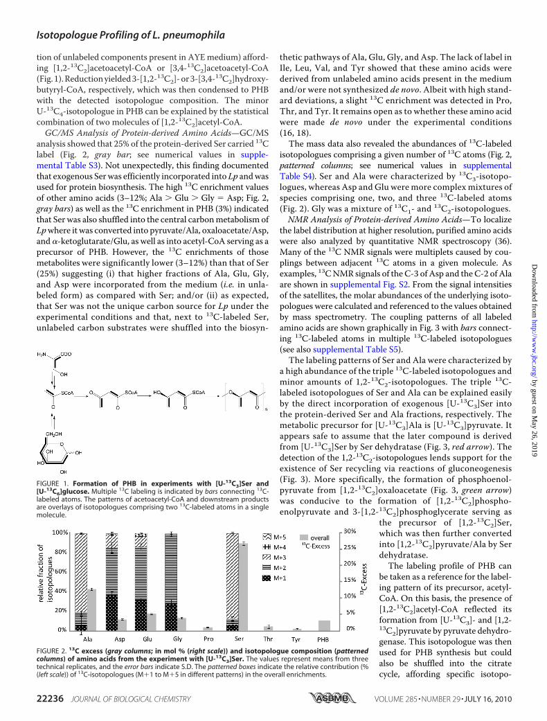
analysis showed that 25% of the protein-derived Ser carried 13Clabel (Fig. 2, gray bar; see numerical values in supple-mental Table S3). Not unexpectedly, this finding documentedthat exogenous Serwas efficiently incorporated into Lp andwasused for protein biosynthesis. The high 13C enrichment valuesof other amino acids (3–12%; Ala � Glu � Gly � Asp; Fig. 2,gray bars) as well as the 13C enrichment in PHB (3%) indicatedthat Ser was also shuffled into the central carbonmetabolism ofLpwhere it was converted into pyruvate/Ala, oxaloacetate/Asp,and�-ketoglutarate/Glu, as well as into acetyl-CoA serving as aprecursor of PHB. However, the 13C enrichments of thosemetabolites were significantly lower (3–12%) than that of Ser(25%) suggesting (i) that higher fractions of Ala, Glu, Gly,and Asp were incorporated from the medium (i.e. in unla-beled form) as compared with Ser; and/or (ii) as expected,that Ser was not the unique carbon source for Lp under theexperimental conditions and that, next to 13C-labeled Ser,unlabeled carbon substrates were shuffled into the biosyn-
thetic pathways of Ala, Glu, Gly, and Asp. The lack of label inIle, Leu, Val, and Tyr showed that these amino acids werederived from unlabeled amino acids present in the mediumand/or were not synthesized de novo. Albeit with high stand-ard deviations, a slight 13C enrichment was detected in Pro,Thr, and Tyr. It remains open as to whether these amino acidwere made de novo under the experimental conditions(16, 18).The mass data also revealed the abundances of 13C-labeled
isotopologues comprising a given number of 13C atoms (Fig. 2,patterned columns; see numerical values in supplementalTable S4). Ser and Ala were characterized by 13C3-isotopo-logues, whereas Asp and Glu were more complex mixtures ofspecies comprising one, two, and three 13C-labeled atoms(Fig. 2). Gly was a mixture of 13C1- and 13C2-isotopologues.NMR Analysis of Protein-derived Amino Acids—To localize
the label distribution at higher resolution, purified amino acidswere also analyzed by quantitative NMR spectroscopy (36).Many of the 13C NMR signals were multiplets caused by cou-plings between adjacent 13C atoms in a given molecule. Asexamples, 13CNMR signals of the C-3 of Asp and the C-2 of Alaare shown in supplemental Fig. S2. From the signal intensitiesof the satellites, the molar abundances of the underlying isoto-pologues were calculated and referenced to the values obtainedby mass spectrometry. The coupling patterns of all labeledamino acids are shown graphically in Fig. 3 with bars connect-ing 13C-labeled atoms in multiple 13C-labeled isotopologues(see also supplemental Table S5).The labeling patterns of Ser and Ala were characterized by
a high abundance of the triple 13C-labeled isotopologues andminor amounts of 1,2-13C2-isotopologues. The triple 13C-labeled isotopologues of Ser and Ala can be explained easilyby the direct incorporation of exogenous [U-13C3]Ser intothe protein-derived Ser and Ala fractions, respectively. Themetabolic precursor for [U-13C3]Ala is [U-13C3]pyruvate. Itappears safe to assume that the later compound is derivedfrom [U-13C3]Ser by Ser dehydratase (Fig. 3, red arrow). Thedetection of the 1,2-13C2-isotopologues lends support for theexistence of Ser recycling via reactions of gluconeogenesis(Fig. 3). More specifically, the formation of phosphoenol-pyruvate from [1,2-13C2]oxaloacetate (Fig. 3, green arrow)was conducive to the formation of [1,2-13C2]phospho-enolpyruvate and 3-[1,2-13C2]phosphoglycerate serving as
the precursor of [1,2-13C2]Ser,which was then further convertedinto [1,2-13C2]pyruvate/Ala by Serdehydratase.The labeling profile of PHB can
be taken as a reference for the label-ing pattern of its precursor, acetyl-CoA. On this basis, the presence of[1,2-13C2]acetyl-CoA reflected itsformation from [U-13C3]- and [1,2-13C2]pyruvate by pyruvate dehydro-genase. This isotopologue was thenused for PHB synthesis but couldalso be shuffled into the citratecycle, affording specific isotopo-
FIGURE 1. Formation of PHB in experiments with [U-13C3]Ser and[U-13C6]glucose. Multiple 13C labeling is indicated by bars connecting 13C-labeled atoms. The patterns of acetoacetyl-CoA and downstream productsare overlays of isotopologues comprising two 13C-labeled atoms in a singlemolecule.
FIGURE 2. 13C excess (gray columns; in mol % (right scale)) and isotopologue composition (patternedcolumns) of amino acids from the experiment with [U-13C3]Ser. The values represent means from threetechnical replicates, and the error bars indicate S.D. The patterned boxes indicate the relative contribution (%(left scale)) of 13C-isotopologues (M�1 to M�5 in different patterns) in the overall enrichments.
Isotopologue Profiling of L. pneumophila
22236 JOURNAL OF BIOLOGICAL CHEMISTRY VOLUME 285 • NUMBER 29 • JULY 16, 2010
by guest on May 26, 2019
http://ww
w.jbc.org/
Dow
nloaded from

logue profiles in �-ketoglutarate and oxaloacetate, which serveas precursors for Glu andAsp, respectively (Fig. 3). Because of aSi-specific citrate synthase, label from [1,2-13C2]acetyl-CoA(Fig. 3, red bar) was transferred to positions 4 and 5 of �-keto-glutarate and glutamate. Following the reactions of the citratecycle, label from �-[4,5-13C2]ketoglutarate afforded [1,2-13C2]succinate and [1,2-13C2]fumarate (Fig. 3, red bars).Because of the intrinsic symmetry of fumarate, malate synthaseyielded a 0.5:0.5 mixture of [1,2-13C2]- and [3,4-13C2]malate
from [1,2-13C2] fumarate. (Fig. 3,green bars). The same isotopologuemixture resulted in oxaloacetateand its amination product, aspartate(Fig. 3, green bars and M�2 valuesfrom the mass spectrum). The pres-ence of triple 13C-labeled isotopo-logues in Asp (Fig. 3, blue bars andM�3 values) also reflected the for-mation of oxaloacetate by carboxyl-ation of [U-13C3]pyruvate (Fig. 3,blue arrow). Primarily, this reactionyielded from [U-13C3]pyruvate[1,2,3-13C3]oxaloacetate, which wasthen converted into [1,2,3-13C3]Asp.The fact that [2,3,4-13C3]oxalo-acetate/Asp also was observed atsimilar abundances as found for the1,2,3-13C3-isotopologue can be ex-plained by rapid equilibriumbetween oxaloacetate/malate/fu-marate due to reversible malatedehydrogenase, malate synthase,and possibly also succinate dehy-drogenase. Because of the symme-try of fumarate and succinate, thetriple label was then randomized,affording the observed symmet-ric isotopologue distribution inoxaloacetate/Asp.Labeled oxaloacetate also served
as an acceptor of unlabeled acetyl-CoA. As shown in Fig. 3, label from[3,4-13C2]oxaloacetate was trans-ferred to positions 1 and 2 of�-ketoglutarate/Glu (green bar),and [1,2-13C2]oxaloacetate yielded[3-13C1]Glu. Label from position 1of oxaloacetate was lost as 13CO2during dehydrogenation of isoci-trate. [1,2,3-13C3]- and [2,3,4-13C3]oxaloacetate gave rise to 2,3-13C2- and 1,2,3-13C3-labeled speciesin �-ketoglutarate/Glu, respec-tively. Notably, all of these isotopo-logues were detected in glutamate,although [1,2,3-13C3]Glu could beidentified only by mass spectrome-try (as M�3 species).
Lp Is Able to Use Glucose as a Carbon Source—To obtaininformation about a potential usage of glucose, Lp was grownin AYE medium or CDM supplemented with 11 mM
[U-13C6]glucose and harvested at stationary phase. A samplework-up and analysis were done as described above. Fig. 4 indi-cates that the same set of amino acids labeled from [13C]Ser alsoacquired significant 13C label (�1% 13C enrichment; Ala �Glu � Asp � Pro � Ser) from [U-13C6]glucose supplementedeither to the AYE medium or to CDM. The percentage of
FIGURE 3. Metabolic model for Ser utilization by Lp Paris grown in culture. The scheme shows labelingpatterns due to the incorporation of exogenous [U-13C3]Ser. Labeling patterns were detected in protein-derived amino acids and PHB (shown in boxes). Multiple 13C-labeled isotopologues determined by NMR spec-troscopy are indicated as bars connecting 13C-labeled atoms in a given molecule. The numbers indicate therespective molar abundances. The molar abundances of isotopomer groups comprising one, two, or three13C-labeled atoms, as determined by mass spectrometry (M�1, M�2, and M�3, respectively), are also listed inthe boxes. PEP, phosphoenolpyruvate; OAA, oxaloacetate; �-KG, �-ketoglutarate.
Isotopologue Profiling of L. pneumophila
JULY 16, 2010 • VOLUME 285 • NUMBER 29 JOURNAL OF BIOLOGICAL CHEMISTRY 22237
by guest on May 26, 2019
http://ww
w.jbc.org/
Dow
nloaded from
enrichment values of amino acids was remarkably similar,pointing to glucose metabolism irrespective of the culturemedium used. In contrast to the small differences in the 13Cenrichments of amino acids, we noticed a rather large differ-
ence in the 13C incorporation of [U-13C6]glucose into PHB.More specifically, 13C enrichment of PHB isolated from cellsgrown in AYE medium was �3-fold higher than the corre-sponding value in the experiments using CDM. The labelingpatterns from the experiment with AYE medium are discussedin detail below.Elevated Rate of Glucose Incorporation into PHB—The 13C
NMR signals of PHB detected in the dichloromethane extractof the labeled cells (supplemental Fig. S1B) displayed the samecoupling pattern as observed previously for PHB labeled from[U-13C3]Ser. However, the intensities of the coupling satellitesrelative to the central signals were higher in the experimentwith [U-13C6]glucose (Table 3). The analysis of the signals cor-roborated the fact that [1,2-13C2]- and [3,4-13C2]PHB wereagain the dominant species, each with abundances of approxi-mately 5%. As described above in detail, this isotopologue mix-ture can be explained by PHB biosynthesis from a mixture of1,2-13C2-labeled and unlabeled acetyl-CoA. The minor[U-13C4]PHB species was again assembled from two labeledmolecules of acetyl-CoA. In summary, these data demonstratedthat [U-13C2]acetyl-CoA can be made from [U-13C3]Ser or[U-13C6]glucose, indicating that Ser and glucose catabolismmerge at a certain stage of the intermediary metabolism (i.e.prior to acetyl-CoA formation). During growth in AYEmedium, glucose appeared to be used preferably as a PHB pre-cursor. Under these conditions more than 6 mol % of PHB wasderived from exogenous [13C]glucose (as compared with 0–5mol % in amino acids).Amino Acids—The 13C incorporation into the bacterial
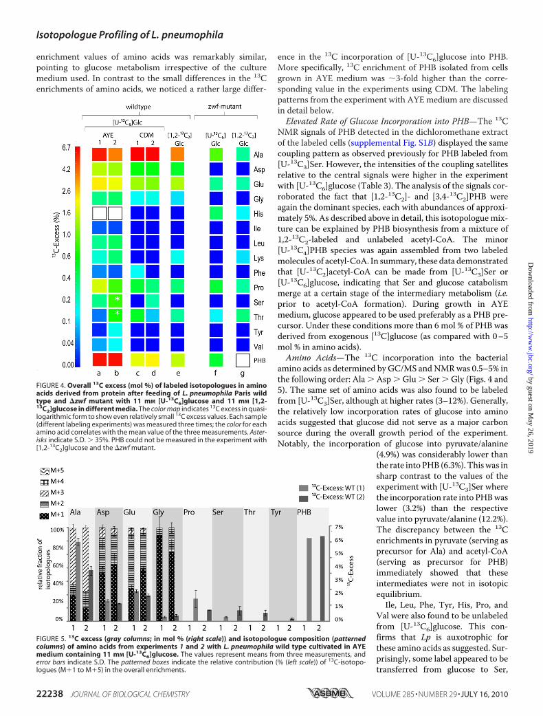
amino acids as determined by GC/MS andNMRwas 0.5–5% inthe following order: Ala � Asp � Glu � Ser � Gly (Figs. 4 and5). The same set of amino acids was also found to be labeledfrom [U-13C3]Ser, although at higher rates (3–12%). Generally,the relatively low incorporation rates of glucose into aminoacids suggested that glucose did not serve as a major carbonsource during the overall growth period of the experiment.Notably, the incorporation of glucose into pyruvate/alanine
(4.9%) was considerably lower thanthe rate into PHB (6.3%). Thiswas insharp contrast to the values of theexperiment with [U-13C3]Ser wherethe incorporation rate into PHBwaslower (3.2%) than the respectivevalue into pyruvate/alanine (12.2%).The discrepancy between the 13Cenrichments in pyruvate (serving asprecursor for Ala) and acetyl-CoA(serving as precursor for PHB)immediately showed that theseintermediates were not in isotopicequilibrium.Ile, Leu, Phe, Tyr, His, Pro, and
Val were also found to be unlabeledfrom [U-13C6]glucose. This con-firms that Lp is auxotrophic forthese amino acids as suggested. Sur-prisingly, some label appeared to betransferred from glucose to Ser,
FIGURE 4. Overall 13C excess (mol %) of labeled isotopologues in aminoacids derived from protein after feeding of L. pneumophila Paris wildtype and �zwf mutant with 11 mM [U-13C6]glucose and 11 mM [1,2-13C2]glucose in different media. The color map indicates 13C excess in quasi-logarithmic form to show even relatively small 13C excess values. Each sample(different labeling experiments) was measured three times; the color for eachamino acid correlates with the mean value of the three measurements. Aster-isks indicate S.D. � 35%. PHB could not be measured in the experiment with[1,2-13C2]glucose and the �zwf mutant.
FIGURE 5. 13C excess (gray columns; in mol % (right scale)) and isotopologue composition (patternedcolumns) of amino acids from experiments 1 and 2 with L. pneumophila wild type cultivated in AYEmedium containing 11 mM [U-13C6]glucose. The values represent means from three measurements, anderror bars indicate S.D. The patterned boxes indicate the relative contribution (% (left scale)) of 13C-isotopo-logues (M�1 to M�5) in the overall enrichments.
Isotopologue Profiling of L. pneumophila
22238 JOURNAL OF BIOLOGICAL CHEMISTRY VOLUME 285 • NUMBER 29 • JULY 16, 2010
by guest on May 26, 2019
http://ww
w.jbc.org/
Dow
nloaded from

although exogenous Ser had been shown to be incorporatedefficiently into proteins in the previous experiment. Moreover,a weak 13C enrichment of Thr also could not be excluded on thebasis of our experimental data.The isotopologue distribution in each of the labeled amino
acids was then determined by mass spectrometry and NMR
spectroscopy as described before.Ala was characterized by theU-13C3-isotopologue. This isotopo-logue can be explained by glucoseutilization via glycolysis, the PPpathway, and/or the ED pathway(Fig. 6). At lower abundances, Serwas also present as a U-13C3-isoto-pologue, suggesting that 3-phos-phoglycerate acquired at least some13C label by glycolysis. [1,2-13C2]Alawas observed as a minor isotopo-logue, suggesting that a smallfraction of phosphoenolpyruvate/pyruvate was made from [1,2-13C2]oxaloacetate by decarbox-ylation (Fig. 6, green arrow). Thelabeling patterns in Asp and Glusupported carbon flux via the com-plete citrate cycle as already shownby the respective labeling profilesfrom [U-13C3]Ser. Thus, formationof oxaloacetate from pyruvate wasagain detected on the basis of 13C3-isotopologues in Asp and 1,2,3-13C3- and 2,3-13C2-isotopologues inGlu.Glucose Is Catabolized by the Ent-
ner-Doudoroff Pathway in Lp—Todetermine the glucose utilizationpathway, we performed a labelingexperiment with Lp Paris (Lp) inAYEmediumsupplementedwith 11mM [1,2-13C2]glucose. The 13Cenrichments and patterns in aminoacids and PHB (Fig. 4, lane e) fol-lowed the same rules as describedabove. As shown in supplemen-tal Fig. S3, label from [1,2-13C2]glucosewas transferred at highrates to [1,2-13C2]Ala via the EDpathway but not to a 2,3-13C2-la-beled specimen representing ahypothetical product via glycolysis.Moreover, the apparent lack of labelin Ser was also in line with the EDpathway, because glycolysis shouldhave generated [2,3-13C2]Ser. ThePP pathway was also excluded as amajor pathway for utilizing glucosebecause no or only single labeledpyruvate/Ala (13C at C-3) should
have occurred when [1,2-13C2]glucose was catabolized by thisroute. The low, single 13C enrichment at C-1 of pyruvate/Alarather reflected minor carbon flow from [13C]oxaloacetate asalready outlined above (supplemental Fig. S3). Pyruvate dehy-drogenase should yield [1-13C1]acetyl-CoA from [1,2-13C2]pyruvate. [1-13C1]Acetyl-CoA was then conducive to the
FIGURE 6. Metabolic model for glucose utilization in Lp Paris grown in AYE medium supplemented with[U-13C6]glucose. Labeling patterns were detected in protein-derived amino acids and PHB (shown in boxes).Multiple 13C-labeled isotopologues determined by NMR spectroscopy are indicated as bars connecting 13C-labeled atoms in a given molecule. The numbers indicate the respective molar abundances. The molarabundances of isotopomer groups comprising one, two, or three 13C-labeled atoms as determined by massspectrometry (M�1, M�2, and M�3, respectively) are also listed for comparison. GAP, glyceraldehyde-3-phosphate dehydrogenase; 6-P-Glcn, 6-phosphogluconate; Pyr, pyruvate; OAA, oxaloacetate; �-KG,�-ketoglutarate.
Isotopologue Profiling of L. pneumophila
JULY 16, 2010 • VOLUME 285 • NUMBER 29 JOURNAL OF BIOLOGICAL CHEMISTRY 22239
by guest on May 26, 2019
http://ww
w.jbc.org/
Dow
nloaded from

detected [1-13C1]- and [4-13C1]PHB specimens. The labelingpatterns of Asp and Glu were also in full accordance with thecarbon fluxes described above for the earlier experiments.Thus, our results identified the EDpathway as the predominantroute of glucose catabolism in Lp.Construction, Properties, and Isotopologue Profiling of a �zwf
Mutant Strain—To further investigate the role of the ED path-way for glucose utilization in Lp, we constructed amutant of LpParis in whichwe deleted the gene lpp0483 (�zwf), which codesa key enzyme of the EDpathway necessary for the conversion ofglucose 6-phosphate into 6-phosphogluconolactone (Fig. 7).To investigate whether the putative zwf operon (lpp0483–0488) is expressed as a polycistronicmRNA, we conducted RT-PCR experiments. Indeed, the genes lpp0483 to lpp0487 wereco-transcribed; however, gene lpp0488 encoding a putative glu-cose transporter was transcribed separately (Fig. 7). To excludepossible downstream effects of the deletion of zwf, we analyzedthe transcription of the downstream genes, which confirmedthat there were no polar effects due to the mutation (data notshown).The �zwf mutant was then grown in AYE medium contain-
ing [U-13C6]- or [1,2-13C2]glucose as described above for thewild type strain. The �zwf mutant strain showed stronglyreduced incorporation rates (by an approximate factor of 10)
into PHB and amino acids (Fig. 4, lanes f and g). On the basis ofthis drastic modulation, we concluded (i) that the proteinencoded by lpp0483 was functionally involved in glucosemetabolism andmost probably catalyzed the presumed conver-sion of glucose 6-phosphate into 6-phosphogluconolactoneand (ii) that the ED pathway was the predominant route forglucose catabolism.Nevertheless, the occurrence of 13C label inAla and PHB (clearly indicated by themass spectrum of alanineand the coupling satellites in the NMR spectrum of alanine andPHB; cf. supplemental Fig. S1 and Table 3) showed that[U-13C6]glucose was still converted (albeit at very low rates)into pyruvate and acetyl-CoA serving as precursors for alanineand PHB, respectively. It can be hypothesized that the minorflux of glucose into pyruvate and acetyl-CoA existed via glycol-ysis in themutant. Notably, the enzymes required for glycolyticconversion of glucose are present in the genome of Lp.Hints for a Functional Role of Carbohydrate Utilization by Lp—
To better understand the role of glucose metabolism in the lifecycle of Lp, we ran infections of A. castellanii. The deletion ofzwf in Lp did not significantly affect replication within the host(Fig. 8A). However, this was different when replication duringsuccessive rounds of infection was analyzed. After a first infec-tion, which lasted 3 days, the mixture (comprising amoebalysate and Lp WT or �zwf mutant strain) was kept for 3 addi-
FIGURE 7. A, proposed EMP, ED, and PP pathways. The names of putative enzymes are shown; beneath them are the encoding open reading frames of Lp Paris(lpp), and the FC value of microarray analysis in vitro (28) are given in parentheses. FC, fold-change values (exponential phase versus stationary phase). Enzymesmarked with an asterisk have no annotated homologues in the Lp genome. Genes determined to be co-transcribed are highlighted in gray and dark gray,respectively. Glk, glucokinase; Pgi, phosphoglucose isomerase; Pfk, phosphofructokinase; Fba, fructose-bisphosphate aldolase; TpiA, triose-phosphate isomer-ase; Gap, glyceraldehyde-3-phosphate dehydrogenase; Pgk, phosphoglycerate kinase; Pgm, phosphoglycerate mutase; Eno, enolase; PykA, pyruvate kinase;Zwf, glucose-6-phosphate dehydrogenase; Pgl, phosphogluconolactonase; Edd, phosphogluconate dehydratase; Eda, 2-keto-3-deoxy-phosphogluconatealdolase; Gnd, 6-phosphogluconate dehydrogenase; Rpe, ribulose phosphate-3-epimerase; RpiA, ribose-5-phosphate isomerase; TktA, transketolase; Tal,transaldolase; Gcd, glucose dehydrogenase; Gnt, gluconate transporter (modified from Ref. 16). B, schematic overview of the genes lpp043–lpp0488 (right) andlpp0150 –lpp0154 (left). mRNA transcripts were determined via RT-PCR. lpp0483, zwf, lpp0484, pgl; lpp0485, edd; lpp0486, glk; lpp0487, eda; lpp0488 (putativesugar transport protein); lpp0150, sdhB (substrate of the Dot/Icm system); lpp0151, pykA; lpp0152, pgk; lpp0153, gap; lpp0154, tktA.
Isotopologue Profiling of L. pneumophila
22240 JOURNAL OF BIOLOGICAL CHEMISTRY VOLUME 285 • NUMBER 29 • JULY 16, 2010
by guest on May 26, 2019
http://ww
w.jbc.org/
Dow
nloaded from

tional days. Then, we ran another infection using 1 ml of a1:1000 dilution of the previousmixture in fresh infection bufferwith fresh amoebae. To follow infection kinetics, each samplewas plated on BCYE and/or BCYE kanamycin plates. Althoughthere was only a minor difference during the second and thirdround of infection of amoebae (Fig. 8B), we noted that the via-bility of the �zwfmutant strain dropped during the lag period.We then verified this observation by an experiment with a lagperiod of 20 days between the first and second rounds of co-infection and by evaluating the recovery percentage (ratios of�zwf/�zwf � WT) of both the mutant and the wild type strain(Fig. 8C). Then, we performed competition experiments withsuccessive rounds of infection using the�zwfmutant strain andtheWTstrain. Again, themutant strain showed less fitness, andthe observed effect accumulated with each additional round ofinfection until the mutant strain was outcompeted by the WTstrain (Fig. 8D). Altogether, these results indicate an importantrole of the ED pathway (glucose-6-phosphate dehydrogenase)for the survival of Lp in the environment.
DISCUSSION
Lp survives within amoebae and macrophages because of itsability to establish a replication vacuole that is derived from theendoplasmic reticulum. Within this vacuole, Lp differentiatesinto the replicative form and multiplies. It was proposed thatwhen nutrients become limiting, a regulatory cascade triggersthe differentiation to a motile spore-like form. After the bacte-
ria are released from the host cells, these forms of Lp are wellprepared to persist for long periods in the environment. It isknown that Legionella exhibits a strictly respiratory form ofmetabolism and does not grow anaerobically (18). It is also cur-rent knowledge that Lp uses amino acids as primary energy andcarbon sources (4, 10–12, 14) and that metabolic genes areexpressed mainly in the exponential growth phase during invivo growth within A. castellanii (28).In this study, the metabolism of Lp was analyzed for the first
time by comprehensive isotopologue profiling under cultureconditions. Metabolic fluxes were estimated on the basis of theobserved labeling profiles in amino acids and PHB. It had beenreported previously that Ser is activelymetabolized by legionel-lae and that Ser is necessary for the growth of Lp (10, 12, 14–16,18, 19, 27). In the genome of strain Paris genes encoding for�12 ABC transporters and amino acid permeases, e.g. a puta-tive Ser transport protein (Lpp2269) and the putative aminoacid transporters Lpp0026 and Lpp0357 are predicted. Indeed,using [U-13C3]Ser as a supplement to AYE medium at a con-centration of 3 mM, label was transferred to protein-derived[13C3]Ser at �25%. Assuming that yeast extract present in AYEmedium contributes unlabeled Ser at a similar concentration asthe added 13C-labeled Ser (see also supplemental Table S1), theincorporation of exogenous Ser into protein-derived Ser can beestimated as 50%. This high value supports the view that aminoacid transporters, accepting Ser as a substrate, are active in Lp.
FIGURE 8. Analysis of L. pneumophila Paris (WT) and �zwf mutant strain (zwf�) in co-cultures with A. castellanii. Bacteria were used to infect monolayersof A. castellanii at a multiplicity of infection of 0.01 with (A) Lp Paris or the �zwf mutant strain for 96 h (A); Lp Paris or �zwf mutant strain for 3 days, resuspendedand incubated for a further 3 days, diluted to �103 bacteria/ml, and used to infect fresh amoebae (B). Four rounds of infection were performed. C, L. pneumo-phila �zwf and WT strain survival over a 20-day period after co-infection of A. castellanii cells at two different ratios (circles, 50:50; diamonds, 75:25). D, infectionwas done as described in B, but A. castellanii cells were infected with both strains (WT and �zwf) at the same time (in competition). At various time pointspostinoculation, bacteria were quantitated by plating aliquots on BCYE agar (see “Experimental Procedures”). Results are means � S.D. of duplicate samplesand are representative of at least three independent experiments.
Isotopologue Profiling of L. pneumophila
JULY 16, 2010 • VOLUME 285 • NUMBER 29 JOURNAL OF BIOLOGICAL CHEMISTRY 22241
by guest on May 26, 2019
http://ww
w.jbc.org/
Dow
nloaded from

Albeit at lower levels of 13C enrichment (0–13%), label from[U-13C3]Ser was also distributed to other amino acids (Ala, Glu,Gly, and Asp) as well as to PHB. This lends evidence that Ser iscatabolized to pyruvate by Ser dehydratase, known to be activein cell extracts of Lp (20). The enrichment values and isotopo-logue profiles of the storage compound, PHB, and amino acidsderived from intermediates of the citrate cycle (i.e. Asp andGlu) also demonstrate that a carbon flux exists frompyruvate toacetyl-CoA serving as a precursor of PHB and a substrate of acomplete citrate cycle with Si specificity of citrate synthase.Our data did not show any evidence for a functional glyoxylatebypass, corroborating data from genome sequence analysis (18,25, 26).The labeling profiles reflected only minor flux from pyruvate
to oxaloacetate as well as from oxaloacetate to phosphoenol-pyruvate/pyruvate by reactions of gluconeogenesis (includingformation of [13C2]Ser indicating that Ser can bemadede novo).This is surprising because it was believed that the EMPpathwayis used in the direction of gluconeogenesis and that Ser or pyru-vate is required tomaintain a pool of oxaloacetate (high activityof pyruvate carboxylase (20)). In addition, our results could notcorroborate minor flux from pyruvate to acetyl-CoA, as sug-gested earlier on the basis of low activity for pyruvate dehydro-genase in cell lysates of Lp (20). However, at this time it is notpossible to reach a conclusion on the importance of gluconeo-genesis for Lp. Experiments are under way to investigate thisquestion further.The fact that many amino acids (Leu, Ile, Val, Phe, Tyr, Met,
Arg, and His) were unlabeled corroborate that Lp is auxotro-phic formany amino acids, as suggested by genome analysis (16,18), and that amino acids (including Ser) can act as major car-bon substrates for Lp. On the other hand, the detection ofdiluted label from exogenous [U-13C3]Ser in metabolitesderived from downstream central intermediates (i.e. acetyl-CoA, oxaloacetate, and �-ketoglutarate) lends support for theuse of additional non-amino acid carbon substrates.Inspired by this observation, we performed labeling experi-
ments with 11 mM [U-13C6]- or [1,2-13C2]glucose as a supple-ment to AYE medium or CDM. In both experimental settings,glucose was incorporated into amino acids and PHB, affording13C enrichments up to 10% in pyruvate/Ala. Again, minoramounts of Ser were synthesized de novo in this experiment.Generally, the transfer of label from glucose to central meta-bolic intermediates providing precursors of the labeled aminoacid is surprising, because it is believed that glucose or carbo-hydrates are not utilized by Lp (12, 15, 18, 20). Although theglycolytic pathway appeared to be complete (as suggested fromthe sequenced genomes), enzymatic assays indicated very low,if any,metabolic flux through this route (14, 16, 20). In addition,we confirmed that Lp does not exhibit a functional PP pathway,corroborated by the lack of orthologues of 6-phosphoglu-conate dehydrogenase and transaldolase within the recentlysequenced Lp genomes. The ED pathway was also not thoughtto be active (16, 18). However, in our experiments, the labelingprofiles of PHB and amino acids demonstrated carbon fluxfrom glucose to pyruvate via the ED pathway and not via theEMP and/or the PP pathway. As further strong evidence, the�zwfmutant of Lp Paris, impaired in the key reaction of the ED
pathway, was strongly reduced in its glucose utilization. How-ever, the deletion of the zwf gene would also affect the catabo-lism of glucose via the PP pathway; but this seems to be a neg-ligible concern, because our results (WT strain) demonstratedthat the PP pathway was not used for glucose catabolism by Lp.Further analysis of the putative zwf operon demonstrated
that the genes lpp0483 to lpp0487 are transcribed as onemRNAunit. The putative glucose transport protein encoding genelpp0488 is transcribed separately as a monocistronic mRNA. Itis also noteworthy that the glucokinase (glk, lpp0486) is locatedwithin the zwf operon encoding for genes of the ED pathway.The products of glucose degradation by the encoded enzymesof the zwf operon would be glyceraldehyde 3-phosphate andpyruvate, which could then enter into the lower part of glycol-ysis and the citrate cycle, respectively (see Fig. 7).Further analysis of the�zwfmutant strain demonstrated that
the mutant strain was outcompeted by the wild type strain in acombined replication/survival assay with successive rounds ofinfection (Fig. 8D). In the first round of infection, the �zwfmutant strain replicated aswell as thewild type strain; however,during repeated infection cycles, the fitness of the �zwfmutantstrain was reduced in the presence of the wild type strain. Thus,the activity of glucose-6-phosphate dehydrogenase (ED path-way) and of glucose catabolism in general is important for fullfitness of Lp.In this context, it is important to note that Lp is also able to
degrade cellulose (29), andwe have identified a glucoamylase inLp Paris the activity of which is responsible for starch and gly-cogen degradation of this strain.6 Moreover, it has been shownthat amutation in the chitinase gene (chiA) has a negative effecton the virulence of the mutant strain as compared with theisogenic wild type strain (40). Preliminary labeling experimentsof A. castellanii, the host organism, were successful. Thus wenow have an excellent basis for in vivo infection experimentsusing Lp and “prelabeled” A. castellanii cells to analyze theintracellular metabolism of the human pathogen Lp. This willprovide further information for better understanding themechanisms of intracellular pathogens and how Lp gets accessto essential nutrients during intracellular parasitism ofamoebae.In summary, we were able to demonstrate that (i) Ser is
used efficiently as a carbon source but is also synthesized denovo by Legionella, although Ser is absolutely required for invitro and in vivo growth of Lp; (ii) glucose is metabolizedmainly via the ED pathway, which is active during in vitrogrowth, and not via the EMP or PP pathway; (iii) carbon fromglucose is incorporated preferably into the storage com-pound PHB; (iv) the citrate cycle is complete and active; (v)Lp is not able to synthesize Ile, Leu, Val, Phe, Met, Arg, andTyr; and (vi) glucose metabolism via the ED pathway is nec-essary for full fitness of Lp during its life cycle.
Acknowledgments—We thank Kerstin Rydzewski and ChristineSchwarz for technical assistance.
6 V. Herrmann, A. Eidner, K. Rydzewski, I. Bladel, M Jules, C. Buchrieser, W.Eisenreich, and K. Heuner, submitted for publication.
Isotopologue Profiling of L. pneumophila
22242 JOURNAL OF BIOLOGICAL CHEMISTRY VOLUME 285 • NUMBER 29 • JULY 16, 2010
by guest on May 26, 2019
http://ww
w.jbc.org/
Dow
nloaded from

REFERENCES1. Garduno, R. A., Garduno, E., Hiltz, M., and Hoffman, P. S. (2002) Infect.
Immun. 70, 6273–62832. Hammer, B. K., Tateda, E. S., and Swanson, M. S. (2002) Mol. Microbiol.
44, 107–1183. Molofsky, A. B., and Swanson, M. S. (2003)Mol. Microbiol. 50, 445–4614. Molofsky, A. B., and Swanson, M. S. (2004)Mol. Microbiol. 53, 29–405. Swanson, M. S., and Hammer, B. K. (2000) Annu. Rev. Microbiol. 54,
567–6136. Greub, G., and Raoult, D. (2003) Res. Microbiol. 154, 619–6217. Anderson, A. J., and Dawes, E. A. (1990)Microbiol. Rev. 54, 450–4728. Anderson, A. J., Haywood, G. W., and Dawes, E. A. (1990) Int. J. Biol.
Macromol. 12, 102–1059. Steinert, M., Emody, L., Amann, R., and Hacker, J. (1997) Appl. Environ.
Microbiol. 63, 2047–205310. Pine, L., George, J. R., Reeves, M. W., and Harrell, W. K. (1979) J. Clin.
Microbiol. 9, 615–62611. Reeves, M. W., Pine, L., Hutner, S. H., George, J. R., and Harrell, W. K.
(1981) J. Clin. Microbiol. 13, 688–69512. Ristroph, J. D., Hedlund, K.W., andGowda, S. (1981) J. Clin.Microbiol. 13,
115–11913. Tesh, M. J., and Miller, R. D. (1982) Can. J. Microbiol. 28, 1055–105814. Tesh, M. J., Morse, S. A., and Miller, R. D. (1983) J. Bacteriol. 154,
1104–110915. Weiss, E., Peacock, M. G., and Williams, J. C. (1980) Curr. Microbiol. 4,
1–616. Fonseca, M. V., Sauer, J. D., and Swanson, M. S. (2008) in Legionella-
Molecular Microbiology (Heuner, K., and Swanson, M. S., eds) pp.213–226, Horizon Scientific Press, Norfolk, United Kingdom
17. Hoffman, P. S. (1984) in Proceedings of the 2nd International Symposiumon Legionella (Thronsberry, C., Balows, A., Feeley, J. C., and Jakubowsky,W., eds) pp. 61–67, American Society for Microbiology, Washington,D. C.
18. Hoffman, P. S. (2008) in Legionella pneumophila: Pathogenesis and Immu-nity (Hoffman, P. S., Klein, T., and Friedman, H., eds) pp. 113–131,Springer Publishing Corp., New York
19. Wieland, H., Ullrich, S., Lang, F., and Neumeister, B. (2005) Mol. Micro-biol. 55, 1528–1537
20. Keen, M. G., and Hoffman, M. S. (1984) Curr. Microbiol. 11, 81–8821. George, J. R., Pine, L., Reeves, M. W., and Harrell, W. K. (1980) J. Clin.
Microbiol. 11, 286–29122. Ristroph, J. D., Hedlund, K. W., and Allen, R. G. (1980) J. Clin. Microbiol.
11, 19–2123. Weiss, E., and Westfall, H. N. (1984) Appl. Environ. Microbiol. 48,
380–38524. Tesh, M. J., and Miller, R. D. (1981) J. Clin. Microbiol. 13, 865–86925. Cazalet, C., Rusniok, C., Bruggemann, H., Zidane, N., Magnier, A., Ma, L.,
Tichit, M., Jarraud, S., Bouchier, C., Vandenesch, F., Kunst, F., Etienne, J.,Glaser, P., and Buchrieser, C. (2004) Nat. Genet. 36, 1165–1173
26. Chien, M., Morozova, I., Shi, S., Sheng, H., Chen, J., Gomez, S. M., Asa-mani, G., Hill, K., Nuara, J., Feder, M., Rineer, J., Greenberg, J. J., Stesh-enko, V., Park, S. H., Zhao, B., Teplitskaya, E., Edwards, J. R., Pampou, S.,Georghiou, A., Chou, I. C., Iannuccilli, W., Ulz, M. E., Kim, D. H.,Geringer-Sameth, A., Goldsberry, C.,Morozov, P., Fischer, S. G., Segal, G.,Qu, X., Rzhetsky, A., Zhang, P., Cayanis, E., De Jong, P. J., Ju, J., Kalachikov,S., Shuman, H. A., and Russo, J. J. (2004) Science 305, 1966–1968
27. Sauer, J. D., Bachman, M. A., and Swanson, M. S. (2005) Proc. Natl. Acad.Sci. U.S.A. 102, 9924–9929
28. Bruggemann, H., Hagman, A., Jules, M., Sismeiro, O., Dillies, M. A.,Gouyette, C., Kunst, F., Steinert, M., Heuner, K., Coppee, J. Y., andBuchrieser, C. (2006) Cell. Microbiol. 8, 1228–1240
29. Pearce, M. M., and Cianciotto, N. P. (2009) FEMS Microbiol. Lett. 300,256–264
30. Glockner, G., Albert-Weissenberger, C., Weinmann, E., Jacobi, S., Schun-der, E., Steinert, M., Hacker, J., and Heuner, K. (2008) Int. J. Med. Micro-biol. 298, 411–428
31. Banerji, S., Aurass, P., and Flieger, A. (2008) Int. J. Med. Microbiol. 298,169–181
32. Flieger, A., Rydzewski, K., Banerji, S., Broich, M., and Heuner, K. (2004)Infect. Immun. 72, 2648–2658
33. Schunder, E., Adam, P., Higa, F., Remer, K. A., Lorenz, U., Bender, J.,Schulz, T., Flieger, A., Steinert, M., and Heuner, K. (2010) Int. J. Med.Microbiol. 300, 313–323
34. Zamboni, N., Fendt, S. M., Ruhl, M., and Sauer, U. (2009) Nat. Protoc. 4,878–892
35. Sauer, U., and Eikmanns, B. J. (2005) FEMS Microbiol. Rev. 29, 765–79436. Eisenreich,W., Slaghuis, J., Laupitz, R., Bussemer, J., Stritzker, J., Schwarz,
C., Schwarz, R., Dandekar, T., Goebel, W., and Bacher, A. (2006) Proc.Natl. Acad. Sci. U.S.A. 103, 2040–2045
37. Eylert, E., Schar, J., Mertins, S., Stoll, R., Bacher, A., Goebel,W., and Eisen-reich, W. (2008)Mol. Microbiol. 69, 1008–1017
38. Heuner, K., Dietrich, C., Skriwan, C., Steinert, M., and Hacker, J. (2002)Infect. Immun. 70, 1604–1608
39. Lee,W. N., Byerley, L. O., Bergner, E. A., and Edmond, J. (1991) Biol. MassSpectrom. 20, 451–458
40. DebRoy, S., Dao, J., Soderberg,M., Rossier, O., andCianciotto, N. P. (2006)Proc. Natl. Acad. Sci. U.S.A. 103, 19146–19151
Isotopologue Profiling of L. pneumophila
JULY 16, 2010 • VOLUME 285 • NUMBER 29 JOURNAL OF BIOLOGICAL CHEMISTRY 22243
by guest on May 26, 2019
http://ww
w.jbc.org/
Dow
nloaded from

Carmen Buchrieser, Wolfgang Eisenreich and Klaus HeunerEva Eylert, Vroni Herrmann, Matthieu Jules, Nadine Gillmaier, Monika Lautner,
GLUCOSE AS CARBON SUBSTRATES: ROLE OF SERINE ANDLegionella pneumophilaIsotopologue Profiling of
doi: 10.1074/jbc.M110.128678 originally published online May 4, 20102010, 285:22232-22243.J. Biol. Chem.
10.1074/jbc.M110.128678Access the most updated version of this article at doi:
Alerts:
When a correction for this article is posted•
When this article is cited•
to choose from all of JBC's e-mail alertsClick here
Supplemental material:
http://www.jbc.org/content/suppl/2010/05/04/M110.128678.DC1
http://www.jbc.org/content/285/29/22232.full.html#ref-list-1
This article cites 37 references, 17 of which can be accessed free at
by guest on May 26, 2019
http://ww
w.jbc.org/
Dow
nloaded from