Embed Size (px)
Citation preview

J. clin. Path., 1971, 24, 53-56
Isolation of fastidious mycoplasma from humansources
ELLI JANSSON
From the Municipal Bacteriological Laboratory, Aurora Hospital, Helsinki, Finland
SYNOPSIS A method for the isolation of fastidious mycoplasma from human sources is described.
It is well known that various mycoplasma specieshave quite different growth requirements. The sapro-phytic Mycoplasma laidlawii is able to grow withoutcholesterol. The most fastidious human mycoplasmais M. pneumoniae which Chanock, Hayflick, andBarile (1962) succeeded in growing on cell-freemedia after a modification in the usual pleuro-pneumonia-like organism (PPLO) media. This wasthe end of an 18-year history of Eaton agent as avirus.
Because mycoplasma in tissue culture can multi-ply intracellularly and thus resemble the trueparasitism of viruses (Clyde, 1963), the requirementof optimal nutrients on primary isolation is stillhigher. Tissue culture cell lines are contaminatedwith mycoplasma in most laboratories. Therefore,cell-free media are to be preferred. Embryonatedhens' eggs also may harbour avian mycoplasma,although seldom (Eaton, Farnham, Levinthal, andScala, 1962).
In our laboratory we have made great efforts forfive years to discover a better cell-free medium forfastidious human mycoplasma. The culture methodthat will be presented here made it possible to isolatemycoplasma from tissue specimens or joint fluidof many patients with rheumatoid arthritis and alsofrom leukaemic bone marrow specimens.
Material and Methods
MEDIUM
Bacto brain heart infusion broth was enriched with20% pooled inactivated human serum, 2 5% yeastextract, I % glucose, and 20 ,ug per ml DNA.For the human serum pool, serum from 10 blooddonors was used. The yeast extract was preparedand stored as described by Chanock et al (1962).DNA was added according to Klieneberger-NobelReceived for publication 18 June 1970.
(1961). Egg yolk pasteurized at +60°C for 50 minwas added in amounts of 0-1 ml per 10 ml enrichedmedium into broth medium. The medium wasthus improved as suggested by Marmion (1967)for antigen production. For solid medium I1* %agar was used and the egg yolk was omitted. Inorder to avoid bacterial contamination, 500 unitsof penicillin per ml and thallium acetate to a finalconcentration of 1:2,000 were added. The pH of themedium was adjusted to 7-8.
CULTIVATIONThe tissue specimens with some broth added werecrushed in a mortar. Then a loopful of this mixtureor 0 5 ml of undiluted synovial fluid was inoculatedinto 10 ml of enriched.brain-heart infusion broth.This rather large volume of broth was used in orderto dilute out possible tissue-bound antibodies. Thebroth cultures were incubated at +37°C for 20days and subcultures were made after five, 10, 15,and 20 days by inoculating 0-1 ml on solid medium.Small Petri dishes, 5 cm in diameter, were used.The cultures on solid media were incubated at +37°Cunder anaerobic condition for 10 days. In addition,every specimen was inoculated on two blood agarplates for possible bacterial growth and incubatedunder aerobic and anaerobic conditions for fourto five days. Blind passages were done three timesby inoculating 0-5 ml of the broth culture into 10ml of enriched broth medium and incubating againfor 20 days.
MICROSCOPICAL EXAMINATIONThe agar plates were examined for growth under alow-power microscope ( x 100). The suspected growthwas stained in situ with drops of 10% Dienes stainas suggested by Hers (1965). With this exception,the method described by Madoff (1960) for identi-fication of mycoplasma by the stained agar tech-
53
on 24 July 2018 by guest. Protected by copyright.
http://jcp.bmj.com
/J C
lin Pathol: first published as 10.1136/jcp.24.1.53 on 1 F
ebruary 1971. Dow
nloaded from

Elli Jansson
nique was followed. An agar block about 1 to 1-5cm2 was cut off, mounted on a microscope slide with ta cover glass, and examined with 100-fold magnifi-cation by a Leitz Orthoplan microscope with the - *condensor Achr 0-70/L4. Suspected colonieswere examined with 1,000-fold magnification andan oil-immersion lens.
Results
Using the method just described for studies of 6 Xmycoplasma in rheumatoid arthritis, we isolated ,in our laboratory a mycoplasma on primary cultureand in the first or second passages in several cases(unpublished data). Had we not done blind passages,not so many specimens would have given positivefindings. Also a mycoplasma was cultivated fromthe bone marrow specimens of some patients withleukaemia.
In the primary cultures and in the first passagesas few as one to four tiny colonies were observedas a rule in the agar block cut off for examinationunder the wide-field Orthoplan microscope. In the Fig. 1 Strain 152-M, slater passages the number of the colonies increased the joint fluid in rheumaand many colonies could be seen in the same field.
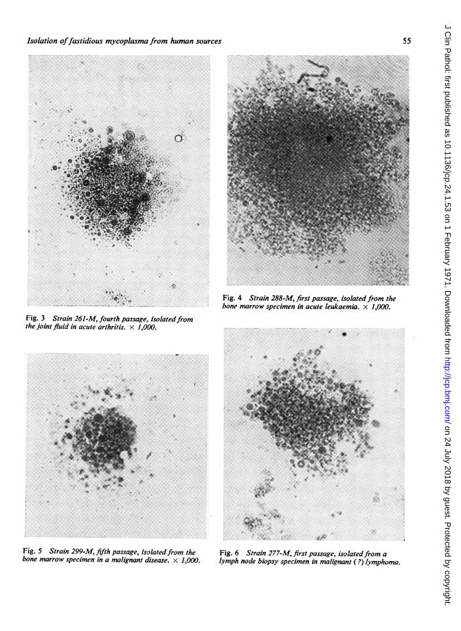
In the primary culture the colonies were usuallytiny, only about 1/10 to 1/100 of the large colonyof mycoplasma. Using Dienes stain and an oil-immersion lens the fragile granular structure of themycoplasma colonies could be seen clearly. Figures1-6 illustrate the microscopical appearance of someisolates.
Large cells or PPLO cells are often seen in sub-cultures. Inside these large forms several small cellsare formed. When all these are released and broken t A
up, a fragile, pleomorphic granular material re- gmains which stains blue or lilac with Dienes stain.In later passages a fairly abundant growth of fragile 0 .4granules tends to form on agar resembling a mono-layer rather than individual colonies. The growthcycle of the isolates seemed very much like thatdescribed by Klieneberger-Nobel (1961).The isolates did not convert into bacteria when
penicillin and thallium acetate were omitted. Theyrequired sterol for their growth.
Metabolically, the strains isolated by the methodjust reported were all glucose- and urea-negative,but some were able to split arginine, especially '3 'when tested just after their primary isolation. P4
Discussion
The following points seem to be important in tryingto isolate fastidious mycoplasma from human Fig. 2 Strain 194-M, isources: (1) a very good culture medium, like the joint fluid in rheumatoid
'eventh passage, isolatedfromitoid arthritis. x 1,000.
OU
D,~'.~.
4
'4
12th passage, isolatedfrom theIarthritis. x 1,000.
54
on 24 July 2018 by guest. Protected by copyright.
http://jcp.bmj.com
/J C
lin Pathol: first published as 10.1136/jcp.24.1.53 on 1 F
ebruary 1971. Dow
nloaded from
Isolation offastidious mycoplasma from human sources
a
n *¼.
Fig. 3 Strain 261-M, fourth passage, isolatedfromthe joint fluid in acute arthritis. x 1,000.
Fig. 4 Strain 288-M, first passage, isolated from thebone marrow specimen in acute leukaemia. x 1,000.
S
O.:. .:.t; il<,4!....
a* .t 4 *
S..i*.
.a;
4a-..
.*
et4.4
f
II
.i~ O.WR.. *.
Fig. 5 Strain 299-M, fifth passage, isolatedfrom thebone marrow specimen in a malignant disease. x 1,000.
Fig. 6 Strain 277-M, first passage, isolatedfrom alymph node biopsy specimen in malignant (?) lymphoma.
55
.,3...£
on 24 July 2018 by guest. Protected by copyright.
http://jcp.bmj.com
/J C
lin Pathol: first published as 10.1136/jcp.24.1.53 on 1 F
ebruary 1971. Dow
nloaded from
Elli Jansson
one just described; (2) first-class microscopicalequipment, preferably a wide-view microscope;(3) long personal experience with mycoplasma.As regards the culture medium, we found that the
diphasic culture medium described by Marmion(1967) was a very good one for isolation of M.pneumoniae from throat specimens. Instead ofPPLO broth we used brain-heart infusion broth.Thereafter we have used his medium further enrichedwith pasteurized egg yolk for subcultures of ourisolates, and their growth has been good. Also theaddition of Hepes buffer (Manchee and Taylor-Robinson, 1969) into the culture medium seems tohave a favourable effect.Some investigators (Duthie, Stewart, Alexander,
and Dayhoff, 1967; Alexander, Stewart, and Duthie,1968; Stewart, Alexander, and Duthie, 1969) havefound diphtheroids from rheumatoid tissues. Whensome of our isolates in their first passages weregrown in enriched broth without egg yolk, theysometimes showed fragile longer forms. Smith,Alexander, and Duthie (1957) observed the con-version of M. hominis type 2 strain Campo intodiphtheroid organisms in 1957. We made a similarobservation some years ago when we were culti-vating a pure culture of the same strain in PPLObroth enriched with rabbit serum and cholesterolbut with thallium acetate omitted.
The skilled technical assistance of Miss SirkkaLiisa Tuuri is gratefully acknowledged.
This study was supported by grants from theSigrid Juselius Foundation, the State MedicalCouncil, and the University of Helsinki.
References
Alexander, W. R. M., Stewart, S. M., and Duthie, J. J. R. (1968).Aetiological factors in rheumatoid arthritis. In RheunmaticDiseases, edited by J. J. R. Duthie and W. R. M. Alexander,pp. 155-164. Edinburgh University Press, Edinburgh.
Chanock, R. M., Hayflick, L., and Barile, M. F. (1962). Growth onartificial medium of an agent associated with atypical pneu-monia and its identification as a PPLO. Proc. nat. Ac ad.Sci. (Wash.), 48, 41-49.
Clyde, W. A., Jr. (1963). Studies on growth of Eaton's agent in tissueculture. Proc. Soc. exp. Biol. (N. Y.), 112, 905-909.
Duthie, J. J. R., Stewart, S. M., Alexander, W. R. M., and Dayhoff,R. E. (1967). Isolation of diphtheroid organisms from rheuma-toid synovial membrane and fluid. Lancet, 1, 142-143.
Eaton, M. D., Farnham, A. E., Levinthal, J. D., and Scala, A. R.(1962). Cytopathic effect of the atypical pneumonia organismin cultures of human tissue. J. Bacteriol., 84, 1330-1337.
Hers, J. Ph. (1965). Personal communication.Klieneberger-Nobel, E. (1962). Pleuropneunionia-like Organisms
(PPLO). Mycoplasmataceae. Academic Press, London andNew York.
Madoff, S. (1960). Isolation and identification of PPLO. Ann. N. Y.Acad. Sci., 79, 383-392.
Manchee, R. J., and Taylor-Robinson, D. (1969). Enhanced growthof T-strain mycoplasmas with N-2-hydroxyethylpiperazine-N"-2-ethanesulfonic acid buffer. J. Bacteriol., 100, 78-85.
Marmion, B. P. (1967). The mycoplasmas: new information on theirproperties and their pathogenicity for man. In Recent Adiuancesin Medical Microbiology, edited by A. P. Waterson, pp.170-253. Churchill, London.
Smith, P. F., Peoples, D. M., and Morton, H. E. (1957). Conversionof pleuropneumonialike organisins to bacteria. Proc. Soc.exp. Biol. (N. Y.), 96, 550-553.
Stewart, S. M., Alexander, W. R. M., and Duthie, J. J. R. (1969).Isolation of diphtheroid bacilli from synovial membrane andfluid in rheumatoid arthritis. Ann. rheum. Dis., 28, 477-487.
56
on 24 July 2018 by guest. Protected by copyright.
http://jcp.bmj.com
/J C
lin Pathol: first published as 10.1136/jcp.24.1.53 on 1 F
ebruary 1971. Dow
nloaded from