Embed Size (px)
Citation preview

Isolation of an antimicrobial compoundproduced by bacteria associated withreef-building corals
Jean-Baptiste Raina1,2,3,4,5, Dianne Tapiolas2, Cherie A. Motti2,Sylvain Foret3,6, Torsten Seemann7, Jan Tebben8,9, Bette L. Willis3,4 andDavid G. Bourne2,4
1 Climate Change Cluster (C3), University of Technology Sydney, Sydney, NSW, Australia2 Australian Institute of Marine Science, Townsville, QLD, Australia3Australian Research Council Centre of Excellence for Coral Reef Studies, James Cook University,
Townsville, QLD, Australia4Marine Biology and Aquaculture, College of Science and Engineering, James Cook University of
North Queensland, Townsville, QLD, Australia5 AIMS@JCU, James Cook University, Townsville, QLD, Australia6 Research School of Biology, Australian National University, Canberra, ACT, Australia7 Victorian Life Sciences Computation Initiative, University of Melbourne, Melbourne, Victoria,
Australia8 Section Chemical Ecology, Alfred Wegener Institute, Bremerhaven, Germany9 University of New South Wales, Sydney, NSW, Australia
ABSTRACTBacterial communities associated with healthy corals produce antimicrobial
compounds that inhibit the colonization and growth of invasive microbes and
potential pathogens. To date, however, bacteria-derived antimicrobial molecules
have not been identified in reef-building corals. Here, we report the isolation of an
antimicrobial compound produced by Pseudovibrio sp. P12, a common and
abundant coral-associated bacterium. This strain was capable of metabolizing
dimethylsulfoniopropionate (DMSP), a sulfur molecule produced in high
concentrations by reef-building corals and playing a role in structuring their
bacterial communities. Bioassay-guided fractionation coupled with nuclear
magnetic resonance (NMR) and mass spectrometry (MS), identified the
antimicrobial as tropodithietic acid (TDA), a sulfur-containing compound likely
derived from DMSP catabolism. TDA was produced in large quantities by
Pseudovibrio sp., and prevented the growth of two previously identified coral
pathogens, Vibrio coralliilyticus and V. owensii, at very low concentrations
(0.5 mg/mL) in agar diffusion assays. Genome sequencing of Pseudovibrio sp. P12
identified gene homologs likely involved in the metabolism of DMSP and
production of TDA. These results provide additional evidence for the integral role of
DMSP in structuring coral-associated bacterial communities and underline the
potential of these DMSP-metabolizing microbes to contribute to coral disease
prevention.
Subjects Marine Biology, Microbiology
Keywords Coral-associated bacteria, Disease, Alphaproteobacteria, Antimicrobial compounds
How to cite this article Raina et al. (2016), Isolation of an antimicrobial compound produced by bacteria associated with reef-building
corals. PeerJ 4:e2275; DOI 10.7717/peerj.2275
Submitted 15 May 2016Accepted 19 July 2016Published 18 August 2016
Corresponding authorJean-Baptiste Raina,
Academic editorMauricio Rodriguez-Lanetty
Additional Information andDeclarations can be found onpage 14
DOI 10.7717/peerj.2275
Copyright2016 Raina et al.
Distributed underCreative Commons CC-BY 4.0
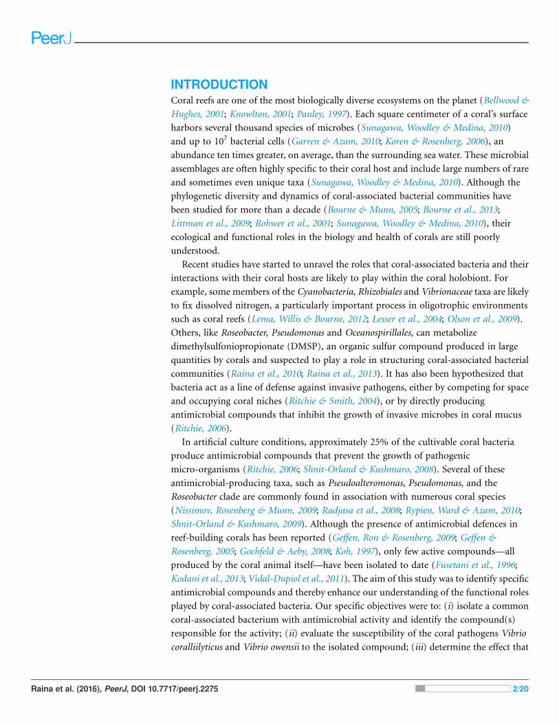
INTRODUCTIONCoral reefs are one of the most biologically diverse ecosystems on the planet (Bellwood &
Hughes, 2001; Knowlton, 2001; Pauley, 1997). Each square centimeter of a coral’s surface
harbors several thousand species of microbes (Sunagawa, Woodley & Medina, 2010)
and up to 107 bacterial cells (Garren & Azam, 2010; Koren & Rosenberg, 2006), an
abundance ten times greater, on average, than the surrounding sea water. These microbial
assemblages are often highly specific to their coral host and include large numbers of rare
and sometimes even unique taxa (Sunagawa, Woodley & Medina, 2010). Although the
phylogenetic diversity and dynamics of coral-associated bacterial communities have
been studied for more than a decade (Bourne & Munn, 2005; Bourne et al., 2013;
Littman et al., 2009; Rohwer et al., 2001; Sunagawa, Woodley & Medina, 2010), their
ecological and functional roles in the biology and health of corals are still poorly
understood.
Recent studies have started to unravel the roles that coral-associated bacteria and their
interactions with their coral hosts are likely to play within the coral holobiont. For
example, some members of the Cyanobacteria, Rhizobiales and Vibrionaceae taxa are likely
to fix dissolved nitrogen, a particularly important process in oligotrophic environments
such as coral reefs (Lema, Willis & Bourne, 2012; Lesser et al., 2004; Olson et al., 2009).
Others, like Roseobacter, Pseudomonas and Oceanospirillales, can metabolize
dimethylsulfoniopropionate (DMSP), an organic sulfur compound produced in large
quantities by corals and suspected to play a role in structuring coral-associated bacterial
communities (Raina et al., 2010; Raina et al., 2013). It has also been hypothesized that
bacteria act as a line of defense against invasive pathogens, either by competing for space
and occupying coral niches (Ritchie & Smith, 2004), or by directly producing
antimicrobial compounds that inhibit the growth of invasive microbes in coral mucus
(Ritchie, 2006).
In artificial culture conditions, approximately 25% of the cultivable coral bacteria
produce antimicrobial compounds that prevent the growth of pathogenic
micro-organisms (Ritchie, 2006; Shnit-Orland & Kushmaro, 2008). Several of these
antimicrobial-producing taxa, such as Pseudoalteromonas, Pseudomonas, and the
Roseobacter clade are commonly found in association with numerous coral species
(Nissimov, Rosenberg & Munn, 2009; Radjasa et al., 2008; Rypien, Ward & Azam, 2010;
Shnit-Orland & Kushmaro, 2009). Although the presence of antimicrobial defences in
reef-building corals has been reported (Geffen, Ron & Rosenberg, 2009; Geffen &
Rosenberg, 2005; Gochfeld & Aeby, 2008; Koh, 1997), only few active compounds—all
produced by the coral animal itself—have been isolated to date (Fusetani et al., 1996;
Kodani et al., 2013; Vidal-Dupiol et al., 2011). The aim of this study was to identify specific
antimicrobial compounds and thereby enhance our understanding of the functional roles
played by coral-associated bacteria. Our specific objectives were to: (i) isolate a common
coral-associated bacterium with antimicrobial activity and identify the compound(s)
responsible for the activity; (ii) evaluate the susceptibility of the coral pathogens Vibrio
coralliilyticus and Vibrio owensii to the isolated compound; (iii) determine the effect that
Raina et al. (2016), PeerJ, DOI 10.7717/peerj.2275 2/20

thermal stress might have on its production; and (iv) investigate the natural abundance
of the antimicrobial compound in coral extracts.
MATERIALS AND METHODSBacterial isolationHealthy colonies of the corals Pocillopora damicornis, Acropora millepora and Montipora
aequituberculata (one colony per species) were collected in November 2011 from Davies
Reef, Great Barrier Reef, Australia (latitude, 18�51′S; longitude, 147�41′E, Great BarrierReef Marine Park Authority permit G12/35236.1) and maintained in aquaria for six days
at the Australian Institute of Marine Science (Townsville, Queensland, Australia). Five
replicate coral fragments (approximately 25 mm in length, containing 60–70 polyps) were
collected from each colony and washed in sterile artificial seawater (ASW) to remove
loosely attached microbes. Tissue slurries were produced by airbrushing (80 lb/in2) each
coral fragment into 5 mL of ASW to remove coral tissues and associated microbes. These
tissue slurries were homogenized to break down tissue clumps, and a dilution series was
plated immediately on bacteriological agar (1%) in 1 L ASW supplemented with 0.3%
casamino acids and 0.4% glucose (Hjelm et al., 2004). After two days of incubation at
28 �C, single colonies were transferred into Marine Broth (MB; Difco, BD, Franklin Lakes,
NJ) and grown overnight. Liquid cultures were re-plated on minimal marine agar and the
procedure was repeated until pure cultures were obtained.
Well diffusion assay with bacterial isolatesFifty bacteria isolated from the coral tissue slurries of the three species (A. millepora = 16,
P. damicornis = 17, M. aequituberculata = 17) were tested for growth-inhibitory
activity against the known coral pathogens Vibrio coralliilyticus P1 (LMG23696) and
V. owensiiDY05 (LMG25443) in a well diffusion agar assay. In brief, the Vibrio strains were
seeded into two different batches of minimal marine agar (after the agar temperature
cooled to 40 �C). Following solidification, wells (diameter 5 mm) were cut into the agar
and loaded with 20 mL of overnight cultures (108 cells/mL) of the test isolates grown in
MB (28 �C, 170 rpm). Plates were incubated at 28 �C and monitored every 24 h for a
period of 72 h for inhibition zones. Phaeobacter strain 27-4 was used as a positive
antagonistic control on each plate because of its broad spectrum inhibitory activity
against Vibrio (Bruhn, Gram & Belas, 2007; Hjelm et al., 2004).
DNA extraction, gene sequencing genomic analysesOne strain, P12 isolated from Pocillopora damicornis, produced the strongest
growth-inhibitory activity against the two target Vibrio strains. High molecular weight
genomic DNA from P12 was extracted using a miniprep phenol-chloroform based
extraction. Briefly, 5 mL of overnight liquid culture of P12 (108 cells/mL) were spun in a
micro-centrifuge (10,000 rcf) for 2 min. The pellet was then resuspended in 567 mL of
TE buffer, 30 mL of 10% SDS and 3 mL of 20 mg/mL proteinase K. The tube was
shaken thoroughly and incubated for 1 h at 37 �C. One hundred microliters of 5 M NaCl
was subsequently added and the sample thoroughly mixed before adding 80 mL of
Raina et al. (2016), PeerJ, DOI 10.7717/peerj.2275 3/20
CTAB/NaCl (10% CTAB in 0.7 M NaCl). The solution was incubated for 10 min at 65 �C,extracted with an equal volume of phenol/chloroform/isoamyl alcohol and centrifuged for
10 min (10,000 rcf). The supernatant was then extracted with an equal volume of
chloroform/isoamyl alcohol and centrifuged again for 10 min. The aqueous phase was
transferred to a new tube, DNA precipitated with equal volume of ice-cold isopropanol,
washed with 70% ethanol and dried.
The near complete 16S rRNA gene of the strain was PCR amplified with bacterial
specific primers 63F and 1387R, as outlined in Marchesi et al. (1998). Amplified PCR
products were visualized by electrophoresis on 1% agarose gel stained with ethidium
bromide. The amplified DNA was dried in a vacuum centrifuge (Savant DNA 120) and
sequenced (Macrogen, Inc., Seoul, Korea). The 16S rRNA gene sequence of isolate P12 was
used for phylogenetic comparisons and Maximum Likelihood trees were constructed
using the ARB software.
We produced a draft genome assembly of P12. A paired-end library was prepared using
the Illumina Truseq protocol (Illumina, San Diego, CA, USA), with an insert size of
169 bp and a read size of 150 bp. The library was sequenced on an Illumina MiSeq
instrument at Monash University (Melbourne, Australia). The genome was assembled
with the SPAdes assembler (v2.4.0) (Bankevich et al., 2012) and annotated with the Prokka
software (v1.5.2) (Seemann, 2014). The presence of the genes involved in DMSP
metabolism (dmdA, dddD, dddL, dddP, dddY, dddQ, dddW) and TDA production
(tdaA-tdaH) was investigated by searching for homologs of the corresponding genes
using reciprocal best blast hits.
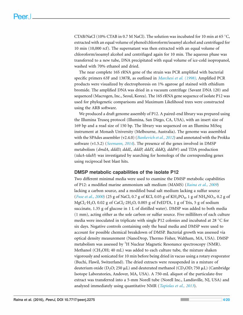
DMSP metabolic capabilities of the isolate P12Two different minimal media were used to examine the DMSP metabolic capabilities
of P12: a modified marine ammonium salt medium (MAMS) (Raina et al., 2009)
lacking a carbon source, and a modified basal salt medium lacking a sulfur source
(Fuse et al., 2000) (25 g of NaCl, 0.7 g of KCl, 0.05 g of KH2PO4, 1 g of NH4NO3, 0.2 g of
MgCl2·H2O, 0.02 g of CaCl2·2H2O, 0.005 g of FeEDTA, 1 g of Tris, 5 g of sodium
succinate, 1.35 g of glucose in 1 L of distilled water). DMSP was added to both media
(1 mm), acting either as the sole carbon or sulfur source. Five milliliters of each culture
media were inoculated in triplicate with single P12 colonies and incubated at 28 �C for
six days. Negative controls containing only the basal media and DMSP were used to
account for possible chemical breakdown of DMSP. Bacterial growth was assessed via
optical density measurement (NanoDrop, Thermo Fisher, Waltham, MA, USA). DMSP
metabolism was assessed by 1H Nuclear Magnetic Resonance spectroscopy (NMR).
Methanol (CH3OH; 40 mL) was added to each culture tube, the mixture shaken
vigorously and sonicated for 10 min before being dried in vacuo using a rotary evaporator
(Buchi, Flawil, Switzerland). The dried extracts were resuspended in a mixture of
deuterium oxide (D2O; 250 mL) and deuterated methanol (CD3OD; 750 mL) (Cambridge
Isotope Laboratories, Andover, MA, USA). A 750-mL aliquot of the particulate-free
extract was transferred into a 5-mm Norell tube (Norell Inc., Landisville, NJ, USA) and
analyzed immediately using quantitative NMR (Tapiolas et al., 2013).
Raina et al. (2016), PeerJ, DOI 10.7717/peerj.2275 4/20

Preparation of crude extracts for antagonist assaysAn overnight culture of P12 (8 mL) was used to inoculate 4� 250 mL of MB (total culture
volume = 1 L). Bacterial cells were incubated for two days at 28 �C (120 rpm); the
culture broth was then acidified to pH 2 with sulphuric acid before being exhaustively
extracted with ethyl acetate (3 � 1.5 L). The extract was washed three times with MilliQ
H2O and dried in vacuo using a rotary evaporator (Buchi). The dried extract was then
weighed and resuspended in CH3OH (which was chosen for its ability to solubilize a wide
range of compounds, its volatility and its innocuity in small volume towards both
V. coralliilyticus and V. owensii) and tested in well-diffusion assays to confirm the
extraction of the antimicrobial compound(s).
Purification and characterization of active compoundPurification of the crude extract was carried out using solid phase extraction on a reversed
phase C18 flash vacuum column (Septra C18-E, Phenomenex, Torrance, CA, USA).
Eleven fractions were eluted sequentially with 20, 40, 60, 80 and 90% CH3OH in H2O and
100% CH3OH, followed by 20, 50 and 100% dichloromethane (CH2Cl2) in CH3OH,
40% hexane in CH2Cl2 and finally 100% hexane. Each fraction was dried and resuspended
in CH3OH (1 mg mL-1). Well diffusion assays were prepared as described above. On
each plate, test wells were inoculated with 20 mL of each chromatographic fraction, or
20 mL of CH3OH as a control, and Vibrio growth monitored. The most active faction
(80% CH3OH) presented an intense yellow color. Fine orange-red needles were
crystallized from this active fraction to yield compound 1 (2.1 mg, 1.7% dry weight of
organic extract).
NMR and FTMS analysisIdentification and structural elucidation of compound 1 was achieved using liquid
chromatography–mass spectrometry (LC-MS), NMR, and Fourier Transform mass
spectrometry (FTMS). Likewise these techniques were used to monitor for the presence of
compound 1 in extracts and fractions. LC-MS analyses were performed on a Thermo
Fisher Scientific Ultra High Performance Liquid Chromatography system connected to an
LTQ Orbitrap XL mass spectrometer (Thermo Fisher Scientific, San Jose, CA, USA).
Samples were separated on a ACQUITY UPLC BEH RP-C18 column (130 A, 1.7 mM,
2.1 � 100 mm, solvents A = aqueous 0.1% formic acid and B = acetonitrile, gradient
elution 80% A: 20% B for 0.5 min ramped up to 100% B over 10 min, then held for 4 min,
400 mL) and detected by positive mode electrospray ionisation using two different m/z
ranges: 150–1,500 and 170–400. 1H and 13C NMR spectra of compound 1 were acquired
in a 5 mm 509-UP Norell NMR tube on a Bruker Avance 600 MHz NMR
spectrometer (Bruker, Germany) with a TXI cryoprobe using standard Bruker pulse
sequences. NMR spectra were referenced to residual 1H and 13C resonances in deuterated
chloroform (CDCl3). High resolution mass spectra of compound 1 were measured with a
Bruker BioApex 47e FTMS fitted with an Analytica of Branford ESI source; ions were
detected in negative mode within a mass range m/z 200–1,000 via direct infusion at
120 mL h-1.
Raina et al. (2016), PeerJ, DOI 10.7717/peerj.2275 5/20
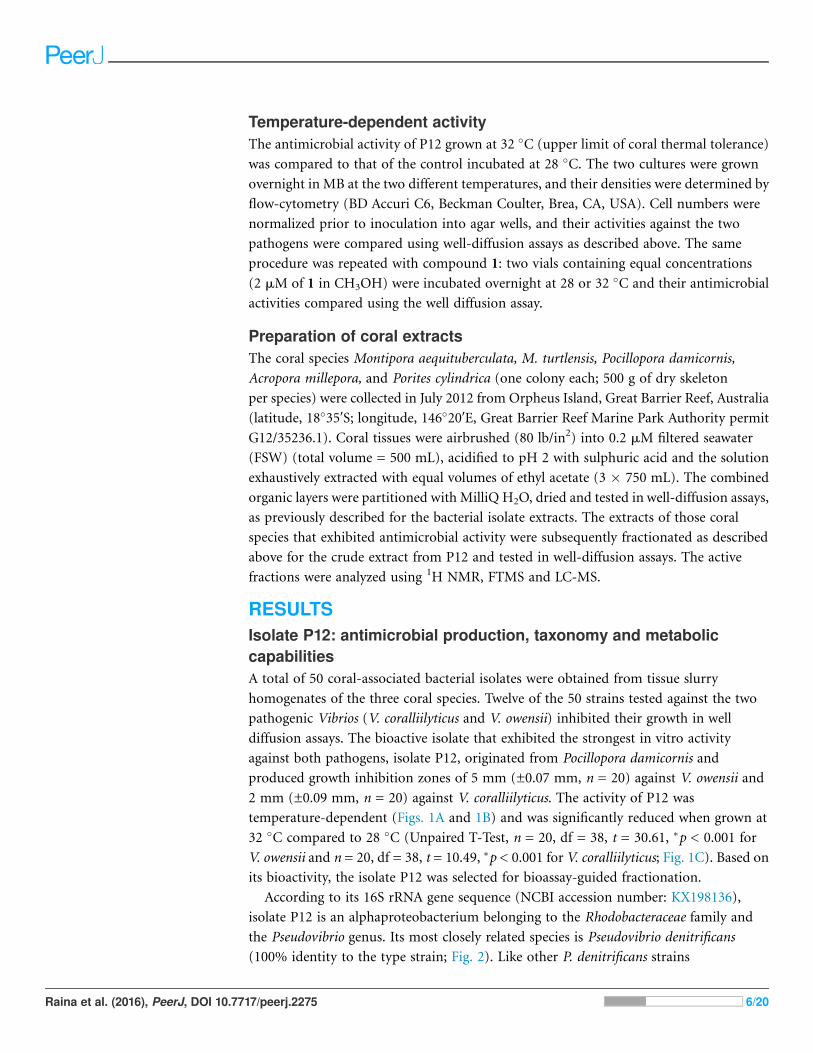
Temperature-dependent activityThe antimicrobial activity of P12 grown at 32 �C (upper limit of coral thermal tolerance)
was compared to that of the control incubated at 28 �C. The two cultures were grown
overnight in MB at the two different temperatures, and their densities were determined by
flow-cytometry (BD Accuri C6, Beckman Coulter, Brea, CA, USA). Cell numbers were
normalized prior to inoculation into agar wells, and their activities against the two
pathogens were compared using well-diffusion assays as described above. The same
procedure was repeated with compound 1: two vials containing equal concentrations
(2 mM of 1 in CH3OH) were incubated overnight at 28 or 32 �C and their antimicrobial
activities compared using the well diffusion assay.
Preparation of coral extractsThe coral species Montipora aequituberculata, M. turtlensis, Pocillopora damicornis,
Acropora millepora, and Porites cylindrica (one colony each; 500 g of dry skeleton
per species) were collected in July 2012 from Orpheus Island, Great Barrier Reef, Australia
(latitude, 18�35′S; longitude, 146�20′E, Great Barrier Reef Marine Park Authority permit
G12/35236.1). Coral tissues were airbrushed (80 lb/in2) into 0.2 mM filtered seawater
(FSW) (total volume = 500 mL), acidified to pH 2 with sulphuric acid and the solution
exhaustively extracted with equal volumes of ethyl acetate (3 � 750 mL). The combined
organic layers were partitioned with MilliQ H2O, dried and tested in well-diffusion assays,
as previously described for the bacterial isolate extracts. The extracts of those coral
species that exhibited antimicrobial activity were subsequently fractionated as described
above for the crude extract from P12 and tested in well-diffusion assays. The active
fractions were analyzed using 1H NMR, FTMS and LC-MS.
RESULTSIsolate P12: antimicrobial production, taxonomy and metaboliccapabilitiesA total of 50 coral-associated bacterial isolates were obtained from tissue slurry
homogenates of the three coral species. Twelve of the 50 strains tested against the two
pathogenic Vibrios (V. coralliilyticus and V. owensii) inhibited their growth in well
diffusion assays. The bioactive isolate that exhibited the strongest in vitro activity
against both pathogens, isolate P12, originated from Pocillopora damicornis and
produced growth inhibition zones of 5 mm (±0.07 mm, n = 20) against V. owensii and
2 mm (±0.09 mm, n = 20) against V. coralliilyticus. The activity of P12 was
temperature-dependent (Figs. 1A and 1B) and was significantly reduced when grown at
32 �C compared to 28 �C (Unpaired T-Test, n = 20, df = 38, t = 30.61, �p < 0.001 for
V. owensii and n = 20, df = 38, t = 10.49, �p < 0.001 for V. coralliilyticus; Fig. 1C). Based on
its bioactivity, the isolate P12 was selected for bioassay-guided fractionation.
According to its 16S rRNA gene sequence (NCBI accession number: KX198136),
isolate P12 is an alphaproteobacterium belonging to the Rhodobacteraceae family and
the Pseudovibrio genus. Its most closely related species is Pseudovibrio denitrificans
(100% identity to the type strain; Fig. 2). Like other P. denitrificans strains
Raina et al. (2016), PeerJ, DOI 10.7717/peerj.2275 6/20

(Enticknap et al., 2006), P12 colonies formed brown mucoid colonies when grown on
Marine Agar. The brown color was absent when the strain was grown on minimal marine
agar, with colonies appearing white. This strain was able use DMSP as either a sole
carbon or sole sulfur source (Fig. 3). The complete utilization of DMSP from the liquid
media after 2–3 days of incubation, as well as the presence of its metabolic byproduct
dimethylsulfide (DMS), were confirmed by 1H NMR. However acrylate, another possible
byproduct of DMSP metabolism, was not observed.
Among the seven different DMSP degradation pathways currently identified (Moran
et al., 2012), the full DMSP cleavage pathway (dddD, dddB, dddC, dddT, dddR; Table 1),
involved in the conversion of DMSP into DMS without formation of acrylate (Todd et al.,
2007) (Table 1), was identified in P12. We also identified possible orthologs for the
demethylation pathway (dmdA, dmdB, dmdC and dmdD) used by marine bacteria to
assimilate sulfur from DMSP, though these gene have low sequence identity to the genes
A B
28
28
28
32
32
32
28
28
2832
32
32
C
0
1
2
3
4
5
6
Inhi
bitio
n zo
ne (m
m)
Vibrio owensii Vibrio coralliilyticus
28oC
32oC
Pure TDAVibrio owensii Vibrio coralliilyticus
Pseudovibrio sp. P12
**
_ _
Figure 1 Representative well diffusion assays of (A) Pseudovibrio sp. P12 and (B) pure TDA,
incubated at two different temperatures (28 and 32 �C) and then inoculated onto agar plates with
embedded Vibrio owensii ((-) Negative control). (C) Comparison of the radius of inhibition zones
between the two temperature treatments for both Pseudovibrio sp. P12 (Unpaired T-Test, n = 20, df = 38,
t = 30.61, �p < 0.001 for V. owensii and n = 20, df = 38, t = 10.49, �p < 0.001 for V. coralliilyticus) and pure
TDA (2 mM, Unpaired T-Test, n = 20, df = 38, t = -0.94, p = 0.355 for V. owensii and n = 20, df = 38, t =
0.632, p = 0.531 for V. coralliilyticus).
Raina et al. (2016), PeerJ, DOI 10.7717/peerj.2275 7/20

originally identified in Ruegeria pomeroyi DSS-3 (Howard et al., 2006; Reisch et al., 2011)
(Table 1). The presence of these two gene pathways corroborates the 1H NMR
measurements: the observed production of DMS without acrylate formation following
DMSP metabolism (DddD pathway); and the ability to use DMSP as sole sulfur source
(DmdA pathway) (Table 1).
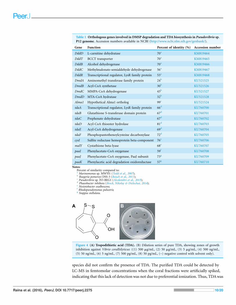
Identification of antimicrobial compounds produced by P12Well diffusion assays revealed that the crude extract from P12 retained the antimicrobial
properties of the strain against both Vibrio species. Purification of the active fractions
using reverse phase liquid chromatography yielded compound 1: optically inactive
orange-red crystals; 2.1 mg (1.7% dry weight); IR (film) �max 3,420, 1,660, 1,280 cm-1;
UV (PDA, CH3OH) �max 512 nm; 1H NMR spectrum (600 MHz, CD3Cl): � 7.12, 7.44,
7.45 and 16.7; 13C NMR (150 MHz, CD3Cl): � 120.3, 132.0, 136.0, 138. 7, 149.5, 168.7,
171.7, and 183.5; HRESIMS m/z found 210.9534 (calculated for C8H3O3S2- 210.9529,
� 2 ppm). Combined spectroscopic techniques revealed that compound 1 was
tropodithietic acid (TDA) (Brinkhoff et al., 2004; Penesyan et al., 2011) (Fig. 4A).
Orthologs for 11 genes involved in TDA biosynthesis (Geng et al., 2008) were present in
the Pseudovibrio sp. P12 genome (Table 1). The biosynthesis of TDA correlated with
production of the yellow-brown pigmentation in the culture medium and antimicrobial
activity, similar to that previously reported (Brinkhoff et al., 2004; Bruhn et al., 2005;
Porsby, 2010). Both coral pathogens were highly sensitive to TDA, with the pure
compound still visually inhibiting their growth at 0.5 mg/mL (2.35 mM; Fig. 4B). In
contrast to the decrease in antimicrobial activity exhibited by Pseudovibrio sp. P12 after
incubation at 32 �C, TDA activity was not affected by exposure to this temperature
(Unpaired T-Test, n = 20, df = 38, t = -0.94, p = 0.355 for V. owensii and n = 20, df = 38,
t = 0.632, p = 0.531 for V. coralliilyticus; Figs. 1B and 1C).
Investigating the presence of TDA in coral samplesAll of the extracts derived from the coral species investigated exhibited antimicrobial
activity against the two pathogens, with the inhibition zones for P. cylindrica,
P. denitrificans FO-BEG1 [FR716549]
P. denitrificans F71059 [HQ908691]
P. denitrificans DN34 [AY486423]
P. ascidiaceicola [AB175663]
P. japonicus [AB246748]
P. axinellae [JN167515]Phaeobacter sp. 27-4
P. sp. P1267
99
0.01
Isolate NW001 [AF295099]
P. strain JE062 [DQ097238]
Figure 2 Maximum likelihood phylogenetic tree based on 16S rRNA gene sequences showing the
isolate used in this study (P12 in red) and closely associated Pseudovibrio spp. Note: the strain
FO-BEG1 has been fully sequenced. Phaeobacter sp. 27-4 (AJ536669) was used as outgroup. Maximum
parsimony bootstrap values (10,000 replicates) are given when different from 100. The scale bar indicates
the number of substitution per nucleotide position.
Raina et al. (2016), PeerJ, DOI 10.7717/peerj.2275 8/20

M. aequituberculata, M. turtlensis and P. damicornis ranging from 3–5 mm in radius
whilst the inhibition zones for A. millepora were much smaller (1 mm on average).1H NMR, LC-MS and FTMS analyses of the extracts and the active fractions of all coral
�
3.0 2.9 2.83.13.23.33.43.53.6 ppm
Pseudovibrio sp.
Vibrio coralliilyticus
Control
Pseudovibrio sp.
Vibrio coralliilyticus
Control
3.0 2.9 2.83.13.23.33.43.53.6
B
A
ppm
a
a
Figure 3 1H NMR spectra showing DMSP utilization as (A) the sole carbon source and (B) the sole
sulfur source in minimal media at the end of a six-day incubation. The “control” lines in all cases are
the growth medium (with no bacterial inoculation). The black and green spectra show the results from
inoculation with Pseudovibrio sp. P12 and V. coralliilyticus (negative control), respectively. In both cases,
the DMSP signals (within the three boxes, see Tapiolas et al. (2013)) disappeared in the Pseudovibrio
treatment and remain unchanged between the no-bacteria control and the V. coralliilyticus treatment. In
the case of DMSP as a sole sulfur source, Pseudovibrio consumed the DMSP and other carbon sources
present and produced secondary metabolites (appearance of new signals). a: solvent peak (methanol).
Raina et al. (2016), PeerJ, DOI 10.7717/peerj.2275 9/20
species did not confirm the presence of TDA. The purified TDA could be detected by
LC-MS in femtomolar concentrations when the coral fractions were artificially spiked,
indicating that this lack of detection was not due to preferential ionization. Thus, TDAwas
Table 1 Orthologous genes involved in DMSP degradation and TDAbiosynthesis in Pseudovibrio sp.P12 genome. Accession numbers available in NCBI (http://www.ncbi.nlm.nih.gov/genbank/).
Gene Function Percent of identity (%) Accession number
DddD L-carnitine dehydratase 70a KM819464
DddT BCCT transporter 70a KM819465
DddB Alcohol dehydrogenase 70a KM819466
DddC Methylmalonate-semialdehyde dehydrogenase 56a KM819467
DddR Transcriptional regulator, LysR family protein 53a KM819468
DmdA Aminomethyl transferase family protein 24b KU521525
DmdB Acyl-CoA synthetase 30b KU521526
DmdC MMPA-CoA dehydrogenase 43b KU521527
DmdD MTA-CoA hydratase 32b KU521528
Alma1 Hypothetical Alma1 ortholog 99c KU521524
tdaA Transcriptional regulator, LysR family protein 66d KU760700
tdaB Glutathione S-transferase domain protein 67d KU760701
tdaC Prephenate dehydratase 67d KU760702
tdaD Acyl-CoA thioester hydrolase 81d KU760703
tdaE Acyl-CoA dehydrogenase 69d KU760704
tdaF Phosphopantothenoylcysteine decarboxylase 72d KU760705
cysI Sulfite reductase hemoprotein beta-component 76e KU760706
malY Cystathione beta-lyase 68e KU760707
paaI Phenylacetate-CoA oxygenase 59f KU760708
paaJ Phenylacetate-CoA oxygenase, PaaJ subunit 73g KU760709
paaK Phenylacetic acid degradation oxidoreductase 57g KU760710
Notes:Percent of similarity compared to:a Marinomonas sp. MWYl1 (Todd et al., 2007);b Ruegeria pomeroyi DSS-3 (Reisch et al., 2011);c Pseudovibrio sp. FO-BEG1 (Alcolombri et al., 2015);d Phaeobacter inhibens (Brock, Nikolay & Dickschat, 2014);e Nesiotobacter exalbescens;f Rhodopseudomonas palustris;g Stappia stellulata.
Figure 4 (A) Tropodithietic acid (TDA). (B) Dilution series of pure TDA, showing zones of growth
inhibition against Vibrio coralliilyticus: ((1) 500 mg/mL, (2) 50 mg/mL, (3) 5 mg/mL, (4) 500 ng/mL,
(5) 50 ng/mL, (6) 5 ng/mL, (7) 500 pg/mL, (8) 50 pg/mL, (-) negative control with solvent only).
Raina et al. (2016), PeerJ, DOI 10.7717/peerj.2275 10/20

either not present in the coral fractions tested or in concentrations below the LC-MS
detection threshold.
DISCUSSIONWhile previous studies have identified corals as a source of bacteria with antimicrobial
activity (Radjasa et al., 2008; Sulistiyani et al., 2010), this study represents the first
isolation and identification of an antimicrobial compound produced by a reef building
coral-associated bacterium with activity against coral-associated pathogens. TDA was
characterized from a pure culture of Pseudovibrio sp. P12 previously isolated from corals
and strongly inhibited the growth of two coral pathogens. Although we could not confirm
the presence of this molecule in the corals tested, TDA has the capacity to provide
protective antimicrobial properties to the coral host and prevent colonization by invasive
bacterial species.
The strain P12 strongly inhibited the growth of Vibrio coralliilyticus and V. owensii, two
coral pathogens causing white syndrome (a collective term describing rapidly
progressing tissue loss, exposing band-like areas of white skeleton) (Ben-Haim et al., 2003;
Sussman et al., 2008; Ushijima et al., 2012; Willis, Page & Dinsdale, 2004). Vibrio
coralliilyticus exhibits antimicrobial resistance to a wide range of commercial antibiotics
and is also resistant to the activities of a large number of coral-associated bacteria
(Rypien, Ward & Azam, 2010; Shnit-Orland & Kushmaro, 2009; Vizcaino et al., 2010). Its
resistance to commercial antibiotics is considerably greater than that of other marine
pathogens such as V. parahaemolyticus or V. vulnificus, and may contribute to its
competitive advantage within the coral holobiont, as well as its ability to infect corals
(Vizcaino et al., 2010). However, whilst V. coralliilyticus is resistant to many coral-
associated bacteria, its growth was strongly inhibited by the strain P12, emphasizing its
antimicrobial capabilities.
The isolate P12 belongs to the bacterial genus Pseudovibrio (Shieh, Lin & Jean, 2004),
and is ubiquitously found in association with healthy sponges (Enticknap et al., 2006;
Thiel & Imhoff, 2003; Webster & Hill, 2001) and corals (see Table 2). Pseudovibrio are
thought to be involved in symbiotic relationships with various organisms; they are
vertically transmitted in large densities by adult sponges to their larvae (Enticknap
et al., 2006) and their presence is required for the growth of the sulfur-oxidizing
bacteria Beggiatoa in culture (Bondarev et al., 2013). Furthermore, their genome is
organized similarly to that of Rhizobia, a well-characterized symbiotic bacterium
(Bondarev et al., 2013; Enticknap et al., 2006; Kennedy et al., 2009). The full genome
sequences of Pseudovibrio FO-BEG1 (KEGG genome T01669; isolated from a Caribbean
coral, and sharing 100% sequence similarity with P12 based on its 16S rRNA gene
sequence) reveal the presence of genes involved in host-cell adhesion, interactions with
eukaryotic cell machinery, and production of secondary metabolites (Bondarev et al.,
2013), further suggesting that this bacterium is involved in symbiotic relationships with
its hosts.
The Pseudovibrio genus is also known for its antimicrobial properties, especially against
human pathogens such as Mycobacterium tuberculosis, Bacillus cereus, Yersinia
Raina et al. (2016), PeerJ, DOI 10.7717/peerj.2275 11/20

enterocolitica, Listeria monocytogenes or methicillin-resistant Staphylococcus aureus
(O’Halloran et al., 2011; Sulistiyani et al., 2010). To date, three active compounds have
been isolated from different Pseudovibrio strains: heptylprodigiocin in tunicate-associated
P. denitrificans Z143-1 (Sertan-de Guzman et al., 2007), pseudovibrocin in P. denitrificans
strain PaH3.28 (Vizcaino, 2011) and TDA from red alga-associated P. ascidiaceicola
D323 (Penesyan et al., 2011). In the present study, we isolated TDA from P12, a strain
closely related to P. denitrificans. The ability of P12 to produce TDAwas further supported
by the the presence of the genes essential for TDA biosynthesis in the genome of strain
P12 (Geng et al., 2008). TDA inhibits the growth of a wide range of marine pathogens
(Bruhn, Gram & Belas, 2007; Bruhn et al., 2005) and is produced almost exclusively by
bacteria from the Roseobacter clade, especially the genera Phaeobacter, Silicibacter, and
Ruegeria (Brinkhoff et al., 2004; Bruhn et al., 2005; Geng & Belas, 2010; Geng et al., 2008;
Wilson et al., 2016) that are commonly associated with DMSP-producing dinoflagellates
(Miller et al., 2004; Wagner-Dobler & Biebl, 2006) and reef-building corals (Bourne
et al., 2013; Littman et al., 2009; Raina et al., 2009).
Many members of the Roseobacter clade, including coral-associated isolates, have been
implicated in sulfur cycling (Miller et al., 2004; Moran, Gonzalez & Kiene, 2003; Raina
et al., 2010). Interestingly, TDA contains two sulfur atoms (C8H4O3S2) and Pseudovibrio
sp. P12 was able to use DMSP either as sole carbon or sole sulfur source, a common
trait among Alphaproteobacteria and especially the Roseobacter clade (Bruhn et al., 2005;
Wagner-Dobler & Biebl, 2006). Bacteria from this clade preferentially metabolize DMSP
rather than sulphate (SO42-), despite the latter being between 106–107-fold more
abundant in seawater (Geng & Belas, 2010; Kiene et al., 1999). Based on genomic and
chemical analyses, DMSP metabolism in P12 can occur via two likely routes: the cleavage
Table 2 Summary of Pseudovibrio isolated or sequenced from corals, accession numbers are displayed when available.
Host Location Method Reference Accession numbers
Acropora palmata Panama Amplicon Sunagawa, Woodley & Medina (2010) GU118050, GU118108, GU119014
Porites astreoides Panama Amplicon Sunagawa, Woodley & Medina (2010) GU118050, GU118108, GU119014
Acropora cervicornis Panama Amplicon Sunagawa, Woodley & Medina (2010) GU118050, GU118108, GU119014
Montastrea franksi Panama Amplicon Sunagawa, Woodley & Medina (2010) GU118050, GU118108, GU119014
Tubastraea coccinea China Amplicon Yang et al. (2013) JF925014
Pseudopterogorgia americana Puerto Rico Isolated Vizcaino et al. (2010) GQ406787, GQ406798, GQ391966, GQ406786
Platygyra carnosus Hong Kong Isolated Chiu et al. (2012) JF411474, JF411466, JF411439, JF411464
Oculina patagonica Israel Isolated Koren & Rosenberg (2006) DQ416557, AY654776
Montastrea anularis Florida Isolated Rypien, Ward & Azam (2010) FJ952798, FJ952774, FJ952804
Sinularia sp. Indonesia Isolated Sulistiyani et al. (2010) NA
Acropora almata Florida Isolated Ritchie (2006) DQ530540
Sarcophyton sp. Java Isolated Sabdono & Radjasa (2006) NA
Oculina patagonica Israel Isolated Nissimov, Rosenberg & Munn (2009) NA
Lobophytum sp. Taiwan Isolated Chen et al. (2012) JQ342682, JQ342695, JQ342696, JQ342697
Hard coral Florida Isolated Bondarev et al. (2013) CP003147
Notes:NA, not available.
Raina et al. (2016), PeerJ, DOI 10.7717/peerj.2275 12/20

pathway (encoded by dddD, (Todd et al., 2007)) that releases the climate-regulating
molecule DMS, and the demethylation pathway (encoded by dmdA, (Howard et al., 2006))
by which the bacterium can retain the sulfur contained in DMSP molecules.
The biosynthetic pathway of TDA has not been fully elucidated (Brock, Nikolay &
Dickschat, 2014). Both labelling (Cane, Wu & Van Epp, 1992; Thiel et al., 2010) and genetic
dissection (Geng & Belas, 2010) studies have shown that its aromatic skeleton is derived
from phenylacetyl-CoA produced by the shikimate pathway. However, the sulfur donor
allowing the incorporation of the two sulfur atoms into the TDA molecule has not been
clearly identified. It has been proposed that sulfur originating from DMSP metabolism
might be used to synthesize TDA (Bruhn, Gram & Belas, 2007; Bruhn et al., 2005; Geng &
Belas, 2010; Porsby, 2010; Wagner-Dobler & Biebl, 2006). For example, DMSP increases
TDA synthesis two-fold in comparison to other sulfur sources (Geng & Belas, 2010),
suggesting that DMSP is a preferred source of sulfur for TDA biosynthesis. Even though
other sources of sulfur, such as the amino-acids cysteine and methionine present in
artificial media like Marine Broth, might be used to synthesize TDA (Geng & Belas, 2010),
DMSP is by far the most readily available reduced sulfur source in the marine
environment (Simo, 2001). It is therefore likely that in DMSP-rich environments, such as
reef-building corals, DMSP metabolism provides the sulfur needed to produce TDA via
the demethylation pathway (Howard et al., 2006).
The presence of TDA in extracts derived from five coral species (Montipora
aequituberculata,M. turtlensis, Pocillopora damicornis, and Porites cylindrica) could not be
confirmed. Three possibilities can explain this lack of detection: (i) TDA is not
synthesized in corals; (ii) TDA is present in corals in concentrations below the detection
limit of our instruments (which would imply that this compound has a very limited
role in coral defense); and (iii) our sampling effort was not sufficient. Indeed, we only
sampled one colony per species, from a location more than 100 km away from the site
where the TDA-producing bacteria was isolated and without prior characterization of the
bacterial communities present in the colony sampled. Given that TDA-producing
Roseobacters are among the first bacteria to colonize the surface of marine microalgae and
corals (Apprill et al., 2009; Dang & Lovell, 2000; Miller et al., 2004) and can be highly
abundant in some coral species (Raina et al., 2009), it would be premature to rule out
possible TDA biosynthesis in corals.
The activity of P12 against V. coralliilyticus sharply decreased at elevated
temperatures (32 �C), however, the activity of the purified TDA did not. This reveals
that the loss of antimicrobial activity observed for P12 at 32 �C is not due to thermal
sensitivity of TDA but likely to a decrease in its production. Our results are in line with
previously reported decline in the antibacterial activity of other TDA-producing
Roseobacter with temperature increase (Bruhn et al., 2005). Clear links have previously
been identified between warm thermal anomalies and outbreaks of white syndromes
(Bruno et al., 2007; Heron et al., 2010; Maynard et al., 2011). If indeed TDA is
synthesized in vivo, a decrease in its production during anomalously high seawater
temperatures could facilitate pathogen outbreaks in corals following thermal stress,
Raina et al. (2016), PeerJ, DOI 10.7717/peerj.2275 13/20

especially since the virulence of some disease-causing bacteria (i.e. V. coralliilyticus)
increase at 32 �C (Sussman et al., 2008).
This study demonstrates that a common coral-associated bacterium, Pseudovibrio
sp. P12, produces TDA, a potent antimicrobial compound that inhibits the growth of
marine and coral pathogens, including V. coralliilyticus. The bacterium can use DMSP as
a sole sulfur or carbon source and potentially as a precursor in the biosynthesis of TDA.
The production of TDA by Pseudovibrio sp. P12 is greatly reduced at temperatures
causing thermal stress in corals, potentially providing a window of opportunity for the
growth of pathogens. These results provide additional evidence for the integral role of
DMSP in structuring healthy, coral-associated bacterial communities and suggest that
these DMSP-metabolizing communities may contribute to the prevention of coral
diseases.
ACKNOWLEDGEMENTSThe authors would like to thank E. Botte, C. Gao and M. Garren for their laboratory
assistance.
ADDITIONAL INFORMATION AND DECLARATIONS
FundingThis work was supported by the ARC Centre of Excellence for Coral Reef Studies,
AIMS@JCU, James Cook University and the Australian Institute of Marine Science.
The funders had no role in study design, data collection and analysis, decision to publish,
or preparation of the manuscript.
Competing InterestsThe authors declare that they have no competing interests.
Author Contributions� Jean-Baptiste Raina conceived and designed the experiments, performed the
experiments, analyzed the data, wrote the paper, prepared figures and/or tables,
reviewed drafts of the paper.
� Dianne Tapiolas analyzed the data, reviewed drafts of the paper.
� Cherie A. Motti analyzed the data, contributed reagents/materials/analysis tools,
reviewed drafts of the paper.
� Sylvain Foret analyzed the data, reviewed drafts of the paper.
� Torsten Seemann analyzed the data, reviewed drafts of the paper.
� Jan Tebben analyzed the data, reviewed drafts of the paper.
� Bette L. Willis conceived and designed the experiments, contributed reagents/materials/
analysis tools, reviewed drafts of the paper.
� David G. Bourne conceived and designed the experiments, contributed reagents/
materials/analysis tools, reviewed drafts of the paper.
Raina et al. (2016), PeerJ, DOI 10.7717/peerj.2275 14/20

Field Study PermissionsThe following information was supplied relating to field study approvals (i.e., approving
body and any reference numbers):
Great Barrier Reef Marine Park Authority, Permit number: G12/35236.1.
DNA DepositionThe following information was supplied regarding the deposition of DNA sequences:
GenBank: All accession numbers are provided in the main text.
Supplemental InformationSupplemental information for this article can be found online at http://dx.doi.org/
10.7717/peerj.2275#supplemental-information.
REFERENCESAlcolombri U, Ben-Dor S, Feldmesser E, Levin Y, Tawfik DS, Vardi A. 2015. Identification of the
algal dimethyl sulfide–releasing enzyme: a missing link in the marine sulfur cycle. Science
348(6242):1466–1469 DOI 10.1126/science.aab1586.
Apprill A, Marlow HQ, Martindale MQ, Rappe MS. 2009. The onset of microbial associations in
the coral Pocillopora meandrina. The ISME Journal 3(6):685–699 DOI 10.1038/ismej.2009.3.
Bankevich A, Nurk S, Antipov D, Gurevich AA, Dvorkin M, Kulikov AS, Lesin VM, Nikolenko
SI, Pham S, Prjibelski AD, Pyshkin AV, Sirotkin AV, Vyahhi N, Tesler G, Alekseyev MA,
Pevzner PA. 2012. SPAdes: a new genome assembly algorithm and its application to single-cell
sequencing. Journal of Computational Biology 19(5):455–477 DOI 10.1089/cmb.2012.0021.
Bellwood DR, Hughes TP. 2001. Regional-scale assembly rules and biodiversity of coral reefs.
Science 292(5521):1532–1535 DOI 10.1126/science.1058635.
Ben-Haim Y, Thompson FL, Thompson CC, Cnockaert MC, Hoste B, Swings J, Rosenberg E.
2003. Vibrio coralliilyticus sp. nov., a temperature dependent pathogen of the coral Pocillopora
damicornis. International Journal of Systematic and Evolutionary Microbiology 53(1):309–315
DOI 10.1099/ijs.0.02402-0.
Bondarev V, Richter M, Romano S, Piel J, Schwedt A, Schulz-Vogt HN. 2013. The genus
Pseudovibrio contains metabolically versatile bacteria adapted for symbiosis. Environmental
Microbiology 15(7):2095–2113 DOI 10.1111/1462-2920.12123.
Bourne DG, Dennis PG, Uthicke S, Soo RM, Tyson GW,Webster N. 2013. Coral reef invertebrate
microbiomes correlate with the presence of photosymbionts. The ISME Journal 7(7):1452
DOI 10.1038/ismej.2012.172.
Bourne DG, Munn CB. 2005. Diversity of bacteria associated with the coral Pocillopora damicornis
from the Great Barrier Reef. Environmental Microbiology 7(8):1162–1174
DOI 10.1111/j.1462-2920.2005.00793.x.
Brinkhoff T, Bach G, Heidorn T, Liang L, Schlingloff A, Simon M. 2004. Antibiotic production
by a Roseobacter clade-affiliated species from the German Wadden Sea and its antagonistic
effects on the indigenous isolates. Applied and Environmental Microbiology 70(4):2560–2565
DOI 10.1128/AEM.70.4.2560-2565.2003.
Brock NL, Nikolay A, Dickschat JS. 2014. Biosynthesis of the antibiotic tropodithietic acid by the
marine bacterium Phaeobacter inhibens. Chemical Communications 50(41):5487–5489
DOI 10.1039/C4CC01924E.
Raina et al. (2016), PeerJ, DOI 10.7717/peerj.2275 15/20

Bruhn JB, Gram L, Belas R. 2007. Production of antibacterial compounds and biofilm formation
by Roseobacter species are influenced by culture conditions. Applied and Environmental
Microbiology 73(2):442–450 DOI 10.1128/AEM.02238-06.
Bruhn JB, Nielsen KF, Hjelm M, Hansen M, Bresciani J, Schulz S, Gram L. 2005. Ecology,
inhibitory activity, and morphogenesis of a marine antagonistic bacterium belonging to the
Roseobacter clade. Applied and Environmental Microbiology 71(11):7263–7270
DOI 10.1128/AEM.71.11.7263-7270.2005.
Bruno JF, Selig ER, Casey KS, Page CA,Willis BL, Harvell CD, Sweatman H, Melendy AM. 2007.
Thermal stress and coral cover as driver of coral disease outbreaks. PLoS Biology 5(6):e50124
DOI 10.1371/journal.pbio.0050124.
Cane DE, Wu Z, Van Epp JE. 1992. Thiotropocin biosynthesis. Shikimate origin of a sulfur-
containing tropolone derivative. Journal of the American Chemical Society 114(22):8479–8483
DOI 10.1021/ja00048a019.
Chen Y-H, Kuo J, Sung P-J, Chang Y-C, Lu M-C, Wong T-Y, Liu J-K, Weng C-F, Twan W-H,
Kuo F-W. 2012. Isolation of marine bacteria with antimicrobial activities from cultured and
field-collected soft corals. World Journal of Microbiology and Biotechnology 28(12):3269–3279
DOI 10.1007/s11274-012-1138-7.
Chiu JMY, Li S, Li A, Po B, Zhang R, Shin PKS, Qiu J-W. 2012. Bacteria associated with
skeletal tissue growth anomalies in the coral Platygyra carnosus. FEMS Microbiology Ecology
79(2):380–391 DOI 10.1111/j.1574-6941.2011.01225.x.
Dang HY, Lovell CR. 2000. Bacterial primary colonization and early succession on surfaces in
marine waters as determined by amplified rRNA gene restriction analysis and sequences analysis
of 16S rRNA genes. Applied and Environmental Microbiology 66(2):467–475
DOI 10.1128/AEM.66.2.467-475.2000.
Enticknap JJ, Kelly M, Peraud O, Hill RT. 2006. Characterization of a culturable
alphaproteobacterial symbiont common to many marine sponges and evidence for vertical
transmission via sponge larvae. Applied and Environmental Microbiology 72(5):3724–3732
DOI 10.1128/AEM.72.5.3724-3732.2006.
Fuse H, Takimura O, Murakami K, Yamaoka Y, Omori T. 2000. Utilization of dimethyl sulfide
as a sulfur source with the aid of light by Marinobacterium sp. strain DMS-S1. Applied and
Environmental Microbiology 66(12):5527–5532 DOI 10.1128/AEM.66.12.5527-5532.2000.
Fusetani N, Toyoda T, Asai N, Matsunaga S, Maruyama T. 1996. Montiporic acids A
and B, cytotoxic and antimicrobial polyacetylene carboxylic acids from eggs of
the scleractinian coral Montipora digitata. Journal of Natural Products 59(8):796–797
DOI 10.1021/np9604036.
Garren M, Azam F. 2010. New method for counting bacteria associated with coral mucus.
Applied and Environmental Microbiology 76(18):6128–6133 DOI 10.1128/AEM.01100-10.
Geffen Y, Ron EL, Rosenberg E. 2009. Regulation of release of antibacterials from stressed
scleractinians corals. FEMS Microbiology Letters 295(1):103–109
DOI 10.1111/j.1574-6968.2009.01590.x.
Geffen Y, Rosenberg E. 2005. Stress-induced rapid release of antibacterials by scleractinian corals.
Marine Biology 146(5):931–935 DOI 10.1007/s00227-004-1505-5.
Geng H, Belas R. 2010. Expression of tropodithietic acid biosynthesis is controlled by a novel
autoinducer. Journal of Bacteriology 192(17):4377–4387 DOI 10.1128/JB.00410-10.
Geng H, Bruhn JB, Nielsen KF, Gram L, Belas R. 2008. Genetic dissection of tropodithietic
acid biosynthesis by marine roseobacters. Applied and Environmental Microbiology
74(5):1535–1545 DOI 10.1128/AEM.02339-07.
Raina et al. (2016), PeerJ, DOI 10.7717/peerj.2275 16/20

Gochfeld DJ, Aeby GS. 2008. Antibacterial chemical defences in Hawaiian corals provide
possible protection from disease. Marine Ecology Progress Series 362:119–128
DOI 10.3354/meps07418.
Heron SF, Willis BL, Skirving WJ, Eakin CM, Page CA, Miller IR. 2010. Summer hot snaps and
winter conditions: modelling white syndrome outbreaks on Great Barrier Reef corals.
PLoS ONE 5(8):e12210 DOI 10.1371/journal.pone.0012210.
Hjelm M, Bergh O, Riaza A, Nielsen J, Melchiorsen J, Jensen S, Duncan H, Ahrens P,
Birkbeck H, Gram L. 2004. Selection and identification of autochthonous potential probiotic
bacteria from turbot larvae (Scophthalmus maximus) rearing units. Systematic and Applied
Microbiology 27(3):360–371 DOI 10.1078/0723-2020-00256.
Howard EC, Henriksen JR, Buchan A, Reisch CR, Burgmann H, Welsh R, Ye W, Gonzalez JM,
Mace K, Joye SB, Kiene RP, Whitman WB, Moran MA. 2006. Bacterial taxa that limit sulfur
flux from the ocean. Science 314(5799):649–652 DOI 10.1126/science.1130657.
Kennedy J, Baker P, Piper C, Cotter PD, Walsh M, Mooij MJ, Bourke MB, Rea MC,
O’Connor PM, Ross RP, Hill C, O’Gara F, Marchesi JR, Dobson ADW. 2009. Isolation and
analysis of bacteria with antimicrobial activities from the marine sponge Haliclona simulans
collected from Irish waters. Marine Biotechnology 11(3):384–396
DOI 10.1007/s10126-008-9154-1.
Kiene RP, Linn LJ, Gonzalez J, Moran MA, Bruton JA. 1999. Dimethylsulfoniopropionate and
methanethiol are important precursors of methionine and protein-sulfur in marine
bacterioplankton. Applied and Environmental Microbiology 65(10):4549–4558.
Knowlton N. 2001. Coral reef biodiversity–habitat size matters. Science 292(5521):1493–1495
DOI 10.1126/science.1061690.
Kodani S, Sato K, Higuchi T, Casareto BE, Suzuki Y. 2013. Montiporic acid D, a new
polyacetylene carboxylic acid from scleractinian coral Montipora digitata. Natural Product
Research 27(20):1859–1862 DOI 10.1080/14786419.2013.768992.
Koh EGL. 1997. Do scleractinian corals engage in chemical warfare against microbes? Journal of
Chemical Ecology 23(2):379–398 DOI 10.1023/B:JOEC.0000006366.58633.f4.
Koren O, Rosenberg E. 2006. Bacteria associated with mucus and tissues of the coral Oculina
patagonica in summer and winter. Applied and Environmental Microbiology 75(8):254–259
DOI 10.1128/AEM.00554-06.
Lema KA, Willis BL, Bourne DG. 2012. Corals form characteristic associations with symbiotic
nitrogen-fixing bacteria. Applied and Environmental Microbiology 78(9):3136–3144
DOI 10.1128/AEM.07800-11.
Lesser MP, Mazel CH, Gorbunov MY, Falkowski PG. 2004. Discovery of symbiotic
nitrogen-fixing cyanobacteria in corals. Science 305(5686):997–1000
DOI 10.1126/science.1099128.
Littman RA, Willis BL, Pfeffer C, Bourne DG. 2009. Diversities of coral-associated bacteria differ
with location, but not species, for three acroporid corals on the Great Barrier Reef. FEMS
Microbiology Ecology 68(2):152–163 DOI 10.1111/j.1574-6941.2009.00666.x.
Marchesi JR, Sato T, Weightman AJ, Martin TA, Fry JC, Wade WG. 1998. Design and evaluation
of useful bacterium-specific primers that amplify genes coding for 16S rRNA. Applied and
Environmental Microbiology 64(2):795–799.
Maynard JA, Anthony KRN, Harvell DC, Burgman MA, Beeden R, Sweatman H, Heron SF,
Lamb JB, Willis BL. 2011. Predicting outbreaks of a climate-driven coral disease in the Great
Barrier Reef. Coral Reefs 30(2):485–495 DOI 10.1007/s00338-010-0708-0.
Raina et al. (2016), PeerJ, DOI 10.7717/peerj.2275 17/20

Miller TR, Hnilicka K, Dziedzic A, Desplats P, Belas R. 2004. Chemotaxis of Silicibacter sp. strain
TM1040 toward dinoflagellate products. Applied and Environmental Microbiology 70(8):4692–
4701 DOI 10.1128/AEM.70.8.4692-4701.2004.
Moran MA, Gonzalez JM, Kiene RP. 2003. Linking a bacterial taxon to sulfur cycling in the sea:
studies of the marine Roseobacter group. Geomicrobiology Journal 20(4):375–388
DOI 10.1080/01490450303901.
Moran MA, Reisch CR, Kiene RP, Whitman WB. 2012. Genomic insights into bacterial DMSP
transformations. Annual Review of Marine Science 4(1):523–542
DOI 10.1146/annurev-marine-120710-100827.
Nissimov J, Rosenberg E, Munn CB. 2009. Antimicrobial properties of resident coral mucus
bacteria of Oculina patagonica. FEMS Microbiology Letters 292(2):210–215
DOI 10.1111/j.1574-6968.2009.01490.x.
O’Halloran JA, Barbosa TM, Morrissey JP, Kennedy J, O’Gara F, Dobson ADW. 2011. Diversity
and antimicrobial activity of Pseudovibrio spp. from Irish marine sponges. Journal of Applied
Microbiology 110(6):1495–1508 DOI 10.1111/j.1365-2672.2011.05008.x.
Olson ND, Ainsworth TD, Gates RD, Takabayashi M. 2009. Diazotrophic bacteria associated
with Hawaiian Montipora corals: diversity and abundance in correlation with symbiotic
dinoflagellates. Journal of Experimental Marine Biology and Ecology 371(2):140–146
DOI 10.1016/j.jembe.2009.01.012.
Pauley G. 1997. Diversity and distribution of reef organisms. In: Birkeland C, ed. Life and Death of
Coral Reefs. New York: Chapman and Hall, 298–345.
Penesyan A, Tebben J, Lee M, Thomas T, Kjelleberg S, Harder T, Egan S. 2011. Identification
of the antibacterial compound produced by the marine epiphytic bacterium Pseudovibrio sp.
D323 and related sponge-associated bacteria. Marine Drugs 9(12):1391–1402
DOI 10.3390/md9081391.
Porsby CH. 2010. Antagonism of Roseobacter clade bacteria against pathogenic bacteria. PhD
thesis. Technical University of Denmark.
Radjasa OK, Wiese J, Sabdono A, Imhoff JF. 2008. Coral as source of bacteria with antimicrobial
activity. Journal of Coastal Development 11(3):121–130.
Raina J-B, Dinsdale EA, Willis BL, Bourne DG. 2010. Do the organic sulfur compounds DMSP
and DMS drive coral microbial associations? Trends in Microbiology 18(3):101–108
DOI 10.1016/j.tim.2009.12.002.
Raina J-B, Tapiolas D, Willis BL, Bourne DG. 2009. Coral-associated bacteria and their role in the
biogeochemical cycling of sulfur. Applied and Environmental Microbiology 75(11):3492–3501
DOI 10.1128/AEM.02567-08.
Raina J-B, Tapiolas DM, Foret S, Lutz A, Abrego D, Ceh J, Seneca FO, Clode PL, Bourne DG,
Willis BL, Motti CA. 2013. DMSP biosynthesis by an animal and its role in coral thermal stress
response. Nature 502(7473):677–680 DOI 10.1038/nature12677.
Reisch CR, Stoudemayer MJ, Varaljay VA, Amster IJ, Moran MA, Whitman WB. 2011. Novel
pathway for assimilation of dimethylsulphoniopropionate widespread in marine bacteria.
Nature 473(7346):208–211 DOI 10.1038/nature10078.
Ritchie KB. 2006. Regulation of microbial populations by coral surface mucus and mucus-
associated bacteria. Marine Ecology Progress Series 322:1–14 DOI 10.3354/meps322001.
Ritchie KB, Smith GW. 2004. Microbial communities of coral surface mucopolysaccharide layer.
In: Rosenberg E, Loya Y, eds. Coral Health and Disease. Berlin: Springer-Verlag, 259–263.
Raina et al. (2016), PeerJ, DOI 10.7717/peerj.2275 18/20

Rohwer F, Breitbart M, Jara J, Azam F, Knowlton N. 2001. Diversity of bacteria
associated with the Caribbean coral Monastera franksi. Coral Reefs 20(1):85–91
DOI 10.1007/s003380100138.
Rypien KL, Ward JR, Azam F. 2010. Antagonistic interactions among coral-associated
bacteria. Environmental Microbiology 12(1):28–39 DOI 10.1111/j.1462-2920.2009.02027.x.
Sabdono A, Radjasa OK. 2006. Antifouling activity of bacteria associated with soft coral
Sarcophyton sp. against marine biofilm-forming bacteria. Journal of Coastal Development
10(1):55–62.
Seemann T. 2014. Prokka: rapid prokariotic genome annotation. Bioinformatics 30:2068–2069
DOI 10.1093/bioinformatics/btu153.
Sertan-de Guzman AA, Predicala RZ, Bernardo EB, Neilan BA, Elardo SP, Mangalindan GC,
Tasdemir D, Ireland CM, Barraquio WL, Concepcion GP. 2007. Pseudovibrio denitrificans
strain Z143-1, a heptylprodigiosin-producing bacterium isolated from a Philippine tunicate.
FEMS Microbiology Letters 277(2):188–196 DOI 10.1111/j.1574-6968.2007.00950.x.
Shieh WY, Lin Y-T, Jean WD. 2004. Pseudovibrio denitrificans gen. nov., sp. nov., a marine,
facultative anaerobic, fermentative bacterium capable of denitrification. International Journal of
Systematic and Evolutionary Microbiology 54(6):2307–2312 DOI 10.1099/ijs.0.63107-0.
Shnit-Orland M, Kushmaro A. 2008. Coral mucus bacteria as a source of antibacterial activity.
In: Proceedings of the 11th International Coral Reef Symposium, Fort Lauderdale, 257–259.
Shnit-Orland M, Kushmaro A. 2009. Coral mucus-associated bacteria: a possible first line of
defense. FEMS Microbiology Ecology 67(3):371–380 DOI 10.1111/j.1574-6941.2008.00644.x.
Simo R. 2001. Production of atmospheric sulfur by oceanic plankton: biogeochemical, ecological
and evolutionary links. Trends in Ecology and Evolution 16(6):287–294
DOI 10.1016/S0169-5347(01)02152-8.
Sulistiyani S, Nugraheni SA, Radjasa OK, Sabdono A, Khoeri MM. 2010. Antibacterial activities
of bacterial symbionts of soft coral Sinularia sp. against tuberculosis bacteria. Journal of Coastal
Development 14(1):45–50.
Sunagawa S, Woodley CM, Medina M. 2010. Threatened corals provide underexplored microbial
habitats. PLoS ONE 5(3):e9554 DOI 10.1371/journal.pone.0009554.
Sussman M, Willis BL, Victor S, Bourne DG. 2008. Coral pathogens identified for White
Syndrome (WS) epizootics in the Indo-Pacific. PLoS ONE 3(6):e2393
DOI 10.1371/journal.pone.0002393.
Tapiolas DM, Raina J-B, Lutz A, Willis BL, Motti CA. 2013. Direct measurement of
dimethylsulfoniopropionate (DMSP) in reef-building corals using quantitative nuclear
magnetic resonance (qNMR) spectroscopy. Journal of Experimental Marine Biology and Ecology
443:85–89 DOI 10.1016/j.jembe.2013.02.037.
Thiel V, Brinkhoff T, Dickschat JS, Wickel S, Grunenberg J, Wagner-Dobler I, Simon M,
Schulz S. 2010. Identification and biosynthesis of tropone derivatives and sulfur volatiles
produced by bacteria of the marine Roseobacter clade. Organic & Biomolecular Chemistry
8(1):234–246 DOI 10.1039/B909133E.
Thiel V, Imhoff JF. 2003. Phylogenetic identification of bacteria with antimicrobial activities
isolated from Mediterranean sponges. Biomolecular Engineering 20(4–6):421–423
DOI 10.1016/S1389-0344(03)00069-8.
Todd JD, Rogers R, Li YG, Wexler M, Bond PL, Sun L, Curson ARJ, Malin G, Steinke M,
Johnston AWB. 2007. Structural and regulatory genes required to make the gas dimethyl sulfide
in bacteria. Science 315(5812):666–669 DOI 10.1126/science.1135370.
Raina et al. (2016), PeerJ, DOI 10.7717/peerj.2275 19/20

Ushijima B, Smith A, Aeby GS, Callahan SM. 2012. Vibrio owensii induces the tissue loss disease
Montipora white syndrome in the Hawaiin reef coral Montipora capitada. PLoS ONE 7(10):
e46717 DOI 10.1371/journal.pone.0046717.
Vidal-Dupiol J, Ladriere O, Destoumieux-Garzon D, Sautiere P-E, Meistertzheim A-L,
Tambutte E, Tambutte S, Duval D, Foure L, Adjeroud M, Mitta G. 2011. Innate immune
responses of a scleractinian coral to vibriosis. Journal of Biological Chemistry 286(25):22688–
22698 DOI 10.1074/jbc.M110.216358.
Vizcaino MI. 2011. The chemical defense of Pseudopternogorgia americana: a focus on the
antimicrobial potential of a Pseudovibrio sp. PhD thesis. University of South Carolina.
Vizcaino MI, Johnson WR, Kimes NE, Williams K, Torralba M, Nelson KE, Smith GW, Weil E,
Moeller PDR, Morris PJ. 2010. Antimicrobial resistance of the coral pathogen Vibrio
coralliilyticus and sister phylotypes isolated from a diseased octocoral. Microbial Ecology
59(4):646–657 DOI 10.1007/s00248-010-9644-3.
Wagner-Dobler I, Biebl H. 2006. Environmental biology of the marine Roseobacter lineage.
Annual Reviews of Microbiology 60:255–280 DOI 10.1146/annurev.micro.60.080805.142115.
Webster NS, Hill RT. 2001. The culturable microbial community of the Great Barrier Reef
sponge Rhopaloeides odorabile is dominated by an Alphaproteobacterium. Marine Biology
138(4):843–851 DOI 10.1007/s002270000503.
Willis BL, Page CA, Dinsdale EA. 2004. Coral disease on the Great Barrier Reef. In: Rosenberg E,
Loya Y, eds. Coral Health and Disease. Berlin, Heidelberg: Springer, 69–104.
Wilson MZ, Wang R, Gitai Z, Seyedsayamdost MR. 2016. Mode of action and resistance studies
unveil new roles for tropodithietic acid as an anticancer agent and the �-glutamyl cycle as a
proton sink. Proceedings of the National Academy of Sciences of the United States of America
113(6):1630–1635 DOI 10.1073/pnas.1518034113.
Yang S, Sun W, Zhang F, Li Z. 2013. Phylogenetically diverse denitrifying and ammonia-oxidizing
bacteria in corals Alcyonium gracillimum and Tubastraea coccinea. Marine Biotechnology
15(5):540–551 DOI 10.1007/s10126-013-9503-6.
Raina et al. (2016), PeerJ, DOI 10.7717/peerj.2275 20/20