Embed Size (px)
Citation preview
I
Isolation of Aerobic Bacteria from Pneumonic Lung of Camels
By
Asmhan Fadl Alla Mohammed Elhilali
B. Sc. (2003)
Faculty of Veterinary Medicine
University of Khartoum
Supervisor
D. Awadelkarim A. Ibrahim��
A thesis submitted to the University of Khartoum in partial fulfillment of the requirements for the Degree of M. Sc. in Microbiology
University of Khartoum
Faculty of Veterinary Medicine
Department of Microbiology
January, 2012
Please purchase PDFcamp Printer on http://www.verypdf.com/ to remove this watermark.

II
Dedication
To my parents, my brothers and sisters
To all whom I love
Please purchase PDFcamp Printer on http://www.verypdf.com/ to remove this watermark.

First of all I would like to express my sincere gratitude and
thankfulness to my supervisor D. Awadelkarim A. Ibrahim, for
his help, guidance, suggestions and cooperation throughout this work.
My deepest thanks to
I am also grateful for all staff of the Department of
Microbiology, for providing all the necessary facilities for smooth
and effective execution
Finally my thanks are extended to my wonderful family for
their continuous encouragement and endless support.
III
First of all I would like to express my sincere gratitude and
thankfulness to my supervisor D. Awadelkarim A. Ibrahim, for
his help, guidance, suggestions and cooperation throughout this work.
My deepest thanks to prof. Sanousi for his great help.
I am also grateful for all staff of the Department of
providing all the necessary facilities for smooth
and effective execution of this work.
Finally my thanks are extended to my wonderful family for
continuous encouragement and endless support.
First of all I would like to express my sincere gratitude and
thankfulness to my supervisor D. Awadelkarim A. Ibrahim, for
his help, guidance, suggestions and cooperation throughout this work.
his great help.
I am also grateful for all staff of the Department of
providing all the necessary facilities for smooth
Finally my thanks are extended to my wonderful family for
Please purchase PDFcamp Printer on http://www.verypdf.com/ to remove this watermark.
IV
ŭƄŤřŪƆƃŒ
ŚƔũŠŌ�ƑƆŷ�ŽũŸśƅŔ�ŽŧƎŗ�řŬŔũŧƅŔ�ƋŨƍ�ŌƛŔ�Ƒž�řƊƈŲśƈƅŔ�řƔœŔƏƎƅŔ�ŕƔũƔśƄŗƅŔ�ŵŔƏƊƚƅ�řƔƏœũƅŔ�Śŕž�¿ŗ�řżƅŕŗƅŔ�ƙŔƇŕƊŬƅŔ�ŘŧƔţƏ�¿ŗ���ƇƜŬƅŔ�ťƆŬƈ�Ƒž�řţƏŗŨƈƅŔŗƇƏųũŦƅŔ�řƔƛƏ��ƓžŘũśſƅŔ�ƔŔũŗž�Ɖƈ�Ƒƅŏ�ũ�ƏƔŕƈ��������
�ũŧŰƈƙŔřƔƏŷũ�ƀųŕƊƈ�Ɖƈ�¿ŗ���řƆƈŠ�Ɖƈ�����ŧŠƏ�řŬŔũŧƅŔ�Řũśž�¿ƜŦ�ŕƔœŔƏŮŷ�ŕƎŰţž�Ƈś�řœũ����������ŕƎŗŔžřţŲŔƏ�řÄƏœũ�Śŕ��ŚŕžŔ�ŧƏŠƏ�Ŷƈ�ƐƏœũƅŔ�ŖŕƎśƅƛŕŗ�řŗŕŰƈƅŔ�ŚŕœũƅŔ�ŶƔƈŠ��ŚƆƈƏŷ
�ŕƔŠƏƅƏƔũśƄŗ�ƏƇś��ƏƈƊ�ƑƆŷ�¿ƏŰţƅŔŔ�ŕƔũśƄŗƅ�Ɠž����řƊƔŷ��������řƆƈŠ�Ɖƈ�ŚŕƊƔŷ��ŚŕœũƅŔƐƏœũƅŔ�ŖŕƎśƅƛŕŗ�řŗŕŰƈƅŔ���
¿Ūŷ�Ƈś�ŧƁƏ�ŧŧŷ�����ƗŔ�ƐƏśŬƈ�ƑƆŷ�ŕƍŧƔŧţś�Ƈś�ƑśƅŔƏ�řƔũƔśƄŗ�řƅŪŷƏƊŗ�ŵŔŐ�ŚŕſŰƅŔ�ƇŔŧŦśŬ
ƙŔƏ�řƔŷũŪƈƅŔ�ŚŔũŕŗśŦ�řƔœŕƔƈƔƄƏƔŗƅŔƏŚƆƈŮ: ��
(12.7%) Coagulate- negative Staphylococci ϭ (8%) Staphylococcus aureusϭ
(17.8%) Streptococcus spp. ��ϭ(7.4%) Micrococcus spp.ϭ
(8.1%)Corynebacterium spp. �ϭ(14.1) Actinomyces spp. (15.5%) Bacillus spp.ϭ
(3%) Escherichia coli �ϭ (3%) Klebsiella pneumoniaϭ (3.7%) Aeromonas
hydrophila�ϭ (3.7�) Proteus mirabilissϭ (1.5�) Alcaligenes faecalisϭ(3.7%)
Kingella kingae.
�ŜŧţƔ�ŧƁ�ũŦƛŔ�űŸŗƅŔƏ�řƔŬſƊś�űŔũƈŔ�Ŝŧţś�ƉŔ�ŶƔųśŬś�ŕƔũśƄŗƅŔ�ƋŨƍ�űŸŗŌ�¿Ŵ�Ƒž�řƔŬſƊś�űŔũƈ
řųŻŕŲƅŔ�ŽƏũŴƅŔ�
Please purchase PDFcamp Printer on http://www.verypdf.com/ to remove this watermark.

V
Abstract
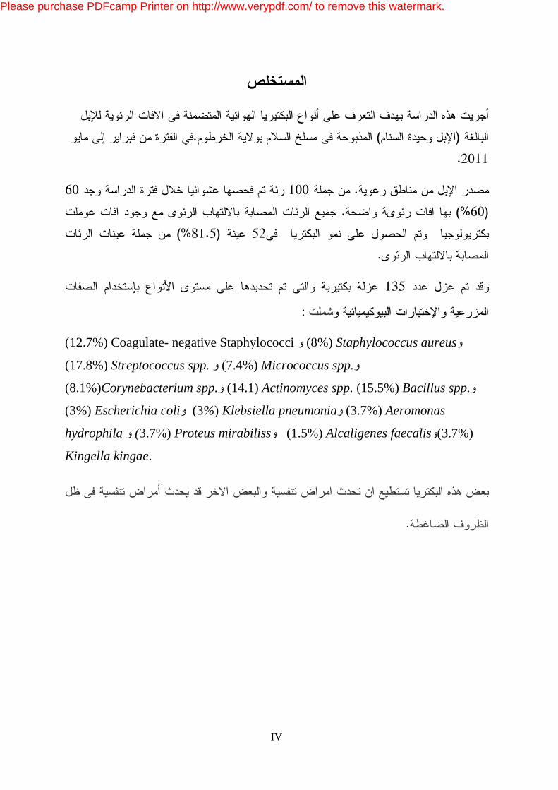
This study was carried out with the aim of identifying aerobic bacterial
species involved in lung lesions of one -humped adult camels
(Camelusdromedarius) slaughtered at Elsalam abattoir enterprise, Khartoum
State .The study was conducted from February to May 2011.
All camels were originated from various pastoral areas. A total of 100 camel
lungs were inspected at random during the study period, of which 60 (60%)
possessed gross pulmonary lesions. All pneumonic lungs with lesions were
processed for bacteriology and bacterial growth was only obtained from
52(81.5%) of the pneumonic lung samples.
A total of 135 bacterial species were isolated and identified to the species
level using cultural characteristics and biochemical tests. These included
coagulase- negative staphylococci (12.7%), Staphylococcus aureus(8%),
Streptococcus spp. (17.8%), Micrococcus spp. (7.4%) Corynebacteria
spp.(8.1%), Actinomyces spp. (14.1) Bacillus spp. (15.5%) Escherichia coli
(3%) Klebsiella pneumonia (3%), Aeromonas hydrophila(3.7%), Proteus
mirabilis (3.7%) Alcaligenes faecalis(1.5%) Kingella kingae(3.7%). Some of
these are potential pathogens that could induce respiratory diseases and other
isolates might induce Respiratory diseases under stressful conditions.
Please purchase PDFcamp Printer on http://www.verypdf.com/ to remove this watermark.
VI
Table of contents
Page DEDICATION
II
ACKNOWLEDGEMENTS
III
ARABIC ABSTRACT
IV
ABSTRACT
V
TABLE OF CONTENTS
VI
LIST OF TABLES
XVI
LIST OF FIGURES
XVII
Chapter one
1
Introduction
1
Chapter two 3
Literature Review
3
2. 1 Introduction 3
2.2. Anatomy of camels lung 3
2.3. Respiratory tract infections in camels 4
2.4. Common bacteria associated with pneumonia 5
2.4.1. Gram positive bacteria 5
2.4.1.1. Staphylococci 5
2.4.1.1.1. Staphylococcus aureus 5
2.4.1.2. Micrococci 6
2. 4.1.3. Streptococci 6
Please purchase PDFcamp Printer on http://www.verypdf.com/ to remove this watermark.
VII
2.4.1.3.1. Group B streptococci 7
a. Classification 7
b. Lance field's grouping
7
c. haemolysis production
7
2.4.1.4. Corynebacterium spp
7
2.4.1.5. Actinomycetes
8
2.4.1.6. Bacillus spp
8
2.4.2. Gram negative bacteria
9
2.4.2.1. Enterobacteriaceae
9
2.4.2.1.1. Escherichia coli
9
2.4.2.1.2. Klebsiella spp
9
2.4.2.1.3. Proteus spp
10
2.4.2.1.3.1. Proteus mirabilis
10
2.4.2.2. Aeromonas hydrophilia
11
2.4.2.3. Alcaligenes SPP
11
2.4.2.4. Kingella spp
11
2.5. The important bacterial respiratory diseases
11
2.5.1. Pasteurellosis
11
2.5.2. Tuberculosis
12
2.5.3. Mycoplasmosis
13
2.6. Bacteria isolated from infected respiratory tract of camels 13
Please purchase PDFcamp Printer on http://www.verypdf.com/ to remove this watermark.

VIII
2.7. Bacteria isolated from respiratory tract of apparently healthy camels
17
Chapter three
19
Materials and methods
19
3.1. Study area
19
3.2. Collection of samples
19
3.3. Isolation of organism
19
3.4. Sterilization
20
a. Flaming
20
b. Red heat
20
c. Hot air oven
20
d. Moist heat
20
3.5. Reagents and Indicators
20
3.5.1.1. Sodium Chloride
20
3.5.1.2 Alpha-naphthol solution
21
3.5.1.3. Potassium hydroxide
21
3.5.1.4. Hydrogen peroxide
21
3.5.1.5. Kovac's reagent
21
3.5.1.6. Methyl red Solution
21
3.5.1.7. Tetramethyl-p-phenylaminedihydrochloride
21
3.5.1.8. Nitrate test reagent
22
Please purchase PDFcamp Printer on http://www.verypdf.com/ to remove this watermark.

IX
3.5.2. Indicators
22
3.5.2.1 Andrade's indicator
22
3.5.2.2 Bromothymol blue
22
3.6. Preparation of Culture Media
22
3.6.1. Liquid media
23
3.6.1.1. Nutrient broth
23
3.6.1.2. Peptone water
23
3.6.1.3. Peptone water sugar
24
3.6.1.4. Glucose- Phosphate medium
24
3.6.1.5. Nitrate medium
25
3.6.1.6. Serum Water sugars
25
3.6.2. Semi solid media
25
3.6.2.1. Motility test medium- Cragie tube medium
25
3.6.2.2 Oxidation-fermentation medium
26
3.6.3. Solid media
27
3. 6. 3.1. Ammonium salt sugars
27
3.6.3.2. Bile Esculin Azide Agar
27
3.6.3.3. Collection of blood for enriched media
28
3.6.3.4. Blood agar
28
3.6.3.5. MacConkey's agar
29
3.6.3.6. Milk agar 29
Please purchase PDFcamp Printer on http://www.verypdf.com/ to remove this watermark.
X
3.6.3.7. Nutrient agar
30
3.6.3.8. Nutrient Gelatin
30
3.6.3.9. Simmons citrate agar
31
3.6.3.10. Starch
31
3.6.3.11. Urea agar Base
32
3.7. Culture media
33
3.7.1. Blood agar
33
3.7.2. MacConkey agar
33
3.8. Isolation
33
3.9. Purification
33
3.10. Preservation
33
3.11. Microscopic Examination
34
3.12. Biochemical Tests
34
3.12.1. Sugar fermentation test
34
3. 12.2. Oxidase test
34
3.12.3. Catalase test
34
3.12.4. Coagulase test
35
3.12.5. The oxidation- fermentation test
35
``3.12.6. Indole production test
35
3.12.7. Methyl red test
36
Please purchase PDFcamp Printer on http://www.verypdf.com/ to remove this watermark.

XI
3.12.8. Voges- Proskauer test
36
3.12.9. Nitrate reduction
36
3.12.10. Urease activity test
37
3.12.11. Citrate utilization
37
3.12.12. Aesculin hydrolysis
37
3.12.13. Hydrogen sulphide (H2s) production
37
3.12.14. Ammonium salt sugar test
37
3.12.15. Gelatin hydrolysis
38
3.12.16. Mobility test
38
3.12.17. Digestion of casein
38
3.12.18. Starch hydrolysis
38
Chapter four
39
Result
39
4.1. Postmortem findings
39
4.2. Isolation of organisms
39
4.3. Gram- positive bacteria
39
4.31. Staphylococcus spp.
40
a. Morphology and staining
40
b. Cultural characteristic
40
1. Blood agar medium
40
2. Nutrient agar 40
Please purchase PDFcamp Printer on http://www.verypdf.com/ to remove this watermark.
XII
3. Manitol salt agar
40
c. Biochemical reactions
41
4.3.2 Streptococcus spp.
41
a. Morphology and staining
41
b. Cultural characteristic
41
1. Blood agar medium
41
b. Biochemical reaction
41
4.3.3.Corynebacterium spp.
42
a. Morphology and staining
42
b. Cultural characteristic
42
1. Blood agar medium
42
2. Nutrient agar
42
c. Biochemical reactions
42
4. 3. 4. Actinomyces spp
42
a. Morphology and staining
42
b. Cultural characteristic
43
c. Biochemical reactions
43
4.3.5. Bacillus spp
43
a. Morphology and staining
43
b. Cultural characteristic
43
c. Biochemical reactions 43
Please purchase PDFcamp Printer on http://www.verypdf.com/ to remove this watermark.
XIII
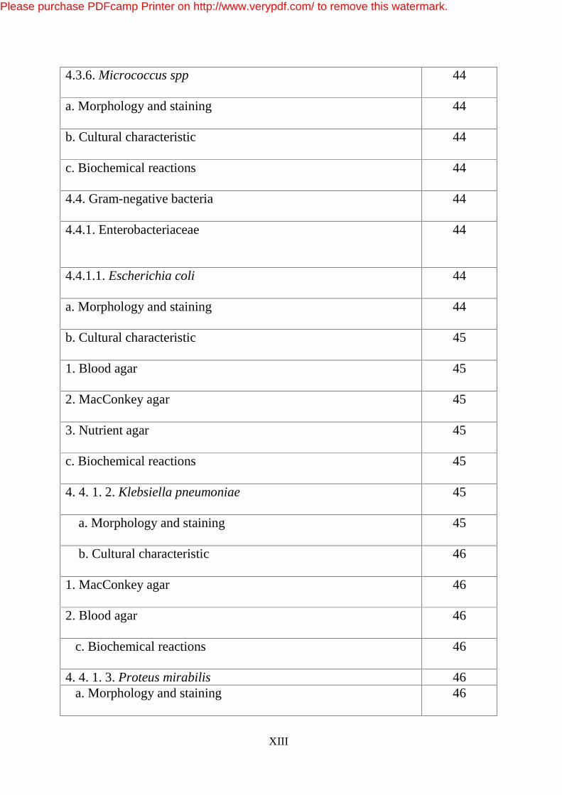
4.3.6. Micrococcus spp
44
a. Morphology and staining
44
b. Cultural characteristic
44
c. Biochemical reactions
44
4.4. Gram-negative bacteria
44
4.4.1. Enterobacteriaceae
44
4.4.1.1. Escherichia coli
44
a. Morphology and staining
44
b. Cultural characteristic
45
1. Blood agar
45
2. MacConkey agar
45
3. Nutrient agar
45
c. Biochemical reactions
45
4. 4. 1. 2. Klebsiella pneumoniae
45
a. Morphology and staining
45
b. Cultural characteristic
46
1. MacConkey agar
46
2. Blood agar
46
c. Biochemical reactions
46
4. 4. 1. 3. Proteus mirabilis 46 a. Morphology and staining
46
Please purchase PDFcamp Printer on http://www.verypdf.com/ to remove this watermark.
XIV
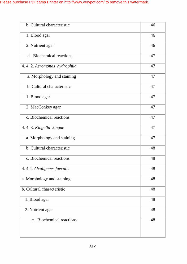
b. Cultural characteristic
46
1. Blood agar
46
2. Nutrient agar
46
d. Biochemical reactions
47
4. 4. 2. Aeromonas hydrophila
47
a. Morphology and staining
47
b. Cultural characteristic
47
1. Blood agar
47
2. MacConkey agar
47
c. Biochemical reactions
47
4. 4. 3. Kingella kingae
47
a. Morphology and staining
47
b. Cultural characteristic
48
c. Biochemical reactions
48
4. 4.4. Alcaligenes faecalis
48
a. Morphology and staining
48
b. Cultural characteristic
48
1. Blood agar
48
2. Nutrient agar
48
c. Biochemical reactions
48
Please purchase PDFcamp Printer on http://www.verypdf.com/ to remove this watermark.

XV
Chapter five
66
Discussion
66
Conclusions
72
Recommendations
73
References 74
Please purchase PDFcamp Printer on http://www.verypdf.com/ to remove this watermark.

XVI
List of Tables
Table Page
1 Types of bacteria isolated from pneumonic lungs 49
2 Staphylococci isolated from pneumonic lungs 51
3 Micrococci isolated from pneumonic lungs of camels 52
4 streptococci isolated from pneumonic lungs of camels 53
5 Actinomyces isolated from pneumonic lungs of camels 54
6 Corynebacteria spp. isolated from pneumonic lungs of
camels
55
7 Bacillus spp. isolated from pneumonic lungs of camels 56
8 Gram negative bacteria isolated from pneumonic lungs
of camels
57
9 Characters and Biochemical reactions of isolated Gram
positive cocci
58
10 Characters and Biochemical reactions of isolated
Corynebacterium spp.
59
11 Characters and Biochemical reactions of isolated
Actinomyces spp.
60
12 Characters and biochemical reactions of isolated
Enterobacteriae.
61
13 Characters and biochemical reactions of isolated
Aeromonas hydrophila
61
Please purchase PDFcamp Printer on http://www.verypdf.com/ to remove this watermark.

XVII
List of figures
No. of Figure Title of figure Page
Figure 1 Gram stained smear showing Actinomyces 64
Figure 2 Growth of Actinomyces in blood agar
65
Please purchase PDFcamp Printer on http://www.verypdf.com/ to remove this watermark.
1
CHAPTER 1
INTRODUCTION
Camels are the most capable animal species in utilizing marginal
areas and in survival and production under harsh environmental
conditions (Knoess, 1977; Gauthier-Pilters and Dagg, 1981; Hjort and
Hussein, 1986; Abbas and Tilley, 1990 and Schwartz, 1992). Many
pastoral groups and communities in the Sudan and diverse eco-zones
throughout the world are depending on camels for their livelihood.
Camels were, and still are, valued for carrying baggage, hair, hides
and as well as the best food providers in the arid areas (Sweet, 1965).
Sudan and Somalia have 70% of the total Africa camels and 55% of that
of the world camel’s population (Wilson, 1984). Moreover, increased
frequencies of drought recurrence, shrinkage and deterioration of the
rangeland by desert encroachment together with increasing aridity are the
major governing factor for the movement of dromedary camels into the
savannah belt. Thus contact between camels and other farm animals has
been increasingly noticeable from the early eighties of the last century.
Camel is a comparatively sturdy animal and is less susceptible to many
of the diseases that affect other livestock species in the same areas
(Schwartz and Dioli, 1992 and Dirie and Abdurhaman, 2003). However,
it is apparent that little is known about the diseases from which it suffers.
Camel of Sudan has suffered from various diseases like trypanosomosis,
camel pox, mange, respiratory disease complex, hemorrhagic septicemia,
pustular dermatitis, dermatomycosis, gastrointestinal parasites and acute
plant poisoning (Agab and Abbas, 1999).
Please purchase PDFcamp Printer on http://www.verypdf.com/ to remove this watermark.
2
Flora of respiratory tract of apparently healthy and infected camels
was reported by different authors (Farrag et al., 1953; Shigidi, 1973;
Chauchan et al., 1986; Alani et al., 1990 and Mustafa, 1992).
Respiratory tract infections are of common occurrence in various species
of domestic and farm animals (Mohamed and Abdelsalam, 2008).
Viruses, bacteria, fungi and parasites have been incriminated as the main
causative agents of pneumonia in mammals (Jubb et al., 19
93 and Cortan et al., 1999). These agents may represent risk to camels,
other livestock and even human population (Abou, 2000; Ogusan et al.,
2000; Bardonn et al., 2002 and Teshom et al., 2003).
Few studies were conducted on the extent of respiratory problems of
camels compared to other livestock species. Respiratory diseases have
been a major threat to the camel population. Respiratory disease outbreak
in camels that is characterized by sudden death has occurred in Ethiopia
(Awol et al., 2011). The most important predisposing factors for camel
diseases are sudden climatic changes, poor management practices,
exposure to various diseases, traveling and low grade nutrition (Schwartz
and Dioli, 1992 and Abubakar et at., 2008). Despite the versatile uses in
the pastoralists' areas, camel has been largely neglected by international
agencies and local governments regarding to improvement of its health
and productivity (Bekele, 1999). Yet there is a need to identify the causes
of respiratory diseases in camels in order to design better control
strategies.
This work was done to identify the etiology of aerobic bacteria
associated with pneumonic lungs of camels slaughtered in Elsalam
abattoir in Khartoum State.
Please purchase PDFcamp Printer on http://www.verypdf.com/ to remove this watermark.
3
Chapter 2
Literature Review
2.1. Introduction:
Pneumonia is an inflammation of pulmonary parenchyma usually
accompanied by inflammation of bronchioles and often with pleurisy
(Blood, Radostitis and Henderson, 1983). It is manifested clinically by
increase in respiratory rate, cough and abnormal breathing sounds. Most
of the bacterial agents of pneumonia are considered to be of aerogenous
orign reaching the lungs through the upper respiratory tract. Infection of
the latter causes inflammation, either acute or chronic (Blood and
Henderson, 1974).
The response of the respiratory tract to disease is determined largely
by the structural and functional complexity of the system. Most of the
diseases of the respiratory system are caused by damaging agents that
arrive either air borne (aerogenous) or blood borne (haematogenous). Air
borne infectious agents commonly cause respiratory diseases and in some
intensive systems of animal husbandry they constitute the most important
cause of morbidity and mortality.
2.2. Anatomy of camel Lungs:
Camel lungs are soft and spongy and characterized by absence of
lobulation and fissures and resemble those of horses. The right lung
consists of cranial lobe, caudal lobe, and accessory lobe while it lacks the
middle lobe. The left lung consists of cranial and caudal lobes only. The
tracheal bronchus, which aerates the apical lobe, is also found. The apical
lobe of the right lung takes its bronchus directly from the trachea.
Please purchase PDFcamp Printer on http://www.verypdf.com/ to remove this watermark.
4
Histologically, the alveolar wall is relatively thick. The type-1
pneumocytes are elongated, flattened cells with densely stained nuclei.
The type -11 pneumocytes are large cuboidal or round cells having large,
round nuclei with prominent nucleolus and vacuolated cytoplasm. The
interlobular septa are prominent. They divide the lung into lobules. Large
amounts of connective tissue are present between lobules which are
composed of collagen and elastin. (Alani, 2004)�
Camel pleura are characteristic and consist of visceral and parietal
layers occupying normally by a small amount of pleural fluid. The pleural
cavities are completely separated. The line of pleural reflection for left
lung extends from the first to twelfth rib on the dorsal aspect while the
cupulae epleurae of the right side extend beyond the first rib. The
phringes, they were once called, or extensions of pleura are at the
periphery of both lungs. These are composed of collagenous and elastic
fibers. They contain blood capillaries, lymph vessels, arterioles, venules
and group of inflammatory cell (Alani, 2004).
2.3. Respiratory tract infections in camels:
The normal flora of upper respiratory tract is important in that it
prevent adherence and colonization by invading bacteria (Jubb and
Kennedy, 1985). Respiratory infection in camels may be due to a variety
of microorganisms that are present in respiratory tract as commensals and
play pathogenic role when the general immune response of the host is
lowered (Shigidi, 1972).
Bacteria can affect respiratory tract of camels causing rhinitis,
pharyngitis, trachitis, bronchitis, pneumonia and lung abscesses.
Exudation and consolidation of lungs characterize bacterial pneumonia
(Jubb and Kennedy, 1985). Haematogenous infection by bacteria results
Please purchase PDFcamp Printer on http://www.verypdf.com/ to remove this watermark.

5
in a varying number of septic foci, which may enlarge to form lung
abscesses (Blood and Henderson, 1983). Pneumonia occurs when these
abscesses rupture inducing a secondary bronchopneumonia.
Arcanobacterium pyogenes and Staphylococcus aureus were isolated
from Lung abscesses (Abubakr et al., 2010).
2.4. Common bacteria associated with pneumonia:
2.4.1. Gram-positive bacteria:
2.4.1.1. Staphylococci
The staphylococci are Gram-positive spherical cells, usually arranged
in grape- like irregular clusters although organisms may appear as single
cells, pairs or short chains (Murray et al., 1998). Staphylococci are non-
motile and do not form spores. They grow readily on most bacteriological
media under aerobic conditions. They grow most rapidly at 37°�C but form
pigment best at room temperature (20- 25°�C).
2.4.1.1.1. Staphylococcus aureus:
Staphylococcus aureus is non- motile, non sporing, aerobic and
facultative anaerobic bacteria, it is catalase positive (Barrow and Feltham,
1993). It is able to grow in a medium containing salt such as manitol salt
agar, which used as a selective medium for isolation of staphylococci that
ferment manitol and colonies appear yellow to cream 1.2 mm in diameter.
The pathogenic Staphylococcus aureus often hemolyse blood, coagulase
plasma and produce a variety of extra- cellular enzymes and toxins.
Coagulase is in extra- cellular protein, which binds to prothrombin to
form a complex called staphylothrombin (Todar, 2002).
Coagulase is a traditional marker for identifying Staphylococcus
Please purchase PDFcamp Printer on http://www.verypdf.com/ to remove this watermark.

6
aureus in the clinical microbiology laboratory. However, there is no
overwhelming evidence that coagulase is a virulence factor, although it is
reasonable to speculate that the bacteria could protect themselves from
phagocytes and immune defenses by causing localized clotting (Todar,
2002).
2.4.1.2. Micrococci
Micrococcus is Gram-positive cocci that are 0.5 to 3.5 micrometers
in diameter and usually arranged in tetrads or irregular clusters. Defining
characteristics of Micrococcus are the ability to aerobically produce acid
from glucose, glycerol. They hydrolyse aesculin and arginine and reduce
nitrate to nitrite (Smith et al. 1999). They attack sugars oxidatively or not
at all (Barrow and Feltham, 1993). Micrococcus species are part of
normal flora of the nasopharynx of domestic animals, they are frequently
found in soil, water, dust, air and skin and on articles of daily use (Carter,
1986). Occasional strains were associated with disease (Barrow and
Feltham, 1993).
2.4.1.3. Streptococci
They are Gram-positive cocci, usually non-motile, smaller than
staphylococci in diameter, aerobic and facultatively anaerobic, catalase-
negative, oxidase negative.They attack sugars fermentatively without gas
production and some may produce haemolysis (Barrow and Feltham,
1993).
Please purchase PDFcamp Printer on http://www.verypdf.com/ to remove this watermark.
7
2.4.1.3.1. Group B streptococci
a. Classification
b. Lance field’s groupings
Serologically, streptococci are classified on the basis of a cell surface
carbohydrate antigen in the cell wall called component C, into serotypes
A to T (Carter et al., 1985 and David Green et al., 2002).
c. Haemolysis production
�- haemolytic streptococci produce a clear zone of haemolysis on
sheep blood agar, whereas a�-haemolytic streptococci produce partial
haemolysis with zone of greenish discoloration, ?�- haemolytic
streptococci produce no hemolysis on blood agar (Carter et al., 1985).
Many isolates, however, are only weakly haemolytic and anaerobic
incubation often increases the extent of haemolysis with such strains
(Barrow and Feltham, 1993). Streptococcus pyogenes secretes different
toxins and enzymes that are associated with its virulence. These include
streptolysin O which is oxygen labile, hylouridase which increases tissue
permeability and leucocidin which destroys leucocytes (Barrow and
Feltham, 1993).
2.4.1.4. Corynebacterium spp.:
They are Gram-positive, catalase positive, non-spore-forming, non-
motile, rod-shaped bacteria that are straight or slightly curved (Collins
and Cummins 1986). Both toxin and non-toxin producing strains of C.
dephtheriae can adhere to and colonize the mucosal tissue of the upper
respiratory tract (Jensen and Warigh, 1998). The bacteria group together
in a characteristic way, which has been described as the form of a "V",
Please purchase PDFcamp Printer on http://www.verypdf.com/ to remove this watermark.
8
"palisades", or "Chinese letters". They may also appear elliptical. They
are aerobic or facultatively anaerobic.
2.4.1.5. Actinomycetes:
Actinomycetes appear as Gram-positive bacilli (Hall, 2006) which
comprise a group of branching unicellular microorganisms. They produce
branching mycelium which may be of two kinds substrate mycelium and
aerial mycelium. Members of the actinomycetes, which live in marine
environment, are poorly understood and only few reports are available
pertaining to actinomycetes from mangroves (Siva Kumar, 2001;
Vikineswari et al., 1997; Rathana Karla and Lakshmanaperumalsamy,
1978). Actinomyces pyogenes, normally commensal bacterium, resides on
mucous membranes of cattle, sheep, swine, and other economically
important animals (Carter et al., 1991).
2.4.1.6. Bacillus spp.: Bacillus species are Gram-positive rods often arranged in pairs or
chains with rounded or square ends and usually have a single endospore.
The endospores are generally oval and are very resistant to adverse
conditions. They have different stabilities and degrees of resistance to
heat, radiation, chemicals, desiccation, and other hostile conditions.
Sporulation is not repressed by exposure to air (Holt et al., 1994).
Bacillus spp. can be broadly divided in three groups based on the
morphology of the spore (Berkeley et al., 1997). The genus Bacillus
currently comprises more than in excess of 60 species, commonly found
in the environment and as laboratory contaminants (Konemann et al.,
1997).
Please purchase PDFcamp Printer on http://www.verypdf.com/ to remove this watermark.
9
2.4.2. Gram-negative bacteria:
2.4.2.1. Enterobacteriaceae:
Members of this family are Gram negative bacilli. They are non-
motile or motile with peritrichous flagella and do not form spores. All
members where growing aerobically and are facultative anaerobes on a
variety of non-selective media (blood agar) and selective media
(MacConkey’s agar medium). Enterobacteriaceae have simple nutritional
requirements, ferment glucose, reduce nitrate. They are catalase- positive
and oxidase- negative (Murray et al., 1998).
2.4.2.1.1. Escherichia coli:
It is Gram-negative rods methyl red positive and 80% of strains are
motile. It can grow on simple media containing glucose as the sole carbon
source (Geo et al., 1998). E. coli is Gram-negative rods that may form
chains under unfavorable conditions (exposure to penicillin). Capsules or
microcapsules are produced by many strains.
2.4.2.1.2. Klebsiella pneumoniae :
It is Gram negative bacillus with a very thick mucoid capsule that
inhibits phagocytosis (Mac Sween and Whaly, 1992). Klebsiella
pneumoniae is present in the respiratory tract causes a small proportion of
bacterial pneumonias (Geo et al., 1995). It is opportunistic pathogen; it
may produce pyogenic lesions like abscesses, infection of wound or
respiratory tract (Satish, 1995). It is the main species of medical
importance . Four sub-species of Klebsiella pneumoniae are recognized
K .pneumoniae sub sp pneumoniae, K. pneumoniae sub-sp. aerogenes, K
.pneumoniae sub-sp ozaenae, K. pneumoniae sub-sp. rhinoscleromatis
(Cheesbrough, 2000).
Please purchase PDFcamp Printer on http://www.verypdf.com/ to remove this watermark.
10
2.4.2.1.3. Proteus species
They are Gram-negative rods, motile, aerobic and facultatively
anaerobic, catalase positive, oxidase negative. They attack sugars
attacked fermentatively usually with gas production and hydrolyze urea
and gelatin (Barrow and Feltham, 1993).
Proteus cultures have a distinctive smell (fishy smell) with swarming
on non-inhibitory solid media such as nutrient agar and blood agar. Most
strains swarms with periodic cycles of migration producing concentric
zones, or spread in a uniform film over moist surfaces of nutrient media.
Proteus species are free living saprophytes in soil, vegetation, water and
sewage, and are found in the intestine in many healthy persons. These
organisms occur naturally in the environment of animals and man and
particularly in the intestines, animal manure, human sewage, soil and
water.
2.4.2.1.3.1. Proteus mirabilis:
It is the main species of medical importance. Many strains of P.
mirabilis produce bacteriocins (proticins) which have a lethal action
against other strains.
Senior (1977) described a highly discriminating method of typing
strains by determination of their proticin production and sensitivity. The
method was made more discriminating when it was used in combination
with O serotyping (Senior, 1977).
Please purchase PDFcamp Printer on http://www.verypdf.com/ to remove this watermark.
11
2.4.2.2. Aeromonas hydrophila:
It is Gram- negative rod, motile and non-motile, aerobic and
facultatively anaerobic, catalase positive, oxidase positive. It attack
sugars fermentatively and gas may be produced (Barrow and Feltham,
1993). It is opportunistic pathogen of fish, reptile and rarely mammals
(Quinn et al., 2002). Camels might acquire it from contaminated drink
water.
2.4.2.3. Alcaligenes spp.:
Gram- negative rods, motile, aerobic, catalse- positive, oxidase-
positive do not produce acid from sugars even in suitable media (some
strains produce acid from ethanol), (Barrow and Feltham, 1993).
2.4.2.4. Kingella spp.:
They are Gram- negative short rods, non-motile, aerobic or
facultatively anaerobic. catalas- negative and oxidase- positive they attack
sugars by fermentation in suitable media and do not produce pigment or
hydrolyse arginine. (Barrow and Feltham, 1993).
2.5. The Important bacterial Respiratory diseases:
2.5.1. Pasteurellosis:
Acute infection with Pasteurella multocida causes hemorrhagic
septicemia in camels. The disease was reported in Africa, India, and
USSR (Macgrane and Higgins, 1985). They described three forms of the
disease. The per- acute form, which is associated with sudden death. The
acute form, which is characterized by fever, anorexia, edematous swells
in pharyngeal and prescapular region. She camels sometimes abort and
Please purchase PDFcamp Printer on http://www.verypdf.com/ to remove this watermark.
12
death occurrs within 2-5 days. The third form of the disease shows
bloody diarrhea as the predominant symptom.
In Ethiopia, Mannheimia haemolytica was identified as the most
important organism involved in an outbreak of camel respiratory disease
(Mersie, 1997).
In Pakistan, an outbreak of a respiratory disease occurred in the
dromedary population of Greater Cholistan desert. The organism isolated
from representative clinical and morbid specimens was Pasteurella
multocida sub spp.multocida. The duration of outbreak was more than a
month (Fraz Munir Khan, 2012).
2.5.2. Tuberculosis:
The disease was first reported in Egypt by Littlewood in (1888). This
was followed by the findings of Archibald (1910) who isolated acid-fast
organisms from lung lesions resembling miliary tuberculosis. The disease
was apparently rare among the camels of nomads, whereas it tended to
occur among camels kept for farming and in close proximity to cattle.
The incidence of camel tuberculosis in Egypt along with typing of the
causal agent was reported by El-Afifi et al. (1953)
Camel tuberculosis in India was first mentioned by Lingard, (1906)
and then by Leese, (1908) who considered it to be comparatively rare,
except in old camels where the usual pulmonary form and occasionally a
generalized form. The disease was reported in Ethiopia by Richard,
(1975) and a spontaneous case of camel tuberculosis in Somaliland has
been described by Pellegrini, (1942-1945). It was characterized by
progressive debility, coughing and death within six months.
Please purchase PDFcamp Printer on http://www.verypdf.com/ to remove this watermark.
13
2.5.3. Mycoplasmosis:
There is very little in the literature with regards to the role played by
mycoplasmas in the etiology of pneumonia in camels.
Elfaki et al. (2002) isolated M. arginini along with several other
bacteria from camel lungs with lesions suggestive of chronic interstitial
pneumonia.
In Egypt, Abeer El-metwally et al. (2010), detected M. arginine from
lung, uterus, cervix, vagina and mammary gland of she camels. Isolation
rate of mycoplasma from lung was 6%.
2.6. Bacteria Isolated From Infected Respiratory Tract of
Camels:
In different regions of Sudan, Mustafa (1992) studied pneumonia in
camels. The examination was carried clinically, bacteriologically and
histopathologically. Speciemens were collected, including nasal swabs,
tracheal swabs and pneumonic lungs. Bacteria isolated were Bacillus
spp., Staphylococcus aureus, Streptococcus pyogenes, Actinomyces.
pyogenes, coagulase- negative staphylococci, diplococci, Klebsiella
pneumoniae, E. coli, diphteroid and mixed flora. The most severe lung
lesions were found to be caused by Actinomyces. pyogenes,
Staphylococcus aureus, Streptococcus pyogenes and Klebsiella
pneumoniae. Also, Tigani et al. (2006). carried out bacteriological and
pathological studies in Central region of the Sudan (Tamboul abattoir)
and Western region (Nyala abattoir) to investigate the condemnation
causes of camel lungs. Camel lungs were inspected at post-mortem in
both areas. The survey revealed various causes of camel’s lung
condemnation with slight variations between the two areas of survey. The
Please purchase PDFcamp Printer on http://www.verypdf.com/ to remove this watermark.
14
microorganisms isolated from Pneumonic cases were Staphylococcus
spp., Corynebacterium spp., Streptococcus spp., Bacillus spp.,
Pneumococcus spp., Enterobacteria spp., Micrococcus spp.,
Haemophilus spp., Actinomyces spp., Pasteurella spp. and
Pseudomonas spp.
Another study of Respiratory infections of the camels (Camelus
dromedarius) in Sudan by Nasr, (2003) has been carried out in camels
brought to Tamboul Market -Butana region, East Central Sudan- for
slaughter. Specimens were collected and they consisted of nasal swabs,
tracheal swabs, lung tissue and bronchial lymph nodes. Bacteria isolated
from nasal cavity in order of abundance, were: Coagulase- negative
staphylococci, Bacillus spp., E. coli, diphteroids, alpha-hemolytic
streptococci, coagulase- positive staphylococci, Actinomyces. pyogenes,
Klebsiella pneumoniae, Alcaligenes faecalis, Streptococcus pyogenes,
Micrococcus luteus, Diplococcus pneumoniae, Bordetella
bronchiseptica, Mannhemia haemolytica, Haemophillus somnus, mixed
flora, Micrococcus kristinae and Pasteurella multocida. Bacteria
isolated from trachea were: Bacillus spp., coagulase- negative
staphylococci, diphteroids, E. coli, Actinomyces pyogenes, Bordetella
bronchiseptica, Alcaligenes faecalis, coagulase- positive staphylococci,
alpha haemolytic streptococci, Klebsiella pneumoniae, Diplococcus
pneumoniae, Micrococcus luteus, Micrococcus kristinae, Haemophillus
somnus and mixed flora. Bacteria isolated from lung tissue were: A.
pyogenes, Bacillus spp., coagulase- negative staphylococci, diphteroids,
coagulase -positive staphylococci, Streptococcus pyogenes, Klebsiella
pneumoniae, alpha-hemolytic streptococci, Mannheimia haemolytica,
Diplococcus pneumoniae and E. coli. Bacteria isolated from bronchial
lymph nodes were: Coagulase-negative staphylococci, Bacillus spp.,
Please purchase PDFcamp Printer on http://www.verypdf.com/ to remove this watermark.

15
diphteroids, coagulase-positive staphylococci, A. pyogenes, alpha-
hemolytic streptococci, Streptococcus pyogenes, Corynebacterium equi,
Pasteurella multocida, Mannheimia haemolytica Klebsiella pneumoniae
and Citrobacter koseri. In this study Haemophillus somnus, Micrococcus
spp. and Bordetella bronchiseptica were isolated for the first time from
respiratory tract of camels.
In Egypt, Farrag et al. (1953) studied pneumonia in camels
slaughtered at Cairo abattoir. Specimens taken were parts of lungs
showing different stages of pneumonia. Bacteria isolated were
Corynebacterium pyogenes, alpha-hemolytic streptococci, Alcaligenes
faecalis, and B. pyocyaneus. Mahamoud et al. (1988) carried out
bacteriological and pathological examination of affected camel lungs in
Egypt. Microorganisms isolated were Klebsiella spp., Proteus vulgaris,
Staphylococcus aureus, Streptococcus pyogenes, E. coli and proteus
rettergi. Another study was carried out in Egypt by Ibtsam (2006). She
investigated bacterial pneumonia in camels. The study included camels
of both sexes, 9 month-2 years old. Bacteria isolated were Streptococcus
pneumoniae, Staphylococcus aureus, E. coli, Mannheimia haemolytica
and Streptococcus pyogenes.
In 1973, Arora and Karla described a case of bronchopneumonia in a
camel in India. The bacteria isolated from affected lungs were
Diplococcus and Klebsiella pneumoniae.
Respiratory infection of camels in Iraq was studied by Alani, (1990).
The isolates from nasal cavity were Pasteurella multocida,
Staphylococcus spp., A. pyogenes, E. coli and Mannheimia haemolytica
The isolates from infected lungs were Pasteurella multocida, Neisseria
spp., Pseudomonas spp., Staphylococcus spp. and A. pyogenes.
Please purchase PDFcamp Printer on http://www.verypdf.com/ to remove this watermark.

16
Moalin and Zessin, (1990) studied Diseases of camels in Somali
Microorganisms isolated from infected lungs were Staphylococcus spp.,
Psudomonas aeroginosa and Citrobacter freundii.
Aldoughaym et al. (1999) studied etiology of pneumonia in camels.
Specimens including nasal swabs, tracheal swabs and pneumonic lung
tissue were collected. Bacteria isolated were Staphlococcus auerus, A.
pyogenes, Streptococcus pyogenes, Coagulase-negative staphylococcus,
Klebsiella pneumoniae, Diplococcus pneumoniae, E. coli, diphteriods
and mixed organisms. The percentage of isolation of Staph.aureus, A.
pyogenes, Streptococcus pyogenes and Klebsiella pneumoniae from
pneumonic tissue was the highest as compared with that from nasal cavity
and the trachea. In contrast the isolation of Bacillus spp., coagulase
negative staphylococcus and diphtheriods from pneumonic lung tissue
was markedly less as compared to that isolated from nasal cavity and
trachea. The isolation percentage of E. coli and Diplococcus pneumoniae
was nearly similar from pneumonic lung tissue, nasal cavity and trachea.
Al-Tarazi (2001) carried out bacteriological and pathological study
on pneumonia in the One-Humped Camels in Jordan. Specimens taken
were lung tissues of slaughtered camels and bacteria isolated from
pneumonic lungs were E. coli, Klebsiella spp., Staphylococcus aureus,
Pseudomonas aeruginosa, A. pyogenes, Mannheimia haemolytica,
Hemolytic streptococci, Proteus spp. and Bacillus spp.
Abubakar et al. (2008) studied camel pneumonia epidemiology and
bacterial flora in normal and diseased lungs of camels in Kano and
Sokoto main abattoirs in northwestern of Nigeria, Their findings in
normal lungs were Staphlococcus aureus, Corynebacterium spp.,
Arcanobacterium pyogenes, Streptococcus spp., Micrococcus spp.,
Please purchase PDFcamp Printer on http://www.verypdf.com/ to remove this watermark.

17
Klebsiella pneumoniae, Bacillus spp., Proteus spp., Pasteurella
multocida and Mannheimia haemolytica. Bacteria isolated from
pneumonic lungs were Staphlococcus aureus, Corynebacterium spp.,
Streptococcus spp., Micrococcus spp., Klebsiella pneumoniae, Bacillus
spp., Proteus spp., Pasteurella multocida and Mannhemia haemolytica.
In Ethiopia Bacteriological study on pulmonary lesions of camels
was carried out. Bacteria isolated were E. coli, Flavobacterium spp.,
Rhodococcus equi, Bordetella bronchoseptica, Aeromonas hydrophila,
Neisseria spp., Streptococcus agalactiae, Staphlococcus aureus,
Pasteurella trehalosi, Pasteurella anatipestifer, Pseudomonas
aeruginosa, and Micrococcus spp. ( Nesibu et al., 2010).
2.7. Bacteria Isolated From Respiratory Tract of Apparently
Healthy Camels:
In Sudan, Shigidi (1972) studied aerobic flora of respiratory tract of
apparently healthy camels. Specimens collected were nasal swabs, lung
tissues and bronchial lymph nodes. Organisms isolated were Bacillus
spp., coagulase-negative staphylococci, diphteroids, Aspergillus spp., A.
pyogenes, alpha- hemolytic Streptococci, Streptomyces spp., coagulase-
positive staphylococcci, E. coli and Enterobacter aerogenes
Bacterial flora of upper respiratory tract of apparently healthy camels
was studied in India. Specimens taken were nasal swabs and bacteria
isolated were coagulase-negative staphylococci, E. coli, Klebsiella
pneumoniae, A. pyogenes, coagulase-positive staphylococci,
Corynebacterium equi, beta-hemolytic streptococci, hemolytic
Diplococci, Anthracoids and Corynebacterium hemolytica (Chauhan et
al., 1986).
Please purchase PDFcamp Printer on http://www.verypdf.com/ to remove this watermark.
18
In 1999, Elhendi studied nasal microflora of upper respiratory tract of
apparently healthy camels under two conditions in Saudi Arabia. First
group contained apparently healthy camels were presented from slaughter
house and second group contained apparently healthy camels of a local
herd of Research Unit at King Faisal University Farm. All camels of both
groups were sampled for a period of six months at appropriate intervals.
Bacteria isolated in first group were Staphlococcus auerus, E. coli,
Streptococcus spp. Bacteria isolated in second group were Staphlococcs
auerus, E. coli, Streptococcus spp., Klebsiella spp and Corynebacterium
spp.
In Iran Azizollah et al., (2008) studied the aerobic bacteria of the
respiratory passageways flora of apparently healthy camels. Specimens
were taken from the nasal cavity, trachea, tonsils and lungs. Bacteria
isolated were staphylococci, followed by Neisseria spp., Bacillus spp. and
streptococci. The majority of the isolates colonized all the anatomical
sites investigated with the exception of E. coli and, streptococci, which
were absent from the lungs, and Klebsiella which was absent from the
nasal tract. Gram-positive bacteria were dominant in this environment,
followed by Neisseria spp., E. coli and Klebsiella spp.
Please purchase PDFcamp Printer on http://www.verypdf.com/ to remove this watermark.
19
Chapter three
Materials and Methods
3.1. Study area:
Specimens were collected from a total of 100 lungs of slaughtered one
humped adult camels of both sexes, they were examined for pneumonic
lesion at Elsalam abattoir about 20 kilometers western of Khartoum. The
study was conducted from February to May 2011.
3.2. Collection of Samples:
Immediately after slaughter, lung tissue of surface area greater than 10
x 10 cm2 showing pneumonic lesions was collected using sterile forceps
and scissors or scalpel blade. Specimens were then transported to the
veterinary laboratory using ice box.
3.3. Isolation of organism:
Immediately after arrival the surface of tissue specimens was burned
by hot spatula and aspirates were taken from the periphery of the lesion.
Aspirates were inoculated onto appropriate primary media (blood agar
and MacConkey’s agar). The cultures were incubated at 37°C for 24 to 72
h depending on the type of bacteria. Bacterial colonies were characterized
by their color, size, edge, surface, consistency, odor, transparency, and
emulsification. Single colonies were taken and smears were prepared for
Gram’s reaction. Finally, identification of bacteria to the species level
was performed using various biochemical tests.
Please purchase PDFcamp Printer on http://www.verypdf.com/ to remove this watermark.
20
3.4. Sterilization:
a. Flaming: It was used to sterilize glass slides, needles, scalpels, points of
scissors and mouth of culture tubes by passing them through Bunsen
burner flame without allowing it to become red hot. ��b. Red heat: It was used to sterilize loop wires, points of forceps and searing
spatulas by holding them over Bunsen burner flame until became red-
hot. ��
c. Hot air oven:
It was used to sterilize glass wares such as tubes, graduated
pipettes, flasks, forceps and cotton swabs. The holding period was
one hour and oven temperature was 160 °�C. ��
e. Moist heat (Autoclave):
Autoclaving at 121 °�C (151b/inch²) for 15 minutes was used for
sterilization of media and plastic wares. Autoclaving at 115 °�C
(101b/inch²) for 10 minutes was used for sterilization of sugars. ��
3.5. Reagents and Indicators:
3.5.1.1 Reagents:
Most reagents were obtained from British Drug House (BDH).
3.5.1.2. Sodium Chloride/ normal saline
Normal, physiological, or isotonic saline was prepared as described in
Oxide Manual by dissolving 8.5 g of sodium chloride in 1 liter of distilled
water to obtain 0.85% concentration.
Please purchase PDFcamp Printer on http://www.verypdf.com/ to remove this watermark.
21
3.5.1.2. Alpha-naphthol solution (Hopkin and Williams):
This solution was prepared as 5% aqueous solution for Voges-Proskaur
(VP) test.
3.5.1.3. Potassium hydroxide (Hopkins and Williams):
This reagent was prepared as 40% solution for Voges-Proskaur (VP) test.
3.5.1.4. Hydrogen peroxide (H2O2) (BDH):
This reagent was prepared as 3% aqueous solution and used for catalase
test.
3.5.1.5. Kovac's reagent:
This reagent was made of 5 g para- dimethyl amino benzaldehyde, 75ml
amyl alcohol and 25 ml concentrated hydrochloride acid. The aldehyde
was dissolved in alcohol gently in a water bath at temperature of 50-55°�C,
then cooled and the acid was added with care. The reagent was protected
from light and stored at 4°�C for indole test.
3.5.1.6. Methyl red Solution (BDH):
This solution was prepared by dissolving0.04 g of methyl red in 40 ml
ethanol and the volume was made to 100 ml with distilled water. It was
used for methyl red test.
3.5.1.7.Tetramethyl-p-phenylamine dihydrochloride
(Hopkin and Williams)
This reagent was prepared as 1% aqueous solution and it was used for
oxidase test.
Please purchase PDFcamp Printer on http://www.verypdf.com/ to remove this watermark.
22
3.5.1.8. Nitrate test reagent:
Nitrate test reagent was consisted of two solutions which were prepared
according to Barrow and Feltham (1993). Solution A composed of 0.33%
sulphanilic acid was dissolved by gentle heating in 5N- acetic acid.
Solution B composed of 0.6% dimethyl amine-alpha-naphthylamine
dissolved by gentle heating in 5N- acetic acid.
3.5.2. Indicators:
3.5.2.1. Andrade's indicator:
It was composed of 5 g of acid fuchsin, 1 liter of distilled water, and
150 ml of N-NaOH. The acid fuchsin was dissolved in distilled water,
and then the alkali solution was added, mixed and allowed to stand at
room temperature for 24 hour with frequent shaking until the color
changed from red to brown.
3.5.2.2. Bromothymol blue:
It was obtained from (BDH). The solution was prepared by dissolving
0.2 g of the powder in 100ml distilled water.
3.6. Preparation of Culture Media:
All media were dispensed under aseptic conditions in a laminar air flow
cabinet type 11 (Prettl R., Germany) provided with fan, ultra violet lamp
and flame . Media were either prepared from the original ingredients or
obtained in a dehydrated form.
Please purchase PDFcamp Printer on http://www.verypdf.com/ to remove this watermark.
23
3.6.1. Liquid media
3.6.1.1. Nutrient broth (Biomark Code No. B274)
FORMULA
1- Peptic digest of animal tissue …….………5g
2- Beef extract …………………………..1.50 g
3- Sodium chloride …………………………5 g
4- Yeast extract ………………………….1.50 g
pH 7.4 ± 0.2
Thirteen grams of Nutrient broth were added to one liter of distilled
water, mixed well and distributed in 3 ml amounts into clean test tubes,
then sterilized by autoclaving at 121°�C for 15 minutes.
3.6.1.2. Peptone water (HIMEDIA Code No. M 028)
FORMULA :
1- Peptic digest of animal tissue ………...10.0 g
2- Sodium chloride ………………………5.0 g
pH 7.2 ± 0.2
15 g were suspended in 1000 ml distilled water, mixed well, dispensed
into tubes and sterilized by autoclaving at 121°�C for 15 minutes.
Please purchase PDFcamp Printer on http://www.verypdf.com/ to remove this watermark.
24
3.6.1.3. Peptone water sugar (Barrow and Feltham, 1993):
FORMULA
1- Peptone water ……………… 10 g
2- Carbohydrates ………………10 g
3- Andrade's indicator...….…….10 g
4- Sugar ……………………..…10 g
Solid ingredients were dissolved in 1000 ml distilled water. The pH was
adjusted to 7.1- 7.3 before the addition of andrade's indicator. The
complete medium was mixed, distributed into 2 ml volumes into test
tubes containing Darham's tube (for gas trapping in case of glucose) and
sterilized by autoclaving at 115°�C for 10 minutes.
3.6.1.4. Glucose- Phosphate medium (MR-VP test medium)
(Oxoid Code NO. 43)
FORMULA
1- Peptone (Oxoid L49) ….………...…5 g
2- Dextrose ……………….…………...5 g
3- Phosphate buffer (k2HO4)……….…5 g
pH 7.5
15 g of the powder were added to 1 liter of distilled water. Mixed well,
distributed into final containers and sterilized by autoclaving at 121°�C for
15 minutes.
Please purchase PDFcamp Printer on http://www.verypdf.com/ to remove this watermark.

25
3.6.1.5. Nitrate medium (Barrow and Feltham, 1993):
FORMULA
1- Potassium nitrate …………………………0.2 g
2- Peptone …………………………….……..5.0 g
Solid ingredients were dissolved into 1000 ml distilled water. The
medium mixture was distributed in 5 ml amounts in test tubes and then
autoclaved for 15 minutes at 121°�C.
3.6.1.6. Serum Water sugars (Barrow and Feltham, 1993):
FORMULA
1- Peptone …………………………….……. 4 g
2- Na2HPO2………..………………….…...0.8 g
3- Distilled water ………………..………. 800 ml
4- Sterile Serum ………………..…...…… 200 ml
5- Bromcresol purple, 02% aq. Sol. ….....….10 ml
Dissolve the peptone and phosphate in the water, steam at 100°�C for 15
min and filter. Add the serum and steam for a further 15 min. adjust pH to
7.5- 7.8 and add the indicator. Sterilize at 115°�C for 10 min.
Please purchase PDFcamp Printer on http://www.verypdf.com/ to remove this watermark.
26
3.6.2. Semi solid media:
3.6.2.1 Motility test medium- Cragie tube medium (Barrow
and Feltham, 1993)
FORMULA :
1- Nutrient broth (Oxoid) …….….13 g
2- Agar (Oxoid agar No. 1)………..5 g
13 g of dehydrated nutrient broth, were added to 5 g of agar and were
dissolved in 1 liter of distilled water. The pH was adjusted to 7.4. The
medium was dispended in volumes of 5 ml into 20 ml capacity test tubes
containing Cragie tube. The medium was then sterilized by autoclaving in
121°�C for 15 minutes.
3.6.2.2. Oxidation-fermentation medium (Barrow and
Feltham, 1993)
FORMULA
1- Peptone ………………………………………….2.0 g
2- Sodium Chloride ………………………………...5.0 g
3- Di- potassium hydrogen orthophosphate ………..0.3 g
4- Dextrose ……………………….……………….10.0 g
5- Agar (Oxoid agar No. 1) …….………………...…3.0 g
6- Bromothymole blue …………….………………15 ml
pH 7.1
Please purchase PDFcamp Printer on http://www.verypdf.com/ to remove this watermark.

27
The solid ingredients were dissolved in 1000 ml distilled water and the
pH was adjusted before the indicator and agar were added. The medium
was then dispensed in tubes and sterilized by autoclaving at 121°�C for 15
min.
3.6.3. Solid media:
3.6.3.1. Ammonium salt sugars (ASS) (Barrow and Feltham,
1993):
FORMULA
1- (NH4)2HPO4 ……………………….1.0 g
2- KCL ………………………………....0.2 g
3- MgSO4.7H2O ………….……………0.2 g
4- Yeast extract ………………..……….0.2 g
5- Agar ………………………………...20.0 g
6- Distilled water ………………...……10.0 g
7- Bromocrysol purple 0.2%………… 0.04ml.
Solids were added to distilled water and dissolved by steaming then
indicator was added before autoclaving at 115°�C for 20 minutes. The
basal medium was allowed to cool to about 60°�C and then appropriate
carbohydrate was added as sterilized solution to give final concentration
1%, mixed and distributed aseptically into sterile tubes that allowed to
solidify in an inclined position so that the medium set in slope and butt.
Please purchase PDFcamp Printer on http://www.verypdf.com/ to remove this watermark.
28
3.6.3.2. Bile Esculin Azide Agar (Barrow and Feltham,
1993):
FORMULA
1- Ox bile, dehydrated ………………………40.0 g
2- Esculin ..................................................…...1.0 g
3- Ferric citrate …………….…………….…...0.5 g
4- Sodium azide ……………………….…..…0.2 g
5- Agar …………………………….….….…15.0 g
6- Nutrient broth …………………….….….1000 g
All ingredients except esculin were dissolved by heating. Allowed to
cool and then esculin was added. The medium was then distributed in
screw capped bottles, sterilized at 115°�C for 20 minutes and allowed to
set as slopes.
3.6.3.2.Collection of blood for enriched media:
Blood for enriched media was collected aseptically into sterile flasks
containing glass beads by venipuncture of jugular vein of a healthy sheep
kept for this purpose. The blood was defibrinated by shaking the sterile
flask containing the glass beads.
3.6.3.4. Blood agar ( Scharlau Code NO. 01-352)
Blood agar base
FORMULA
1- Meat extract ……………………………10.0 g
Please purchase PDFcamp Printer on http://www.verypdf.com/ to remove this watermark.
29
2- Tryptone ………………………….….....10.0 g
3- Sodium chloride ………………….………5.0 g
4- Agar …………………………………….15.0 g
pH 7.3± 0.2
Forty grams of blood agar base were suspended in one liter of distilled
water, dissolved by boiling mixed and sterilized by autoclaving at 121°�C
for 15 minutes. Then cooled to about 50°�C and defibrinated sheep blood
(10%) was added, mixed gently and 20 ml of complete medium were
poured into each sterile petri dish. The plates were allowed to solidify at
room temperature on a leveled surface.
3.6.3.5. MacConkey's agar (Scharlau Code No. 01-118)
FORMULA
1- Peptone ……………………….20.0 g
2- Lactose ……………………..…...10.0 g
3- Bile salt ……..……………….…...5.0 g
4- Sodium chloride …….……………5.0 g
5- Neutral red ……………………..0.03 g
6- Crystal violet …………………...0.001 g
7- Agar …………………...……..….15.0 g
pH 7.1
51.5 g of the powder, were suspended in 1 liter of distilled water,
brought to boiling until dissolved completely then sterilized by
autoclaving at 121°�C for 15 minutes. The medium was then cooled to
about 50 °�C and aseptically poured into sterile petri dishes in 20 ml
amounts.
Please purchase PDFcamp Printer on http://www.verypdf.com/ to remove this watermark.

30
3.6.3.6. Milk agar (Barrow and Feltham, 1993):
FORMULA
1- Skim milk ……………………….500 ml
2- Nutrient Agar, double- strength….500 ml
The skim-milk was sterilized by heating at 115°�C for 10 minutes. Cooled
to about 50°�C and add to the double strength Nutrient agar which melted
and cooled to 50- 55°�C. The mixture distributed in petri dishes.
3.6.3.7. Nutrient agar (Scharlau Code No. 01-144)
FORMULA
1- Peptone…………………………. 5 g
2- Meat Extract .………………….. .3 g
3- Agar ……………………………15 g
pH 7.0 ± 0.2
23 g nutrient agar and 5 g of sodium chloride were suspended in 1 liter
of distilled water, brought to boiling until dissolved completely then
sterilized by autoclaving at 121°�C for 15 minutes. The medium was then
cooled to about 50°�C and aseptically distributed in 20 ml amount per
sterile petri dish.
3.6.3.8.Nutrient Gelatin (Oxoid):
FORMULA
1- Beef extract ………………….3 g
Please purchase PDFcamp Printer on http://www.verypdf.com/ to remove this watermark.
31
2- Peptone ……………………..15 g
3- Gelatin ………….………….120 g
4- Distilled water….….……1000 ml
pH 6.8 ±0.2
An amount of 128 grams of Nutrient gelatin powder were added to one
liter of distilled water, boiled to dissolve, mixed well, then poured into
sterile Bijou bottles in 2ml amount and sterilized by autoclaving at 121°�C
for 15 minutes.
3.6.3.9. Simmons citrate agar (Scharlau Code No. 01-177)
FORMULA
1- Magnesium sulfate …………….…………..0.2 g
2- Manoammonium dihydrogen phosphate ….1.0 g
3- Dipotassium phosphate ………………...…..1.0 g
4- Sodium citrate …………………….………..2.0 g
5- Sodium chloride …………….…………...…5.0 g
6- Bacto- agar ………….…………………….15.0 g
7- Brom thymol blue …………………….….0.08 g
24.2 g were suspended in 1000 ml distilled water and heated to boiling
until dissolved completely and the pH was adjusted to 7.0. The medium
was then dispended into Bijou bottles in portions of 5 ml each and
sterilized in the autoclave for 15 minutes at 121°�C. Then the medium was
left to solidify in slope position.
Please purchase PDFcamp Printer on http://www.verypdf.com/ to remove this watermark.
32
3.6.3.10. Starch agar (Barrow and Feltham, 1993)
FORMULA
1- Potato starch …………….… 10 g
2- Distilled water …..….……..50 ml
3- Nutrient agar ………...….1000 ml
The starch was suspended with water to a smooth cream, and added to
the molten nutrient agar. Then Mixed, and sterilized at 115°�C for 10
minutes. Finally it was distributed into petri dishes.
3.6.3.11. Urea agar Base (OxoidCode No. CM53)
FORMULA
1- Peptone (Oxoid L37)……………………….….1.0 g
2- Dextrose ………………………………….……1.0 g
3- Sodium chloride ………………………………5.0 g
4- Disodium phosphate .…………………………..1.2 g
5- Potassium dihydrogen phosphte ………………0.8 g
6- Phenol red ……………..….…………………0.012 g
7- Agar No.3 (Oxoid L13)…….…………….……15.0 g
pH 6.8
2.4 g were suspended in 95 ml of distilled water, brought to boiling
until dissolved completely. The medium was then sterilized by
autoclaving at 115°�C for 20 min, cooled to 50°�C and aseptically 5 ml
sterilized distilled water to which 2 g of urea extra pure were dissolved,
Please purchase PDFcamp Printer on http://www.verypdf.com/ to remove this watermark.
33
were dissolved, and were added to it. Mixed well and distribution in 7 ml
amounts into sterilized containers and allowed to set in slope position.
3.7. Culture media:
3.7.1. Blood agar:
It was used as an enriched, non- inhibiting medium for primary
isolation of bacteria and for determination of colonial morphology and
hemolytic activity of streptococci, staphylococci and other organisms.
3.7.2. MacConkey’s agar:
This medium was used in this study for isolation of coliform bacteria.
3.8. Isolation:
Lung tissue aspirates were inoculated onto 10% defibrinated sheep
blood agar and MacConkey's agar and incubated aerobically at 37°�c for
48 h as described by Barrow and Feltham (1993).
3.9. Purification:
All bacteria isolated were purified by subculturing from single well
separated colony on separate nutrient agar plates or blood agar plates for
those which do not grow on nutrient agar. Then examined for purity
microscopically as described later.
3.10. Preservation:
The isolates were preserved for further studies to determine their cultural
and biochemical characterizations. Preservation was made by sub
culturing the purified isolates on fresh sheep blood agar plates weakly.
Cultures were kept at 4°�C.
Please purchase PDFcamp Printer on http://www.verypdf.com/ to remove this watermark.
34
3.11 Microscopic Examination:
Smears were made from purified colonies, fixed by heating and stained
by Gram stain method (Barrow and Feltham, 1993). Then examined
microscopically for purity and morphology characterization of bacteria.
3.12. Biochemical Tests:
3.12.1. Sugar fermentation test:
The tests were carried out as described by Barrow and Feltham (1993).
The peptone water sugar was inoculated with organism under the test,
organism incubated at 37°�C and then examined daily for 7 days. Acid
production was indicated by appearance of reddish color, while gas
production was indicated by presence of an empty space in the inverted
Durham's tubes.
3.12. 2. Oxidase test:
The method of Barrow and Feltham (1993) was followed. Strips of filter
paper was soaked in 1% solution of tetramethyl- p-phenylenediamine
dihydrochloride and dried in hot air oven and then placed on a clean glass
slide with sterile forceps. A fresh young test culture on nutrient agar was
picked off with sterile glass rod and rubbed on the filter paper strips. If a
purple color developed within 5-10 seconds, the reaction was considered
positive.
3.12.3. Catalase test:
The test was carried out as described by Barrow and Feltham (1993). A
drop of 3% aqueous solution of hydrogen peroxide was placed in a clean
glass slide. A colony of test culture on nutrient agar was picked off and
Please purchase PDFcamp Printer on http://www.verypdf.com/ to remove this watermark.
35
put on the drop of hydrogen peroxide. Evolution of gas and appearance of
bubbles indicated positive test.
3.12.4. Coagulase test:
The test was performed as described by Barrow and Feltham (1993).
0.1ml of 18-24 h old broth culture of the tested organism was added to
0.5 ml of 1:10 dilution of human plasma in saline, then incubated at 37 °�C
and examined after 2-24 hrs for coagulation. Definite clot formation
indicated a positive result.
3.12.5. The oxidation- fermentation (O/F) test:
The test was carried out as described by Barrow and Feltham (1993).
Duplicate test tubes OD Hugh and Leifeson's medium were inoculated by
the test organism with straight wire. To one of the test tube a layer of
sterile melted soft paraffin oil was added to a depth of 3cm above the
medium to seal it from air. The inoculated tubes were incubated at 37°�C
and examined daily for fourteen days. Yellow color in the open tube only
indicated oxidation of glucose, yellow color in both tubes showed
fermentation reaction and blue or green color in open tube and green
color in the sealed tube indicated production of alkali.
3.12.6. Indole production test:
Indole production test was carried out as described by Barrow and
Feltham (1993). The test organism was inoculated into peptone water and
incubated at 37c for 48h. One milliliter of the Kovac's reagent was run
down along side of the test tube. Appearance of pink color within a
minute indicated positive reaction.
Please purchase PDFcamp Printer on http://www.verypdf.com/ to remove this watermark.

36
3.12.7. Methyl red (MR) test:
Methyl red test was carried out as described by Barrow and Feltham
(1993). The test organism was inoculated into glucose phosphate medium
(MR-VP medium) then incubated at 37c for 48h. Two drops of methyl
red reagent were added, shaken well and examined. Appearance of red
color indicated positive reaction, whereas orange or yellow color
indicated a negative reaction.
3.12.8. Voges- Proskauer (VP) test:
The test was performed as described by Barrow and Feltham (1993). The
test culture was inoculated into glucose phosphate medium (MR-VP
medium) then incubated at 37°�C for 48h. 0.6 ml of 5% alpha-naphthol
solution and 0.2 ml of 4% potassium hydroxide were added and shaken
well. The tube was sloped and examined after 15 min and 1hr. A positive
reaction was indicated by strong red color.
3.12.9. Nitrate reduction:
Nitrate broth was inoculated with the test organism, incubated at 37°�C for
2 up to five days. Then1 ml of nitrate reagent solution A was added
followed by one ml of nitrate reagent solution B. Positive reaction was
indicated by the development of red colour. To tubes not showing red
colour within five minutes, powdered zinc was added up to 5 gram/ml of
culture and allow standing. The development of red colour meant nitrate
was not reduced by the bacteria (i.e. negative) while absence of red
colour meant the nitrate was reduced to nitrite and then further reduced to
nitrogen.
Please purchase PDFcamp Printer on http://www.verypdf.com/ to remove this watermark.

37
3.12.10. Urease activity test:
The test was carried out as described by Barrow and Feltham (1993). The
test organism was inoculated heavily onto a slope of urea agar medium
and then incubated at 37°�C for two days. Appearance of red color
indicated a positive reaction.
3.12.11. Citrate utilization:
The test was performed as described by Barrow and Feltham (1993). The
test culture was inoculated as a single streak over the surface of a slope of
Simmons' citrate medium and examined daily for 7 days. Growth of the
organism and chang of color indicated a positive test.
3.12.12 Aesculin hydrolysis:
The procedure of Barrow and Feltham (1993) was followed. The test
organism was inoculated into Aesculin broth, incubated at 37°�C and
examined daily for up to five days. Blackening indicated a positive
reaction. Alternatively, aesculin agar was incubated at 37°�C. Blackening
in and around the bacterial colonies indicated a positive reaction.
3.12.13 Hydrogen sulphide (H2S) production:
The method of Barrow and Feltham (1993) was followed. The test culture
was inoculated into nutrient broth; filter paper impregnated with 10%
lead acetate solution was placed in the neck of the tube and incubated. A
positive reaction was indicated by blacking of the tip of the filter paper.
3.12.14 Ammonium salt sugar test:
The test was performed as described by Barrow and Feltham (1993). The
test organism was inoculated onto a slope of ASS medium and incubated
Please purchase PDFcamp Printer on http://www.verypdf.com/ to remove this watermark.
38
at 37°�Cfor up to 7 days. The medium was examined on alternative days
for growth and acid production.
3.12.15. Gelatin hydrolysis:
Gelatin hydrolysis test was carried out as described by Barrow and
Feltham (1993). The test culture was stabbed into nutrient gelatin and was
incubated at 37°�C for up to 14 days. The inoculated tube was placed in a
refrigerator for 2h and examined every 2-3 days. Liquefaction of gelatin
indicated a positive test.
3.12.16. Mobility test:
The test was performed as described by Barrow and Feltham (1993). The
tube of motility medium was stabbed by inoculum to depth of about 5mm
and incubated at 37°�C for 18 hrs. Motile organisms migrated through the
medium, which became turbid while the growth of non-motile organisms
was confined to the stab inoculums.
3.12.17. Digestion of casein:
Plates of Casein Agar was inoculated and examined at intervals for up to
14 days for clearing of the medium around the bacterial growth.
3.12.18. Starch hydrolysis:
Starch agar was inoculated lightly and incubated at 30°�C for 5 days. The
plates were hooded with lugol's iodine solution. The medium remained
blue where starch had not been hydrolysed; clear colorless zones
indicated hydrolysis.
Please purchase PDFcamp Printer on http://www.verypdf.com/ to remove this watermark.
39
Chapter 4
Results
4.1. Postmortem findings:
Post-mortem inspection revealed various types of pneumonia, red and
grey hepatization, consolidation and pulmonary abscesses in pneumonic
lungs.
From a total of 100 camel lungs inspected randomly at post-mortem
only 60 (60%) lungs were found to have pneumonic lesions. From the
sixty lung samples cultured for bacteriological examination, only 52
(86.7%) specimens gave bacterial growth on blood agar.
4.2. Isolation of Organisms:
The 52 lung tissue specimens yielded bacterial colonies and among
these 81. 5% (n =110) were Gram-positive and 18, 5% (n = 25) were
Gram-negative bacteria.
4.3. Gram- positive bacteria:
Isolated organisms included Staphylococcus spp. (18.5%),
Streptococcus spp. (17.8%), Bacillus spp. (15.5%), Corynebacterium spp.
(8.1%), Micrococcus spp. (7.4%), and Actinomyces spp. (14.1%)
(Table1).
Staphylococci represented the highest percentage of the total Gram
positive bacteria isolated 25 (18.5%). They were identified as- coagulase-
negative staphylococci this comprised 12.7 % of the total Gram positive
isolates and 56% of the genus Staphylococcus. Coagulase-positive
Staphylococci represented 10% of the total Gram positive isolates and
44% of the genus staphylococci.
Please purchase PDFcamp Printer on http://www.verypdf.com/ to remove this watermark.
40
4.3.1. Staphylococcus spp.
The criteria used for the identification of the isolates were as follows:
a. Morphology and staining: The organism appeared, in stained smear as Gram positive cocci,
arranged in clusters. Staphylococci were non- motile.
b. Cultural characteristics:
1. Blood agar medium:
On primary isolation, the culture was incubated at 37 °�C for 24 hours.
Colonies produced were circular, smooth, opaque, low convex, soft and
easily to be emulsified when touch by the wire loop. They formed a zone
of hemolysis. Different strains of Staphylococcus aureus colonies with
grey and cream colourless colonies and beta hemolysis.
2. Nutrient agar:
Within 24 hours incubation, Staphylococcus aureus produced
round, smooth and low convex colonies.
3. Manitol salt agar:
After 24 hours incubation at 37°�C, colonies were round, smooth
and low convex. Most strains fermented mannitol and produced yellow
colour in colonies.
Please purchase PDFcamp Printer on http://www.verypdf.com/ to remove this watermark.
41
c. Biochemical reaction: All examined isolates were catalase positive and usually oxidase
negative. Aerobic and facultatively anaerobic. Attacked sugars by
fermentation and produced acid with no gas from glucose, sucrose,
lactose and fructose but not from xylose and raffinose. (Table 9).
4.3.2. Streptococcus spp.:
These represented 24(17.8%) from the total isolates.
a. Morphology and staining:
Isolates of this organism were Gram-positive cocci, non-motile, non-
capsulated, non-sporing, arranged in chains and some strains were
capsulated.
b. Cultural characteristics:
1. Blood agar medium:
After 24 hours incubation at 37°�C, streptococcus pyogenes colonies
were always dew-drop like surrounded with a narrow zone of complete
haemolysis. Whereas Streptococcus viridans colonies were surrounded
by an area of partial haemolysis and green-brown colour, it was described
as alpha- haemolytic streptococci (a�-H.S).
c. Biochemical reaction: The isolates of streptococci isolated were catalase and oxidase-
negative and aerobic and facultatively anaerobic. Indole and hydrogen
sulphide were not produced and nitrate was not reduced. They attacked
carbohydrates fermentatively and produced acid with no gas from
glucose, sucrose, and trehalose but they did not attack sorbitol or
arabinose (Table 9 ).
Please purchase PDFcamp Printer on http://www.verypdf.com/ to remove this watermark.

42
4.3.3. Corynebacterium spp.:
a. Morphology and staining:
They were Gram-positive small rods, exhibiting a tendency to form
clumps and palisade arrangement simulating Chinese letters. The
organism was non –motile.
b. Cultural characteristics:
1. Blood agar medium:
On primary isolation, the cultures were incubated at 37°�C, for 48 hours.
Growth was in the form of small tiny colonies (one mm in diameter).
They were white, opaque and flat with matt surface.
2. Nutrient agar: Growth was obtained after 48 hours incubation and the colonies
appeared small and splashed.
c. Biochemical reactions: All isolates tested were positive for catalase and negative for oxidase.
They attacked carbohydrates fermentatively without gas from glucose
and maltose but did not attacked lactose and manitol. (Table 10)
Please purchase PDFcamp Printer on http://www.verypdf.com/ to remove this watermark.
43
4.3.4. Actinomyces spp.:
a. Morphology and staining: They were Gram positive, short to filamentous rods with branching.
Non- motile.
b. Cultural characteristics: Actinomyces spp. required enriched culture and growth was enhanced
by an atmosphere with 6 - 10% added carbon dioxide. The optimum
growth temperature was 37°C. Colonies appeared after 3 - 7 days of
incubation. They were described often as ‘breadcrumb’colonies. Colonies
were powdery appearance with convex, concave or flat surface and color
ranged from white, gray to pinkish and yellowish.
c. Biochemical reactions: All examined isolates were negative for catalase and oxidase, indole
and aesculin. They were variable in gelatin liquefaction. They fermed
sugars and produced acid with no gas from lactose and maltose. They did
not attack sorbitol, sucrose and manitol. (Table 11).
4.3.5. Bacillus spp.:
a. Morphology and staining:
The isolates were Gram-positive rods that grew aerobically on nutrient
agar and formed resistant endospores. Most were motile.
b. Cultural characteristics:
The isolates grew cans occur on all the ordinary media. In broth
media, a surface scum formed with or without turbidity.
c. Biochemical reactions: All isolated were oxidase variable and catalase positive. attacked
glucose and cellobiose but not attacked glactose, raffinose and xylose.
Please purchase PDFcamp Printer on http://www.verypdf.com/ to remove this watermark.

44
4.3.6. Micrococcus spp.:
a. Morphology and staining:
The isolates were Gram- positive cocci resembling staphylococci
morphologically. They were pigmented. They were non –motile.
b. Cultural characteristics: The isolates grew on ordinary media and usually halotolerant (5%
NaCl). Optimum growth temperature was 25- 37°C and colonies were
usually pigmented with yellow or red shade.
c. Biochemical reactions: All isolates were catalase positive and usually oxidase positive. They
did not reduce nitrate but attacked sugars oxidatively or not at all. They
were variable in sugars fermentation.
4.4. Gram-negative bacteria:
Gram- negative bacteria isolated were Klebsiella pneumoniae (3%),
Escherichia coli (3%), Aeromonas hydrophila (3.7), proteus
mirabilis(3.7), Kingella kingae (3.7) and Alkaligenes faecalis (1.5). as
shown in ( Table1).
4.4.1. Enterobacteriaceae
They included Eshericha coli, Klebsiella Pneumoniae and Proteus
Mirabilis.
Please purchase PDFcamp Printer on http://www.verypdf.com/ to remove this watermark.
45
4.4.1.1. Escherichia coli:
a. Morphology and staining:
Isolates of E. coli were Gram-negative rods, occurred singly, long and
thin. Thin were motile and non-spore forming.
b. Cultural characteristics:
1. Blood agar:
The isolates formed colonies after 24 hours incubation at 37°�C
were white to yellowish, moist, glistening, opaque and circular with an
entire edge. Some isolates of E. coli were haemolytic.
2. MacConkey’s agar:
The isolates formed colonies after 24 hours incubation at 37°�C
were smooth, shiny, large (2-4 mm in diameter) and pink in colour.
(Lactose fermenting).
3. Nutrient agar:
Colonies formed after 24 hours incubation at 37°�C, were small,
White and low convex.
d. Biochemical reactions: All isolates were catalase and indole positive, reduced nitrate,
hydrolyzed aesculin and were MR positive. They were oxidase
negative.they did not produce acetoin or hydrogen sulphide or
liquefy gelatin .They produced acid with gas due to fermentation of
glucose, lactose, fructose, maltose, xylose and manitol. (Table 12).
Please purchase PDFcamp Printer on http://www.verypdf.com/ to remove this watermark.

46
4.4.1.2 Klebsiella pneumoniae:
a. Morphology and staining: Isolates of Klebsiella pneumoniae were Gram-negative rods,
capsulated, short and thick. They were non-motile.
b. Cultural characteristics:
1. MacConkey agar:
Colonies formed after 24 hours incubation at 37°�C, were large,
(2-4mm in diameter), mucoid and pink in colour (Lactose fermenting).
2. Blood agar:
Colonies formed after24 hours incubation at 37°�C, were large, mucoid
and grey-white in colours.
c. Biochemical reactions: All examined isolates were catalase positive and oxidase negative.
They were indole and MR negative. They utilized citrate and urease.
They prduced and acid with gas due to fermentation of glucose, sucrose,
lactose, fructose, maltose, xylose and manitol. (Table 12)
4.4.1.3. Proteus mirabilis:
a. Morphology and staining: Isolates of Proteus mirabilis were Gram-negative rods. They were
motile.
b. Cultural characteristics:
1. Blood agar:
After 24 hours incubation at 37°�C. They showed swarming
appearance. Most strains produced fishy smell and wavy culture.
Please purchase PDFcamp Printer on http://www.verypdf.com/ to remove this watermark.

47
2. Nutrient agar:
Growth was similar to that shown on blood agar.
Biochemical reactions: All examined isolates were indole and oxidase-negative; they were
catalase and MR positive. They produced hydrogen sulphide and acid
with gas due to fermentation of glucose, trehalose and xylose. (Table 12).
4.4.2. Aeromonas hydrophila:
a. Morphology and staining: Isolates of Aeromonas hydrophila were Gram-negative rods, motile but
some were non-motile.
b. Cultural characteristics:
1. Blood agar:
Colonies formed after 24 hours incubation at 37°�C, were large,
greyish, white, translucent, moist, circular and flat with either a� or �
haemolysis. They turned into light green with age.
2. MacConkey’s agar:
Colonies formed after 24 hours incubation at 37°�C were non-
lactose fermenting.
c. Biochemical reactions: All examined isolates were catalase and oxidase positive, reduced nitrate
and were indole positive. They produced acid with gas due to
fermentation of glucose, sucrose, lactose, maltose (Table13).
Please purchase PDFcamp Printer on http://www.verypdf.com/ to remove this watermark.
48
4.4.3. Kingella kingae:
a. Morphology and staining: The isolates were Gram-negative rods or coccobacilli occurring in pairs
or chains and non- motile.
b. Cultural characteristics:
Colonies formed after48 hours incubation at 37°�C, were
smooth,convex and haemolytic.
c. Biochemical reactions:
All examined isolates were oxidase positive, catalase negative.
Urease negative. They attacked carbohydrates fermentatively and
produced acid with no gas.
4.4.4 Alcaligenes Faecalis:
a. Morphology and staining: The isolates were Gram- negative rods, motile, and motile.
b. Cultural characteristic:
1. Blood agar: After 24 h, colonies were usually 1.0-1.5mm in diameter flat, often
with a characteristic spreading periphery.
2. Nutrient agar: Colonies formed were circular, non-pigmented to grayish-white,
translucent to opaque, flat to convex, usually smooth and with margin
that was usually entire.
Please purchase PDFcamp Printer on http://www.verypdf.com/ to remove this watermark.

49
c. Biochemical reactions: The isolates were oxidase and catalase positive. They did not produce
Indole or utilize carbohydrates they produce alkali from several organic
salts and amides.
Please purchase PDFcamp Printer on http://www.verypdf.com/ to remove this watermark.

50
Table 1: Types of bacteria isolated from pneumonic lungs of camels
Isolated organism
Number of isolates
Percentage %
Staph spp.
25
18.5
Strepto spp.
24
17.8
Bacillus spp.
21
15.5
Actinomyces spp.
19
14.1
Micrococcus spp.
10
7.4
A. hydrophyla
5
3.7
K. kingae
5
3.7
P. mirabilis
5
3.7
Please purchase PDFcamp Printer on http://www.verypdf.com/ to remove this watermark.
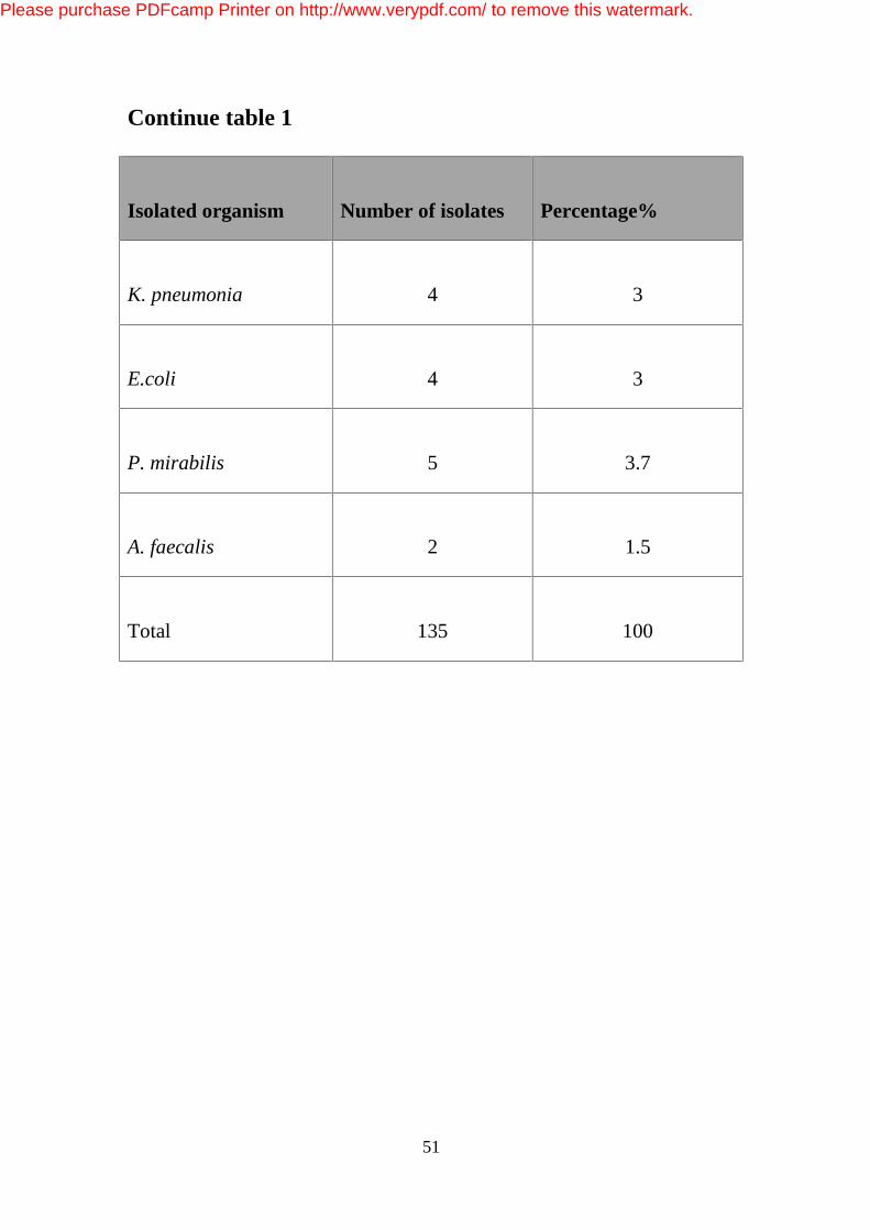
51
Continue table 1
Isolated organism
Number of isolates
Percentage%
K. pneumonia
4
3
E.coli
4
3
P. mirabilis
5
3.7
A. faecalis
2
1.5
Total
135
100
Please purchase PDFcamp Printer on http://www.verypdf.com/ to remove this watermark.
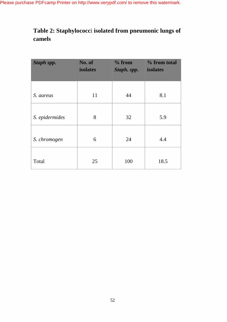
52
Table 2: Staphylococci isolated from pneumonic lungs of camels
Staph spp.
No. of isolates
% from Staph. spp.
% from total isolates
S. aureus
11
44
8.1
S. epidermides
8
32
5.9
S. chromogen
6
24
4.4
Total
25
100
18.5
Please purchase PDFcamp Printer on http://www.verypdf.com/ to remove this watermark.
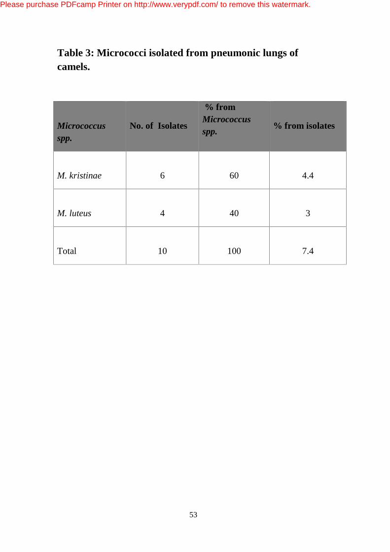
53
Table 3: Micrococci isolated from pneumonic lungs of camels.
Micrococcus spp.
No. of Isolates
% from Micrococcus spp.
% from isolates
M. kristinae
6
60
4.4
M. luteus
4
40
3
Total
10
100
7.4
Please purchase PDFcamp Printer on http://www.verypdf.com/ to remove this watermark.
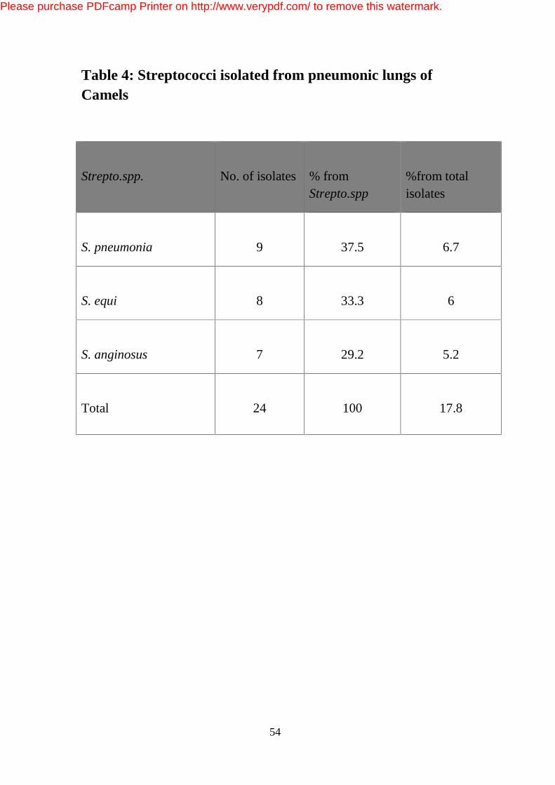
54
Table 4: Streptococci isolated from pneumonic lungs of Camels
Strepto.spp.
No. of isolates
% from Strepto.spp
%from total isolates
S. pneumonia
9
37.5
6.7
S. equi
8
33.3
6
S. anginosus
7
29.2
5.2
Total
24
100
17.8
Please purchase PDFcamp Printer on http://www.verypdf.com/ to remove this watermark.
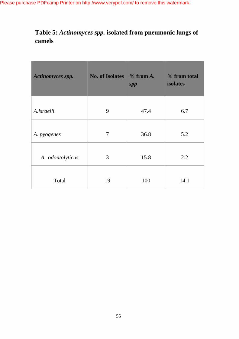
55
Table 5: Actinomyces spp. isolated from pneumonic lungs of camels
Actinomyces spp.
No. of Isolates
% from A. spp
% from total isolates
A.israelii
9
47.4
6.7
A. pyogenes
7
36.8
5.2
A. odontolyticus
3
15.8
2.2
Total
19
100
14.1
Please purchase PDFcamp Printer on http://www.verypdf.com/ to remove this watermark.
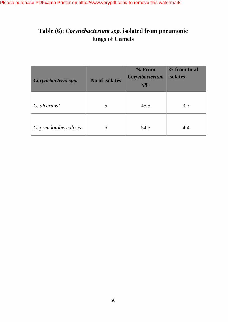
56
Table (6): Corynebacterium spp. isolated from pneumonic lungs of Camels
Corynebacteria spp.
No of isolates
% From Corynbacterium
spp.
% from total isolates
C. ulcerans’
5
45.5
3.7
C. pseudotuberculosis
6
54.5
4.4
Please purchase PDFcamp Printer on http://www.verypdf.com/ to remove this watermark.
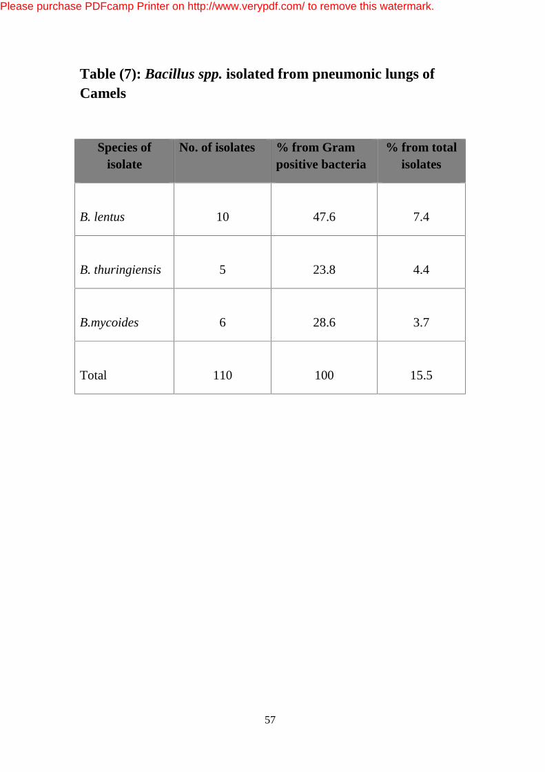
57
Table (7): Bacillus spp. isolated from pneumonic lungs of Camels
Species of isolate
No. of isolates % from Gram positive bacteria
% from total isolates
B. lentus
10
47.6
7.4
B. thuringiensis
5
23.8
4.4
B.mycoides
6
28.6
3.7
Total
110
100
15.5
Please purchase PDFcamp Printer on http://www.verypdf.com/ to remove this watermark.
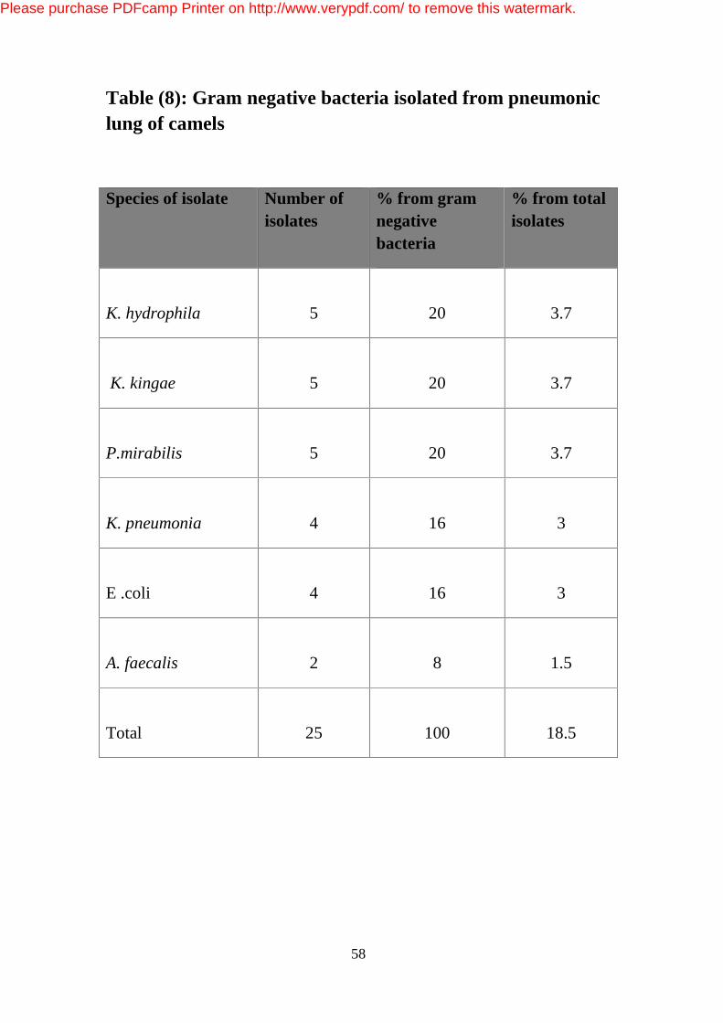
58
Table (8): Gram negative bacteria isolated from pneumonic lung of camels
Species of isolate Number of isolates
% from gram negative bacteria
% from total isolates
K. hydrophila
5
20
3.7
K. kingae
5
20
3.7
P.mirabilis
5
20
3.7
K. pneumonia
4
16
3
E .coli
4
16
3
A. faecalis
2
8
1.5
Total
25
100
18.5
Please purchase PDFcamp Printer on http://www.verypdf.com/ to remove this watermark.

59
Table (9): Characters and Biochemical properties of isolated Gram positive cocci
Test genus Staphylococcus Micrococcus Streptococcus
Growth in air + + +
Anaerobic growth + - -
Motility - - -
Oxidase - + +
Catalase + + +
Acid from glucose + - +
O/F F O F
V: variable +ve: Positive -ve: Negative O: Oxidative F: Fermentative
Please purchase PDFcamp Printer on http://www.verypdf.com/ to remove this watermark.
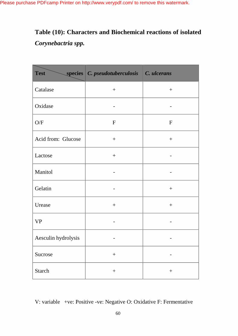
60
Table (10): Characters and Biochemical reactions of isolated
Corynebactria spp.
V: variable +ve: Positive -ve: Negative O: Oxidative F: Fermentative
Test species C. pseudotuberculosis C. ulcerans
Catalase + +
Oxidase - -
O/F F F
Acid from: Glucose + +
Lactose + -
Manitol - -
Gelatin - +
Urease + +
VP - -
Aesculin hydrolysis - -
Sucrose + -
Starch + +
Please purchase PDFcamp Printer on http://www.verypdf.com/ to remove this watermark.
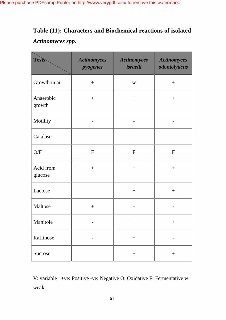
61
Table (11): Characters and Biochemical reactions of isolated
Actinomyces spp.
Tests Actinomyces pyogenes
Actinomyces israelii
Actinomyces odontolyticus
Growth in air + w +
Anaerobic growth
+ + +
Motility - - -
Catalase - - -
O/F F F F
Acid from glucose
+ + +
Lactose - + +
Maltose + + -
Manitole - + +
Raffinose - + -
Sucrose - + +
V: variable +ve: Positive -ve: Negative O: Oxidative F: Fermentative w:
weak
Please purchase PDFcamp Printer on http://www.verypdf.com/ to remove this watermark.
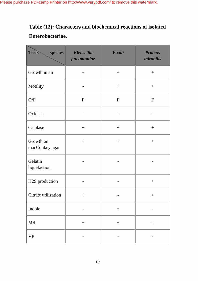
62
Table (12): Characters and biochemical reactions of isolated
Enterobacteriae.
Tests species Klebseilla pneumoniae
E.coli Proteus mirabilis
Growth in air + + +
Motility - + +
O/F F F F
Oxidase - - -
Catalase + + +
Growth on macConkey agar
+ + +
Gelatin liquefaction
- - -
H2S production - - +
Citrate utilization + - +
Indole - + -
MR + + -
VP - - -
Please purchase PDFcamp Printer on http://www.verypdf.com/ to remove this watermark.
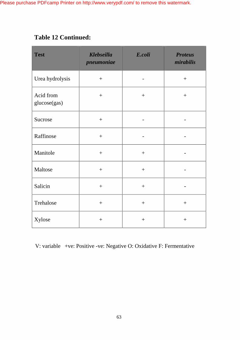
63
Table 12 Continued:
Test Klebseilla pneumoniae
E.coli Proteus mirabilis
Urea hydrolysis + - +
Acid from glucose(gas)
+ + +
Sucrose + - -
Raffinose + - -
Manitole + + -
Maltose + + -
Salicin + + -
Trehalose + + +
Xylose + + +
V: variable +ve: Positive -ve: Negative O: Oxidative F: Fermentative
Please purchase PDFcamp Printer on http://www.verypdf.com/ to remove this watermark.
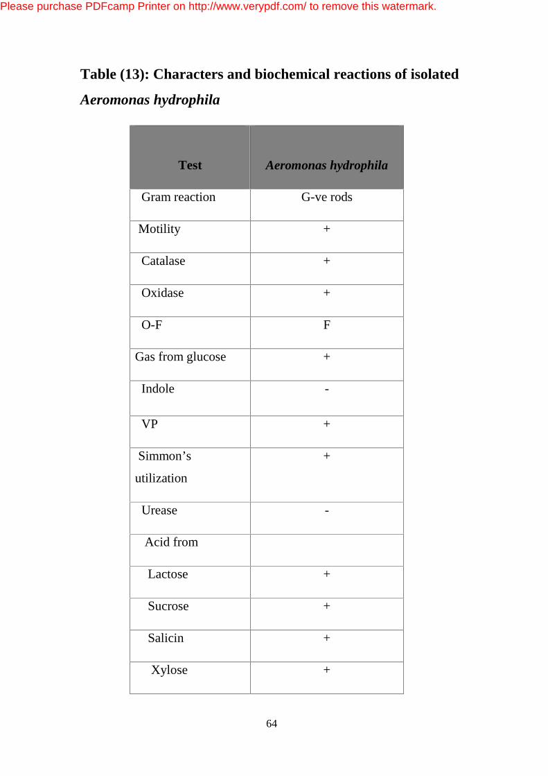
64
Table (13): Characters and biochemical reactions of isolated
Aeromonas hydrophila
Test
Aeromonas hydrophila
Gram reaction G-ve rods
Motility +
Catalase +
Oxidase +
O-F F
Gas from glucose +
Indole -
VP +
Simmon’s
utilization
+
Urease -
Acid from
Lactose +
Sucrose +
Salicin +
Xylose +
Please purchase PDFcamp Printer on http://www.verypdf.com/ to remove this watermark.
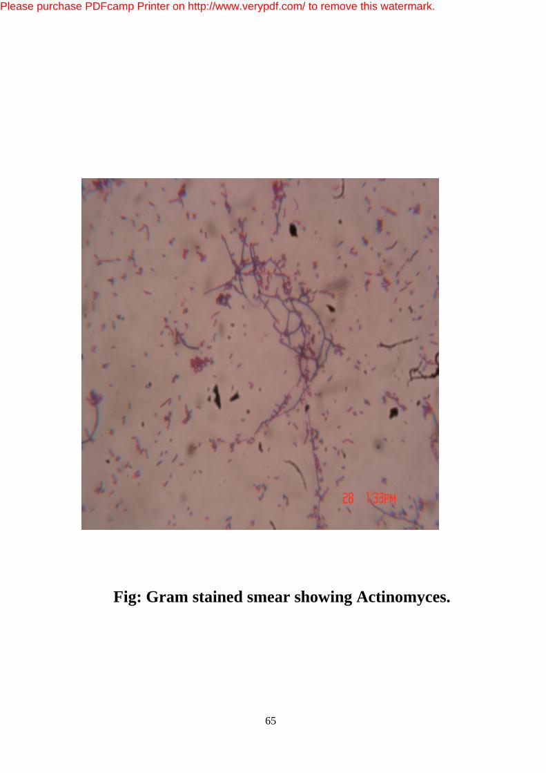
65
Fig: Gram stained smear showing Actinomyces.
Please purchase PDFcamp Printer on http://www.verypdf.com/ to remove this watermark.

66
Fig : Growth of Actinomyces in blood agar
Please purchase PDFcamp Printer on http://www.verypdf.com/ to remove this watermark.
67
Chapter 5
Discussion
In holy Quran it is said "Do they not look at the Camels, how they are
made? And at the Sky, how it is raised high? And at the Mountains, how
they are fixed firm? And at the Earth, how it is spread out. In this honored
verses, the Creator, who knows the secrets of his creatures, advises
people to think and contemplate in creating camels as a creature that
witnesses Allah's Glory, power and planning.
The normal flora of the upper respiratory tract is important as they
prevent adherence and colonization by pathogenic organisms (Jubb and
Kennedy, 1985). There are large numbers of microorganisms that are
present in the respiratory tract of camels as commensals and play
pathogenic role when general immune responses of the host are lowered
(Shigidi, 1973).
This study assessed the types of aerobic bacteria involved in different
types of pulmonary lesions encountered in camel lungs slaughtered at
Elsalam abattoir, in which a relatively high number of bacterial isolates
(135) was isolated from 52 pneumonic lungs. The remaining 8 lung
tissues with observable lesions, failed to yield microorganisms on
primary culture, these infections may be probably due to the involvement
of other pathogens such as anaerobic bacteria, virus, Mycoplasma and
fungi. Lopéz (2001) reported that normal lungs were ideally supposed to
be sterile as the pulmonary macrophages continue to scavenge microbial
agents in the lungs there by making the lung free from pathogens as a
defensive mechanism
Please purchase PDFcamp Printer on http://www.verypdf.com/ to remove this watermark.
68
The isolated organisms included Gram positive and
Gram negative bacteria which were Staphylococcus spp.,
Streptococcus spp., Micrococcus spp., Corynebacteria
spp., Actinomyces spp., Bacillus spp., Escherichia spp.,
Klebsiella spp., Aeromonas spp., Proteus spp., Alcaligenes
spp. and Kingella spp.
Gram positive bacteria represented the majority of the
bacterial isolates which was similar to that observed by
different workers (Farrag et al., 1953; Shigidi, 1972;
Chauchan et al., 1986; Alani, 1989 and Mustafa, 1992).
Staphylococcus spp. represented 18.5% of the total Gram positive
bacteria isolated, most of them were identified as Coagulase negative
staphylococci (CNS) they were the commonest bacteria isolated from
10.3% of lungs Lower than those of Tigani et al. (2006) who recovered
them from 27.9% of pneumonic lungs of camels but considerably higher
than the results of Al- Doughaym et al. (1999) and Al-Tarazi (2001) who
recovered them from 6.5% and 4% respectively. It was suggested that
they exist as part of the normal micro flora of respiratory tract of camels
(Shigidi, 1972 and Chauchan 1987). Coagulase positive staphylococci
were recovered from 8.1% of lungs lower than the results of Al-
Doughaym et al. (1999) and Al-Tarazi (2001) who isolated them from
24.8% and 10.7% of lungs respectively. Also they had been reported in
pneumonic camels by different workers (Chauchan et al., 1987;
Mahmoud et al., 1988 and Moalin and Zessin, 1990). Staph aureus were
isolated from pneumonic cases in other studies (Farrag et al., 1953;
Ghawi, 1978 and Elmaghauri et al., 1986).
Please purchase PDFcamp Printer on http://www.verypdf.com/ to remove this watermark.

69
On the other hand, the frequency of isolation of Streptococcus spp.
was 20.7%. It was similar to that reported by Nesibu Awol (2011) who
isolated them from 19.3%. In contrast Azizollah et al. (2009) and Zubair
et al. (2004) reported a frequency of 4.5% and 7% only respectively.
Streptococcus pyogenes was reported to occur in pneumonic camels by
various authors, among these (Farrag et al., 1953, Elmaghauri et al.
(1986) and Mahmoud et al. (1988).
Buxton and Fraser, (1977) indicated that Streptococcus spp. are widely
distributed in nature and live as commensals in the respiratory tract of
many species of domestic animals.
In this study Corynebacterium spp. were isolated from pneumonic
lungs this was in agreement with the previous findings of (Farrag et al.,
1953; Ghawi1978; Elmaghauri et al., 1986; Moalin and Zessin 1990).
Other Gram-positive bacteria incriminated in pneumonic lungs were
Arcanobacterium pyogenes 5.2% which were isolated from pulmonary
abscesses and this was similar to that reported in Iraq. The same organism
was reported in cases of pneumonia by various authors (Farrag et al.,
1953; Shigidi, 1972; Chauchan et al., 1987; Mahmoud et al., 1988; Alani
et al., 1989 and Mustafa, 1992).
A. pyogenes has been previously reported in camels as a respiratory
pathogen. The organism enters the blood stream and causes septic
arthritis, lesions and abscesses in various organs and tissues, mainly in
the lungs Tolle et al., (1983). Further more A. pyogenes is often isolated
from the abscesses in the lungs of ruminants, pigs and sometimes people
(Hinto, 1972 and Hommez et al., 1991). The same organism was reported
in cases of pneumonia by various authors (Farrag et al., 1953; Shigidi,
Please purchase PDFcamp Printer on http://www.verypdf.com/ to remove this watermark.

70
1972; Chauchan et al., 1987; Mahmoud et al., 1988; Alani et al.1989 and
Mustafa, 1992).
Moreover, the frequency of isolation of Aeromonas hydrophila was
3.5% which was similar to the previous report of Nesibu et al. (2011).
There are no previous reports of isolation of this organism in the Sudan,
and this is the first time to be isolated from camels.
The presence of E. coli is not uncommon, this could be due to
inhalation of dry faeces or from water containers as it is a normal resident
of intestinal tract. In this study four isolates 3% were identified, this is
much lower than the results of Al-Tarazi, (2001) and Nesibu et al. (2004).
This pathogen was isolated by (Elmaghauri et al. (1986); Omer, (1987);
Mahmoud et al. (1988) and Al- Ani, 1989).
Klebsiella pneumoniae was microorganisms commonly isolated from
pneumonic lesions in camels (Zubair et al., 2004; El-Tigani et al., 2004
and Kane et al., 2005). In this study they were isolated at a rate of 3%.
The same organism was stated by Arora and Karla, (1973); Ghawi,
(1978); Mahmoud et al.,1988; Zubair et al., 2004; El-Tigani et al.,
2004 and Kane et al., 2005).
Despite the fact that Micrococcus species are considered non-
pathogenic (Quinn et al., 1999). Its isolation from pneumonic lung
specimens of animals was reported by various workers (Al-Tarazi, 2001;
Nasr, 2003; Kane et al., 2005 and Tigani et al., 2006). It could have a role
in the development of respiratory infections. In this study Micrococcus
spp. was isolated from 7.4% of lungs so these findings supported those of
Nasr (2003) who isolated the same organism for the first time in the
Sudan.
Please purchase PDFcamp Printer on http://www.verypdf.com/ to remove this watermark.

71
Bacillus spp. were isolated by Farrag et al. (1953); Shigidi, 1987;
Omer (1987) and Mustafa, 1992). Bacillus spp. isolated during this study
are comparable to those reported from apparently healthy camels
(Shigidi, 1973 and Chauhan et al. 1987). Thus a variety of
microorganisms may reside the respiratory tract which under suitable
predisposing conditions could invade the different parts of the tract and
cause pathological lesions. In this study Bacillus and Proteus isolates
were isolated from mixed infection of pneumonic lungs. Such isolates
were previously mentioned by others and were considered less pathogenic
(AL-Doughaym et al., 1999 and Mohamed and Farah, 1994).
Alcaligenes faecalis was isolated from 1.5% of lungs, it is considered
to be alkaline tolerant as indicated in the result. Farrag et al. (1953) also
isolated the same organism and he suggested their entrance through blood
or lymph or they might be a contaminant during slaughter.
Pasteurella spp. and Mannheimia hemolytica were not isolated in this
study and this is in agreement with the reports from Egypt, where
Mannheimia haemolytica was not isolated from any pathological lesions
of pneumonic lungs (Mahmoud et al., 1988).
In the previous studies, different bacterial species were isolated from
pneumonic lung specimens of camels. In addition, Azizollah et al. (2009)
isolated various bacterial species from lung lesions of apparently healthy
camels. Variation in the frequencies isolation of different bacteria could
be due to variation in sample size, sample collection and processing,
types of pneumonia or geographical areas.
Al-Tarazi, (2001) reported that, interstitial pneumonia that affected
young camels was caused by Klebsiella spp. and E. coli. Our isolates
Please purchase PDFcamp Printer on http://www.verypdf.com/ to remove this watermark.

72
Klebsiella spp. and E. coli isolated from pneumonic lungs of camel in this
study could probably be involved in induction of these pneumonias.
Lung abscesses are caused by pyogenic organisms. The majority of
bacterial pneumonia cases had multifactorial causes.
Please purchase PDFcamp Printer on http://www.verypdf.com/ to remove this watermark.

73
Conclusions
1- In this study aerobic bacteria isolated and identified included
Staphylococcus spp., Micrococcus spp., Streptococcus spp.,
Actinomyces spp., Corynebacterium spp., Bacillus spp., Klebsiella
spp., Escherichia spp., Aeromonas spp., Kingella spp., Proteus spp.
and Alcaligenes spp.
2- Some of these bacteria reside the lungs normally and under stress
conditions they might become pathogenic.
3- Arcanobacterium pyogenes were associated with pulmonary
abscesses.
4- In this study Aeromonas hydrophila was isolated from lesions of
camel lungs for the first time in Sudan.
Please purchase PDFcamp Printer on http://www.verypdf.com/ to remove this watermark.
74
Recommendations
1- It is highly needed to determine the pulmonary pathologies and
associated bacterial agents and possibly normal bacterial flora of
respiratory tract of camels in Sudan.
2- The role of A. hydrophila in pneumonia and respiratory tract infection
in camels has not previously been studied or documented thus it requires
further studies.
Please purchase PDFcamp Printer on http://www.verypdf.com/ to remove this watermark.

75
References
Abbas, B. and Tilley, P. (1990). Pastoral management for protecting
ecological balance in Halaib District, Red Sea Province, Sudan. Nomadic
Peoples. 29: 77 – 86.
Alhendi, A.A.B. (1999). Nasal microflora of camel (Camelus
dromedaries) under two different conditions. Pakistan Vet. J. 19: 164-
167
Abdurahman, O.A.Sh. and Bornstein, S. (1991). Diseases of camels
(Camelus dromedarius) in Somalia and prospects for better
health.Nomadic Peoples. 29: 104 – 112.
Abeer, E. El-Metwally; Yassin, M. H. and Hanaa, A. El- Hallawany.
(2010). Mycoplasmosis in Slaughtered She- camels: Pathological Studies
Global Veterinarian 4: 474- 482
Abou. E. A. (2000). Brucellosis in Camels and Its relation to Public
health. Assiut Vet. Med. J. 44: 86-105.
Abubakar, M. S; Fatihu, M. Y; Ibrahim, N. D. G; Oladele, S. B. and
Abubakar, M. B. (2008). Camel pneumonia in Nigeria: Epidemiology
and bacterial flora in normal and diseased lung. Afri J Microbiol Re 4:
2479-2483.
Agab, H. and Abbas, B. (1999). Epidemiological studies on camel
diseases in eastern Sudan. World Animal review. 92: 42-51
Alani, F.K. (1990). Common respiratory diseases of Iraqi camels. In:
Proc. Symposium on Camel Breeding, Diseases and their Control.
Please purchase PDFcamp Printer on http://www.verypdf.com/ to remove this watermark.

76
283-287. Arab Organization for Agricultural Development (AOAD),
Alger, Algeria.
Al-ani, F.K. (2004). The Respiratory System In: Camel Management and
Diseases, ed.1st, Al-ani, F.K. (ed) p. 272. Dar ammar book publisher.
Amman, Jordan
Al-Doughaym, A.M; Mustafa, K.M. and Mohamed, G. E. (1999).
Aetiological study on pneumonia in camel (Camelus dromedaries) and
in- vitro antibacterial sensitivity pattern of the isolates. Pakistan J. Biol.
Sc. 4: 1102-1105.
Al-Tarazi, Y. H. (2001). Bacteriological and pathological study on
pneumonia in the One-Humped Camel (C. dromedarius) in Jordan. Revue
Élev.Méd.vét. Pays trop. 54: 93-97.
Archibald, R.G. (1910). Acid-fast bacilli in a camel's lung. J. comp.
Path. Therap. 23: 56-57.
Arora, R.G. and Karla, D.S. (1973). A note on isolation of Klebsiella
pneumoniae and Diplococci from a case of broncho- pneumonia in
camels. Ind. J. Anim. Sc. 43: 1095-1096.
Awol, N; Ayelet, G ; Jenberie, Sh; Gelaye, E; Sisay, T and Nigussie,
H. (2010). Bacteriological studies on pulmonary lesions of camel (C.
dromedarius) slaughtered at Addis Ababa abattoir, Ethiopia . Afri J
Microbiol Res. 5: 522-527.
Azizollah, E; Bentol-hoda, M. and Razieh, K. (2009). The aerobic
bacterial population of the respiratory passageways of healthy
dromedaries in Najaf-abbad abattoir, Central Iran. J Camelid Sc. 2: 26-
29.
Please purchase PDFcamp Printer on http://www.verypdf.com/ to remove this watermark.

77
Bardonn, K; Piarroux, R; Dia, L; Schneegans, F; Beurdele, A;
Godot, V. and Vuitton, D.A. (2002). Combined Ecoepidemiological and
Molecularbiology approaches to assess Echinococcus granulosus
transmissionto human in Mauritania: Occurrence of Camel strain and
humancystic echinococcosis. Royal Soc. Trop. Med. Hyg. 96: 383-386.
Barrow, G.I. and Feltham, R.K.A. (1993). Cowan and Steel’s Manual
for the Identification of Medical Bacteria, 3(rd) Ed., Cambrige University
press.
Bekele, T. (1999). Studies on respiratory disease ‘sonbobe’ in camels in
the eastern lowlands of Ethiopia. Trop. Anim. Hlth. Prod. 31: 333-345.
Bergey's, Manual of Systemic Bacteriology, (1984). 1: 409-570.
Baltimore, MD, USA, William & Wilkins.
Berkeley, R.C.W. and Logan, N.A. (1997). Bacillus,
Alicyclobacillus and Paenibacillus. In: Principles and Practice of
Clinical Bacteriology. Emmerson, A.M., Hawkey, P.M. and
Gillespie, S.H. (eds). pp. 185-207. John Wiley & Sons Ltd.
Blood, D.C. and Henderson, J.A. (1974). Blood and Henderson
Veterinary Medicine.4thed Bailliene Tindall-London.
Blood, D. C; Radostits, O. M. and Henderson, J. A. (1983). Veterinary
Medicine: A textbook of disease of cattle, sheep, pigs, goats and
horse.6thed, Great Britain. Pitman Press.
Burgemeister, V. R; Leyk, W; Gossler, R. (1975). Investigation on the
occurrence of parasites and bacterial and virus infection in Southern
Tunisian dromedaries Dtsch. Tierarztl.Wschr. 82: 341–348.
Please purchase PDFcamp Printer on http://www.verypdf.com/ to remove this watermark.
78
Buxton, A. and Frazer, G. (1977). Streptococci. In: Animal
Microbiology I. pp.1 65-171. Blackwell Scientific Publications,
Edinburgh.
Carter, G.R. (1986). Essencials of veterinary Bacteriology and
Mycology.3rd ed. Lea and Febiger, Philadephia, U.S.A.
Carter, G.R. and Chengappa, M.M. (1991). Essentials of veterinary
bacteriology and Mycology.4th ed. Lea and Febiger, Philadelphia, Pa.
Chauchan, R.S; Gupta, S.C; Satija, K.C; Kulshrestha, R.C. and
Kaushik, R. K. (1986). Bacterial flora of the upper respiratory tract in
apparently healthy camels. Ind. J. Anim. Sc. 57: 424 - 426.
Cheesbrough, M. (2000). District Laboratory Practice in tropical
countries. 1sted. The Press Syndicate of University of Cambridge, part 2,
Cambridge.
Collins, M.D. and Cummins, C.S. (1986). Genus Corynebacterium
Lehmann and Neumann .In: Bergey's Manual of Systematic
Bacteriology. Sneath,P. H. A; Mair,N. S; Sharpe, M. E.& Holt, J.
G.(eds). 2:1266–1276. Baltimore: Williams &Wilkins.
Cotran, R. S; Kumar, V. and Collins, T. (1999). Robbins Pathologic
Basis of Disease.6th ed. W.B. Saunders, Philadelphia.
David-Green, W; Richard, C; Siack, B; John, F.and Peutherer.
(2002). Medical Microbiology, A guide to microbial infection,
pathogensity immunity, laboratory, diagnosis and control.16thed.
Dirie, M.F and Abdurahman, O. (2003). Observations on little known
diseases of camels (Camelus dromedarius) in the Horn of Africa. Rev.
Sci. Tech. OIE. 22: 1043-1049.
Please purchase PDFcamp Printer on http://www.verypdf.com/ to remove this watermark.

79
Eisa, M.I. (1998). Serological survey against some viral diseases in
camels in Sharkia Governorate, Egypt. In: Proceedings of the Third
Annual Meeting for Animal Production Under Arid Conditions. 1: 167–
173.United Arab Emirates University.
EL-afif, A; ZAKI, R. and Farrag, H. (1953). Incidence and typing of
tuberculosis in camels in Egypt. Vet. Med. J., Giza, 1: 1
Elfaki, M.G; Abbas, B; Mahmoud, O. M. and Kleven, S.H. (2002).
Isolation and characterization of Mycoplasma arginini from camels
(Camelus dromedarius) with Pneumonia. Comparative Immunology,
Microbiology and Infectious Diseases. 25: 49-57.
EL Tigani, A; Abdulkhalig, B. and Adam, A. (2004). Studies on
Pathological changes of condemned lungs of One-humped camels
(Camelus dromedarius). Rural Poverty Reduction Through Research for
Development. Deutscher Tropentag, October 5-7, Berlin.
Farag, H; Zaki, R. and El Hindawi, M.R.(1953). Pneumonia in camels.
Br.Vet. J. 109: 119-122.
Khan, F.M. 2012. Field epidemiology of an outbreak of hemorrhagic
septicemia in dromedary population of greater Cholistan desert , Pakistan.
Pak Vet J. 32: 31-34.
Gauthier-Pilters, H. and Dagg, A.I. (1981). The camel: it's evolution,
ecology, behavior and relationship to man. Chicago Univ. Press. USA.
Geo, F. B; Janet, S. B. and Stephen, A. M. (1995). Medical
Microbiology 20(ed).United State of America.
Geo, F. B; Janet, S. B. and Stephen, A. M. (1998). Medical
Microbiology 21(ed). pp. 197-235. International UK Limited London
Please purchase PDFcamp Printer on http://www.verypdf.com/ to remove this watermark.

80
Hall, V. (2006). Anaerobic Actinomycetes and Related Organisms. In:
Principles and Practice of Clinical Bacteriology. Hawkey, P.M and
Gillespie, S.H. (eds). 2nd ed. 575-86. John Wiley and Sons Ltd.
Hinto, M. (1972). Bovine abortion associated with Corynebacterium
pyogenes. Vet. Bull. 42: 753.
Hjort, A. and Hussein, M. A. (1986). Camel herd dynamics in southern
Somalia: Long term development and milk production implications.
Camel Forum. 18: 1 – 28.
Holt, J. G; Krieg N. R; Sneath, P. H.A; Staley J. T. and Williams S.
T. (1994). Bergey's Manual of Determinative Bacteriology. 9th ed.
Philadeliphia: Williams and Wilkins.
Hommez, J; Deuriese, L.A; Miry, C and Castrycy, F. (1991).
Characterization of 2 groups of Actinomyces like-bacteria isolated from
purullent lesions in Pigs. J. Vet. Med. Series B. 38: 575-580.
Ibtisam, M.I. and Azam, A. (2006). Laboratory investigation of
bacterial pneumonia in camels. Animal Health Research Institute (AHRI)
jornal Cairo (Egypt). Serial title: Egyptian J. Compara pathol and clin
pathol.
Intisar, K. S; Ali, Y. H; Khalafalla, A.I; Taha, K. M. and Mahasin, E.
A. Rahman. (2010). Adenovirus type 3 infections in camels in Sudan
Afri J Microbiol Res. 4:1356-1358.
Intisar, K.S; Ali, Y.H; Khalafalla, A.I; Mahasin, E.A; Amin, A.S. and
Taha, K.M. (2009). Respiratory infection of camels associated with
parainfluenza virus 3 in Sudan. J. Virol.Methods. 163: 82-6.
Please purchase PDFcamp Printer on http://www.verypdf.com/ to remove this watermark.
81
Jensen, M. and Waright, D.W. (1998). introduction to Microbiology for
the Health Sciences. 2 ed.
Jubb, K.V.F. and Kennedy, P.C. (1985). Pathology of domestic
animals. 3rd ed. Nigel palmer, New York, USA.
Jubb, K. V. F; Kennedy P. C. and Palmer, N. (1993). Pathology of
Domestic Animal.3 rd ed. Academic Press, New York.
Kane, Y; Kadja, M.C; Bada, A.R; Bezeid, O.E; Akakpo, J.A and
Kaboret, Y. (2005). Lung lesions and bacteria of the one humped camel
(Camelusdromedaries) at Nouakchott slaughter house in Mauritania.
Rev. Elev. Med. Vet. Pays.Trop. 58: 164-171.
Knoess, K. H. (1977).The camel as a meat and milk animal. World
Animal Review. 22 : 39 – 44.
Konemann, E.W; Allen, S. D; Janda, W. M; Schreckenberger, P.
C. and Winn, W. J. (1997).Color Atlas and Textbook of Diagnostic
Microbiology. 5th (ed). pp. 651-708. Philadelphia: Lippincott
Williams & Wilkins.
Lakshmanaperumalsamy, P. (1978). Studies on actinomycetes with
special reference to antagonistic streptomycetesfrom sediments of
Porto Novo coastal zone.Ph.D. thesis, Annamalai University, India.
LEESE, A.S. (1908). - Camel tuberculosis. Ann. Rep. Officer
Investigating Camel Diseases, India.
Lingard, A. (1906). Ann. Rep. Imperial Bacteriologist. p. 11 India.
Littlewood, (1888). Camel tuberculosis.Egyptian Official Gazette.
Lopéz, A. (2001). Respiratory system, Thoracic cavity, and pleura. In:
McGavin, M.D; Carlton, W.W. and Zachary, J.F. (Eds.) Thomson’s
Please purchase PDFcamp Printer on http://www.verypdf.com/ to remove this watermark.

82
Special Veterinary pathology 3 rd (ed). pp. 125-191. Mosby, A Harcourt
Health Sciences Company, London.
MacSween, R.N.M. and Whaley, K. (1992). Mair,s textbook of
pathology 13(ed). Edward Arnold.
MAHMOUD A.Z; SABAH. I. M and EL-YAS. A. H., 1988.
A study on lung affections of camels (Camelus dromedarius) in
Assiut governerate. Assiut vet. Med. J. 20: 93-99.
��
McGrane, J.J. and Higgins, A.J. (1985).The camel in health and
diseases.Infectious diseases of camel, viral bacterial and fungal.
Brit. Vet. J 141: 529-547. ��
Mersie, A. (1997). Diseases profile of camels in eastern Ethiopia. Camels
in eastern Ethiopia. Camel News letter. 16: 50-51.
Moalin, A. S. M. and Zessin, K. H. (1990). Note on diseases of the
dromedaries at Beletweyne Abbatoir of central Somalia. Camel News
letter 7: 69.
Mohamed, R.A. and Abdelsalam, E.B. (2008). A review on pneumonic
pasteurellosis (respiratory manheimiosis) with emphasis on
pathogenesis. Virulence mechanisms and predisposing factors. Bulgaria
J. Vet. Med. 11:139-�160.
Murphy, F.A; Gibbs, E.P; Horzinek, M.C and Studdert, M.J. (1999).
Paramyxoviridae. In: Veterinary Virology, 3rd (ed). p. 423. Academic
press, U.S.A.
Murry, P. R; Rosenthal, K. S; Kobayashi, G. S. and Pfaller, M. A.
(1998). Medical Microbiology.3rd ed. Mosby, Inc. U.S.A.
Please purchase PDFcamp Printer on http://www.verypdf.com/ to remove this watermark.
83
Mustafa, K. E, (1992). Study on clinical, aetiological and pathological
aspect of pneumonia in camels. M.V.Sc. Thessis. Faculty of
Veterinary Science, University of Khartoum, Sudan. ��
Nasr, N.A. (2003). Aerobic bacteria associated with respiratory
infections of camels, isolation and identification. M.V. Sc. Thesis. Fac.
Of Vet. Med. University of Khartoum, Sudan.
Nawal, M.A.Y; Gabry, G.H; Hussein, M. and Omayma, A.A.S.
(2003). Occurrence of Parainfluenza type 3 and bovine herpes virus type
1 (BHV-1) viruses (mixed infection) among camels. Egypt. J. Agric. Res.
81 : 781–791.
Ogunsan A; Umari, O; Bannor , T.T. and Majiyagbe, K.A. (2000).
Hydatidosis in Slaughtered camels in Sokoto State, Nigeria. Nig. Vet. J.
21: 1-9.
Pellegrini, D. (1942-1945). Tuberculosis spontanea del Camello in
somalia, Ricerche diagnostiche espermentali. Racc.Stud. Vet. Part.
Somali, 1:33-41.
Quinn, P.J; Carter, M.E; Markey, B. K. and Carter, G.R. (1999).
Clinical Veterinary Microbiology. pp. 641-648. Harcourt Publishers
Limited, Edinburgh, London.
Quinn, P.J; Markey, B.K; Carter, M.E; Donnelly, W.J. and
Leonard, F.C. (2002). Veterinary Microbiology and Microbial Disease.
pp. 137-143. Black well Science, London.
Rathna- Kala, R. and Chandrika, V. (1978). Effect of different media
for isolation, growth and maintenance of actinomycetes from mangrove
sediments. Indian J. Mar.Sc. 22: 297 -� 299.
Please purchase PDFcamp Printer on http://www.verypdf.com/ to remove this watermark.
84
Richard, D. (1975). Etude de la pathologie du dromadaire dans la sous-
province du Borana (Ethiopie). Thèse Doct.Vét., ENV Alfort, No. 75. In
English: IEMVT, Maisons- Alfort.
Satish, G. (1995). Short Textbook of medical Microbiology .6th (ed).
Lordson Publishers (P) Ltd; Delhi.
Schwartz, H.J. (1992). Productive performance and productivity of
dromedaries (Camleus dromedarius). Anim Res Develop. 35: 86 – 98.
Schwartz, H.J. and Dioli, M. (1992).The One-humped Camel (Camelus
dromedarius) in Eastern Africa: A pictorial guide to diseases, health care
and management. Verlag Josef Margraf, Weikersheim, Germany.
Senior, B.W. (1977).Typing of Proteus strains by proticine Production
and sensitivity. J. Med. Microbiol 10: 7-17.
Shigidi, A.M. (1973). Aerobic microflora of respiratory tract of camels.
Sudan J. Vet. Sc. Anim. Husb. 14: 9-14.
Siva- Kumar, K. (2001). Actinomycetes of an Indian Mangrove
(Pichavaram) environment : An Inventory. Ph.D. thesis, Annamalai
University, India.
Smith, K.J; Neafie, R; Yeager, J. and Skelton, H.G.
(1999)."Micrococcus folliculitis in HIV-1 disease."Brit J Dermatol, 141:
558-561.British.
Sweet, L. E. (1965). Camel pastoralism in Northern Arabia. IN: Man,
culture and animals. Leeds, A.&Vayda, A.P. pp. 129-152.Washington.
Teshome, H; Molla, B. and Tibbo, M. (2003). A seroprevalence
Please purchase PDFcamp Printer on http://www.verypdf.com/ to remove this watermark.
85
Study of Camel brucellosis in three Camel rearing regions of Ethiopia. J.
Anim. Hlth. Prod. 35:381-390
Tigani, T.A; Hassan, A.B. and Abaker, A.D. (2006). Bacteriological
and pathological studies on condemned lungs of one humped camels
(Camelus dromedarius) slaughtered in Tamboul and Nyala abattoirs,
Sudan. pp. 1-13.
Todar, K. (2002). Text Book of Bacteriology. Net/. University of
Wisconsin, Madison Department of Bacteriology.
Tolle, A; Franke, V and Reichmuth, J. (1983).Corynebacterium
pyogenes mastitis-Bacteriological aspects. Deut. Tierarztl. Woch. 90:
256-260.
Vikineswary, S; Nadaraj, P; Wong, W. H. and Balabaskaran, S.
(1997). Actinomycetes from a tropical mangrove ecosystem -� Antifungal
activity of selected strains. Asia Pacific J. Mol. Biol. Boitec.5: 81 -� 86.
Wilson, R.T. (1984).The camel origin and domestication of camels.1 -
4.Long man group publications, London and New York.
Zubair, R.Khan, A.M.Z, Sabri, M.A .(2004). Pathology in Camel
Lungs. J Camel Sc. 1: 103-106.
Please purchase PDFcamp Printer on http://www.verypdf.com/ to remove this watermark.