Embed Size (px)
Citation preview
JOURNAL OF CLINICAL MICROBIOLOGY, May 1993, p. 1127-11350095-1137/93/051127-09$02.00/0Copyright ©) 1993, American Society for Microbiology
Isolation of Gram-Positive Rods That Resemble but AreClearly Distinct from Actinomyces pyogenes from
Mixed Wound InfectionsJURG WUST,* GLADYS MARTINETTI LUCCHINI, JACQUELINE LUTHY-HOTFlENSTEIN,
FRANZISKA BRUN, AND MARTIN ALTWEGG
Department ofMedical Microbiology, University of Zurich, Gloriastrasse 32,CH-8028 Zurich, Switzerland
Received 16 November 1992/Accepted 4 February 1993
Beginning in 1990, gram-positive rods resembling Actinomyces pyogenes were found with increasingfrequency in mixed cultures from various infectious processes, most of them from patients with otitis,empyema, pilonidal cysts, perianal abscesses, and decubitus ulcers. Ribotyping and hybridization showed thatthese gram-positive rods could be divided into five groups not related to known Actinomyces species.Biochemical markers for reliable differentiation into these groups, however, could not be found. Therefore,naming new species is not warranted unless parameters are discovered that allow identification without DNAhybridization. These gram-positive rods have been isolated only in mixed cultures with anaerobes, Staphylo-coccus aureus, Streptococcus "mileri," enterococci, and gram-negative rods. Their exact role in these possiblysynergistic infections needs further investigation.
Gram-positive rods are found with increasing frequency aspathogens. In recent years, bacteria such as nontoxigenicCorynebacterium diphtheriae, C. jeikeium, C. pseudodiph-theriticum, Arcanobacterium haemolyticum, Rhodococcusequi, and bacteria of Centers for Disease Control group D2(recently named C. urealyticum [37]) have been recognizedas important pathogens in patients with endocarditis, intra-vascular devices, prostheses, or encrusted cystitis and inimmunosuppressed patients (9, 29, 35, 44).As a consequence, gram-positive rods are now being
identified to the species level more often in clinical microbi-ological laboratories. During 1990, five strains morphologi-cally and biochemically resembling Actinomyces (formerlyCorynebacterium)pyogenes (38), a species capable of grow-ing well aerobically, were isolated from specimens submittedto the Department of Medical Microbiology of the Univer-sity of Zurich. Identical strains were found 30 times during1991 and 36 times in the first 6 months of 1992, always inmixed cultures with other bacteria. This report contains datafrom a detailed phenotypic and genotypic analysis of 12strains isolated consecutively from specimens taken fromdeep-wound infections between August and October 1991.
MATERIALS AND METHODS
Bacterial strains, media, and growth conditions. Clinicalspecimens were cultured aerobically at 37°C with 5% CO2 onColumbia agar with 5% sheep blood, Columbia agar with 5%sheep blood, colistin and nalidixic acid, and Columbiachocolate agar; MacConkey agar was incubated aerobicallywithout CO2. Anaerobic cultures were set up on brucellaagar with 5% sheep blood, kanamycin-vancomycin agar withlaked blood (42), and phenylethyl blood agar (42) (all mediawere from Becton Dickinson Microbiology Systems, Cock-eysville, Md., unless otherwise specified). Bacteria growingaerobically were identified by standard methods (14, 15, 20,27); obligate anaerobes were identified by RapID ANA II
* Corresponding author.
(Innovative Diagnostic Systems, Atlanta, Ga.), API 20A, orATB 32A (both from API, La-Balme-les-Grottes, France)combined with gas-liquid chromatography of volatile andnonvolatile fatty acids from prereduced, anaerobically ster-ilized (PRAS) chopped-meat broth containing carbohydrates(23); if anaerobes could not be identified by these means,they were only identified to the genus level, e.g., Bacte-roides sp.A. pyogenes-like (APL) gram-positive rods were desig-
nated APL1 to APL12. Five A. pyogenes isolates of animalorigin fitting the description of Reddy et al. (38) (AP13 toAP17) were obtained from J. Nicolet, University of Bern,Bern, Switzerland. Another typical A. pyogenes strain(AP18) was isolated from multiple blood cultures from apatient with septicemia and submitted as a reference culturefor identification from a laboratory in Fribourg, Switzerland;a further typical strain (AP20) was isolated later during thestudy from a case of whitlow. The type strain ofA. pyogenes(NCTC 5224 = ATCC 19411), AP19, was obtained throughthe Swiss National Collection of Type Cultures in Lausanne.
Strains representing the following 11 Actinomyces species(numbered 21 to 31) were included in ribotyping and hybrid-ization experiments (obtained either through the AmericanType Culture Collection or the Swiss National Collection ofType Cultures; type strains are designated by the letter T):A. israelii ATCC 10048, A. odontolyticus ATCC 17982, A.naeslundii ATCC 12104T, A. viscosus LA (Lausanne) 762,A. meyeri ATCC 33972, A. bovis ATCC 13683T, A. gerenc-seriae ATCC 23860T, A. georgiae ATCC 49285T, A. horde-ovulneris ATCC 35275T, A. denticolens ATCC 43322T, andA. howellii ATCC 43323T. The Arcanobactenum haemolyti-cum strain used in hybridization experiments was type strainATCC 9345.The strains were maintained in skim milk at -70°C. Two
of the atypical isolates resembling A. pyogenes (APL5 andAPL9) were unfortunately lost after basic biochemical ex-amination but before they could be investigated genotypi-cally and by extended PRAS biochemistry.
Biochemical and enzymatic characterization. Strains were
1127
Vol. 31, No. 5
on May 2, 2020 by guest
http://jcm.asm
.org/D
ownloaded from

1128 WUST ET AL.
identified in accordance with the criteria outlined by Coyleand Lipsky (9) and Hollis and Weaver (24). Media used forbiochemical reactions were prepared as described by Nashand Krenz (36); basal media were obtained from BectonDickinson. Cystine Trypticase agar (CTA) media contained1% of the carbohydrates and were supplemented with 5%rabbit serum. Gelatin degradation was tested by adding filmstrips (Diagnostics Pasteur, Marnes-la-Coquette, France) toa dense suspension (McFarland no. 5) of the bacteria insaline. Degradation of casein, tyrosine, and xanthine wasdetermined as described by Land et al. (32). Nitrate reduc-tion was tested in nitrate broth, and esculin hydrolysis wastested on mannitol-esculin agar slants (both from Difco,Detroit, Mich.). Starch hydrolysis was evaluated by check-ing for lack of blueness around growth after addition ofaqueous iodine solution to colonies grown on Mueller-Hinton agar. Lecithinase and lipase reactions were done onmodified McClung-Toabe agar (36). Catalase activity wasdetermined by adding 30% hydrogen peroxide solution toTrypticase soy broth cultures. The CAMP reaction wasperformed on Columbia sheep blood agar with Staphylococ-cus aureus ATCC 25923. DNase test agar with methyl green(Difco) was used with addition of 0.1 ml of sterile rabbitserum to 4 ml of medium.
Incubation was done at 37°C without CO2 for tests depen-dent on changes in pH but otherwise at 37°C with 5% C02;anaerobic incubations were done at 37°C in an anaerobicchamber (Jouan, Nantes, France) containing a mixture of10% hydrogen, 10% CO2, and 80% nitrogen or in anaerobejars (Oxoid, Basingstoke, England).
Additionally, a battery of reactions in PRAS media wastested as described by Johnson et al. (25) and Holdeman etal. (23); PRAS media were obtained from Carr-ScarboroughMicrobiologicals (Stone Mountain, Ga.). In preliminary ex-periments, cultures were tested for maximum growth inpeptone-yeast extract-glucose broth medium with and with-out Tween 80 (0.02%, vol/vol) and inoculated (i) anaerobi-cally under CO2, (ii) aerobically and then restoppered (tointroduce a small amount of oxygen), and (iii) aerobicallywith tubes covered with sterile aluminium foil (25). Eventu-ally, strains were inoculated and incubated either anaerobi-cally or microaerobically, depending on the optimal condi-tions for acid formation for a particular strain. Tween 80 wasadded to all of the media.The commercial identification systems API CORYNE (16,
18), API ZYM (22) (both from API), and RapID ANA II(Innovative Diagnostic Systems) were used in accordancewith the instructions of the manufacturers.
Gas-liquid chromatography. Volatile and nonvolatile fattyacids from fermentation were determined by gas-liquid chro-matography of PRAS chopped-meat broth with carbohy-drates, in which bacteria were grown, gassed with CO2 (23).Cellular fatty acids were analyzed by the Microbial Identifi-cation System (MIS) of Hewlett-Packard, currently mar-keted by Microbial ID, Inc. (Newark, Del.), from culturesgrown for 24 h at 37°C with 5% CO2 on Trypticase soy agar(Becton Dickinson) with 5% sheep blood (39, 43). Thesoftware used was version 3.5 of both the clinical and thegeneral Trypticase soy blood agar libraries. The broth pro-cedure described for use with the anaerobe MIS library(version 3.5) was also used (19, 25). For the broth procedure,10 ml of PRAS peptone-yeast-glucose medium with Tween80 (PYG-T; Carr-Scarborough) was inoculated with 0.2 ml ofan actively growing culture in PRAS chopped-meat broth(23); after overnight incubation at 37°C, another subcultureto several tubes of PYG-T medium was made to obtain
sufficient cell mass. After centrifugation, the pellet waswashed with 3 ml of 0.7% aqueous MgSO4 and centrifugedagain. The cells were lysed and saponified, and the fattyacids were methylated, extracted, washed, and analyzed bythe MIS as described by Ghanem et al. (19).
Streptococcal antigens. Agglutination for streptococcal an-tigens found in A. pyogenes (30) was done by the PhadebactStreptococcus Test (Karo Bio Diagnostics AB, Huddinge,Sweden) and the Prolex Streptococcal Latex Kit (Pro-LabDiagnostics, Richmond Hill, Ontario, Canada).
Susceptibility to antimicrobial agents. MICs were deter-mined by the E-test procedure (AB Biodisk, Solna, Sweden)on Mueller-Hinton agar with 5% sheep blood (26). The plateswere swabbed with an inoculum corresponding to the tur-bidity of a McFarland 0.5 standard, and incubation was doneat 37°C without CO2.DNA analysis. Total genomic DNA was isolated by the
standard miniprep procedure described by Ausubel et al. (1),except that cells were incubated in proteinase K and sodiumdodecyl sulfate (SDS) for 4 to 6 h.
Ribotyping was performed essentially as described byMartinetti and Altwegg (34), with restriction enzymes ClaI,SmaI, and PvuII (Boehringer, Mannheim, Federal Republicof Germany).For slot hybridizations (1), 1.8 ,ug of chromosomal DNA
was bound per slot to a supported Nylon 66 membrane(BiodyneB; Pall Biosupport, East Hills, N.Y.) and hybrid-ized with 50 ng of probe strain genomic DNA labelled with32P by using a random-primer labelling kit (Boehringer).Prehybridization and hybridization were done in 5x SSC(lx SSC is 0.15 M NaCl plus 0.015 M sodium citrate)-SxDenhardt's solution-50 mM sodium phosphate buffer (pH6.5)-0.5% SDS-250 mg of denatured herring sperm DNA perml-50% formamide. On the basis of an average G+C-content of 63% (range, 57 to 69%) for actinomycetes (40),hybridization temperatures were 45°C (optimal conditionscorresponding to 25 to 30°C below the Tm) and 60°C (strin-gent conditions corresponding to 10 to 15°C below the Tm).Membranes were washed three times for 5 min each time ina solution containing 2x SSC and 0.1% SDS at roomtemperature and twice for 15 min each time in 0.1x SSC-0.1% SDS at 50°C. Hybrids were detected by autoradiogra-phy using NiF RX film (Fuji Photo Film Co., Ltd., Tokyo,Japan).For quantitative analysis, pieces of membrane carrying
the DNA of one slot were cut out and each piece wasintroduced into a reaction tube containing 10 ml of toluol asscintillation liquid. Counts per minute were assessed during10 min on a liquid scintillation analyzer (Packard).
RESULTS
Clinical data. Clinical data and the bacteria isolated fromthe specimens are summarized in Table 1. Gram-positiverods resembling A. pyogenes were always isolated in mixedculture with anaerobes, S. aureus, S. "milleri," enterococci,or gram-negative rods. Pure cultures were never found. Thiswas also the case for all of the other 59 specimens fromwhich they had been isolated between January 1990 and June1992. The clinical spectrum of these infections was similar tothat of the 12 cases shown in Table 1. Overall, the 71 strainswere isolated from the following: 22 pilonidal cysts, 19perianal abscesses, seven intra-abdominal infections, fourwound infections of the female genital tract, three chronicotitis ear discharge fluid samples, three superficial abdominalwound infections, three cases of suppurative hydradenitis,
J. CLIN. MICROBIOL.
on May 2, 2020 by guest
http://jcm.asm
.org/D
ownloaded from

ACTINOMYCES PYOGENES-LIKE GRAM-POSITIVE RODS 1129
TABLE 1. Origins of and accompanying bacteria isolated from specimens containing APL gram-positive rods
Isolate Sex', age Diagnosis Aerobically growing Obligately anaerobic(yr) bacteria bacteria
APL1 F, 30 Perianal abscess APL, Enterobacter cloacae, Bacteroides fragilis, Bacte-Klebsiella oxytoca, Citro- roides sp., Fusobacteriumbacter amalonaticus, En- nucleatum, Peptostrepto-terococcus faecalis coccus sp.
APL2 F, 68 Subhepatic abscess after lap- APL, Escherichia coli Bacteroidesfragilis, Pep-aroscopic cholecystectomy tostreptococcus sp., Veil-
lonella sp., Lactobacillus sp.APL3 M, 29 Pilonidal cyst APL Peptostreptococcus anaero-
bius, Peptostreptococcusasaccharolyticus, Pep-tostreptococcus sp.
APL4 M, 26 Pilonidal cyst APL, Streptococcus "milleri," Bacteroides fragiliscoagulase-negative staphylo-cocci
APL5 (lost) M, 29 Pilonidal cyst APL Peptostreptococcus anaero-bius, Peptostreptococcusasaccharolyticus, Pep-tostreptococcus sp.
APL6 F, 4 Chronic otitis media APL, Pseudomonas aerugi- Peptostreptococcus asaccha-nosa rolyticus, Peptostreptococ-
cus sp., Prevotella melani-nogenica, Bacteroides sp.
APL7 M, 32 Chronic otitis media APL, Pseudomonas aerugi- Peptostreptococcus asaccha-nosa, Proteus mirabilis, En- rolyticusterococcus faecalis
APL8 M, 32 Perianal abscess APL Bacteroides fragilis, Prevo-tella melaninogenica, Bacte-roides sp., Lactobacillus sp.
APL9 (lost) M, 25 Pilonidal cyst APL, Streptococcus "milleri" Peptostreptococcus sp.APL10 M, 54 Perianal abscess APL, coagulase-negative None
staphylococciAPL11 M, 66 Drainage after lung resection APL, Staphylococcus aureus NoneAPL12 M, 59 Pleural empyema APL Bacteroides sp., Fusobacteinum
nucleatum, Propionibacte-rium acnes, Lactobacillussp., Veillonella sp.
a F, female; M, male.
two mammillary abscesses, two cases of pleural empyema,one bronchial secretion sample, one inguinal abscess, onegluteal abscess, one decubital ulcer, one case of whitlow,and one patient with osteomyelitis.DNA analysis. Ribotyping (Fig. 1) and slot hybridization
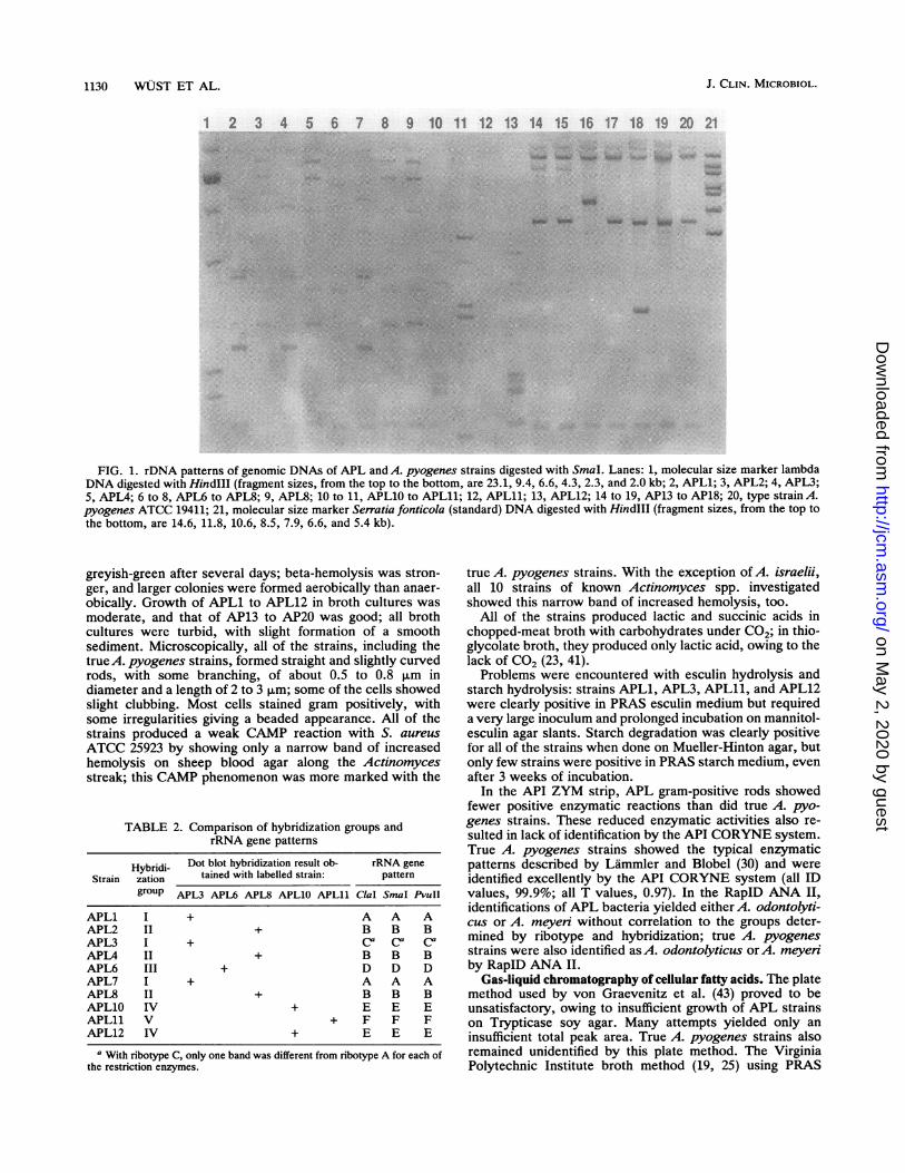
analysis (see Fig. 3) clearly indicated that the 10 strainsresembling A. pyogenes were very diverse and could bearranged in five different groups with one to three strains pergroup, as summarized in Table 2. Hybridization group Icomprised strains APL1, APL3, and APL7; group II in-cluded APL2, APL4, and APL8; group III included APL6;group IV included APL10 and APL12; and group V includedAPL1l. Cross-hybridization between groups was minimal(see Fig. 3). Strains belonging to the same group showedidentical, or at least very similar, rDNA patterns. StrainsAPL10 and APL12 were identical with each of the threeenzymes used, and the same was true for strains APL2,APL4, and APL8. Strain APL3 was different from strainsAPL1 and APL7; however, the difference was restricted to asingle band with each enzyme. Strains APL6 and APL11 hadunique rDNA patterns and made up single-member hybrid-ization groups. Slot hybridization of all 10 APL strains withArcanobacterium haemolyticum showed no hybridization.
In contrast to the APL strains, true A. pyogenes strains(Fig. 1, lanes 14 to 20) represented a relatively homogeneousgroup. Hybridization results were very similar under both
stringent and optimal hybridization conditions (see Fig. 3).This homogeneity was confirmed by rDNA patterns (Fig. 1),which were very similar for these strains but definitivelydifferent from all of the other strains analyzed. There wereminor differences in the rDNA patterns of strains AP15 andAP17 (Fig. 1, lanes 16 and 18), although biochemically thesetwo strains did not differ from the other true A. pyogenesstrains.The representative strains of the 11 known Actinomyces
species showed different ribotypes (Fig. 2). APL strains didnot hybridize significantly with these other Actinomycesstrains (Fig. 3), with the exception of APL11, which had 56%homology with A. odontolyticus; all of the other hybridiza-tion homologies visible in Fig. 3 were below 30%. ForAPL11, A. odontolyticus could not be excluded with cer-tainty by comparing its biochemical reactions with thosepreviously published (25).
Cultural and biochemical properties. The biochemicalproperties of the bacterial isolates tested are shown in Table3. The gram-positive rods resembling A. pyogenes showedthree colony types (Table 3). All three types showed nohemolysis when incubated anaerobically. Except for thecrumbly colonies of APL1l, all of the colonies were smooth.Colony sizes were equal under aerobic and anaerobic incu-bations. In contrast, true A. pyogenes strains grew largerwhite colonies 0.8 to 1 mm in diameter which turned
VOL. 31, 1993
on May 2, 2020 by guest
http://jcm.asm
.org/D
ownloaded from
1130 WUST ET AL.
1 2 3 4 5 6 7 8 9 10 11 12 13 14 15 16 17 18 19 20 21
.- . -
.- .- _
FIG. 1. rDNA patterns of genomic DNAs of APL and A. pyogenes strains digested with SmaI. Lanes: 1, molecular size marker lambdaDNA digested with HindIII (fragment sizes, from the top to the bottom, are 23.1, 9.4, 6.6, 4.3, 2.3, and 2.0 kb; 2, APL1; 3, APL2; 4, APL3;5, APL4; 6 to 8, APL6 to APL8; 9, APL8; 10 to 11, APL10 to APL11; 12, APL11; 13, APL12; 14 to 19, AP13 to AP18; 20, type strain A.pyogenes ATCC 19411; 21, molecular size marker Serratia fonticola (standard) DNA digested with HindIlI (fragment sizes, from the top tothe bottom, are 14.6, 11.8, 10.6, 8.5, 7.9, 6.6, and 5.4 kb).
greyish-green after several days; beta-hemolysis was stron-ger, and larger colonies were formed aerobically than anaer-obically. Growth of APL1 to APL12 in broth cultures wasmoderate, and that of AP13 to AP20 was good; all brothcultures were turbid, with slight formation of a smoothsediment. Microscopically, all of the strains, including thetrueA. pyogenes strains, formed straight and slightly curvedrods, with some branching, of about 0.5 to 0.8 ,um indiameter and a length of 2 to 3 ,um; some of the cells showedslight clubbing. Most cells stained gram positively, withsome irregularities giving a beaded appearance. All of thestrains produced a weak CAMP reaction with S. aureusATCC 25923 by showing only a narrow band of increasedhemolysis on sheep blood agar along the Actinomycesstreak; this CAMP phenomenon was more marked with the
TABLE 2. Comparison of hybridization groups andrRNA gene patterns
Hybridi- Dot blot hybridization result ob- rRNA geneStrain zation tained with labelled strain: pattern
group APL3 APL6 APL8 APL1O APL11 ClaI SmaI PvuIIAPL1 I + A A AAPL2 II + B B BAPL3 I + Ca Ca Ca
APL4 II + B B BAPL6 III + D D DAPL7 I + A A AAPL8 II + B B BAPL10 IV + E E EAPL11 V + F F FAPL12 IV + E E E
a With ribotype C, only one band was different from ribotype A for each ofthe restriction enzymes.
true A. pyogenes strains. With the exception of A. israelii,all 10 strains of known Actinomyces spp. investigatedshowed this narrow band of increased hemolysis, too.
All of the strains produced lactic and succinic acids inchopped-meat broth with carbohydrates under C02; in thio-glycolate broth, they produced only lactic acid, owing to thelack of CO2 (23, 41).Problems were encountered with esculin hydrolysis and
starch hydrolysis: strains APL1, APL3, APL11, and APL12were clearly positive in PRAS esculin medium but requireda very large inoculum and prolonged incubation on mannitol-esculin agar slants. Starch degradation was clearly positivefor all of the strains when done on Mueller-Hinton agar, butonly few strains were positive in PRAS starch medium, evenafter 3 weeks of incubation.
In the API ZYM strip, APL gram-positive rods showedfewer positive enzymatic reactions than did true A. pyo-genes strains. These reduced enzymatic activities also re-sulted in lack of identification by the API CORYNE system.True A. pyogenes strains showed the typical enzymaticpatterns described by Lammler and Blobel (30) and wereidentified excellently by the API CORYNE system (all IDvalues, 99.9%; all T values, 0.97). In the RapID ANA II,identifications of APL bacteria yielded either A. odontolyti-cus or A. meyen without correlation to the groups deter-mined by ribotype and hybridization; true A. pyogenesstrains were also identified as A. odontolyticus or A. meyenby RapID ANA II.
Gas-liquid chromatography of cellular fatty acids. The platemethod used by von Graevenitz et al. (43) proved to beunsatisfactory, owing to insufficient growth of APL strainson Trypticase soy agar. Many attempts yielded only aninsufficient total peak area. True A. pyogenes strains alsoremained unidentified by this plate method. The VirginiaPolytechnic Institute broth method (19, 25) using PRAS
J. CLIN. MICROBIOL.
JiAl
.7.- sffl 7...,
.......
4 kkOw
on May 2, 2020 by guest
http://jcm.asm
.org/D
ownloaded from

ACTINOMYCES PYOGENES-LIKE GRAM-POSITIVE RODS 1131
kb 1 2 3 4 5 6 7 8 9 10 11 1223.1 -
94 - m
6.6- m-- _
4.3 - -
2.32.0-
FIG. 2. rDNA patterns of genomic DNAs of Actinomyces spe-cies digested with SmnaI. Lanes: 1, A. israelii ATCC 10048; 2, A.odontolyticus ATCC 17982; 3, A. naeslundii ATCC 12104T; 4, A.viscosus LA ausanne) 762; 5, A. meyeri ATCC 33972; 6, A. bovisATCC 13683 ;7, A. gerencseriae ATCC 23860T; 8, A. georgiaeATCC 49285T 9, A. hordeovulneris ATCC 35275T; 10, A. dentico-lens ATCC 43322T; 11, A. howellii ATCC 43323T; 12, molecular sizemarker S. fonticola (standard) DNA digested with HindIII. Thesizes of the S. fonticola molecular size marker fragments are givenin Fig. 1. Positions of fragments of molecular size marker lambdaDNA digested with Hindlll are indicated at the left.
PYG-T medium resulted in acceptable total interpretablepeak areas of 51,000 to 93,000. As shown in Table 4,identifications were, with one exception (APL2 was identi-fied as a Lactobacillus sp.) within the genusActinomyces butat similarity levels of <0.500. The true A. lpyogenes strainsAP13 to AP20 were identified as either A. georgiae (similar-ity index, 0.214 to 0.375), A. .meyeri (0.270), A. naeslundii(0.243 to 0.317), Actinomyces sp. strain D01 (0.176 to0.402), or bifidobacteria (0.113 to 0.335) and occasionally asstreptococci (0.078 to 0.138). It must be mentioned that A.pyogenes is not included in the MIS data base for anaerobes.APL strains had higher contents than did true A. pyogenesstrains of 16:1 cis-9 fatty acid methyl ester (FAME), 18:1cis-9 FAME, and the fatty acids combined in feature 10(18:1C11/T9/T6 FAME or un-17.834 equivalent chain length[ECLU) and lower amounts of 16:0 FAME and 18:0 FAME.
In summary, gram-positive rods resembling A. pyogeneswere distinguished from true A. pyogenes strains primarilyby formation of smaller colonies; weaker beta-hemolysis andCAMP reactions; negative casein,, DNA, and gelatin degra-dation; negative agglutination with Streptococcus group B
FIG. 3. Slot blot of genomic DNAs isolated from APL strains, A.pyogenes strains, and strains of other Actinomyces species hybrid-ized at 45°C. (A) Schematic illustration of the positions of thegenomic DNAs of the strains. This schema remains constant in allslot blots. Slots numbered 1 to 12 represent APL strains; thehybridization groups are designated with roman numerals. Slots: 21,A. israelii ATCC 10048; 22, A. odontolyticus ATCC 17982; 23, A.naeslundii ATCC 12104T; 24, A. viscosus LA (Lausanne) 762; 25, A.meyeri ATCC 33972; 26, A. bovis ATCC 13683T; 27, A. gerencseriaeATCC 23860T; 28, A. georgiae ATCC 49285T; 29, A. hordeovulnenisATCC 35275T; 30, A. denticolens ATCC 43322T; 31, A. howelliiATCC 43323T Slot 32 is a negative control containing 2x SSCwithout DNA. The strain designations above the slot blot panelscorrespond to the labelled probes used for hybridization.
and G antibodies in both of the agglutination kits used;negative pyrrolidonyl arylamidase; and overall fewer enzy-matic reactivities. The cellular fatty acid content was higherin unsaturated 16:1 cis-9 FAME and 18:1 cis-9 FAME andlower in saturated 16:0 FAME and 18:0 FAME.
DISCUSSION
Beginning in 1990, facultatively anaerobic gram-positiverods resembling A. pyogenes were found with increasingfrequency in mixed cultures from various infectious pro-cesses. These bacteria seem to be widely distributed, as 7 of71 isolates were reference cultures submitted for identifica-tion by other laboratories up to 60 km away from Zurich.These bacteria could not be identified by the commonly usedcommercial system API CORYNE. When the traditionalbiochemical schemes of Coyle and Lipsky (9) and Hollis andWeaver (24) were used, the bacteria looked very much likeA. pyogenes, negative gelatin hydrolysis being the onlyatypical reaction. RapID ANA II and gas-liquid chromatog-raphy by the MIS failed on both APL bacteria and true A.pyogenes strains by identifying them as other Actinomycesspp., although A. pyogenes is included in the RapID ANA IIdata base; it is, however, not included in the MIS anaerobelibrary.
APL 3
4,_m I--
A1A 1uN 27
:2Ai 12/1V 28
3A 21 29
4/1 22 30
6/11 23 31
7A 24 32
8/1 25
1 OVf 26_j
APL6
oom
Mahlip
dow M.
40 .04.01 .-,
".." 040
APL 8 APL 10 APL 11
VOL. 31, 1993
on May 2, 2020 by guest
http://jcm.asm
.org/D
ownloaded from

1132 WUST ET AL.
TABLE 3. Characteristics of APL bacteria and A. pyogenesa
Result obtained with hybridization group: Result obtainedTest I II III IV v with AP13-
APL1 APL3 APL7 APL2 APL4 APL8 (APL6) APL11APL12 (APL11) AP20
Colony typea i i iiHemolysis aerobically, 2 days _b _ WHemolysis anaerobically, 2 daysCAMP reaction, 2 days w w wTriple sugar iron agar A/A A/A A/ACystine Trypticase agar:Glucose A A AMaltose A A AMannitol - ASucrose A A AXylose A A A
Esculin hydrolysis, 2 daysEsculin hydrolysis, 5 days w wCasein degradationDNase, 5 daysGelatin degradation (film strips)Starch hydrolysis (Mueller-Hin- + + +
ton agar)PRAS:AmygdalinArabinose A ACellobiose A AErythritolEsculin acid - wEsculin hydrolysis + +Fructose A A AGalactose A AGlucose A A AGlycogen w w AInositol A ALactose A AMaltose A A AMannitol w wMannose A A AMelezitose A AMelibiose A ARaffinose A ARhamnose - - -Ribose A A ASalicin A ASorbitolSucrose w A ATrehalose - AXylose A A AGelatin hydrolysis, 3 wkMilk, curd formation + +Starch degradation, 3 wk + - +
API ZYMAlkaline phosphatasecEsterase (C4) w w wEsterase lipase (C8) w w wLeucine arylamidase + + +Valine arylamidase - +Trypsin -
Acid phosphatase - +a-Galactosidase - +1-Galactosidase - +13-Glucuronidase -
ct-Glucosidase w +,B-Glucosidase - +,B-Glucosamidase - +ca-Fucosidase - +
API CORYNEPyrazinamidase + + +Pyrrolidonyl arylamidase
ii ii ii ii1w 1w 1w 1w
w w w wA/A A/A A/A A/A
A A A AA A A A
A A A AA A A A
w w - A
A A - A_ - A -
A A A A
- _ A -
A A A A
w A A A
- A - _
A A A A
A A A A
A A w A
_ - w -
w - - ww w - w
_ - w -
- w + w
- - + -
ii1w -
w w
A/A A/A
A AA A
A AA A
- w
+ +
- A
- A
A w- w
+
A A- A
A A
A A- A
A A- w
- A
- A
- A
- A
A w- A
A w- w
A A
+
+ -
w ww w+ +
- w
w +
w -
iii1w
w
A/A
iv
1w to 1+A/A
A AA A- 7A, 1-A AA A
w _+
+
_+
+ +
+_
A -_ AA A_ A- 6A, 2-_ Aw A- lw, 7-- 4A, 4-- 2A, 6-
_ A
- 6A, 2-A A_ Aw A_
+
-7+,l1-- 1+,7-
w ww w
+ +
w wto +- w to +
_ +
- w to ++ +
_ +
+ + + __- +
Continued on following page
J. CLIN. MICROBIOL.
on May 2, 2020 by guest
http://jcm.asm
.org/D
ownloaded from

ACTINOMYCES PYOGENES-LIKE GRAM-POSITIVE RODS 1133
TABLE 3-Continued
Result obtained with hybridization group:Result obtained
Test I II III IV v with AP13-
APL1 APL3 APL7 APL2 APLA APL8 (APL6) APL1O APL12 (APLll) AP2O
Agglutination with Strep B anti- - - - - - - - - - - wbody
Agglutination with Strep G anti- - - - - - - - - - - +body
MIC range (mg/liter) of:Clindamycin <0.016-0.047d 0.023-0.047Erythromycin <0.016d <0.016Gentamicin 0.19-2d 0.38-1Penicillin G '0.016.0.032d<0.016-0.016Tetracycline 0.094-0.25d 0.125-0.25Vancomycin 0.190.75d 0.19-0.75
a All strains were negative for catalase, urease, nitrate reduction, and indole formation; tyrosine, xanthine, lipase, lecithinase, milk digestion (all after 3 weeksof incubation); API ZYM lipase, cystine arylamidase, chymotrypsin, phosphohydrolase, and a-mannosidase.
b i, whitish, slightly dull, circular, low convex colonies about 0.3 to 0.5 mm in diameter after 48 h of incubation aerobically with 5% CO2 on sheep blood agarwith no hemolysis; ii, similar to i but more greyish and more transparent with weak beta-hemolysis; iii, slightly yellow and opaque with weak beta-hemolysis;iv, white colonies 0.8 to 1 mm in diameter with pronounced beta-hemolysis which turn greyish green after several days of incubation. +, positive reaction; A,acid formation (in PRAS media, pH reduction of .0.8); w, weakly positive reaction (in PRAS media, pH reduction of 0.5 to 0.7); -, negative reaction (in PRASmedia, pH reduction of s0.4).
c API ZYM reactions: w, color intensity 1 to 2; +, color intensity 3 to 5 according to manufacturer's color chart.d Result obtained for all strains in all five hybridization groups.
Most isolates were from pilonidal cysts, perianal ab-scesses, intraabdominal infections, and, further, from pa-tients with chronic otitis, empyema, and various otherabscesses. Although the bacteriology of these infections hasbeen investigated thoroughly (3, 13), A. pyogenes and re-lated bacteria have rarely been mentioned, whereas the roleof P. aeruginosa, S. aureus, S. "milleri," and the obligateanaerobes has been well described.
Ribotyping and hybridization showed that these gram-positive rods could be divided into five groups not related toknownActinomyces spp., with the possible exception of oneof those groups showing 56% hybridization with A. odon-tolyticus. The morphologically and biochemically similarspecies Arcanobacterium haemolyticum could be excludedby lack of hybridization with the APL strains, as well asabsence of an antagonistic hemolytic effect with S. aureus
TABLE 4. Fatty acids of APL bacteria and A. pyogenes'
% of total fatty acids in hybridization group: % of total fatty acids in AP13
to AP20Component I II III IV t
APLI APL3 APL7 APL2 APL4 APL8 (APL6) APL1O APL12 (APLll) Range i ± SD
10:0 FAME 4.3 5.3 5.9 5.1 4.8 3.2 4.7 5.7 3.8 1.9 2.7-3.5 3.11 ± 0.3112:0 FAME 2.5 2.9 1.5 1.2 1.3 1.0 1.3 1.5 2.0 4.0 0.9-2.0 1.61 + 0.4114:1 cis-9 FAME 1.6 1.8 1.414:0 FAME 6.1 7.4 3.0 2.5 3.0 2.4 2.6 2.8 5.5 19.6 5.0-10.2 7.94 ± 2.5016:1 cis-7 FAME 0.7 1.2 0.9-3.6 2.33 ± 0.8916:1 cis-9 FAME 10.9 12.1 8.0 6.9 6.4 6.4 6.3 6.7 10.4 4.1 2.3-2.9 2.64 + 0.2116:0 FAME 10.9 13.4 11.0 9.2 10.8 12.3 12.7 11.1 10.3 18.3 28.0-37.7 32.56 ± 3.38Feature 8b 1.5 1.4 1.618:2 cis-9,12 FAME 1.9 1.8 2.0 1.318:1 cis-9 FAME 43.8 38.8 56.8 60.8 59.5 60.1 59.3 58.4 44.1 42.2 30.4-40.7 35.98 ± 3.70Feature 10b 10.8 11.8 8.2 7.3 7.6 8.5 7.8 7.7 11.1 5.9 3.1-5.2 4.05 ± 0.7118:1 cis-11 DMAC 1.6 1.6un-18.199 18:Oa 2.4 2.7 2.5 2.0 2.1 2.3 0.9-1.1 1.0 + 0.08DMA
Feature 12b 1.6 1.3 1.4 1.1 1.2 1.618:0 FAME 2.6 2.1 3.3 3.0 2.7 3.1 1 2.2 1.5 5.9-15.0 8.96 ± 3.13
a Identification given by the MIS with similarity indices: APLI1, 0.313 forA. odontolyticus serotype II, 0.183 for Fusobacterium sp. strain A, 0.162 forA. meyeri;APL2, 0.417 for Lactobacillus crispatus, 0.355 for L. acidophilus, 0.328 for Gemella morbillorum CFA G3; APL3, 0.277 for A. odontolyticus serotype II, 0.145for Peptostreptococcus asaccharolyticus, 0.132 for Fusobacterium sp. strain A; APL4, 0.413 for A. odontolyticus serotype II, 0.380 for G. morbillorum, 0.327for A. meyeri; APL6, 0.480 for A. odontolyticus serotype II, 0.380 for A. meyeri, 0.332 for A. odontolyticus serotype I; APL7, 0.382 for A. meyeri, 0.355 for A.odontolyticus serotype II, 0.324 for L. acidophilus; APL8, 0.488 for A. odontolyticus serotype II, 0.443 for Actinomyces sp. strain DO1, 0.334 for A. meyeri;APL10, 0.392 for A. odontolyticus serotype II, 0.351 for A. meyeri, 0.317 for L. acidophilus; APL11, 0.483 for A. odontolyticus serotype I, 0.398 forBifidobacterium sp. strain DO5, 0.231 for Clostridium malenominatum; APL12, 0.333 forA. odontolyticus serotype II, 0.160 for Fusobacterium sp. strain A, 0.139for Actinomyces sp. strain DO1.
b Feature 8, 17:1 cis-9 FAME or 17:2 FAME (16.803 ECL). Feature 10, 18:1C11/T9/T6 FAME or un-17.834 ECL. Feature 12, un-18.622 ECL or 19:0 FAME.c DMA, dimethylacetyl.
VOL. 31, 1993
on May 2, 2020 by guest
http://jcm.asm
.org/D
ownloaded from

1134 WUST ET AL.
ATCC 25923, the positive xylose reaction, and the mostlynegative acid phosphatase and beta-glucosamidase results ofAPL strains (5, 9, 31).
True A. pyogenes strains are a relatively homogeneousgroup. There was some minor variation in the rDNA pat-terns of two strains, as has to be expected since ribotypinghas allowed subtyping within most of the species analyzed sofar (21, 34).The minimal cross-hybridization between the groups of
APL bacteria shows that they are new species rather thansubspecies of A. pyogenes. Naming new species is notwarranted unless there are biochemical parameters thatallow their identification without DNA hybridization. Bio-chemical markers for reliable differentiation into thesegroups, however, could not be found. Further investigationof these strains will require analysis of cell walls (40), DNAbase composition, and isoprenoid quinone type (8).
In recent years, numerous new Actinomyces species havebeen described from human (A. georgiae and A. gerencse-nae) (25) and animal (A. denticolens, A. hordeovulneris, A.howellii, and A. slackii) (6, 10-12) sources. As stated byJohnson et al. (25), it continues to be difficult to differentiatebetween Actinomyces species by the usual biochemicaltests, owing to great variations within a species defined byDNA homology and serology. A recent study using theRapID ANA II and API ZYM systems has shown that thereare probably more organisms similar to Actinomyces spp.which require further study (5).Owing to the mixed nature of the infections, the patho-
genic role of gram-positive rods resembling A. pyogenes isdifficult to define. A. pyogenes itself is a well-known animalpathogen and has been described as a causative agent ofbovine mastitis and other pyogenic infections in variousdomestic animals (17, 31, 33). Reports of human infectionsare rare: an epidemic of leg ulcers in school children hasbeen reported from Thailand (28). There are single-casereports of empyema and bacteremia (2, 7); our isolate AP18also originated from a bacteremic patient living on a farm.Gahrn-Hansen and Frederiksen recently reported 11 humancases of A. pyogenes infections, mainly various abscesses,collected since 1968 (17).Our report is meant to draw the attention of bacteriologists
to the fact that gram-positive rods resembling A. pyogenesare isolated with increasing frequency from clinical speci-mens. Part of this increase is probably due to higher aware-ness of infections by gram-positive rods. The exact patho-genic potential of APL bacteria has to be evaluated further.It may well be that the different DNA groups do not have thesame virulence potential. At least it seems that APL bacteriaare implicated in synergistic infections with staphylococci,streptococci, gram-negative bacteria, and especially anaer-obes.One problem of correct recognition of manyActinomyces
spp. is posed by their ability also to grow aerobically to someextent and the fact that this property is not taken intoaccount by most of the traditional identification schemesused in clinical laboratories (9, 29) or used in conjunctionwith determinations of cellular fatty acids (4). As manyActinomyces spp. can grow fairly well aerobically (40), theyshould be included in tables and commercial systems usedfor identification of aerobically isolated gram-positive rods.
ACKNOWLEDGMENT
We thank A. von Graevenitz for careful review of the manuscript.
REFERENCES1. Ausubel, F. M., R. Brent, R. E. Kingston, D. D. Moore, J. G.
Seidman, J. A. Smith, and K. Struhl. 1989. Current protocols inmolecular biology. John Wiley & Sons, Chichester, England.
2. Barnham, M. 1988. Actinomyces pyogenes bacteremia in apatient with carcinoma of the colon. J. Infect. 17:231-234.
3. Bartlett, J. G. 1990. Anaerobic bacteria: general concepts, p.1828-1842. In G. L. Mandell, R. G. Douglas, and J. E. Bennett(ed.), Principles and practice of infectious diseases, 3rd ed.Churchill & Livingstone, New York.
4. Bernard, K. A., M. Bellefeuille, and E. P. Ewan. 1991. Cellularfatty acid composition as an adjunct to the identification ofasporogenous, aerobic gram-positive rods. J. Clin. Microbiol.29:83-89.
5. Brander, M. A., and H. R. Jousimies-Somer. 1992. Evaluation ofthe RapID ANA II and API ZYM systems for identification ofActinomyces species from clinical specimens. J. Clin. Micro-biol. 30:3112-3116.
6. Buchanan, A. M., J. L. Scott, M. A. Gerencser, B. L. Beaman,S. Jang, and E. L. Biberstein. 1984. Actinomyces hordeovulnerissp. nov., an agent of canine actinomycosis. Int. J. Syst. Bacte-riol. 34:439-443.
7. Chlosta, E. M., G. K. Richards, E. Wagner, and J. F. Holland.1970. An opportunistic infection with Corynebacterium pyo-genes producing empyema. Am. J. Clin. Pathol. 53:167-170.
8. Collins, M. D., D. Jones, R. M. Kroppenstedt, and K. H.Schleifer. 1982. Chemical studies as a guide to the classificationof Corynebacterium pyogenes and "Corynebacterium hae-molyticum." J. Gen. Microbiol. 128:335-341.
9. Coyle, M. B., and B. A. Lipsky. 1990. Coryneform bacteria ininfectious diseases: clinical and laboratory aspects. Clin. Micro-biol. Rev. 3:227-246.
10. Dent, V. E., and R. A. D. Williams. 1984. Actinomyces howellii,a new species from the dental plaque of dairy cattle. Int. J. Syst.Bacteriol. 34:316-320.
11. Dent, V. E., and R. A. D. Williams. 1984. Actinomyces denti-colens Dent & Williams sp. nov.: a new species from the dentalplaque of cattle. J. Appl. Bacteriol. 56:183-192.
12. Dent, V. E., and R. A. D. Williams. 1986. Actinomyces slackiisp. nov. from dental plaque of dairy cattle. Int. J. Syst.Bacteriol. 36:392-395.
13. Eykyn, S. J., and I. Phillips. 1989. Miscellaneous anaerobicinfections, p. 567-589. In S. M. Finegold and L. W. George(ed.), Anaerobic infections in humans. Academic Press, SanDiego.
14. Facklam, R. R., and J. A. Washington II. 1991. Streptococcusand related catalase-negative gram-positive cocci, p. 238-257.In A. Balows, W. J. Hausler, Jr., K. L. Herrmann, H. D.Isenberg, and H. J. Shadomy (ed.), Manual of clinical microbi-ology, 5th ed. American Society for Microbiology, Washington,D.C.
15. Farmer, J. J., III, and M. T. Kelly. 1991. Enterobacteriaceae, p.360-383. In A. Balows, W. J. Hausler, Jr., K. L. Herrmann,H. D. Isenberg, and H. J. Shadomy (ed.), Manual of clinicalmicrobiology, 5th ed. American Society for Microbiology,Washington, D.C.
16. Freney, J., M. T. Duperron, C. Courtier, W. Hansen, F. Allard,J. M. Boeufgras, D. Monget, and J. Fleurette. 1991. Evaluationof API Coryne in comparison with conventional methods foridentifying coryneform bacteria. J. Clin. Microbiol. 29:38-41.
17. Gahrn-Hansen, B., and W. Frederiksen. 1992. Human infectionswith Actinomyces pyogenes (Corynebacterium pyogenes). Di-agn. Microbiol. Infect. Dis. 15:349-354.
18. Gavin, S. E., R. B. Leonard, A. M. Briselden, and M. B. Coyle.1992. Evaluation of the rapid CORYNE identification systemfor Corynebacterium species and other coryneforms. J. Clin.Microbiol. 30:1692-1695.
19. Ghanem, F. M., A. C. Ridpath, W. E. C. Moore, and L. V. H.Moore. 1991. Identification of Clostridium botulinum, Clostnid-ium argentinense, and related organisms by cellular fatty acidanalysis. J. Clin. Microbiol. 29:1114-1124.
20. Gilardi, G. L. 1991. Pseudomonas and related genera, p.429-441. In A. Balows, W. J. Hausler, Jr., K. L. Herrmann,
J. CLIN. MICROBIOL.
on May 2, 2020 by guest
http://jcm.asm
.org/D
ownloaded from
ACTINOMYCES PYOGENES-LIKE GRAM-POSITIVE RODS 1135
H. D. Isenberg, and H. J. Shadomy (ed.), Manual of clinicalmicrobiology, 5th ed. American Society for Microbiology,Washington, D.C.
21. Grimont, F., and P. A. D. Grimont. 1986. Ribosomal ribonucleicacid gene restriction patterns as potential taxonomic tools. Ann.Inst. Pasteur 137B:165-175.
22. Gruner, E., A. von Graevenitz, and M. Altwegg. 1992. The APIZYM system: a tabulated review from 1977 to date. J. Micro-biol. Methods 16:101-118.
23. Holdeman, L. V., E. P. Cato, and W. E. C. Moore (ed.). 1977.Anaerobe laboratory manual, 4th ed. Department of AnaerobicMicrobiology, Virginia Polytechnic Institute and State Univer-sity, Blacksburg.
24. Hollis, D. G., and R. E. Weaver. 1981. Gram-positive organisms:a guide to identification. Special Bacteriology Section, Centersfor Disease Control, Atlanta.
25. Johnson, J. L., L. V. H. Moore, B. Kaneko, and W. E. C. Moore.1990. Actinomyces georgiae sp. nov., Actinomyces gerencse-riae sp. nov., designation of two genospecies of Actinomycesnaeslundii, and inclusion of A. naeslundii serotypes II and IIIand Actinomyces viscosus serotype II in A. naeslundii geno-species 2. Int. J. Syst. Bacteriol. 30:273-286.
26. Jorgensen, J. H., A. W. Howell, and L. A. Maher. 1991.Quantitative antimicrobial susceptibility testing of Haemophilusinfluenzae and Streptococcus pneumoniae by using the E-test.J. Clin. Microbiol. 29:109-114.
27. Kloos, W. E., and D. W. Lambe. 1991. Staphylococcus, p.222-237. In A. Balows, W. J. Hausler, Jr., K. L. Herrmann,H. D. Isenberg, and H. J. Shadomy (ed.), Manual of clinicalmicrobiology, 5th ed. American Society for Microbiology,Washington, D.C.
28. Kotrajaras, R., and H. Tagami. 1987. Corynebacterium pyo-genes. Its pathogenic mechanism in epidemic leg ulcers inThailand. Int. J. Dermatol. 26:45-50.
29. Krech, T., and D. G. Hollis. 1991. Corynebacterium and relatedorganisms, p. 277-286. In A. Balows, W. J. Hausler, Jr., K. L.Herrmann, H. D. Isenberg, and H. J. Shadomy (ed.), Manual ofclinical microbiology, 5th ed. American Society for Microbiol-ogy, Washington, D.C.
30. Lammier, C., and H. Blobel. 1986. Tentative identification ofActinomyces pyogenes with antisera against group G strepto-cocci. Zentralbl. Bakteriol. Hyg. A 262:357-360.
31. Lammler, C., and H. Blobel. 1988. Comparative studies onActinomyces pyogenes and Arcanobacterium haemolyticum.Med. Microbiol. Immunol. 177:109-114.
32. Land, G. L., M. R. McGinnis, J. Staneck, and A. Gatson. 1991.Aerobic pathogenicActinomycetales, p. 340-359. In A. Balows,
W. J. Hausler, Jr., K. L. Herrmann, H. D. Isenberg, and H. J.Shadomy (ed.), Manual of clinical microbiology, 5th ed. Amer-ican Society for Microbiology, Washington, D.C.
33. Madsen, M., G. H. Sorensen, B. Aalbaek, J. W. Hansen, and H.Bjorn. 1992. Summer mastitis in heifers-studies on the sea-sonal occurrence of Actinomyces pyogenes, Peptostreptococ-cus indolicus and Bacteroidaceae in clinically healthy cattle inDenmark. Vet. Microbiol. 30:243-255.
34. Martinetti, G., and M. Altwegg. 1990. rRNA gene restrictionpatterns and plasmid analysis as a tool for typing Salmonellaenteritidis. Res. Microbiol. 141:1151-1162.
35. Morris, A., and I. Guild. 1991. Endocarditis due to Corynebac-teium pseudodiphtheriticum. Rev. Infect. Dis. 13:887-892.
36. Nash, P., and M. M. Krenz. 1991. Culture media, p. 1226-1288.In A. Balows, W. J. Hausler, Jr., K. L. Herrmann, H. D.Isenberg, and H. J. Shadomy (ed.), Manual of clinical microbi-ology, 5th ed. American Society for Microbiology, Washington,D.C.
37. Pitcher, D., A. Soto, F. Soriano, and P. Valeroguillen. 1992.Classification of coryneform bacteria associated with humanurinary tract infection (group D2) as Corynebacterium urealyti-cum sp. nov. Int. J. Syst. Bacteriol. 42:178-181.
38. Reddy, C. A., C. P. Cornell, and A. M. Fraga. 1982. Transfer ofCorynebacterium pyogenes (Glage) Eberson to the genus Acti-nomyces as Actinomyces pyogenes (Glage) comb. nov. Int. J.Syst. Bacteriol. 32:419-429.
39. Sasser, M., and M. D. Wichman. 1991. Identification of micro-organisms through use of gas chromatography and high-perfor-mance liquid chromatography, p. 111-118. In A. Balows, W. J.Hausler, Jr., K. L. Herrmann, H. D. Isenberg, and H. J.Shadomy (ed.), Manual of clinical microbiology, 5th ed. Amer-ican Society for Microbiology, Washington, D.C.
40. Schaal, K. P. 1986. Genus Actinomyces, p. 1383-1418. InP. H. A. Sneath, N. S. Mair, M. E. Sharpe, and J. G. Holt (ed.),Bergey's manual of systematic bacteriology, vol. 2. The Wil-liams & Wilkins Co., Baltimore.
41. Schaal, K. P. (University of Bonn, Bonn, Germany). Personalcommunication.
42. Sutter, V. L., D. M. Citron, M. A. C. Edelstein, and S. M.Finegold. 1985. Wadsworth anaerobic bacteriology manual, 4thed. Star Publishing Co., Belmont, Calif.
43. von Graevenitz, A., G. Osterhout, and J. DicL 1991. Grouping ofsome clinically relevant gram-positive rods by automated fattyacid analysis. APMIS 99:147-154.
44. Zuber, P. L. F., E. Gruner, M. Altwegg, and A. von Graevenitz.1992. Invasive infection with non-toxigenic Corynebacteriumdiphtheriae among drug users. Lancet 339:1359.
VOL. 31, 1993
on May 2, 2020 by guest
http://jcm.asm
.org/D
ownloaded from