Embed Size (px)
Citation preview

Isolation, Characterization, and Cloning ofa-L-Arabinofuranosidase Expressed duringFruit Ripening of Japanese Pear
Akira Tateishi*, Hitoshi Mori, Junya Watari, Kenji Nagashima, Shohei Yamaki, and Hiroaki Inoue
College of Bioresource Sciences, Nihon University, Fujisawa, Kanagawa 252–8510, Japan (A.T., K.N., H.I.);and Graduate School of Bioagricultural Science, Nagoya University, Nagoya, Aichi 464–8601, Japan(H.M., J.W., S.Y.)
a-L-Arabinofuranosidase (a-L-arafase) was purified from fruit of Japanese pear (Pyrus pyrifolia). The enzyme solubilized fromthe cell wall by NaCl and Triton X-100 had the homogeneity of a single 62-kD polypeptide on SDS-PAGE after purificationthrough the steps of hydroxyapatite, anion-exchange chromatography, and size-exclusion chromatography. A related cDNAclone was isolated (PpARF2). The transcript and related protein were detected solely in the ripening fruit corresponding to theincrease of a-L-arafase activity. Transcripts of PpARF2 were not detected in buds, leaves, roots, or shoots of the Japanese pear.The deduced amino acid sequences of PpARF2 had low identity with those of other plants or bacteria. This a-L-arafasebelonged to glycoside hydrolase family 3, which includes some b-xylosidases. The purified enzyme hydrolyzed mainlyp-nitrophenyl a-L-arabinofuranoside and also reacted bifunctionally with p-nitrophenyl b-D-xylopyranoside. However, itreleased only arabinose from native cell wall polysaccharides prepared from Japanese pear and from sugar beet arabinan. Theenzyme did not release xylose from arabinoxylan and xylan. The only activity of the a-L-arafase presented here washydrolyzing the arabinosyl residue from native polysaccharides, whereas it showed bifunctional activity against artificialsubstrates. According to the expression pattern and properties of the enzyme, it is a new member of the glycoside hydrolasefamily 3 isolated from fruit, and it may be responsible for modification of the cell wall architecture during fruit softening.
The modification of cell wall architecture is involvedin plant growth, development, and formation of shape.The cooperative biosynthesis and degradation of sev-eral cell wall components are necessary, and numerouscell wall-related enzymes are implicated in these pro-cesses. Fruit softening or textural changes are impor-tant factors that decide fruit quality, and they arecaused by modification of cell wall polysaccharidearchitecture during fruit ripening. Several cell wall-metabolizing enzymes contribute to the changes in cellwall architecture (Fischer and Bennett, 1991). Duringfruit ripening, pectic and some hemicellulosic poly-saccharides become increasingly soluble and depoly-merize with the release of neutral sugar residues fromside chains of matrix polysaccharides (Huber andO’Donoghue, 1993; Brummell and Labavitch, 1997;Sakurai and Nevins, 1997; Brummell and Harpster,2001). It seems that the loss of the neutral sugar resi-dues of the side chains occurs during the early stage ofripening associated with pectin solubilization (Sakuraiand Nevins, 1997; Rose et al., 1998). The initial solu-bilization of pectic polysaccharides during ripeningoccurs without changes in the degree of polymeriza-tion (Dawson et al., 1992; Redgwell et al., 1992).
Side chains of matrix polysaccharides are removedby the actions of several glycosidases (Fry, 1995). Therelease of neutral sugar residues during fruit ripeningwas assumed to increase the sensitivity of enzymaticdegradation or accessibility of other glycan hydrolasesbecause almost all pectic or hemicellulosic polysaccha-ride backbones constituting the cell wall are associatedwith branched side chains, except for homogalactur-onan. For example, b-galactosidase II, one of the gly-cosidases isolated from tomato (Lycopersicon esculentum)fruit, possesses the ability to release galactosyl resi-dues from pectic side chains (Pressey, 1983). Recently,Smith et al. (2002) demonstrated that b-galactosidaseII, which is encoded by TBG4, had a significant effecton fruit softening by using antisense suppression of arelated gene. Therefore, the release of galactosyl res-idues from side chains of pectic polysaccharides, at least,is involved in fruit softening in addition to the pectindepolymerization caused by polygalacturonase activityat the later stage of ripening. However, the actual roleof other glycosidases in vivo remains to be established.
Besides the release of galactosyl residues, the loss ofarabinosyl residues during fruit softening was also ob-served in many kinds of fruits (Gross, 1984; Gross andSams, 1984). Terminal arabinosyl residues are widelydistributed in pectic and hemicellulosic polysaccharidessuch as arabinan, arabinogalactan, arabinoxylan, ara-binoxyloglucan, and glucuronoarabinoxylan (Beldmanet al., 1997; Saha, 2000; Sozzi et al., 2002b). a-L-Arabinofuranosidase (a-L-arafase; a-L-arabinofurano-
* Corresponding author; e-mail [email protected]; fax81–466–84–3622.
Article, publication date, and citation information can be found atwww.plantphysiol.org/cgi/doi/10.1104/pp.104.056655.
Plant Physiology, July 2005, Vol. 138, pp. 1653–1664, www.plantphysiol.org � 2005 American Society of Plant Biologists 1653
Dow
nloaded from https://academ
ic.oup.com/plphys/article/138/3/1653/6103098 by guest on 16 January 2022

side arabinofuranohydrolase, EC 3.2.1.55) is an en-zyme that is able to hydrolyze nonreducing arabino-furanosyl residues. Yamaki et al. (1979) havereported the loss of arabinosyl residues associatedwith fruit ripening in Japanese pear (Pyrus pyrifolia).Indeed, a-L-arafase activity increased during ripeningof Japanese pear fruit (Tateishi et al., 1996) and its risewas also observed in ripening fruits of apple (Malusdomestica; Yoshioka et al., 1995), avocado (Persea amer-icana; Tateishi et al., 2001b), tomato (Sozzi et al., 2002a),
persimmon (Diospyros kaki; Xu et al., 2003), and peach(Prunus persica; Brummell et al., 2004a). Recently, Sozziet al. (2002b) characterized a-L-arafase isoforms fromtomato fruit and reported their different hormonalregulation and contribution to fruit development andripening. One of the a-L-arafase isoforms is regulatedby ethylene and releases arabinosyl residues from thepectic fraction. This indicates that the isoform plays animportant role in arabinose (Ara) metabolism duringripening of tomato fruit.
Figure 1. Elution profiles of a-L-arafase activityby hydroxyapatite (A), Q-Sepharose (B), andSephacryl S-200 (C) chromatography. The activitywas represented by a black circle (d) and theprotein by a white circle (s). Fractions elutedfrom hydroxyapatite were collected (fraction nos.18–23; 36 mL), dialyzed, and loaded onto aQ-Sepharose anion-exchange column. Then frac-tion numbers 10 to 12 (15 mL) were collected,concentrated, and then loaded onto a SephacrylS-200 gel-filtration chromatograph. Fractionswere collected in 2-mL aliquots.
Tateishi et al.
1654 Plant Physiol. Vol. 138, 2005
Dow
nloaded from https://academ
ic.oup.com/plphys/article/138/3/1653/6103098 by guest on 16 January 2022

To date, we have found a-L-arafase cDNA sequencesfrom higher plants in the DDBJ/EMBL/GenBankdatabases; however, except for arabinoxylan arabino-furanohydrolase (AXAH; Ferre et al., 2000; Lee et al.,2001) and a-L-arafase (Lee et al., 2003) purified frombarley (Hordeum vulgare), they appear to be defined asa-L-arafase on the basis of low sequence homologyto bacterial a-L-arafase. There is no evidence that theprotein translated from them actually possessesa-L-arafase activity. We have already reported the in-crease in a-L-arafase activity with the ripening ofJapanese pear fruit and the purification of the enzyme(Tateishi et al., 1996). However, it was difficult tofurther analyze the enzyme properties due to lowrecovery of the enzyme protein after purification. Inthis study, we repurified a-L-arafase from ripeningJapanese pear fruit, characterized its properties againstnative cell wall polysaccharides, and, moreover, iso-lated a related cDNA clone by elucidating the primarystructure of the enzyme.
RESULTS
Purification of a-L-Arafase from Cell Walls of
Japanese Pear Fruit
In a previous study, approximately 30% of a-L-arafaseactivity was solubilized from cell walls usingtrans-1,2-diaminocyclohexane-N,N,N#,N#-tetraaceticacid (CDTA) for 38 h (Tateishi et al., 1996). Using thebuffer containing NaCl and Triton X-100 for solubili-zation, 20 times the activity was recovered comparedto that solubilized by CDTA (Tateishi and Inoue, 2000).The condition also contributed to stabilization of theenzyme. a-L-Arafase was purified by hydroxyapatite,Q-Sepharose anion-exchange chromatography, andSephacryl S-200 gel-filtration chromatography (Fig. 1;Table I). Through the purification steps, we could notuse ammonium sulfate or ultrafiltration membrane topurify or concentrate the enzyme because the recoverywas too low (data not shown). The enzyme was elutedby gel media with a delay, compared to ideal elutionposition, regardless of the addition of 0.2 M NaCl(data not shown); this is similar to a case previouslyreported by Sozzi et al. (2002b). After size-exclusionchromatography, a-L-arafase was purified 106-foldcompared to the activity of the crude enzyme, and
the activity against p-nitrophenyl a-L-arabinofuranoside(pNPA) became 6.95 mmol h21. The purified a-L-arafase appeared as a single protein with a molecularmass of 62 kD by staining with silver nitrate after SDS-PAGE (Fig. 2). It was larger than that obtained in theprevious study (Tateishi et al., 1996). Proteolytic deg-radation may have occurred during long-term incu-bation with CDTA. The pH optimum of a-L-arafaseagainst pNPA was 4.5, and the activity was detected atpH 4.0 to 6.5 (data not shown). Cu21 and Hg21 werefound to be potent inhibitors (data not shown). Theexact Km value of a-L-arafase against pNPA was notdetermined because pNPA could not be dissolved atconcentrations higher than approximately 20 mM. Allthe kinetic parameters assayed were almost the sameas those of a previous study (Tateishi et al., 1996).
Isolation of a PpARF2 cDNA Clone
We determined the amino acid sequences of severaltryptic peptide fragments generated from purifieda-L-arafase (Fig. 3). Degenerate primers were designedbased on the obtained amino acid sequences and PCRwas performed using a combination of all primers. Theprimer set of ARA-Fr39 and ARA-Fr57C gave a singlecDNA fragment of 350 bp (data not shown). The
Table I. Typical purification steps of a-L-arafase from the cell wall of Japanese pear fruit
Solubilized enzyme extract from the cell wall was defined as the crude enzyme.
Step Activity Protein Specific Activity Fold Yield
mmol h21 mg mmol h21 mg protein21 %
Crude enzyme 150.4 50.4 2.99 1.0 100Hydroxyapatite 121.3 5.79 20.9 7.0 80.6Q-Sepharose FF 38.58 0.49 77.9 26.1 25.6Sephacryl S-200 6.95 0.02 315 105.7 4.62
Figure 2. SDS-PAGE of each purification step of a-L-arafase andwestern-blot analysis. Marker (lane M), crude enzyme (lane 1),hydroxyapatite (lane 2), Q-Sepharose FF (lane 3), and SephacrylS-200 gel filtration (lane 4). The gel was stained with silver nitrate.LaneW is a western-blot of a-L-arafase protein using anti-Japanese peara-L-arafase IgG raised from recombinant protein.
Characterization of a-L-Arabinofuranosidase from Fruit
Plant Physiol. Vol. 138, 2005 1655
Dow
nloaded from https://academ
ic.oup.com/plphys/article/138/3/1653/6103098 by guest on 16 January 2022

deduced amino acid sequence from the clone obtainedmatched the internal amino acid sequence of purifieda-L-arafase. Based on the sequence obtained, the5#- and 3#-ends of the cDNA fragment were deter-mined using RACE-PCR (see ‘‘Materials and Methods’’).Finally, we obtained a full-length cDNA clone encod-ing Japanese pear a-L-arafase (named PpARF2; acces-sion no. AB195230; Fig. 3). The cloned cDNA fragment(2,575 bp) contained an untranslated region at both the5#- and the 3#-ends, and a presumptive coding se-quence of 2,322 nucleotides. The molecular masscalculated from the deduced amino acid sequencewas 84 kD, consisting of 774 amino acids (nucleotides37–2,358), which had a calculated pI of 8.29. TheN-terminal amino acid sequence from purified a-L-arafase was Arg-Pro-Pro-Phe-Ala-Cys-Asp-Pro-Arg-Asn, which corresponds to the amino acid sequenceof 29 to 38 deduced from PpARF2. This indicates thata signal sequence existing in the N terminus ofPpARF2 protein would be cleaved in the matureprotein. The sequences of a total of 41 amino acidresidues from three different tryptic peptides exactlymatched the sequence deduced from PpARF2 (Fig. 3).PpARF2 was subcloned into an expression vector and
protein expression was induced by the addition ofisopropylthio-b-galactoside. Under our conditions, theinduced protein was recovered as an inclusion bodywithout activity (see ‘‘Materials and Methods’’).Therefore, we dissolved the protein and used it as anantigen to raise an antibody against a-L-arafase. Theprepared antibody recognized a single polypeptide inthe crude extract and also reacted with purified a-L-arafase after gel-filtration chromatography (Fig. 2).
Two N-glycosylation sites were predicted from thededuced amino acid sequence of PpARF2. PSORT(Nakai and Kanehisa, 1991) and TargetP (Emanuelssonet al., 2000) programs predicted that the encodeda-L-arafase would be exported to the apoplast, indi-cating an enzyme would act on cell wall substrates.Both nucleotide and deduced amino acid sequencesshowed low identity with those of other plant andbacterial a-L-arafases. However, they showed rela-tively higher similarity with putative b-glucosidaseand b-xylosidase cDNA clones isolated from Chenopo-dium rubrum and Arabidopsis (Arabidopsis thaliana),respectively, and extremelyhigh identity witha Japanesepear cv Nijisseiki b-xylosidase-like gene expressedduring fruit ripening and senescence (Itai et al.,
Figure 3. Amino acid sequence of a-L-arafase (PpARF 2) from Japanese pearfruit. The shade-boxed amino acidsare the sequences of N-terminalamino acids and tryptic peptidefragments generated from purifieda-L-arafase. White-boxed amino acidsequences exhibited possible N-gly-cosylation sites. The cleaved signalpeptide is indicated by an underline.The putative catalytic nucleophile(amino acid position 292) and cata-lytic acid/base (amino acid position498) are circled.
Tateishi et al.
1656 Plant Physiol. Vol. 138, 2005
Dow
nloaded from https://academ
ic.oup.com/plphys/article/138/3/1653/6103098 by guest on 16 January 2022
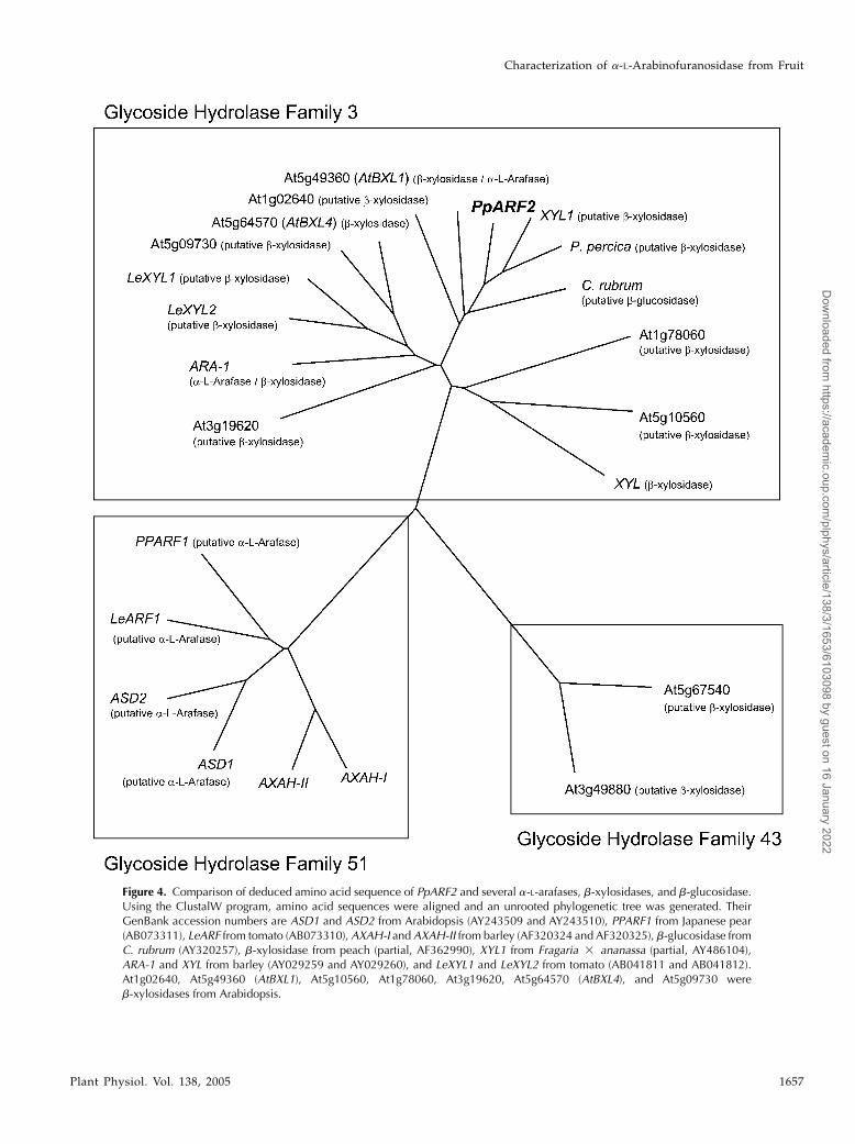
Figure 4. Comparison of deduced amino acid sequence of PpARF2 and several a-L-arafases, b-xylosidases, and b-glucosidase.Using the ClustalW program, amino acid sequences were aligned and an unrooted phylogenetic tree was generated. TheirGenBank accession numbers are ASD1 and ASD2 from Arabidopsis (AY243509 and AY243510), PPARF1 from Japanese pear(AB073311), LeARF from tomato (AB073310), AXAH-I and AXAH-II from barley (AF320324 and AF320325), b-glucosidase fromC. rubrum (AY320257), b-xylosidase from peach (partial, AF362990), XYL1 from Fragaria 3 ananassa (partial, AY486104),ARA-1 and XYL from barley (AY029259 and AY029260), and LeXYL1 and LeXYL2 from tomato (AB041811 and AB041812).At1g02640, At5g49360 (AtBXL1), At5g10560, At1g78060, At3g19620, At5g64570 (AtBXL4), and At5g09730 wereb-xylosidases from Arabidopsis.
Characterization of a-L-Arabinofuranosidase from Fruit
Plant Physiol. Vol. 138, 2005 1657
Dow
nloaded from https://academ
ic.oup.com/plphys/article/138/3/1653/6103098 by guest on 16 January 2022

1999). Bioinformatics analysis of deduced aminoacid sequences of PpARF2 also revealed that purifieda-L-arafase was grouped into the glycoside hydro-lase (GH) family 3 (http://afmb.cnrs-mrs.fr/CAZY;Fig. 4), which contains b-glucosidase, b-xylosidase,and a-L-arafase cDNA clones.
The Activity and Gene Expression of a-L-Arafasewith Fruit Development and Ripening
Western-blot analysis revealed that a-L-arafase pro-tein accumulated as ripening progressed in fruit (120and 135 days after full bloom [DAF]; Fig. 5B). RNA gel-blot analysis also detected accumulation of PpARF2transcripts only in the ripening stage (Fig. 5C). We alsoassayed a-L-arafase activity during fruit developmentand ripening (Fig. 5A). The highest activity on a pergram fresh weight basis was detected in both youngfruit (15 DAF) and fully ripened fruit (135 DAF).To exclude the dilution of the activity with fruit en-largement, the fluctuation of activity on a whole-fruitbasis is also described in the same figure. The activity
increased vigorously with fruit ripening after fruitenlargement (Fig. 5A). Moreover, PpARF2 transcriptswere detected solely in ripening fruit, but not in buds,young and mature leaves, and shoots (Fig. 6). Theincrease of the activity, accumulation of the enzymeprotein, and expression of PpARF2 were stronglyspecific to ripening fruit.
Figure 5. Changes in a-L-arafase ac-tivities, accumulation of protein, andexpression of mRNA during fruit de-velopment and ripening. A, Activity ofa-L-arafase solubilized from the cellwall using pNPA as substrate. Theactivity on a per gram fresh weightbasis is indicated by a black box (n)and the activity on a whole-fruit basisis indicated by a black circle (d). Fruitgrowth was indicated by a white circle(s) with a dashed line. The values arethe means of three independent ex-periments. Vertical bars indicate theSE. B, Accumulation of a-L-arafase pro-tein. C, Ripening-specific expressionof PpARF2. The probe was preparedfrom mainly the 3#-untranslated re-gion of PpARF2. The hybridizationtemperature was 68�C and the finalwashing condition was 0.13 SSC,0.1% SDS at 68�C. rRNAs stainedwith ethidium bromide were used asa loading control.
Figure 6. Fruit-specific expression of PpARF2. RNA samples wereprepared from buds (lane B), leaves (lane L), roots (lane R), shoots (laneS), and ripe fruit (lane F). Ethidium bromide-stained gel was used asa loading control (indicated rRNA).
Tateishi et al.
1658 Plant Physiol. Vol. 138, 2005
Dow
nloaded from https://academ
ic.oup.com/plphys/article/138/3/1653/6103098 by guest on 16 January 2022

Activities against Native Cell Wall Polysaccharides and
Substrates Containing Xylose
We assayed the activity against several Ara-containingpolysaccharides because the activity against anartificial substrate does not necessarily reflect thatagainst polysaccharides in vivo (Fig. 7). a-L-Arafasereleased only Ara as a consequence of hydrolysisof alcohol-insoluble substances (AIS) prepared fromthe cell wall of Japanese pear fruit. a-L-Arafasealso possessed the activity of hydrolyzing sugar beetarabinan to Ara. However, no Ara could be detectedin the reaction mixture incubated up to 72 h withwheat (Triticum aestivum) arabinoxylan. a-L-Arafasealso hydrolyzed an arabino-oligosaccharide, arabino-hexaose, to Ara within 72 h (Fig. 7). The relativeactivity against arabinan and/or arabinohexaose seemsto be lower than that against pNPA estimated from theAra spot of thin-layer chromatography.
Bioinformatics analysis revealed that purified a-L-arafase is grouped into GH family 3 involving mainly
plant b-xylosidases. Therefore, we also assayed theactivity against substrates containing xylose (Xyl).a-L-Arafase had not hydrolyzed xylan to Xyl after along incubation (72 h; Fig. 8), and we also did not detectXyl in the reaction mixture with Japanese pear AISand wheat arabinoxylan (Fig. 7). On the other hand,a-L-arafase also hydrolyzed xylohexaose to Xyl; how-ever, some hydrolysis reactions were still in progressafter a 96-h incubation (Fig. 8). As an enzymaticproperty, a-L-arafase possessed hydrolysis activityagainst p-nitrophenyl b-D-xylopyranoside (pNPX)that was 31% of that against pNPA. It was clear thatthe rate of hydrolysis of xylohexaose to Xyl was slowerthan that of arabinohexaose to Ara.
DISCUSSION
We purified an a-L-arafase to the homogeneity ofa single polypeptide on SDS-PAGE. We designed
Figure 7. Hydrolysis of polysaccharides and ara-binohexaose by purified a-L-arafase. Releasedmonosaccharides were separated using thin-layerchromatography. AIS isolated from Japanese pearfruit, arabinan from sugar beet, and arabinoxylanfrom wheat were used as polysaccharide sub-strates. Lane M indicates standard sugars.
Characterization of a-L-Arabinofuranosidase from Fruit
Plant Physiol. Vol. 138, 2005 1659
Dow
nloaded from https://academ
ic.oup.com/plphys/article/138/3/1653/6103098 by guest on 16 January 2022

degenerate primers based on internal amino acidsequences and then obtained the cDNA clone encod-ing a-L-arafase (PpARF2; Fig. 4). The sequences of thethree independent peptide fragments were actuallyfound in the deduced amino acid sequence of PpARF2.The N-terminal amino acid sequence also exactlymatched the deduced amino acid sequence of PpARF2(below amino acid position 29). Furthermore, theantibody raised against recombinant a-L-arafase fromPpARF2 recognized purified a-L-arafase (Fig. 2). Thus,we determined the complete primary structure ofa-L-arafase from corresponding cDNA. The calculatedmolecular mass of PpARF2 was 84 kD, which isconsiderably larger than that of the a-L-arafase ob-tained on SDS-PAGE, 62 kD. A single 62-kD poly-peptide was detected by immunoblot analysis whenthe frozen sample was inactivated immediately inSDS-denatured sample buffer. Therefore, it is hard toconsider that proteolytic degradation of the proteinwas caused by isolation. This discrepancy may be dueto a process of the C-terminal region of the proteinduring maturation, besides cleaving of the N-terminalsignal peptide. A similar case has been reported inbarley a-L-arafase (Lee et al., 2003). A hydrophobicsignal peptide is present at the N terminus, and bothPSORT and TargetP programs predicted that the pro-tein would be exported to the outside of the plasmamembrane. These results imply that a-L-arafase worksactively in cell wall degradation.
Based on amino acid sequences of active sites ratherthan their substrate specificities, a-L-arafases wereclassified into five GH families (family 3, 43, 51, 54,and 62), and we can find a-L-arafase sequence in-formation for several plants in families 3, 43, and 51(Fig. 4). Almost all a-L-arafases were found in family51 and indeed were cloned from fruit of Japanesepear (PpARF1, AB073311), tomato (LeARF; Itai et al.,2003), and Arabidopsis (ASD1 and ASD2; Fulton and
Cobbett, 2003). Ferre et al. (2000) and Lee et al. (2001)reported that a-L-arafases belonging to family 51encoded AXAH protein. According to the substratespecificity, the a-L-arafase purified from Japanese pearshould be separated in other a-L-arafase groups. In-deed, Japanese pear a-L-arafase (PpARF2), which wasnot classified in family 51, could not hydrolyze arabi-noxylan (Figs. 4 and 7) and it was isolated fromripening fruit.
Japanese pear a-L-arafase belongs to GH family 3,which includes b-glucosidase and b-xylosidase as wellas a-L-arafase. Due to limits in the availability fromplants, it has not been elucidated whether the proteinsencoded by the clones possessed b-xylosidase orb-glucosidase activity or not, except for b-xylosidasesfrom barley and Arabidopsis (Goujon et al., 2003; Leeet al., 2003; Minic et al., 2004). Recently, Lee et al. (2003)reported that a-L-arafase purified from barley wasclassified into GH family 3 and that it possessedb-xylosidase activity besides a-L-arafase activity.Moreover, most recently, it was reported that one ofthe b-xylosidases of Arabidopsis was classified intoGH family 3 and also possesses a-L-arafase activity(Goujon et al., 2003; Minic et al., 2004). Therefore, wedetermined the properties of Japanese pear a-L-arafaseas b-xylosidase and b-glucosidase. The purified en-zyme did not possess b-glucosidase activity; however,through all the purification steps, elution profilesof b-xylosidase activity corresponded with that ofa-L-arafase (data not shown). Purified a-L-arafasealso hydrolyzed xylohexaose at a relatively slowrate compared to arabinohexaose (Figs. 7 and 8).This indicates that the enzyme possesses bifunctionalactivity as a-L-arafase/b-xylosidase as an enzymaticproperty. Bifunctional activity has been reported inplant a-L-arafases purified from radish (Raphanussativus) seeds (Hata et al., 1992) and spinach (Spinaciaoleracea) leaf (Hirano et al., 1994) besides barley (Lee
Figure 8. Hydrolysis activity against Xyl-containing substrate. Xylan from birchwood andxylohexaose were used as substrates. Releasedmonosaccharides were separated using thin-layerchromatography. Lane M indicates standardsugars.
Tateishi et al.
1660 Plant Physiol. Vol. 138, 2005
Dow
nloaded from https://academ
ic.oup.com/plphys/article/138/3/1653/6103098 by guest on 16 January 2022

et al., 2003). It is not clear whether these a-L-arafasesbelong to GH family 3 or not; however, it may bereasonable that several b-xylosidases grouped family3 were called a-L-arafase rather than b-xylosidase. Itshould be noted that the activities of bifunctionala-L-arafase against pNPA are 3-fold higher than thoseof b-xylosidase activity against pNPX.a-L-Arafase isolated from Japanese pear actually
released Ara and Xyl hydrolyzed bifunctionally fromboth p-nitrophenyl (pNP) aglycon and oligosacchar-ides. Surprisingly, only Ara was detected in hydroly-sates from AIS prepared from Japanese pear fruit,which contains xylosyl-polysaccharides (Yamaki et al.,1979). Moreover, the a-L-arafase did not release Xylfrom xylan (Fig. 8). The case of the barley bifunctionala-L-arafase was different (Lee et al., 2003). The bi-functional b-xylosidase from Arabidopsis releasedAra and Xyl from arabinoxylan polysaccharides(Minic et al., 2004), but our a-L-arafase did not. Oneof the possible reasons is that there are few xylosylsites, which would be cleaved by a-L-arafase, in na-tive substrates such as AIS or xylan compared to thosein pNPX or xylohexaose. However, Japanese peara-L-arafase did not release Xyl from xylan that had beenpreviously partially hydrolyzed by trifluoroacetic acid(data not shown). According to substrate specificity,we consider that the a-L-arafase we isolated from pearfruit was a distinct type of bifunctional a-L-arafase/b-xylosidase and may release only arabinosyl residuesfrom cell wall polysaccharides in situ. In fact, PpARF2is expressed highly in ripening fruit (Figs. 5 and 6),whereas barley ARA-1 is expressed in grain, root, andleaf, and AtBXL1 is expressed highly in stem. More-over, during fruit ripening, including Japanese pear,the loss of arabinosyl residues from the fruit cell wallwas generally observed, but loss of xylosyl residueswas not. The release of arabinosyl residues fromJapanese pear was observed in an acid-soluble hemi-cellulose fraction where uronic acids were also de-tected (Yamaki et al., 1979) during the over-ripeningstage. a-L-Arafase may act in the hydrolysis of thehighly branched side chain of pectic and hemicellulo-sic polysaccharides and contribute to a part of thefruit-softening process.
In this study, we purified a-L-arafase from the cellwall of ripening pear fruit and determined that itsmRNA is expressed highly only in ripening fruit. Itwas also determined that PpARF2 is a new member ofthe GH family 3, according to the primary structure ofthe enzyme and its activity against native substrates.However, we did not elucidate how the loss of ara-binosyl residues during fruit ripening affects fruitsoftening. The existence of arabinosyl residues in wallpolysaccharides seems to affect the adhesion of eachpolysaccharide or cell. In apple fruit, Ara content de-creased during the over-ripening stage (Pena andCarpita, 2004) and a decrease of arabinosyl residuesfrom cell wall polysaccharides was observed in thedevelopment of mealiness of apple fruit during post-harvest storage (Nara et al., 2001). On the other hand,
in peach fruit, extensive loss of arabinosyl residuesfrom both loosely and tightly bound matrix glycanswas observed in normal ripening fruit, but did notoccur in mealy fruit, which showed a decline in theloss of arabinosyl residues containing polysaccharidesfirmly attached to cellulose (Brummell et al., 2004a,2004b). A larger amount of tightly bound arabinosyl-containing polysaccharides was observed in softening-suppressed, colorless, nonripening (Cnr) mutanttomato fruit, which has a mealy texture (Thompsonet al., 1999; Orfila et al., 2001, 2002). It was also reportedthat the existence of arabinosyl residues in pectic sidechains affected cell-to-cell adhesion in cultured cells(Iwai et al., 2001). Fruit softening is a complex processand several cell wall-metabolizing enzymes contri-bute to modification of cell wall architecture. Therefore,a-L-arafase, in particular, may play an important rolein alteration of fruit texture during softening.
MATERIALS AND METHODS
Plant Material
Japanese pear (Pyrus pyrifolia) cv Housui fruit was harvested from
Shinohara Orchard in Yokohama City, Kanagawa Prefecture, Japan. Fruit
was picked 15, 30, 45, 60, 75, 90, 105, 120, and 135 DAF. The fruit harvested on
135 DAF was at optimum maturity for eating. Buds, leaves, roots, and shoots
were sampled to use in RNA gel-blot analysis. Peeled and cored fruit and the
other organs were immediately frozen in liquid nitrogen and then stored
at 285�C.
Enzyme Extraction and Purification
The extraction of a-L-arafase was carried out at 4�C, and all buffers
contained 10 mM 2-mercaptoethanol, unless stated otherwise. The cell wall-
bound enzyme fraction was prepared by the method described previously
(Tateishi et al., 1996). To solubilize this enzyme from the cell wall, the cell wall
fraction was resuspended in 10 mM borate buffer, pH 9.0, containing 1.0 M
NaCl and 0.1% (v/v) Triton X-100, and gently stirred for 1 h. The suspension
was centrifuged at 15,000g for 30 min, and the resultant supernatant was de-
fined as the crude enzyme. The crude enzyme was then dialyzed against
10 mM potassium phosphate buffer, pH 8.0, containing 0.1% (v/v) Triton X-100
(buffer A). It was then applied to a hydroxyapatite (Wako Pure Chemical
Industries, Osaka) column (2.5 3 6.1 cm) previously equilibrated with buffer
A. The column was washed with buffer A and eluted with a linear gradient of
0 to 400 mM potassium phosphate buffer, pH 8.0, containing 0.1% (v/v) Triton
X-100. Active fractions were pooled and dialyzed against 10 mM Tris-HCl
buffer, pH 8.5, containing 0.1% (v/v) Triton X-100. After dialysis, the dialysate
was applied to a Q-Sepharose FF (Amersham-Pharmacia Biotech, Uppsala)
column (1.6 3 10 cm) previously equilibrated with 10 mM Tris-HCl buffer, pH
8.5, containing 0.01% (v/v) Triton X-100 (buffer B). The column was washed
by the same buffer and the enzyme was eluted with a linear gradient of 0 to
0.5 M NaCl in buffer B. The active fractions were inserted into a dialysis tube
and concentrated to 3 mL using polyethylene glycol (average Mr 20,000) sur-
rounding the tube. Finally, the concentrated protein was loaded onto a Seph-
acryl S-200 (Amersham-Pharmacia Biotech) size-exclusion chromatography
column previously equilibrated with buffer B containing 0.2 M NaCl. Assay of
a-L-arafase activity using pNPA (Sigma, St. Louis) was performed according to
the method described previously (Tateishi et al., 1996).
Protein Determination and SDS-PAGE
The protein concentration of the fractions was determined using the
Coomassie Brilliant Blue dye-binding method (Bradford, 1976) with bovine
serum albumin as a standard. SDS-PAGE was carried out by the method of
Characterization of a-L-Arabinofuranosidase from Fruit
Plant Physiol. Vol. 138, 2005 1661
Dow
nloaded from https://academ
ic.oup.com/plphys/article/138/3/1653/6103098 by guest on 16 January 2022

Laemmli (1970) on 8.5% separation gel, and the protein bands were stained
with silver nitrate (Wray et al., 1981).
Activity against Native Substrate and Oligosaccharides
Cell wall polysaccharides from Japanese pear fruit were prepared
according to the method previously described (Tateishi et al., 2001b). Briefly,
fruit (105 DAF) was homogenized in ice-cold 80% ethanol, and enzymes
included in precipitates were inactivated in Tris-buffered phenol (Huber and
O’Donoghue, 1993). The ethanol-insoluble materials were washed by ethanol
and reprecipitated by 80% (v/v) ethanol. The resultant cell wall precipitate
was freeze dried and defined as AIS. Xylan from birchwood was purchased
from Sigma. Sugar beet arabinan, wheat arabinoxylan, arabinofurano-
hexaose, and xylopyranohexaose were purchased from Megazyme (Bray,
Ireland).
Purified a-L-arafase was dialyzed against 5 mM sodium-citrate buffer, pH
4.5, and then concentrated using polyethylene glycol surrounding the dialysis
tube. Reaction mixtures consisting of 0.5% (w/v) substrate, 5 mM sodium-
citrate buffer (pH 4.5), and purified a-L-arafase, equal to 1 mmol h21 activity
against pNPA were incubated at 37�C for 72 h for native substrates, and 96 h
for oligosaccharides. Toluene (20 mL) was added to each reaction mixture to
prevent bacterial effects.
The monosaccharide product released from native substrates during
enzymatic hydrolysis by a-L-arafase was determined by using thin-layer
chromatography on silica plates (silica gel 64 F254; Merck, Rahway, NJ). After
incubation, the reaction mixture was centrifuged at 15,000 rpm for 15 min.
Then ethanol was added to the supernatants (200 mL) to the final concentra-
tion of 80% (v/v), and the mixture was centrifuged at 15,000 rpm for 30 min.
The supernatant was collected and evaporated to dryness with a rotary
vacuum evaporator, then dissolved again in 5 mL of water. A silica plate was
sprayed with 0.2 M NaH2PO4, dried in air for more than 12 h, and dried
completely at 110�C. Concentrated samples (0.5 mL) were spotted onto the
plate and developed in n-propanol:water (85:15). Sugars were visualized by
heating the plate at 95�C after spraying ethanol:sulfuric acid:p-anisaldehyde
(95:2.5:2.5) on the plate. For analysis of the products of oligosaccharide
substrates, 1.0 mL of the reaction mixture was spotted on a silica plate (no
pretreatment) and developed in ethyl acetate:acetic acid:water (2:1:1) for
arabinohexaose or in n-propanol:ethanol:water (7:1:2) for xylohexaose.
Determination of Internal Amino Acid Sequence
Protein purified by SDS-PAGE was electroblotted onto a polyvinylidene
difluoride membrane (Bio-Rad Laboratories, Hercules, CA) and the
N-terminal amino acid residues were sequenced with an ABI automated pro-
tein sequencer using Edman degradation chemistry. In addition, purified
a-L-arafase was extracted from the gel and digested by trypsin. The peptide
fragments generated were separated with reversed-phase HPLC and the
sequences of the peptide fragments were also determined.
Cloning of a-L-Arafase cDNA
Total RNA was extracted from ripe Japanese pear fruit using the hot borate
extraction method (Wan and Wilkins, 1994); the extraction buffer contained
1% Nonidet P-40, 2% polyvinylpyrrolidone, and 1% sodium deoxycholate
(Tateishi et al., 2001a). First-strand cDNA was synthesized from 1 mg total
RNA using SuperScript II (Invitrogen, Carlsbad, CA) reverse transcriptase
with a NotI-oligo d(T)18 primer (5#-ACCTGGAAGAATTCGCGGCCGCAG-
GAA(T)18-3#). The degenerate primers were designed based on amino acid
sequences from purified Japanese pear a-L-arafase fragments; ARA-Fr39
(5#-CCNGGNCAYCARCARGARTTRGC-3#, upstream) and ARA-Fr57C
(5#-CCCATNCCRAANGGRAANCANAC-3#, downstream) were used. The
temperature program was 1 cycle of 3 min at 94�C; 30 cycles of 1 min at
94�C, 1 min at 52�C, and 1 min at 72�C; 1 cycle of 20 min at 70�C. PCR products
were analyzed on 1% agarose gel and the cDNA fragments were recovered
from the gel using QIAEX II (Qiagen, Valencia, CA) followed by cloning into a
pGEM-T easy vector (Promega, Madison, WI), according to the manufactur-
er’s instructions. After the sequence was confirmed, the 5#-end of the PpARF2
cDNA was amplified using the 5#-RACE system (version 2.0; Invitrogen);
ARA-GSP95C (5#-TCACATCAATAGGGCCGCCT-3#) was used as the gene-
specific primer. After incubating oligo-dC with terminal deoxynucleotidyl
transferase, the dC-tailed cDNA was purified and amplified by PCR using the
Japanese pear-specific nested primer ARA-GSP44C (5#-AAGCCCTGGC-
CACCCTAGAC-3#) and the abridged anchor primer supplied with the kit.
An amplified DNA fragment was cloned into a pGEM-T easy vector and
sequenced. The full-length PpARF2 cDNA was synthesized using high-fidelity
DNA polymerase (Platinum Pfx DNA polymerase; Invitrogen). The primers,
ARA-START2 (5#-GGGAGAAAAATACACAATATTCC-3#, sense) designed
from the obtained sequence of 5#-RACE and NotI-oligo d(T)18 (antisense)
were used, incubated with a temperature program of 1 cycle of 2 min at 94�C;
30 cycles of 1 min at 94�C, 2 min at 50�C, and 3 min at 68�C, and then incubated
at 70�C for 20 min after addition of 1 unit of Taq DNA polymerase to the
reaction tube. An approximately 2.6-kb band was recovered from the agarose
gel and cloned into a pGEM-T easy vector.
DNA Sequencing and Sequencing Analysis
Sequencing was carried out by the primer walking method with an ALF
Express II DNA sequencer (Amersham-Pharmacia Biotech). BLAST programs
were used for searching homology against nucleotide sequence databases and
deducing amino acids. The targets of the deduced protein were analyzed with
PSORT (Nakai and Kanehisa, 1991) and TargetP (Emanuelsson et al., 2000),
and the motifs were subjected to the PROSITE program. The ClustalW
program was used for alignment of the deduced amino acid sequences and
drawing the unrooted phylogenetic tree.
RNA Gel-Blot Analysis
Total RNA (1.5 mg per lane) was separated by electrophoresis on 0.8%
formaldehyde denaturing agarose gel and blotted onto a positively charged
nylon membrane. The blotted membrane was prehybridized at 68�C for 1 h in
a solution containing 53 SSC, 50% formamide, 1% (w/v) blocking reagent
(Roche Diagnostics, Mannheim, Germany), 0.1% (v/v) N-lauroylsarcosine,
and 0.02% (w/v) SDS. Then overnight hybridization followed in the same
solution containing a digoxygenin (DIG)-labeled RNA probe that was pre-
pared from the 3#-untranslated region of a full-length a-L-arafase cDNA clone
(nucleotide sequence corresponding to 2,265–2,575) with a DIG RNA-labeling
kit (Roche Diagnostics). After hybridization, the membrane was washed twice
in 23 SSC containing 0.1% (w/v) SDS for 5 min, followed by two washes in
0.13 SSC containing 0.1% (w/v) SDS at 68�C for 15 min. The hybridized DIG-
labeled probe was detected by a DIG nucleic acid detection kit using CDP-Star
(Tropix, Bedford, MA).
Expression of PpARF2 and Western Blotting
The DNA fragment excluding signal peptides of PpARF2 was amplified
using the primers of SalI ARA103 (5#-ACGCGTCGACATGGCAGTTG-
TGCATGCTCG-3#, sense) and NotI-oligo d(T)18 (antisense) with Pfx DNA
polymerase. The temperature program was 1 cycle of 2 min at 94�C; 30
cycles of 1 min at 94�C, 2 min at 57�C, 3 min at 68�C, and incubation at
70�C for 5 min. The PCR fragment was digested by SalI and NotI and
subcloned into a pPROEX expression vector (Gibco-BRL, Cleveland) pre-
viously digested by the same endonuclease. The subcloned insert was
sequenced to confirm that no nucleotide error had occurred during PCR
amplification. The subcloned PpARF2 was cultured and the protein was
induced with isopropylthio-b-galactoside, according to the manufacturer’s
instructions. The induced protein obtained as an inclusion body was
solubilized with urea and purified using a HisTrap kit (Amersham-
Pharmacia Biotech). The purified protein was injected into rabbit to raise
the antibody that was used for immunoblot analysis. Proteins separated in
the gel by SDS-PAGE were electroblotted on PVDF membrane, and the
membrane was blocked in 1% blocking reagent in Tris-buffered saline plus
Tween buffer. The blocked membrane was incubated for 1 h with the
generated polyclonal antibody (1,000-fold dilution) in the same buffer. After
washing steps, the membrane was incubated with anti-rabbit IgG-alkaline
phosphatase-conjugated goat antibody. The signal was visualized using
nitroblue tetrazolium/5-bromo-4-chloro-3-indolyl phosphate color develop-
ment after the washing steps.
Sequence data from this article have been deposited with the DDBJ/
EMBL/GenBank data libraries under accession number AB195230.
Tateishi et al.
1662 Plant Physiol. Vol. 138, 2005
Dow
nloaded from https://academ
ic.oup.com/plphys/article/138/3/1653/6103098 by guest on 16 January 2022

ACKNOWLEDGMENTS
The authors are very grateful to Prof. Naoki Sakurai, Hiroshima Univer-
sity, for helpful discussions and advice. We also thank Mr. Minoru Shinohara,
Yokohama City, Kanagawa Prefecture, for assisting in the cultivation of
Japanese pear.
Received November 16, 2004; revised February 24, 2005; accepted March 14,
2005; published June 17, 2005.
LITERATURE CITED
Beldman G, Schols HA, Pitson SM, Searle-van Leeuwen MJF, Voragen
AGJ (1997) Arabinans and arabinan degrading enzymes. In RJ Sturgeon,
ed, Advances in Macromolecular Carbohydrate Research, Vol 1. Jai
Press, Greenwich, CT, pp 1–64
Bradford MM (1976) A rapid and sensitive method for the quantitation of
microgram quantities of protein utilizing the principle of protein-dye
binding. Anal Biochem 72: 248–254
Brummell DA, Cin VD, Crisosto CH, Labavitch JM (2004a) Cell wall
metabolism during maturation, ripening and senescence of peach fruit.
J Exp Bot 55: 2029–2039
Brummell DA, Cin VD, Lurie S, Crisosto CH, Labavitch JM (2004b) Cell
wall metabolism during the development of chilling injury in cold-
stored peach fruit: association of mealiness with arrested disassembly of
cell wall. J Exp Bot 55: 2041–2052
Brummell DA, Harpster MH (2001) Cell wall metabolism in fruit softening
and quality and its manipulation in transgenic plants. Plant Mol Biol 47:
311–340
Brummell DA, Labavitch JM (1997) Effect of antisense suppression of
endopolygalacturonase activity on polyuronide molecular weight in
ripening tomato fruit and in fruit homogenates. Plant Physiol 115:
717–725
Dawson DM, Melton LD, Watkins CB (1992) Cell wall changes in
nectarines (Prunus persica). Solubilization and depolymerization of
pectic and neutral polymers during ripening and in mealy fruit. Plant
Physiol 100: 1203–1210
Emanuelsson O, Nielsen H, Brunak S, von Heijne G (2000) Predicting
subcellular localization of proteins based on their N-terminal amino
acid sequence. J Mol Biol 300: 1005–1016
Ferre H, Broberg A, Duus JØ, Thomsen KK (2000) A novel type of
arabinoxylan arabinofuranohydrolase isolated from germinated barley.
Eur J Biochem 267: 6633–6641
Fischer RL, Bennett AB (1991) Role of cell wall hydrolases in fruit ripening.
Annu Rev Plant Physiol Plant Mol Biol 42: 675–703
Fry SC (1995) Polysaccharide-modifying enzymes in the plant cell wall.
Annu Rev Plant Physiol Plant Mol Biol 46: 497–520
Fulton LM, Cobbett CS (2003) Two a-L-arabinofuranosidase genes in
Arabidopsis thaliana are differentially expressed during vegetative
growth and flower development. J Exp Bot 54: 2467–2477
Goujon T, Minic Z, Amrani AEI, Lerouxel O, Aletti E, Lapierre C,
Joseleau J-P, Jouanin L (2003) AtBXL1, a novel higher plant (Arabidopsis
thaliana) putative beta-xylosidase gene, is involved in secondary cell
wall metabolism and plant development. Plant J 33: 677–690
Gross KC (1984) Fractionation and partial characterization of cell walls
from normal and non-ripening mutant tomato fruit. Physiol Plant 62:
25–32
Gross KC, Sams CE (1984) Changes in cell wall neutral sugar composition
during fruit ripening: a species survey. Phytochemistry 23: 2457–2461
Hata K, Tanaka M, Tsumuraya Y, Hashimoto Y (1992) a-L-Arabino-
furanosidase from radish (Raphanus sativus L.) seeds. Plant Physiol
100: 388–396
Hirano Y, Tsumuraya Y, Hashimoto Y (1994) Characterization of spinach
leaf a-L-arabinofuranosidases and b-galactosidases and their synergistic
action on an endogenous arabinogalactan protein. Physiol Plant 92:
286–296
Huber DJ, O’Donoghue EM (1993) Polyuronides in avocado (Persea
americana) and tomato (Lycopersicon esculentum) fruits exhibit markedly
different patterns of molecular weight downshifts during ripening.
Plant Physiol 102: 473–480
Itai A, Ishihara K, Bewley JD (2003) Characterization of expression, and
cloning, of b-D-xylosidase and a-L-arabinofuranosidase in developing
and ripening tomato (Lycopersicon esculentum Mill.) fruit. J Exp Bot 54:
2615–2622
Itai A, Yoshida K, Tanabe K, Tamura F (1999) A b-D-xylosidase-like gene is
expressed during fruit ripening in Japanese pear (Pyrus pyrifolia Nakai).
J Exp Bot 50: 877–878
Iwai H, Ishii T, Satoh S (2001) Absence of arabinan in the side chains of the
pectic polysaccharides strongly associated with cell walls of Nicotiana
plumbaginifolia non-organogenic callus with loosely attached constituent
cells. Planta 213: 907–915
Laemmli UK (1970) Cleavage of structural proteins during the assembly of
the head of bacteriophage T4. Nature 227: 680–685
Lee RC, Burton RA, Hrmova M, Fincher GB (2001) Barley arabinoxylan
arabinofuranohydrolases: purification, characterization and determina-
tion of primary structures from cDNA clones. Biochem J 356: 181–189
Lee RC, Hrmova M, Burton RA, Lahnstein J, Fincher GB (2003) Bi-
functional family 3 glycoside hydrolases from barley with a-L-arabino-
furanosidase and b-D-xylosidase activity. J Biol Chem 278: 5377–5387
Minic Z, Rihouey C, Do CT, Lerouge P, Jouanin L (2004) Purification and
characterization of enzymes exhibiting b-D-xylosidase activities in stem
tissues of Arabidopsis. Plant Physiol 135: 867–878
Nakai K, Kanehisa M (1991) Expert system for predicting protein local-
ization sites in gram-negative bacteria. Proteins 11: 95–110
Nara K, Kato Y, Motomura Y (2001) Involvement of terminal-arabinose and
-galactose pectic compounds in mealiness of apple fruit during storage.
Postharvest Biol Tech 22: 141–150
Orfila C, HuismanMMH,Willats WGT, van Alebeek G-JWM, Schols HA,
Seymour GB, Knox JP (2002) Altered cell wall disassembly during
ripening of Cnr tomato fruit: implications for cell adhesion and fruit
softening. Planta 215: 440–447
Orfila C, Seymour GB, Willats WGT, Huxham IM, Jarvis MC, Dover CJ,
Thompson AJ, Knox JP (2001) Altered middle lamella homogalactur-
onan and disrupted deposition of (1/5)-a-L-arabinan in the pericarp of
Cnr, a ripening mutant of tomato. Plant Physiol 126: 210–221
Pena MJ, Carpita NC (2004) Loss of highly branched arabinans and
debranching of rhamnogalacturonan I accompany loss of firm texture
and cell separation during prolonged storage of apple. Plant Physiol
135: 1305–1313
Pressey R (1983) b-Galactosidases in ripening tomatoes. Plant Physiol 71:
132–135
Redgwell RJ, Melton LD, Brasch DJ (1992) Cell wall dissolution in
ripening kiwifruit (Actinidia deliciosa). Solubilization of the pectic
polymers. Plant Physiol 98: 71–81
Rose JKC, Hadfield KA, Labavitch JM, Bennett AB (1998) Temporal
sequence of cell wall disassembly in rapidly ripening melon fruit. Plant
Physiol 117: 345–361
Saha BC (2000) a-L-Arabinofuranosidases: biochemistry, molecular biology
and application in biotechnology. Biotechnol Adv 18: 403–423
Sakurai N, Nevins DJ (1997) Relationship between fruit softening and wall
polysaccharides in avocado (Persea americana Mill) mesocarp tissues.
Plant Cell Physiol 38: 603–610
Smith DL, Abbott JA, Gross KC (2002) Down-regulation of tomato
b-galactosidase 4 results in decreased fruit softening. Plant Physiol
129: 1755–1762
Sozzi GO, Fraschina AA, Navarro AA, Cascone O, Greve LC, Labavitch
JM (2002a) a-L-Arabinofuranosidase activity during development
and ripening of normal and ACC synthase antisense tomato fruit.
HortScience 37: 564–566
Sozzi GO, Greve LC, Prody GA, Labavitch JM (2002b) Gibberellic acid,
synthetic auxins, and ethylene differentially modulate a-L-arabino-
furanosidase activities in antisense 1-aminocyclopropane-1-carboxylic
acid synthase tomato pericarp discs. Plant Physiol 129: 1330–1340
Tateishi A, Inoue H (2000) Purification and characterization of
a-L-arabinofuranosidase from Japanese pear fruit. Acta Hortic 517:
397–403
Tateishi A, Inoue H, Shiba H, Yamaki S (2001a) Molecular cloning of
b-galactosidase from Japanese pear (Pyrus pyrifolia) and its gene
expression with fruit ripening. Plant Cell Physiol 42: 492–498
Tateishi A, Inoue H, Yamaki S (2001b) Fluctuation of the activities of three
b-galactosidase isoforms from avocado (Persea americana) fruit with fruit
ripening and different activities against its cell wall polysaccharides.
J Jpn Soc Hortic Sci 70: 586–592
Tateishi A, Kanayama Y, Yamaki S (1996) a-L-Arabinofuranosidase from
cell walls of Japanese pear fruits. Phytochemistry 42: 295–299
Characterization of a-L-Arabinofuranosidase from Fruit
Plant Physiol. Vol. 138, 2005 1663
Dow
nloaded from https://academ
ic.oup.com/plphys/article/138/3/1653/6103098 by guest on 16 January 2022

Thompson AJ, Tor M, Barry CS, Vrebalov J, Orfila C, Jarvis MC,
Giovannoni JJ, Grierson D, Seymour GB (1999) Molecular and genetic
characterization of a novel pleiotropic tomato-ripening mutant. Plant
Physiol 120: 383–389
Wan C-Y, Wilkins TA (1994) A modified hot borate method significantly
enhances the yield of high-quality RNA from cotton (Gossypium hirsu-
tum L.). Anal Biochem 223: 7–12
Wray W, Boulikas T, Wray VP, Hancock R (1981) Silver staining of
polyacrylamide gels. Anal Biochem 118: 197–203
Xu CG, Nakatsuka A, Kano H, Itamura H (2003) Changes in ethylene
production and activities of cell wall degrading enzymes during rapid fruit
softening of Japanese persimmon ‘Saijo’. J Jpn Soc Hortic Sci 72: 460–462
Yamaki S, Machida Y, Kakiuchi N (1979) Changes in cell wall polysac-
charides and monosaccharides during development and ripening of
Japanese pear fruit. Plant Cell Physiol 20: 311–321
Yoshioka H, Kashimura K, Kaneko K (1995) b-D-Galactosidase and
a-L-arabinofuranosidase activities during the softening of apples. J Jpn
Soc Hortic Sci 63: 871–878
Tateishi et al.
1664 Plant Physiol. Vol. 138, 2005
Dow
nloaded from https://academ
ic.oup.com/plphys/article/138/3/1653/6103098 by guest on 16 January 2022