Embed Size (px)
Citation preview
Mansoura Veterinary Medical Journal 20:1 (2019) 27-34
Original Article Virology
Isolation and identification of Bovine coronavirus (BCoV) from diarrheatic calves in Dakahlia Governorate, Egypt El-Kenawy, A. A.1, Shahein, M. A. 2, Yassen, H. A.3
1Department of Virology, Faculty of Veterinary Medicine, Mansoura University, Egypt. 2Animal Health Research Institute, Dokki, Giza, Egypt. 3Animal Health Research Institute, Dakahlia, Egypt.
ABSTRACT
In the current study, a total of 107 fecal samples were collected from diarrhetic calves from 1 day to 3 weeks of age from different localities in Dakahlia governorate, Egypt (Gamasa, Belkas, Sherbin, Nabaroh, Talkha, Almanzala, Algamalia, and Aga) during the period from year 2015 to year 2018. These calves were suffering from watery diarrhea, dehydration, weakness and recumbency. All samples were taken from calves which were born from vaccinated cows. The fecal samples from 107 cases were screened for the presence of BCoV by using Ag-ELISA and RT-PCR. From ELISA and PCR positive identified samples, we visualized the Coronavirus particles when negatively stained by transmission electron microscopy. From ELISA and PCR positive identified samples, virus isolation was done via MDBK tissue culture, six passages were carried out followed by identification of BCoV in harvested tissue culture via DFA test and indirect immunoperoxidase technique. Furthermore, nucleotide sequence analysis of amplified N gene of the identified BCoV nucleic acid was done and compared with published reference sequences on GeneBank. The results of ELISA and RT-PCR revealed that: out of 107 tested fecal samples, 4 samples showed positive results (3.7%) and 103 samples showed negative results (96.2%). The viral particles showed pleomorphism with characteristic radial projections forming a corona, by transmission electron microscopy. All tested samples gave positive results with DFAT as yellowish green coloration was detected in stained MDBK cells which increased gradually with increased passages. All tested samples gave positive results with IP as brownish coloration was detected in stained MDBK cells which increased gradually with increased the period of incubation. further studies is required for better understanding the genetic diversity among BCoV circulating in Egyptian farms based on full genome sequencing.
Keywords: Antigen captures ELISA (Ag-ELISA), Bovine coronavirus (BCoV), Direct fluorescent antibody test (DFA), Reverse transcriptase polymerase chain reaction (RT-PCR), immunoperoxidase (IP).
1. INTRODUCTION
Bovine coronavirus infection is one of the many causes of diarrhea which resulting in a high seroprevalence in cattle herds (Saif, 2004). Bovine coronavirus was first identified as a possible cause of calf scours in 1972 and is now recognized as one of the etiologic agents associated with neonatal enteritis in calves. The disease characteristically affects young calves of 3 to 21 days of age, producing a profuse watery diarrhea which often results in dehydration, acidosis and death (Robert Anton Heckert et al., 1990).
Bovine coronavirus is an enveloped, positive-stranded RNA virus classified as an antigenic group II member of the family Coronaviridae (Spaan et al., 1988) and is implicated in both respiratory and enteric disease, including calf diarrhea, winter dysentery in adult dairy cows and respiratory infections in cattle of all ages (Alenius et al., 1991).
The aims of the present study were detection of BCoV antigen by using Ag-ELISA and molecular characterization by RT-PCR. Detection of the BCoV by Transmission Electron Microscopy directly in fecal samples. Trials for isolation of BCoV circulating in newly born calves population using MDBK cell culture. Identification of BCoV using Direct Fluorescent Antibody test (DFAT) and indirect immunoperoxidase technique (IP).
Nucleotide sequence and amino acid analysis of amplified BCoV N gene to study the genetic relationship between the virus isolates in this study and reference strains recorded on the Gene Bank.
2. MATERIALS AND METHODS
2.1. Clinical specimens:

22 A. El-kenawy et al. 2019
Mans Vet Med J 20:1 (2019) 27-34
In the current study, a total of one hundred and seven (107) fecal samples were collected from calves which suffering from diarrhea from 3 days to 3 weeks of age from different localities in Dakahlia Governorate, Egypt (Gamasa, Belkas, Sherbin, Nabaroh, Talkha, Almanzala, Algamalia, Aga) during the period from year 2015 to year 2018.These calves were suffering from watery diarrhea, dehydration, weakness and recumbency. All samples were taken from calves which were born from vaccinated cows.
2.2. Reference (positive) BCoV strain:
The BCoV strain was propagated on MDBK tissue culture (virus titer was 10
6 TCID50 /ml) and used as control positive
sample. It was kindly obtained from Virology Department, Animal Health Research Institute, Dokki, Egypt.
2.3. Preparation of collected samples:
The collected fecal samples from calves suffering from diarrhea were prepared according to Takiuchi et al., (2005). Fecal suspensions were prepared at 10% (w/v) conc. in 0.01 M PBS, pH 7.2 and centrifuged at 3,000 rpm for 15 min at 4 ºC. Antibiotic antimycotic mixture was added. The supernatants were placed in eppendorf tubes, labelled and kept at -20 until used in different diagnostic methods.
2.4. Detection of BCoV antigen from the collected field samples using ELISA test:
One hundred and seven (107) fecal samples collected from calves suspected to be infected with BCoV were screened for the presence of bovine coronavirus antigen by using commercially available Monoscreen Ag-ELISA Bovine coronavirus BIO K 344/2 Sandwich test. Interpretation of the technique: The positive control antigen yields a difference in the optical density within 10 minutes that is greater than 1.312. The O.D value express it as a percentage (>5%) cosidered be positive.
2.5. Molecular detection of BCoV on collected field samples by conventional PCR depending on N gene:
It was carried out for molecular diagnosis of BCoV from 107 fecal samples collected from diarrhetic calves suspected to be infected with BCoV. Oligonucleotide Primers were designed for amplification of 236 bp of N gene of BCoV according to Amer et al., 2013 and were supplied from Metabion (Germany). Sequences of the used primer were as follow: forward 5 - TGGATCAAGATTAGAGTTGGC - 3` and reverse 5`- CCTTGTCCATTCTTCTGACC -3`. RNA extraction was done by using a commercial kit QIAamp Viral RNA Minikit according to the procedure in the kit handbook (April, 2010).. One-Step RT- PCR was performed by using a commercial kit My Taq One-Step RT-PCR Kit according to kit instructions. RT-PCR was done as the following: 1 cycle at 50 °C for 30 minutes;
then 1 cycle at 95 °C for 5 minutes; followed by 35 cycles at 94 °C for 30 s, 58 °C for 30 s, and 72 °C for 30 s; and then a final cycle at 72 °C for 7 min. Amplicons of PCR reaction were visualized using agarose gel electrophoresis.
2.6. Detection of BCoV from previously positive samples by Transmission Electron Microscopy (Negative staining):
It was done according to Catroxo et al., (2010). For specimen processing, fecal samples were suspended in phosphate buffer 0.1 M, pH 7.0. Drops of the obtained suspension were placed in contact with carbon coated grid. Next, the grids were drained with filter paper till drying and negatively stained with 2% phosphotungestic acid, pH 5.0, then examined under electron microscope on EM-Unit in Faculty of Agriculture, Mansoura University.
2.5. Isolation of BCoV from previously positive samples by ELISA and PCR in MDBK cells:
MDBK cell line was used for trials of isolation of the virus in the collected samples that were previously positive by ELISA and PCR. The propagation of the virus from field samples was carried out according to OIE (2013). MDBK cell line was kindly obtained from virology department, Animal Health Research Institute, Dokki, Egypt. It was grown with Earl’s Minimal Essential Medium (EMEM). Prescription flasks, either inoculated or control, were incubated in CO2 incubator at 37
oC for 1 hour, for the inoculum adsorption. Then
maintenance media was added (10ml at each flask). Flasks were incubated at 37
oC with daily examination for recording
the development of CPE. The inoculated plates were examined daily for 7 days for recording the cytopathic changes. After 7 days, the prescriptions were frozen and thawed several times then the suspension was passaged for 5 successive passages by the same way.
2.6. Identification of the isolated BCoV in inoculated MDBK by DFAT:
It was carried out according to Gunn, (1992). For detection of BCoV in inoculated MDBK cells, DFAT was done on 24h infected MDBK cell culture in lab-Tek glass chamber slides. MDBK cell line was prepared in sterile 16 micro wells Lab-Tek Glass Chamber Slides. Slides with confluent monolayer of MDBK were inoculated as follow: First 2 wells were inoculated only with maintenance medium and left as negative control, second 2 wells inoculated with 50μl/well of reference BCoV strain and considered as positive control, the rest of wells inoculated with 50μl/well of each suspected isolate from the 6th passage and incubated at 37
0C for one
hour in CO2 incubator for virus adsorption. The inoculums were removed and 200μl/well of maintenance media was added to each well and incubated at 37
0C overnight. Then the
maintenance media was discarded; Slides were rinsed three
22 A. El-kenawy et al. 2019
Mans Vet Med J 20:1 (2019) 27-34
times with PBS and fixed in cold acetone 100% for 30 minutes, then acetone was discarded and slides were air dried. Anti-bovine coronavirus monoclonal antibody conjugated with FITC was diluted 1/300 and added by 30μl/well also then incubated for 20-30min. at 37
0 C. Excess of the conjugate was discarded;
slides were rinsed 3 times with PBS and air dried. Wells were examined for presence of intra-cytoplasmic greenish yellow fluorescence using fluorescent microscopy.
2.7. Identification of the isolated BCoV in inoculated MDBK by indirect Immunoperoxidase test:
It was carried out according to Schacherer et al., (1988) using Immunoperoxidase kit (Power-StainTM 1.0 Poly HRP DAB Kit) Cat No. 54-0017. For detection of BCoV in inoculated MDBK cells, IP was done on 24h infected MDBK cell culture in 16 micro wells lab-Tek glass chamber slides.The slide was examined under ordinary microscope. A test sample was considered positive if stained cells were observed.
2.8. Sequencing of N gene of BCoV analysis:
Purification of the RT-PCR Products was done according to QIAquik PCR product purification protocol, using QIAquick PCR Purification Kit. (Qiagen Inc. Valencia CA). Sequencing of purified PCR product, master mix was prepared using Big dye Terminator V3.1 cycle sequencing kit. Purification of the sequence reaction was done using Centrisep (spin column):Cat. No. CS-901 of 100 reactions according to the instruction of the manufacture. The loaded plate was placed in the sequencer (Applied Biosystem S3130 genetic analyzer, USA). A BLAST® analysis (Basic Local Alignment Search Tool) was initially performed to establish sequence identity to GenBank accessions. A comparative analysis of sequences was performed using the CLUSTAL W multiple sequence alignment program, version 1.83 of MegAlign module of Lasergene DNA Star software Pairwise, which was designed by Thompson et al., (1994) and Phylogenetic analyses were done using neighbour joining in MEGA6 (Tamura et al., 2013).
3. RESULTS
3.1. Ag-ELISA technique for detection of BCoV antigen:
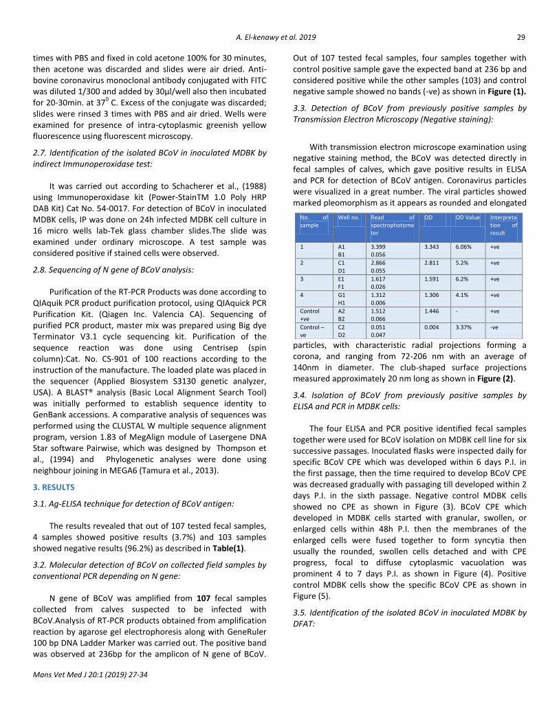
The results revealed that out of 107 tested fecal samples, 4 samples showed positive results (3.7%) and 103 samples showed negative results (96.2%) as described in Table(1).
3.2. Molecular detection of BCoV on collected field samples by conventional PCR depending on N gene:
N gene of BCoV was amplified from 107 fecal samples collected from calves suspected to be infected with BCoV.Analysis of RT-PCR products obtained from amplification reaction by agarose gel electrophoresis along with GeneRuler 100 bp DNA Ladder Marker was carried out. The positive band was observed at 236bp for the amplicon of N gene of BCoV.
Out of 107 tested fecal samples, four samples together with control positive sample gave the expected band at 236 bp and considered positive while the other samples (103) and control negative sample showed no bands (-ve) as shown in Figure (1).
3.3. Detection of BCoV from previously positive samples by Transmission Electron Microscopy (Negative staining):
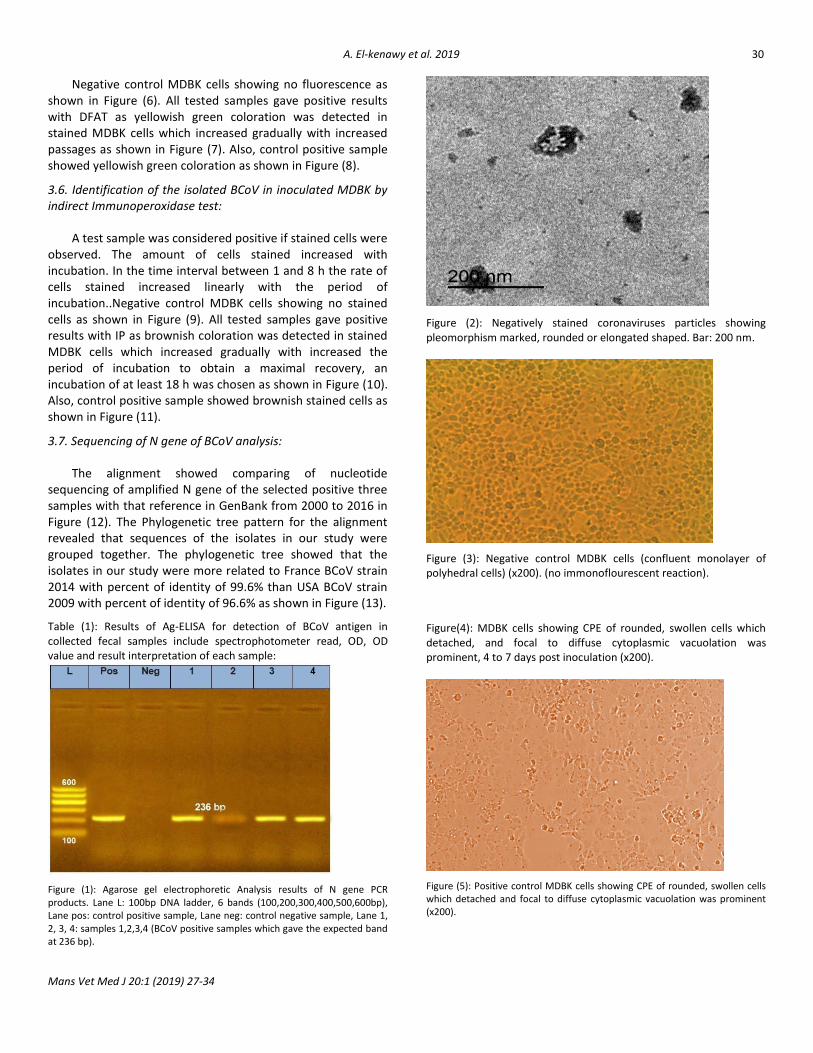
With transmission electron microscope examination using negative staining method, the BCoV was detected directly in fecal samples of calves, which gave positive results in ELISA and PCR for detection of BCoV antigen. Coronavirus particles were visualized in a great number. The viral particles showed marked pleomorphism as it appears as rounded and elongated
particles, with characteristic radial projections forming a corona, and ranging from 72-206 nm with an average of 140nm in diameter. The club-shaped surface projections measured approximately 20 nm long as shown in Figure (2).
3.4. Isolation of BCoV from previously positive samples by ELISA and PCR in MDBK cells:
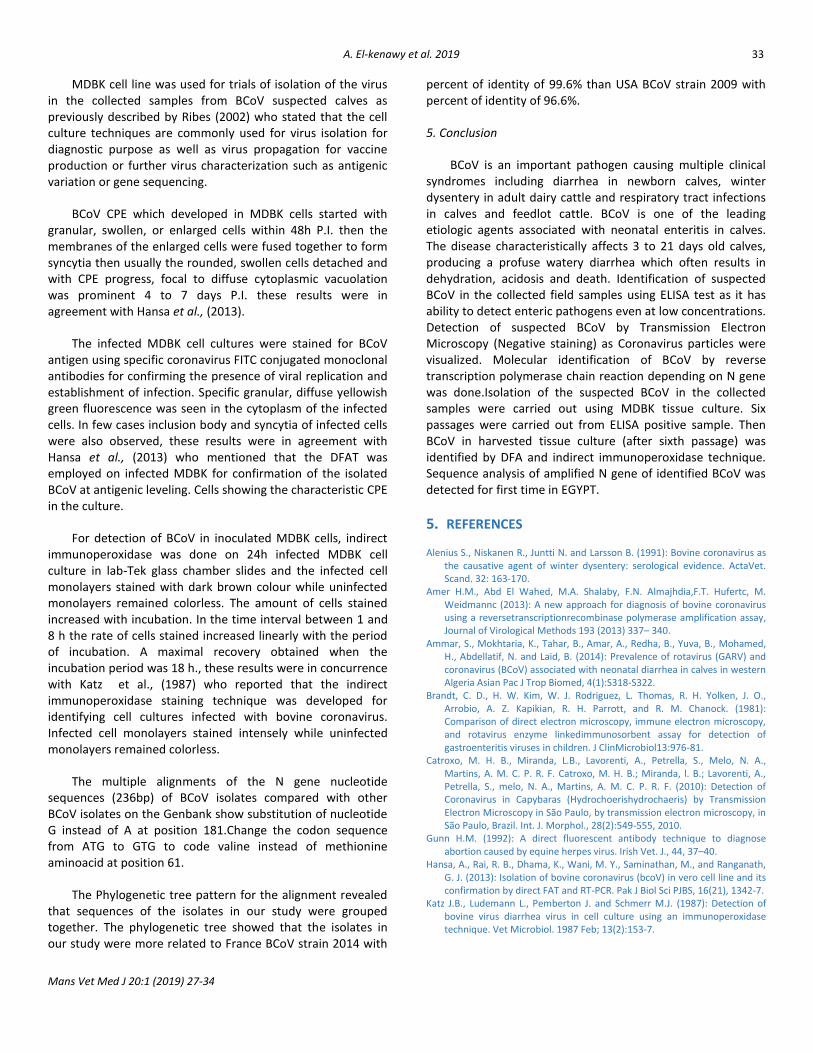
The four ELISA and PCR positive identified fecal samples together were used for BCoV isolation on MDBK cell line for six successive passages. Inoculated flasks were inspected daily for specific BCoV CPE which was developed within 6 days P.I. in the first passage, then the time required to develop BCoV CPE was decreased gradually with passaging till developed within 2 days P.I. in the sixth passage. Negative control MDBK cells showed no CPE as shown in Figure (3). BCoV CPE which developed in MDBK cells started with granular, swollen, or enlarged cells within 48h P.I. then the membranes of the enlarged cells were fused together to form syncytia then usually the rounded, swollen cells detached and with CPE progress, focal to diffuse cytoplasmic vacuolation was prominent 4 to 7 days P.I. as shown in Figure (4). Positive control MDBK cells show the specific BCoV CPE as shown in Figure (5).
3.5. Identification of the isolated BCoV in inoculated MDBK by DFAT:
No. of sample
Well no. Read of spectrophotometer
OD OD Value Interpretation of result
1 A1 B1
3.399 0.056
3.343 6.06% +ve
2 C1 D1
2.866 0.055
2.811 5.2% +ve
3 E1 F1
1.617 0.026
1.591 6.2% +ve
4 G1 H1
1.312 0.006
1.306 4.1% +ve
Control +ve
A2 B2
1.512 0.066
1.446 - +ve
Control –ve
C2 D2
0.051 0.047
0.004 3.37% -ve
30 A. El-kenawy et al. 2019
Mans Vet Med J 20:1 (2019) 27-34
Negative control MDBK cells showing no fluorescence as shown in Figure (6). All tested samples gave positive results with DFAT as yellowish green coloration was detected in stained MDBK cells which increased gradually with increased passages as shown in Figure (7). Also, control positive sample showed yellowish green coloration as shown in Figure (8).
3.6. Identification of the isolated BCoV in inoculated MDBK by indirect Immunoperoxidase test:
A test sample was considered positive if stained cells were observed. The amount of cells stained increased with incubation. In the time interval between 1 and 8 h the rate of cells stained increased linearly with the period of incubation..Negative control MDBK cells showing no stained cells as shown in Figure (9). All tested samples gave positive results with IP as brownish coloration was detected in stained MDBK cells which increased gradually with increased the period of incubation to obtain a maximal recovery, an incubation of at least 18 h was chosen as shown in Figure (10). Also, control positive sample showed brownish stained cells as shown in Figure (11).
3.7. Sequencing of N gene of BCoV analysis:
The alignment showed comparing of nucleotide sequencing of amplified N gene of the selected positive three samples with that reference in GenBank from 2000 to 2016 in Figure (12). The Phylogenetic tree pattern for the alignment revealed that sequences of the isolates in our study were grouped together. The phylogenetic tree showed that the isolates in our study were more related to France BCoV strain 2014 with percent of identity of 99.6% than USA BCoV strain 2009 with percent of identity of 96.6% as shown in Figure (13).
Table (1): Results of Ag-ELISA for detection of BCoV antigen in collected fecal samples include spectrophotometer read, OD, OD value and result interpretation of each sample:
Figure (1): Agarose gel electrophoretic Analysis results of N gene PCR products. Lane L: 100bp DNA ladder, 6 bands (100,200,300,400,500,600bp), Lane pos: control positive sample, Lane neg: control negative sample, Lane 1, 2, 3, 4: samples 1,2,3,4 (BCoV positive samples which gave the expected band at 236 bp).
Figure (2): Negatively stained coronaviruses particles showing pleomorphism marked, rounded or elongated shaped. Bar: 200 nm.
Figure (3): Negative control MDBK cells (confluent monolayer of polyhedral cells) (x200). (no immonoflourescent reaction).
Figure(4): MDBK cells showing CPE of rounded, swollen cells which detached, and focal to diffuse cytoplasmic vacuolation was prominent, 4 to 7 days post inoculation (x200).
Figure (5): Positive control MDBK cells showing CPE of rounded, swollen cells which detached and focal to diffuse cytoplasmic vacuolation was prominent (x200).

31 A. El-kenawy et al. 2019
Mans Vet Med J 20:1 (2019) 27-34
Figure (6): Negative control MDBK cells by fluorescent microscope showing no immunoflourescent reaction (no yellowish green color).
Figure (7): MDBK cells (sixth passage) show intracytoplasmic yellowish green fluorescence by fluorescent microscope (x200).
Figure (8): Positive control MDBK cells show intracytoplasmic yellowish green fluorescence by fluorescent microscope (x200).
Figure (9): Negative control MDBK cells showing brownish stained cells by ordinary microscope (x200).
Figure (10): MDBK cells (sixth passage) showing positive result of high average of brownish stained cells by IP (x200) after 18 hours.
Figure (11): Positive control MDBK cells showing positive result of high average of brownish stained cells by IP (x200).

32 A. El-kenawy et al. 2019
Mans Vet Med J 20:1 (2019) 27-34
Figure (12): The multiple alignments of the N gene nucleotide sequences (236bp) of BCoV isolates compared with other BCoV isolates on the Genbank show substitution of nucleotide G instead of A at position 181.
Figure(13): Phylogenetic tree of deduced amino acid sequence of N gene of our isolates compared with previous described related genomic sequences in GenBank from year 2000 to year 2016.
4. DISCUSSION
The aims of the present study were trials for isolation and identification of BCoV circulating in newly born calves population using MDBK cell culture and identification methods. In the present study, a total of 107 fecal samples were collected from diarrhetic calves from 1 day to 3 weeks of age from different localities in Dakahlia governorate, Egypt (Gamasa, Belkas, Sherbin, Nabaroh, Talkha, Almanzala, Algamalia, Aga) during the period from 2015 to 2018.These calves were suffering from watery diarrhea, dehydration, weakness and recumbency. Such clinical findings similar to that of BCoV were described by Lefèvre et al., (2010).
The collected field fecal samples from 107 calves were screened for the presence of BCoV Ag by using commercially available Monoscreen Ag ELISA Bovine coronavirus BIO K 344/2 Sandwich test. This test was used due to the facts that most of the other immunodiagnostic tests developed for detection of enteric pathogen have either low specificity and/ or sensitivity. ELISA also has ability to detect enteric pathogens even at low concentrations (Selim et al., 1991; Kelkar et al., 2004).
The results revealed that: out of 107 tested collected fecal samples, 4 samples showed positive results and 103 samples were negative. The overall prevalence of BCoV in Dakahlia governrate as detected by ELISA was 3.7% of collected samples, which may be due to decreasing of passive immunity and the absence of the natural resistance against infection for BCoV as mentioned by Uhde et al., 2008 and Ammar et al., 2014.
For further confirmation after traditional method, molecular tool RT- PCR by targeting N gene was done for rapid detection and confirmation of BCoV. Standardization of RT-PCR was done by selecting viral N gene based primers as it is conserved among BCoV strains. The estimated specific viral amplicons with sizes of 236bp were obtained confirming virus detection in clinical samples. The results of RT-PCR gave four positive samples while the other samples (103) are negative. This result was similar to the result of ELISA test and that was in agreement with Tsunemtitsu et al., (1999) who detect the BCoV RNA in fecal samples by RT-PCR and they also chose the N gene as target region for amplification.
The Electron Microscopy is commonly used for BCoV detection and identification based on morphological characteristics as Brandt et al., (1981) mentioned.
Through this study, it was found that the size of bovine coronavirus ranging from 72–206 nm with an average of 140 nm which was in harmony with the previous reports (Stair et al., 1972; Rai, 1983;).
33 A. El-kenawy et al. 2019
Mans Vet Med J 20:1 (2019) 27-34
MDBK cell line was used for trials of isolation of the virus in the collected samples from BCoV suspected calves as previously described by Ribes (2002) who stated that the cell culture techniques are commonly used for virus isolation for diagnostic purpose as well as virus propagation for vaccine production or further virus characterization such as antigenic variation or gene sequencing.
BCoV CPE which developed in MDBK cells started with granular, swollen, or enlarged cells within 48h P.I. then the membranes of the enlarged cells were fused together to form syncytia then usually the rounded, swollen cells detached and with CPE progress, focal to diffuse cytoplasmic vacuolation was prominent 4 to 7 days P.I. these results were in agreement with Hansa et al., (2013).
The infected MDBK cell cultures were stained for BCoV antigen using specific coronavirus FITC conjugated monoclonal antibodies for confirming the presence of viral replication and establishment of infection. Specific granular, diffuse yellowish green fluorescence was seen in the cytoplasm of the infected cells. In few cases inclusion body and syncytia of infected cells were also observed, these results were in agreement with Hansa et al., (2013) who mentioned that the DFAT was employed on infected MDBK for confirmation of the isolated BCoV at antigenic leveling. Cells showing the characteristic CPE in the culture.
For detection of BCoV in inoculated MDBK cells, indirect immunoperoxidase was done on 24h infected MDBK cell culture in lab-Tek glass chamber slides and the infected cell monolayers stained with dark brown colour while uninfected monolayers remained colorless. The amount of cells stained increased with incubation. In the time interval between 1 and 8 h the rate of cells stained increased linearly with the period of incubation. A maximal recovery obtained when the incubation period was 18 h., these results were in concurrence with Katz et al., (1987) who reported that the indirect immunoperoxidase staining technique was developed for identifying cell cultures infected with bovine coronavirus. Infected cell monolayers stained intensely while uninfected monolayers remained colorless.
The multiple alignments of the N gene nucleotide sequences (236bp) of BCoV isolates compared with other BCoV isolates on the Genbank show substitution of nucleotide G instead of A at position 181.Change the codon sequence from ATG to GTG to code valine instead of methionine aminoacid at position 61.
The Phylogenetic tree pattern for the alignment revealed that sequences of the isolates in our study were grouped together. The phylogenetic tree showed that the isolates in our study were more related to France BCoV strain 2014 with
percent of identity of 99.6% than USA BCoV strain 2009 with percent of identity of 96.6%.
5. Conclusion
BCoV is an important pathogen causing multiple clinical syndromes including diarrhea in newborn calves, winter dysentery in adult dairy cattle and respiratory tract infections in calves and feedlot cattle. BCoV is one of the leading etiologic agents associated with neonatal enteritis in calves. The disease characteristically affects 3 to 21 days old calves, producing a profuse watery diarrhea which often results in dehydration, acidosis and death. Identification of suspected BCoV in the collected field samples using ELISA test as it has ability to detect enteric pathogens even at low concentrations. Detection of suspected BCoV by Transmission Electron Microscopy (Negative staining) as Coronavirus particles were visualized. Molecular identification of BCoV by reverse transcription polymerase chain reaction depending on N gene was done.Isolation of the suspected BCoV in the collected samples were carried out using MDBK tissue culture. Six passages were carried out from ELISA positive sample. Then BCoV in harvested tissue culture (after sixth passage) was identified by DFA and indirect immunoperoxidase technique. Sequence analysis of amplified N gene of identified BCoV was detected for first time in EGYPT.
5. REFERENCES Alenius S., Niskanen R., Juntti N. and Larsson B. (1991): Bovine coronavirus as
the causative agent of winter dysentery: serological evidence. ActaVet. Scand. 32: 163-170.
Amer H.M., Abd El Wahed, M.A. Shalaby, F.N. Almajhdia,F.T. Hufertc, M. Weidmannc (2013): A new approach for diagnosis of bovine coronavirus using a reversetranscriptionrecombinase polymerase amplification assay, Journal of Virological Methods 193 (2013) 337– 340.
Ammar, S., Mokhtaria, K., Tahar, B., Amar, A., Redha, B., Yuva, B., Mohamed, H., Abdellatif, N. and Laid, B. (2014): Prevalence of rotavirus (GARV) and coronavirus (BCoV) associated with neonatal diarrhea in calves in western Algeria Asian Pac J Trop Biomed, 4(1):S318-S322.
Brandt, C. D., H. W. Kim, W. J. Rodriguez, L. Thomas, R. H. Yolken, J. O., Arrobio, A. Z. Kapikian, R. H. Parrott, and R. M. Chanock. (1981): Comparison of direct electron microscopy, immune electron microscopy, and rotavirus enzyme linkedimmunosorbent assay for detection of gastroenteritis viruses in children. J ClinMicrobiol13:976-81.
Catroxo, M. H. B., Miranda, L.B., Lavorenti, A., Petrella, S., Melo, N. A., Martins, A. M. C. P. R. F. Catroxo, M. H. B.; Miranda, l. B.; Lavorenti, A., Petrella, S., melo, N. A., Martins, A. M. C. P. R. F. (2010): Detection of Coronavirus in Capybaras (Hydrochoerishydrochaeris) by Transmission Electron Microscopy in São Paulo, by transmission electron microscopy, in São Paulo, Brazil. Int. J. Morphol., 28(2):549-555, 2010.
Gunn H.M. (1992): A direct fluorescent antibody technique to diagnose abortion caused by equine herpes virus. Irish Vet. J., 44, 37–40.
Hansa, A., Rai, R. B., Dhama, K., Wani, M. Y., Saminathan, M., and Ranganath, G. J. (2013): Isolation of bovine coronavirus (bcoV) in vero cell line and its confirmation by direct FAT and RT-PCR. Pak J Biol Sci PJBS, 16(21), 1342-7.
Katz J.B., Ludemann L., Pemberton J. and Schmerr M.J. (1987): Detection of bovine virus diarrhea virus in cell culture using an immunoperoxidase technique. Vet Microbiol. 1987 Feb; 13(2):153-7.

34 A. El-kenawy et al. 2019
Mans Vet Med J 20:1 (2019) 27-34
Kelkar S. D., Bhide V. S., Ranshing S. S. and Bedekar S. S. (2004): Rapid ELISA for the diagnosis of rotavirus. Indian Journal of Medical Research, 119, 60-65.
Lefèvre P.C., Blancou J. and Chermette R. (2010): Infectious and Parasitic Diseases of Livestock, Chapter: Bovine coronavirus.
OIE (2013): Manual of Diagnostic Tests and Vaccines for Terrestrial Animals. Office International des Epizooties (OIE)., standards commission and adopted by the International Committee of the OIE 4th edition. Publ. Paris, France. pp. 77-81.
Rai R.B., A. Hansa, K. Dhama, M.Y. Wani, M. Saminathan and G.J. Ranganath (2013):solation of Bovine Coronavirus (BCoV) in Vero Cell Line and its Confirmation by Direct FAT and RT-PCR, Pakistan Journal of BiologicalSciences, Volume 16 (21): 1342-1347, 2013.
Ribes, J. A., J. P. Seabolt, and S. B. Overman (2002): Performance characteristics of VIDAS and directigen respiratory syncytial virus (RSV) antigen detection assays and culture for the identification of RSV in respiratory specimens. J ClinMicrobiol40:1818-20.
Robert Anton Heckert, B.Sc. (Agr), D.V.M. (1990):Bovine coronavirus in calves: Epidemiology, diagnosis and antibody isotype responses to structural viral proteins.The Ohio State University.
Saif L.J. (2004): Rev. sci. tech. Off. int. Epiz., 2004, 23 (2), 643-660 Animal coronaviruses: what can they teach us about the severe acute respiratory syndrome? Food Animal Health Research Program, Ohio Agricultural Research & Development Center (OARDC), Ohio State University, Wooster, OH 44691, United States of America.
Schacherer C., Braun W., Bauer G. and Doerr H. W. (1988): Detection of cytomegalovirus in bronchial lavage and urine using a monoclonal antibody to an HCMV early nuclear protein. Infection, 16(5), 288-292.
Selim, S.A., Aziz, K.M.S., Sarker, A.J. and Rahman, H. (1991): Rotavirus infection in calves in Bangladesh. Res. Opin. Anim. Vet. Sci., 2013, 3(8), 225-234.
Spaan, W., Cavanagh, D., Horzinek, M.C., (1988): Coronaviruses: structure and genome expression. Journal of General Virology 69, 2939–2952.
Stair, S.L., Rhodes, M.B., White, R.G. and Mebus, C.A. (1972): Neonatal calf diarrhoea: purification and electron microscopy of coronavirus-like agent. American Journal of Veterinary Research, 33: 1147-1156.
Takiuchi E., Danilo T. Stipp, Alice F. Alfieri and Amauri A. Alfieri (2005): Improved detection of bovine coronavirus N gene in faeces of calvesinfected naturally by a semi-nested PCR assay and an internal control.Box 6001, 86051-990, Londrina, Paran´a, Brazil,accepted 18 August 2005.
Tamura K., Stecher G., Peterson D., Filipski A. and Kumar S. (2013): MEGA6: molecular evolutionary genetics analysis version 6.0. Mol. Biol. Evol. 30, 2725–2729.
Thompson J.D., Higgins D.G. and Gibson T.J. (1994): CLUSTAL W: improving the sensitivity of progressive multiple sequence alignment through sequence weighting, position-specific gap penalties and weight matrix choice. Nucleic Acids Research, 22(22):4673-4680.
Tsunemitsu H., D.R. Smith and L.J. Saif (1999): Experimental inoculation of adult dairy cows with bovine coronavirus and detection of coronavirus in feces by RT-PCR. Arch. Virol., 144: 167-175.
Uhde, F.L., Kaufmann, T., Sager, H., Albini, S., Zanoni, R., Schelling, E. and Meylan, M. (2008): Prevalence of four enteropathogens in the faeces of young diarrhoeic dairy calves in Switzerland. Veterinary Record, 163: 362-366.
.