Embed Size (px)
Citation preview

Proc. Nall. Acad. Sci. USAVol. 84, pp. 7473-7477, November 1987Biochemistry
Isolation and sequence of a eDNA clone for human tyrosinase thamaps at the mouse c-albino locusBYOUNG S. KWON*t, ASIFA K. HAQ*, SEYMOUR H. POMERANTZt, AND RUTH HALABAN§*Molecular Genetics Laboratory, Guthrie Research Institute, Sayre, PA 18840; tDepartment of Biological Chemistry, University of Maryland School ofMedicine, Baltimore, MD 21201; and §Department of Dermatology, Yale University School of Medicine, New Haven, CT 06510
Communicated by Aaron B. Lerner, July 16, 1987
ABSTRACT Screening of a Xgtll human melanocytecDNA library with antibodies against hamster tyrosinase(monophenol, L-dopa:oxygen oxidoreductase, EC 1.14.18.1)resulted in the isolation of 16 clones. The cDNA inserts from 13of the 16 clones cross-hybridized with each other, indicatingthat they were from related mRNA species. One of the cDNAclones, Pmel34, detected one mRNA species with an approxi-mate length of 2.4 kilobases that was expressed preferentiallyin normal and malignant melanocytes but not in other celltypes. The amino acid sequence deduced from the nucleotidesequence showed that the putative human tyrosinase is com-posed of 548 amino acids with a molecular weight of 62,610.The deduced protein contains glycosylation sites and histidine-rich sites that could be used for copper binding. Southern blotanalysis of DNA derived from newborn mice carrying lethalalbino deletion mutations revealed that Pmel34 maps near or atthe c-albino locus, the position of the structural gene fortyrosinase.
Tyrosinase (monophenol, L-dopa:oxygen oxidoreductase,EC 1.14.18.1) is a copper-based oxidoreductase that cata-lyzes the oxidation of tyrosine to dopa and the oxidation ofdopa to dopaquinone (1). It is a key enzyme in melaninbiosynthesis. Oculocutaneous albinism, a group of auto-somal-recessive diseases in humans (2) and animals, ischaracterized by reduced or absent melanin in skin, hair, andeyes. Tyrosinase-negative albino melanocytes have notyrosinase activity in vitro.
Genetic control of pigmentation has been extensivelystudied in mice. There is evidence that the c-albino locus atchromosome 7 codes for the structural gene for tyrosinase (3,4). Mutations at this locus affect both tyrosinase activity andcoat color (5, 6).A nucleic acid probe for tyrosinase would be an invaluable
tool for studies of the regulation of tyrosinase, of themolecular basis of human albinism, and of various mousemutations affecting coat and eye color. We report here theisolation and sequence ofacDNA clone for human tyrosinasethat maps at or near the mouse c-locus.¶
MATERIALS AND METHODSCell Culture. Normal human melanocytes, melanotic mel-
anoma cells (LG), and neuroblastoma cells (SK-N-SH) werecultured as described (7-9). The murine neuroblastoma cellline NIE115 was obtained from X. 0. Breakefield (E. K.Shriver Center, Waltham, MA). Proteins of normal melano-cytes were radiolabeled with [355]methionine (Amersham)(100 ,XCi/ml, 1390 Ci/mmol; 1 Ci = 37 GBq) as described(10).cDNA Libraries and Screening. RNA from normal human
melanocytes was prepared, and poly(A)+ RNA was purified
on an oligo[d(T)I-cellulose column (11, 12). A cDNA liwas prepared employing a Xgtll cloning vector (13-15)Xgtll library contained 1.7 x 10⁶ independent phagesimmunobiological screening and analysis of the fusionteins produced by Xgtll cDNA clones were carried cdescribed by Young and Davis (15). The rabbit anti-haltyrosinase antibodies and their application in the stuntyrosinases have been described in detail (10, 16).RNA Blot Hybridization. Poly(A)+ RNA from n(
human melanocytes, melanoma cells, neuroblastomalines, HL-60 (human promyelocytic leukemia cell line)HepG2 (human hepatocarcinoma cell line) was fractiolon a 1.2% (wt/vol) formaldehyde denaturing gel (17), tferred to a GeneScreenPlus membrane (New Englandclear), and hybridized with 32P-labeled cDNA pr(Pmel14-2, a cDNA clone that was isolated from our himelanocyte cDNA library, was used as a control pbecause the corresponding RNA was detectable in si5amounts in all human and murine cells tested.Genomic DNA Blot Analysis. Newborn mice of the g
types C3H/C3H and cch/cH were provided by SalGluecksohn-Waelsch (Albert Einstein College of MedicNewborn mice provided by M. Lynn Lamoreux (Texas AUniversity, College Station, TX) were littermates ofgenotypes C14COs/C14Cos cCh/c14CWs, and cch/cch. Thesewere descended from three mice of the genotype C6H/Cthat were given to M. L. Lamoreux by S. GluecksWaelsch. These stocks were crossed twice onto JU/Ct/+c/+c mice, then once onto C57BL/6J mice, then for egenerations onto a C57BL/6J-cch/cch stock descended Imice provided by D. Townsend (University of MinnesMinneapolis), and then sibling mated for six generationHigh molecular weight DNAs of newborn mice of var
genotypes were prepared as described (18). Restricendonuclease digests ofDNA were electrophoresed in a 0agarose gel at 40C, transferred (19) to GeneScreenPlus,hybridized with the 32P-labeled cDNA probes. Pmell7-cDNA clone that was isolated from our human melanoicDNA library, was utilized as a control probe for the purrof estimating the amounts of DNA in each lane bectPmell7-1 detected a single EcoRI fragment in all mouse Dtested. The intensity of the hybridizing bands was meastby a densitometer at 500 nm (Beckman DU-8BUV/Spectrophotometer).DNA Sequencing. DNA restriction fragments subclone
M13 vectors (20) were sequenced by the dideoxy chtermination technique (21), with modifications made tocommodate 2'-deoxyadenosine 5'-[a-[35S]thio]triphospt(22). A forward primer (New England Biolabs) compleirtary to the lacZ sequence adjacent to the 5'-side of the Ec
tTo whom reprint requests should be addressed.9This sequence is being deposited in the EMBL/GenBank data t(Bolt, Beranek, and Newman Laboratories, Cambridge, MA,Eur. Mol. Biol. Lab., Heidelberg) (accession no. J03581).
The publication costs of this article were defrayed in part by page chargepayment. This article must therefore be hereby marked "advertisement"in accordance with 18 U.S.C. §1734 solely to indicate this fact.

Proc. Natl. Acad. Sci. USA 84 (1987)
site in Xgtll was used for the direct sequencing of cDNAinsert end points in Xgtll (23).
RESULTS
Initial screening of 500,000 recombinant phage plaques withthe rabbit anti-tyrosinase antibodies identified 16 indepen-dent clones. The cDNA inserts varied in size from 0.2 to 1.6kilobase pairs. The longest cDNA insert (-1.6 kilobase pairs)from Xme134 hybridized to 12 other cDNA inserts and sharedan overlapping restriction enzyme pattern (Fig. la). Theother three cDNA inserts (not included in the figure) were notrelated to the 13 cDNA clones. A cDNA insert contained inone of these, Xmell7-1, was utilized as a control probe. ThecDNA inserts of Xmell6, Xmel34, Xmel4O, and Xmell7-1 weresubcloned into pBR322 to yield Pmell6, Pmel34, Pmel4O, andPmell7-1, respectively, and used for further characterization
a 0 200 400 600 800 1000 1200 1400 1600 180 2000 2200 2400
A"34A me 33Xmel44A me 16
Xmel 4
A mM 52Xmel 16me 106
mel?A me 20
C
1 2 3 41 2
Mr
IKdl
94-
67-b4 -
43-
20-
of the cDNA clones. A lysogen of Xmell6 was prepared, andthe bacterial lysate was analyzed by 6% NaDodSO4/poly-acrylamide gel electrophoresis and by competitive immuno-precipitation assay. Xmell6 was used for this experimentbecause the Xmell6 plaques produced the strongest signal inplaque screening with anti-tyrosinase antibodies even thoughXmel6 contained an incomplete cDNA of -0.7 kilobasepairs. A fusion protein was produced in Y1089/Xmell6 thathad a relative size of 140 kDa (Fig. lb). The fusion proteinwas 25 kDa larger than Escherichia coli 8-galactosidase,indicating that the incomplete cDNA in Xmell6 was fused to.3-galactosidase gene in frame. Synthesis of both /3-galacto-sidase and the fusion protein was dependent on inductionwith isopropyl 13-D-thiogalactopyranoside. The production ofimmunologically reactive tyrosinase protein in the lysate ofXmell6 lysogen was tested by competition assay usingmetabolically labeled melanocyte cell extracts as a source fortyrosinase. Bacterial lysates of the Xmell6 lysogen (2 x 10⁷bacterial cells in 100 pil) competed with roughly 70% of theanti-tyrosinase antibodies as judged from the intensity of thetyrosinase bands and the radioactivity in the relevant gelslices (Fig. lc).To examine whether mRNA homologous to Pmel34 is ex-
pressed preferentially in melanocytes, RNA gel blot analysis ofpoly(A)+ mRNA from cells ofmelanocytic and nonmelanocyticlineage was performed. Pmel34 hybridized to 21S (=2.4kilobase) mRNA species from normal human melanocytes andhuman melanotic melanoma cells (LG) but not from HepG2,HL-60, human or murine neuroblastoma (Fig. 2), human andmouse fibroblasts, or lymphocytes (data not shown).The skin of mice carrying the radiation-induced albino
alleles, such as c3Hlc3H, had no tyrosinase activity (4). Thetyrosinase activity levels in the skin of mice heterozygousbetween the lethal albino deletion and chinchilla were shownto be intermediate between the normal and mutant homo-zygotes, consistent with the murine albino locus encoding thestructural gene of tyrosinase (unpublished observation of S.Gluecksohn-Waelsch, reviewed in ref. 4). If the c-albinolocus codes for the structural gene for tyrosinase, tyrosinasecDNA should not detect any hybridizing band in DNAextracted from c3H/c3H or c14Cos/c14Cos mice but should detecthybridizing bands of normal and half intensity in DNA fromhomozygote (cch/cch) and heterozygotes (cch/c3H and cch/c)4Cos) respectively.
1 2 3 4 5 6 7 8
28S-
FIG. 1. Isolation of human tyrosinase cDNA by antibody screen-ing. (a) Alignment of Xmel34-related cDNA inserts. Inserts from 13cDNA clones whose gene products bound to anti-tyrosinase anti-bodies were aligned, based upon restriction mapping and partialnucleotide sequencing. (b) Analysis of lacZ-cDNA fusion proteins.Bacterial lysates were prepared from the lysogens of Y1089/Xgt1l(lanes 1 and 2) and Y1089/Xmell6 (lanes 3 and 4) that were culturedin the absence (lanes 1 and 4) or presence (lanes 2 and 3) of isopropylf3D-thiogalactopyranoside. Samples were electrophoresed on a 6%NaDodSO4/polyacrylamide gel and stained with Coomassie brilliantblue. The isopropyl /3-D-thiogalactopyranoside-dependent produc-tion of 3-galactosidase (lane 2) and a - 140-kDa Xmell6 fusion protein(lane 3, arrow) was noted. Protein sizes in kDa (Kd) are at the left.(c) Competition immunoprecipitation assay. The lysate of Xmell6 orXgtll lysogen was used to compete with metabolically labeled humanmelanocyte cell extract for anti-tyrosinase antibodies. Equalamounts of lysates of Xgtll lysogen and Xmell6 lysogen (-2 x 10⁷bacterial cells in 100-1,l volume) were incubated with 5 ,ul of a 1:100dilution of anti-tyrosinase antibodies. The respective supernatantswere used to immunoprecipitate [35S]methionine-labeled humanmelanocyte extract (6 x 10⁶ cpm in protein per tube). The elutedimmune complexes were separated on 8.5% NaDodSO4/polyacryl-amide gel. In lanes 1 and 2, the antibody preparation waspreabsorbed with lysogens Xgtll and Xmell6, respectively.
pmel 3418S
pmel 14-2----e-
FIG. 2. Blot analysis of poly(A)+ RNA derived from cultures ofmelanocytic and nonmelanocytic lineage. Four micrograms ofpoly(A)+ RNA of normal human melanocyte (lane 1), LG (lane 2),HepG2 (lane 3), and HL-60 (lane 4) cells, and 10 ,ug of poly(A)+ RNAof normal human melanocytes (lane 5), LG (lane 6), human neuro-blastoma (lane 7), and murine neuroblastoma cells (lane 8) werefractionated on a 1.2% (wt/vol) formaldehyde denaturing agarosegel, blotted, and hybridized with 32P-labeled Pmel34. The same filterwas used to hybridize to 32P-labeled Pmel14-2 to show that each lanecontained RNA as indicated above and that the RNA was relativelyintact.
bMr
IKdI
200 -
116 -
66.2 -40 --
7474 Biochemistry: Kwon et al.

Proc. Natl. Acad. Sci. USA 84 (1987) 7475
Si
ka U ,
izeMarker_IKbi
21.7
5.2- _
1.6-
FIG. 3. Southern blot analysis of genomic DNA of murine lethalalbino deletion mutants. Genomic DNA from newborn mice ofstrains cch/cch, cc /c3H, c3H/c3H, c/C14Cos, and c)4Cos/cI4Cos wasdigested with EcoRI, electrophoresed on a 0.8% agarose gel, trans-ferred to GeneScreenPlus, and hybridized to Pmel34 (a). The samefilter was probed with Pmell7-1 after removal of the Pmel34 probe(b). Spectrophotometric analysis of the intensity of Pmell7-1 bandsrevealed that there was -20% more cch/cch DNA compared with thatin the other four lanes. Kb, kilobases.
As shown in Fig. 3a, the Pmel34 probe detected threehybridizing cch/cch genomic DNA fragments whose sizeswere 4.5, 12.0, and 14.5 kb. The different extent of hybrid-ization of the three bands appears to be due to the hetero-geneity between the human probe and the mouse genes. Thesame three bands were detected at half intensity in cch/c3Hand cch/c14Cos DNA when normalized with respect to thePmell7-1 band (Fig. 3b). No hybridizing fragments weredetected in c3H/C3H or c14CoS/c14C0s DNA even after pro-longed exposure. Therefore, the murine genes whose se-quences are homologous to cDNA contained in Pmel34 arelocated at or near the albino locus.The nucleotide sequence of three overlapping cDNA clones
(Pmel34, Pmell6, and Pmel40) was determined according to the
0 200 400 600 800 1000 1200 1400
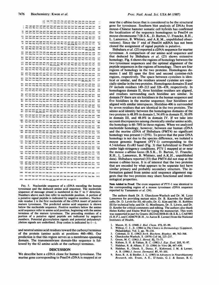
strategy shown in Fig. 4. Most of the other 10 clones were alsosequenced. The nucleotide sequence of tyrosinase cDNA re-vealed a single long open reading frame, beginning with the firstnucleotide after the EcoRI linker. Direct sequence analysis witha Xgtll forward primer (23) revealed that this open readingframe is in frame with the lacZ gene of the Xgtll vector. Wefound that other clones were also fused in frame with the IacZgene. This property was helpful in assigning the open readingframe even though the cDNAs did not start with the first ATGcodon. The open reading frame coded for a polypeptide of 566amino acids with a molecular weight of 63,549 (Fig. 5). Thecodon specifying carboxyl-terminal leucine was followed by thetranslation termination codon TAA (nucleotide residues1645-1647). No nucleotide differences were observed amongthe three cDNA clones except that they differed in length. The3'-untranslated sequence determined from PmeI34, Pmell6, andPmel4O did not extend as far as the poly(A)+ tail. However,Pmel4O contained a potential polyadenylylation signal of AT-TAAA (24, 25) (underlined nucleotide residues 1822-1827) thatappears upstream of the consensus polyadenylylation signalAATAAA.The deduced sequence of the first 12 amino acid residues
of the tyrosinase has characteristics of the signal peptide ofsecretory and membrane-associated proteins (26), whichmainly contains hydrophobic amino acids (10 out of 12residues) and terminates with serine (27) (Fig. 5). We puta-tively assign the first 12 amino acids as a part of a signalpeptide. A possible site cleavage of the signal peptide of thetyrosinase precursor is after the serine residue at phenylal-anine-1 (Fig. 5). Thus, the protein backbone of processedtyrosinase is composed of 548 amino acids with a molecularweight of 62,160.As tyrosinase is a glycoprotein, we have looked for
possible N-glycosylation sites and found five potential as-paragine-linked glycosylation signals (28, 29) at positions 73,98, 148, 217, and 324 as underlined in Fig. 5.The protein contains two cysteine-rich domains (Fig. 6a,
Pmel34P). There are 17 cysteine residues; 10 residues areclustered within the first 100 amino acids, and 5 residues areclustered between amino acids 231 and 308.The tyrosinase molecule contains two copper atoms (30,
31), and histidine residues serve as copper binding sites (31,32). There are 15 histidine residues in the deduced amino acidsequence of human tyrosinase. Four of these appearedbetween the two cysteine-rich domains (histidine-130, -167,-189, and -198), and 6 residues appeared after the secondcysteine-rich domain (histidine-350, -354, -360, -377, -390,and -406). Histidine residues at positions 350, 354, 360, and377 had an arrangement similar to that of bovine superoxidedismutase, another copper-containing enzyme (32). There isa stretch of 27 amino acids that contains only hydrophobic
1600 1800 2000 2200 2400
I~> 2 - .2* 2 :A.< < -i co <:
A
No di_ _ _ _ _la IN 10 ON 44 -
so*t- lo
FIG. 4. Partial restrictionmap and sequencing strategy forhuman tyrosinase cDNA. Thetyrosinase-coding region is indi-cated by an open box. Horizon-tal arrows under the three insertsshow the direction and extent ofsequencing used to generate thesequence presented in Fig. 5.Restriction sites used for se-quencing are indicated. Thescale at the top indicates thenucleotide number.
5, F.: .;a2.-I -C
X mel 34
X mel 16
X mel 40
Biochemistry: Kwon et al.
9h
7476 Biochemistry: Kwon et al.
-42 GAA TTC CTG CTC CTG GCT GTT TTG TAC TGC CTG CTG TGG AGT -1-12 ------- Leu Leu Leu Ala Val Leu Tyr Cys Leu Lett TrpSr -
1 TTC CAG ACC TCC GCT GGC CAT TTC CCT AGA GCC TGT GTC TCC TCT AAG AAC CTC ATG GAG 601 Phe Gln Thr Ser Ala Gly His Phe Pro Arg Ala Cys Val Ser Ser Lvs Asn Leu Met Gltt 20
61 AAG GM TGC TGT CCA CCG TGG AGC GGG ACA GGA GTC TGT (GC CAG CTT TCA GGC AGA GGT 12n21 Lys Glu Cys Cys Pro Pro Trp Ser Gly Thr Gly Val Cys Gly Gln Leu Ser Gly Are Glv 4n
121 TCC TGT CAG AAT ATC CTT CTG TCC MT GCA CCA CTT GCG CCT CM TTT CCC TTC ACA GGG 10041 Ser Cys Gln Asn Ile Leu Leot Ser Asn Ala Pro Leu Gly Pro Gln Phe Pro Phe Ther Glv 60
181 GTG GAT GAC CGG GAG TCG TGG CCT TCC GTC TTT TAT AAT AGG ACC TGC CAG TGC TCT GGC 24061 Val Asp Asp Arg Glu Ser Trp Pro Ser Val Phe Tyr Asn Arg Thr Cys Gln Cys Ser Glv 80
241 MC TTC ATG GGA TTC MC TGT GGA MC TGC MCG mTT GGC TmT TGG GGA CCA MC TGC ACA 30081 Asn Phe Met Gly Phe Asn Cys Gly Asn Cys Lys Phe Gly Phe Trp Gly Pro Asn Cys Thr 100
301 GAG AGA CGA CTC TTG GTG AGA AGA MC ATC TTC GAT TTG AGT GCC CCA GAG MG GAC AA 360101 Glu Arg Arg Leu Leu Val Arg Arg Asn Ile Phe Asp Leu Ser Ala Pro Glu Lys Asp Lys 12C361 mTT TTT GCC TAC CTC ACT TTA GCA AAG CAT ACC ATC AGC TCA GAC TAT GTC ATC CCC ATA 420121 Phe Phe Ala Tyr Leu Thr Leu Ala Lys His Thr Ile Ser Ser Asp Tyr Val Ile Pro Ile 141
421 GGG ACC TAT GGC CAA ATG MA AAT GGA TCA ACA CCC ATG mr MC GAC ATC MT ATT TAT 080141 Gly Thr Tyr Gly Gln Met L-s Asn Gly Ser Thr Pro Met Phe Asn Asp Ile Asn Ile Tyr 160
481 GAC CTC TmC GTC TGG ATG CAT TAT TAT GT, TCA ATG CAT GCA CTG CTT GGG GGA TAT CAA 541161 Asp Leu Phe Val Trp Met His Tyr Tyr Val Ser Met Asp Ala Leu Leu Gly Gly Tyr Glu 180541 ATC TGG AGA GAC ATT GAT TTT GCC CAT GAA GCA CCA GCT TTT CTG CCT TGG CAT AGA CTC 605o181 Ile Trp Arg Asp Ile Asp Phe Ala His Glu Ala Pro Ala Phe Leu Pro Trp His Arg Lett 20o601 TTC TTG TTG CGG TGG GAA CM CAA ATC CAG MG (TG ACA GGA GAT GM MC TTC ACT ATT 460201 Phe Lett Leu Arg Trp Glu Gln Glt Ile Gln Lys Leu Thr Gly Asp Gli Asn Phe Thr Ile 220
661 CCA TAT TGC GAC TGG CGG GAT GCA GM MtC TGT GAC ATT TGC ACA GAT 4AG TAC ATG GGA 72n221 Pro Tyr Trp Asp Trp Arg Asp Ala Glu Lys Cos Asp Ile Cys Thr Asp Glu Tyr Met Gly 240
721 GGT CAG CAC CCC ACA MT CCT MC TTA CTC AGC CCA GCA TCA TTC TTC TCC TCT TGG CAG 780241 Gly Gln His Pro Thr Asn Pro Asn Leu Leu Ser Pro Ala Ser Phe Phe Ser Ser Trp Gln 260
781 ATT GTC TGT AGC CGA TTG GAG GAG TAC MC AGC CAT CAG TCT TTA TGC MT GIA ACG CCC 840261 Ile Val Cys Ser Arg Leu Glu Glu Tyr Asn Ser His Gln Ser Leu Cys Asn Gly Thr Pro 280
841 GAG GGA CCT TTA CGG CGT AAT CCT GGA MC CAT GAC MA TCC ACA ACC CCA AGG CTC CCC 900281 Glu Gly Pro Leu Arg Arg Asn Pro Gly Asn His Asp Lys Ser Thr Thr Pro Arg Leu Pro 300
901 TCT TCA GCT GAT GTA GM ('C TGC CTG AGT TTG ACC CM TAT GAA TCT GGT TCC ATG GAT 9f0301 Ser Ser Ala Asp Val Glu Phe Cys Leu Ser Leu Thr Oln Tyr Glu Ser Gly Ser Met Asp 120
961 MA GCT GCC MT TTC AGC TTT AGA MT ACA CTG GM GGA TTT GCT AGT CCA CTT ACT GGG 1020321 Lys Ala Ala Asn Phe Ser Phe Arg Asn Thr Leu Glu Gly Phe Ala Ser Pro Leu Thr Gly 340
1021 ATA GCG GAT GCC TCT CAA AGC AGC ATG CAC AAT (CC TTG CAC ATC TAT ATG MT GGA CAT 1080341 Ile Ala Asp Ala Ser Gln Ser Ser Met His Asn Ala Leu His Ile Tyr Met Asn Gly Hit 341
1081 GTC CCA GGT ACA GGA TCT GCC MC GAT CGT ATC TTC CTT CTC ACC ATG CAT TTG TTG ACA 1140361 Val Pro Gly Thr Gly Ser Ala Asn Asp Arg Ile Phe Leu Leu Thr Met Hit Leu Leu Thr 380
1141 GTA TTT TTG AGG CAG TGG CTC CM AGG CAC CGT CCT CTT CM GM GTT TAT CCA GM (CC 1200381 Val Phe Leu Arg Gln Trp Leu Gln Arg His Arg Pro Leu Gln Glu Val Tyr Pro Glu Ala 400
1201 MT GCA CCC ATT GGA CAT AAC CGG GM TCC TAC ATG GTT CCT TTT ATA CCA CTG TAC AGA 126(0401 Asn Ala Pro Ile Gly His Asn Arg Glu Ser Tyr tet Val Pro Phe Ile Pro Leu Tyr Arg 420
1261 MT GGT GAT TTC mTI ATT TCA TCC MA CAT CTG GGC TAT GAC TAT ACC TAT CTA CM GAT 1320421 Asn Gly Asp Phe Phe Ile Ser Ser Lys Asp Leu Gly Tyr Asp Tyr Ser Tyr Leu Gln Asp 440
1321 TCA GAC CCA GAC TCT TTT CM GAC TAC ATT MG TCC TAT TTG GAA CAA GCG AGT CGG ATC 1381441 Ser Asp Pro Asp Ser Phe Gln Asp Tyr Ile Lys Ser Tyr Leu Glu Gln Ala Ser Arg Ile 460
1381 TGG TCA TGG CTC CTT GGG GCG GCG ATG GTA GGG GCC GTC CTC ACT GCC CTG CTG GCA GGGI 1440461 Trp Ser Trp Leu Leu Gly Ala Ala Met Val Gly Ala Val Leu Thr Ala Leu Leu Ala Gly 480
1441 CCT GTG AGC TTG CTG TGT CGT CAC MG AGA AAG CAG CTT CCT GM GM MG CAG CCA CTC 1500481 Pro Val Ser Leu Leu Cvs Art His Lys Arg Lys Gln Leu Pro Glu Glu Lys Gln Pro Leu 550
1501 CTC ATG GAG AM GM GGA TTA CCA CAG CTT GTA TCA GAG CCA TTT ATA AAA GGC TTA GGC 1560501 Leu Met Glu Lys Glu Gly Leu Pro Gln Leu Val Ser Glu Pro Phe Ile Lys Gly Leu Gly 5201561 AAT AGA GTA GGG CCA MA ACC CCT GAC CTC ACT CTA ACT CAA AGT MT GTC CAG GTT CCA 1620521 Asn Arg Val Gly Pro Lys Ser Pro Asp Leu Thr Leu Thr Gln Ser Asn Val Gln Val Pro 540
1621 GAG AAT ATC TGC TGG TAT TTT CTG TAA AGA CCA TTT GCA AM TTG TM CCT MAT ACA MG 160n541 Glu Asn Ile Cys Trp Try Phe Leu ---
1681 TGT AGC CTT CTT CCA ACT CAG GTA GM CAC ACC TGT CTT TGT CTT GCT GTT TTC ACT CAG 17'.0
1741 CCC TTT TAA CAT TTT CCC CTA AGC CCA TAT GTC TM CIA AAG GAT GCT ATT TGG TM TGA 1800
1801 GGA ACT GTT ATT TGT ATG TGA ATTMA AGT GCT CTT AGG MT TC
FIG. 5. Nucleotide sequence of a cDNA encoding the humantyrosinase and the deduced amino acid sequence. The nucleotidesequence of message strand is numbered in the 5' to 3' direction.Numbers above each line refer to nucleotide position. A portion ofa putative signal peptide is indicated by negative numbers. Nucleo-tide residue 1 is the first nucleotide of the cDNA insert of putativemature tyrosinase. The predicted amino acid sequence is shownbelow the nucleotide sequence. Positive numbers below the aminoacid sequence refer to amino acid position, beginning with the aminoterminus of the mature tyrosinase. The preceding residues of a
portion of a putative signal peptide are indicated by negativenumbers. Potential glycosylation signals and potential polyadenyl-ylation signals are underlined. Stop codon is indicated by (---).
and neutral amino acid residues toward the carboxyl terminusof the protein (amino acids at positions 460-486). Ourprediction is that this region serves as a membrane-spanningdomain. The transmembrane domain-like sequence is fol-lowed by the 62 amino acids at the carboxyl terminus.
DISCUSSIONWe describe here a cDNA clone for human tyrosinase. Themurine gene corresponding to Pmel34 cDNA is mapped at or
Proc. Natl. Acad. Sci. USA 84 (1987)
near the c-albino locus that is considered to be the structuralgene for tyrosinase. Southern blot analysis of DNAs frommouse-Chinese hamster somatic cell hybrids has confirmedthe localization of the sequence homologous to Pmel34 onmouse chromosome 7 (B.S.K., D. Barton, U. Francke, R.H.,L. Lamoreux, B. Whitney, and A.K.M., unpublished obser-vations). Since the 5' end of Pmel34 mRNA has not beencloned the assignment of signal peptide is putative.
Shibahara et al. (25) reported a cDNA sequence for murinetyrosinase. A comparison of our amino acid sequence andthat deduced by Shibahara et al. (25) shows extensivehomology. Fig. 6 shows the regions ofhomology between thetwo tyrosinase sequences and the optimal alignment of thepeptide sequences in the regions of homology. There are fourregions of homology in the two proteins. Homologous do-mains I and III span the first and second cysteine-richregions, respectively. The space between cysteines is iden-tical or similar, and the residues around cysteine are espe-cially similar in the two proteins. Homologous domains II andIV include residues 149-213 and 326-439, respectively. Inhomologous domain II, three histidine residues are aligned,and residues surrounding each histidine are similar. Indomain IV there are six histidines in the human sequence andfive histidines in the murine sequence; four histidines arealigned with similar interspaces. Histidine-406 is surroundedby seven residues that are identical in the two proteins. Theamino acid identity between the homologous domains of thetwo proteins is 48.8% in domain I, 54.6% in domain II, 50.0%in domain III, and 48.0% in domain IV. If we take intoaccount discrepancies among chemically similar amino acids,the homology is 60-70% in these regions. When we analyzednucleotide homology, however, between our human cDNAand the murine cDNA of Shibahara (PMT4) no significanthomology was present (<25%). To prove that the poor DNAhomology is not due to the species difference, we isolated amouse genomic fragment (PTY-1) corresponding to the4.5-kilobase EcoRI band (Fig. 3) that hybridized to Pmel34under high-stringency conditions. PTY-1 mapped at or nearthe mouse c-albino locus (B.S.K., D. Barton, U. Francke,R.H., L. Lamoreux, B. Whitney, and A.K.H., unpublisheddata). Shibahara reported (33) that PMT4 did not map at themouse c-albino locus. It is of interest that the two proteinsthat are encoded by what appears to be separate loci havesimilar primary and probably secondary structure. The in-formation gained from amino acid sequence alignment sug-gests that the two proteins may share functional and immu-nological properties.Note Added in Proof. The exon sequence of PTY-1 was identical tothe corresponding region of a mouse tyrosinase cDNA sequencereported by Yamamoto et al. (34).The authors thank Dr. S. Glueckson-Waelsch and Dr. M. Lynn
Lamoreux for providing mutant mice; Dr. B. Knowles for HepG2cells; Dr. D. Levitt for HL-60 cells; Dr. G. Kim and Mr. H. RobbinsIII for technical help; and Dr. G. Moellmann, Dr. S. Litwin, and Dr.D. Kestler for critical comments and editing. The authors also thankHelen Kelley and Elaine Wall for typing the manuscript. This workwas supported in part by Grants lR23AI23058-01 (B.S.K.), CA07093(S.H.P.), and CA04679 (R.H., to Aaron B. Lerner) from the NationalInstitutes of Health.
1. Mason, H. S. (1948) J. Biol. Chem. 172, 83-99.2. Witkop, C. J., Jr. (1984) in The Clinics in Dermatology (Lippincot,
Philadelphia), Vol. 2, pp. 70-134.3. Coleman, D. E. (1962) Arch. Biochem. Biophys. 69, 562-568.4. Gluecksohn-Waelsch, S. (1979) Cell 16, 225-237.5. Green, M. C. (1961) J. Hered. 52, 73-75.6. Kidson, S. H. & Fabian, B. C. (1981) J. Exp. Zool. 215, 91-97.7. Halaban, R. & Alfano, F. D. (1984) In Vitro 20, 447-450.8. Halaban, R., Ghosh, S., Duray, P., Kirkwood, J. M. & Lerner,
A. B. (1986) J. Invest. Dermatol. 87, 95-101.9. Ross, R. A. & Biedler, J. L. (1985) in Advances in Neuroblastoma
Research, eds. Evans, A. E., D'Ansio, G. J. & Seeser, R. C.

Proc. Natl. Acad. Sci. USA 84 (1987) 7477
a 100AA a I I I
S CHO CH8,
pmelT34P NH2
pMT4P NH2 I
200 300 400 500
COOH
cys-nch region cys-rich region
I II in wHomologous Region I
Pnei 34 P: (8-138) F P R A V S S K N L - - M E r P P W S G - - T G V
*** * I** * * ** *
PMT 4 P: (2-137) F P R E A N I E A L R R G V P D L L P S S G P G T D P
CG Q L S G R G S Q N - I L L S N A P L G P Q F P F T G V
I*i* ***** 1*1 * * * + +* *G S S S G R G R V A V I A D S R P H S R H Y P H D G K
D D R E S W P S V F Y N R T Q S G N F M G F N G N K
****+** ~*+***J* * 1i ** * *l *j * * +
D D R E A W P L R F F N R T C Q N D N F S G H N G T R
F G F W - G P N T E R R L L V R R N I F D L S A P E K D K* * *+ * ++ * *** *** ** +*
PG-WRGAA NQ+KIL*TV*R*RNLLD*LS;- E-E+K
--F F - A Y L T L A K H T I S S D Y V I* * * +* *+ * + +* *
S H F V R A- L D M A K R T T H P Q F V I
Homologous Region II
Pmel 34 P:(149-213) G S T PM F - N D I N I Y D L F V W M Y Y V S M D A L L* ** * * * +*+ *** 1*1** * *
PMT 4 P; (150-215) G N T P Q F E N - I S V Y N Y F V W T H Y Y - S V K K T F L
G G Y E I W R D I D F A E A P A F L P W 'R L F L L R W* * * + *+**+I I*+*s***+* * * **
G TCGQ ES FCGDV D F SJEGP A FLT W HR YNHL LQL
E Q E I Q K - L* + + * *
E R DMQ E ML
Homologous Region III
Pmel 34 P: (231-309) D I T D E Y M G G Q H P T N P N L L S P A S F F S S W QI*I*+I*I**+ **+ ++ *+** * ** *
PMT 4 P: (234-313) D V C T D D L M GS R S N F D S T L I S P N S V F S Q W R
I V S R L E E Y N S H Q S L N G T P E G - P L R R N P+ *I*lI** * ** * ++ +5*1*15*+ * * *5 s+s* *V V E S - L E E Y D T L G T L|N S T - E G G P I R R N P
-G N H D K S T T P R L P S S A D V E F L** +++ *5* + ** 11
A G N V G R P A V Q R L P E P Q D V T Q L
Homologous Region IV
Pmel 34-P:(326-439) S F R N T L E G F - A S P L T G I A D - A S Q S S M i N A L5** ** *+*.* * * * +1*1* *
PMT 4-P: (330-443) S F R N T V E G Y S A - P - T G K Y D P A V R S - L N - L
- I Y M N G H V P G- T G - S A N D R I F - L L T M L L
11+++* * + . i * * * * * * 1*A L F L N G T - G G Q T H L S P N D P I F V L L - T F
T --V F L R Q W L Q R H R P L Q E V Y P - E A N APIG* ** + ** * + + * * * *** *1*
T D A V F D - E W L R R Y N A D I S T F P L E - N A P I G i
N R E S Y - M V P F I P L Y R N G D F F I S S K D - L G Y D* * + * * * * * * * + + * . * * * *
N R Q - Y N M V P F W P P V T N T E M F V T A P D N L G Y A
Y S Y L Q
Y E V - Q
FIG. 6. Primary structure of the deduced protein of Pmel34 in the single-letter amino acid code. (a) Diagram showing the primary structureof the deduced protein of Pmel34 (Pmel34P) and regions of homology between Pmel34P and the reported putative mouse tyrosinase (PMT4P).The putative signal sequence (S), positions of cysteine residues (*), positions of possible copper ligands (H), possible glycosylation sites (CHO),and a putative transmembrane region (TM) are indicated. NH2, amino-terminus; COOH, carboxyl terminus. The numbers of identical residues(expressed as percentage) in a given segment are indicated between the two proteins. The numbers along with Pmel34P and PMT4P indicatethe positions ofamino acids in the putative mature proteins. (b) Alignment of Pmel34P and PMT4P in the homologous regions. (*) Identical aminoacids in these proteins. (+) Chemically similar amino acids found in both sequences. Residues in boxes with asterisks are registered residuesused for optimum alignment (cysteine or histidine).
(Raven, New York), pp. 249-259.10. Halaban, R., Pomerantz, S. H., Marshall, S., Lambert, D. T. &
Lerner, A. B. (1983) J. Cell Biol. 97, 480-488.11. Chirgwin, J. M., Przybyla, A. E., MacDonald, R. J. & Rutter,
W. J. (1978) Biochemistry 18, 5294-5299.12. Aviv, H. & Leder, P. (1972) Proc. NatI. Acad. Sci. USA 69,
1408-1412.13. Land, H., Grez, M., Hauser, H., Lindenmaier, W. & Schutz, G.
(1981) Nucleic Acids Res. 9, 2251-2261.14. Kwon, B. S., Kim, G. S., Prystowsky, M. B., Lancki, D. W.,
Sabath, D. E., Pan, J. & Weissman, S. M. (1987) Proc. Natl. Acad.Sci. USA 84, 2896-2900.
15. Young, R. A. & Davis, R. W. (1983) Proc. Natl. Acad. Sci. USA80, 1194-1198.
16. Biossy, R. E., Moellmann, G. E. & Halaban, R. (1986) J. Invest.Dermatol. 88, 292-300.
17. Thomas, P. S. (1980) Proc. Natl. Acad. Sci. USA 77, 5201-5205.18. Grass-Bellard, M., Oudet, P. & Chambon, P. (1973) Eur. J.
Biochem. 36, 32-38.19. Southern, E. M. (1975) J. Mol. Biol. 98, 503-517.20. Messing, J., Crea, R. & Seeburg, P. H. (1981) Nucleic Acids Res.
9, 309-322.
21. Sanger, F., Nicklen, S. & Coulson, A. R. (1977) Proc. Natl. Acad.Sci. USA 74, 5463-5467.
22. Biggin, M., Gibson, T. & Hung, G. (1983) Proc. Natl. Acad. Sci.USA 80, 3963-3965.
23. Mehra, V., Sweetser, D. & Young, R. A. (1986) Proc. Natl. Acad.Sci. USA 83, 7013-7017.
24. Goeddel, D. V., Leung, D. W., Dull, T. J., Gross, M., Lawn, R. M.,McCandliss, R., Seeburg, P. H., Ullrich, A., Yelverton, E. & Gray,P. W. (1981) Nature (London) 290, 20-26.
25. Shibahara, S., Tomita, Y., Sakakura, T., Nager, C., Chaudhuri, B.& Muller, R. (1986) Nucleic Acids Res. 14, 2413-2427.
26. Blobel, G. & Dobberstein, B. (1975) J. Cell Biol. 67, 852-862.27. Steiner, D. F., Quinn, P. S., Chan, S. J., Marsh, J. & Tager, H. S.
(1980) Ann. N. Y. Acad. Sci. 343, 1-16.28. Marshall, R. D. (1974) Biochem. Soc. Symp. 40, 17-26.29. Bause, E. (1983) Biochem. J. 209, 331-336.30. Nishioka, K. (1978) Eur. J. Biochem. 85, 137-146.31. Lerch, K. (1976) FEBS Lett. 69, 157-160.32. Richardson, J. S., Thomas, K. A., Rubin, B. H. & Richardson,
D. C. (1975) Proc. Natl. Acad. Sci. USA 72, 1349-1353.33. Shibahara, S. (1987) in Advances in Pigment Cell Research, ed.
Bagnara, J. T. (Liss, New York), in press.34. Yamamoto, H., Takeuchi, S., Kudo, T., Makino, K., Nakata, A.,
Shinoda, T. & Takeuchi, T. (1987) Jpn. J. Genet. 62, 271-274.
b
Biochemistry: Kwon et al.
II