Embed Size (px)
Citation preview
Etiology
Isolation and Characterization of a Rod-Shaped, Whitefly-Transmissible, DNA-Containing Plant Virus
I. Sela, I. Assouline, E. Tanne, S. Cohen, and S. MarcoThe first two authors are at The Hebrew University, Faculty of Agriculture, Rehovot, Israel; and the latter three authors are with theAgriculture Research Organization, The Volcani Center, Bet Dagan, Israel.
Accepted for publication 27 July 1979.
ABSTRACTSELA, 1., I. ASSOULINE, E. TANNE, S. COHEN, and S. MARCO. 1980. Isolation and characterization of a rod-shaped, whitefly-transmissible, DNA-containing plant virus. Phytopathology 70:226-228.
A whitefly-transmissible virus, cucumber vein yellowing virus (CVYV), particles are rod-shaped (740-800 X 15-18 mm) and have a sedimentationwas purified and characterized. The virus is unstable, forms various coefficient of about 220 S. CVYV consists of double-stranded DNA and aaggregates, and its infectivity and particle integrity depend greatly on the single proteinaceous subunit (39,000 daltons). These characteristicsionic strength of the medium used in extraction and purification. The virus indicate that CVYV is a type of virus that has not been described previously.
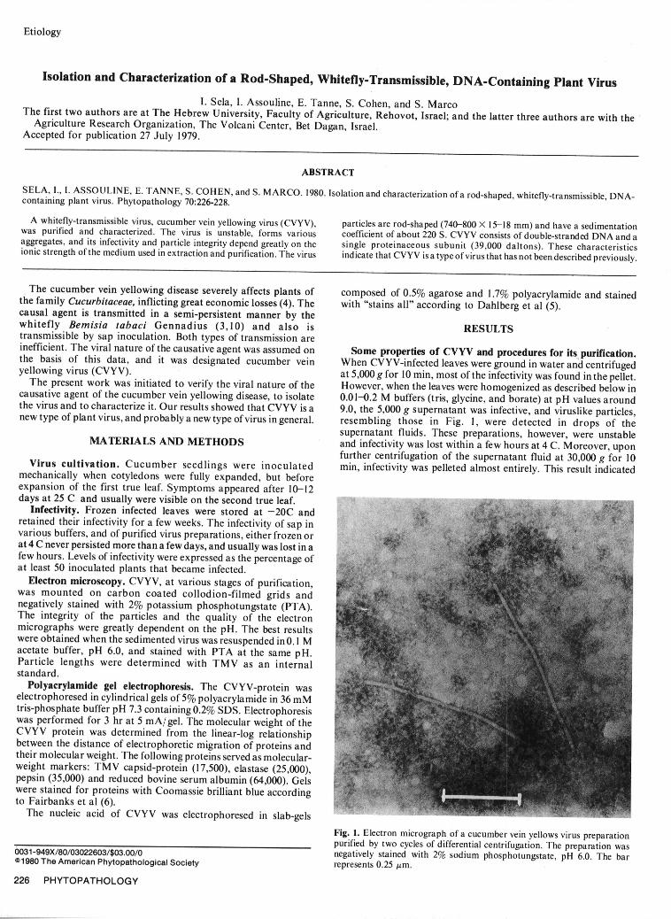
The cucumber vein yellowing disease severely affects plants of composed of 0.5% agarose and 1.7% polyacrylamide and stainedthe family Cucurbitaceae, inflicting great economic losses (4). The with "stains all" according to Dahlberg et al (5).causal agent is transmitted in a semi-persistent manner by thewhitefly Bemisia tabaci Gennadius (3,10) and also is RESULTStransmissible by sap inoculation. Both types of transmission areinefficient. The viral nature of the causative agent was assumed on Some properties of CVYV and procedures for its purification.the basis of this data, and it was designated cucumber vein When CVYV-infected leaves were ground in water and centrifugedyellowing virus (CVYV). at 5,000 g for 10 min, most of the infectivity was found in the pellet.The present work was initiated to verify the viral nature of the However, when the leaves were homogenized as described below incausative agent of the cucumber vein yellowing disease, to isolate 0.01-0.2 M buffers (tris, glycine, and borate) at pH values aroundthe virus and to characterize it. Our results showed that CVYV is a 9.0, the 5,000 g supernatant was infective, and viruslike particles,new type of plant virus, and probably a new type of virus in general. resembling those in Fig. 1, were detected in drops of the
supernatant fluids. These preparations, however, were unstableMATERIALS AND METHODS and infectivity was lost within a few hours at 4 C. Moreover, uponfurther centrifugation of the supernatant fluid at 30,000 g for 10Virus cultivation. Cucumber seedlings were inoculated min, infectivity was pelleted almost entirely. This result indicated
mechanically when cotyledons were fully expanded, but beforeexpansion of the first true leaf. Symptoms appeared after 10-12days at 25 C and usually were visible on the second true leaf.
Infectivity. Frozen infected leaves were stored at -20C andretained their infectivity for a few weeks. The infectivity of sap invarious buffers, and of purified virus preparations, either frozen orat 4 C never persisted more than a few days, and usually was lost in afew hours. Levels of infectivity were expressed as the percentage ofat least 50 inoculated plants that became infected.
Electron microscopy. CVYV, at various stages of purification,was mounted on carbon coated collodion-filmed grids andnegatively stained with 2% potassium phosphotungstate (PTA).The integrity of the particles and the quality of the electronmicrographs were greatly dependent on the pH. The best resultswere obtained when the sedimented virus was resuspended in 0.1 Macetate buffer, pH 6.0, and stained with PTA at the same pH.Particle lengths were determined with TMV as an internalstandard.
Polyacrylamide gel electrophoresis. The CVYV-protein waselectrophoresed in cylindrical gels of 5% polyacrylamide in 36 mMtris-phosphate buffer pH 7.3 containing 0.2% SDS. Electrophoresiswas performed for 3 hr at 5 mA/gel. The molecular weight of theCVYV protein was determined from the linear-log relationshipbetween the distance of electrophoretic migration of proteins andtheir molecular weight. The following proteins served as molecular-weight markers: TMV capsid-protein (17,500), elastase (25,000),pepsin (35,000) and reduced bovine serum albumin (64,000). Gelswere stained for proteins with Coomassie brilliant blue accordingto Fairbanks et al (6).
The nucleic acid of CVYV was electrophoresed in slab-gels
Fig. 1. Electron micrograph of a cucumber vein yellows virus preparationpurified by two cycles of differential centrifugation. The preparation was0031-949X/80/03022603/$03.00/0 negatively stained with 2% sodium phosphotungstate, pH 6.0. The bar
01980 The American Phytopathological Society represents 0.25 ym.
226 PHYTOPATHOLOGY
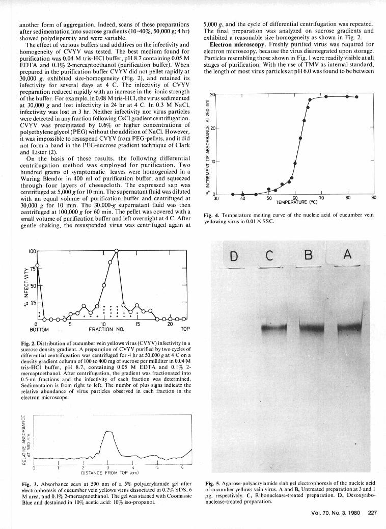
another form of aggregation. Indeed, scans of these preparations 5,000 g, and the cycle of differential centrifugation was repeated.after sedimentation into sucrose gradients (10-40%, 50,000 g; 4 hr) The final preparation was analyzed on sucrose gradients andshowed polydispersity and were variable, exhibited a reasonable size-homogeneity as shown in Fig. 2.
The effect of various buffers and additives on the infectivity and Electron microscopy. Freshly purified virus was required forhomogeneity of CVYV was tested. The best medium found for electron microscopy, because the virus disintegrated upon storage.purification was 0.04 M tris-HCl buffer, pH 8.7 containing 0.05 M Particles resembling those shown in Fig. 1 were readily visible at allEDTA and 0.1% 2-mercaptoethanol (purification buffer). When stages of purification. With the use of TMV as internal standard,prepared in the purification buffer CVYV did not pellet rapidly at the length of most virus particles at pH 6.0 was found to be between30,000 g, exhibited size-homogeneity (Fig. 2), and retained itsinfectivity for several days at 4 C. The infectivity of CVYVpreparation reduced rapidly with an increase in the ionic strength 30of the buffer. For example, in 0.08 M tris-HC1, the virus sedimentedat 30,000 g and lost infectivity in 24 hr at 4 C. In 0.3 M NaCl, C0infectivity was lost in 3 hr. Neither infectivity nor virus particleswere detected in any fraction following CsCl gradient centrifugation.CVYV was precipitated by 0.6% or higher concentrations of 20-polyethylene glycol (PEG) without the addition of NaCl. However, zit was impossible to resuspend CVYV from PEG-pellets, and it did Crnot form a band in the PEG-sucrose gradient technique of Clark 0and Lister (2).
On the basis of these results, the following differential 010-centrifugation method was employed for purification. Two !hundred grams of symptomatic leaves were homogenized in a I
wWaring Blendor in 400 ml of purification buffer, and squeezed a:through four layers Of cheesecloth. The expressed sap was zcentrifuged at 5,000 g for 10 min. The supernatant fluid was diluted 1 o.with an equal volume of purification buffer and centrifuged at 30 40 50 60 70 80 9030,000 g for 10 min. The 30,000-g supernatant fluid was then TEMPERATURE (0C)
centrifuged at 100,000 g for 60 min. The pellet was covered with asmall volume of purification buffer and left overnight at 4 C. After Fig. 4. Temperature melting curve of the nucleic acid of cucumber veingentle shaking, the resuspended virus was centrifuged again at
100 ,o,, / ''D C B A>-
L) 50LiiLLz
-~25-
4+ + + + 4
0 5 10 15 20BOTTOM FRACTION NO. TOP
Fig. 2. Distribution of cucumber vein yellows virus (CVYV) infectivity in asucrose density gradient. A preparation of CVYV purified by two cycles of .differential centrifugation was centrifuged for 4 hr at 50,000 g at 4 C on adensity gradient column of 100 to 400 mg of sucrose per milliliter in 0.04 Mtris-HCI buffer, pH 8.7, containing 0.05 M EDTA and 0.1% 2-mercaptoethanol. After centrifugation, the gradient was fractionated into0.5-ml fractions and the infectivity of each fraction was determined.Sedimentaion is from right to left. The numbr of plus signs indicate therelative abundance of virus particles observed in each fraction in theelectron microscope.
0Uf)E
0 1 2 3 .456
DISTANCE FROM TOP (cm)
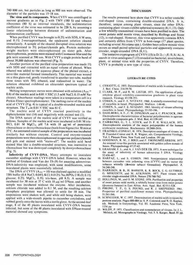
Fig. 3. Absorbance scan at 590 nm of a 5% polyacrylamide gel after Fig. 5. Agarose-polyacrylamide slab gel electrophoresis of the nucleic acidelectrophoresis of cucumber vein yellows virus dissociated in 0.2% SDS, 6 of cucumber yellows vein virus. A and B, Untreated preparation at 3 and IM urea, and 0.1% 2-mercaptoethanol. The gel was stained with Coomassie pg, respectively. C, Ribonuclease-treated preparation. D, Desoxyribo-Blue and destained in 10% acetic acid: 10% iso-propanol. nuclease-treated preparation.
Vol. 70, No. 3,1980 227
740-800 nm, but particles as long as 900 nm were observed. The DISCUSSIONdiameter of the particles was 15-18 nm.
The virus and its components. When CVYV was centrifuged in The results presented here show that CVYV is a rather unstablesucrose gradients as in Fig. 2 with TMV (180 S) and tobacco rod-shaped virus, containing double-stranded DNA. It is,ribosomes (80 S) as internal standards, the sedimentation therefore, unique among plant viruses, since the other DNA-coefficient of the virus was estimated to be 220 S, by assuming a containing plant viruses exhibit spherical symmetry (8,11,13). Onlylinear relationship between distance of sedimentation and a few whitefly-transmitted viruses have been purified to date. Thesedimentation coefficient. sweet potato mild mottle virus, described by Hollings and Stone
When purified CVYV was brought to 0.2% with SDS, 6 M urea, (12), is rod-shaped. However, this virus apparently contains RNAand 0.1% 2-mercaptoethanol, the opalescent solution cleared and certainly differs from CVYV in host-range (17 plant species,immediately. A sample of the cleared disrupted virus solution was including several Nicotiana spp.). Golden bean yellow mosaic viruselectrophoresed in 5% polyacrylamide gels. Protein molecular- occurs as small paired spherical particles and apparently containsweight markers were electrophoresed on sister gels. After circular, single-stranded DNA (8,11).electrophoresis, proteins were stained with Coomassie blue and the The tentative cryptogram of CVYV (15) is D/ 2;-/ -;E/ E; S/Al. Agels were scanned at 590 nm. With CVYV a single protein band of search in virus catalogues (7,13) revealed no bacterial, invertebrate,about 39,000 daltons was observed (Fig. 3). plant, or animal virus with the properties of CVYV. Therefore,
Another portion of the purified virus preparation was made 1% CVYV is probably a new type of virus.with SDS and extracted with an equal volume of phenol. Whenethanol was added to the aqueous phase of the phenol extract, asieve-like material formed immediately. This material was woundon a thin glass-rod, gently transferred to another test tube, washed LITERATURE CITEDthree times with 70% ethanol, and redissolved in water. Thesolution obtained exhibited an ultraviolet spectrum typical of 1. CERIOTTI, G. 1955. Determination of nucleic acids in animal tissues.nucleic acids. J. Biol. Chem. 214:59-70.
Melting temperature curves were obtained with solution (A260 = 2. CLARK, M. F., and R. M. LISTER. 1971. The application of poly-0.6) of the nucleic acid in 0.01 X SSC (1.5 mM NaCI; 0.15 mM Na- ethylene glycol solubility-concentration gradients in plant viruscitrate). Absorbance readings were taken in a temperature cell in a research. Virology 43:338-351.Perkin-Elmer spectrophotometer. The melting curve of the nucleic 3. COHEN, S., and F. E. NITZANY. 1960. A whitefly-transmitted virusacid of CVYV (Fig. 4) is typical of a double-stranded nucleic acid of cucurbits in Israel. Phytopathol. Mediterr. 1:44-46.structure. The Tm in 0.01 X SSC was 61 C. 4. COHEN, S., and F. E. NITZANY. 1963. Identity of viruses affecting
The nucleic acid of CVYV reacted positively in the cucurbits in Israel. Phytopathology 53:193-196.diphenylamine test (14) and negatively with orcinol test (1). 5. DAHLBERG, A. E., C. W. DINGMAN, and A. C. PEACOCK, 1969.Electrophoretic characterization of bacterial polyribosomes in agarose-
The DNA nature of the nucleic acid of CVYV was verified as acrylamide composite gels. J. Mol. Biol. 41:139-147.follows. Samples of the nucleic acid were incubated in 0.01 M tris- 6. FAIRBANKS, G., T. L. STECK, and D. F. H. WALLACH. 1971.HCI, 0.01 M MgC12, pH 7.6 with 10 j•g/ml of pancreatic Electrophoretic analysis of the major polypeptides of the humanribonuclease or deoxyribonuclease (both from Sigma) for 30 min at erythrocyte membrane. Biochemistry 10:2606-2616.37 C. An untreated control sample of the preparation was incubated 7. FRAENKEL-CONRAT, H. 1974. Descriptive catalogue of viruses. in:similarly but without enzyme. Control and enzyme-treated H. Fraenkel-Conrat and R. R. Wagner, eds. Comprehensive Virology,preparations were then electrophoresed in agarose-polyacrylamide Vol. I. Plenum Press. New York and London. 191 pp.slab gels and stained with "stains-all". The nucleic acid band 8. GOODMAN, R. M., J. BIRD, and P. THONGMEEARKOM. 1977.slaib lugelsandstained wt "ou straindeds- .thre, w inulensiccide bd An unusual virus-like particle associated with golden yellow mosaic ofstained blue like a double-stranded structure, was insensitive to beans. Phytopathology 67:37-42.ribonuclease but was destroyed completely by deoxyrobonuclease 9. GRAHAM, F. L., and A. J. VAN DER EB. 1973. A new technique for(Fig. 5). the assay of infectivity of human adenovirus 5 DNA. Virology
Infectivity of CVYV-DNA. Many attempts to inoculate 52:456-467.cucumber seedlings with CVYV-DNA failed. However, when the 10. HARPAZ, 1., and S. COHEN. 1965. Semipersistent relationshipmethod of Graham and Van der Eb (9) for assaying adenovirus- between cucumber vein yellowing virus (CVYV) and its vector theDNA-infectivity was employed, with some modifications, some tobacco whitefly (Bemisia tabacci Gennadius). Phytopathol. Z.inoculated plants were successfully infected. 54:240-248.
The DNA of CVYV (A260 = 10) was dialysed against a modified 11. HARRISON, B. B., H. BARKER, K. R. BOCK, E. G. GUTHRIE,G. MEREDITH, and M. ATKINSON. 1977. Plant viruses withTBS buffer (0.8 NaC1; 0.04% KCI; 0.012% Na 2HPO 4"2 H 20; 0.1% circular single-stranded DNA. Nature 270:760-762.glucose; 0.2% MgCI 2, 0.3% tris-base, pH 8.5). A sample was 12. HOLLINGS, M., and 0. M. STONE. 1976. Purification and propertiesincubated for 30 min at 37 C with 10 Mg/ml DNase, and another of sweet potato mild mottle, a whitefly borne virus from sweet potatosample was incubated without the enzyme. After incubation, (Ipomoea batatas) in East Africa. Ann. Appl. Biol. 82:511-528.calcium chloride was added to 0.1 M, and the resulting calcium 13. PIRONE, T. P., G. S. POUND, and R. J. SHEPHERD. 1961.phosphate precipitate was allowed to settle for 10 min. The Properties of purified cauliflower mosaic virus. Phytopathologysuspensions were shaken well, and while still being shaken, were 51:541-546.applied with a wide-mouth pipette to cucumber cotyledons, and 14. SCHNEIDER, W. C. 1957. Determination of nucleic acids in tissues byrubbed gently onto the leaves with a sterile glove. At the second-leaf pentose analysis. Pages 680-684 in: S. P. Colowick and N. 0. Kaplan,
eds. Methods in Enzymology, Vol. IlL Academic Press, New York.stage, 8 of the 45 plants inoculated with CVYV-DNA became 1154 pp.infected, but none of the 48 plants inoculated with DNase-treated 15. WILDY, P. 1971. Classification and nomenclature of viruses. In: J. L.material showed any symptoms. Melnick, ed. Monographs in Virology, Vol. 5. S. Karger, Basel. 81 pp.
228 PHYTOPATHOLOGY