Embed Size (px)
Citation preview

THE JOURNAL OF BIOLOGICAL CHEMISTRY 0 1988 by The American Society for Biochemistry and Molecular Biology. Inc.
Vol. 263, No. 15, Issue of May 25, pp. 7000-7006,1988 Printed in U. S. A.
Isolation and Characterization of a Chondroitin Sulfate-degrading Endo-&glucuronidase from Rabbit Liver*
(Received for publication, July 21, 1987)
Keiichi Takagaki, Toshiya Nakamura, Mitsuo MajimaS, and Masahiko Endo From the Department of Biochemistry, Hirosaki University School of Medicine, 5 Zaifu-cho, Hirosaki 036, Japan
An endo-&glucuronidase acting on chondroitin sul- fate was isolated from rabbit liver and purified about 550-fold, using a combination of ammonium sulfate fractionation, DEAE-cellulose chromatography, gel filtration on Sephacryl 5-300, affinity chromatogra- phy through heparin-Sepharose CL-GB and prepara- tive polyacrylamide gel electrophoresis. The pH opti- mum of this enzyme was 4.0 and the K , value 7 X M for chondroitin sulfate (Mr 40,000). The isoelectric point of the enzyme was found to be at pH 5.4. The molecular weight, estimated by gel filtration through Sephacryl S-200 and by sodium dodecyl sulfate-poly- acrylamide gel electrophoresis, was 35,000. This en- zyme, which was found in the liver, kidney, spleen, and lung, hydrolyzed the glucuronyl galactose linkage of the linkage region of chondroitin sulfate possessing a very small peptide segment. The enzyme did not hydrolyze proteoglycan. It was concluded that an endo- &glucuronidase is involved in the catabolism of pro- teoglycan chondroitin sulfates.
The catabolism of proteoglycans in tissues has not yet been fully elucidated. However, it seems likely that initially the protein moiety is digested by proteolytic enzymes, liberating glycosaminoglycan chains bearing small peptides. In turn, the glycosaminoglycan chains may be degraded by exoglycosi- dases such as /?-glucuronidase and /3-N-acetylhexosaminidase, resulting in stepwise removal of the monosaccharide residues from the nonreducing termini. In addition, endoglycosidases, as exemplified by hyaluronidase, cleave the internal bonds of the glycosaminoglycan chains, increasing the number of target sites available for exoglycosidases (1).
Endoglucuronidases, which cleave the nonterminal internal uronide linkages of glycosaminoglycan chains, have been found in leech (2), human placenta (3), human blood platelets (4,5), mouse mastocytoma (6), metastatic melanoma cells (71, rat liver cells (8), and in rat ovarian granulosa cells (9). However, the presence of an endoglucuronidase acting specif- ically on chondroitin sulfates has not hitherto been reported.
Majima et al. (10-12) and Matsue and Endo (13) showed that urinary chondroitin sulfate and chondroitin bear in part either glucuronic acid or iduronic acid residues at the reducing termini. These observations demonstrated that nonterminal
* This work was supported by Grants-in-Aid 61219002 for Scien- tific Research, 60127010 for Special Project Research, Inborn Errors of Metabolism, and 61304063, Glycoconjugates as Markers of Cell Function, from the Ministry of Education, Science and Culture of Japan, and from the Karhoji Medical Foundation. The costs of publication of this article were defrayed in part by the payment of page charges. This article must therefore be hereby marked “aduer- tisement” in accordance with 18 U.S.C. Section 1734 solely to indicate this fact.
$ Present address: Kushiro Junior College, Kushiro 085, Japan.
internal glucuronide and iduronide linkages of chondroitin sulfates were cleaved in tissues.
Recently Takagaki et al. (14) found that Ch-S’-degrading endo-/?-glucuronidase activity was present in rabbit liver. Apparently, this enzyme cleaves the nonterminal internal glucuronide linkage of a chondroitin sulfate chain and exposes a new glucuronic acid residue at its reducing terminus. The present report describes the purification and characterization of this enzyme.
EXPERIMENTAL PROCEDURES
Materials Japanese White rabbits (12-month-old male) were supplied by the
Institute for Animal Experiments, Hirosaki University School of Medicine. Sephadex G-15, G-25, and G-100, Sephacryl S-200 and S- 300, Sepharose CL-2B and CL-GB, heparin-Sepharose CL-GB, chro- matofocusing gel PBE 94, Polybuffer 74, and calibration kits for gel filtration and electrophoresis were purchased from Pharmacia Fine Chemicals, Uppsala, Sweden. AG 5OW-X8 (200-400 mesh) and AG 1-X2 (200-400 mesh) were from Bio-Rad, DEAE-cellulose (DE-32) was obtained from Whatman Chemical Separation Ltd. (Maidstone, England), and Diaflow PM-10 membrane for ultrafiltration was pur- chased from Amicon Corp. (Lexington, MA).
Bovine testicular hyaluronidase, bovine liver &glucuronidase, jack bean p”N-acetylhexosaminidase, Escherichia coli @-galactosidase, and Dactylium dendroides galactose oxidase were purchased from Sigma. Pronase P from Streptomyces griseus was obtained from Kaken Kagaku Co., Tokyo, Japan. Chondroitinase AC from Arthrobacter aurescens and 4,5-unsaturated disaccharides (ADi-4S, ADi-6S, and ADi-OS) were purchased from Seikagaku Kogyo Co., Tokyo, Japan. Streptomyces hyaluronidase from S. hyalurolyticus was purchased from Amano Pharmaceutical Co. Ltd., Nagoya, Japan.
The p-nitrophenyl derivatives of @-glucuronic acid, @-galactose, @- xylose, 0-N-acetylgalactosamine, 8-N-acetylglucosamine, and inor- ganic sulfate were purchased from Sigma. Phenyl-a-L-iduronide was purchased from Zeria Shinyaku Kogyo Co. Ltd., Tokyo. Glucuronic acid, gulonic acid, idonic acid, gulonolactone, and idonolactone were described previously (10). Sodium [3H]horohydride (specific activity, 360 mCi/mmol) was purchased from Du Pont-New England Nuclear. Other reagents were of analytical grade.
Analytical Methods
Chemical Analysis-Total hexuronic acid was determined by the method of Bitter and Muir (15). Free hexuronic acid and the reducing terminal hexuronic acid of glycosaminoglycans were determined by a
The abbreviations used are: Ch-S, chondroitin sulfate; Ch-64, chondroitin 6-sulfate; Ch-4-S, chondroitin 4-sulfate; proteo-Ch-6-S, proteoglycan chondroitin 6-sulfate; peptide-Ch-6-S, peptidoglycan chondroitin 6-sulfate; SDS, sodium dodecyl sulfate; PAGE, polyacryl- amide gel electrophoresis; ADi-4S, 2-acetamido-2-deoxy-3-0-(&~- gluco-4-enepyranosyluronic acid)-4-O-sulfo-D-galactopyranose; ADi- 6S, 2-acetamido-2-deoxy-3-O-(~-~-gluco-4-enepyranosyluronic acid)- 6-0-sulfo-D-galactopyranose; ADi-OS, 2-acetamido-2-deoxy-3-0-(@- D-g~uco-4-enepyranosy~uronic acid)-D-galactopyranose. All sugars mentioned in this paper are of D configuration except for iduronic acid, gulonic acid, gulonolactone, and idonolactone which are of L configuration.
7000

Chondroitin Sulfate-degrading Endo-/3-glucuronidase 7001
modification (11) of the method of Milner and Avigad (16). Among the constituent monosaccharides of the glycosaminoglycans, hexu- ronic acids showed a high color yield by this method, while xylose (0.4%), galactose (0.1%), and N-acetylhexosamine (0%) yielded a negligible degree of color. However, the color yield of the hexuronic acid at the reducing termini of oligosaccharide (70%) was less than that of free hexuronic acids (100%) (11). The N-acetylhexosamine content of the nonreducing termini of glycosaminoglycans was deter- mined by the method of Reissig et at. (17). Protein was determined by the method of Lowry et al. (18) using bovine serum albumin as a standard.
Preparation of Glycans
Proteo-Ch-6-S-Proteo-Ch-6-S was extracted from human umbil- ical cord and purified by a modification of the method of Danishefsky and Abraham (19). Protease inhibitors (EDTA and phenylmethylsul- fonyl fluoride) were used throughout the preparation. To remove contaminating hyaluronic acid, the proteo-Ch-6-S preparations were treated with Streptomyces hyaluronidase by the method of Ohya and Kaneko (20), followed by chromatography using a Sephacryl S-300 column. Proteo-Ch-6-S prepared as above was dialyzed against water and lyophilized.
Peptide-Ch-6-S-To obtain a Ch-6-S chain attached to the very small segment of a peptide, the proteo-Ch-6-S was digested with Pronase in 0.1 M Tris-HC1 buffer (pH 8.0) containing 10 mM CaC12 at 37 "C for 24 h. After digestion, the peptide-Ch-6-S was separated by DEAE-cellulose column chromatography and then purified by Sephacryl S-300 chromatography.
Reduced Ch-6-S-The peptide-Ch-6-S was reduced with 1.0 M sodium borohydride in 0.05 M NaOH at 45 "C for 24 h. The reaction was terminated by adjusting the pH to 4.0 with 2 M acetic acid. The resulting reaction mixture was passed through AG 50W-X8 (H' form) and then evaporated to dryness five times with methanol. The dried eluate was dialyzed and lyophilized. The reduced Ch-6-S, which had xylitol at the reducing terminus of the Ch-6-S chain, was used as a substrate for endo-@-glucuronidase.
Oligosaccharides from Ch-6-S-Tetra- and hexasaccharides, (4GlcA@1-3GalNA~(6-OSO;)@l-)~ and (4GlcA@1-3GalNAc (6- OSO;)@l-),, were prepared from Ch-6-S digested with testicular hy- aluronidase, as described by Knudson et al. (21).
Other Glycosami~oglycans-Ch-6-S from shark cartilage and Ch- 4-S from whale cartilage were purchased from Seikagaku Kogyo Co. Hyaluronic acid was prepared from human umbilical cord by the method of Danishefsky and Bella (22), and chondroitin was prepared from Ch-6-S by the method of Kantor and Schubert (23). The reducing termini of glycosaminoglycans were reduced as described above.
3H Labeling of Glycans Preparation of Ch-6-S-[3H]Xylitol-Proteo-Ch-6-S (1 mg) was
dissolved in 0.2 ml of 0.05 M NaOH, 1.0 M sodium borohydride containing 1.0 M sodium [3H]borohydride (2.5 mCi). The resulting mixture was kept a t 45 "C for 24 h. The reaction was terminated by adjusting the pH to 4.0 with 2 M acetic acid. The resulting reaction mixture was passed through a 0.5 X 2-cm column of AG 50W-X8 (H' form) and repeatedly evaporated to dryness with methanol five times.
3H Labeling of Reducing Terminal of Glycans-To tritiate the reducing termini of glycans, which were exposed newly after enzyme digestion, the glycans were reduced with 1.0 M sodium [3H]borohy- dride in 0.1 M borate buffer (pH 8.0) at 22 "C for 3 h. The reaction was terminated by adjusting the pH to 4.0 with 2 M acetic acid. The reaction mixture was evaporated to dryness under reduced pressure, and boric acid was removed by repeated evaporations with methanol.
Labeling of Galactose at Nonreducing Terminus-The nonre- ducing terminus of the oligosaccharide fraction derived from the linkage region of peptide-Ch-6-S was tritiated using a slight modifi- cation of the method of Morel1 et al. (24) as follows. The oligosaccha- ride fraction dissolved in 0.1 M phosphate buffer (pH 7.0) was incu- bated with galactose oxidase at 37 "C for 24 h, and sodium [3H] borohydride was added. The resulting mixture was then incubated at 22 "C for 3 h. Salt and boric acid were removed as described above.
Enzyme Assays Endo-@-glucuronidase-Enzyme activity was routinely monitored
by colorimetric means using reduced Ch-6-S as a substrate. The reaction mixture consisted of enzyme, the reduced Ch-6-S (2 mg) and 0.1 M acetate buffer (pH 4.0) in a total final volume of 1.0 ml. A drop
of toluene was added as a preservative, and after sealing the mixture was incubated at 37 "C for 18 h. The reaction was terminated by adding 0.25 ml of 25% trichloroacetic acid, and insoluble materials were removed by centrifugation. To the supernatant were added 4 volumes of ethanol saturated with NaCl. The resulting precipitates were collected by centrifugation, washed with ethanol and ether, and dried in uacuo. The dried residue was dissolved in 0.25 ml of water, and the newly exposed glucuronic acid residues at the reducing terminus were quantified by a modification of the method of Milner and Avigad (16). One unit of enzyme activity was defined as the amount of enzyme yielding 1 nmol of glucuronic acid residue at the reducing terminus per 1 h at 37 "C.
Assays for Glycosidase Actiuity-@-Glucuronidase (exo-type), @-N- acetylglucosaminidase, P-N-acetylgalactosaminidase, @-galactosidase, @-xylosidase, and sulfatase were determined using the corresponding p-nitrophenyl derivatives (25). a-L-Iduronidase was measured by the method of Liem and Hooghwinkel (26) using phenyl-a-L-iduronide as a substrate.
Molecular Weight Determination-SDS-polyacrylamide gel elec- trophoresis was performed in 12.5% polyacrylamide gels using 0.75 M Tris-HC1 buffer (pH 8.8) containing 0.2% SDS at 3 mA/tube (27). The gels were stained with Coomassie Brilliant Blue R-250. Molecular weight standards used for calibration were: phosphorylase b (M, = 94,000), albumin ( M , = 67,000), ovalbumin ( M , = 43,000), carbonic anhydrase ( M , = 30,000), trypsin inhibitor ( M r = 20,100), and a- lactoalbumin (Mr = 14,400). Estimation of the molecular weight was also carried out by gel filtration on a Sephacryl S-200 column (1.8 X 103 cm, using 0.005 M phosphate buffer (pH 7.3) containing 0.1 M NaCl and 0.02% sodium azide). Molecular weight standards used for calibration were: catalase (M, = 210,000), aldolase ( M , = 158,000), albumin (M, = 67,000), ovalbumin (M, = 43,000), and chymotrypsin- ogen A ( M , = 25,000).
Paper Chromatography-Paper chromatography was carried out on Whatman No. 1 paper using a descending technique. The following solvents were used solvent 1, 1-butanol/acetic acid/water (50:12:25); solvent 2, 1-butanol/pyridine/water (6:4:3); solvent 3, 1-butanol/ acetic acid/l N ammonium hydroxide (2:3:1); solvent 4, n-butyric acid/0.5 N ammonium hydroxide (5:3); solvent 5, 1-butanol/acetone/ water (2:71), by volume, respectively. Sugars were detected using the alkaline silver nitrate method (28).
Determination of Radwactiuity-Paper strips hearing radioactive derivatives were cut into 1-cm pieces, immersed in scintillation fluid (29), and radioactivities were determined using a liquid scintillation spectrometer (Aloka, Model LSC-683).
Purification of Endo-0-glucuronidase-Unless indicated otherwise, all procedures were conducted at 4 "C; all centrifugation steps were performed at 10,000 X g for 20 min; 0.005 M sodium phosphate buffer (pH 7.4) was used throughout the purification procedures. The en- zyme-containing fraction was concentrated by lyophilization or by an ultrafiltration system using Amicon PM-10 membrane.
Frozen rabbit liver tissue (160 g) was thawed, freed from connective tissue, minced, and homogenized in a Waring blender at 3000 rpm for 1 min in an ice-bath. The phosphate buffer contained 0.1 mM phenylmethylsulfonyl fluoride. The resulting homogenate was ad- justed to pH 7.4, centrifuged, and the supernatant was treated in the manner described below. Protamine sulfate solution (pH 7.4) was added to the supernatant fluid to a final concentration of 0.2%. After 15 min, the mixture was centrifuged and the precipitate discarded. To the supernatant was added solid ammonium sulfate up to 50% saturation. After 15 min the mixture was centrifuged, and the con- centration of ammonium sulfate in the supernatant fluid was in- creased to 70% by further addition of ammonium sulfate. The result- ing mixture was allowed to stand for 1 h, and then the precipitate was collected by centrifugation and dissolved in and dialyzed against phosphate buffer. After dialysis the solution was adjusted to pH 4.0 with 1 M acetic acid, centrifuged, and then the supernatant was dialyzed against the phosphate buffer. The dialyzed solution was applied to a DEAE-cellulose column (3.8 X 60 cm, chloride form) equilibrated with phosphate buffer, and the column was washed with 800 ml of phosphate buffer. The bulk of the enzyme was then eluted from the column in phosphate buffer containing 0.15 M NaCl.
The fractions containing enzyme activity were concentrated using an ultrafiltration system. The concentrated enzyme preparation was applied to a Sephacryl S-300 column (2.2 X 167 cm) previously equilibrated with phosphate buffer and the column developed with the same buffer (Fig. 1). Fractions from 89 to 99 were pooled, dialyzed against phosphate buffer, and concentrated. The concentrated retent was applied to a heparin-Sepharose CL-GB column (1.8 X 12 cm)

7002 Chondroitin Sulfate-degrading Endo-@-glucuronidase
FRACTION NUMBER
FIG. 1. Gel filtration of crude endo-&glucuronidase on Sephacryl S-300. The pooled fractions from the DEAE-cellulose column containing enzyme were combined and applied to a column (2.2 X 167 cm) of Sephacryl S-300. The column was eluted with 0.005 M phosphate buffer (pH 7.4). The flow rate was 16 ml/h, and 10-ml fractions were collected. 0, absorbance at 280 nm; 0, enzyme activity.
TABLE I Purification of endo-&glucuronida.se from rabbit liver
mg units unitsfmg '% -fold Crude extract 15,600 4,990 0.32 100 1.0 Protamine 9,200 5,200 0.57 104 1.8 Ammonium 5,740 4,250 0.74 85 2.3
pH 4 treatment 2,490 3,040 1.22 61 3.8 DEAE-cellu- 533 1,740 3.26 35 10.2
Sephacryl S- 91 1,447 15.97 29 49.9
Heparin-Seph- 26.3 947 36.04 19 112.6
Preparative 1.7 299 175.88 6 549.6
sulfate
lose
300
arose CL-GB
PAGE
equilibrated with phosphate buffer, and the column washed with the same buffer. The enzyme was then eluted from the column with phosphate buffer containing 0.15 M NaCI. The fractions containing enzyme activity were pooled, dialyzed against phosphate buffer, and concentrated. The concentrated enzyme preparation was subjected to polyacrylamide gel electrophoresis in 7.5% gels in glass tubes (0.5 X 10 cm) as described by Davis (30). Electrophoresis was carried out at 4 "C for 4 h a t 2 mA/tube until bromphenol blue used as a marker had migrated to the end of the gel. The gels were either stained with Coomassie Brilliant Blue and used as a guide or cut into 2-mm-thick slices, each of which was extracted individually for 24 h with phos- phate buffer, and the enzyme activity was measured in each fraction. Fractions containing enzyme activity were combined and used as the most highly purified enzyme preparation.
RESULTS
Purification
The overall purification scheme is summarized in Table I. A 550-fold purification was achieved. Analysis of the heparin- Sepharose CL-GB affinity chromatography fraction by PAGE revealed the presence of three Coomassie Brilliant Blue- stained bands. After the preparative gel electrophoresis step, only a single band was observed on PAGE (Fig. 2). The following enzyme activities were not detected: @-glucuronidase (exo-type), a-L-iduronidase, j3-galactosidase, @-N-acetylgalac- tosaminidase, /3-N-acetylglucosaminidase, sulfatase, and hy- aluronidase (data not shown).
(3 . I . ,
FIG. 2. Polyacrylamide gel electrophoretogram of the pu- rified enzyme preparation. The enzyme preparation was subjected to polyacrylamide gel electrophoresis in 7.5% gels (0.5 X 10 cm) at 4 "C for 4 h at 2 mA/tube. The gel was stained for protein with Coomassie Brilliant Blue.
pH
FIG. 3. Effect of pH on endo-&glucuronidase activity. A standard spectrophotometric assay was done at 37 "C except that a final concentration of 0.1 M was used for both glycine buffer (pH 2.0- 3.5) and sodium acetate buffer (pH 3.0-6.5), in the presence of 0.2 M NaCl(0) and in the absence of NaCl(0).
Properties of Endo-@-glucuronidase
Stability-After incubation in 0.1 M acetate buffer (pH 4.0) at 50 "C for 30 min, 80% of the enzyme activity was lost. The enzyme was stable at 4 "C for at least 5 days in phosphate buffer but retained full activity when stored at 4 "C for 1 month in phosphate buffer containing 20% glycerol. At -20 "C it was stable for about 2 months.
Effect of pH-The optimum pH for the enzyme activity approximated pH 4.0 (Fig. 3). In the range from pH 2.0 to 6.5, addition of 0.2 M NaCl reduced the enzyme activity (60% at pH 4).
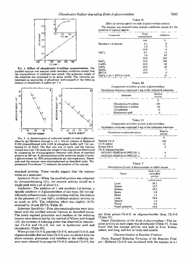
Effect of Substrate Concentration-As shown in Fig. 4, the velocity of the enzyme reaction changed depending on the concentration of reduced Ch-6-S. Since the molecular weight of the reduced Ch-6-S was about 40,000, as calculated by gel filtration through Sephacryl S-200 and S-300, the K,,, value was estimated to be 7 x M by Lineweaver-Burk plot.
Moleculcrr Weight-The molecular weight of the native enzyme approximated 35,000 (Fig. 5 ) as determined by gel filtration on Sephacryl S-200 and SDS-PAGE calibrated with
Chondroitin Sulfate-degra Iding Endo-&glucuronidase 7003
TABLE I1 Effect of various agents on endo-@-glucuronidase actiuity
The enzyme was assayed under normal conditions except for the presence of various agents.
-200 0 200 400 600 8M) I/S (mM ‘ 1
FIG. 4. Effect of chondroitin 6-sulfate concentration. The purified enzyme was assayed under standard conditions except that the concentration of substrate was varied. The molecular weight of the substrate was estimated to be about 40,000. The velocities are expressed as nanomoles of glucuronic acid exposed at the reducing termini of chondroitin 6-sulfate per 1 h.
0 0.2 0.4 0.6 0.8 1.0 FRACTION NUMBER RELATIVE MOBILITY
FIG. 5. A , determination of molecular weight of endo-@-glucuron- idase by gel filtration through a 1.8 X 103-cm column of Sephacryl S-200 preequilibrated with 0.005 M phosphate buffer (pH 7.4) con- taining 0.1 M NaCl. The flow rate was 12 ml/h, and the fraction volume was 3 ml. The molecular weight of the enzyme was determined by comparing its chromatographic behavior with those of standard proteins at 280 nm. B, determination of the molecular weight of endo- @-glucuronidase by SDS-polyacrylamide gel electrophoresis. Stand- ards and the enzyme were electrophoresed as described under “Ex- perimental Procedures.” 0 indicates the position of the enzyme.
standard proteins. These results suggest that the enzyme exists as a monomer.
Isoelectric Point-When the purified protein was subjected to chromatofocusing (31), the enzyme activity eluted as a single peak with a PI of about 5.4.
Inhibitors-The addition of 1 mM saccharo-1,4-lactone, a specific inhibitor of @-glucuronidase of exo-type, did not sig- nificantly influence endo-&glucuronidase activity. Incubation in the presence of 1 mM HgClz inhibited catalytic activity by as much as 43%. The inhibition effect was slightly (21%) reversed by 10 mM EDTA (Table 11).
Substrate Specificity-Four glycosaminoglycans were incu- bated with the purified enzyme fraction as described above. The newly exposed glucuronic acid residues at the reducing termini were determined by the method of Milner and Avigad (16). An increase of reducing power was observed in commer- cial Ch-6-S and Ch-443, but not in hyaluronic acid and chondroitin (Table 111).
When proteo-Ch-6-S, peptide-Ch-6-S, reduced Ch-63, and oligosaccharides derived from Ch-6-S were incubated with the above enzyme, glucuronic acid residues at the reducing ter- mini were released from peptide-Ch-6-S, reduced Ch-6-S, but
Compound concentration Inhibition Final
SnCl, c u s o 4 CdCl, ZnSO, EDTA HgCI, (1.0) + EDTA (10.0)
rnM
0.1 1.0
10.0 0.1 1.0
10.0 10.0 10.0 10.0 10.0 10.0
% 0 0 0
14 43
100 100 40 25 7 0
21
TABLE 111 Comparatiue activities of endo-(3-glucuronidase
Incubation mixtures contained 2 mg of the indicated substrate.
Substrate Relative activitv
%
Chondroitin 6-sulfate 100 Chondroitin 4-sulfate 84 Chondroitin 12 Hyaluronic acid 0
TABLE IV Comparatiue activities of endo-P-glucuronidase
Incubation mixtures contained 2 mg of the indicated substrate.
Chondroitin 6-sulfate derivatives Relative activity
% Peptide-Ch-S 100 Ch-S-xylitol 41 Proteo-Ch-S 3 Ch-S-oligosaccharides
( ~ G ~ C A @ ~ - ~ G ~ ~ N A C ( ~ - O S O & ~ ~ - ) ~ 0 (4GlcA@l-3GalNAc(6-OSO~~@l-)3 0
TABLE V Distribution of endo-P-glucuronidase in rabbit tissues
Tissue Endo-P-giu- curonidase
activity
unitsfg tissue Liver 30.7 Spleen 16.7 Kidney 36.3 Brain 3.9 Heart 1.1 Lung 30.4 Muscle 1.3 Testis 8.2 Small intestine 10.8
not from proteo-Ch-6-S, or oligosaccharides from Ch-6-S (Table IV).
Organ Distribution of the Endo-0-glucuronidase-The en- zyme activity in each organ was determined (Table V). It was found that the enzyme activity was high in liver, kidney, spleen, and lung, and low in brain and muscle.
Characterization of Reaction Products Newly Exposed Reducing Terminus of the Reaction Prod-
uct-Reduced Ch-6-S was incubated with the enzyme in 0.1

7004 Chondroitin Sulfate-degrading Endo-P-glucuronidase
M acetate buffer (pH 4.0) containing 0.1 mM phenylmethyl- sulfonyl fluoride after addition of a drop of toluene as a preservative. After incubation, the reaction was terminated by the addition of trichloroacetic acid. Insoluble materials were removed by centrifugation. The supernatant was washed with ether several times and passed through a Sephadex G- 100 column to remove low molecular weight material. The recovered Ch-6-S was reduced with sodium [3H]borohydride (t3H]Ch-6-S). [3H]Ch-6-S was divided into three portions: two were used for acid hydrolysis, and the third portion was digested with chondroitinase AC. The first aliquot of [3H]Ch- 6-S was hydrolyzed with trifluoroacetic acid and then treated with nitrous acid according to the method of Majima et al. (9). The hydrolysate was passed through AG 50W-X8, and then the nonadsorbed fraction (89% of total radioactivity of [3H]Ch-6-S) was treated with 0.25 M ammonium hydroxide to de-lactonize the reduced hexuronic acid in the hydrolysate, followed by passage through AG 1-X2. The radioactive ma- terial eluted with 2 M acetic acid (78% of total radioactivity of [3H]Ch-6-S) was treated with 1 N HC1 to convert the hexuronic acid back to the lactone. This sample was subjected to descending paper chromatography using solvent 1 (Fig. 6A) . The radioactive area corresponding to standard gulono- lactone was recovered and rechromatographed in solvent 2 (Fig. 6B). A radioactive spot was found that co-migrated with authentic gulonolactone, the compound anticipated from re- duction and lactonization of the product of glucuronic acid. No idonic acid, a derivative of iduronic acid, was detected.
The second aliquot was hydrolyzed directly with 4 N HC1 at 100 "C for 8 h. The hydrolysate was applied to a AG 50W- X8 column, and the 3H-labeled fraction was eluted with 1 N HC1 and subsequently subjected to paper chromatography (solvents 1 and 2). No [3H]galactosaminitol was detected.
The third aliquot of [3H]Ch-6-S was digested with chon- droitinase AC (32) and the digest was chromatographed on a Sephadex G-15 column (Fig. 7) . One radioactive peak (Peak I) with a K,, of 0.32 and another hexuronate-positive peak (Peak 11) with a K,, of 0.48 were observed. Peak I1 was eluted at a position corresponding to that of the standard unsatu- rated disaccharide, ADi-6s. The material included in this peak was identified as ADi-6S by paper chromatography (33). Peak 1 containing the radioactive material was eluted at a position intermediate between a tetrasaccharide derived from Ch-6-S and authentic ADi-6S, actually at the position corre-
OO - IO 20 30 40 DISTANCE (crnl
FIG. 6. Paper chromatographic identification of the aldono- lactone derived from the newly exposed reducing terminus of Ch-6-S liberated by endo-&glucuronidase. The sample was de- veloped on paper. A, solvent l, l-butanol/acetic acid/water (5012:25); and B, solvent 2, 1-butanol/pyridine/water (643). The chromato- gram was cut into 1-cm pieces, and radioactivity of each piece was determined with a liquid scintillation counter. Gulonic acid ( I ) , idonic acid (ZZ), gulonolactone (ZIZ), and idonolactone (IV) served as ref- erence standards.
t I
P a b 1 1
"
a Peak I Peak U
.05 I-
0
'0 40 60 0 - I FRACTION NUMBER
FIG. 7. Degradation of endo-8-glucuronidase-digested Ch- 6-S with chondroitinase AC. Endo-8-glucuronidase-digested Ch- 6-S was reduced with [3H]borohydride and then digested with chon- droitinase AC. The digest was subjected to gel filtration chromatog- raphy through a 1.3 X 145-cm column of Sephadex G-15. Elution was effected at a rate of 6 ml/h. The effluent fractions (2 ml) were analyzed for total hexuronic acid (0) and for radioactivity (0). The elution positions of saturated tetrasaccharide derived from Ch-6-S ( a ) and unsaturated disaccharide ( b ) (ADi-6s) are indicated by arrows.
FRACTION NUMBER
FIG. 8. Degradation of 'H-labeled chondroitin sulfate with endo-#?-glucuronidase. Ch-6-S with [3HJxylitol at the reducing termini was incubated with endo-B-glucuronidase, and subjected to gel filtration chromatography on a 1.1 X 110-cm column of Sephadex G-25. The column was eluted with 0.1 M acetic acid at a rate of 6 ml/ h. The effluent fractions (2 ml) were analyzed for radioactivity before (0) and after (0) digestion, respectively. 1 , galactose; 2, maltose; 3, maltotriose.
sponding to authentic maltotriose. This material was sub- jected to paper chromatography (solvent 3). A radioactive spot was detected that migrated slower than the standard unsaturated disaccharides (ADi-4S, ADi-6S, or ADi-OS). The radioactive material recovered from the paper was hydrolyzed with trifluoroacetic acid, treated with nitrous acid, and then subjected again to paper chromatography (solvent 1). Only gulonic acid was detected. We therefore concluded that the newly exposed reducing terminal sugar was glucuronic acid.
Identification of the Oligosaccharide Released from Ch-6-S by Endo-p-glucuronidase-The oligosaccharide released from the reducing terminal site of Ch-6-S by endo-@-glucuronidase was characterized as follows. Ch-6-S-[3H]xylitol, containing [3H]xylitol at the reducing terminus, was digested with this enzyme and subjected to gel filtration on Sephadex G-25 (Fig. 8). An aliquot of the recovered 3H-labeled oligosaccharide was digested successively with @-N-acetylhexosaminidase (34) and @-glucuronidase (35) and then passed through a Sephadex G- 15 column. The elution profiles before and after digestion with the two enzymes remained unchanged.
A second aliquot of the oligosaccharide was hydrolyzed with 2 N HC1 at 100 "C for 2 h. After removal of HC1 in vacuo, the hydrolysate was reduced with [3H]borohydride and subjected to paper chromatography (solvent 2). Only [3H]galactitol and f3H]xylitol were detected.
A third aliquot was incubated with galactose oxidase and reduced with [3H]borohydride, thereby labeling the galactose residue at the nonreducing terminus of the oligosaccharide

Chondroitin Sulfate-degrading Endo-@-glucuronidase 7005
DISTANCE (cm)
FIG. 9. Paper chromatographic identification of the nonre- ducing terminal sugar of trisaccharide released by digestion with endo-&glucuronidase. The trisaccharide recovered from gel filtration chromatography (Fig. 8) was treated with galactose oxidase followed by reduction with [3H]borohydride. A, product of hydrolysis of the trisaccharide with 2 N HC1 at 100 "C for 2 h. Solvent 5, 1- butanol/acetone/water (2:7:1). B, rechromatography of the sugar recovered from the same area as spot I on Plate A. Solvent 2, 1- butanol/pyridine/water (6:4:3). C, product of digestion with /3-galac- tosidase of trisaccharide. Solvent 5, galactose ( I ) ; xylitol (ZZ).
(24). An aliquot of the resulting compound was desalted, hydrolyzed with 2 N HC1 at 100 "C for 2 h, and subjected to paper chromatography (solvent 4). Only [3H]galactose and [3H]xylitol were detected (Fig. 9, A and B ) . Then, an aliquot of the latter oligosaccharide was digested with @-galactosidase (36) and examined by paper chromatography (solvent 4). Only [3H]galactose and [3H]xylitol were detected (Fig. 9C).
From the results of these experiments we concluded that the endo-8-glucuronidase cleaved the internal glucuronide linkage of a chondroitin sulfate chain and that the glucuronic acid was linked to a galactose residue of the nonreducing terminus of a neutral oligosaccharide. It was further concluded that this oligosaccharide is a trisaccharide because of its elution position on gel filtration, probably consisting of Gal- Gal-Xyl and representing the linkage region between the chondroitin sulfate chain and peptide chain (37).
DISCUSSION
The present Ch-S-degrading endo-@-glucuronidase was pu- rified from rabbit liver, and yielded a single protein band when analyzed by SDS-PAGE. Separation of the endo-@- glucuronidase from glycosidases such as exo-@-glucuronidase, 8-N-acetylhexosaminidase, and hyaluronidase but not @-ga- lactosidase was achieved by gel chromatography on Sephacryl S-300. @-Galactosidase activity was successfully removed by heparin-Sepharose CL-GB chromatography.
Exo-P-glucuronidase from rabbit liver (38) and the endo-@- glucuronidase described in the present paper catalyze hydrol- ysis of the glucuronic acid linkage. However, the endo-@- glucuronidase was different from the exo-P-glucuronidase with regard to the following properties: 1) The endo-P-glucu- ronidase cleaved neither p-nitrophenyl-@-D-glucuronide nor oligosaccharides with a glucuronic acid residue at the nonre- ducing terminus. 2) The present enzyme was partially inhib- ited by NaCl over a wide pH range. 3) The enzyme was not
inhibited by saccharo-1,4-lactone which is an inhibitor of exo- &glucuronidase.
Examination of the specificity of the endo-P-glucuronidase revealed that 1) the enzyme acts on long-chain but not short- chain oligosaccharides of Ch-S; 2) the enzyme exposes glu- curonic acid residues at the reducing terminus of Ch-S; 3) the enzyme requires the presence of the linkage region between the Ch-S and peptide moieties, as demonstrated by the for- mation of an oligosaccharide having a galactose residue at its nonreducing terminus; 4) The enzyme catalyzes hydrolysis of Ch-S attached to a short peptide derived from the core protein of a proteo-Ch-S moiety, but not the proteo-Ch-S complex.
The presence of an endo-@-glucuronidase which acts on Ch- S has not hitherto been reported. Majima et al. (10-12) and Matsue and Endo (13) demonstrated the presence of a glu- curonic acid residue at the reducing termini of a low molecular weight Ch-S and a low-sulfated Ch-S in human urine. These findings strongly suggested that an endo-@-glucuronidase act- ing on Ch-S was present in human tissues, and this was confirmed in the present study.
Endo-@-glucuronidases, which act on hyaluronic acid, hep- arin, and heparan sulfate, have been found in several tissues and cells. Some of these enzymes act on the process of glycosaminoglycan biosynthesis (6, 9), and others act on the process of catabolic degradation of glycosaminoglycans (3,4). We conclude from the specificity of the endo-@-glucuronidase described here that this enzyme is associated with the degra- dation of Ch-S, and that it very likely acts at the early stage of catabolic degradation of proteo-Ch-S.
REFERENCES 1. RodCn, H. (1980) in The Biochemistry of Glycoproteins and Pro-
teoglycans (Lennarz, w. J., ed), 1st ed., pp. 267-371, Plenum Publishing Corp., New York
2. Yuki, H., and Fishman, W. H. (1963) J. Biol. Chem. 238 , 1877- 1879
3. Klein, U., and von Figura, K. (1976) Biochem. Biophys. Res. Commup. 73,569-576
4. Oldberg, A., Heldin, C.-H., Wasteson, A,, Busch, C., and Hook, M. (1980) Biochemistry 19,5755-5762
5. Oosta, G. M., Favreau, L. V., Beeler, D. L., and Rosenberg, R. D. (1982) J. Biol. Chem. 257,11249-11255
6. Thunberg, L., Backstrom, G., Wasteson, A,, Robinson, H. C., Ogren, S., and Lindahl, U. (1982) J. Biol. Chem. 257 , 10278- 10282
7. Nakajima, M., Irimura, T., Di Ferrante, N., and Nicolson, G. L. (1984) J. Bid. Chem.-259,2283-2290 ~
8. Hook, M., Wasteson, A., and Oldberg, A. (1975) Biochem. Bio- phys. Res. Commun. 67,1422-1428
9. Yanagishita, M. (1985) J. Biol. Chem. 260 , 11075-11082 10. Majima, M., Nakamura, T., Igarashi, S., Matsue, H., and Endo,
11. Majima, M., Takagaki, K., Igarashi, S., Nakamura, T., and Endo,
12. Majima, M., Nakamura, T., and Endo, M. (1985) Tohoku J. Exp.
13. Matsue, H., and Endo M. (1987) Biochim. Biophys. Acta 9 2 3 ,
14. Takagaki, K., Nakamura, T., Majima, M., and Endo, M. (1985)
15. Bitter, T., and Muir, H. M. (1962) Anal. Biochem. 4 , 330-334 16. Milner, Y., and Avigad, G. (1967) Carbohydr. Res. 4,359-361 17. Reissig, J. L., Strominger, J. L., and Leloir, L. F. (1955) J. Biol.
18. Lowry, 0. H., Rosebrough, N. J., Farr, A. L., and Randall, R. J.
19. Danishefsky, I., and Abraham, G. (1971) Prep. Biochem. 1, 133-
20. Ohya, T., and Kaneko, Y. (1970) Biochim. Biophys. Acta 198 ,
21. Knudson, W., Gundlach, M. W., Schmid, T. M., and Conrad, H.
M. (1984) J. Biochem. Biophys. Methods 9 , 245-249
M. (1984) J. Biochem. Biophys. Methods 10, 143-151
Med. 146,83-88
470-477
FEBS Lett. 181,271-274
Chem. 2 17,959-966
(1951) J. Biol. Chem. 193, 265-275
140
607-609
E. (1984) Biochemistry 23,368-375

7006 Chondroitin Sulfate-degrading Endo-P-glucuronidase
22. Danishefsky, I., and Bella, A., Jr. (1966) J. Biol. Chem. 241, 29. Patterson, M. S., and Greene, R. C. (1965) Anal. Chem. 37, 854-
23. Kantor, T. G., and Schubert, M. (1957) J. Am. Chem. SOC. 79, 30. Davis, B. J. (1964) Ann. N. Y. ACd. SCi 1217404-427
24, &forell, A, G., van Den Hamer, c, J. A,, Scheinberg, 1. H., and 32. Yamagata, T., Saito, H., Habuchi, o., and Suzuki, s. (1968) J .
25. Barrett, A. J. (1972) in Lysosomes (Dingle, J. T., ed) pp. 110-126, 33’ Oegema7 T’ R.y Jr’7 E’ L‘7 G‘ w’, and Van
26. Liem, K. o-, and Hooghwinkel, G. J. M. (1975) Ch. Chim. Acta 35. L., and Armand, G. (1966) J, B ~ L , them, 241, 65-70 34. Li, Y.-T., and Li, S.-C. (1972) Methods Enzymol. 28, 702-713
27. Laemmli, U. K. (1970) Nature 227, 680-685 37. Hassell, J. R., Kimura, J . H., and Hascall, V. C. (1986) Annu. 28. Trevelyan, W. E., Procter, D. P., and Harrison, J. S. (1950) Rev. Biochem. 55,539-567
143-146 857
152-153 31. Fenger, M. (1982) J. Chromatogo. 240,173-179
Ashwell, G. (1966) J. Bwl. Chem. 241,3745-3749
North-Holland Publishing Co., Amsterdam
Biol. Chem. 243, 1523-1535
T. R. (1984) J. Bid. Chem. 259, 1720-1726
60,259-262 36. Lindahl, U., and Rodbn, L. (1966) J. Bwl. Chem. 241,2113-2119
Nature 166,444-445 38. Dean, R. T. (1974) Biochem. J. 138,395-405