Embed Size (px)
Citation preview
Islamic University of GazaFaculty of MedicineSpring, 2012-2013

o ~1100 genes are thought to be located on X chr.o ~40% are
presently known to be associated with disease phenotypes.
2 Medical Genetics (2012-2013)

o Two female heterozygotes for an X-linked disease may have very different clinical presentations
o Example: Duchenne muscular dystrophyoFemale carriers exhibit typical
mosaic expression
Dystrophin immunostaining
Normal female
Affected male
Carrier female
3 Medical Genetics (2012-2013)
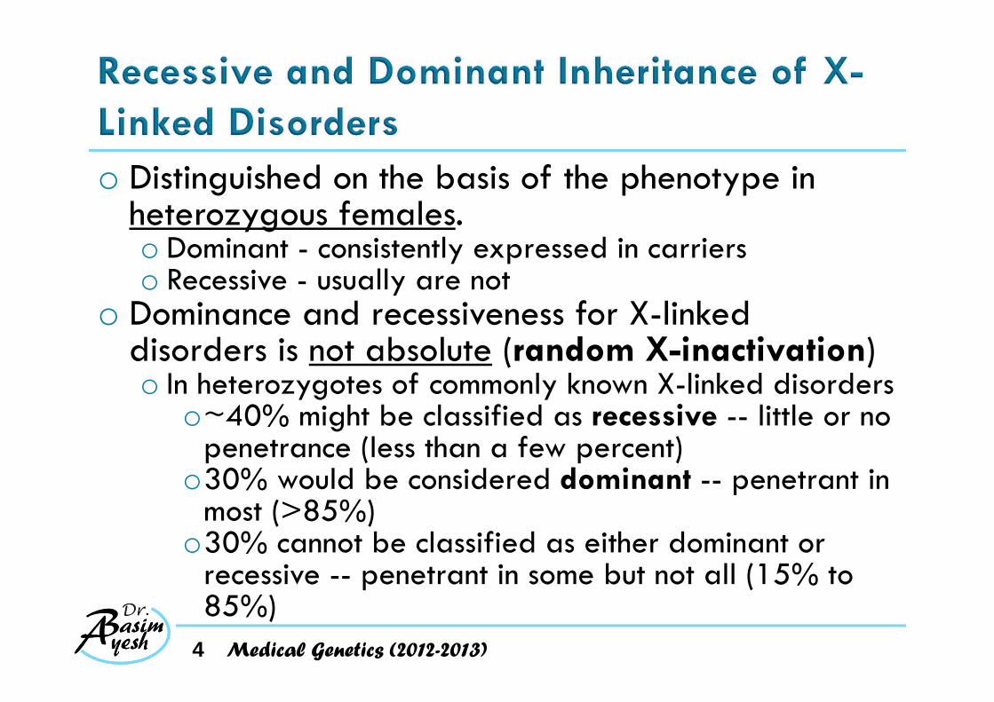
o Distinguished on the basis of the phenotype in heterozygous females. o Dominant - consistently expressed in carrierso Recessive - usually are not
o Dominance and recessiveness for X-linked disorders is not absolute (random X-inactivation)o In heterozygotes of commonly known X-linked disorders
o~40% might be classified as recessive -- little or no penetrance (less than a few percent)
o30% would be considered dominant -- penetrant in most (>85%)
o30% cannot be classified as either dominant or recessive -- penetrant in some but not all (15% to 85%)
4 Medical Genetics (2012-2013)

o Incidence of the trait in males >>> females.o Heterozygous females are usually unaffected
o some may express variable levels of condition (X inactivation)
o Transmitted from an affected manthrough all hisdaughterso never transmitted
directly from father to son
5 Medical Genetics (2012-2013)

o Transmitted through a series of carrier femaleso the affected males in a kindred are related through
females.o A significant
proportion ofisolated casesare due to newmutation.
6 Medical Genetics (2012-2013)

o Hemophilia A is a classic X-linked recessive disordero For the coagulation factor VIII gene
o XH = wild-type allele o Xh = mutant allele causes hemophilia A
o The expected genotypes would be as follows:
7
Genotypes PhenotypesMales Hemizygous XH Unaffected
Hemizygous Xh AffectedFemales Homozygous XH/XH Unaffected
Heterozygous XH/Xh Unaffected (usually)Homozygous Xh/Xh Affected
Medical Genetics (2012-2013)

AFFECTED MALE BY NORMAL FEMALE: Xh/Y × XH/XH
XH XH
Xh XH/Xh XH/Xh Daughters: all carriers
Y XH/Y XH/Y Sons: all unaffected
NORMAL MALE BY CARRIER FEMALE: XH/Y × XH/Xh
XH Xh
XH XH/XH XH/Xh Daughters: 1/2 normal, 1/2 carriers
Y XH/Y Xh/Y Sons: 1/2 normal, 1/2 affected
8 Medical Genetics (2012-2013)
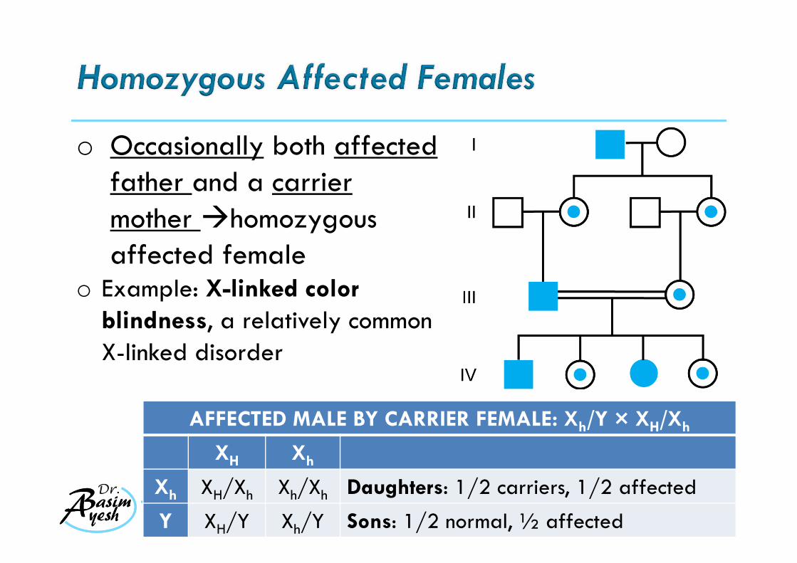
o Occasionally both affected father and a carrier mother homozygous affected female
o Example: X-linked colorblindness, a relatively common X-linked disorder
9 Medical Genetics (2012-2013)
AFFECTED MALE BY CARRIER FEMALE: Xh/Y × XH/Xh
XH Xh
Xh XH/Xh Xh/Xh Daughters: 1/2 carriers, 1/2 affected
Y XH/Y Xh/Y Sons: 1/2 normal, ½ affected
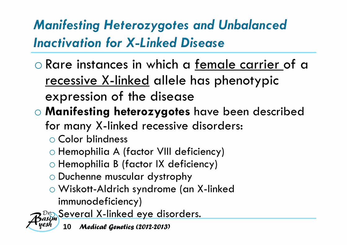
o Rare instances in which a female carrier of a recessive X-linked allele has phenotypic expression of the disease
o Manifesting heterozygotes have been described for many X-linked recessive disorders:o Color blindnesso Hemophilia A (factor VIII deficiency) o Hemophilia B (factor IX deficiency)o Duchenne muscular dystrophyo Wiskott-Aldrich syndrome (an X-linked
immunodeficiency)o Several X-linked eye disorders.
10 Medical Genetics (2012-2013)

o Random X inactivationo Established at <100 cells stage of embryonic
developmento Unbalanced or "skewed" X inactivation - the
deleterious allele located on the active X in relevant tissues (non-random X-inactivation for a tissue or organ)
o Different degrees of disease penetrance and expression in females with the same degree of skewed inactivation o Because of the underlying physiological functioning of
the genesMedical Genetics (2012-2013)11

o Example: Hunter syndromeo lysosomal storage disease caused by iduronate
sulfatase deficiencyoCells carrying active X with normal gene can
export the enzyme to the extracellular space it is picked up by cells carrying the mutant allele the defect is corrected in those cells
oThe penetrance for Hunter syndrome in female heterozygotes is extremely low
12 Medical Genetics (2012-2013)
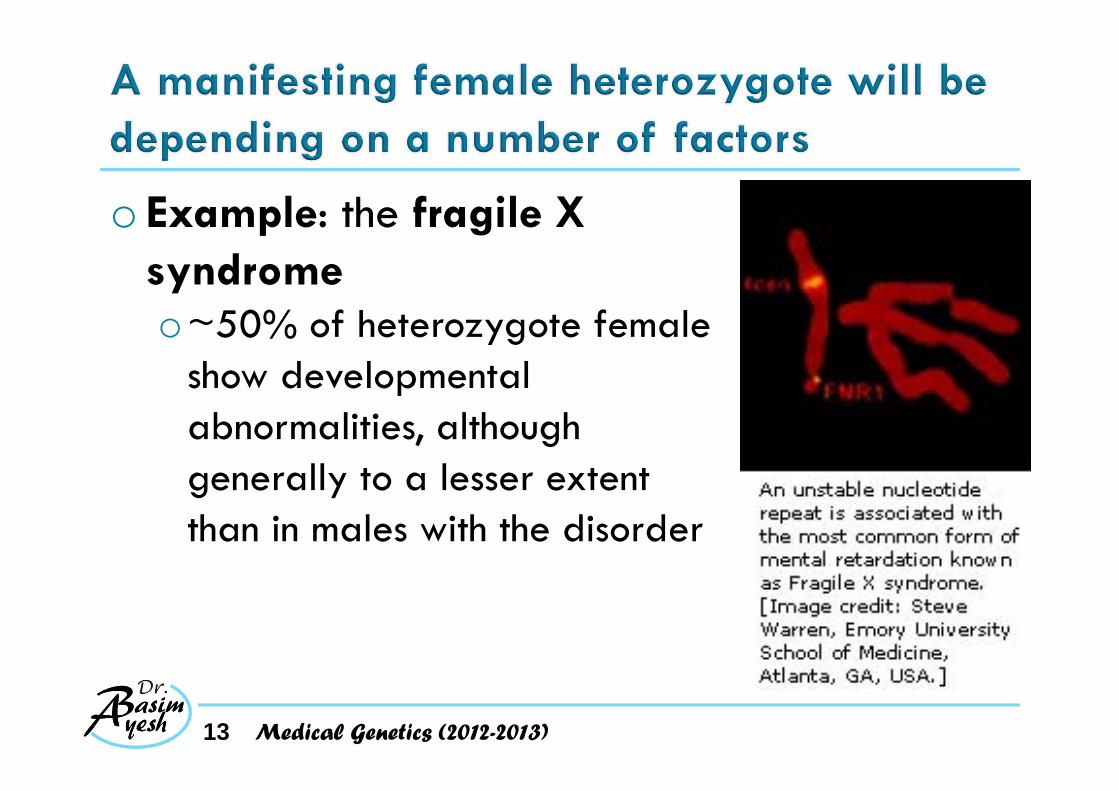
o Example: the fragile X syndromeo~50% of heterozygote female
show developmental abnormalities, although generally to a lesser extent than in males with the disorder
13 Medical Genetics (2012-2013)
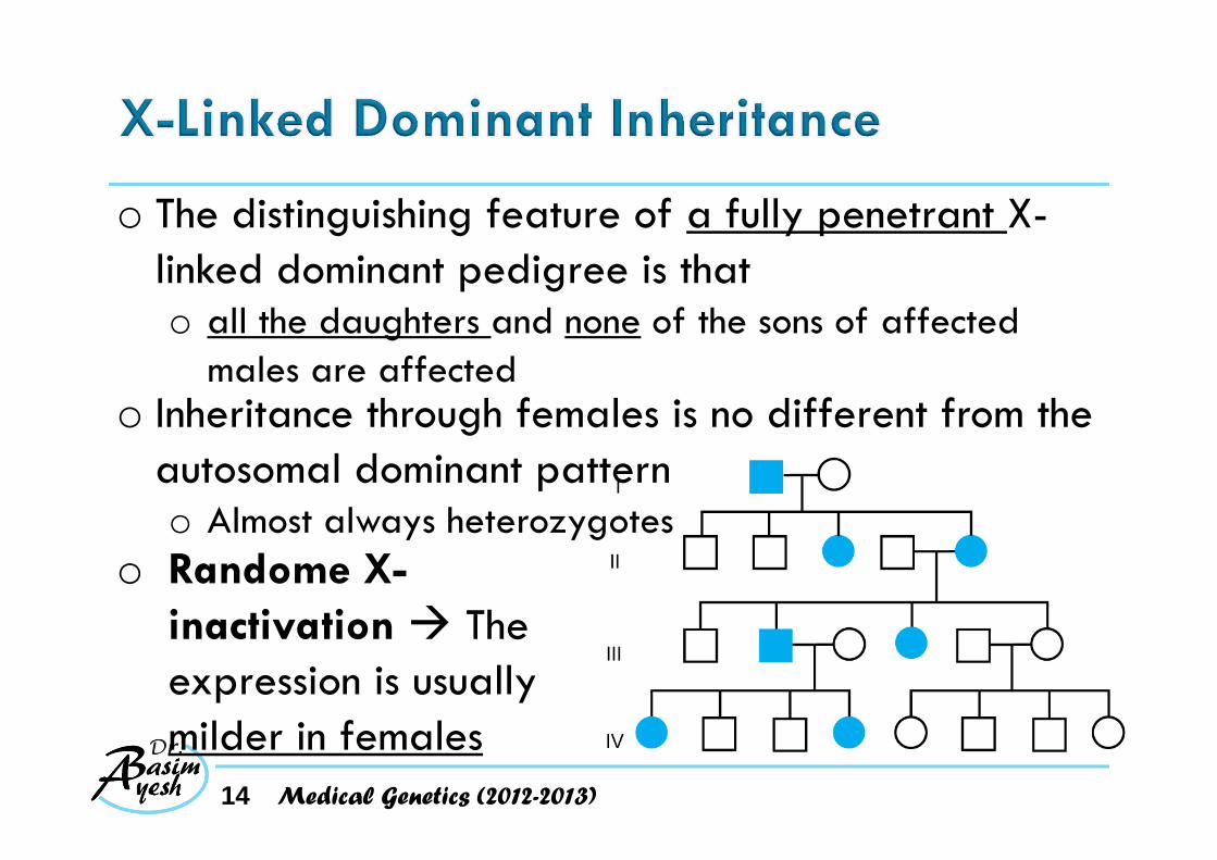
o The distinguishing feature of a fully penetrant X-linked dominant pedigree is that o all the daughters and none of the sons of affected
males are affectedo Inheritance through females is no different from the
autosomal dominant patterno Almost always heterozygotes
o Randome X-inactivation The expression is usually milder in females
14 Medical Genetics (2012-2013)
o Affected males with normal mates have no affected sons and no normal daughters.
o Both male and female offspring of female carriers have a 50% risk of inheriting the phenotype. The pedigree pattern is similar to that seen with autosomal dominant inheritance.
o Affected females are about twice as common as affected males
o Affected females typically have milder (although variable) expression of the phenotype.
15 Medical Genetics (2012-2013)

o Example: X-linked hypophosphatemic rickets(also called vitamin D-resistant rickets)o Impaired kidney tubules ability to reabsorb filtered
phosphateo The defective gene is a member of a family of
endopeptidases that activate or degrade a variety of peptide hormones.
o Fits the criterion of an X-linked dominant disordero Although both sexes are affected, the serum phosphate
level is less depressed and the rickets less severe in heterozygous females than in affected males.
16 Medical Genetics (2012-2013)
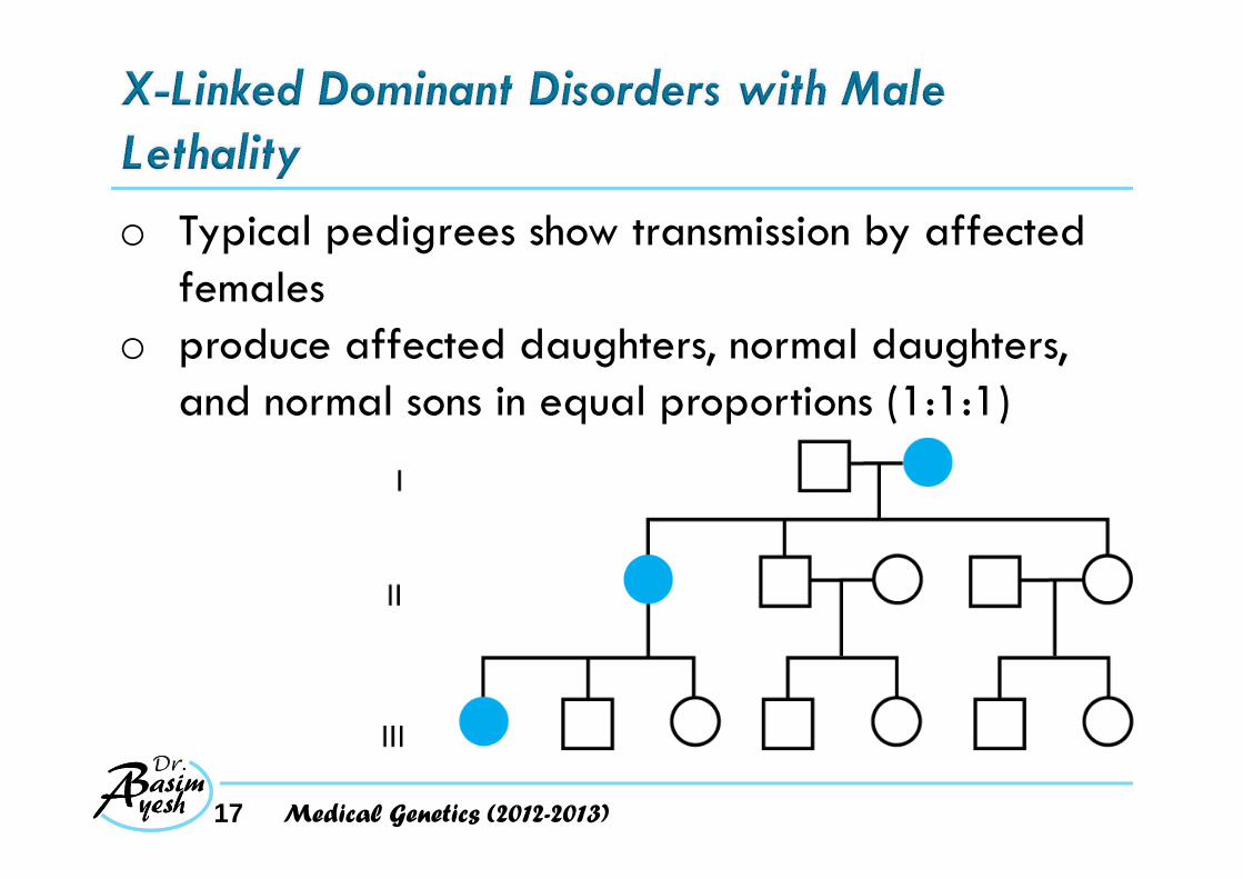
o Typical pedigrees show transmission by affected females
o produce affected daughters, normal daughters, and normal sons in equal proportions (1:1:1)
17 Medical Genetics (2012-2013)

o Example: Rett syndrome oX-linked dominant disorder that is usually lethal
in hemizygous malesoNormal prenatal and neonatal growth and
developmentoRapid onset of neurological symptoms in 6 -18
months oSpastic and ataxicoAutistic features and irritable behaviour with
outbursts of crying
18 Medical Genetics (2012-2013)
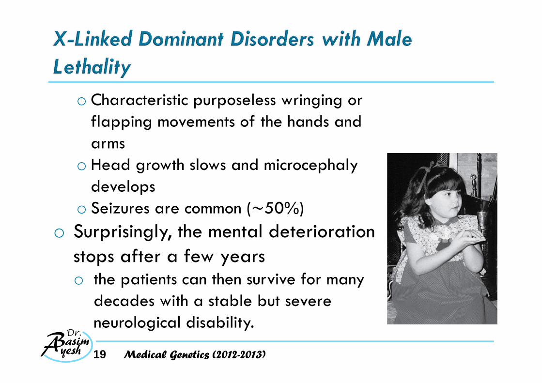
o Characteristic purposeless wringing or flapping movements of the hands and arms
o Head growth slows and microcephaly develops
o Seizures are common ( 50%)o Surprisingly, the mental deterioration
stops after a few years o the patients can then survive for many
decades with a stable but severe neurological disability.
19 Medical Genetics (2012-2013)
o Most cases are caused by spontaneous mutations in an X-linked gene, MECP2 (methyl-CpG-binding protein 2)o Abnormalities in the regulation of a set of genes in the
developing brain. o Most female heterozygotes have full-blown Rett
syndrome.o Males who survive
o have two X chromosomeso 47,XXY Klinefelter individual o XX males: 46,X,der(X) male with the male-determining SRY gene
translocated from the Y to an X
o Mosaic for a mutation that is absent in most of their cells
20 Medical Genetics (2012-2013)

o A few apparently unaffected women have given birth to more than one child with Rett syndromeo the mother may be heterozygous for an MECP2
mutation with highly skewed X inactivation pattern
o Alternatively, the phenotypically normal mother of more than one child affected with Rett syndrome can be a germline mosaic
odoes not have the mutant gene in her own somatic tissues
21 Medical Genetics (2012-2013)

o In males, genes for X-linked disorders are exposed to selection against mutant alleles ocomplete for some disorders, partial for others,
and absent for still othersodepending on the fitness of the genotype.
o New mutations constitute a significant fraction of isolated cases of many X-linked diseases
22 Medical Genetics (2012-2013)

o Patients affected with a severe X-linked recessive disease, such as Duchenne muscular dystrophy, cannot reproduce (i.e., selection is complete; fitness =~0)o The mutant alleles they carry are lost from the
population. o Because the incidence of DMD is not changing
o mutant alleles lost through failure of the affected males to reproduce are continually replaced by new mutations.
o For hemophilia, in which reproduction is reduced but not eliminated (fitness =~0.70)o A proportionately smaller fraction of cases will be due
to new mutation.23 Medical Genetics (2012-2013)

o Alleles for genes in the pseudoautosomalregion can show male-to-male transmission, omimic autosomal inheritanceo they can cross over from the X to the Y during
male gametogenesis and be passed on from a father to his male offspring.
24 Medical Genetics (2012-2013)

o Example: DyschondrosteosisoDominantly inherited skeletal dysplasiaoDisproportionate short statureoDeformity of the forearm oPrevalence: females > males,
suggesting an X-linked dominant disorder,
oThe presence of male-to-male transmission clearly ruled out strict X-linked inheritance
25 Medical Genetics (2012-2013)

Inheritance of dyschondrosteosis
A male who inherited the trait on his Y chromosome from his father
26 Medical Genetics (2012-2013)

oMutations in the SHOX gene encoding a homeodomain-containing transcription factor have been found responsible for this condition
oSHOX is located in the pseudoautosomal region on Xp and Yp and escapes X inactivation
27 Medical Genetics (2012-2013)

28 Medical Genetics (2012-2013)
• Mutations arising in a single cell in either prenatal or postnatal life• Can give rise to clones of cells genetically
different from theoriginal zygote

o Did the mutation occur before or after the separation of germline cells from somatic cells?o Pure somatic mosaicism: The mutation is restricted to
the somatic tissues but not in the gameteso Pure germline mosaicism: the mutation is restricted to
the gamete lineage only and no where else o The mutation is present in both somatic lineages and
the germlineo There are ~30 mitotic divisions in the cells of the
germline before meiosis in the female and several hundred in the male
29 Medical Genetics (2012-2013)
o Might be manifested as a segmental or patchyabnormalityo depending on the stage and the lineage of the somatic
cell in which it originated.o Example: Segmental NF1- affecting only one
part of the body. o Normal parents (a postzygotic mutation)o If his/her child is affected typical NF1, not
segmental. oThe mutation must have occurred before separation
of germline cells from the somatic cell line30 Medical Genetics (2012-2013)
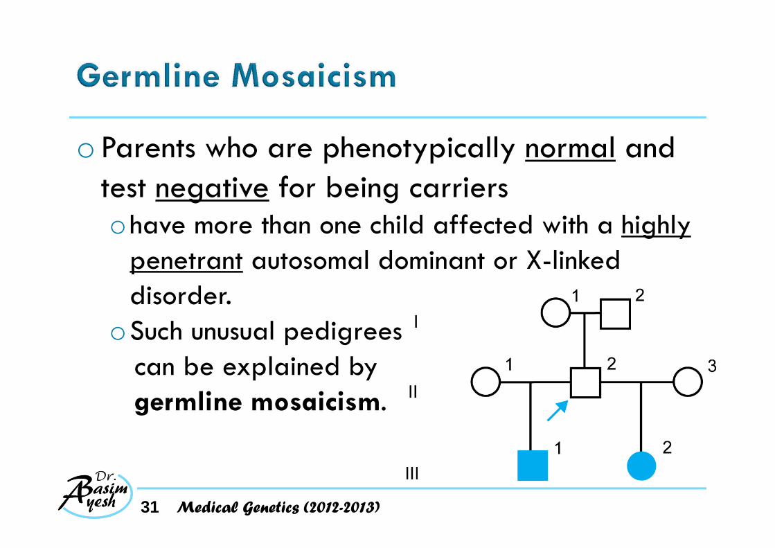
o Parents who are phenotypically normal and test negative for being carriersohave more than one child affected with a highly
penetrant autosomal dominant or X-linked disorder.
oSuch unusual pedigrees can be explained by germline mosaicism.
31 Medical Genetics (2012-2013)

o Well documented in ~ 6% of severe, lethal forms of the autosomal dominant disorder osteogenesis imperfectaoMutations in type I collagen genesoAbnormal collagenoBrittle bonesoFrequent fractures.
32 Medical Genetics (2012-2013)
o Have also been reported for several other well-known disordersohemophilia A, hemophilia B, and DMD
o Have only very rarely been seen in other dominant diseaseso such as achondroplasia
o Estimated highest incidence is in DMD, oup to 15% of the mothers of isolated cases
33 Medical Genetics (2012-2013)

o Example: the rare condition Albright hereditary osteodystrophy (AHO).o Obesity, short stature, subcutaneous
calcifications, and brachydactyly, particularly of the 4th and 5th metacarpal bones
o Inherited as a fully penetrant autosomal dominant trait.
o In families of individuals affected by AHO, some but not all of the affected patients have an additional clinical disorder known as pseudohypoprathyroidism (PHP)
34 Medical Genetics (2012-2013)

o In PHP, an abnormality of calcium metabolism o Elevated levels of parathyroid hormone secondary to
renal tubular resistance to the effects of parathyroid hormone.
o PHP in an individual with the AHO phenotype is known as pseudohypoparathyroidism type1a (PHP1a).
o AHO with or without PHP is caused by a defect in the GNAS geneo GNAS is involved in transmitting the parathyroid
hormone signal from the surface of renal cells to inside the cell
35 Medical Genetics (2012-2013)

o PHP1 pedigrees:o Some individuals have AHO only, without the calcium
and renal problemso others have the physical characteristics as a component
of PHP1a o When AHO occurs without the renal tubular
dysfunction in families in which other relatives have PHP1a, it is often referred to as pseudopseudohypoparathyroidism (PPHP)
36 Medical Genetics (2012-2013)

o When PPHP and PHP1a occur within the same family, o affected brothers and sisters in any one sibship either
all have PPHP or all have PHP1ao Why???
37 Medical Genetics (2012-2013)

o The defective gene (GNAS) in PHP1a and PPHP is imprinted only in certain tissues, including renal tubular cellso Maternally expressed in these cells paternally silent.o PHP1a therefore occurs only when an individual inherits
an inactivating mutation in GNAS from his or her mother
o In the tissues without GNAS imprinting, heterozygotes for one mutant GNAS allele all develop AHO, which is passed on as a simple autosomal dominant trait
38 Medical Genetics (2012-2013)

Disorder Phenotype Molecular BasisAHO Obesity, short stature,
subcutaneous calcifications, brachydactyly
Constitutional haploinsufficiency for GNAS
PHP1a AHO with pseudohypoparathyroidism, hypothyroidism, growth hormone deficiency
Constitutional haploinsufficiency for GNAS inherited from a female parent, which also causes complete loss of expression in critical renal and endocrine tissues
PPHP AHO alone in a member of a family in which PHP1a is also occurring
Constitutional haploinsufficiency for GNAS inherited from a male parent, which leaves intact expression of the maternal copy in critical renal and endocrine tissues
39 Medical Genetics (2012-2013)
40 Medical Genetics (2012-2013)

o > dozen diseases are known to result from unstable repeat expansions. o All are primarily neurological. o Inherited as dominant, X-linked, or recessive
o The degree of expansion is variable in different diseases
o Anticipation: The disease appears to develop at an earlier and earlier age when it is transmitted through the pedigree
o Premutations: repeat alleles at the upper limits of normal that do not cause disease but are capable of expanding into the disease-causing range
41 Medical Genetics (2012-2013)

Medical Genetics (2012-2013)42
Inheritance Pattern
Repeat Gene Affected
Location in Gene
Normals Intermediate Affected
Autosomal dominant
CAG HD coding region <36 36-39 usually affected
>40
Paternal transmission bias:• Expansion occurs most frequently during male gametogenesis• The severe early-onset juvenile form of the disease, seen with
the largest expansions (70 to 121 repeats), is always paternally inherited.

43
Inheritance Pattern
Repeat Gene Affected
Location in Gene Normals Intermediate Affected
X-linked CGG FMR1 5' untranslated <60 60-200 usually unaffected*
>200
*May have tremor-ataxia syndrome or premature ovarian failure
Penetrance of the full mutation in a female is approximately 50%
Medical Genetics (2012-2013)

44
Inheritance Pattern
Repeat Gene Affected
Location in Gene Normals Intermediate Affected
Autosomal dominant
CTG DMPK 3' untranslated <30 50-80 may be mildly affected
80-2000
• Myotonia• Muscular dystrophy• Cataracts• Hypogonadism• Diabetes• Frontal balding• Changes in the
electroencephalogram.Expansion is almost always inherited from an affected mother
Medical Genetics (2012-2013)
45
Inheritance Pattern
Repeat Gene Affected
Location in Gene
Normals Intermediate Affected
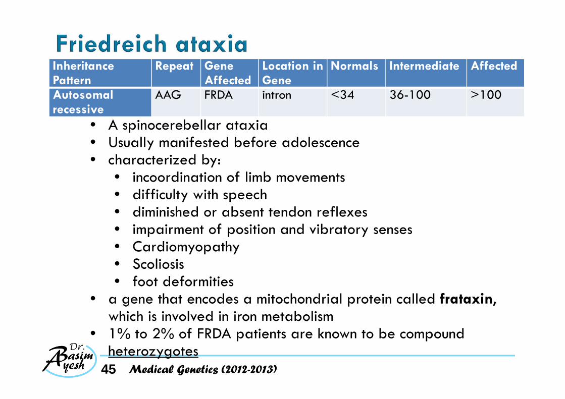
Autosomal recessive
AAG FRDA intron <34 36-100 >100
• A spinocerebellar ataxia• Usually manifested before adolescence • characterized by:
• incoordination of limb movements• difficulty with speech• diminished or absent tendon reflexes• impairment of position and vibratory senses• Cardiomyopathy• Scoliosis• foot deformities
• a gene that encodes a mitochondrial protein called frataxin, which is involved in iron metabolism
• 1% to 2% of FRDA patients are known to be compound heterozygotes
Medical Genetics (2012-2013)

46
Disease Inheritance Pattern
Repeat Gene Affected
Location in Gene
Normals Intermediate Affected
Huntington disease
Autosomal dominant
CAG HD coding region
<36 36-39 usually affected
>40
Fragile X X-linked CGG FMR1 5' untranslated
<60 60-200 usually unaffected*
>200
Myotonicdystrophy
Autosomal dominant
CTG DMPK 3' untranslated
<30 50-80 may be mildly affected
80-2000
Friedreichataxia
Autosomal recessive
AAG FRDA intron <34 36-100 >100
Medical Genetics (2012-2013)

47 Medical Genetics (2012-2013)
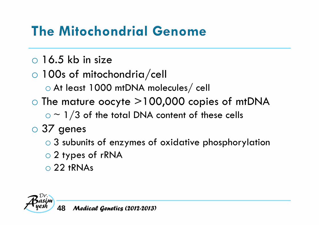
o 16.5 kb in sizeo 100s of mitochondria/cell
o At least 1000 mtDNA molecules/ cello The mature oocyte >100,000 copies of mtDNA
o ~ 1/3 of the total DNA content of these cellso 37 genes
o 3 subunits of enzymes of oxidative phosphorylationo 2 types of rRNAo 22 tRNAs
48 Medical Genetics (2012-2013)

o >100 different rearrangements and 100 different point mutations have been identified in mtDNA that can cause human diseaseo Often involving the central nervous and musculoskeletal
systemso Show a distinctive pattern of inheritance because
of o replicative segregationo homoplasmy and heteroplasmyo maternal inheritance
49 Medical Genetics (2012-2013)

50 Medical Genetics (2012-2013)

o At cell division oThe multiple copies of mtDNA in each of the
mitochondria in a cell Replication sort randomly among newly synthesized mitochondria.
oThe mitochondria, are distributed randomly between the two daughter cells
51 Medical Genetics (2012-2013)
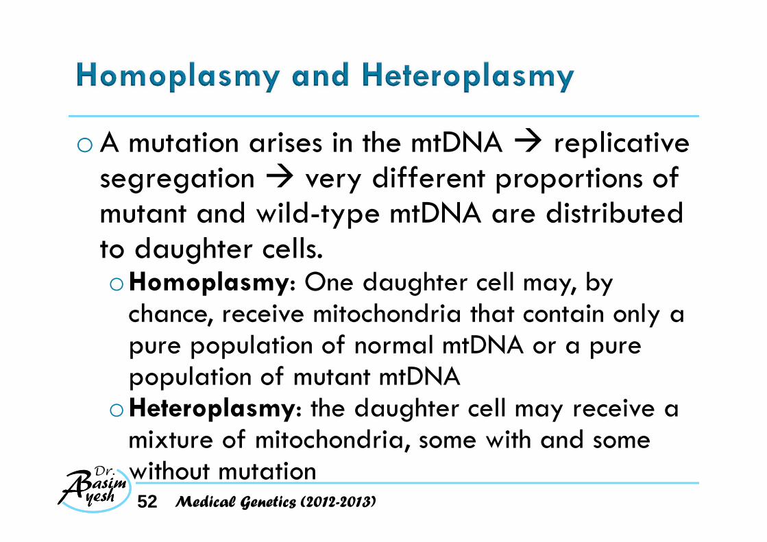
o A mutation arises in the mtDNA replicative segregation very different proportions of mutant and wild-type mtDNA are distributed to daughter cells.oHomoplasmy: One daughter cell may, by
chance, receive mitochondria that contain only a pure population of normal mtDNA or a pure population of mutant mtDNA
oHeteroplasmy: the daughter cell may receive a mixture of mitochondria, some with and some without mutation52 Medical Genetics (2012-2013)
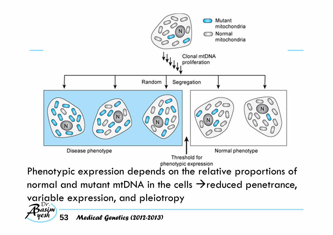
53
Phenotypic expression depends on the relative proportions of normal and mutant mtDNA in the cells reduced penetrance, variable expression, and pleiotropy
Medical Genetics (2012-2013)
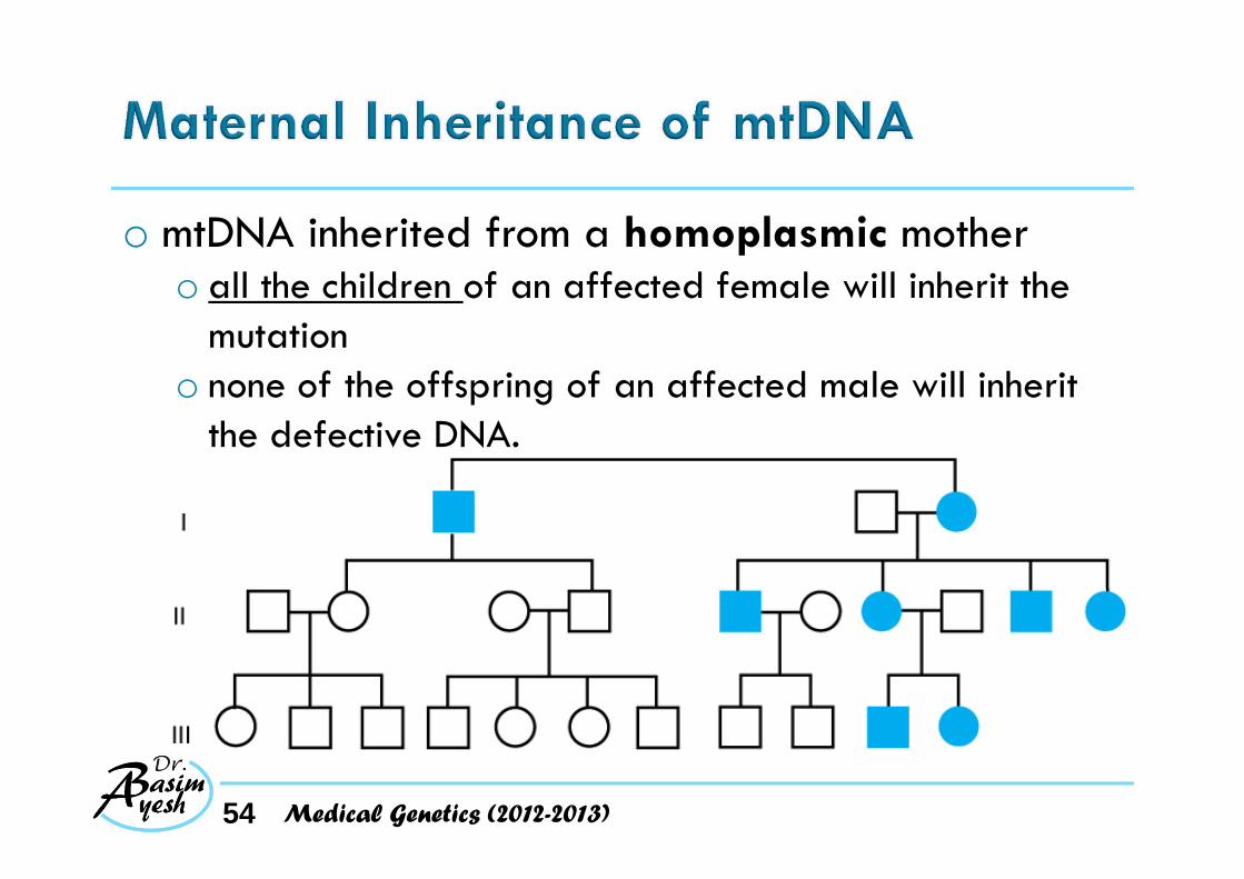
o mtDNA inherited from a homoplasmic mothero all the children of an affected female will inherit the
mutationo none of the offspring of an affected male will inherit
the defective DNA.
54 Medical Genetics (2012-2013)
o In the presence of heteroplasmy:oAs a result of mitochondrial genetic bottleneck
ovariability in the % of mutant mtDNA molecules seen in the offspring of a mother with heteroplasmy for a mtDNA mutation
o Mothers with a high proportion of mutant mtDNA eggs with a higher proportion of mutant mtDNA more likely to have clinically affected offspring
55 Medical Genetics (2012-2013)
o Exception to maternal inheritanceo when the mother is heteroplasmic for deletion mutation
in her mtDNAo for unknown reasons, deleted mtDNA molecules are
generally not transmitted from clinically affected mothers to their children
o At least one instance of paternal inheritance of mtDNA has occurred in a patient with a mitochondrial myopathy.o in patients with apparently sporadic mtDNA mutations,
the rare occurrence of paternal mtDNA inheritance must be considered
56 Medical Genetics (2012-2013)