Embed Size (px)
Citation preview
With the advent of affordable CBCT, oral surgeons are adding 3D imaging systems
to their practices in increasing numbers.
Is it just a trend? How CBCT is impacting the practice?
RXDigitalRX Soluciones de ImagenologíaRX Soluciones de Imagenología
RxDigitalDentalwww.rxdigitalsoluciones.com

The ability to look at a tooth, pathology or an anatomical situation in any direction and orientation, as well as in 3D, eliminates much of the guesswork commonly experienced with 2D radiographs.
“ “ Dr. Nick Fahey
2
RxDigitalDentalwww.rxdigitalsoluciones.com RXDigitalRX Soluciones de ImagenologíaRX Soluciones de Imagenología

Images of multiple planes are taken to make up the composite panoramic image. So the anatomical structures are superimposed and distorted.
The anatomical structures are clearly examined with each sectional view. It helps in gaining a better view of bone structures and supports a wide range of diagnosis and treatment plans.
A, B, C , D
A B
C D
3
RXDigitalRX Soluciones de ImagenologíaRX Soluciones de Imagenología
RxDigitalDentalwww.rxdigitalsoluciones.com

Mes ial
Dis tal
With 2D image, you may lose vital information on “Buccal & Lingual“ side ! And it may be able to lead you to misdiagnosis.
2D 3D
As you can see below, it looks like the impacted tooth is touching the mandibular nerve in 2D panoramic view but, in 3D, you can verify the tooth does not touch the nerve. This way, 3D helps you remove the accident in treatment.
3D
4
RxDigitalDentalwww.rxdigitalsoluciones.com RXDigitalRX Soluciones de ImagenologíaRX Soluciones de Imagenología

Length 13.5mm 10.0mm
Length 12.96mm
9.60mm
The difference between 2D & 3D
2D
Distortions inherent in panoramic imaging because of projection geometry produce discrepancies in the angular measurements in the mandibular third molar regions on panoramic radiographs. Interpretation of third molar angulation from panoramic radiographs is often unreliable and may not accurately reflect the true orientation of the tooth. Apparent tilting of the tooth across the arch on the panoramic radiograph exacerbates the problem and appears to correlate with the bucco-lingual inclination of the third molar as visualized on reformatted CT images.
3D
Implant Fixture + 0.54mm Length to canal - 0.40mm
� Reference : Thesis ‘Accuracy of angular measurements and assessment of distortion in the mandibular third molar region on panoramic radiographs.
5
RXDigitalRX Soluciones de ImagenologíaRX Soluciones de Imagenología
RxDigitalDentalwww.rxdigitalsoluciones.com

2D
3D
In 2D panorama view, palatal bone is superimposed. So it is hard to figure out sinus, septa and other adjacent structures.
Single sectional images are available with 3D. So it is easy to gain information like septum of the sinus.
These information is very vital for successful maxillary sinus implant surgery.
6
RxDigitalDentalwww.rxdigitalsoluciones.com RXDigitalRX Soluciones de ImagenologíaRX Soluciones de Imagenología

Without question, 3D imaging allows us to directly interpret the anatomy and challenges of the area of interest before direct visualization.
This information allows us to optimally create and sequence the best possible surgical and prosthetic treatment plan.
Most importantly, overall morbidity can be decreased as a result.
“ “
7
RXDigitalRX Soluciones de ImagenologíaRX Soluciones de Imagenología
RxDigitalDentalwww.rxdigitalsoluciones.com

Improve Treatment Outcomes with Pre-simulation
Appropriate use of CBCT absolutely improves treatment outcomes in the oral surgery practice. There is no doubt that using CBCT makes you to be better prepared going into a surgical procedure than with 2D imaging alone, and your results will reflect it.
#2 Make the position accurately in 2D slice images
#1 Get real feedback from 3D; reducing risk of incidents
#3 Get real feedback from 3D; checking bone density
#4 Check the overall view and be con�dent!
8
RxDigitalDentalwww.rxdigitalsoluciones.com RXDigitalRX Soluciones de ImagenologíaRX Soluciones de Imagenología

“3D diagnosis is so essential to doing the best for our patients that most of us will provide the service at a reduced cost, or even gratis in some cases. We then have an opportunity to provide confident, high quality care.”
As you know well overall 3D evaluation of patients’ present anatomy, as well as identify potential incidental pathologic findings is imperative.
The information 2D provides you is limited and there is high probability of doing treatment just what patients ask.
This is the best care you can do for patients.
- Dr. Michael A. Pikos -
9
RXDigitalRX Soluciones de ImagenologíaRX Soluciones de Imagenología
RxDigitalDentalwww.rxdigitalsoluciones.com

“I use a large �at panel monitor and take time to explain what the images represent in 3D. This is an invaluable experience as they understand and take ownership of their condition, diagnosis and proposed treatment.”
- Dr. Michael A. Pikos -
It’s a far cry from days when you consult with 2D. Showing a patient what needs to be done with 3D is necessary and invaluable, and a far cry from the days when a dentist would hold up a tiny black and white film image and expect the patient to understand it. It absolutely raise the acceptance rate and improve satisfaction.
10
RxDigitalDentalwww.rxdigitalsoluciones.com RXDigitalRX Soluciones de ImagenologíaRX Soluciones de Imagenología

My partners and I use CBCT for a variety surgical procedures such as third molar impactions, impacted maxillary canines, supernumeraries, and so on.
The volume of cases I do translates into a good return to justify cost of this technology.
Beyond this, having an in-office unit gives me many benefits.
“ “
11
RXDigitalRX Soluciones de ImagenologíaRX Soluciones de Imagenología
RxDigitalDentalwww.rxdigitalsoluciones.com

Such as cysts, dentoalveolar lesions, root resorption, periapical disease
Such as bony fractures, tooth fractures, sinus treatment
Such as third molars, imacted maxillary canines, supernumeraries, mesiodens
12
� Reference : Pajdds.wordpress.com / www.endomicrosurg.net
RxDigitalDentalwww.rxdigitalsoluciones.com RXDigitalRX Soluciones de ImagenologíaRX Soluciones de Imagenología

I’ve come to realize that we canonly ensure our patients get the very best treatment journey, catering to all of their needs, if everything can be done in-house. Up until last year we could offer in practice every service, except for 3D imaging.
Unfortunately, we had issues where we were referring patients out for scans and patients weren’t getting the kind of service that we would like them to receive.
“ “
Dr. Ajay Murgai
13
RXDigitalRX Soluciones de ImagenologíaRX Soluciones de Imagenología
RxDigitalDentalwww.rxdigitalsoluciones.com

It is essential to have an in-office unit for immediate utilization to eliminate the logistics and inherent problems with referral and delay for an external diagnostic
Patients think it looks like space age technology. It helps to show that you are cutting edge.
Both clinicians and patients have been realizing that 3D scan is truly necessary thing for treatment. Having 3D imaging in your office speaks a lot more than other promotions.
14
RxDigitalDentalwww.rxdigitalsoluciones.com RXDigitalRX Soluciones de ImagenologíaRX Soluciones de Imagenología

However, Still many clinicians have difficulties
to utilize 3D in their practice.
Why is that? What makes them be passive to use it?
RXDigitalRX Soluciones de ImagenologíaRX Soluciones de Imagenología
RxDigitalDentalwww.rxdigitalsoluciones.com

Many clinicians have thoughts like “It is too di�cult to utilize everyday”
These are not wrong questions because there are some obstructive factors making These questions make sense. It is true that 3D software is difficult to deal with and it results in not just making many clinicians to be stressful but also underutilizing.
What are the obstructive factors and how they are changed?
“Is 3D S oftware really for me? ”
16
RxDigitalDentalwww.rxdigitalsoluciones.com RXDigitalRX Soluciones de ImagenologíaRX Soluciones de Imagenología

Actually, about 70% of clinicians have difficulty to utilize 3D software quite freely because axes moving and getting to ROI is too complicated and confusing.
To get accurate sectional images of ROI(Region of Interest), you are required to adjust axial, coronal, sagittal axes and draw the panoramic curve but in this stage you just get lost.
Even though you are accustomed to this process, it takes much time from you and staff.
What if you do not need to adjust axes anymore? What if you do not need to draw panoramic curve anymore?
Axes moving makes clinicians get lost and confusing to get the sectional images of ROI
A X I A LS A G I T T A LC O R O N A L
17
RXDigitalRX Soluciones de ImagenologíaRX Soluciones de Imagenología
RxDigitalDentalwww.rxdigitalsoluciones.com

Save time with this simple workflow and see more patients.
Draw panoramic curve
Check Bone Density
Select the Fixture
Measure Length
Check Position on VR
Position the Fixture
Position the axes
Measure Length
Insert Implant
Position Navigator Box
www.vatechglobal.com
One Click S ectioningS implifies the whole process.
19
It’s Navigator Box!
On volume panorama(It’s 3D!), there is a navigator box. Just position the box! Then the sectional images of the ROI will be shown below. That’s it! It removes the process of adjusting axes and drawing the panoramic curve. Yes, right. You don’t need to try to understand complicated axes anymore.Let’s compare conventional and current process in detail.
18
One Click S ectioning S implifies the whole process.
RxDigitalDentalwww.rxdigitalsoluciones.com RXDigitalRX Soluciones de ImagenologíaRX Soluciones de Imagenología

Save time with this simple workflow and see more patients.
Draw panoramic curve
Check Bone Density
Select the Fixture
Measure Length
Check Position on VR
Position the Fixture
Position the axes
Measure Length
Insert Implant
Position Navigator Box
One Click S ectioning S implifies the whole process.
19
RXDigitalRX Soluciones de ImagenologíaRX Soluciones de Imagenología
RxDigitalDentalwww.rxdigitalsoluciones.com

Change the paradigm of Diagnosis & Consultation
When your patients get a scanning PaX-i3D Smart gives you two image; 2D Panorama and 3D Volume. What’s greater thing is you can see both images in one 3D viewer together.
We call that 2D Panorama image Auto Pano. Do a quick diagnosis first with Auto Pano and examine closely where you are interested in. Also it will be very helpful when you have consultation with patients.
The panorama image you saw in previous pages is not 2D, it’s 3D image.For you who are accustomed to 2D, Ez3D-i stretches the volume. As it is 3D, it provides you accurate information on anatomy, also as it is 2D, you can verify the condition of dentition at a glance.
20
RxDigitalDentalwww.rxdigitalsoluciones.com RXDigitalRX Soluciones de ImagenologíaRX Soluciones de Imagenología

“Buying small FOV CT looks good choice. It has reasonable price and it can be enlarged with stitching.”
Is that right?
Of course small FOV imaging machine is inexpensive than large FOV ones because normally FOV size is determined by the size of detector. However, until when you do single implant surgery only?
To maximize your income, it is important how many patients you see, but if you can do multiple implant surgery from one patient, it will be more efficient. Even if you purchased small FOV one now, someday you will upgrade and it absolutely will make you invest much money.
Then, you might consider double scanning to enlarge the image called ‘stitching’.Do you really think you can have large image from stitching?
21
RXDigitalRX Soluciones de ImagenologíaRX Soluciones de Imagenología
RxDigitalDentalwww.rxdigitalsoluciones.com

S titching Technology causes…
Double Time-consuming
Unnecessary X-ray exposure
Possible to image imperfection
Image stitching is the process of combining multiple small images with overlapping fields of view to produce a large single image.
Over Dose
Double scanning means scan time will be double and it means your time to see other patients will reduce.
Stitching has no choice but to require overlapped area for accuracy. It means patients be under over dose.
One of processes is image registration. Matching each point cannot be sure perfect all the time.
It will be concluded to bad impression to patients
22
RxDigitalDentalwww.rxdigitalsoluciones.com RXDigitalRX Soluciones de ImagenologíaRX Soluciones de Imagenología

With FOV 8x8, it’s not possible to takefull arch image.
Normally it requires you to choose one capturing mode; capturing area. Every time when the area is changed, your staff need to change the position. Not just that. As it does not cover the full arch you will have limitation on your diagnosis range.
� Surgical guides � Complex extractions � Bone grafting
� Surgical guides � 3rd Molar extraction � Sinus lifts for both sinuses � Oral and maxillofacial surgery for mandibular
arch reconstruction
FOV 8x8
FOV 12x9
“Is FOV really enough? ”
23
RXDigitalRX Soluciones de ImagenologíaRX Soluciones de Imagenología
RxDigitalDentalwww.rxdigitalsoluciones.com

Arch-shaped volume show the dentition of FOV 12x9 Normally, a FOV 10x8.5 image shows tooth #8. However, when the tooth is lying on its side, there is a high possibility that the tooth will be cut out of the image. With this image, it is not possible to check the correlations between the tooth and the mandibular nerve and increases the risk of medical accident. As you cannot expect which patient would visit your clinic, it is good for you to have CBCT which guarantees to show the hidden-possible area.
Find Hidden Area with Anatomical FOV, 12x9
Current FOV 10x8.5 Anatomical FOV 12x9
PaX-i3D Smart
24
RxDigitalDentalwww.rxdigitalsoluciones.com RXDigitalRX Soluciones de ImagenologíaRX Soluciones de Imagenología

Normally, patients who are considered to take a CT for implant treatment might have some or many metals in their mouths. In this case, what is most disturbing you? Can you make a diagnosis with these kinds of images as below?
You might want to diagnose with a 3D image that does not get affected by metal artifacts. That is why many imaging manufacturers develop the Metal Artifact Reduction technology. But in reality, still many practitioners are challenged by unclear and affected image by various types of metal in patients’ mouths such as prosthesis, implants or dental �llings.
“I feel I’m losing diagnostic confidence”
25
RXDigitalRX Soluciones de ImagenologíaRX Soluciones de Imagenología
RxDigitalDentalwww.rxdigitalsoluciones.com

As Dr. Todd says, even though you can avoid small amount of artifacts spending some time and energy, it is not the case always happened. Sometimes you might have to tell your patients “Oh, you have lots of metals in your mouth, so let’s take a CT one more”.Although the patient is not sensitive to radiation, taking a new scan will be troublesome work to your staff and it takes up your time to see other visitors.
• Simplify workflow of surgery• Reduce possibility of accidents• Reduce visiting count and consultation hours• Reduce operation time and inconvenience of patients
In effects, Metal Artifacts…
Produce dark bands or streaks on images
Hinder visualization
Reduce diagnostic confidence
Lead making misdiagnosis
Making it difficult to merge 3D and STL data
If you have ever considered using surgical guide, it is hard to have benefits below.
26
RxDigitalDentalwww.rxdigitalsoluciones.com RXDigitalRX Soluciones de ImagenologíaRX Soluciones de Imagenología

This metal artifact reducing solution is much developed than before. The accuracy to figure out metals and high-density anatomies such as enamel is higher. It finds out real metals more delicately. It means the side effect resulted from misrecognition is reduced.
Before this solution, users are required to control the on or off of the MAR function case by case. But sometimes it brings mistake and it results in side effects on the image quality.
In contrast, the clinicians using PaX-i3D Smart do not need that. The algorithm automatically makes decision applying the solution or not.
High Accuracy to Apply MAR Solution
AUTOMATIC
Streamlined Workflow
� Clear image gives you less stress and more confidence � Leads to accurate diagnosis for implant planning � No extra discomfort to create surgical guide
� It automatically activates the function based on patients’ dental conditions
Be Confident with MAR S olution
27
RXDigitalRX Soluciones de ImagenologíaRX Soluciones de Imagenología
RxDigitalDentalwww.rxdigitalsoluciones.com

Compare images; SMART MAR Off vs. SMART MAR On
28
RxDigitalDentalwww.rxdigitalsoluciones.com RXDigitalRX Soluciones de ImagenologíaRX Soluciones de Imagenología

10
There are quite many things you need to check before purchasing 3D. But the most important factor is, after all, image quality.
Low Dose Mode 95kV / 8.7mA / Scan Time 18sec. / 0.2voxel
This is No.1 Point to Check ; Image Quality
29
RXDigitalRX Soluciones de ImagenologíaRX Soluciones de Imagenología
RxDigitalDentalwww.rxdigitalsoluciones.com

Ultra Low Dose Mode 80kV / 5mA / Scan Time 18sec. / 0.2voxel
30
RxDigitalDentalwww.rxdigitalsoluciones.com RXDigitalRX Soluciones de ImagenologíaRX Soluciones de Imagenología

Hybrid Sensor with Small Pixel Size
Key to Clear Image Quality
One-Time Reconstruction Ten-Time Reconstruction
VATECH’s SMART Iterative reconstruction
VATECH developed this conventional reconstruction algorithm into smarter way. It repeats the reconstruction process abou10 times at nd it produces high quality of the image as a result. Under the same exposure condition, the image reconstructed using this solution has better quality with the reduced noise and emphasized contrast.
PaX-i3D Smart utilizes 49.5㎛ high resolution X-raysensor. With the latest 49.5㎛ pixel size in X-ray sensor,it is the smallest pixel/high resolution dynamic X-ray sensor for CBCT available on the market.
This hybrid sensor is one of the factors to make the image quality fantastic. Physically the smaller the sensor size is, the better the image quality will be. In addition, because of this slot-scan, it reduces X-ray scattered too.
31
RXDigitalRX Soluciones de ImagenologíaRX Soluciones de Imagenología
RxDigitalDentalwww.rxdigitalsoluciones.com

Accessory Box
Wheelchair Accessible Accessory Cradle
Stable Temple Support
PaX-i3D Smart has not just state-of-the art features but also user-friendly design. Black and white color will create modern atmosphere in your clinic and detailed structures will help staff utilize PaX-i3D Smart very handily.
32
RxDigitalDentalwww.rxdigitalsoluciones.com RXDigitalRX Soluciones de ImagenologíaRX Soluciones de Imagenología

Function CT(with Auto Pano) + Pano + Ceph
Focal Spot 0.5 mm
CT FOV Size Adult 10x8.5 cm
Child 10x7 cm
Voxel Size 0.2 mm / 0.3 mm
Scan Time
CT 18.2 sec
Pano 13.6 sec / 7 sec (Optional with Magic PAN)
Ceph Scan : 12.9 sec / One-Shot : 0.7 sec
Gray Scale 14 bit
Tube Voltage / Current 50~99 kVp / 4~16 mA
33
RXDigitalRX Soluciones de ImagenologíaRX Soluciones de Imagenología
RxDigitalDentalwww.rxdigitalsoluciones.com
6
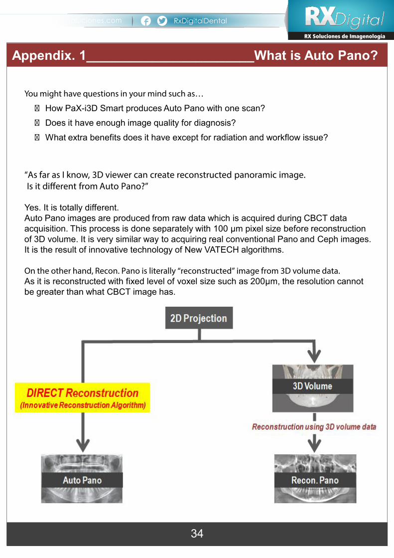
You might have questions in your mind such as…
� How PaX-i3D Smart produces Auto Pano with one scan? � Does it have enough image quality for diagnosis? � What extra benefits does it have except for radiation and workflow issue?
“As far as I know, 3D viewer can create reconstructed panoramic image. Is it di�erent from Auto Pano?”
Yes. It is totally different. Auto Pano images are produced from raw data which is acquired during CBCT data acquisition. This process is done separately with 100 µm pixel size before reconstruction of 3D volume. It is very similar way to acquiring real conventional Pano and Ceph images. It is the result of innovative technology of New VATECH algorithms.
On the other hand, Recon. Pano is literally “reconstructed” image from 3D volume data. As it is reconstructed with fixed level of voxel size such as 200µm, the resolution cannot be greater than what CBCT image has.
What is Auto Pano?Appendix. 1_______________________What is Auto Pano?
34
RxDigitalDentalwww.rxdigitalsoluciones.com RXDigitalRX Soluciones de ImagenologíaRX Soluciones de Imagenología

The different process of Auto Image and Recon. Image makes different results as below.
Auto Image Recon. Image
Image Quality Higher resolution Lower resolution
Metal Artifact No metal artifact Seen in CBCT image
Image Size Same size with real conventional panoramic image
Limited image size in 3D volume size (FOV)
What is Auto Pano?
Auto Pano
Recon. Pano
Appendix. 1_______________________What is Auto Pano?
35
RXDigitalRX Soluciones de ImagenologíaRX Soluciones de Imagenología
RxDigitalDentalwww.rxdigitalsoluciones.com

VATECH’s SMART MAR Technology
VATECH’s MAR Solution addresses those problems resulted from metal artifacts.This metal artifact reducing technology finds out the location of metals when radiation is exposed. And it deletes the metal in projection data artificially and fills that part with the similar values to surrounding area. At the same time, it remembers the original location and status of metals and then it inpaints the area with new value which has no streaks.
Refer to the [Figure 1] below will help you understand easier. The numbers in the box is the values of the image. ‘2’ is the value of dentition areaand ‘9’ is metal area. VATECH Algorithm erases the metal, so ‘9’ becomes ‘0’.‘0’ area is �lled with ‘2’ and it remembers that locations as metal ‘n’.Then, it replaces ‘2’ to ‘9’.
This algorithm absolutely is approved than *previous solutions with high probability to find out real metal; 70% to more than 90% and the high accuracy to inpaint the values.
Appendix. 2_____________________________SMART MAR
R X S O L U C I O N E S D E I M A G E N O L O G Í A S . A . d e C . V .Av. Moctezuma 3515-1B, Col. Ciudad del Sol. Zapopan, Jalisco. MÉXICO
VISITE Nuestro Sitio Weby vea nuestra Línea deProductos Completa
RX Soluciones de ImagenologíaRX Soluciones de Imagenología
TEL: (33) 3880-1262 CEL: (33) 3499-7095 / 1300-5813
RxDigitalDentalwww.rxdigitalsoluciones.comcontacto@ rxdigitalsoluciones.comRXDigital2
01608
RxDigitalDentalwww.rxdigitalsoluciones.com RXDigitalRX Soluciones de ImagenologíaRX Soluciones de Imagenología

Thank you.
R X S O L U C I O N E S D E I M A G E N O L O G Í A S . A . d e C . V .Av. Moctezuma 3515-1B, Col. Ciudad del Sol. Zapopan, Jalisco. MÉXICO
VISITE Nuestro Sitio Weby vea nuestra Línea deProductos Completa
RX Soluciones de ImagenologíaRX Soluciones de Imagenología
TEL: (33) 3880-1262 CEL: (33) 3499-7095 / 1300-5813
RxDigitalDentalwww.rxdigitalsoluciones.comcontacto@ rxdigitalsoluciones.comRXDigital2
01608
RXDigitalRX Soluciones de ImagenologíaRX Soluciones de Imagenología
RxDigitalDentalwww.rxdigitalsoluciones.com