Embed Size (px)
Citation preview
Iron—Too Much, Too Little, Too Late
Marc J. Kahn, MD, MBA Peterman-Prosser Professor
Sr. Associate Dean Tulane University School of Medicine
Case 1.
A 30 year old woman with menorrhagia is referred for persistent iron deficiency anemia despite reported compliance with oral ferrous sulfate 325 mg TID. Her current ferritin is 6 ng/mL. Her Hgb is 7.2 g/dL.
Iron Critical Functions
• Hemoglobin
• Myoglobin
• Electron transport
• Immune function
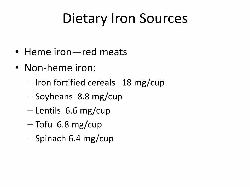
Dietary Iron Sources
• Heme iron—red meats
• Non-heme iron:
– Iron fortified cereals 18 mg/cup
– Soybeans 8.8 mg/cup
– Lentils 6.6 mg/cup
– Tofu 6.8 mg/cup
– Spinach 6.4 mg/cup
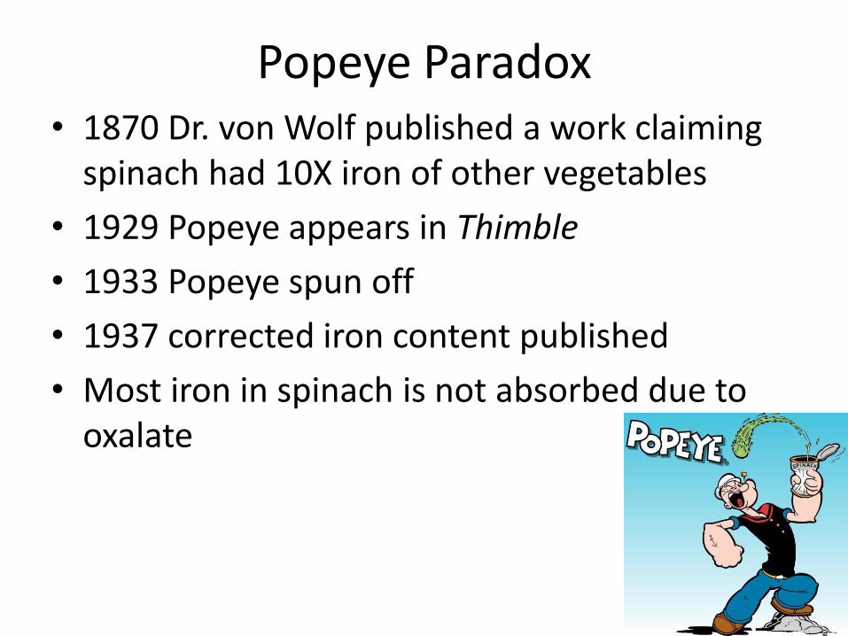
Popeye Paradox • 1870 Dr. von Wolf published a work claiming
spinach had 10X iron of other vegetables
• 1929 Popeye appears in Thimble
• 1933 Popeye spun off
• 1937 corrected iron content published
• Most iron in spinach is not absorbed due to oxalate
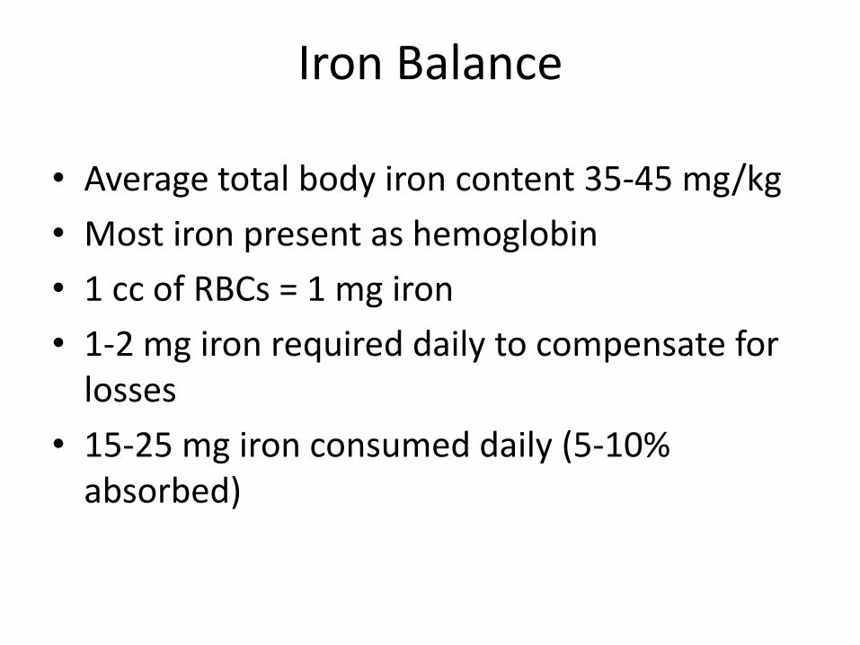
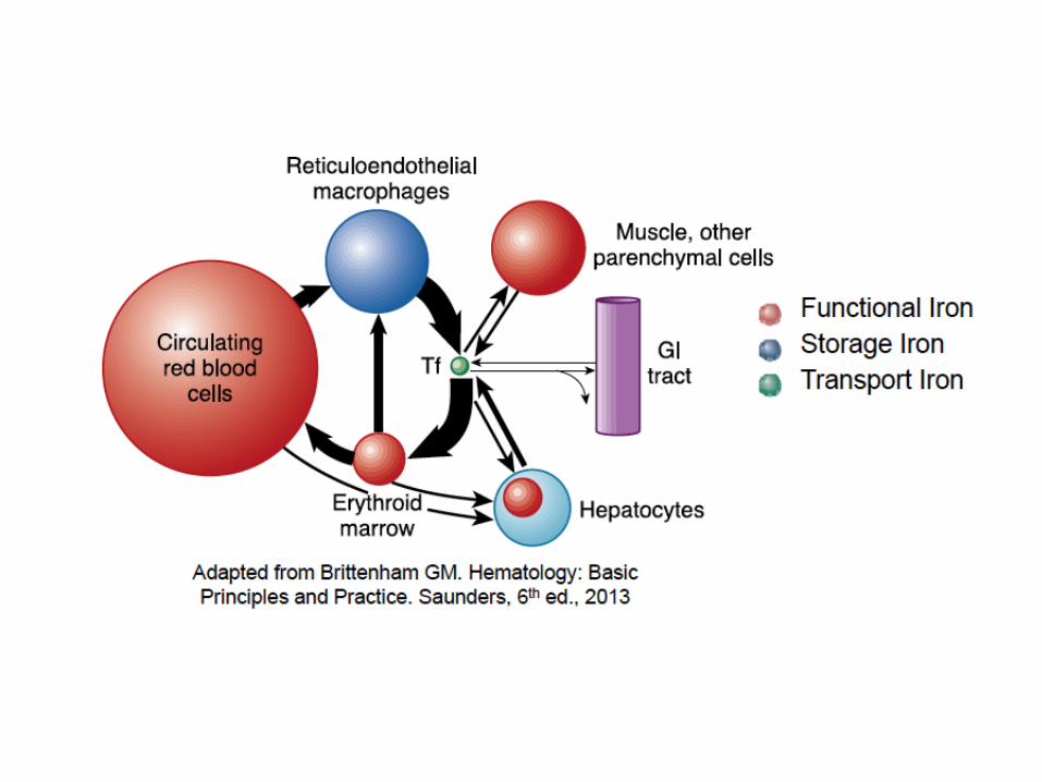
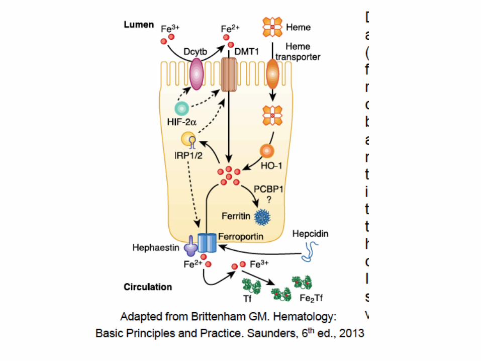
Iron Balance
• Average total body iron content 35-45 mg/kg
• Most iron present as hemoglobin
• 1 cc of RBCs = 1 mg iron
• 1-2 mg iron required daily to compensate for losses
• 15-25 mg iron consumed daily (5-10% absorbed)
Which of the following is the major regulator of iron balance?
A. Ferroportin
B. HFE
C. Transferrin receptor 2
D. Ferritin
E. Hepcidin
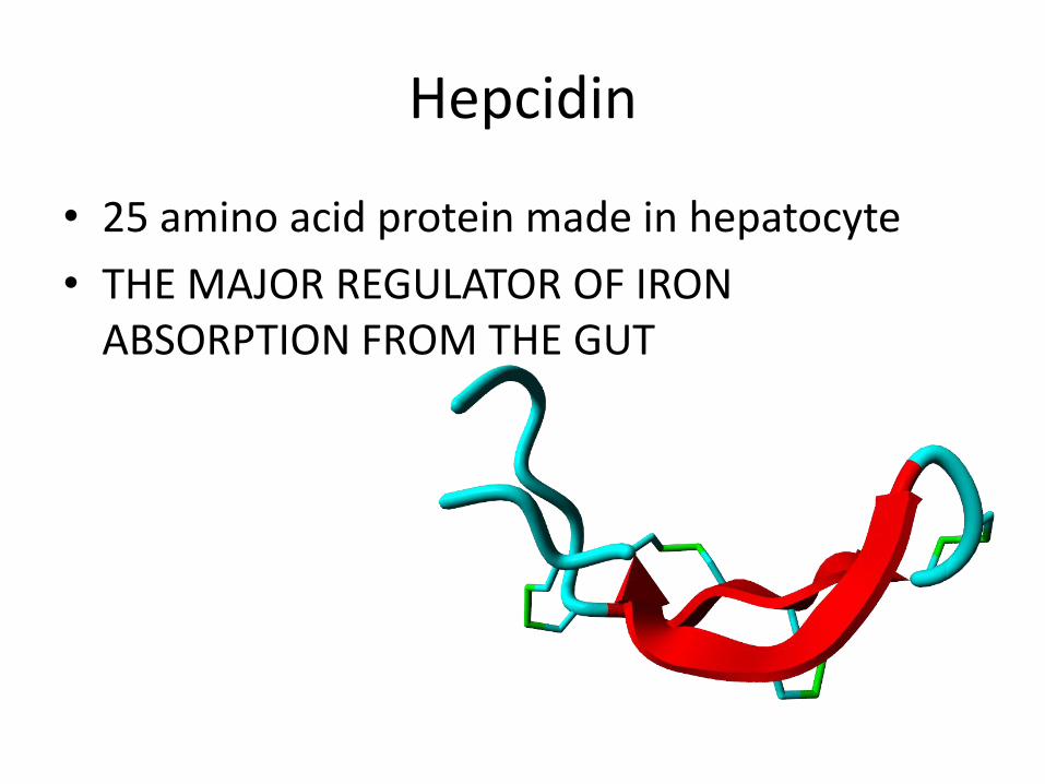
Hepcidin
• 25 amino acid protein made in hepatocyte
• THE MAJOR REGULATOR OF IRON ABSORPTION FROM THE GUT

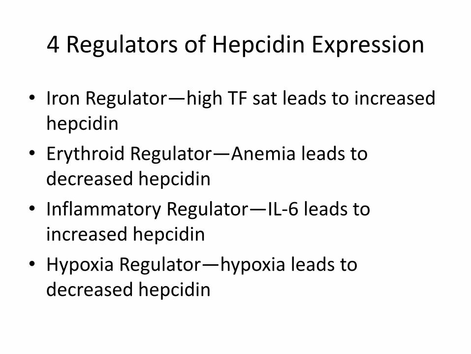
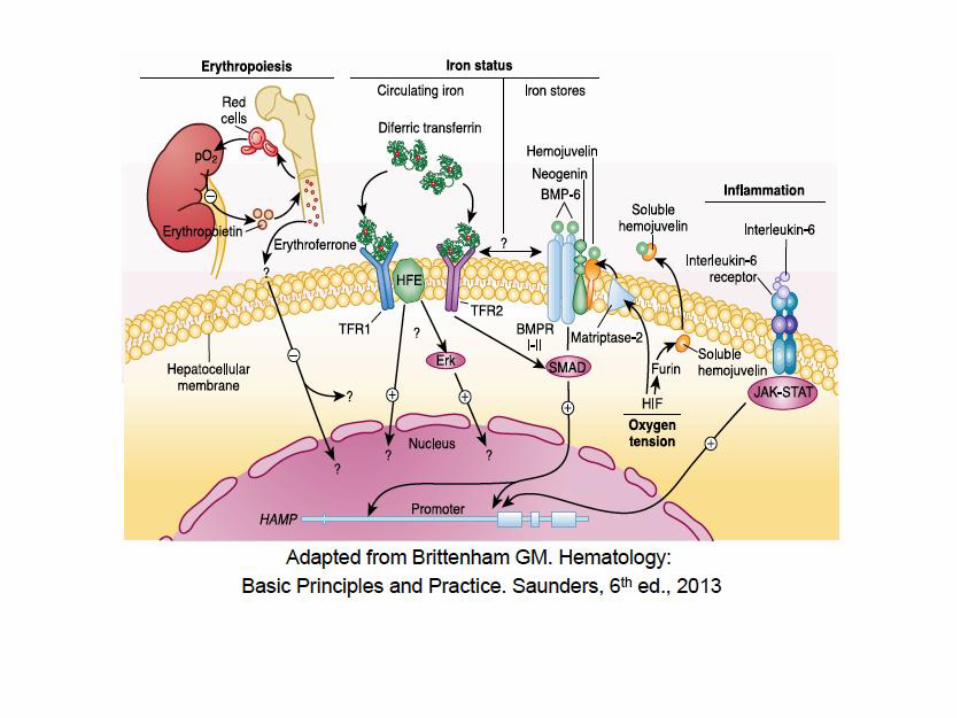
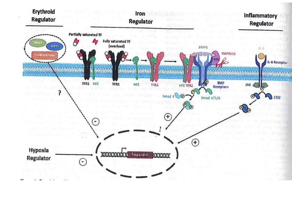
4 Regulators of Hepcidin Expression
• Iron Regulator—high TF sat leads to increased hepcidin
• Erythroid Regulator—Anemia leads to decreased hepcidin
• Inflammatory Regulator—IL-6 leads to increased hepcidin
• Hypoxia Regulator—hypoxia leads to decreased hepcidin


Back to the case
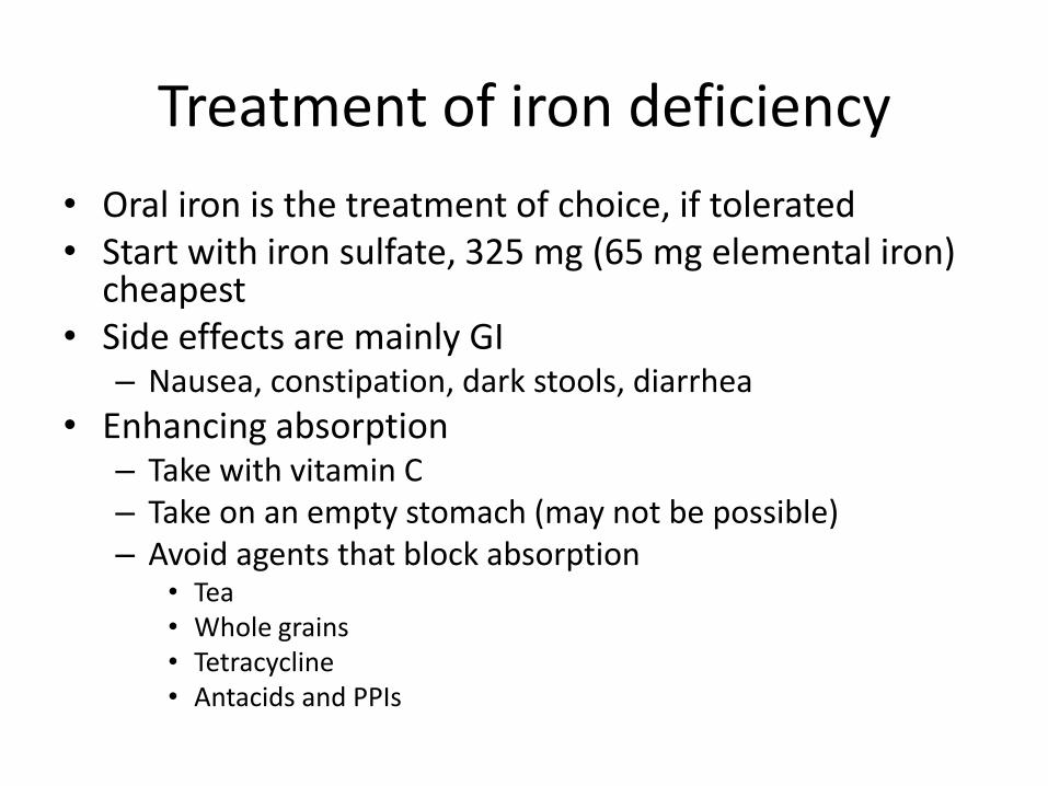
Treatment of iron deficiency
• Oral iron is the treatment of choice, if tolerated • Start with iron sulfate, 325 mg (65 mg elemental iron)
cheapest • Side effects are mainly GI
– Nausea, constipation, dark stools, diarrhea
• Enhancing absorption – Take with vitamin C – Take on an empty stomach (may not be possible) – Avoid agents that block absorption
• Tea • Whole grains • Tetracycline • Antacids and PPIs
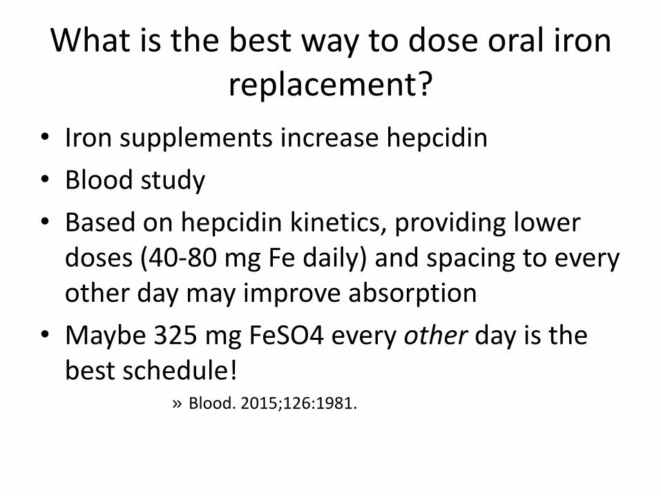
What is the best way to dose oral iron replacement?
• Iron supplements increase hepcidin
• Blood study
• Based on hepcidin kinetics, providing lower doses (40-80 mg Fe daily) and spacing to every other day may improve absorption
• Maybe 325 mg FeSO4 every other day is the best schedule!
» Blood. 2015;126:1981.

Case 2.
A 44 year old woman with Crohn colitis presents with severe iron deficiency anemia. Her hgb is 6.8 g/dL with an MCV of 56 fL. Her iron studies reveal a ferritin of 2 ng/mL. She reports intolerance to oral iron.

How to calculate iron deficit
mg iron deficit = 2.21 [desired hemoglobin-observed hemoglobin] x lean body weight in kg + (0.26 x lean body wt).
OR:
Wt (lbs) x (hgb desired – current hgb)
Add 600 mg for women and 1000 mg for men to replace stores
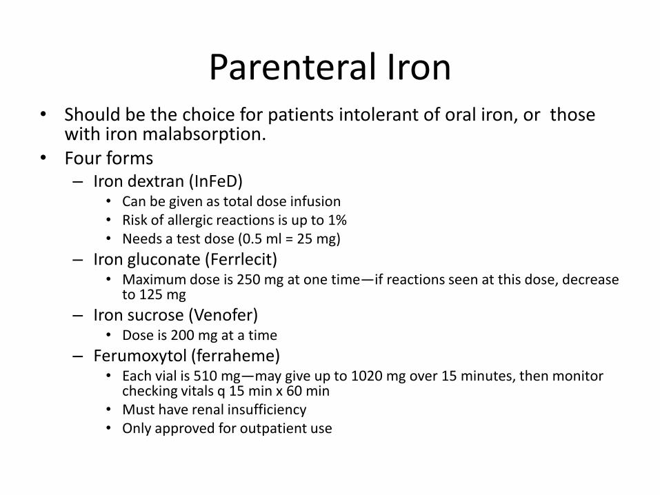
Parenteral Iron • Should be the choice for patients intolerant of oral iron, or those
with iron malabsorption. • Four forms
– Iron dextran (InFeD) • Can be given as total dose infusion • Risk of allergic reactions is up to 1% • Needs a test dose (0.5 ml = 25 mg)
– Iron gluconate (Ferrlecit) • Maximum dose is 250 mg at one time—if reactions seen at this dose, decrease
to 125 mg
– Iron sucrose (Venofer) • Dose is 200 mg at a time
– Ferumoxytol (ferraheme) • Each vial is 510 mg—may give up to 1020 mg over 15 minutes, then monitor
checking vitals q 15 min x 60 min • Must have renal insufficiency • Only approved for outpatient use
Case 3
• 24 y.o. Caucasian woman with no PMHx presents with iron deficiency. She has heavy menses. Not on OCPs.
• Her Hgb is 9.3. MCV 72. Ferritin is 4
• She is started on iron sulfate 325 mg
• 3 mo later, hgb is 8.5. She swears she is taking her iron.
• What are the next steps?
Iron challenge test
• Check serum iron. Give 325 mg iron sulfate. Check serum iron 1 hour later. Should go up by 100.
• Pt’s iron level rose from 32 to 355. She does not have iron malabsorption. Admits to getting nausea with meds. Hasn’t been taking iron.
Case 4 • 45 y.o. man has had iron deficiency for 4
years. Hgb hovers around 10. He feels terrible.
• Colonoscopy has been negative. Ferritin is 10.
• Tissue transglutaminase IgA is positive
• Anti-endomysial antibody is positive
• EGD shows flattening of villi. Gluten free diet leads to remarkable improvement in sense of well-being.
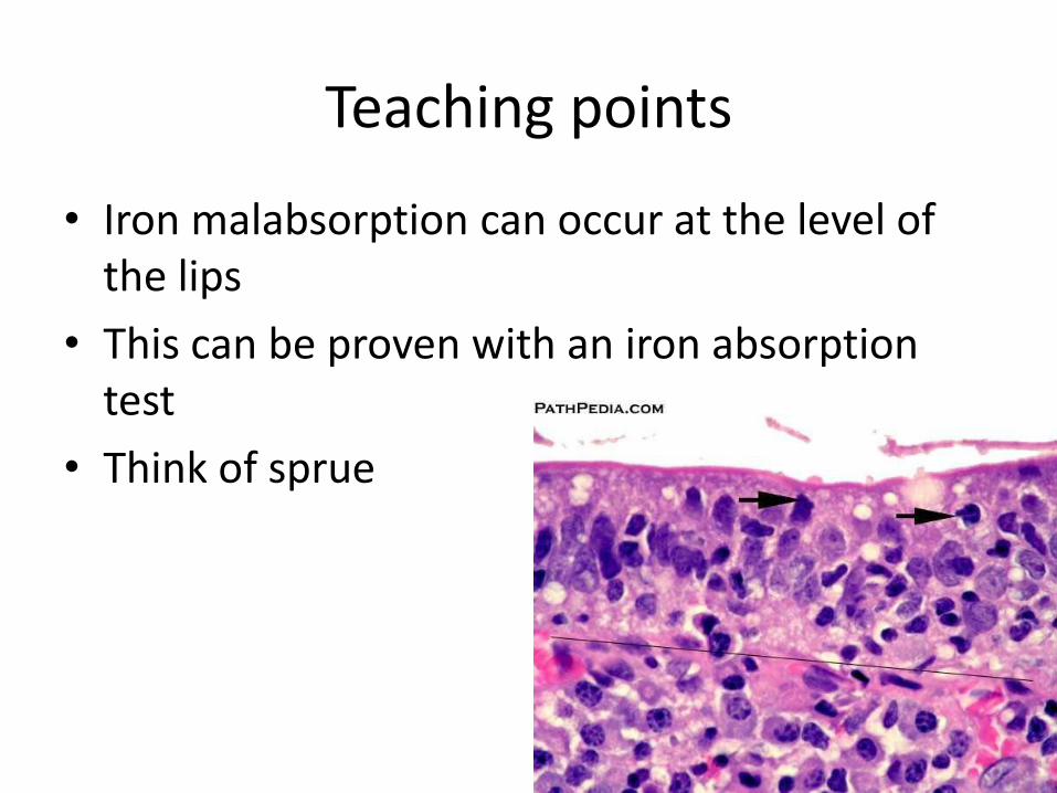
Teaching points
• Iron malabsorption can occur at the level of the lips
• This can be proven with an iron absorption test
• Think of sprue
Case 4
• 55 yo man with neurofibromatosis presents for evaluation of iron deficiency anemia. Hgb is 12 g/dL. Ferritin is 10 ng/mL. MCV has fallen steadily for the past 5 years—now 10 fL.
• Colonoscopy and EGD are negative.
• What is going on?
• He is not a vegetarian.
Answers and teaching points
• He is a regular blood donor for the past 20 years.
• He only eats food out of vending machines—never eats vegetables—never eats meat.
• So a good dietary history is important.
• Not all blood loss is involuntary!!
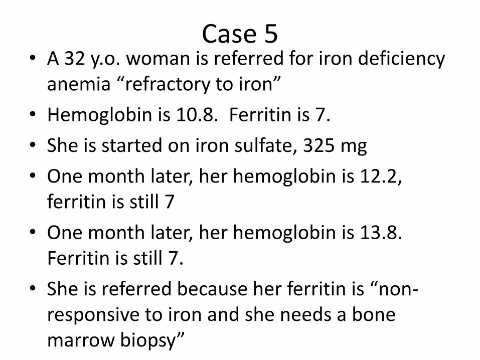
Case 5 • A 32 y.o. woman is referred for iron deficiency
anemia “refractory to iron”
• Hemoglobin is 10.8. Ferritin is 7.
• She is started on iron sulfate, 325 mg
• One month later, her hemoglobin is 12.2, ferritin is still 7
• One month later, her hemoglobin is 13.8. Ferritin is still 7.
• She is referred because her ferritin is “non-responsive to iron and she needs a bone marrow biopsy”
Case 5
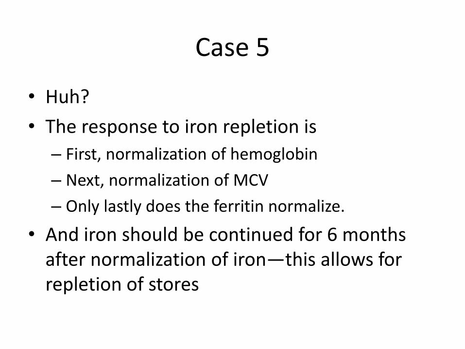
• Huh?
• The response to iron repletion is
– First, normalization of hemoglobin
– Next, normalization of MCV
– Only lastly does the ferritin normalize.
• And iron should be continued for 6 months after normalization of iron—this allows for repletion of stores
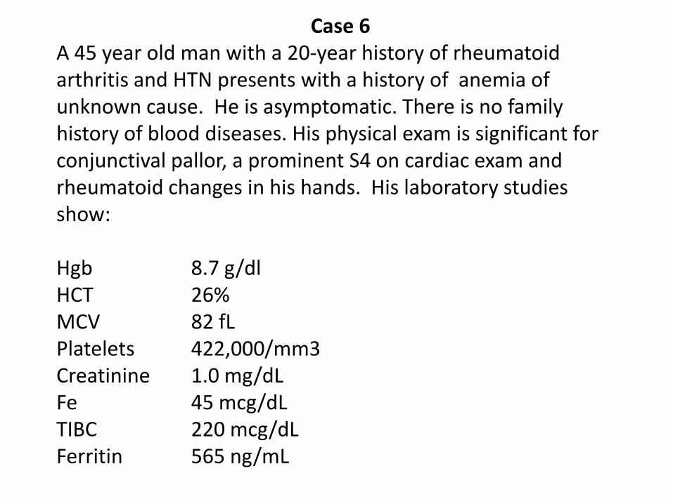
Case 6 A 45 year old man with a 20-year history of rheumatoid arthritis and HTN presents with a history of anemia of unknown cause. He is asymptomatic. There is no family history of blood diseases. His physical exam is significant for conjunctival pallor, a prominent S4 on cardiac exam and rheumatoid changes in his hands. His laboratory studies show: Hgb 8.7 g/dl HCT 26% MCV 82 fL Platelets 422,000/mm3 Creatinine 1.0 mg/dL Fe 45 mcg/dL TIBC 220 mcg/dL Ferritin 565 ng/mL
What do you want to do next?
ALWAYS look at the peripheral smear
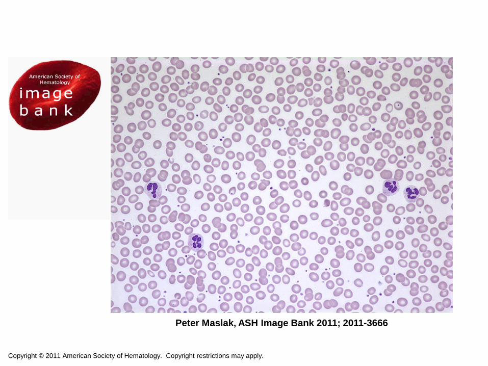
Copyright © 2011 American Society of Hematology. Copyright restrictions may apply.
Peter Maslak, ASH Image Bank 2011; 2011-3666
What is your diagnosis?
A. Anemia of chronic disease
B. Iron deficiency
C. Thalassemia trait
D. Hereditary spherocytosis
E. Sideroblastic anemia
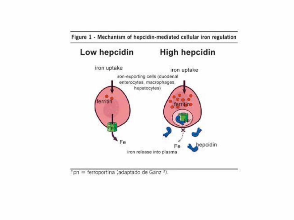
Anemia of chronic disease
• Modulated by hepcidin
• Small peptide
• Increased in response to inflammation
• Causes internalization and proteolysis of membrane channel ferroportin
• No commercially available assay for hepcidin

hepcidin enterocyte
macrophage
Reduced iron absorption
Increased iron accumulation
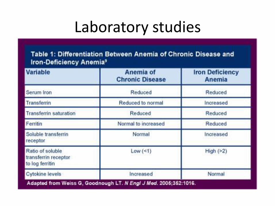
Laboratory studies
Case 7
A 78 year old man presents with unexplained elevation in his transaminases. His uncle and father both died of alcoholic hepatitis. He has developed glucose intolerance. His iron studies are significant for a ferritin of 775 ng/mL, iron sat of 55%.
Hereditary Hemochromatosis
• First described in mid 19th century
• Iron overload, bronzing of skin, endocrinopathies, arthropathies
• HFE mutations most commonly associated with hemochromatosis
• HFE described in 1996 but mechanisms not elucidated until more recently
• Original investigations focused on HFE interactions with gut

Skin “bronzing”
What are hepcidin levels in patients with hereditary hemochromatosis?
A. High
B. Low
C. Normal
D. Don’t know
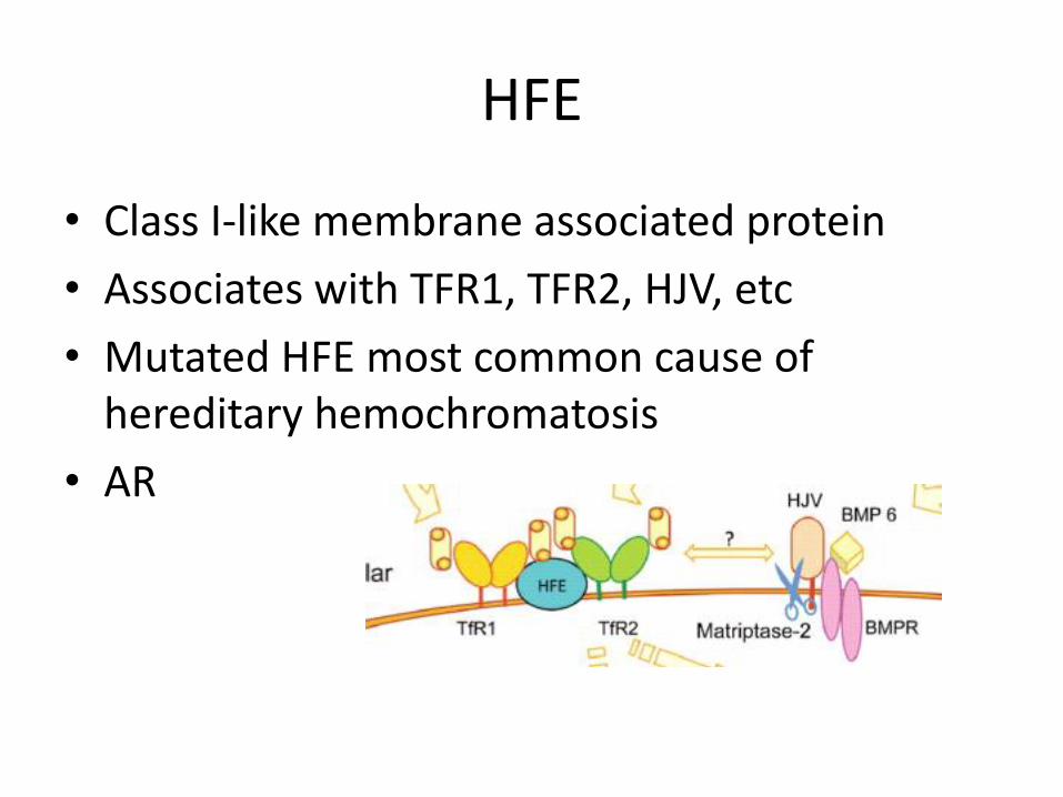
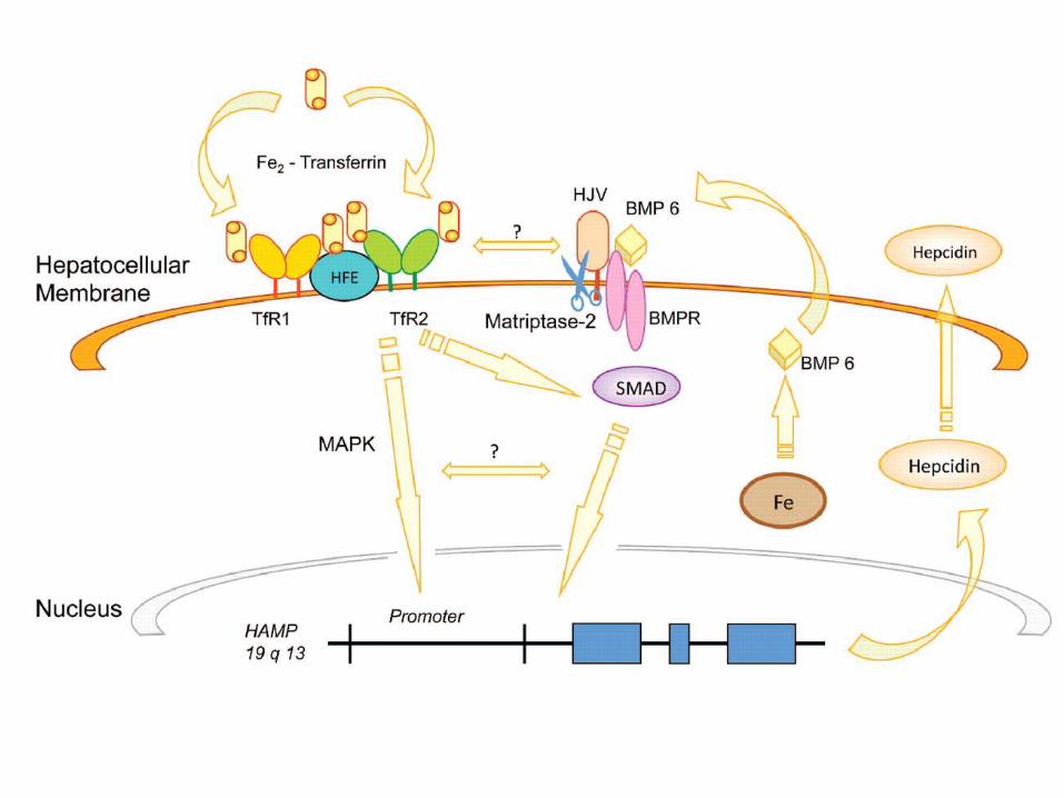
HFE
• Class I-like membrane associated protein
• Associates with TFR1, TFR2, HJV, etc
• Mutated HFE most common cause of hereditary hemochromatosis
• AR
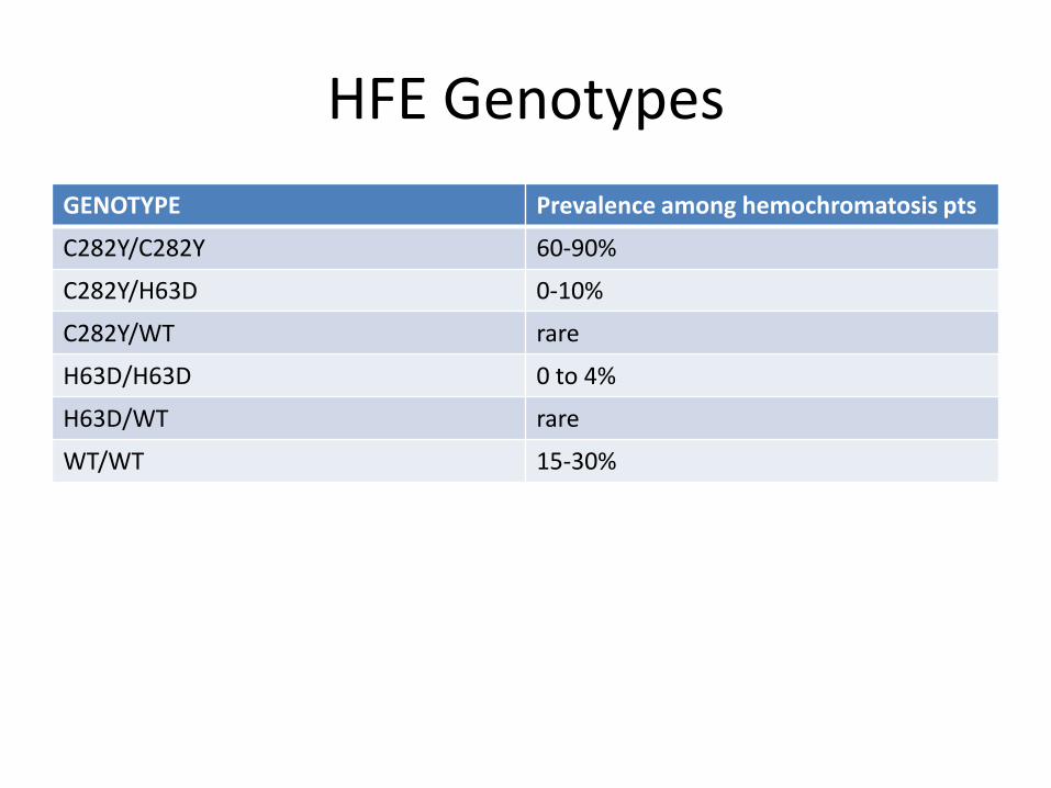
HFE Genotypes
GENOTYPE Prevalence among hemochromatosis pts
C282Y/C282Y 60-90%
C282Y/H63D 0-10%
C282Y/WT rare
H63D/H63D 0 to 4%
H63D/WT rare
WT/WT 15-30%
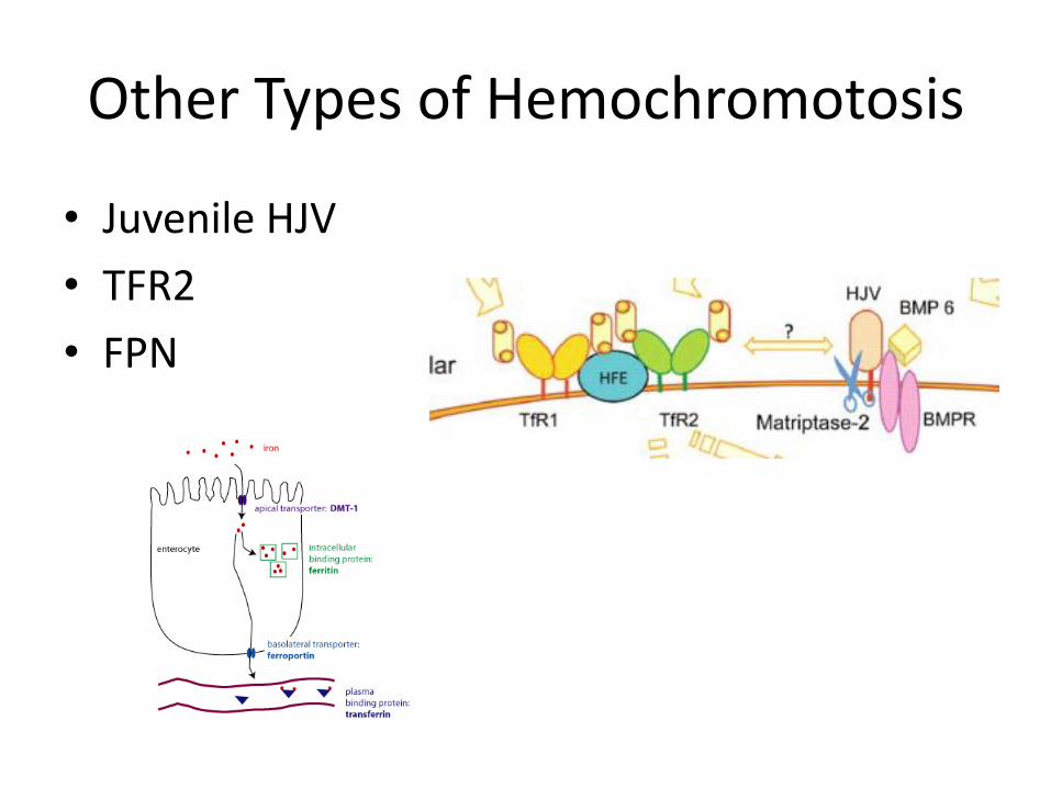
Other Types of Hemochromotosis
• Juvenile HJV
• TFR2
• FPN
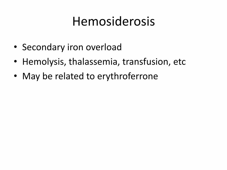
Hemosiderosis
• Secondary iron overload
• Hemolysis, thalassemia, transfusion, etc
• May be related to erythroferrone
ALL Associated with LOW Hepcidin
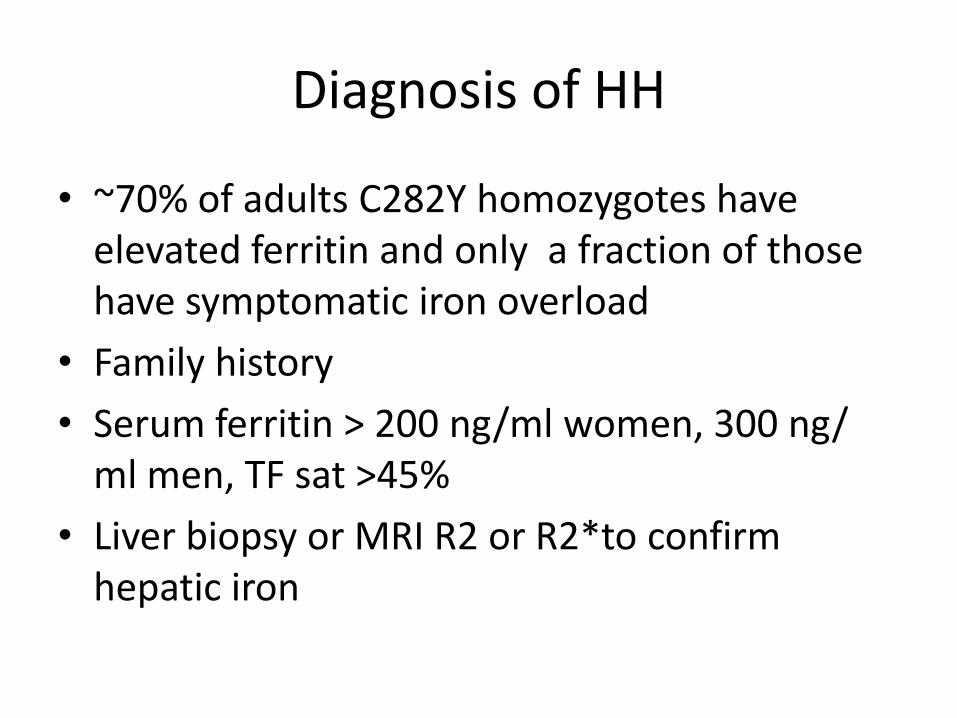
Diagnosis of HH
• ~70% of adults C282Y homozygotes have elevated ferritin and only a fraction of those have symptomatic iron overload
• Family history
• Serum ferritin > 200 ng/ml women, 300 ng/ ml men, TF sat >45%
• Liver biopsy or MRI R2 or R2*to confirm hepatic iron
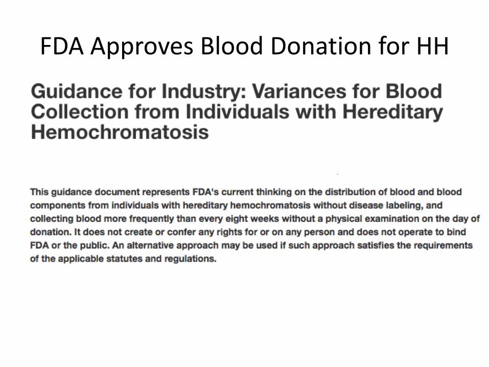
FDA Approves Blood Donation for HH
Treatment of Iron Overload
• Hemochromatosis—phlebotomy
• Hemosiderosis—chelation
• Historically used parenteral chelator deferoxamine
• Chelates in 1:1 ratio
Oral Iron Chelators
• Deferiprone (ferriprox®)—introduced in 1980’s
• Side effects: agranulocytosis (up to 5%), GI symptoms (33%), arthralgias and arthritis (30-40%),
Deferasirox (Exjade®)
• Well tolerated (GI symptoms most common)
• Well-studied
• COST: deferasirox > deferiprone> deferoxamine
• Can combine for added chelation
New Targets for Iron Overload
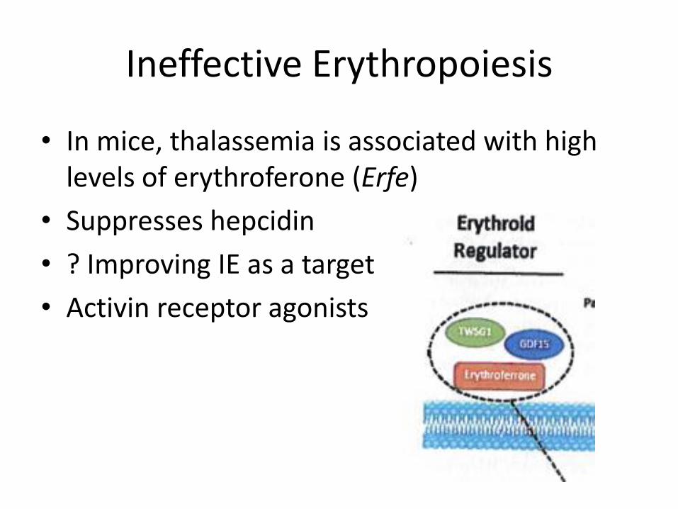
Ineffective Erythropoiesis
• In mice, thalassemia is associated with high levels of erythroferone (Erfe)
• Suppresses hepcidin
• ? Improving IE as a target
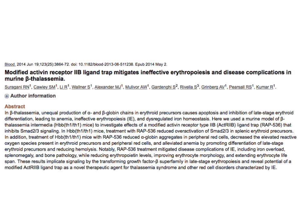
• Activin receptor agonists
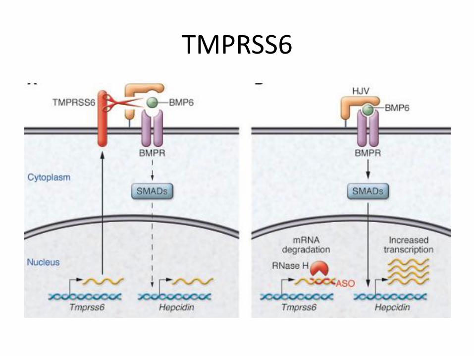
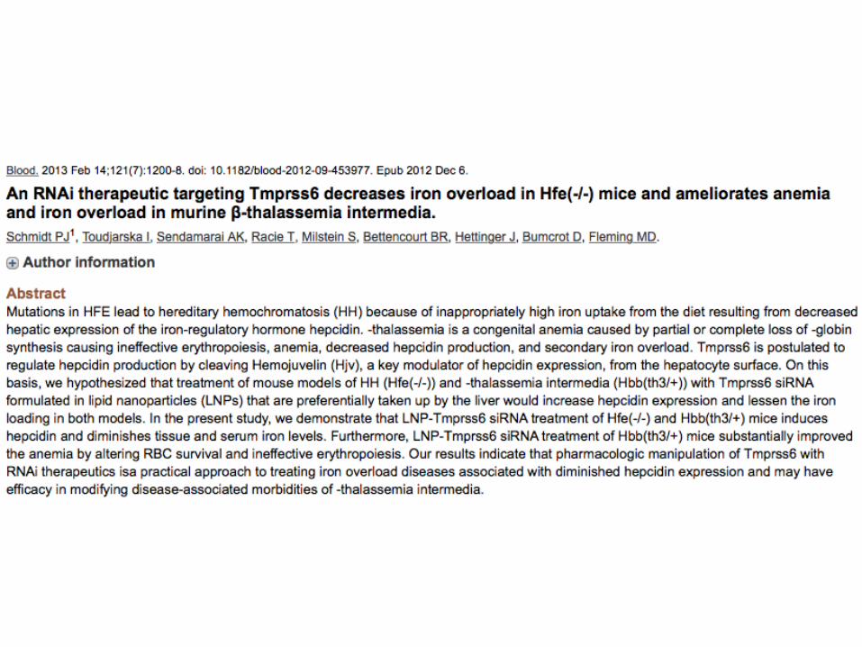
TMPRSS6
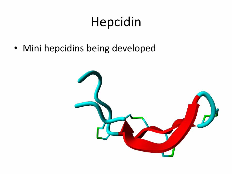
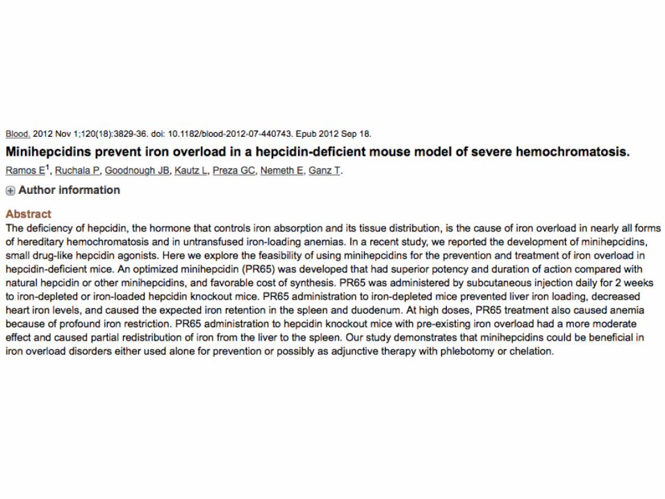
Hepcidin
• Mini hepcidins being developed
Pearls
• Best iron replacement may be 325 mg FeSO4 every other day
• Iron replacement: wt (kg) X D Hgb
• Iron challenge test can identify true non-absorbers
• Ferritin can take weeks to normalize
• Hemochromatosis is associated with low hepcidin