Embed Size (px)
Citation preview

Iron in the Liver: A Review for Surgical Pathologists
Hans Popper Society
USCAP 2010
Michael Torbenson, MD
Department of Pathology
Johns Hopkins University School of Medicine
1. DEFINITIONS
The clinical and basic science research communities have made significant progress over the
past several decades in understanding the causes and significance of iron accumulation in the liver. This
update is designed to summarize the major advances and also to synthesize the current literature in a
manner that is relevant to the practice of surgical pathology.
To start, some nomenclature issues: at times there have been inconsistent use of terminology
related to iron in the liver, which can be somewhat confusing. For example, what is precisely meant by
the term “hemochromatosis”? The term sometimes refers to any degree of tissue iron accumulation,
sometimes only to those cases with sufficient iron accumulation to cause tissue damage, sometimes to
those cases where the iron is only (or predominately) in the hepatocytes, and sometimes to any degree
of iron accumulation as long as there is evidence of genetic mutations. Likewise, the terms “primary”
and “secondary” iron accumulation are not always used in a consistent fashion. In this review, we shall
adopt for practical purposes the following: the term “hemochromatosis” indicates hepatic iron
accumulation in the setting of a genetic mutation, the term “siderosis” indicates hepatic iron
accumulation without genetic mutations, and the terms “primary” and “secondary” will be avoided. We
will also use the term “genetic non-hemochromatotic iron overload disorder” which has been proposed
for a range of rare genetic disorders that lead to iron accumulation that is primarily deposited in Kupffer
cells and macrophages.
Major proteins and cells involved in iron metabolism
There are many proteins and cells involved in iron metabolism that we will not be able to cover in this
review. However, the major ones are listed below for quick reference.
Proteins
DMT-1: Dimetal transporter-1. Transports iron from gut lumen to enterocyte cytoplasm
Ferritin: Protein that has a enormous capacity to bind iron; located in the cell cytoplasm
and a major physiological storage form of iron
Ferroportin: Transports iron out of cells into the blood stream (principally enterocytes and
macrophages, also hepatocytes)
Hemojuvelin: The precise role of this membrane bound protein is not clear. However, it
appears to interact with important signaling pathways (BMP, SMAD) that have
hepcidin as a down stream target. Without hemojuvelin, these signaling
pathways are not able to active hepcidin gene synthesis in a normal fashion.
Hemosiderin: Abnormal deposits of iron
Transferrin: Protein that transports iron in blood
Cells
Enterocytes: Absorption and short term storage of iron
Hepatocytes: Major producer of ferritin, hepcidin
Major organ for storage of iron in the form of ferritin
Macrophages: Main recycler of old/damage red blood cells
Major cell type for storage of iron in the form of ferritin
2. DIETARY CONSIDERATIONS
The recommend daily allowance (RDA) for iron is 8 mg per day for adult men and
postmenopausal women and 18 mg per day for premenopausal women. The “Tolerable Upper Limit of
Intake” for dietary supplementation is about 45 mg of iron per day in adults before there is
gastrointestinal distress. A detailed resource on iron and dietary considerations in health and general
growth and development is available free of charge from the United States Department of Agriculture
(USDA) at (http://desearch.nal.usda.gov/cgi-bin/dexpldcgi?qry1227124502;2). This report was written
by the National Academy of Sciences in 2001, so it is a bit dated, but it still has a wealth of information.
The latest available iron RDA, published in 2005, keeps to essentially the same RDA as noted above but
with a much greater breakdown by age, gender, and caloric intake
(http://www.cnpp.usda.gov/DGAs2005Guidelines.htm). Updated RDA for iron should be published by
the USDA in 2011 (http://www.cnpp.usda.gov/dietaryguidelines.htm).
Iron in the diet comes in two principal forms: heme iron from meats, including poultry and fish,
and non-heme iron from grains, legumes, vegetables and fruits. Heme-iron is more readily absorbed
than non-heme iron. Some fruits and vegetables that are naturally high in iron content include green
leafy vegetables such as spinach and broccoli; most dried beans such as lima beans, kidney beans, etc;
dried fruit such as raisins and prunes; and citrus such as oranges, lemons, and grapefruit. Vitamin C can
enhance iron absorption.
3. OVERVIEW OF NORMAL IRON METABOLISM
The normal adult body contains a total of 3-5 grams of iron. About 20 mg of iron is needed each
day for normal physiological functions, largely heme synthesis, but the majority of this daily need is met
through recycling of damaged and no longer properly functioning red blood cells. Because of the
efficiency of this red blood cell recycling, only 1 to 2 mg per day are needed in a healthy diet, though the
Recommended Daily Allowance (RDA) is somewhat higher at 8-18 mg of iron.
Iron is important in a number of metabolic processes outside of heme synthesis, including
oxidative phosphorylation and DNA synthesis. Despite this, iron can be toxic at high levels and iron
levels within the body are tightly regulated. The human body has no physiological way to excrete iron
and regulatory mechanisms are instead focused on iron absorption from the intestine. Separate, but
integrated, controls also tightly regulate blood iron levels.
Iron Absorption
Most iron is absorbed in the duodenum and proximal jejunum by a protein called DMT-1, where
it is first sequestered into the cytoplasm of enterocytes. Iron can than be exported by a protein called
ferroportin into the blood stream where it is carried by the protein transferrin to sites of principal usage,
including the bone marrow for hemoglobin and the muscle for myoglobin. In healthy individuals, the
blood contains much more transferrin protein than iron and the transferrin levels are approximately
30% saturated with iron. As blood iron levels increase, the excess transferrin proteins serve as a sort of
buffer and will bind more iron to prevent excess free iron in the blood. Thus, increased serum
transferrin levels can serve as a sensitive early indicator of excess iron absorption. All nucleated cells
have transferrin receptors that can uptake transferrin bound iron to meet cellular needs.
Iron storage
If there is excess iron in the body, it can be incorporated into ferritin molecules for storage,
largely in hepatocytes and macrophages. Ferritin is produced principally by the liver and is found in the
liver cytoplasm, where it can hold up to 4500 atoms of iron per ferritin protein complex. Ferritin is
typically not observed on Perls’ Prussian Blue stain, but occasionally it can be seen as a diffuse blush of
blue in hepatocyte cytoplasm. The iron in ferritin can be rapidly accessed for physiological needs. If
ferritin levels are excessive over a sufficiently long period of time, hemosiderin deposits can then
develop.

Hemosiderin is typically granular and golden brown on H&E staining and is composed of iron
and various proteins, principally degraded ferritin. The vast majority of the metal in hemosiderin is iron,
but small amounts of copper and calcium can also be detected. Despite an identical H&E appearance of
hemosiderin in both genetic and non-genetic causes of iron overload, there are differences in both the
metallic as well as the organic components at the molecular level.1 In contrast to ferritin, the iron in
hemosiderin is not as readily available for biological needs.
In sum, there are two important reservoirs of iron that can both be tapped to keep iron levels in
the blood at physiologically correct levels: (1) a short term reservoir of iron stored within enterocytes
and (2) a longer term reservoir of iron stored as ferritin, principally in hepatocytes and macrophages.
Both reservoirs have separate but interconnected control mechanisms that serve to regulate iron flow
into the blood. If they both are unable to meet the demands for iron, then anemia develops; if they
have dysregulated (mutated) control mechanisms, then hemochromatosis can develop.
4. CONTROL OF IRON TRAFFICKING
Iron absorption
Iron is absorbed primarily in the duodenum and proximal jejunum. Heme-iron is taken up by
the enterocytes after disassociation from globin, while non-heme iron is first reduced from a ferric to a
ferrous state and then transported across the cell membrane into the enterocytes by a protein called
DMT-1. There are several additional iron transport mechanisms for getting luminal iron into the
enterocyte cytoplasm that we wont be able to discuss today. Once iron is within the enterocytes, it can
have several fates. If the body has sufficient iron stores, then the iron remains within the cytoplasm of
the enterocytes. When the enterocyte eventually dies, the iron within the cell’s cytoplasm is lost within
the fecal stream; this is a major control mechanism to prevent iron overload.
If the body needs iron, then the iron is transported out of the enterocyte by ferroportin, with
some help by accessory proteins including ceruloplasmin and hephaestin, and enters the blood stream
where it is bound by transferrin and circulates within the blood. Individual cells have mechanisms to
determine if they have sufficient iron stores within their cytoplasm to meet their needs. If not, then the
cells increase their expression of transferrin receptors (there are two, conveniently named transferrin
receptor 1 and transferrin receptor 2; receptor 1 is on all nucleated cells, while receptor 2 is primarily
found in the liver) and take in more transferrin bound iron. Hepatocytes, with their abundant
transferrin receptors, take up any excess iron which then can be stored in the form of ferritin and, in
times of great excess, as hemosiderin.
Control of iron release from stores in the enterocytes, liver, and macrophages
When blood levels are low, iron is released from enterocytes where it has been freshly absorbed
and released from hepatocytes and macrophages where it has been stored as ferritin. However, when

blood iron levels are sufficient, then iron is blocked from being released from these two compartments.
Hepcidin is a major controller of iron metabolism: it blocks the release of iron from hepatocytes,
macrophages, and enterocytes. When hepcidin levels are low, there is increased iron absorption from
the gut and increased release of iron into the blood.
Hepcidin is produced mainly by hepatocytes and is an acute phase reactant. Because it is an
acute phase reactant, hepcidin levels can be elevated in a variety of inflammatory and infectious
conditions. In addition to inflammation, hepcidin levels are also increased by excess body iron stores
and by tissue hypoxia. The main physiological role of hepcidin in healthy individuals is to lower blood
iron levels by blocking transfer of iron from enterocytes to the blood and by blocking the release of iron
stores from the liver and macrophages into the blood. Hepcidin accomplishes this by causing
degradation of feroportin, the protein that transports iron from enterocytes and macrophages into the
blood.
Recent findings have shown the central role of hepcidin in hemochromatosis. In fact, many of
the mutations that lead to hemochromatosis, whether in HFE, HAMP, HJV, TfR2, all lead to decreased
hepcidin production or impaired hepcidin function.2 The lack of hepcidin first manifests as increased
serum transferrin saturation levels. Later, increased serum ferritin levels are found and eventually
increased serum iron levels are seen. This chronic excess of blood iron levels eventually leads to the
accumulation of hemosiderin deposits in the liver and other organs.
5. MUTATIONS IN IRON RELATED GENES
“Cliff notes” version of genetic hemochromatosis
There are a number of mutations that lead to hemochromatosis. The number will probably
continue to grow with time. Despite this, these conditions share a core set of common findings as listed
below. I have found it very useful to understand genetic iron diseases by remembering these basic
observations:
(1) As noted previously, a common mechanism is that all mutations, at least in part, involve abnormally
low levels or dysfunction of hepcidin
(2) Most mutations are inherited—new sporadic mutations appear to very rare.
(3) Most mutations are recessively inherited.
(4) The clinical consequences include iron deposition in the liver, heart, joints, and endocrine organs.
There is an established increased risk for hepatocellular carcinoma and possibly increased risks for
cholangiocarcinoma as well as other non-hepatic malignancies.
(5) Blood findings progress in severity from elevated transferrin saturation levels, to elevated ferritin
levels, to elevated iron levels.
(6) Histologically, iron is deposited primarily in hepatocytes. Classic findings on iron stains for
hemochromatosis include a zone 1 distribution of iron deposits, a peri-canalicular pattern of iron
deposits within the hepatocyte cytoplasm, and iron deposits in bile duct epithelial cells. These features
are all fun to find, but they are neither very sensitive nor specific for hemochromatosis.
(7) Clinical management revolves around phlebotomy, which can be life saving as it can prevent the
clinical sequelae listed above (No. 4). Individuals have intact erythropoiesis so tolerate phlebotomy
well.
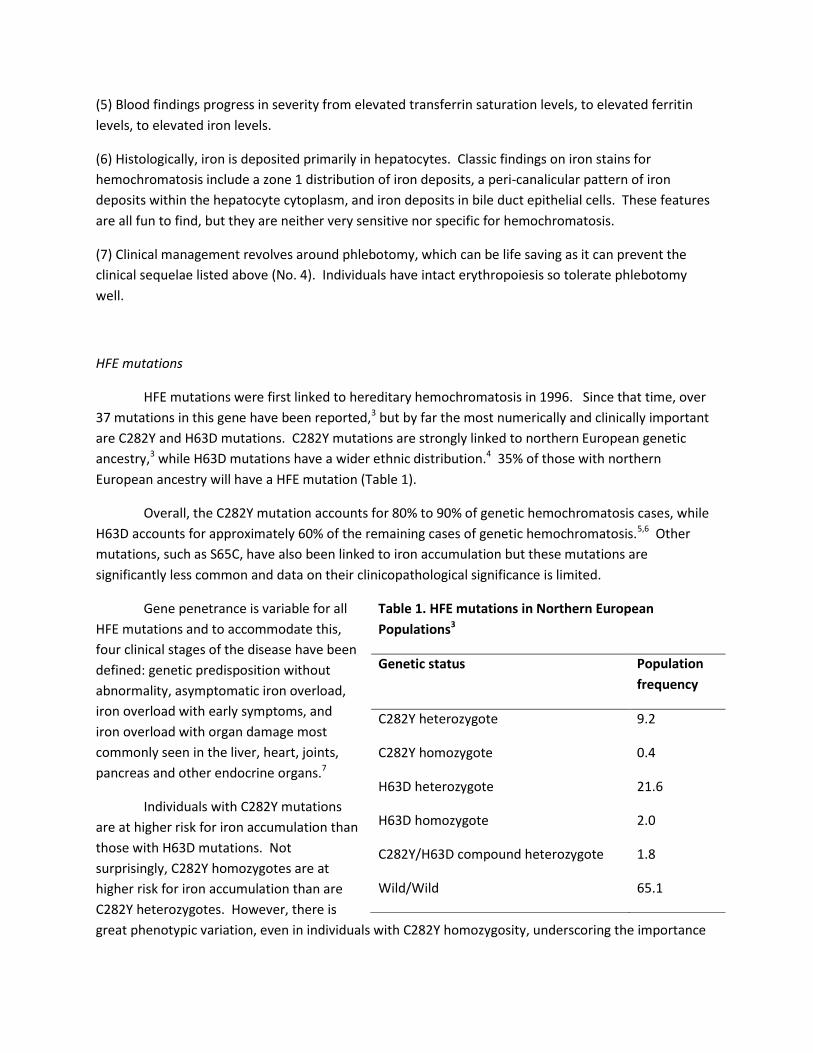
HFE mutations
HFE mutations were first linked to hereditary hemochromatosis in 1996. Since that time, over
37 mutations in this gene have been reported,3 but by far the most numerically and clinically important
are C282Y and H63D mutations. C282Y mutations are strongly linked to northern European genetic
ancestry,3 while H63D mutations have a wider ethnic distribution.4 35% of those with northern
European ancestry will have a HFE mutation (Table 1).
Overall, the C282Y mutation accounts for 80% to 90% of genetic hemochromatosis cases, while
H63D accounts for approximately 60% of the remaining cases of genetic hemochromatosis.5,6 Other
mutations, such as S65C, have also been linked to iron accumulation but these mutations are
significantly less common and data on their clinicopathological significance is limited.
Gene penetrance is variable for all
HFE mutations and to accommodate this,
four clinical stages of the disease have been
defined: genetic predisposition without
abnormality, asymptomatic iron overload,
iron overload with early symptoms, and
iron overload with organ damage most
commonly seen in the liver, heart, joints,
pancreas and other endocrine organs.7
Individuals with C282Y mutations
are at higher risk for iron accumulation than
those with H63D mutations. Not
surprisingly, C282Y homozygotes are at
higher risk for iron accumulation than are
C282Y heterozygotes. However, there is
great phenotypic variation, even in individuals with C282Y homozygosity, underscoring the importance
Table 1. HFE mutations in Northern European
Populations3
Genetic status Population
frequency
C282Y heterozygote 9.2
C282Y homozygote 0.4
H63D heterozygote 21.6
H63D homozygote 2.0
C282Y/H63D compound heterozygote 1.8
Wild/Wild 65.1

of other factors such as polymorphisms or mutations in other genes, environmental influences, and
demographics such as age and gender. For example, in a major population-based study from Australia,
203 individuals who were homozygous for C282Y mutations were followed for 12 years. Twenty-eight
percent of men, but only 1% of women, developed iron-overload related diseases.8 This same research
group also examined C282Y/H63D compound heterozygotes and found that only 1/82 men and none of
95 women developed iron overload related disease over a 12 year study interval.9 This and other data
argues for a strong protective effect for female gender. However, this does not appear to be solely due
to physiological blood loss and other gender associated polymorphisms appear likely (reviewed in Wood
et. al.).10
Table 2. Morbidity in individuals with clinical HFE
hemochromatosis
Data Milman
et al11
Niederau
et al12
Fargion et
al13
Number of deaths 147 69 44
Length of follow-up 8.5 yrs,
median
14 yrs,
mean
4 yrs,
median
Ethnicity Danish German Italian
Causes of Death (%)
Cirrhosis, no cancer 32 20 23
Hepatocellular
carcinoma
23 28 45
Non-liver cancer 11 12 14
Cardiovascular disease 11 20 7
Cerebrovascular
disease
5 Not stated Not stated
Respiratory disease 5 Not stated Not stated

The mechanism by which HFE
mutations lead to iron accumulation are
incompletely understood. At this time there are two major theories. The first suggests that the HFE
protein is critical in determining the enterocytes internal “set-point” for determining its cellular iron
state. With HFE mutations, the enterocyte set-point incorrectly indicates the cell is iron-deficient,
leading to increased enterocyte absorption of iron. The second theory focuses on the observation that,
for incompletely understood reasons, individuals with HFE mutations have abnormally low plasma
hepcidin levels. These low levels of hepcidin then lead to gradual excess iron absorption and deposition
in the hepatocytes and other organ tissues. Both theories have supporting data from animal models as
well as human observations, suggesting that both will be at least partially correct in the end.
Causes of death in HFE related hemochromatosis
Clinical follow-up studies have consistently identified liver de-compensation from cirrhosis as
well as hepatocellular carcinoma as leading causes of death in individuals who are untreated or
incompletely treated for HFE hemochromatosis (Table 2). However, there is also an increased risk for
morbidity from heart failure and complications of diabetes. An increased risk for non-liver cancer has
also been identified in some but not all studies. Treatment by phlebotomy can substantially lower the
risk of death. A single unit of blood can safely remove 200-450 mg of iron and over a period of time,
usually a year or two, phlebotomy can restore safe levels of iron within the blood.2
Liver transplantation for HFE iron overload
Overall, hereditary hemochromatosis is an uncommon indication for liver transplantation.
An early study of liver transplant outcomes that examined 5,180 liver transplantations reported
only 56 (1%) of the transplantations were for hemochromatosis.14
This and other early studies
reported an overall decreased post transplant survival rate for patients with hereditary
hemochromatosis compared to those transplanted for other causes of chronic liver disease, with
major causes of mortality including infection, cardiac failure, and cancer.14-16
However, a more
recent study has shown great improvement over the last decade in the survival of individuals
transplanted for hemochromatosis.17
This increased survival likely reflects better patient
selection and better pre and post-transplant management. Despite this, cardiovascular disease
continues to be an important cause of morbidity and mortality.17
Hemojuvelin mutations (usually children/early onset). Hemojuvelin mutations are the most common
cause of juvenile hemochromatosis. Nevertheless, this remains are relatively rare disease. There can be
marked hepatocellular iron overload and the disease typically runs a severe clinical course.
sepsis 3 Not stated Not stated
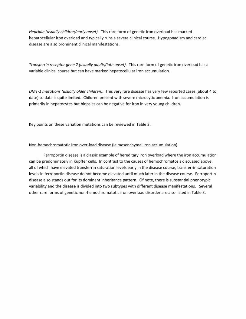
Hepcidin (usually children/early onset). This rare form of genetic iron overload has marked
hepatocellular iron overload and typically runs a severe clinical course. Hypogonadism and cardiac
disease are also prominent clinical manifestations.
Transferrin receptor gene 2 (usually adults/late onset). This rare form of genetic iron overload has a
variable clinical course but can have marked hepatocellular iron accumulation.
DMT-1 mutations (usually older children). This very rare disease has very few reported cases (about 4 to
date) so data is quite limited. Children present with severe microcytic anemia. Iron accumulation is
primarily in hepatocytes but biopsies can be negative for iron in very young children.
Key points on these variation mutations can be reviewed in Table 3.
Non-hemochromatotic iron over-load disease (ie mesenchymal iron accumulation)
Ferroportin disease is a classic example of hereditary iron overload where the iron accumulation
can be predominately in Kupffer cells. In contrast to the causes of hemochromatosis discussed above,
all of which have elevated transferrin saturation levels early in the disease course, transferrin saturation
levels in ferroportin disease do not become elevated until much later in the disease course. Ferroportin
disease also stands out for its dominant inheritance pattern. Of note, there is substantial phenotypic
variability and the disease is divided into two subtypes with different disease manifestations. Several
other rare forms of genetic non-hemochromatotic iron overload disorder are also listed in Table 3.

Table 3. Overview of Genetic iron diseases involving the liver
Gene AKA (some names used in the
literature)
Chromo-
some
Transmission Onset Iron location
HFE Hemochromatosis type 1 6p21.3 Recessive Late Hepatocytes >
Kupffer cells
HJV (hemojuvelin) Juvenile hemochromatosis type
2A
1p21 Recessive Early Hepatocytes >
Kupffer cells
HAMP (hepcidin) Juvenile hemochromatosis type
2B
19q13.1 Recessive Early Hepatocytes >
Kupffer cells
TfR2 Hemochromatosis type 3 7q22 Recessive Late Hepatocytes >
Kupffer cells
SCL11A2
(DMT-1)
None yet 12q13 Recessive Early Hepatocytes >
Kupffer cells
SLC40A1
(ferroportin)
Ferroportin disease type B 2q32 Dominant Late Hepatocytes >
Kupffer cells
Diseases with iron deposited primarily in mesenchymal cells
SLC40A1
(ferroportin)
Ferroportin disease type A
(hemochromatosis type 4)
2q32 Dominant Late Kupffer cells>
hepatocytes
Tf
(Transferrin)
Hypotransferrinemia 3q21 Recessive Early Kupffer cells>
hepatocytes
CP
(Ceruloplasmin)
Hypoceruloplasminemia 3q23-35 Recessive Late
Links to other chronic diseases

There are complex genetic, environmental, and dietary variables that determine the penetrance
of disease in individuals with mutations in iron metabolism genes such as HFE. Thus, it is not surprising
that other chronic liver diseases have been linked to iron overload and /or HFE mutations. As discussed
in more detail in section 8, certain diseases such as alpha-1-antitrypsin and cryptogenic cirrhosis (many
of which are now presumably NAFLD related) can show marked iron accumulation, with iron levels that
equal those of genetic hemochromatosis. Whether or not such cases are enriched for HFE mutations is
unclear and will require future studies, but it seems biologically plausible for one HFE mutation to
predispose to iron accumulation, and for that predisposition in turn to become increasingly penetrant in
the setting of a second significant liver disease.
The relationship between disease severity and the presence of iron accumulation and/or the
presence of HFE mutations has been investigated by numerous studies for many of the major chronic
liver diseases, including chronic viral hepatitis C and B, alcohol related liver disease, and non-alcoholic
fatty liver disease. While the data is substantially mixed, there is evidence to support an association
between more severe disease and excess iron accumulation in all of these chronic liver diseases. The
many negative studies highlight the difficulty of identifying what is most likely a modest impact for iron
in the very complex setting of clinical cohort studies where it is very difficult to adequately control for all
of the factors that have been reported to influence iron status.
6. DETECTION OF IRON IN THE LIVER
Significance
The normal adult liver has between 10 to 36 μmol iron/g dry weight of liver. Iron in the range
of 400 μmol and above can cause cirrhosis; lower levels of iron may also be relevant to fibrosis
progression if there are concurrent diseases.
Iron stains
The major histochemical stain used to detect iron in the liver is Perls’ Prussian Blue (note that
the most correct spelling is Perls or Perls’ Prussian Blue, not Perl’s Prussian Blue). This stain is named
after Max Perls, a German pathologist who first suggested the stain. The basic chemistry of Perls’
Prussian Blue is that iron in the ferric state will react with hydrochloric acid to form ferric ferrocyanide,
an insoluble blue compound (Prussian Blue) that can be seen histologically. The distribution and density
of blue staining correlates, albeit imperfectly, with tissue iron concentrations. The stain is not as
sensitive for very low levels of iron but is easier and more reproducible than other methods such as the
Tirmann-Schmeltzers method, which can identify both ferric and ferrous forms of iron.
Ferritin: Normally no ferritin will be seen. However, in cases of elevated serum ferritin levels,
ferritin may be seen as a light, diffuse, blue blush of the hepatocyte or Kupffer cell cytoplasm.

Hemosiderin: Hemosiderin can be seen as brown granular deposits on H&E stains and as a
bright blue granular staining on iron stain. Residual brown granular material is often seen on iron stain
and represents lipofuschin in most cases.
Iron grading systems
There are many iron grading systems that have been proposed over the years. They vary
considerably in their approach: some are based on zonation of iron distribution, some on the lowest
magnification that discernable granules can be seen, some on the percent of hepatocytes positive for
iron. There is a nice summary of these iron grading systems available on line at
(http://tpis1.upmc.com:81/tpis/dlp/DLPHome.html), then click on the Chapter 9 and find Table 9-3. This
book chapter is somewhat dated and does not cover several newer systems, but is still very useful. The
system by Turlin et. al.18 has the advantage of having been validated, but it is too complex to be readily
adopted for routine diagnostic use.
Is one system clearly the best? Probably not, but I personally use a schema (Table 4) based on
the percent of hepatocytes positive for iron, similar to that described by LeSage et. al.19 For routine
diagnostic purposes, I include
the descriptor (e.g. “mild” etc)
in the pathology report but do
not routinely provide the
corresponding numerical grade.
I believe that this simple-to-use
classification system provides
sufficient clinical information
for patient care. But there are
many reasonable alternatives
to consider if you prefer a
different approach. A modified
Scheuer’s system (shown in
Table 5) is also a very useful
and popular system. If
employed, separate numbers
should be given for hepatocellular and the reticuloendothelial iron.
Table 4. My iron scoring system (similar to that of LeSage)19
Grade Description Hepatocytes Lobular Kupffer cells
0 none none none
1 Minimal < 5% < 5%
2 Mild 5-30% 5-30%
3 Moderate 31-60% 31-60%
4 Marked >60% >60%
Note: For studies, I also record the zonal pattern of iron and whether the
distribution is homogenous. For some studies, I also record endothelial iron and
portal macrophage iron.

Table 5. Modified Scheuer’s20
Grade Description
0 Iron granules absent or
Iron granules barely seen at 400X
1 Iron granules resolved at 250X
2 Iron granules resolved at 100X
3 Iron granules resolved at 25X
4 Iron deposits resolved at 10X or
Iron deposits visible without magnification
Quantitative measurement of hepatic iron concentrations
As noted previously, the normal adult liver has between 10 to 36 μmol iron/g dry weight of liver.
Hepatic iron concentrations measured in fresh liver tissues or in paraffin embedded tissues are
equivalent. Thus, paraffin embedded tissues are preferred over fresh tissues in most cases because it
allows direct visualization of the tissue and assures the tissue is representative. This prevents
submission of tissue that is largely composed of collapsed/fibrotic stroma or a nodule that is either
unusually high or low in stainable iron compared to the rest of the tissue. Excess iron accumulation has
been classified as mild (up to 150 μmol iron/g dry weight of liver), moderate (151-300), and marked
(>301).18 Iron levels greater than 400 μmol are the most strongly associated with cirrhosis, but lower
levels of iron also contribute to fibrosis progression in the setting of other liver diseases.
Hepatic Iron Index
Historically, the hepatic iron index was calculated as an aide to interpreting quantitative tissue
iron levels. The hepatic iron index adjusts the total iron concentration for age, based on the observation
that hepatic iron concentrations tend to increase steadily with age in individuals with genetic
hemochromatosis, but not in individuals with “secondary” iron overload. In a non-cirrhotic liver, a
hepatic iron index greater than 1.9 was considered suggestive of genetic hemochromatosis. Given the
advances in understanding the causes of hemochromatosis and the readily available genetic testing for
HFE mutations in many parts of the world, the diagnostic role of the hepatic iron index has diminished in

importance, but direct measurement of hepatic iron concentrations remain useful in guiding therapy
and we still get many requests for blocks to be submitted for quantitative iron analysis.
Non-invasive measurements of hepatic iron
MRI based imaging studies have advanced in recent years to the point that they can reasonably
assess iron accumulation and can also distinguish hepatic from reticuloendothelial iron deposits. There
have been multiple validation studies and MRI has established for itself an important role in measuring
iron in the liver. Recent expert opinion review articles on hemochromatosis have highlighted the
changing role of the biopsy in managing patients with HFE hemochromatosis.2,21 Biopsies continue to be
important in determining the fibrosis stage and to search for any associated lesions (eg evaluation of
mass lesion). However, some experts21 foresee a further diminution of the role for liver biopsies with
the advent of non-invasive markers of liver fibrosis.
7. HISTOLOGICAL FINDINGS
In genetic hemochromatosis, iron classically accumulates initially within zone 1 hepatocytes. A
clear gradient in the amount of iron between zone 1 and zone 3 hepatocytes can often be seen, even
with advanced iron accumulation. In addition, the iron distribution often has a distinctive clustering
around the bile canaliculi. With time, injury and death of hepatocytes will lead to a redistribution of iron
into Kupffer cells and portal macrophages. However, a zone 1 distribution of iron can be seen in other
non-hemochromatosis conditions, particularly once a liver is cirrhotic, and a diagnosis of
hemochromatosis should not be based on recognizing a zonal pattern alone. My personal opinion is
that the zone 1 predominate pattern of iron deposition can be seen whenever there is dysregulation of
hepcidin, either through mutations or through reduced hepcidin production from other causes.
Iron can also be seen in biliary epithelium on iron stain. First, iron is commonly seen in
proliferating bile ductules in areas of subacute parenchymal collapse in cirrhotic or non-cirrhotic livers.
This finding appears to have no association with hemochromatosis. Iron can also be deposited in the
epithelium of the bile duct proper. In my experience, this pattern of iron deposition tracks better with
the overall severity of iron deposition within the liver and less so with HFE mutations per se. However,
there is very little data that examines this specific question.
With iron overload due to transfusion dependent anemias and similar causes, iron is classically
first deposited in Kupffer cells and with time there is involvement of the hepatocytes. However, in
practice most cases show a mixed hepatocellular and Kupffer cell iron staining pattern.
Iron can also be seen in some cases either exclusively in portal endothelial cells or in a
combination of endothelial, hepatocyte, and Kupffer cell iron accumulation. At this time, there has not
been any specific linkage of endothelial iron accumulation to a disease process or genetic mutation. In

one study, endothelial iron positivity was linked to decreased interferon response in individuals with
chronic hepatitis C infection.22
8. CLINICOPATHOLOGICAL SIGNIFICANCE OF IRON OVERLOAD
Iron in explanted livers
Iron can accumulate in cirrhotic livers of individuals who do not have clinical findings of genetic
hemochromatosis. In a classic study by Ludwig et. al., iron stains were positive in 32% of 447 liver
explants with varying underlying liver diseases. For those diseases with at least 5 cases in this study, the
proportions of cases with any degree of positivity by iron stain were as follows: hereditary
hemochromatosis (100%), cryptogenic cirrhosis (65%), alcohol cirrhosis (63%) chronic hepatitis B
cirrhosis (65%), A1AT cirrhosis (56%), chronic hepatitis C cirrhosis (42%), primary biliary cirrhosis (10%),
and primary sclerosing cholangitis (7%).
In this same study, the number of cases with a hepatic iron index of greater than 1.9 were as
follows: HH (100%), A1AT (28%), cryptogenic cirrhosis (19%), alcohol cirrhosis (14%), chronic hepatitis B
cirrhosis (18%), chronic hepatitis C cirrhosis (7%), primary biliary cirrhosis (1%), and cirrhosis from
primary sclerosing cholangitis (1%). This and other data sets document that other diseases can have
iron deposition within the liver and that in alpha-1-antitrypsin deficiency and in cryptogenic cirrhotic
livers, 20% or more of cases can have hepatic iron indexes greater than 1.9. Another important
observation from these data is that biliary cirrhosis is only rarely associated with iron overload.
An important study by Kowdley et al.23 found that patients with significant hepatic iron
accumulation had decreased survival following transplantation regardless of whether they had an HFE
mutation. The reason(s) for this are unclear, but at least in a subset of these individuals, there can be
significant extrahepatic stores of iron at the time of transplantation, often which are clinically
unrecognized.24 The stress of surgery or other post transplant factors may then place this group of
patients at increased risk for heart failure.
When examining an explanted liver with iron overload, any foci (even smaller subcentimeter
foci) with decreased iron deposition should be targeted for sectioning to evaluate for carcinoma. These
“iron free foci” are often associated with dysplastic nodules or with frank carcinoma. They can rarely be
seen on needle biopsies also, and, when present, should be indicated in the report as well as fully
evaluated for malignancy.
Iron in donor liver biopsies
There is very little data on the relevance of iron levels in donor livers (an interesting area that
hopefully will get more attention in the future). One study is available that looked at the significance of
donor iron for subsequent fibrosis progression in individuals transplanted for chronic HCV. Counter-
intuitively, they found a link between female gender, pre-transplant iron content, and risk for fibrosis
progression.25
Iron in liver tissues with chronic hepatitis C Virus infection
Iron deposits, including both hepatocellular as well as reticuloendothelial, are seen in liver
biopsies of approximately 5 to 48% of individuals with chronic HCV.26-31 Overall, the median is
approximately 30% for these studies and the variation presumably reflects differences in gender, viral
genotypes, and the proportion of cirrhotics in the cohort. Livers with genotype 3 infection tend to have
more hepatocellular iron than other genotypes.29 In the majority of cases, the iron deposits are mild,
occasionally moderate, and only very rarely severe.
A large body of literature has been published on the question of the significance of HFE
mutations in chronic HCV. Some of the larger studies are summarized for you in Table 6 on the
following page. Unfortunately, despite all of the work, the literature is substantially mixed on the
question of whether HFE mutations increase the risk for fibrosis progression. This current state of
confusion likely reflects the many different study populations, study designs, as well as variable
penetration of genetic hemochromatosis. Many studies also do not adequately control for potentially
confounding variables such as gender, viral genotypes, duration of HCV infection, etc. Nevertheless, one
reasonable way to synthesize the data is as follows: (1) individuals with chronic HCV do not have an
increased risk for HFE mutations;26,32-34 (2) once an individual has chronic HCV infection, HFE mutations
may increase the rate of fibrosis progression35 and the presence of HFE mutations is associated with
higher fibrosis stages in many26,32-37 but not all studies.27,28 The strength of the association between HFE
mutations and fibrosis has been measured by both relative risks, where a relative risk of 4.6 has been
reported,34 as well as odds ratio, where odds ratios for C282Y heterozygosity has been reported ranging
from 2.5 to 30. 26 32,35 Overall, C282Y alleles appear to have a stronger risk for fibrosis than H63D
alleles.35 With a sufficiently long duration of chronic HCV infection, the risk of cirrhosis is high
regardless of HFE mutational status and the effect of HFE mutations may be harder to discern.35
Interestingly, for unclear reasons, HFE mutations have also been linked to increased
inflammation on liver biopsy in some studies.33,34 Despite the observations linking HFE mutations to
increased fibrosis and less consistently to increased inflammation, HFE mutation status has typically not
been associated with increased iron deposits by histochemical analysis.28,33,34 As an exception, H63D,
but not C282Y mutations, were associated with increased hepatic iron concentrations in one study.35
Most of the data discussed above is from studies that looked at HFE mutations. The question
then naturally arises of the meaning of mild to moderate iron deposits in individuals with chronic HCV

who lack HFE mutations. Unfortunately, the data is no clearer on this point than it is for HFE mutations
and the same “take home message” as above appears to apply: most likely there is either no role or a
very limited role for minimal or very mild iron on a liver biopsy in terms of fibrosis progression; for
moderate iron there is likely a modest role. For marked iron accumulation, a role in fibrosis progression
seems likely even it has not yet been specifically demonstrated.

Table 6. Representative studies with at least 100 biopsies that explore the relationship between HCV and HFE mutations
Study Study design, tissue
N Study location
Demographics
Mean age (yrs); gender
Associations with HFE mutations
Tung35 Cross sectional, biopsies and explants
316 USA 46; 71%M Serum: C282Y: no associations
Serum: H63D: increased iron, TIBC, tran sat, ferritin
Liver: C282Y: odds ratio 30 for advanced fibrosis
Liver: H63D: odds ratio 22 for advanced fibrosis
Liver: any mutation: odds ratio 18 for advanced fibrosis
Geier et al34 Cross sectional, consecutive biopsies
166 Germany 42; 60%M Serum: C282Y: increased iron, ALT, AST,
Serum: H63D: increased iron, tran sat, ferritin
Liver: C282Y: increased inflammation, fibrosis. Not iron stain
Liver: H63D: increased fibrosis. Not inflammation, Not iron stain
Gehrke26 Cross sectional, biopsies
256 Germany 42, 63%M Serum: C282Y: increased ferritin
Serum: H63D: increased ferritin
Liver: C282Y: odds ratio 2.5 for advanced fibrosis, increased stainable iron
Liver: H63D: no association with fibrosis
Erhardt32 Cross sectional, biopsies
401 (217 biopsied)
Germany 48, 60%M Serum: C282Y: increased ferritin
Serum: H63D: increased ferritin, increased trans sat
Liver: C282Y: increased fibrosis
Liver: H63D: increased fibrosis.
Thorburn27 Cross sectional, consecutive biopsies
164 United Kingdom
36, 63%M Blood: no associations
Liver: no associations
Valenti31 Cross sectional, consecutive biopsies
143 Italy 50, 60%M Serum: mutation data combined: associated with increased ferritin and tran sat
Liver: no associations

Negro28 Cross sectional, biopsies
120 Switzerland
42, 67%M Liver: no association with inflammation or fibrosis
Martinelli Cross sectional, biopsies
135 (102 biopsied)
Brazil 36; 100%M Serum: C282Y: increased iron
Serum: H63D: increased iron, tran sat
Liver: mutations data combined: increased inflammation, increased fibrosis; Not iron.
1Median age
Of note, there is only limited longitudinal data or paired biopsy studies that examine the role of
iron in fibrosis progression and it is hoped that future studies will permit a more accurate and nuanced
understanding of the role of iron in fibrosis progression. One of the few paired biopsy studies that
specifically analyzed the role for iron found no association with fibrosis progression in 214 individuals,
but the time interval between biopsies was only 2.5 years, which limits the findings general
applicability.38
Mutations in the TFR1 gene have also been investigated by several groups in the context of
chronic HCV infection, but no relationship to the severity of disease has been found.26,37 TFR2 mutations
appear to be very rare and data is limited.31,34
Iron in non-alcoholic fatty liver disease (NAFLD)
Iron deposition in NAFLD is common, with about 30 to 40% of liver biopsies showing iron
accumulation. As with chronic viral hepatitis, in most cases the siderosis is mild and may involve either
or both of the hepatic and Kupffer cell compartments. Moderate iron accumulation is much less
common and marked iron accumulation is rare. The role of iron in fibrosis progression is even less clear
than with chronic HCV. In one of the first studies to address this question, George et. al. found in a
study of 51 patients that increased iron on Perls iron stain was associated with increased fibrosis, with a
relative risk of 5.5.39 However, several other studies have not been able to identify an increased risk for
fibrosis in cases of siderosis and NAFLD. 40,41 This topic has been recently reviewed in detail by Sumida
et. al. 42
Iron overload and Liver Carcinoma
There is a high risk for hepatocellular carcinoma in individuals with genetic hemochromatosis
and marked iron accumulation. The risk increases further with the combination of iron accumulation
and cirrhosis, but hepatocellular carcinomas can arise even in non-cirrhotic livers. The risk was

previously estimated to be extremely high but more recent data suggests a lower, but still elevated, risk.
Precursor lesions include iron free foci. Most liver carcinomas in genetic hemochromatosis are
hepatocellular carcinomas, but intrahepatic cholangiocarcinomas have also been reported.
9. ACQUIRED IRON OVERLOAD
The topic of siderosis in either the hepatic or Kupffer cell compartment as an acquired condition
is usually considered under the notion of “secondary iron overload”. This classification approach, of
primary versus secondary iron accumulation, has historically been very useful as a tool in classifying iron
accumulation, but it has not been seriously updated to match the current state of knowledge with the
various new genetic mutations. Nevertheless, several broad categories deserve brief consideration
below. While the classic description of iron deposition in these conditions is that of an exclusive or
predominant Kupffer cell or macrophage pattern, in actual practice a mixed pattern of Kupffer cell and
hepatocellular iron accumulation is almost always seen.
Hematological disorders
In routine surgical pathology practice, it is fairly common to see hepatic siderosis in liver
biopsies of patients with various hematological disorders including sickle cell disease, thalessemia, etc.
Liver biopsies in individuals with bone marrow transplants also commonly show excess iron
accumulation.
Anemia of chronic disease
Since hepcidin is an acute phase reactant, chronic inflammatory conditions can lead to mild
siderosis that involves primarily Kuppfer cells and to lesser extent hepatic iron accumulation.
Excess iron intake
Hepatic siderosis secondary to excess dietary intake is unusual, but rare cases do occur. Almost
always such cases are seen in the setting of dietary/vitamin supplements. It is much more common to
see mild siderosis in individuals who have had multiple blood transfusions.
Chronic liver disease
As discussed in more detail above, chronic viral hepatitis and chronic fatty liver disease often
have mild siderosis involving both the Kupffer cells and the hepatocytes. Both individuals with and

without HFE mutations may be affected. The mechanism varies with the underlying liver disease, but as
an example, alcohol has been shown to inhibit hepcidin expression. Other studies have also suggested
that non-alcoholic fatty liver disease may be associated with a relative hepcidin resistance state.
10. SUMMARY
Many new mutations leading to iron overload have been reported
Those that lead to hepatic iron accumulation all have a shared mechanism through their impact on the levels of hepcidin
Hepcidin blocks iron absorption from the gut and blocks iron release from hepatocytes and macrophages/Kupffer cells
Recent data indicates improvement in patient survival after liver transplantation for HFE, but cardiac disease continues to be a cause of morbidity and mortality.
Marked iron overload in an explanted liver can be clinically important, even if HFE mutations are negative.
Individuals with genetic hemochromatosis have an increased risk for hepatocellular carcinoma and possibly for cholangiocarcinoma and other non-liver malignancies.
The role of HFE mutations in disease progression in chronic HCV and non-alcoholic fatty liver disease is controversial: there appears overall to be a modest affect on fibrosis progression.

APPENDIX. FREQUENTLY ASKED QUESTIONS ABOUT IRON
For a biopsy performed to stage and grade chronic viral hepatitis, what is the significance of iron on the
iron stain?
A. The data on this question is quite mixed. Nevertheless, a reasonable distillation of the data is as
follows: iron accumulation most likely has a small but measurable impact on fibrosis progression.
However, other known risk factors for fibrosis progression, such as viral genotype, duration of infection,
gender, etc, appear to have a stronger and more consistent impact on fibrosis than iron accumulation.
The risk for fibrosis progression and iron accumulation can most likely be further stratified by the extent
of iron accumulation, with marked iron have the greatest risk.
I have a liver biopsy where the only iron is present in endothelial cells. What does this mean?
A. While not common, this can be seen in a small proportion of cases, especially if iron stains are
carefully examined at higher magnifications. One study reported that individuals with chronic HCV and
endothelial iron had lower responses to interferon therapy, but this study has not been replicated and
there is no established clinical significance at this time.
Is an iron stain necessary as part of the “standard of care” for evaluating a liver biopsy?
A. I am aware of no evidence based data on this point. I do a routine iron stain in my practice. I suspect
that most pathology practices also include an iron stain as part of the routine evaluation of liver
biopsies. It seems likely that an approach based on ad hoc ordering of iron stains after examining the
H&E stain would be unlikely to miss cases with moderate or marked iron accumulation. Many cases
with minimal or focal mild iron would be missed I suspect, but since the clinical relevance of these lower
grades of iron is not well established, it would seem unlikely to materially impact patient care.
In an explanted liver, what is the significance of findings moderate or marked grades of hepatocellular
iron in the situation where there is already a known cause of the liver disease, such as chronic hepatitis
C?

A. Moderate or marked iron accumulation carries an increased risk of having an HFE mutation.
However, many cases with marked iron, including those with biliary epithelial iron accumulation as well
as those with hepatic iron indexes of greater than 1.9, will not have HFE genetic mutations. Because of
this, genetic testing is required if the patient/clinical team wants to determine the status of the HFE
gene.
Some patients with marked iron accumulation in their explanted livers can also have systemic iron
overloading, even if HFE mutational studies are negative. These individuals have an increased risk of
cardiac iron deposition and some can develop significant cardiac disease post transplantation.
The iron stain shows a diffuse light cytoplasmic staining of the hepatocytes. What does this mean?
A. Typically this is ferritin and is more commonly seen in cases with elevated serum ferritin levels.
Ferritin can also be seen in macrophages.
Do I need to formally grade the iron in liver specimens in routine practice of surgical pathology?
A. It is prudent patient care to provide information on the amount of iron accumulation in the
hepatocellular and Kupffer cell compartments that is sufficiently detailed to be clinically actionable
when appropriate. A description is sufficient for this purpose and there is no data to support an
additional need to provide a formal number based on a specific scoring system.
However, if the pathologists or clinicians prefer a formal numerical assessment, that is fine. Sufficient
scoring system detail should then be provided to allow a reader of the report to determine what the
numbers mean (and should be in the body of the report; a statement that the grading system is “on file”
or “available on request” is suboptimal). As an example, a statement of the sort “iron grade 2” is in
itself fairly useless and is strongly discouraged as neither the magnitude of the scale nor the location of
the iron is apparent from this statement.

What is the best grading system for evaluating histological iron accumulation?
A. There are many adequate grading systems. They can be very useful in research studies and the
specific system can be chosen based on the goals of the study. Please see Tables 4 and 5 for two useful
approaches.
Does iron in the bile duct epithelium have special significance? How about in the bile ductules?
A. Iron in the bile duct epithelium is typically a marker of heavy iron accumulation, but it is not a marker
of HFE mutations per se.
Iron in proliferating bile ductules can be seen particularly in areas of parenchymal collapse, even in livers
with only modest iron accumulation, and does not indicate HFE mutations are present.
References
1. Ward RJ, O'Connell MJ, Dickson DP, et al. Biochemical studies of the iron cores and polypeptide
shells of haemosiderin isolated from patients with primary or secondary haemochromatosis.
Biochim Biophys Acta 1989; 993:131-3.
2. Pietrangelo A. Hemochromatosis: an endocrine liver disease. Hepatology 2007; 46:1291-301.
3. Hanson EH, Imperatore G, Burke W. HFE gene and hereditary hemochromatosis: a HuGE review.
Human Genome Epidemiology. Am J Epidemiol 2001; 154:193-206.
4. Settin A, El-Bendary M, Abo-Al-Kassem R, et al. Molecular analysis of A1AT (S and Z) and HFE
(C282Y and H63D) gene mutations in Egyptian cases with HCV liver cirrhosis. J Gastrointestin
Liver Dis 2006; 15:131-5.
5. Mura C, Raguenes O, Ferec C. HFE mutations analysis in 711 hemochromatosis probands:
evidence for S65C implication in mild form of hemochromatosis. Blood 1999; 93:2502-5.
6. Limdi JK, Crampton JR. Hereditary haemochromatosis. Qjm 2004; 97:315-24.
7. Pietrangelo A. Hereditary hemochromatosis--a new look at an old disease. N Engl J Med 2004;
350:2383-97.
8. Allen KJ, Gurrin LC, Constantine CC, et al. Iron-overload-related disease in HFE hereditary
hemochromatosis. N Engl J Med 2008; 358:221-30.
9. Gurrin LC, Bertalli NA, Dalton GW, et al. HFE C282Y/H63D compound heterozygotes are at low
risk of hemochromatosis-related morbidity. Hepatology 2009; 50:94-101.
10. Wood MJ, Powell LW, Ramm GA. Environmental and genetic modifiers of the progression to
fibrosis and cirrhosis in hemochromatosis. Blood 2008; 111:4456-62.
11. Milman N, Pedersen P, a Steig T, et al. Clinically overt hereditary hemochromatosis in Denmark
1948-1985: epidemiology, factors of significance for long-term survival, and causes of death in
179 patients. Ann Hematol 2001; 80:737-44.
12. Niederau C, Fischer R, Purschel A, et al. Long-term survival in patients with hereditary
hemochromatosis. Gastroenterology 1996; 110:1107-19.
13. Fargion S, Mandelli C, Piperno A, et al. Survival and prognostic factors in 212 Italian patients with
genetic hemochromatosis. Hepatology 1992; 15:655-9.
14. Kilpe VE, Krakauer H, Wren RE. An analysis of liver transplant experience from 37 transplant
centers as reported to Medicare. Transplantation 1993; 56:554-61.
15. Farrell FJ, Nguyen M, Woodley S, et al. Outcome of liver transplantation in patients with
hemochromatosis. Hepatology 1994; 20:404-10.
16. Brandhagen DJ, Alvarez W, Therneau TM, et al. Iron overload in cirrhosis-HFE genotypes and
outcome after liver transplantation. Hepatology 2000; 31:456-60.
17. Yu L, Ioannou GN. Survival of liver transplant recipients with hemochromatosis in the United
States. Gastroenterology 2007; 133:489-95.
18. Deugnier Y, Turlin B. Pathology of hepatic iron overload. World J Gastroenterol 2007; 13:4755-
60.
19. LeSage GD, Baldus WP, Fairbanks VF, et al. Hemochromatosis: genetic or alcohol-induced?
Gastroenterology 1983; 84:1471-7.
20. Turlin B, Deugnier Y. Evaluation and interpretation of iron in the liver. Semin Diagn Pathol 1998;
15:237-45.
21. Deugnier Y, Brissot P, Loreal O. Iron and the liver: update 2008. J Hepatol 2008; 48 Suppl 1:S113-
23.
22. Kaji K, Nakanuma Y, Harada K, et al. Hemosiderin deposition in portal endothelial cells is a
histologic marker predicting poor response to interferon-alpha therapy in chronic hepatitis C.
Pathol Int 1997; 47:347-52.
23. Kowdley KV, Brandhagen DJ, Gish RG, et al. Survival after liver transplantation in patients with
hepatic iron overload: the national hemochromatosis transplant registry. Gastroenterology
2005; 129:494-503.

24. Fenton H, Torbenson M, Vivekanandan P, et al. Marked iron in liver explants in the absence of
major hereditary hemochromatosis gene defects: a risk factor for cardiac failure.
Transplantation 2009; 87:1256-60.
25. Toniutto P, Fabris C, Bortolotti N, et al. Evaluation of donor hepatic iron concentration as a
factor of early fibrotic progression after liver transplantation. J Hepatol 2004; 41:307-11.
26. Gehrke SG, Stremmel W, Mathes I, et al. Hemochromatosis and transferrin receptor gene
polymorphisms in chronic hepatitis C: impact on iron status, liver injury and HCV genotype. J
Mol Med 2003; 81:780-7.
27. Thorburn D, Curry G, Spooner R, et al. The role of iron and haemochromatosis gene mutations in
the progression of liver disease in chronic hepatitis C. Gut 2002; 50:248-52.
28. Negro F, Samii K, Rubbia-Brandt L, et al. Hemochromatosis gene mutations in chronic hepatitis C
patients with and without liver siderosis. J Med Virol 2000; 60:21-7.
29. Sebastiani G, Vario A, Ferrari A, et al. Hepatic iron, liver steatosis and viral genotypes in patients
with chronic hepatitis C. J Viral Hepat 2006; 13:199-205.
30. Pirisi M, Scott CA, Avellini C, et al. Iron deposition and progression of disease in chronic hepatitis
C. Role of interface hepatitis, portal inflammation, and HFE missense mutations. Am J Clin Pathol
2000; 113:546-54.
31. Valenti L, Pulixi EA, Arosio P, et al. Relative contribution of iron genes, dysmetabolism and
hepatitis C virus (HCV) in the pathogenesis of altered iron regulation in HCV chronic hepatitis.
Haematologica 2007; 92:1037-42.
32. Erhardt A, Maschner-Olberg A, Mellenthin C, et al. HFE mutations and chronic hepatitis C: H63D
and C282Y heterozygosity are independent risk factors for liver fibrosis and cirrhosis. J Hepatol
2003; 38:335-42.
33. Martinelli AL, Franco RF, Villanova MG, et al. Are haemochromatosis mutations related to the
severity of liver disease in hepatitis C virus infection? Acta Haematol 2000; 102:152-6.
34. Geier A, Reugels M, Weiskirchen R, et al. Common heterozygous hemochromatosis gene
mutations are risk factors for inflammation and fibrosis in chronic hepatitis C. Liver Int 2004;
24:285-94.
35. Tung BY, Emond MJ, Bronner MP, et al. Hepatitis C, iron status, and disease severity:
relationship with HFE mutations. Gastroenterology 2003; 124:318-26.
36. Smith BC, Gorve J, Guzail MA, et al. Heterozygosity for hereditary hemochromatosis is
associated with more fibrosis in chronic hepatitis C. Hepatology 1998; 27:1695-9.

37. Bonkovsky HL, Troy N, McNeal K, et al. Iron and HFE or TfR1 mutations as comorbid factors for
development and progression of chronic hepatitis C. J Hepatol 2002; 37:848-54.
38. Ryder SD, Irving WL, Jones DA, et al. Progression of hepatic fibrosis in patients with hepatitis C: a
prospective repeat liver biopsy study. Gut 2004; 53:451-5.
39. George DK, Goldwurm S, Macdonald Ga et. al. Increased hepatic iron concentration in in
nonalcholic steatohepatitis is associated with increased fibrosis. Gastroenterology 1998;
114:311-318.
40. Younossi ZM, Gramlich T.,Bacon BR et al. Hepatic iron and nonalcoholic fatty liver disease.
Hepatology 1999; 30:847-50.
41. Angulo P, Keach JC, Batts KP et al. Independent predictors of liver fibrosis in patients with
nonalcoholic steatohepatitis. Hepatology 1999; 30: 1356-62.
42. Sumida Y, Yoshikawa T, Okanoue T. Role of hepatic iron in nonalcoholic steatohepatitis.
Hepatology Research 2009; 39: 213-222.
![Conductivity Measurements of Paddlewheel Dimetal Complexes ...yarrisjm/research/SME-IEEE06-RZZ_Final.pdf · The initial compounds synthesized are a Co3 complex [3] that contains a](https://img.dokumen.tips/doc/110x75/5f0c6a287e708231d43548e7/conductivity-measurements-of-paddlewheel-dimetal-complexes-yarrisjmresearchsme-ieee06-rzzfinalpdf.jpg)