Embed Size (px)
Citation preview

Iron Chelators Improve the Pathophysiology of 13-Thalassemia In Vitro and In Vivo
Natasha Szuber
Department of Physiology
McGill University, Montreal
August 2004
A thesis submitted to McGill University in partial fuI filment of the requirements of the degree of Master of Science
© Natasha Szuber, 2004

1+1 Library and Archives Canada
Bibliothèque et Archives Canada
Published Heritage Branch
Direction du Patrimoine de l'édition
395 Wellington Street Ottawa ON K1A ON4 Canada
395, rue Wellington Ottawa ON K1A ON4 Canada
NOTICE: The author has granted a nonexclusive license allowing Library and Archives Canada to reproduce, publish, archive, preserve, conserve, communicate to the public by telecommunication or on the Internet, loan, distribute and sell theses worldwide, for commercial or noncommercial purposes, in microform, paper, electronic and/or any other formats.
The author retains copyright ownership and moral rights in this thesis. Neither the thesis nor substantial extracts from it may be printed or otherwise reproduced without the author's permission.
ln compliance with the Canadian Privacy Act some supporting forms may have been removed from this thesis.
While these forms may be included in the document page cou nt, their removal does not represent any loss of content from the thesis.
• •• Canada
AVIS:
Your file Votre référence ISBN: 0-494-12548-9 Our file Notre référence ISBN: 0-494-12548-9
L'auteur a accordé une licence non exclusive permettant à la Bibliothèque et Archives Canada de reproduire, publier, archiver, sauvegarder, conserver, transmettre au public par télécommunication ou par l'Internet, prêter, distribuer et vendre des thèses partout dans le monde, à des fins commerciales ou autres, sur support microforme, papier, électronique et/ou autres formats.
L'auteur conserve la propriété du droit d'auteur et des droits moraux qui protège cette thèse. Ni la thèse ni des extraits substantiels de celle-ci ne doivent être imprimés ou autrement reproduits sans son autorisation.
Conformément à la loi canadienne sur la protection de la vie privée, quelques formulaires secondaires ont été enlevés de cette thèse.
Bien que ces formulaires aient inclus dans la pagination, il n'y aura aucun contenu manquant.

Abstract
Thalassemia is a blood disorder requiring lifelong transfusions for survival. Erythrocytes
accumulate toxic iron at their membranes, triggering an oxidative cascade that leads to
their premature destruction. We hypothesized that removing this proximate iron
compartment as a primary treatment using novel iron chelators, could prevent hastened
red cell removal and clinically alleviate the need for transfusion. Novel, highly cell
permeable iron chelators, pyridoxal isonicotinoyl hydrazone (PIH) and pyridoxal ortho
chlorobenzoyl hydrazone (0-108) were compared to the present mainstay,
desferrloxamine (DFO) and deferiprone (LI), in vitro and in vivo. Treatment of human
model ~-thalassemic erythrocytes with chelators resulted in significant depletion of
membrane-associated iron and reduced oxidative stress as indicated by a decrease in
methemoglobin levels. When administered to ~-thalassemic mice, iron chelators
mobilized erythrocyte membrane iron, reduced cellular oxidation, and prolonged
erythrocyte survival. Consistently, these mice showed improved hematological
abnormalities. A beneficial effect as early as the erythroid precursor stage was also
determined by normalized proportions of mature versus immature reticulocytes.
Remarkably, all four chelators reduced iron accumulation in target organs. Most
importantly, 0-108 revealed superior activity, decreasing iron in liver and spleen by
-5-fold and -2-fold, respectively, compared to DFO. Our study demonstrates that iron
chelators ameliorate thalassemia in a human and murine model, and validates their
primary use as an alternative to transfusion therapy.
Résumé
La thalassémie est une hémoglobinopathie héréditaire nécessitant de fréquentes
transfusions sanguines pour assurer la survie du patient. La thalassémie et les
transfusions sanguines causent une accumulation de fer toxique dans les érythrocytes et
déclenchent une cascade oxydative menant à leur destruction prématurée. Nous avons
émis l'hypothèse que l'utilisation de chélateurs de fer comme traitement de premier choix
pourrait réduire ou éliminer le fer dans les globules rouges et ainsi augmenter leur durée
de vie. D'un point de vue clinique, le besoin de transfusions serait réduit sinon écarté.
Deux nouveaux chélateurs de fer, l'hydrazone d'isonicotinoyl de pyridoxal (PIH) et
l'hydrazone ortho-chlorobenzoyl de pyridoxal (0-108), hautement perméables dans les
cellules ont été comparés aux chélateurs de fer actuels, la desferrioxamine (DFO) et la
deferiprone (L 1), in vitro et in vivo. Le traitement des érythrocytes ~-thalassémique
humains in vitro par les nouveaux chélateurs de fer a causé une diminution significative
du fer associé à la membrane et une diminution du niveau de méthémoglobine montrant
ainsi qu'ils protègent aussi contre l'oxydation cellulaire. In vivo, le traitement de souris
~-thalassémiques par ces nouveaux chélateurs de fer a mobilisé le fer des membranes
érythrocytaires, atténué l'oxydation des érythrocytes et prolongé leur demi-vie. Ces
souris ont toutes démontrées une amélioration de leurs paramètres hématologiques. De
plus, un effet bénéfique inattendu de ces chélateurs a été observé au cours de la
maturation érythroïde par la présence de proportions presque normales de réticulocytes
matures et immatures. Des analyses histopathologiques ont montrées que les quatre
chélateurs ont réduit l'accumulation de fer dans les organes cibles. Toutefois, le chélateur
0-108 s'est révélé supérieur à la D FO, diminuant davantage le fer dans le foie et la rate
par -5 fois et -2 fois, respectivement. Notre étude démontre que les chélateurs de fer
améliorent substantiellement le phénotype thalassémique chez les modèles humain et
murin, et soutient ainsi leur utilisation comme traitement de premier choix au lieu de la
thérapie transfusionnelle.
ii

Acknowledgements
Many thanks to Dr. Andrea Romero of the Concordia Chemistry Department for mass
spectrometry analysis, Dr. Mark Scott for his insights on in vitro studies, Dr. Lorraine
Chalifour for instruction of surgi cal technique, Dr. Martin Loignon for instruction of
F ACS, and to the LDI and IRCM Animal Care staff for teaching and reinforcing humane
care of animais. Thanks to Billy Joel's Greatest Hits Vol. 2 for providing the soundtrack
in the mouse house.
My deepest gratitude to Dr. Joan Buss, Shan Soe-Lin, and Alex Sheftel for their
continuai assistance throughout the project and for their support, both on a scientific and
human level. Support for Dr. J. Buss came from the Thalassemia Foundation of Canada,
whose funding made this project possible. I also thank my colleagues in the lab - Carlos,
Lidia, Marc, and Guangiun - for fostering a refreshing learning environment and for
taking the time to listen, teach, and challenge. Sincere thanks to the members of my
graduate committee, Dr. T. Chang, Dr. W. Lapp, Dr. J. Orlowski, and Dr. J. F. Prchal, for
graciously giving of their time and expertise.
r am greatly indebted to Dr. Marie Trudel for her exceptional generosity, her
sound advice, and her strength of spirit. Dr. Trudel kindly donated the animaIs and
taught the techniques necessary to carry out the in vivo experiments. Dr. Trudel's team at
the IRCM - Hady, Hugues, Patricia and Mariette - were also extremely helpful
throughout the course of the in vivo studies.
A very special thank you to "Dearest" Dr. Prem Ponka for what developed into a
joumey that surpassed all my expectations. Thank you for your guidance, patience,
inspiration, infectious curiosity, and your giant heart - and most especially, for al\;vays
encouraging personal development in equal measure with scientific growth.
Finally, infinite gratitude to Mom, Dad, Maria and Liv for their love and
unconditional support, the most fundamental gifts of ail.
III

Table of Contents
Abstract
Resume
Acknowledgements
Table of Contents
List of Tables
List of Figures
Abbreviations
1. Introduction
1.1. Iron Metabolism
1.2. Tha1assemia
1.2.1. Background
1.2.2. Hemoglobin
1.2.3. Classification of Thalassemias
1.2.4. Pathophysiology of p-Thalassemia
1.2.4.1. Excess a-Hemoglobin Chains
1.2.4.2. The Red Blood Cell Membrane
1.2.4.3. The Role ofIron
1.2.4.3.1. Protein Oxidation
1 ? 4 '"I? L· ·d P ·d· ._ .. .J._. lpl erOXI atIOn
1.2.5. Clinical Consequences
1.2.5.1. General
1.2.5.2. Iron Overload
1.2.6. CUITent Treatment
1.2.6.1. Transfusion Therapy
1.2.6.2. Iron Chelation Therapy
Page
11
111
IV
IX
x
xii
1
'"1 .J
3
4
6
7
8
10
Il
12
12
1-l
1-l
15
16
16
17
lV

1.3. Rationale and Hypothesis of Study
1.3.1. Preliminary Supportive Evidence
1.4. Iron Chelators
1.4.1. Mechanism of Action
1.4.2. Criteria for Selection
1.4.3. Iron Chelators Und~r Investigation
1.4.3.1. Desferrioxamine (DFO)
1.4.3.2. Deferiprone (LI)
18
19
20
20
21
22
22
23
1.4.3.3. Pyridoxal Isonicotinoyl Hydrazone (PIH) 24
1.4.3.4. Pyridoxal ortho-Chlorobenzoyl Hydrazone (0-108) 25
1.5. Objectives and Models of~-Thalassemia 26
1.5.1. Testing the Hypothesis: Objectives 26
1.5.2. ln Vitro - Human Model ~-Thalassemic Red Blood CeUs 27
1.5.3. ln Vivo - Animal Model of~-Thalassemia 29
1.5.3.1. Murine ~-Thalassemia 29
l.5.3.2. Hematopoietic Reconstitution Model 32
2. Materials and l\'Iethods 33
2.1. Materials 33
2.1.1. Chemicals and Equipment 33
2.1.2. Instruments 35
2.2. AnimaIs 35
2.3. Methods 36
2.3.1. Preparation of Model ~-Thalassemic Erythrocytes 36
2.3.1.1. Purification of a-Hemoglobin Chains 36
2.3.1.2. Protein Concentration of Purified a-Chains 38
2.3.1.3. a-Chain Purity 38
2.3.1.4. Hemoglobin Oxidation 38
2.3.1.5. Osmotic Lysis and Resea1ing 39
2.3.1.6. Determination of Universal Loading 40
v

2.3.2. Characterization of Model p-Thalassernic Erythrocytes 41 and In Vitro Evaluation of Iron Chelators
2.3.2.1. Iron Chelator Solutions 41
2.3.2.2. Incubation Conditions 42
2.3.2.3. Mernbrane-Associated Non-Herne Iron 42
2.3.2.3.1. Ghost RBC Preparation 43
2.3.2.3.2. Ferrozine Assay 43
2.3.2.4. Hernoglobin Oxidation Assay 44
2.3.3. Generation of Mouse Models ofp-Thalassernia 45
2.3.3.1. Bone Marrow Transplantation 45
2.3.3.2. Monitoring Engraftrnent: Glucose Phosphate 46 Isornerase Assay
2.3.4. Preparation of Che1ator Solutions for In Vivo Testing 47
2.3.5. Osmotic Pump Implantation 48
2.3.6. Characterization of Murine p-Thalassemic Erythrocytes 48 and In Vivo Evaluation of Iron Chelators: Erythrocyte and Precursor Pathology
2.3.6.1. Membrane-Associated Non-Herne Iron 49
2.3.6.2. Hemoglobin Oxidation Assay 49
2.3.6.3. Hematological Parameters 49
2.3.6.4. Erythrocyte Survival 50
2.3.6.4.1. In Vivo Biotinylation of Erythrocytes 51
2.3.6.4.2. FITC-Avidin Incubation 51
2.3.6.4.3. Flow Cytometry 51
2.3.7. Characterization of Murine p-Thalassemic Tissues 52 and In Vivo Evaluation of Iron Chelators: Organ Pathology
2.3.7.1. SpleenIBody Weight Ratio 52
2.3.7.2. Liver Iron Content 53
2.3.7.3. Perls' Iron Staining in Liver and Spleen 53
3. Results 55
3.1. In Vitro Studies 55
3.1.1. Purification of a-Hemoglobin Chains 56
VI
3.1.2. Univers aI Loading of a.-Chains 57
3.1.3. Characterization of Model ~-Thalassemic Erythrocytes 58 and In Vitro Evaluation of Iron Chelators
3.1.3 .1. Quantification of Membrane-Associated 59 Non-Herne Iron
3.1.3.2. Hemoglobin Oxidation Assay 60
3.2. In Vivo Studies 61
3.2.1. Mouse Model of p-Thalassemia 62
3.2 .1.1. Donor AnimaIs 63
3.2.1.2. Recipient Hematopoietic Reconstitution 63
3.2.1.3. Treatment Groups 65
3.2.2. In Vivo Administration ofIron Chelators to 65 P-Thalassemic Mice
3.2.2.1. Quantification of Membrane-Associated 66 Non-Herne Iron
3.2.2.2. Hemoglobin Oxidation Assay 67
3.2.2.3. Hematological Parameters 68
3.2.2.4. ErythroC}1e Survival 70
3.2.2.5. Assessment of Organ Pathology and Tissue Iron 71
3.2.2.5.1. SpleenIBody Weight Ratio 72
3.2.2.5.2. Liver Iron Content 73
3.2.2.5.3. Perls' Iron Staining in Liver and Spleen 7-1-
4. Discussion
4.1. Thalassemia: Pathophysiology and CUITent Treatment
4.2. Summary of Hypothesis and Experimental Models
4.3. In Vitro Studies
77
77
80
82
87 4.4. In Vivo Studies
4.5. Comparison of In Vitro and In Vivo Results 97
4.6. Comparison of Iron Chelators in Erythroid and Tissue Compartments 98
4.7. Overall Assessment ofIron Chelator Efficiency
4.8. Future Directions
100
102
vu

5. Reference List 105
Appendix - Compliance Certificates
VU1
List of Tables
Table 1- Characteristics of Iron Chelators Selected for Study
Table 2- Effects of Iron Chelators on Murine ~-Thalassemic RBC Membrane-Associated Non-Herne Iron
Table 3- Effects of Iron Chelators on RBC and Reticulocyte Parameters in p-Thalassemic Mice
Table 4- Effects of Iron Chelators on Organ Pathology and Tissue Iron in p-Thalassemic Mice
ix

List of Figures
General: Thalassemia
Figure 1- Pathways lnvolved in Membrane Damage and Shortened RBC Lifespan in p-Thalassemia
Figure 2- Clinical Consequences of a-Chain Accumulation in p-Thalassemia
S-Thalassemia: l'-Iodels (ln Vitro and In Vivo)
Figure 3- Preparation of Human Modelp-Thalassemic Ery1hrocytes
Figure 4- Generation of a p-Thalassemic Mouse Population
Preparation of i\lodel S-Thalassemic Ervthrocvtes
Figure 5- Standard and Experimental ESI-Mass Spectra of Human a.-Hemoglobin
Figure 6- Sequential Spectrophotometric Scans of Normal Human Hemolysate
Figure 7- Initial and Final (Post-20 h Incubation) Scans ofPurified a.-Hemoglobin
Figure 8- Encapsulation of Purified a-Hemoglobin Chains into Human Erythrocytes
Effects of Iron Chelators on Model S-Thalassemic Ervthrocvtes
Figure 9- Effects of Iron Chelators on Membrane-A!?sociated Non-Heme Iron and Iron-Mediated Hemoglobin Oxidation in Human Control and Model p-Thalassemic RBC
Generation of :\[ouse Models of B-Thalassemia
Figure 10- Glucose Phosphate lsomerase (Gpi) Phenotyping of Recipient Mice 4, 7, and 10 \Veeks Follo\Ving Murine p-Thalassemic Bone MarrowTransplant
x

Effects of Iron Chelators on Murine B-Thalassemic Erythrocvtes and Tissues
Figure 11- Effects of Iron Chelators on Iron-Mediated Hemoglobin Oxidation in Murine p-Thalassemic RBC
Figure 12- Representative RBC Survival Curves from Untreated and Iron ChelatorTreated p-Thalassemic Mice
Figure 13- Perls' Prussian Blue Iron Staining in Liver
Figure 14- Perls' Prussian Blue Iron Staining in Spleen
Xl

Abbreviations
0-108: pyridoxai ortho-chlorobenzoyi hydrazone
AU: absorbance units
Biotin-X-NHS: biotinamidocaproate N-hydroxysuccinimide ester or biotin
B~I: bone marrow
B~IT: bone marrow transplant
BSA: bovine serum albumin
C57BL/6JGPI-la or GPI-lb: mice expressing "a" or "b" form of glucose phosphate isomerase-l enzyme
. CO: carbon monoxide
CO-Hb: carbon monoxide-treated hemoglobin
d: days(s)
ddHzO: double distilled \vater
Da: dalton
Dex: dextran
DFO: desferrioxarnine or desferal or deferoxamine
DMF: dimethyl formamide
DMT1: divalent metai transporter 1
EDT A: ethylene diamine tetraacetic acid
ESI-MS: electrospray ionization mass spectrometry
F ACS: fluorescence-associated cell sorting
Fe: iron
F errozine: 3-(2-p)'Tidyl)-5 ,6-bis( 4-phenylsulfonicacid)-1 ,2,4-triazine
FITC: fluorescein isothiocyanate
xii
FITC-Avidin: fluorescein isothiocyanate-conjugated avidin
FITC-Dex: fluorescein isothiocyanate-conjugated dextran
Gpi: glucose phosphate isomerase
eH]-NEM: 3H-N-ethyl malemide
Hb: hemoglobin
HbbtmlTow IHbbs: designated abbreviation for hemizygous B-globin knockout mice
HBED: N,N-bis(2-hydroxybenzyl)ethylenediamine-N,N-diacetic acid
HBSS: Hank's Balanced Salt Solution
Het: hematocrit
HES-DFO: hydroxyethyl starch derivative of desferrioxamine (high molecular weight)
h: hour(s)
H RETIC: high staining-intensity, designating most immature reticulocytes
IgG: immunoglobulin G
IMDM: Iscove's Modified Dulbelco's Medium
IRE: iron-responsive element(s)
IRP: iron regulatory proteines)
LI: l ,2-dimethyl-3-hydroxypyridin-4-ones or deferiprone
LIP: labile iron pool
L RETIC: low staining-intensity, designating most mature reticulocytes
MCH: mean cell hemoglobin
MCHC: mean cell hemoglobin concentration
MCV: mean cell volume, mature RBC index
MCVr: mean ceU volume, reticulocyte index
X1l1

MDA: malondialdehyde
min: minute(s)
M RETIC: medium staining-intensity, designating intermediate maturation of reticulocytes
mRNA: messenger RNA
MS: mass spectrometry
MTT: 3-( 4,5-dimethylthiazol-2-yl)-2,5-diphenyltetrazolium bromide
MW: molecular weight(s)
NADP: nicotinamide adenine dinuc1eotide phosphate
Nramp2: natural resistance associated macrophage protein 2
NTBI: non-transferrin bound iron
OD: optical density
pRBC: packed red blood celles)
PBS: phosphate-buffered saline
PE: phosphatidylethanolamine
PIH: pyridoxal isonicotinoyl hydrazone
PMB: parahydroxymercuribenzoate
PMS: phenazine methosulfate
PS: phosphatidylserine
PUF A: polyunsaturated fatty acid(s)
RBC: red blood celles)
SH: sulfhydryl
SD: standard deviation
tIn: half-life
XIV

Introduction
1.1. Iron Metabolism
Iron (Fe) is a precious metal essential for life. Serving multiple functions in biological
systems, iron is especially valued for its role in oxygen transport as a key constituent of
the molecule hemoglobin (Hb). However, because of iron's potential to catalyze the
formation of damaging reactive oxygen species, careful orchestration of its acquisition,
transport, and storage is absolutely required in order to maintain body iron homeostasis
and minimize iron toxicity. A series of specialized molecules and mechanisms have
evolved to this end and will be discussed briefly in this section.
The iron cycle may be envisioned as beginning, arbitrarily, in the small intestine,
at its site of absorption from the diet. This is a critical control point, since humans have a
limited capacity to ex crete iron; therefore, steady body iron levels are achieved in a
unique fashion, by exclusive reliance upon iron absorption (1). Though the daily intake
of iron can range from lOto 30 mg, only ~5-1 0% of it is actually absorbed to balance the
~ 1 mg/day lost through non-specifie mechanisms, including cell desquamation (2). Thus,
under normal circumstances, iron absorption is adjusted to the needs of the organism and
is dictated by both the amount of storage iron and the rate of erythropoiesis (2).
Dietary iron absorption (mostly in the Fe3+form), firstly involves iron's reduction
by a ferrireductase, duodenal cytochrome b (3), followed by its transport across the
epithelial cell brush border membrane via natural resistance associated macrophage
protein 2 (Nramp2), also known as divalent metal transporter 1 (DMTl) (1). Iron then
enters a poorly characterized intracellular labile iron pool (LIP), and, following the
ferroxidase activity of the molecule hephaestin (4), is finally transferred across the
1

basolateral membrane by transporter ferroportin 1, also called Iregl (5). Iron circulates in
the plasma bound to the carrier prote in transferrin and is finally delivered to cells via
receptor-mediated internalization of the iron-transferrin complex (1). Iron is released
from transferrin following endosomal acidification and is exported from the endosome by
Nramp2/DMTl (6, 7). The subsequent steps involved in the cellular trafficking of iron ~ ,
however, remain somewhat more elusive, as do the specific characteristics of the UP;
these aspects of iron metabolism continue to be the subject of intense study.
In developing erythroid precursors, by far the most avid consumers of the metal,
the majority of iron (-80-90%) is targeted to the mitochondria and incorporated into
heme for the synthesis of hemoglobin, while the remainder may enter the storage forrn,
ferritin (8). \vnen red blood cells (RBC) become senescent, nearing the end oftheir
120-day course through the circulation, they are engulfed by macrophages predominantly
within the spleen, and their hemoglobin-derived iron may be either stored as ferritin or
hemosiderin, or delivered back to the plasma complexed with transferrin, completing the
cycle. Iron turnover follo\vs this pattern at a rate of 30 mg/day, with an ovef\vhelming
80% of the metal being delivered to the bone marrow to match the demands of
hemoglobin synthesis (1). The rest of iron is found in muscle (mostly as myoglobin),
hepatocytes, and other parenchymal cells, as weIl as in its storage forrn in macrophages
(1).
Cellular iron homeostasis is achieved post-transcriptionally by an elegant system
comp~ised of c)10solic iron regulatory proteins (IRP), and key nucleotide sequences
known as iron-responsive elements (IRE) (9). The latter are stem-Ioop structures found
in the mess enger Rt'J'A (rnRt'J'A) of two principal proteins of iron metabolism, ferritin and
2

transferrin receptor. Iron levels in the LIP are "sensed" and articulated by IRP to IRE.
This occurs within a complex scheme involving differential binding of IRP to either the
5' or 3' untranslated regions of rnRNA (9). For example, in cells where iron is lacking,
an increase in iron acquisition and concomitant decrease in synthesis of the storage fonu
of the metal occurs, reflecting the cells' attempt to restore iron balance. In this case, the
low level of iron in the LIP elicits a response in the IREIIRP system, promoting IRP 1
binding at the 5' and 3' untranslated regions of ferritin and transferrin receptor rnRl"J'A,
respectively, repressing ferritin translation and stabilizing the mRNA of the transferrin
receptor. The reverse occurs in iron-replete cells, where ferritin synthesis is augmented
and transferrin receptor mR..t"'J"A, predictably, is destabilized (9).
The number of processes designed to maintain intracellular and body iron levels
\vithin narrow limits, and the level of sophistication of these processes, underscore the
hazards of iron imbalance and emphasize how relatively easily these scales may be
tipped. Such a condition of imbalance occurs in thalassemia, a disease in which the toxic
effects of excess unbound iron are dearly manifested. This common disorder served as
the focus of our research and will now be discussed in more detail.
1.2. Thalassemia
1.2.1. Background
The thalassemias are a heterogeneous group of blood disorders affecting the synthesis of
the critical oxl'gen-carrying component of red blood cells, hemoglobin. The disease was
first described in 1925 bl' Thomas Cooley and Pearl Lee as a severe form of anemia
occurring in children of Italian descent and characterized by massive splenomegaly and
bone deformities (10). "Thal as sa" stems from the Greek terrn meaning "the sea", in
reference to the Mediterranean Sea. Although thalassemia is thought to have originated
from the Mediterranean region, it is now considered a serious cause of morbidity and
mortality worldwide with a high incidence observed in the Middle East, Africa, India,
Southeast Asia, and even North America, where over two million carriers are estimated in
the United States alone (11). The thalassemias are so widespread, in fact, occurring \.Vith
a gene frequency of up to 10% in sorne areas and affecting thousands of infants each
year; the World Health Organization regards them as the most prevalent group of
inherited blood disorders (12). Developing countries are especially affected as the
decline in childhood mortality rates has increased the number of adult patients, a
phenomenon requiring longer-terrn and more effective thalassemia management. The
drain on health services is made particularly heavy by the fact that no widely available
cure for thalasse;nia exists. Moreover the treatment that is available is cumbersome and
costly in nature. Since the fundamental defect lies at the level ofhemoglobin production,
a short review of hemoglobin and its genetic regulation \vill follow.
1.2.2. Hemoglobin
Hemoglobin is a 64 000 Dalton (Da) tetrameric prote in composed of two a- and two
"non-a" globin polypeptide chains, each covalently linked to an iron-containing heme
prosthetic group. During development, two a- and two y-chains combine to form fetai
hemogiobin (HbF), the predominant hemoglobin as of 10-12 weeks post-conception (13).
The y-chains, however, are replaced shortly after birth by two ~-globin subunits which
bec orne the principal non-a-chains throughout adult life. Normal adult hemoglobin
consists of a major component, HbA, made of two a- and two ~-chains non-covalently
bound, and a minor component, HbA2, comprised of two a- and two D-chains. Normal
4
proportions of hemoglobin types within adult red cells are 98% HbA, -2% HbA2, and
traces ofHbF (14).
The primary function of hemoglobin in vertebrates is to deliver O2 from the lunas o
to the tissues and to carry C02 back to the lungs. This critical role in gas exchange
defines hemoglobin as a molecule essential for life.
The heme iron is responsible for mediating O2 binding, and must be in the ferro us
(Fe2+) oxidation state for this to occur (15). The folded helices of globin, meamvhile,
form a pocket surrounding the heme to protect it against oxidation. Both structures,
therefore, interact in such a way that ensures reversible and co-operative O2 binding.
Because the two originate in distinct cellular compartments (heme in cytosol and
mitochondria, globin on ribosomes in cytoplasm), their synthesis must be co-ordinately
regulated to properly form the final hemoglobin molecule in the nuc1eated cells of the
bone marrow.
Globin formation itself is a complex process relying on key regulatory gene
sequences, transcription factors, and feedback regulation for expression that is
appropriate to the physiological need and developmental stage of the organism. Genes at
the tip of the short arrn of chromosome 16 code for a-chains, while a ~-globin gene
c1uster on chromosome Il codes for "non-a"-chains (16). Subunit production is tightly
regulated to ensure matching of a- and ~-chains in an exact ratio of 1 ± 0.05 (17). Any
disturbance in this balance precipitates a cascade of pathological events related to both
the reduction in cellular hemoglobin levels and tr.e !e~ctivity inherent in the unpaired
subunÏts.
5

1.2.3. Classification ofThalassemias
The thalassemias originate from a state of polypeptide imbalance due to mutations in one
or more of the globin-producing genes. They are classified according to the type of
subunit affected and the extent to which synthesis has failed. The two most common
forms are a- and ~-thalassemia, resulting from decreased a- and ~-chain output
respectively. These nvo types of thalassemia differ not only in terms of the targeted
subunit, but also in the types of mutations they arise from, the RBC membrane properties
impaired, the severity of the phenotypes they produce, and ev en in their geographical
distributions (18). The more widespread and often more severe of the two,
~-thalassemia, was selected as the disease for study and will therefore be the focus for the
remainder ofthis investigation.
The description of ~-thalassemia as minor, intermedia, or major, refers to
progressively decreasing levels of ~-globin subunit and, c0l"!espondingly, increasing
clinical severity (19). Though this classification is practical, it 1S also somewhat
imprecise as the underlying defects and genetic moditiers impart a phenotypic
heterogeneity to the disease that makes distinctions between types more ambiguous than
absolute. Thus, each category includes a broad spectrum of symptoms requiring variable
treatment modalities. lnheriting nvo mutated genes results in the homozygous condition
of ~-thalassemia major or Cooley's anemia, the most severe form being when virtually no
~-chain is produced. The disease is lethal \Vithin the tir st year of life unless patients are
transfused (20). Transfusions are subsequently required throughout the lifetime of
affected individuals to maintain normal Hb levels. Thalassemia intermedia patients range
from completely asymptomatic to moderately anemic individuals, and though sorne
6

require transfusions, many survive without any treatment at aIl. ~-thalassemia minor
patients are heterozygous carriers of the disease and are asymptomatic or only slightly
anemic. Because activity of the normal gene undergoes compensatory upregulation, this
condition is extremely mild and do es not usually necessitate treatment (20).
1.2.4. Pathophysiology of f3-Thalassemia
Over 200 mutations resulting in f3-thalassemia have been characterized to date, the most
common being point and splicing mutations, although frameshift, and occasionally,
deletion mutations also occur (21). AIl share in common the effect of partially or
completely reducing ~-globin chain output. Interestingly, though the defining feature of
the disease is the decreased or absent synthesis of ~-subunits, the pathophl'siology is not
dictated as much bl' this deficiencl' as it is bl' the relative excess of the unaffected
a-chains. While the basic defect in thalassemia is genetic in nature, the pathology
ultimatell' manifests itself at the level oferythroid precursors and mature red cells, whose
premature destruction produces clinical consequences throughout the organism. It should
be noted that while cell death occurs predominantly at the level of erj1hroblasts \\1thin
the marrow, both erythrocl'tes and their precursors share the fundarnental process of
a-chain deposition. The emphasis here will be on mature red cells since the oxidant
injury leading to their hemolysis is more fully understood and because thel' constitute a
more practical tool in \vhich the disorder, and therapeutic approaches to correct it, can be
studied.
7
The pathophysiology of j3-thalassemia is detennined by the accumulation and
instability of a-chains, the susceptibility of RBC membrane components to oxidation,
and the oxidative drive of catalytic iron (17). Each factor will be discussed below.
1.2.4.1. Excess a-Hemoglobin Chains
The globin gene mutation govems the degree of chain imbalance. Several mechanisms
exist to partially counteract the discrepancy in a- and j3-globin chain production. These
compensatory rnechanisrns include a-chain-degrading proteases (22), persistent
production of variable levels ofy-globin (and therefore HbF) into adulthood (23), and the
induction of a recently discovered a-hernoglobin stabilizing protein that acts as an
a-rnonorner chaperone (24). The phenotype may also appear less severe if chain
imbalance is reduced due to co-inheritance of a-thalassemia (25). The methods used to
destroy or neutralize excess a-chains, however, are only partially effective in limiting the
imbalance, and a-chain synthesis persists unbridled.
Unable to pair with their j3-counterparts, "bachelor" chains with hemes still
attached accumulate within the cytoplasrn of red celts and ultirnately precipitate within
the RBC membrane (26). Since they lack the confonnational constraint usually conferred
by a/j3-chain interactions in the tetramer, a-monomers bec orne highly unstable (27).
This instability relates to their abnonnally high susceptibility to autoxidation concurrent
with the 10ss of alj3 "packing" contacts. Free a-chains, in fact, autoxidize and 1iberate
their heme moieties approximately eight times faster than their tetrameric counterparts
(28-30).
8

One possible fate of reactive a-subunits involves their oxidative denaturation to
methemoglobin (metHb), a hemoglobin derivative, in which Fe2+ iron has been oxidized
to Fe3+. Further denaturation to the low-spin ferric derivative, hemichrome, also occurs,
aggregates of which may damage cells directly or precipitate over time (31).
Alternatively, a-chains may be degraded to their constituents, globin and heme, the latter
eventually liberating non-heme iron. Released in close proximity to the membrane, each
of these metabolites has the potential to cause sheer mechanical or oxidative damage to
cytoskeletal and membrane proteins and lipids. Structural alterations of the RBC
membrane will compromise its integrity and lead to premature cell destruction by specific
immune or mechanical processes (32).
Immune removal of red cells primarily involves the binding of hemichrome to the
cy10plasmic domain of integral membrane prote in band 3, a chloride-bicarbonate
ex changer (33). This ab normal association results in co-clustering of band 3 mo1ecules
and subsequent generation of neo-antigenic sites recognized by autologous
immunoglobulin G (IgG) antibodies. Through opsonization and complement binding,
erythrocytes are then targeted for removal via macrophages (34).
The mechanical removal of erythrocytes from the circulation is triggered by the
degradation products of a-chains. Herne and globin may directly associate Vof1th
peripheral, cytoskeIetal, and integral membrane proteins, causing them to cluster and, in
so doing, changing the mechanical properties of the cell (35). They may also intercalate
within the lipid bilayer, increasing phospholipid spacing in the outer leaflet, \vhich
stimulates phagoc)'tosis (32, 36). Most crucial, though, is the action of catalytic iron
liberated from heme, which serves as a nidus for oxidant generation (further described in
9

1.2.4.3.). By relentlessly transmitting oxidative insult to membrane structures, iron
promotes protein cross-linking and lipid peroxidation, compounding the structural
damage already effected by heme and globin (37). These abnonnalities result in RBC
instability and fragmentation, prompting their recognition and mechanical removal by
macrophages.
1.2.4.2. The Red Blood Celll\'Iembrane
In thalassemic red cells and precursors, a direct relationship exists between the size of the
unpaired a-chain pool and 1) membrane protein defects, 2) the degree of hemolysis and,
3) the clinical se verity of the disease (38, 39). This underscores the importance not only
of surplus a-chains in the pathogenesis but also, the location of the damage - the red
blood cell, specifically its membrane. The thalassemic RBC must handle the hazardous
combination of continuous oxygen exposure, reactive heme-containing a-chains, and the
potential toxicity of heme-derived iron. Furthennore, the very nature of the membrane -
an envelope rich in oxidant-sensitive unsaturated lipids - makes the cell particularly
vulnerable to iron-mediated peroxidation.
Thalassemic cells have also lost the protection inherent in hemoglobin structure
which, by its very design, conceals iron and limits its reactivity. Compromised
hemoglobin integrity thus results in iron decompartmentalization, freeing the metal to
initiate oxidative damage. In addition, red blood cells from thalassemic patients produce
higher amounts of superoxide, hydrogen peroxide, and hydroxyl radical species, and are
deficient in antioxidants, such as glutathione and vitamin E (40-4-l-). Though the
mechanisms evolved by RBC to cope \Vith oxidant stress are generally effective, they are
aiso located within the cytoplasm, relatively remote from where the major damage is
10

occurring. Furthennore, the absence of nuclei and mitochondria limits the RBC's
eapacity for de novo synthesis of moleeules involved in membrane repair and
eytoproteetion.
The thalassemic RBC, therefore, represents a highly pathologieal enVlronment in
whieh failure of nonnally fastidious iron eompartmentalization, loss of cytoprotective
mechanisms, and high oxidant generation act synergistically to promote its premature
demise. The final details of these proeesses and the specifie membrane structures
involved will be diseussed next, in the eontext of iron release from heme.
1.2.4.3. The Role of Iron
Iron has long been deemed a biological paradox; it is indispensable for life, yet harbours
a deadly potential - the ability to promote severe oxidative damage \vithin cells (45).
Among its many attributes, iron's interconvertibility bet\veen two redox states, ferro us
(Fe:!"'"") and ferric (Fe3+), is probably its most important. Moreover, its physiological
ubiquity in an array of functions ranging from oxygen transport to DNA synthesis, is a
testament to hovv vital and multifunctional it is. Ho\vever, just as many of iron's essential
functions are fulfilled through its potential to redox-cycle, so too are many of its
destructive effeets.
Iron's abilityto eatalyze the fonnation of toxie free radieals places it at the heart
of the pathophysiology of thalassemia. The process of iron's release from heme begins
with the generation of methemoglobin and superoxide in the heme poeket during a-chain
autoxidation (46). Superoxide is then eonverted to hydrogen peroxide, which can
oxidatively cleave the heme ring and liberate iron in proximity to the membrane. This
iron is now bioavailable for valenee-eycling (47) and, by eatalyzing the classic Fenton
11

reaction (H20 2 + Fe2+ -> OH- + 'OH + Fe3~, produces the most de1eterious of activated
oxygen species, the hydroxyl radical (48). As it attacks virtually any organic molecule in
its path, the deadly reactivity of the hydroxyl radical will be focused on those str~.:,~_~.,)
iron is most intimately associated with: membrane proteins and lipids.
1.2.4.3.1. Protein Oxidation
At the prote in level, the major effects of iron-induced damage include amino acid
modification, abnorrnal prote in cross-linking, and oxidation of protein thiol groups (49).
Excessive oxidation converges on the three major cy10skeletal proteins - spectrin, actin,
and band 4.1 (anchors cy10skeleton to the bilayer), as weIl as peripheral and integrai
membrane proteins, ankyrin and band 3, respectively (49-51). As aresponse, the proteins
undergo abnorrnal aggregation and co-clustering, forrning rigid patches within the
membrane that resemble "islands" of dysfunctionai molecules. This oxidative cross
linking not only causes the cells to lose their deforrnability, but may also expose lipid
regions on their outer surface, marking them for destruction (52, 53). Furtherrnore,
oxidation of the K+/Cr co-transporter causes it to become excessively stimulated,
producing leakiness of the membrane, and ev_entually, cellular dehydration (54).
1.2.4.3.2. Lipid Peroxidation
Membrane phospholipids are aiso a target of iron-initiated oxidative attack. Remarkably,
high-affinity binding of non-heme iron to anionic lipids in both sickle and thalassemic red
cells has been demonstrated, imparting a sponge-like attraction of iron to the membrane
(55-57). As a result, altered phospholipid asymmetry is commonly seen in thalassemic
RBC, with phosphatidyicholine being found in the inner leaflet and both
12
phosphatidylserine (PS) and phosphatidylethanolamine (PE) translocated to the outer
surface (32). One of the consequences of this abnonnal distribution is the exp0 c:""e and
partial release of phosphatidylserine from the outer bilayer leaflet, which activates the
prothrombinase complex and promotes hypercoagulability (58-60). Furthermore, a
selective decrease in the percentage of oxidant-sensitive lipids, PS and PE, as weIl as
polyunsaturated fatty acids (PUF A), notably arachidonic acid, suggests ongoing iron
mediated oxidative stress as the driving force behind lipid damage (61). The dO\YTIstream
effects of excessive lipid peroxidation include decreased lipid fluidity, altered cell
permeability, ceIl stiffness and rigidity, and the permanent "locking" of the cell into an
ab normal configuration (32, 62). Finally, iron-driven lipid peroxidation has been sho\vTI
to generate abnormal Iipid adducts as weIl as elevated levels of the by-product of PUF A
breakdo\YTI, malondialdehyde (MD A), which itself can trigger erythrophagocytosis and
opsonization via a specifie immunoglobulin (63).
Each of these pro cesses contributes to the hastened destruction of RBC, and is
dangerously amplified by the destabilization of normal hemoglobin as it gives up ifs
heme-iron to funher promote oxidation (64). Moreover, the chemistry of iron reactivity
ensures continuous availability of the metal as it is alternately oxidized and reduced,
always free for catalysis and relentlessly feeding into the oxidative cycle.
In summary, therefore, iron triggers a self-amplifying, self-propagating cycle of
redox-reactions producing hydroxyl radicals that target the RBC membrane and incite the
premature death of thalassemic cells. AlI of these defects, and the pathways involved in
their generation, are represented schematically in Fig. 1 (27). As sho\YTI, the ultimate
consequence of iron-driven injury is accelerated cell destruction - either intramedullary
13
(precursors) or peripherai (mature RBC) hemolysis, which in turn, dictates the
downstream clinical manifestations of the disease.
1.2.5. Clinical Consequences
1.2.5.1. General
The phenotypes observed in thalassemic patients are, as the genetic origins, extremely
heterogeneous. Accelerated destruction of erythroid precursors in the bone marrow
occurs both as a result of apoptosis as well as direct iron-mediated cellular modifications
(65, 66). Up to 80% of erythroblasts may be unable to complete maturation and thus
perish \vithin the marrow (17). Many of the surviving cells eventually succumb to the
same oxidative damage while in circulation, and are prematurely destroyed peripherally.
Shortened RBC survival due to hemolysis results in clinical anemia, a hall mark of the
disease. The decrease in oxygen delivery to tissues stimulates the production of
er)thropoietin by the kidneys and massive expansion of the marrow to compensate for the
cellloss.
There is a fundamental fla\-v in the physiologie al response, however, as failure to
correct the basic genetic defect perrnits the oxidative cycle to continue unabated, destined
to repeat itself in each new cell forrned. Thus, increased erythroid production is futile;
and the successive destruction and compensatory production of precursors lead to an
important phenomenon in ~-thalassemia knOWTI as ineffective erythropoiesis (67).
Under this condition of enorrnous erythropoietin-driven pressure to\vards
er)throid differentiation, erythroid mass has been shoWTI to increase up to 30 times its
normal size (27), the consequences of which are manifold, as outlined in Fig.2. Skeletal
deformities result from the grossly expanded marrow, and extensive hypertrophy of both
I~
liver and spleen occurs as a result of RBC entrapment (27, 68) and storage of iron
(discussç;d in i.2.5.2.). Dysregulated iron metabolism is perhaps the most devastatincr o
complication of thalassemia, however. The excessive dietary absorption and tissue
deposition of iron, which in large part determines patient survival, Viill now briefly be
considered.
1.2.5.2. Iron Overload
Iron overload in target tissues is the major cause of morbidity and mortality in
~-thalassemic patients, and has been shOVin to increase proportionally with the rate of
ery1hropoiesis (69). The insidiously high turnover rate of red cells creates a metabolic
drive for continuous iron absorption; however, since no physiological mechanism for iron
excretion exists, the amount of iron in the circulation eventually exceeds the capacity of
its plasma carrier prote in, transferrin, to bind and detoxify it. As iron loading progresses,
an unbound fraction, knov.n as non-transferrin-bound iron (NTBI), appears in plasma
(70). Intracellularly, meanwhile, iron accumulates in parenchymal cells, notably
hepatoCy1eS, from its intracellular storage prote in, ferritin. Cells of the
reticuloendothelial system also amass iron derived from the hemoglobin of senescent
RBC. The iron loading in hepatocytes, macrophages, and other tissues is aggravated by
cellular uptake of iron from NTBI. Moreover, iron from NTBI in circulation is weakly
bound to physiologicalligands (e.g. citrate or albumin), and is available to participate in
the production of membrane-damaging free radicals (71-73). Over time, iron's steady
accumulation in organs su~h as the liver, spleen, he art, endocrine and other tissues, leads
to their extensive damage and, ultimately, organ failure. Cardiac cells are particularly
15

vulnerable and their progressive 10ss of function commonly leads to congestive heart
failure or arrhythmias, both ofwhich can be deadly (74).
Spontaneous iron loading is further exacerbated by the very treatment used to
manage the disease, transfusion therapy. Before describing this in further detail, it is
critical to mention that this systemic iron overload is a phenomenon distinct from, and
unrelated to the primary, intraerythrocytic iron accumulation discussed up to now.
1.2.6. Current Treatment
1.2.6.1. Transfusion Therapy
Transfusion therapy is the current mainstay treatment for thalassemia. Though allogenic
bone marrow transplantation would be curative, it is limited to a small fraction of
individuals for \vhom histocompatible donors Can be found and is, therefore, not an
option for most patients. As no definitive cure exists, disease management demands
continuaI care and must be maintained over a patient' s lifetime to ensure his or her
survival. When thalassemia presents during the first year of life, diagnosis is made by
blood test, appearing as a hypochromic, miCrOCy1ic anemia; thalassemia diagnosis may
then be confirrned by hemoglobin electrophoresis. Upon diagnosis, the transfusion
regime is prescribed according to clinical parameters, the usual requirement being
treatment every 2 to 4 weeks (75). The aim of transfusion is to counteract the anemia,
suppress excessive ery1hropoiesis, and prevent the enhanced dietary absorption of iron
and its subsequent tissue deposition. Ho\vever, the inevitable by-product of repeated
transfusions is the further "tèeding" of iron from transfused cells into patients, leaèing to
secondary, or transfusional, iron overload. Again, since no physiologicaI system exists to
rid the body of the excess metaI, a complementary practice must be instated to administer
16

agents that bind and remove surplus iron before it amounts to dangerous levels; these
compounds are known as iron chelators (discussed in 1.4.).
1.2.6.2. Iron Chelation Therapy
The only currently approved chelator in North America is the compound desferrioxamine
(DFO), a bacterial siderophore (76). Unfortunately, DFO suffers from a number of
serious disadvantages, the most critical, perhaps, being its lack of oral absorption and
short plasma half-life necessitating long, subcutaneous or intravenous infusions. The
drug is thus given continuously over 8-10 hours, 5-7 times a week (77). Due to the
cumbersome and often painful nature of administration, as manY as 50% of patients do
not comply with the therapy protocol; such a lack of compliance leads to serious cardiac
and metabolic abnormalities (78). Other reported side effects of DFO include painful
local reactions or infections at the injection site (79), as well as dose-related toxicity in a
small number of people (80). Finally, it is an extremely costly drug to synthesize,
prohibiting its use by at least 90% of thalassemic patients (81), these being individuals
living in developing countries - ironically, the same patients who need treatment the
most.
Although an exhaustive selection of chelators now exists, including compounds
designed and synthesized since the introduction ofDFO therapy in the 1960's, many have
been subject to controversy regarding their safety and effectiveness, and none have
exhibited a clinical profile favourable enough to replace DFO as the chelator of choice.
Thus, despite its cumbersome mode of delivery, DFO continues to be the gold standard in
iron chelation therapy and the only agent accepted as viable on a long-term basis. The
search continues, then, to find orally available compounds that would maintain the
17

effectiveness of DFO while circumventing the difficulties associated with its
administration. A more comprehensive discussion of iron chelation as ":ell as novel
compounds that have emerged will follow next, in the context of our hypothesis.
1.3. Rationale and Hvpothesis of Studv
Despite major advancements in thalassemia research over the past several decades,
ultimate management of the disease may require alternative, more curative treatment
approaches. The schema of CUITent therapy may be represented as follows:
intraerythrocytic iron ~ pathology
• transfusion
• side effect: secondary iron overload
• iron chelation
side effect: chelatorJelated complications
The goal of replacing defective cells with healthy ones is undoubtedly achieved
using this strategy, however, the complications of secondary iron overload and non-
compliance \vith chelation regimes make it sub-optimal. Moreover, chelation is aimed at
reducing tissue iron and neither accounts for, nor attempts to pursue, the proximate
a-chain-derived iron at the source of the damage. Therefore, given the shortcomings of
current therapy and the magnitude of the effects triggered by intraerythrocytic iron in the
pathophysiology of ~-thalassemia, it is conceivable that targeting this iron for removal
could be of significant therapeutic benefit. Since this is the most proximal iron
compartment in the chain of pathological events, we hypothesize that intercepting it
within erythrocytes may dirninish metal-catalyzed oxidative damage to membrane
18

proteins and lipids, and perhaps slow or even prevent accelerated RBC destruction.
Clinically, this would be expected to enhance RBC survival and, correspondingly,
decrease the transfusion burden in thalassemic patients, ultimately improving their quality
oflife.
1.3.1. Preliminary Supportive Evidence
vVe propose, at this juncture, an alternative treatment paradigm for ~-thalassemia that
would be based on RBC iron chelation priar ta transfusion therapy as a me ans of
circurnventing the cycle of iron-mediated damage and premature ceU clearance. This
could be accomplished in a practical and therapeuticaHy feasible way by using iron
chelators capable of penetrating the red blood ceH membrane and accessing abnormal
metal deposits therein. Preliminary evidence exists to support this concept: iron chelator
deferiprone (L 1), has meaningfully improved RBC pathology in ~-thalassemic mice (82),
and promoted both membrane-bound iron removal (35,83), and enhanced K""'!Cr status in
Ll-treated human thalassemics (84). However, these have been the coly trials to date and
have been limited to the study of LI and DFO. They have also focused exclusively on
red blood ceUs and associated parameters. These preliminary investigations have been
significantly broadened in our laboratory, both in terms of the compounds tested and in
the scope of ceU types examined. Before describing our research objectives and
experimental models, a brief overview of iron chelation and selected chelating agents will
be presented.
19

1.4. Iron Chelators
The term "chelator" is derived from translation of the Greek word for c1aw ("chele"), and
aptly refers to the ability of such compounds to bind metal ions like the claws of a
lobster. Though a somewhat elementary analogy, it does convey the prime objective of
chelators: to bind met aIs specifically and with high-affinity, and in the case of iron
chelators, to keep iron chemically inert and prevent its participation in redox-reactions.
1.4.1. Mechanism of Action
Iron chelators function by binding and inactivating iron either intracellularly, from the
labile iron pool, or extracellularly, usually in the form oftoxic NTBI in plasma. Iron that
is not co-ordinated to high-affinity ligands such as transferrin, or molecules such as
hemoglobin, is amenable to chelation; this includes, importantly, intraery1hrocytic non
heme iron. Once the chelator binds iron, the complex is transported in the circulation and
excreted through feces or urine.
Detoxification by iron chelators requires a biodistribution granting them access to
the "toxic" iron pools, and the formation of stable iron-chelate complexes that are rapidly
excreted to avoid their redistribution. The stability of these complexes directly depends
on how tightly the metal is bound to the chelator. Since iron has six co-ordination sites,
aIl must be liganded to completely prevent it from redox-cycling (85). Hexadentate
chelators, binding iron in al: 1 ratio, thus form the most kinetically stable complexes.
Tridentate and bidentate chelators, on the other hand, bind iron in a 2:1 and 3:1 ratio
respectively. Therefore, at low concentrations of these chelators, the iron neutralization
reaction is incomplete and partial co-ordination complexes containing bioactive iron may
be formed - these complexes are free to generate reactive oxygen species (77).
20
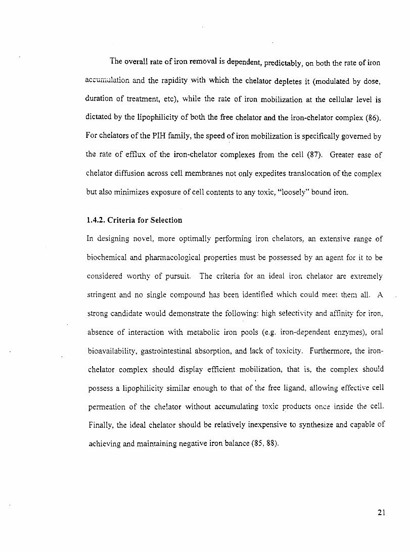
The overall rate of iron removal is dependent, predictably, on both the rate of iron
accümülation and the rapidity with which the chelator depletes it (modulated by dose,
duration of treatment, etc), while the rate of iron mobilization at the cellular level is
dictated by the lipophilicity of both the free chelator and the iron-chelator complex (86).
For chelators of the PIH family, the speed ofiron mobilization is specifically governed by
the rate of efflux of the iron-chelator complexes from the cell (87). Greater ease of
chelator diffusion across cell membranes not only expedites translocation of the complex
but also minimizes exposure of cell contents to any toxic, "loosely" bound iron.
1.4.2. Criteria for Selection
In designing novel, more optimally perfonning iron chelators, an extensive range of
biochemical and pharmacological properties must be possessed by an agent for it to be
considered \vorthy of pursuit. The criteria for an ideal iron chelator are extremely
stringent and no single compound has been identified which could meet Them aIl. A
strong candidate would demonstrate the follo\ving: high selectivity and affinity for iron,
absence of interaction \Vith metabolic iron pools (e.g. iron-dependent enzymes), oral
bioavailability, gastrointestinal absorption, and lack of toxicity. Furthennore, the iron
chelator complex should display efficient rnobilization, that is, the cornplex should
possess a lipophilicity similar enough to that of the free ligand, allowing effective cell
penneation of the chelator without accumulating toxic products once inside the ce Il.
Finally, the ideal chelator should be relatively inexpensive to synthesize and capable of
achieving and rnaintaining negative iron balance (85, 88).
21
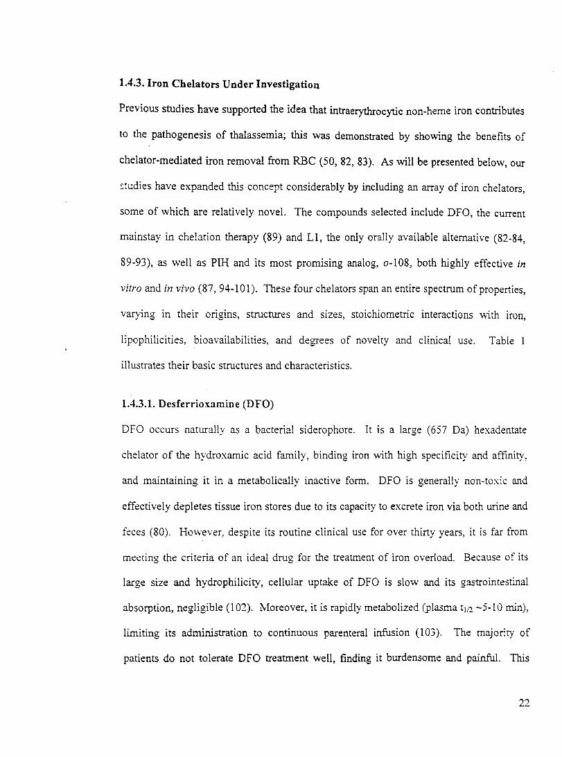
1.4.3. Iron Chelators Under Investigation
Previous studies have supported the idea that intraerythrocytic non-he me iron contributes
to the pathogenesis of thalassernia; this was demonstrated by showing the benefits of
chelator-mediated iron removal from RBC (50, 82, 83). As will be presented below, our
studies have expanded this concept considerably by including an array of iron chelators,
sorne of which are relatively nove!. The cornpounds selected inc1ude DFO, the current
rnainstay in chelation therapy (89) and LI, the only orally available alternative (82-84,
89-93), as well as PIH and its most prornising analog, 0-108, both highly effective in
vitro and in vivo (87, 94-101). These four chelators span an entire spectrum of properties,
varying in their origins, structures and sizes, stoichiometric interactions with iron,
lipophilicities, bioavailabilities, and degrees of novelty and clinical use. Table 1
illustrates their basic structures and characteristics.
1.4.3.1. Desferrioxamine (DFO)
DFO occurs naturally as a bacterial siderophore. It is a large (657 Da) hexadentate
chelator of the hydroxamic acid family, binding iron with high specificity and affinity,
and rnaintaining it in a rnetabolically inactive form. DFO is generally non-toxic and
effectively depletes tissue iron stores due to its capacity to excrete iron via both urine and
feces (80). Ho\vever, despite its routine clinical use for over thirty years, it is far from
meeting the criteria of an ideal drug for the treatrnent of iron overload. Because of its
large size and hydrophilicity, cellular uptake of DFO is slow and its gastrointestinal
absorption, negligible (102). Moreover, it is rapidly metabolized (plasma t1/2 -5-10 min),
lirniting its administration to continuous parenteral infusion (103). The majority of
patients do not tolerate DFO treatrnent weIl, finding it burdensome and painful. This
22

chelation protocol frequently results in a lack of patient compliance, inevitably leading to
a decrease in life expectancy due to iron overload. It has thus been a major goal of
chelator research to identify new formulations of DFO, new methods of its
administration, or new treatment options involving DFO's combination \Vith other
chelators to improve compliance and decrease the risk oftoxicity.
1.4.3.2. Deferiprone (LI)
Deferiprone is a relatively small (139 Da) compound belonging to a class kno\m as
a-ketohydroxypyridinones, and has been used in therapeutical trials for over fifteen years
Cl 04). It is the only chelator approved for oral application, although exclusively available
in Europe and lndia (92). Excretion of iron using LI is predominantly urinary. !ts small
size, neutral charlZe and bidentate nature predict many of its benefits, including fast
gastrointestinal absorption and high cell permeability.
However, the properties of LI are altered once bound to intracellular iron,
resulting in a larger, more hydrophilic and relatively cell impermeable complex that may
accumulate dangerously \vithin the cytosol (l05). LI also has the potential to inhibit
iron-containing enzymes such as ribonucleotide reductase, necessary for DNA synthesis
Cl 06, 107). Another disadvantage of LI is the instability of its iron complex because of
its failure to occupy aIl six iron co-ordination sites. Thus, at low drug concentrations,
biochemically active intennediate products may drive the generation of free r3.dicals
(108). Manifestations of this toxicity and other undesirable side effects of LI in humans
include agranuloc;-10sis and neutropenia (l09), arthritis (80), musculoskeletal pain (lI 0),
and gastric intolerance (93). Independent of these potential risks, LI 's v.idespread
implementation and general acceptance by the biomedical community has been hampered
23
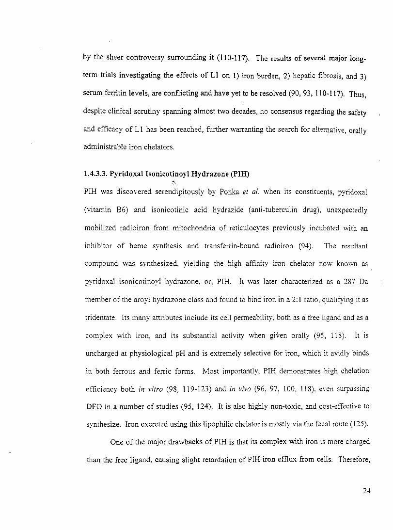
by the sheer controversy surrounding it (110-117). The results of several major long
tenn trials investigating the effects of LIon 1) iron burden, 2) hepatie fibrosis, and 3)
serum ferritin levels, are conflicting and have yet to be resolved (90, 93,110-117). Thus,
despite clinical scrutiny spanning aimost two deeades, no consensus regarding the safety
and efficacy of LI has been reached, further warranting the search for alternative, orally
administrable iron chelators.
1.4.3.3. Pyridoxal Isonicotinoyl Hydrazone (pIH)
PIH was diseovered serendipitously by Ponka et al. when its eonstituents, pyridoxal
(vitamin B6) and isonicotinic aeid hydrazide (anti-tubereulin drug), unexpeetedly
mobilized radioiron from mitochondria of retieuloeytes previously ineubated \vith an
inhibitor of heme synthesis and transferrin-bound radioiron (94). The resultant
compound was synthesized, yielding the high affinity iron chelator now knO\\TI as
pyridoxal isonicotinoyl hydrazone, or, PIH. lt was later characterized as a 287 Da
member of the aroyl hydrazone class and found to bind iron in a 2: 1 ratio, qualifying it as
tridentate. Its many attributes include its cell permeability, both as a free ligand and as a
complex with iron, and its substantial activity when given orally (95, 118). It is
uncharged at physiologie al pH and is extremely selective for iron, which it avidly binds
in both ferrous and ferric fonns. Most importantly, PIH demonstrates high chelation
efficiency both in vitro (98, 119-123) and in vivo (96, 97, 100, 118), even surpassing
DFO in a number of studies (95, 124). It is also highly non-toxie, and eost-effective to
synthesize. Iron excreted using this lipophilic che1ator is mostly via the fecal route (125).
One of the major drawbacks of PIH is that its eomplex \Vith iron is more charged
than the free ligand, causing slight retardation of PIH-iron efflux from cells. Therefore,
24

mobilization of iron by PIH has been postulated to rely on an energy-deper:J~nt carrier
(120, 126). Another disadvantage of PIH is its rapid hydrolysis at acidic pH, such as that
found in the stomach. In fact, because of its administration in a non-soluble form during
Phase 1 clinical trials, the efficacy of PIH has likely been underestimated. New snidies
examining the benefits of PIH administration while fasting or in a coated formulation are
thus needed in order for it to be fairly evaluated (99). Results from trials in animaIs given
PIH on an empty stomach yielded a much higher efficiency, and would support this
contention (95, 100). The present challenge, then, lies in the attempt to maximally
enhance the bioavailability of PIH. Given its relatively few shortcomings and its ability
to successfully bind and detoxify iron, PIH merits further consideration as a potentially
strong candidate for the treatment of ~-thalassemia.
1.4.3.4. Pyridoxal ortho-Chlorobenzoyl Hydrazone (0-108)
Once PIH was identified as a therapeutically viable iron chelator, a series of analogs were
synthesized in the attempt to improve on its chelation efficiency. One such derivative is
0-108 (320 Da) \vhich, like its PIH parent compound, is a tridentate member of the aroyl
hydrazone family. This agent, too, is readily absorbed from the gut, is highly cell
permeable and efficiently mobilizes iron both in vitro (87, 98, 123, 124, 127-129) and in
vivo (97, 100, 101), resulting in fecal excretion. However, because of its modified
structure, it possesses bicchemical and pharmacokinetic properties distinct from PIH. In
0-108, a weaker electron-v.ithdrawing halogenated benzene ring replaces the pyridine of
PIH, rendering it more lipophilic and potentially more adept at accessing intracellular
iron pools. This \vould exp Iain its superior capacity to mobilize iron compared to both
PIH and DFO in the aforementioned studies. Finally, 0-108 is highly non-toxic (128) and
25
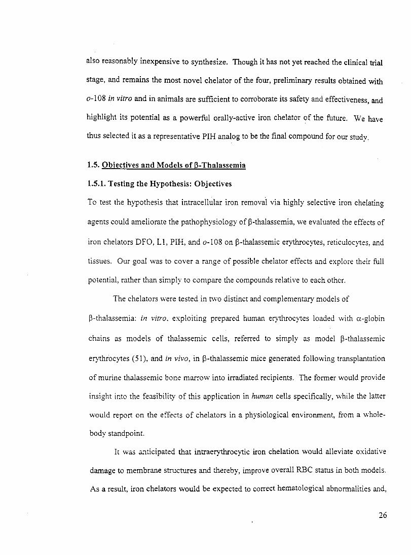
also reasonably inexpensive to synthesize. Though it has not yet reached the clinical trial
stage, and remains the most novel chelator of the four, preliminary results obtained with
0-108 in vitro and in animaIs are sufficient to corroborate its safety and effectiveness, and
highlight its potential as a powerful orally-active iron chelator of the future. We have
thus selected it as a representative PIH analog to be the final compound for our study.
1.5. Objectives and Models of B-Thalassemia
1.5.1. Testing the Hypothesis: Objectives
To test the hypothesis that intracellular iron removal via highly selective iron chelating
agents could ameliorate the pathophysiology of p-thalassemia, we evaluated the effects of
iron chelators DFO, LI, PIH, and 0-108 on p-thalassemic erythrocytes, reticulocytes, and
tissues. Our goal was to cover a range of possible chelator effects and explore their full
potential, rather than simply to compare the compounds relative to each other.
The chelators were tested in t\VO distinct and complementary models of
0-thalassemia: in vitro. exploiting prepared human erythrocytes loaded \vith a-globin
chains as models of thalassemic cells, referred to simply as model 0-thalassemic
erythrocytes (51), and in vivo, in 0-thalassemic mice generated following transplantation
of murine thalassemic bone marrow into irradiated recipients. The former would provide
insight into the feasibility of this application in hum an cells specifically, \vhile the latter
would report on the effects of chelators in a physiological environrnent, from a whole
body standpoint.
It \-vas aaticipated that intraerythrocytic iron chelation would alleviate oxidative
damage to membrane structures and thereby, improve overall RBC status in both models.
As a result, iron chelators would be expected to correct hematological abnormalities and,
26
consequently, perhaps prolong RBC survival in vivo. Fina11y, we reasoned that effects of
chelation might extend beyond red cells to improve conditions in t:.;::- t:-.",,,,~..>vJ.') cuJ.u
even, possibly, within tissues. 1t was our hope that by studying these factors concurrently
in two different models, a more global appreciation of the therapeutic potential of
intraerythrocytic iron chelation would be achieved.
The first goal was to prepare each model and confirm its functionality and
accurate representation of human j3-thalassemia. Second, it was vital to verify that all
chelators were, in fact, able to remove membrane-bound iron, so as to attribute \
downstream consequences to their iron binding and not other, non-specifie properties.
The final objective was to assess various indices of pathology within red eells, and
additionally, in vivo, in el)1hroid precursors and mouse tissues. As described belo\v, the
parameters ehosen for evaluation refleeted the most salient pathologie features of
thalassemic cells, the same ones that would likely be the most amenable to improvement.
Bèfore proceeding to the methodology, both models will briefly be discussed.
1.5.2. In Vitro - Human ~lodelj3-Thalassemic Red Blood Cells
Though ideal in vitro testing would use genuinely pathological eells derived from
thalassemic patients, such cells are difficult to obtain and are not always appropriate for
several reasons. Firstly, thalassernic patients undergo a rigorous regirne of transfusion,
coupled with chelation therapy, to prevent transfusional iron overload. Their red blood
cells would thus not be expected to exhibit the full-blo\\TI damage eharaeteristic of
untreated patients. Moreover, the most pathological cells are often the tirst destroyed and
therefore, would not be present in peripheral blood, causing underestirnation of the level
of damage. Finally, the relevant injury to patient cells would have already occurred in
27
vivo, precluding the study of developmental alterations and the involvement of iron
therein.
In light of these shortcomings, a model of thalassemic RBC was devised :::
which disease biogenesis could be tracked, and therapeutic compounds could be tested
without compromising the validity of the results. It was found that healthy hum an
erythroC)1eS, loaded with purified a-hemoglobin subunits via gentle osmotic lysis and
resealing, display the abnormalities characteristic of actual p-thalassemia (51, 130, 131).
The damage incurred by the modified cells is a direct result of, and proportional to, the
concentration of the encapsulated a-chains. These cells represent an experimentally
generated, and thus, readily available population in which the sequence of oxidative
events, and the effects of iron chelators at each stage, can be monitored. By minimizing
dilution of intracellular constituents and cell membranes during lysis, the resultant
erythroCy1eS are initially, physiologically sound. They display normal morphology,
phospholipid composition and membrane permeability, as \vell as unaltered enzyme
concentrations (ATP, glutathione, catalase), and intact membrane transport systems.
Conceptually, therefore, they act as a "blank canvas" over which the abnormalities
induced by a-chains can be illustrated as they emerge in real-time. As the a-chains
adhere to the membrane, they trigger structural and functional modifications that closely
parallel the pathophysiological changes underlying human p-thalassemia, specifically,
excess membrane-bound iron, exaggerated protein oxidation, and tlnaHy, 105s of
deformability (130, 131). By treating model cells ", .. ith iron chelators, it should be
possible to evaluate their potential protective effects on each of these aspects of cell
dysfunction.
28

In order to generate mode! ~-thalassemic erythrocytes, we first needed to iso!ate
the a.-globin to be entrapped within RBC, ascertaia its purity, then test for its universal
loading; that is, ensure that the majority of cells were indeed being lysed, and that they
successfully engulfed exogenous a-chains. Fig. 3 schematically depicts the steps
involved in model ~-thalassemic RBC preparation.
Human model ~-thalassemic cells would be incubated in the presence or
absence of iro!". chelators, and while untreated cells would reproduce the key pathological
features of the disease, the chelator-treated ones would demonstrate the potential of each
compound to remove membrane iron and curb Hb oxidation. The in vitro stage of ...
testing, therefore, constituted a primary checkpoint for chelator efficiency, ensuring the
basic ability of each compound to ameliorate cell status. In vitro studies also reported on
the response of human cells to iron chelation and, thus, addressed the clinical viability of
this strategy in thalassemic patients. Provided the efficiency of iron chelators in these
initial experiments, we \vould then evaluate them in a pathophysiologically realistic
model in vivo, using ~-thalassemic mice.
1.5.3. In Vivo - Animal Model of ~-Thalassemia
1.5.3.1. Murine ~-Thalassemia
The first mouse model of ~-thalassemia arose through a spontaneous mutation discovered
by Johnson and Lewis in 1981 (131). A nurnber of models have since been generated
through selective deletions in the ~-globin gene locus, which in adult mice nonnally
contains 1\vo distinct ~-globin genes: ~-globin major and ~-globin minor ("diffuse"
mouse haplotype). Due to polymorphisms, another mouse haplotype contains n'lo genes
29

forming an identical p-globin subunit, p-single (133). The murine models ofthalassemia
characterized to date include: 1) mice homozygous for a deletion in p-globin major,
2) mice neterozygous for an insertional disruption of p-globin major (milder phenotype),
and recentIy, 3) mice engrafted with P-giobin null fetai liver cells, displaying the
equivalent of Cooley's anemia (133-135). The last model is the only one representing
true adult thalassemia major and is the most severe, requiring animal euthanasia just 7-9
weeks after the transplant. Yet another model exists which is more severe th an 1) and 2),
but whose lifespan enables more extended study than model 3), making it ideal for our
experiments. This model, generated in 1995 by To\",nes et al. (136), consists of mice
hemizygous for the deletion of both p-major and p-minor genes. The mice are termed
hemizygous P-giobin knockout (though referred to here simply as p-thalassemic), and as
a result of their mutation, produce only 70-75% of the normal arnount of p-globin protein
( 136).
Due to the decreased production o.f p-globin polypeptide chains, mouse models of
thalassemia exhibit anemia, abnormal RBC morphology and hematoIogicai indices,
spontaneous tissue iron deposition, and organ pathology - each with varying degrees of
severity. Importantly, the biochemicai and functional defects observed in the RBC
membranes of these mice also mirror those found in thalassemic hurnans and critically,
include the hall mark intraerythrocytic iron overload, as weil as shortened RBC lit'çspan
(49,82). Combined, these data suggested the suitability ofthese animaIs for study (137-
l39).
Our objectives in vivo were, firstly, to generate a high number ofthalassemic mice
and establish a chelator treatment regimen using the selected compounds. At the cellular
3e

level, our goal was to evaluate the effects of chelators on RBC membrane-associated non-
heme iron and hemoglobin oxidation, as was done in vitro. Next, we planned to examine
hematological parameters and, crucially, track RBC survival for signs of chelator-
mediated improvements. Finally, one of the aims of this study was to explore chelator-
mediated benefits outside the red cell by evaluating hematological indices in late RBC
precursors and iron accumulation and related injury in tissues. Using these animaIs not
only offered the advantage of heightened physiological relevance over isolated cell
systems, but it enabled us to evaluate parameters which were inaccessible in vitro, such
as RBC half-life and organ damage - both important endpoints in determining the overall
impact of the chelators.
Again, similar to in vitro methodology, ~-thalassemic mice were either chelator-
. ,
treated or untreated; the latter serving as "controIs" to confirm the expected disease
phenotype - the touchstone against which chelator-treated groups would ultimately be
compared. This will be further explained in section 3.2.1.
Finally, an important point regarding our very first objective, the generation of the
animal models, should be mentioned at this time. Though our initial intent was to breed
colonies of the hemizygous p-thalassemic mice, this prospect proved extremely difficult,
due in part to the severity of the disease, the affected animaIs breeding so inefficiently, it
\-vas not feasible to generate sufficiently large, age-matched treatment groups. Faced with
this unforeseen challenge, we sought out alternative means of obtaining a meaningful
population of thalassemic mice.
31

1.5.3.2. Hematopoietic Reconstitution Model
Animal studies have often been hindered by the lack of availability of models, and/or by
potentially high variability amongst animaIs due to genetic or age-related heterogeneity.
A strategy used by Trudel and colleagues (140) to attain high animal numbers while
circumventing both of these difficulties, was to utilize bone marrow transplantations
(BMT) to transfer a deficit (sickle cell anemia) into normal mice. Since the marrow
contains the blood-forming cells, then blood disorders may be transmitted from relatively
rare donors expressing the disease, to readily available recipients whose marrow has been
ablated. Using this technique, starting with a single donor mouse, Dr. Trudel's laboratory
generated a homogeneous population of age-matched, hemoglobinopathic mice,
exhibiting virtually identical degrees of disease severity - essentially "siblings" of a lone
pathological "parent" (unpublished). Success was also obtained recently by RÏ\:ella and
colleagues using a similar procedure, fetal liver cell transplants, to generate large
numbers of severely thalassemic mice (135).
We attempted the BMT method, using two hemizygous ~-thalassemic donors, in
hopes of conferring the disease to nearly thirty control mice. Though ByIT may be
increasingly commonly used to generate animal models, particularly when the diseases
are rare and the animaIs scarce, it has never been attempted using this specific mouse
model before. Therefore, a small-scale pilot study was conducted to ensure the long-term
survival of transplanted mice and appropriate manifestation of the disease therein; it was
also vital to confirrn that the features displayed by recipients were identical to those from
donor mice (original colony). Once these questions were resolved, we carried out the
transplants, carefully monitoring the status of recipients at each stage of recovery, and
32

confirming full hematological and phenotypic adoption of the disease. After generating
the mouse models, we proceeded with the investigation of our hypothesis in vivo.
2. l\'Iaterials and l\'lethods
2.1. Materials
2.1.1. Chemicals and Equipment
Michel suture clips were from Aesculap (Tuttlingen, Germany). Thiourea, standard 1 N
NaOH, 1 N HCI, control valve and CGA Teflon sealing washer for CO tank were
obtained from Aldrich (Oakville, Ontario). 2001 Model osmotic pumps and filling tubes
were from Alzet Corporation (La Jo11a, CA). Trichloroacetic acid was from American
Chemicals Ltd. (r.rfontreal, Quebec). NaCI, acetic acid, KPO-l, and MgCb were from
BDH (Toronto, Ontario). BioRad reagent dye, Chelex, and \Vheaton glass tissue
homogenizer were obtained from BioRad (Richmond, CA). Powder HCI, NaOH,
KH:::P O-l , glucose, agar, and reagents for ferrozine assay (with noted exceptions) were
obtained from Bioshop (Burlington, Ontario). Tris was obtained from Boehringer
(Ingelheim, Germany). Biotin-X-NHS was obtained from Calbiochem (San Diego, CA).
Centrifuge tubes, polystyrene tubes for F ACS, and 96-well plates were from Falcon
(Franklin Lakes, NJ). Dialysis tubing (SpectraPor3, 3,500 Da M\V) , sodium
metabisulfite, and aIl chromatography columns were purchased from Fisher (Fair Lawll,
NJ). HBSS, IMDM, and 10,000 U penicillin/lO mg streptomycin were from Gibco
(Burlington, Ontario). Glucose phosphate isomerase (Gpi) assay was perfonned using
Helena UK equipment and reagents (Newcastle, UK). FITC-conjugated Egg-\vrute
Avidin was obtained from Jackson Laboratories (West Grove, PA). Centricon Plus-80
33
centrifugaI filters (5,000 Da MW) were from Millipore Corporation (Bedford, MA). CO,
parahydroxymercuribenzoate (pMB), ~-mercaptoethanol, fluorescein isothiocyanate
dextran (FITC-Dex; 9,500 Da MW), ascorbate, ferrozine, fructose-6-phosphate, 3-(4,5-
dimethylthiazol-2-yl)-2,5-diphenyltetrazolium bromide (MIT), glucose-6-phosphate
dehydrogenase, nicotinamide adenine dinucleotide phosphate (NADP), phenazine
methosulfate (PMS), trizma, glycine, trypan bIue, and aIl other chemicais were obtained
from Sigma (St. Louis, MO). The lysis buffer used for dialysis ofRBC samples in model
thalassemic ceIl preparation consisted of 5 mM KP04 (pH 7.4, ice cold); the resealing
buffer was identical, but with the addition of 170 mM NaCl and 5 mM glucose (pH 7.4,
heated to 37°C). The buffers used in ghost RBC membrane preparation were as follows:
lysis buffer A consisted of 5 mM sodium phosphate + 0.5 mM ethylene diamine
tetraacetic acid (EDTA) pH 8.0, lysis buffer B was identical but without EDTA and with
the pH adjusted to 7.4; both were ice cold. In the ferrozine assay, reagent A (distinct
from lysis buffer A), \vas prepared as 3 g sodium dodecyl sulfate (SDS) in 5 mL 0.2 M
sodium acetate stock buffer (pH 4.5), then brought to 50 ml with iron-free double
distilled water (ddH10). Reagent B was prepared by adding 30 g ascorbic acid and 1 g
sodium metabisulfite to 50 mL 0.2 M acetate stock buffer, then brought to 500 ml with
ddH20 (chelating resin added then removed immediately). The ferrozine reagent
consisted of 0.4 g ferrozine and 2.5 g thiourea brought to 100 ml with iron-free ddH:O.
Gey's solution for BMT (Dr. Marie Trudel, unpublished protocol) was prepared as 8.3 g
NtLCl, 1 g KHC03, and 1 mL phenol red in 1 L ddH10; this was filtered through a 0.22
micron membrane, the pH adjusted to 7.2, and the final solution kept at 4°C. Chelators:
DFO was obtained from Novartis (Dorval, Quebec), LI was a gift from Dr. Robert \V.
34

Grady (Medical College of Comell University, New York, NY), and PIH and 0-108 were
synthesized in our laboratory as previously described (141). A 100% tribromoethanol
(Avertin) stock solution was prepared by combining lOg tribromoethanol with 10 mL
tert-amy1alcoh01, then diluting to 2.5% in warm PBS just before injecting as either 0.017
mL/mg animal body weight (anaesthetic) or 1 mL/animal (letha1 dose).
2.1.2. Instruments
Mass spectrometry was perfonned by Dr. Andrea Romeo, Concordia Chemistry
Department (Montreal, Quebec), using a Finnigan SSQ7000 single quadrupole
electrospray ionization (ESI) mass spectrometer (Finnigan, Bremen, Germany).
Spectrophotometric assays were conducted using a CARY 13E spectrophotometer
(Varian, Australia). Fresh mouse blood samples were shipped to C1inTriais BioResearch
Ltd. (CTBR, Senneville, Quebec) for hematological analysis using an Advia 120
automated analyzer. F ACS was performed using an EPICS XL-MCL flow cytometry
system, Beckman-Couiter Ltd. (Mississauga, Ontario). Fixed mouse tissue sections
underwent routine histological processing followed by staining \vith Peris' Prussian blue
for the detection of tissue iron according to standard methods (142). Staining was
performed at the IRCM (140), by histologist Annie Vallée.
2.2. AnimaIs
Four- to five-month-old male and female C57BLl6JGP1-13 and C57BLl6JGPI-lb controls, as
well as ~-g10bin knockout mice (HbbtmIToWlHbbS) hemizygous for the deletion of murine
~-major and ~-minor globin genes, were used in these studies. The congenic
C57BL/6JGPI-la and C57BL/6fJPI-lb mice were obtained from Jackson Laboratories (Bar
35

Harbor, ME) and mice were used as controis. The hemizygous J3-knockout mice
(HbbtmlTow IHbbS), referred to simply as J3-thalassemic, were originally obtained from Dr.
T. Townes (136) and since backcrossed for> 16 generations to C57BLl6JGPl-lb inbred
animaIs to prec1ude genetic variability between backgrounds. J3-thalassemic mice were
identified following peR amplification and characterized as having intennediate to
severe J3-thalassemia as previously described (143). AU animaIs were generously
provided by Dr. Marie Trudel (140), and were maintained in microisolator cages.
2.3. Methods
In Vitro
2.3.1. Preparation of Model 13-Thalassemic Erythrocytes
2.3.1.1. Purification of a-Hemoglobin Chains
Pure a-globin subunits "vere isolated from whole blood as previously described (144-
146), and as summarized briefly below:
Preparation of RBC Hemolysate:
40 mL of blood was collected ln heparinized tubes from a healthy volunteer and
centrifuged at 1000 g for 5 minutes (min). The plasma was aspirated and red blood cells
were washed three times \.Vith PBS. The cells were then lysedby adding 1 volume of
ddH20 and freeze-tha'Wing three times in acetone/dry ice. The hemolysate was
centrifuged at 15,000 g for 30 min then exposed to CO at 4°C for 1 min while mixing.
36

Dia/ysis of RBC Hemolysate:
The CO-treated hemoglobin was dialyzed against 2 L ddH20 for 4 hours (h) after which
the protein content of the concentrated Hb was measured. KH2PO~ and NaCI were then
added to a final concentration of 10 mM and 80 mM, respectively.
PA1B Treatment of a2fJz-Hb:
The arnount of PMB was calculated 50 as to be in 10 M excess of the heme concentration
present in the hemolysate. The PMB was dissolved in 0.5 mL ddH20 by adding 2 drops
of 1 M NaOH then 2-3 drops of 1 M acetic acid until the precipitate just redissolved. The
PMB was added to the CO-Hb, the pH adjusted to 6.0 with 1 M acetic acid, and the
solution left at 4°C overnight.
Separation of a- and fJ-P:.HB Chains:
The hemolysate was centrifuged at 1000 g for 5 min, reserving and CO-treating the
supematant as previously described. To isolate the a- from the ~-chains, P:VfB-Hb was
passed through a coarse G-25 Sephadex colurnn equilibrated with 10 mM KPO~ buffer
(pH 8.15), and the elution volume concentrated to 20 mL. The concentrate \vas passed
through a \Vhatman DE52 column equilibrated with la mM KPO .. buffer (pH 8.15). The
a-PMB chains, which eluted first, were collected and exposed to CO.
Regeneration of Purified a-Chain -SH Groups:
The a-PMB chains were adjusted to pH 6.5 \'Iith la nu\-! KH2PO .. and loaded cnte a 4 cm
CM23 colurnn equilibrated with la mM KPO .. (pH 6.5). The a-PMB chains were
mobilized cnte the column as a single band and were concentrated through this process.
The column was washed for 15 min with la mM KPO .. (pH 6.5) first containing, then
37

devoid of 15 mM fj-mercaptoethanol. The a-SH chains were finally eluted with 20 mM
Tris-HCl (pH 8.0) then eO-treated, pelleted in liquid nitrogen, and stored at -80oe until
use.
2.3.1.2. Protein Concentration of Purified a-Chains
The final protein concentration of the a-hemoglobin was evaluated using the Bradford
protein assay (147). Briefly, a dye reagent (BioRad) was diluted 1:4 with ddH20 and the
protein solution added in a 1 :500 volume ratio. Samples were incubated for 5 min at
room temperature and read in triplicate at 595 nm. Absorbance units (AU) were recorded
and divided by 0.13 (this number corresponds to the extinction coefficient obtained from
the standard curve using BSA, and corrects for dilution), to yield results in }lg protein/}lL.
2.3.1.3. a-Chain Purity
The a-hemoglobin \Vas analyzed by electrospray ionization-mass spectrometry (ESI-~lS)
in order to confirrn a molecular mass equivalent to previously established values. Fresh
a-globin chain samples \vere diluted 1: 150 with ddH20 and analyzed according to
standard technique (148).
2.3.1.4. Hemoglobin Oxidation
To ensure the hemoglobin subunits were not oxidatively damaged during the purification
process, a well-established spectrophotometric assay (149-151) was used to assess the
relative concentrations of intact (oxy), oxidized (met), and denatured (hemichrome)
hemoglobin present in the a-hemoglobin-derived lysate. The a-chains were diluted
1: 100 with dd.H20, the hemolysate scanned, and absorbance values recorded at 560, 576,
630, and 700 nm. The a-subunits were then incubated ovemight at 25°C and the
38

spectrum re-recorded to examine changes in methemoglobin generation over time. It
should be noted that the response of functional hemoglobin subunits to oxidation is
firstly, the fonnation of metHb, fo11owed eventually by that of readily precipitating
hemichromes, the latter being a prime determinant of anemia severity (31). The
calculations, based on the method ofWinterbourn et al. (151), are as fo11ows:
Using m},,{ extinction coefficients:
oxyhemoglobin (ffil\1) = 29.8(ODs76 - OD700) - 9.8(OD630 - OD700) - 22.2(ODs60 - OD700)
methemoglobin (ffil\1) = 7(OD576 - OD700) + 76.8(OD630 - OD700) - 13.8(OD56o - OD700)
hemichrome (mM)* = 33.2(OD576 - ODiOO) - 36(OD630 - ODioo) - 58.2(OD560 - OD700)
* \',Then no hemichrome \vas present, the concentrations of oxyHb and metHb were added
and their sum considered as total hemoglobin (100%). Each was divided by total Hb
concentration and multiplied by 100 to determine their relative proportions, expressed as
a percentage. The change in metHb generation over time (~ % metHb), \vhen required,
\vas determined by the percent metHb(ttinaD - percent metHb(tinitial).
2.3.1.5. Osmotic Lysis and Resealing
Loading erythrocytes \\ith the purified a-chains was performed according to the method
of Scott et al. (152, 153). Briefly, blood was collected fromhealthy hurnan volunteers in
heparinized tubes. The samples were centrifuged and the plasma and buffy coat
aspirated. Packed ery1hrocytes were washed three times in PBS (pH 7.4) and the
following RBC populations were prepared: 1) control normal, 2) control-resealed, and 3)
a-chain loaded. A 250 ilL aliquot of washed pRBC was reserved as the first control. A
mixture of 500 ilL pRBC and 275 ilL PBS (no a-chains) constituted the second control,
39

undergoing osmotic lysis and resealing in the absence of a-chains to exclude any adverse
effects due to the procedure itself. Finally, 500 JlL pRBC were admixed with 250 JlL
a-chains (33.75 mg) and 25 JlL 10X PBS to eventually comprise the model ~-thalassemic
cell population. After thorough mixing of cells and added solutions, samples 2) and 3)
were dialyzed against 1 L of ice cold 5 l1li\II KP04 lysis buffer (pH 7.4) for 45 min at 4°C
with constant stirring. The membranes \vere then transferred to 1 L of warrn 5 mM KPO~
resealing buffer (pH 7.4) and dialyzed for another 45 min at 37°C with stirrin2:. AH
samples, including reserved controls, were then transferred to centrifuge tubes and
washed 7 times \vith PBS. \Vashed control, resealed, and a-chain loaded pRBC were
resuspended to 5% hematocrit in HBSS supplemented with 5 mM glucose and containing
100 U penicillin/mL and 100 Jlg streptomycin/mL. Samples were then incubated at 37°C
\vith gentle shaking and aliquots removed at 0, 1, 3, 5 and 20 h to deterrnine levels of
hemoglobin oxidation. Samples recovered after 20 h were used to deterrnine erythrocyte
membrane-bound non-heme iron.
2.3.1.6. Determination of universal Loading
Previous studies have ca1culated the efficiency of encapsulation, proportional to the mean
intraerythrocytic a-chain concentration, using a-chains radiolabeled with eH]-NEM.
This value has repeatedly been sho\'tTI to correspond to 3-5% of the total RBC
hemoglobin when resealing 10 mg a-chains/mL packed cells (51, 130, 154). An
alternative test examines whether loading is universal or not, that is, whether a high
enough proportion of cells incorporates exogenous material. The "universality" of
loading can be deterrnined by co-entrapment of a fluorescent dextran (FITC-Dex) of
40
similar molecular weight (MW) to et-chains. Since incorporation of the fluorescent
marker parallels that of exogenous chains, cells exhibiting fluorescence should likewise
contain the subunits. Using this technique, other laboratories found more than 95% of
resealed cells to effectively incorporate a-chains (51, 130). To confinn this value in our
studies, we loaded REC with a-chains as above (2.3.1.5.) in the presence or absence of
5 mg FITC-DeximL pREC. The percentage of cells exhibiting fluorescence, and thus
containing FITC-Dex along with a-chains, was then detennined using F ACS.
2.3.2. Characterization of lVlodel ~-Thalassemic Erythrocytes and In Vitro Evaluation of Iron Chelators
Once the a-chains had been purified and their encapsulation into RBC verified, we
characterized the model ~-thalassemic cells and assessed potential chelator-mediated
benefits therein. \\'e therefore repeated the cell preparation procedure as above, this time
treating samples of a-chain loaded cells with the selected iron chelators. After the 20 h
incubation, control, control-resealed, and model ~-thalassemic RBC (untreated or
chelator-treated) \-vere characterized in terms of: 1) membrane-associated non-heme iron,
and 2) hemoglobin oxidation (metHb generation). These parameters reflect the
importance of intraerythrocytic iron in disease pathophysiology, and represent two of the
most prominent features of ~-thalassemic cells.
2.3.2.1. Iron Chelator Solutions
Stock solutions of 5 mM chelator were prepared in ddH20 (DFO and LI) or 1 M NaOH
(PIH and 0-108, subsequentIy neutralized through the addition of 1 M HCI), and
41
dissolved in HBSS to a final concentration of 50 J.1M. This is within the lower range of
concentrations routinely used to test iron chelators in vitro (50-1000 J.1M) (119-121), and
has been reported as the minimum concentration required to achieve maximal iron release
using radioiron-labeled reticulocytes (155).
2.3.2.2. Incubation Conditions
The process of osmotic lysis and resealing was repeated as above (2.3.1.5.), preparing a
larger volume of a-chain loaded ceUs to be distributed among four iron chelator
treatment groups. Thus, 2.5 mL pRBC were admixed with 1.25 mL a-chains (168.75
mg) dissolved in 125 ilL 10X PBS. Samples were dialyzed against lysis and resealing
buffers, and washed with PBS. An aliquot of a-chain loaded ceUs \vas reserved
(untreated) and the remainder divided into four chelator treatment groups. \Vashed
erythrocytes were suspended at 5% hematocrit in HBSS or HBSS/chelator solution and
incubated for 20 h at 37°C with gentle shaking.
2.3.2.3. Membrane-Associated Non-Herne Iron
Measuring non-heme iron adherent to the RBC membrane was an important objective in
the evaluation of selected iron chelators, as any subsequent chelator action should be
related to their ability to strip this pathological iron from the membrane milieu. Non
heme iron present in RBC membranes was quantified by: 1) preparing ghost RBC
membranes, then 2) using an iron-binding reagent to detennine the exact concentration of
iron in ghost samples. AU buffers and solutions used were rendered iron-free by pre
treatment with chelating resin, Chelex (2.1.1.).
42
2.3.2.3.1. Ghost RBC Preparation
In order to quantify strictly the iron that was membrane-associated, RBC were lysed, their
cytosolic contents expelled, and the resultant "ghost" membranes washed exhaustively
until no further hemoglobin could be removed; this was do ne according to the method of
Dodge et al. (156, 157). Briefly, after 20 h of incubation, RBC sampI es were washed 3
times in PBS. Aliquots (0.5 mL) of pRBC were each lysed in 45 mL ice cold lysis buffer
A and centrifuged at 13,000 rpm for 15 min at 4°C. Supematants were aspirated and
pellets resuspended by drawing and expelling them five times through a 1 mL syringe fit
with a 22-gauge needle, ensuring thorough break-up of ghosts and freeing any
hemoglobin embedded \vithin membranes. Exhaustive washing continued until control
ghosts \vere milky-white (a total of 5 \vashes). Membranes were then washed twice in
lysis buffer B, and finally stored at 50% hematocrit in buffer B at -80 Oc until use.
2.3.2-.3.2. F errozine assay
Free, non-heme iron associated \vith ghost membranes was quantified by convention of
its reaction with the compound, ferrozine, within two minutes of its addition; a standard
method knO\vTI as the ferrozine assay (157). When the ferrozine chromophore binds
ferro us iron, a rich magenta complex is formed whose depth of colouration is
proportional to the concentration of iron in the sample. Ghost membranes, prepared as
above, were resuspended in 5 rru"\! sodium phosphate buffer (pH 7.2) to 1 mg/mL. Three
cuvettes were prepared for each sarnple: buffer blank (100 ilL buffer), ghost blank
(100 ilL iron-free control ghosts), and sample of interest (100 ilL ghost sample). The
first accounts for the colour produced by the buffer itself, the second reports the
absorbance of ghosts in absence of ferrozine, and the last is the colour produced by the
43

ghosts of interest from the reaction with ferrozine. The frr5t two served as controls and
were eventually subtracted from the sample of interest. SDS (275 ilL of 10%), reagent A
(100 ).I.L), and just before reading, reagent B (725 ilL), were added to each sample.
Finally, 100 ilL ferrozine reagent was quickly added (except to ghost blank) and the
sample was read over 2 min at 562 nm. For the ghost blank, 100 ilL phosphate buffer
was added instead of ferrozine in this final step. Calculations used to determine levels of
membrane non-heme iron were based on a protocol by Dr. Mark Scott (146).
Calculations:
[OD ghost sample ofinterest - (OD buffer blank + OD ghost blank)] X 465.9/ 1mg/mL = nmoles non-heme iron/mg
In this equation:
- OD (optical density) denotes absorbance values ranging from 0 to 1.0
- 00 of buffer blank and ghost blank are subtracted from 00 of ghost sample of interest, yielding the absorhance due exclusively to membrane-bound iron
- value 465.9 represents the molar extinction coefficient of the iron-ferrozine complex after correcting for dilution; standard value = 28,000 liters mOrl cm- I (158)
- 1 mg/mL is the protein concentration in each sample
- final results are expressed as nanomoles of non-heme. iron per milligram of membrane protein
2.3.2.4. Hemoglobin Oxidation Assay
Levels of methemoglobin were measured as an index of total cellular oxidative stress,
expected to be elevated in model ~-thalassemic RBC due to iron-catalyzed oxidation of
membrane components, and potentially decreased in chelator-treated cells due to removal
of that iron. During cell incubation, aliquots were removed at 0, l, 3, 5, and 20 h,
44

centrifuged, and packed erythrocytes diluted 1 :500 with ddH20. Using the described
method (2.3.1.4.), RBC lysates were analyzed for their respective concentrations of
oxyhemoglobin and its oxidized form, methemoglobin.
In Vivo
2.3.3. Generation of Mouse l\'Iodels of 13-Thalassemia
2.3.3.1. Bone l\'Iarrow Transplantation
Several mouse models of 13-thalassemia exist (132-138, 159, 160), arising either through
spontaneous mutations or more commonly, targeted deletions of the 13-globin gene locus.
The mice used in this study are hemizygous for the deletion of murine 13-major and
13-minor globin genes, and are characterized as having intermediate to severe
13-thalassemia. Ho\-vever, due to their phenotype and poor breeding efficiency, only a
limited number are available at any one time. Thus, in order to have a population large
enough to form meaningful treatment groups, murine thalassemic bone marrow \-vas
transplanted into a substantial number of control mice. Two 13-thalassemic adult donor
mice were thus sacrificed, and both hind and forelimbs were dissected and thoroughly
cleaned. Using a 3 mL syringe and 26 1/2_gauge needle, BM cells were harvested by
perforating and flushing bones with IMDM containing 5% serum. Cells Viere centrifuged
at 1200 rpm for 5 min at 4°C, the supernatant aspirated, and Gey's solution (2.1.1.) added
to lyse the RBC. Lysis was stopped after 3 min with 3 ml IMD~l containing 5% serum.
The cells v"oere centrifuged again, the supernatant aspirated, and the pellet resuspended in
2 mL serum-free IMDM. Cells were counted using the trypan blue exclusion method and
resuspended in serum-free IMDM in preparation for transfer.
45

Twenty-eight C57BLl6fiPI-la mice were lethally irradiated at 152 rads/minute
for a total of 875 rads (cGy), and following a 4 h recovery period, injected intravenously
(tail vein) with 1.8 million BM cells in 350 ilL volume. The chimeric mice were
monitored for hematopoietic engraftment at 4, 7, and 10 wk post-transplant before
commencing the iron chelator regimen on wk 12.
2.3.3.2. l\'Ionitoring Engraftment: Glucose Phosphate Isomerase Assay
The two murine electrophoretic isoforms of the enzyme, glucose phosphate isomerase-l,
can be separated by their different migration rates on cellulose acetate membranes using
standard Gpi-phenotyping methods (161, 162). Recipient and donor mice expressing
altemate forms of the enzyme, Gpi-la and Gpi-lb, respectively, were thus used in this
study. Peripheral blood of transplanted mice was analyzed at the protein level to
determine the proportion of endogenous RBC remaining, and the proportion contributed
by the donor (Gpi-l b) at different stages following BMT. As per standard protocol from
Dr. Marie Trudel (unpublished), cellulose acetate membranes Viere soaked 60 min in Gpi
running buffer (3.0 g trizma, 14.4 g glycine in 1 L ddH20, pH 8.7). Approximately
50 ilL tail vein blood was collected into heparinized hematocrit tubes and centrifuged for
3 min at 13,000 rpm. The pRBC contained within 0.5 cm of tube were lysed in 125 ilL
ddH20 and 8 ilL of this, transferred onto the membranes. Sarnples migrated from
cathode to anode for 60 min at 200 Volts. Gpi-l bands were then revealed using an
enzymatic colorimetric reaction based on the Gpi-l-mediated conversion of fructose-6-
phosphate to glucose-6-phosphate, which, in the presence of NADP and glucose-6-
phosphate dehydrogenase, drives the reaction toward the formation of product, NAD PH.
46

When NADPH reacts with MIT in the presence of PMS, the latter is reduced, yielding
the intensely purple-coloured product, formazon. Briefly, therefore, a 2% agar
preparation was heated to 55°C and maintained at that temperature until use. A first
reagent was made, consisting of 2 mL of 0.2 M Tris-HCl (pH 8.0), 120 ilL of 0.2 M
MgCh, 150 ilL of 100 mg/mL fructose-6-phosphate, and 120 ilL of 10 mg/mL MIT.
Just before the end of migration, a second reagent was made (120 ilL of 10 mg/mL
NADP, 120 ilL of 2.4 mg/mL PMS, and 4 ilL of glucose-6-phosphate dehydrogenase),
and added to the first. The migration was stopped and the membrane blotted dry.
Finally, 2 mL of the hot agar was added to the above admixed solutions (total volume
-4.5 mL) and, using a 3 mL syringe, distributed evenly over the membrane, brightly
staining Gpi-l bands. Once control bands appeared sharp, membranes \vere fixed in 5%
acetic acid to stop the staining. The intensity of the signal was then quantified by Image
Quant software.
2.3.4. Preparation of Chelator Solutions for In Vivo Testing
The osmotic pumps used for drug delivery in this study were designed to dispense 1 JlL
solutionlhour, and contained a reservoir which could be filled to a maximum of 168 JlL ±
a built-in margin of error of a few ilL (163). The total volume of 168 ilL reflects the
constant delivery of 1 ilL solutionlhour X 24 hours/day X 7 days total. In order to
achieve administration of 50 mglkglday, chelator stock solutions of DFO, LI, PIH, and
0-108 were prepared by dissolving 92 mg ofeach into 2 mL of 25-30% NaOHIPBS (lM
NaOH diluted to 0.25-0.3 M 'With PBS), depending on their solubilities. This
concentration would be considered conservative and highly non-toxic, as
47

phannacological doses routinely administered to humans range between 30-100
mglkglday (89, 99, 111) and those to animaIs, between 100-500 mglkglday (96, 97).
Doses were adjusted to weight by varying the chelator concentration inside the pump, and
solutions sterilize-filtered before filling the pumps to capacity. Pumps containing a
vehicle solution of25% NaOH in PBS were also prepared as controls.
2.3.5. Osmotic Pump Implantation
After filling osmotic purnps, animaIs were anaesthetized with avertin and the pumps
implanted into their backs subcutaneously as per manufacturer' s directions (163).
Briefly, a small incision was made in the skin bet\veen the scapulae. A hemostat was
used to spread apart the connective tissue, creating a small pocket. The pump was
inserted into the pocket \\ith flow moderator end (end from which solution is dispensed),
pointing towards the animal's posterior, and sterile suture clips then used to seal the
wound. New pumps containing fresh chelator solutions were implanted to replace the old
ones every 7 days for a total of 4 procedures.
2.3.6. Characterization of l\-Iurine ~-Thalassemic Erythrocytes and In Vivo Evaluation of Iron Chelators: Erythrocyte and Precursor Pathology
In addition to the tests described below, mice were monitored daily throughout the course
oftreatment for signs of chelator toxicity and/or morbidity, inc1uding: lack of grooming,
changes in appearance of coat, inability to eat or drink, 20% weight loss/gain, and
behavioural abnormalities.
48
2.3.6.1. Membrane-Associated Non-Heme Iron
Following 4 weeks of iron chelator treatment, mice were sacrificed and total blood
volumes collected. RBC ghost membranes were prepared exactly as above (2.3.2.3.1.),
and the non-herne, non-ferritin iron reacting within 2 min in the presence of ferrozine was
assessed as described (2.3.2.3.2.). This test served to deterrnine, in vivo, the effectiveness
of iron chelators ln rnobilizing RBC membrane-bound iron frorn actual murine
thalassemic cells.
2.3.6.2. Hemoglobin Oxidation Assay
Spectrophotometric evaluation of oxidized and reduced components of mouse
hemoglobin was conducted as above (2.3.2.4.) prior to, and follo'oVing the chelation
regime. Methernoglobin generation was initially examined at 10 wk post-transplant by
\vithdra\ving a srnall amount of blood (-150 ilL) via tail snip. Final levels were taken
within 24 h of sacrificing the animaIs. As before, this assay would indicate the potential
of iron chelators to, as a function of their iron-binding capacity, attenuate oxidative stress
in thalassemic erythroc)tes in vivo.
2.3.6.3. Hematological Parameters
Fresh blood samples, collected immediately into EDTA-coated microtubes following
each animal's sacrifice, were shipped to CTBR for complete hematological analysis
(2.1.2.). ErythrOCyte and reticuloc)te parameters were established using flo\v cytometry
based measurernents obtained on an Advia 120 automated blood cell analyzer. The
mouse archetype of multi-species sofnv'are version 2.2.06 was used in analysis (Bayer
Diagnostics, Ta.rryto\m, NY). Hematocrits (Hct) were measured manually, and mean
49

cellular volume (MeV) and red cell Hb concentration were detennined using two (low
and high-angle) light seatter measurements. From the measured Het and Rb
concentration, the mean cell hemoglobin concentration (MCHC) was derived (164).
Reticuloeyte eounts were detennined using the RNA-staining dye, oxazine 750, and
expressed as a percentage of total red cells. Sub-populations of reticuloeytes were
quantified based on their levels of maturation, designated as follows: 1) L RETIC (low
staining-intensity, most mature), 2) M RETIC (medium staining-intensity, intennediate
maturation), and 3) H RETIC (high staining-intensity, most immature). The relative
proportions of these groups provided information regarding the effectiveness of
ery1hropoiesis, and correspondingly, the seve rit y of the disease, since thalassemia is
characterized by fewer mature retieulOCy1eS and abnormally high numbers of immature
ones. Reticulocy1e mean ceIl volume (MCVr), an indicator ofhydration status specific to
reticulocytes, was also measured using the reticulocy1e channel of the analyzer.
2.3.6.4. Erythrocyte Survival
Labeling RBC in vivo using the compound biotin, is a safe and useful alternative to
radiolabeling for the determination of erythrocyte survival. Biotin-labeled cells are
discriminated from newly-produeed, unlabeled ones, using the fluorescent conjugate
FITC-Avidin. FITC-Avidin specifically binds biotin, causing labeled eells to fluoresce,
allowing them to be quantified by F ACS analysis. This technique was used to track RBC
lifespan in the various mouse populations (165), and is briefly summarized here.
50

2.3.6.4.1. In Vivo Biotinylation of Erythrocytes
A biotin solution of 1.2 mg biotin dissolved in 45.6 ilL N, N,-Dimethylfonnamide, then
added to 300 ilL PBS, was prepared for each mouse. AlI mice (except for reserved non
biotinylated controls) received tail vein injections of 280 ilL biotinlmouse/day for three
consecutive days. Twenty-four ho urs following the final injection was regarded as the
100% labeling point, and the first day of F ACS analysis.
2.3.6.4.2. FITC-A vidin Incubation
Tail vein blood (2-3 ilL) was collected on days 1, 3, 4, 5, 6, then on altemate days for a
total of 21 days. 2!J.L blood was measured out from each heparinized capillary tube and
admixed with 4 mL PBS. 1 mL of this \-vas reserved as FITC-negative control while
another 1 mL was transferred into an Eppendorf tube and incubated with 10 ilL FITC
Avidin for 30 min at 4°C. AlI samples were centrifuged at 2500 rpm for 10 min at 4°C,
after which the supematant was aspirated and the small RBC pellet resuspended in fresh
PBS and washed twice. The final pellet was resuspended in 1000 ilL PES, and aIl
samples then analyzed by flow cytometry. Controls included non-biotinylated RBC as
weIl as FITC-negative and FITC-positive biotin-Iabeled ones. An steps involving FITC
A vidin were perforrned in the dark to prevent fluorescence quenching by direct exposure
to light.
2.3.6.4.3. Flow Cytometry
Flow cytometry was used to distinguish the fluorescently labeled cell population from the
unlabeled one. The fate of labeled cells was monitored by the progressive decay of
fluorescence over the course of their destruction. Gates were first set to exc1ude the
51

analysis of aggregated cells or non-cellular debris. Then, background fluorescence of
untreated (non-biotinylated) FITC-incubated syngeneic erythrocytes was counted and
new gates set to exclude inherent cellular fluorescence of control cells. Control
histograms were generated through this process and subsequent histograms gated to
account for the background. Erythrocytes were counted (10,000 cells/sample) and
biotinylated cells expressed as a percentage of the total using F ACScan software. To
deterrnine erythrocyte half-life, the percentage of biotinylated cells was first plotted
against time. The day at which 50% of labeled cells remained in circulation was
calculated from the linear equation derived from each plot using Sigma Plot scientific
graphing software CV ersion 3.0), and established as the RBC tl/2 for that animal.
2.3.7. Characterization of lVlurine ~-Thalassemic Tissues and In Vivo Evaluation of Iron Chelators: Organ Pathology
2.3.7.1. SpleenlBody 'Veight Ratio
AnimaIs \-vere sacrificed by avertin injection and total body, liver, and spleen weights
were deterrnined. The spleen to body weight ratio (spleenlbody weight), calculated as
wet weight of the organ divided by total body weight, is a well-known indicator of
disease severity (137). A high ratio reflects hyperactivity of the spleen during accelerated
hemolysis, as the spleen constitutes the primary organ for destruction of damaged or
senescent RBC in mice.

2.3.7.2. Liver Iron Content
To quantify the accumulation of non-heme iron in mouse liver, a major organ for iron
storage in addition to spleen, a proto col was adapted from Pountney et al. (166). Livers
were dissected and fresh sections were weighed and frozen at -80°C until the time of
assay. Tissues \-vere homogenized with 700 ilL iron-free ddH20 in a 1 mL glass
homogenizer, then boiled in 20 J.l.L 12 N HCI for 10 min after which 200 ilL 50%
trichloroacetic acid was added, and the samples cooled on ice for 15 min. Samples were
finally centrifuged at 14,000 rpm for 15 min, and 40 J.l.L 10 M NaOH added to
supematants (yielding a final pH of 4.5 in the medium). A ferrozine reagent was
prepared (0.88 g ascorbate, 4.3 g sodium-acetate, and 98 mg ferrozine in 50 mL ddH20),
and 500 ilL added to each sample before reading the absorbance over 2 min at 562 nm.
AU solutions were rendered iron-free as above (2.3.2.3.). Finalliver iron concentrations
were expressed as nmoles/mg wet tissue.
2.3.7.3. PerIs' Iron Staining in Liver and Spleen
A standard method used to demonstrate iron accumulation in tissues is the Perls' Prussian
blue stain (142). The iron demonstrable by this assay must be in the ferric form, as most
human and mouse tissue iron is. \Vhole spleens and liver lobes were fixed in formalin
(10% formaldehyde in PBS) ovemight \Vith gentle shaking. The organs were then
transferred to cassettes and kept in PBS at 4°C until the time of histological sample
preparation (no more than one day). The organs were embedded in paraffin, sectioned,
dehydrated in graded alcohol and stained with Prussian blue using a hematoxylin and
eosin counterstain (137). Tissues were scored for blue-positive iron deposits at 20 X
53
magnification using a Zeiss Axiophot image analysis microscope. Five non-overlapping
fields were systematically examined for each organ. Areas staining positive for iron were
quantified by conversion to pixels, which were then expressed as a percentage of total
surface area examined using Northern Eclipse software Version 6.0. The percentage
values from aU five fields assessed were averaged and considered as the total stainable
iron in that particular organ.
Statistical Analysis
The probability of a statistically significant difference between the mean values of 2 data
sets was determined using the unpaired Student t test or Mann-Whitney test (murine RBC
membrane-associated iron).
54
3. Results
3.1. In Vitro Studies
Model ~-thalassemic erythrocytes were an innovative design by Scott et al. that helped
elucidate the sequence of events occurring during the development of the thalassemic
phenotype in RBC (51, 153). In essence, model thalassemic erythrocytes are normal
human red cells manipulated into a diseased state through the insertion of excess, heme
containing a-chains, and therefore, represent a readily available and unique tool in which
the effects of therapeutic agents can be evaluated. Despite extensive characterization of
these cells, hO'vvever, the benefits of intraerythrocytic iron chelation therein have been
minimally explored, with very few compounds having been tested to this end. These
include DFO and its high molecular weight derivative, plasma tripeptide H-Gly-His-Lys
OH, and N,N-bis(2-hydroxybenzyl)ethylenediamine-N,N-diacetic acid, also known as
HBED (146). Because sorne of the more potent iron chelators, including novel
compounds that have since been designed, have never been investigated in this model, the
full potential of intraerythroCy1ic iron chelation in thalassemic RBC is not truly known.
Therefore, the prime objective in vitro was to examine iron chelator-mediated
improvements in human thalassemic type cells using four diverse agents, two of which
constitute the mainstay in treatment, and two comparatively new and promising oral
candidates. The experiments involved, firstly, purifying and characterizing a-Hb
subunits, then encapsulating them into normal erythrocytes. The model thalassemic cells
were then confirmed as appropriate correlates of human disease and the iron chelators
tested therein for their respective abilities to: 1) remove abnormal membrane-bound iron,
and 2) curtail the oxidative destruction of hemoglobin; thereby probing two key features
55

of the disease. Importantly, because model ~-thalassemic RBC were elaborated from
human erythrocytes, they provided insight into the applicability of this strategy to human
cells specifically. Furthermore, findings from the in vitro studies helped to validate and
develop the ensuing animal studies.
3.1.1. Purification of a.-Hemoglobin Chains
Once isolated, the a.-chains were determined to have a protein concentration of
0.135 glmL. This value was similar to those obtained in other laboratories using the same
method (51, 154). In order to confirm the identity of the subunits, ESI-MS, a highly
accurate technique for measuring protein molecular weights, was used. This type of rnass
spectrornetry enables analysis of the different globin chain varieties and he me rnoieties of
hemoglobin as separate entities, as well as any modified forms of either. As a reference,
Fig. 5 A depicts the standard ESI mass spectra of normal hurnan blood diluted 500-fold
(148), while panel B shO\vs the spectra obtained frorn a-chains purified in our laboratory
(diluted 1: 150). Both spectra consist of a characteristic peak at 15,126 Da, due to the
contribution of a.-globin protein (no herne), and a peak at 15,741 Da that represents the
entire a-hernoglobin peptide (globin with attached herne). Both peaks obtained from
isolated a-chains \-vere consistent with the reference peaks and accepted rnass values,
authenticating the prepared subunits as pure a-hemoglobin rnonorners.
Furthermore, analysis of the subunits' initial oxidation state and tirne-dependent
oxidative response helped establish their functional properties. An assay which proved
indispensable in this regard, and which was used throughout the course of both in vitro
and in vivo testing, was the spectrophotornetric rneasurernent of hernoglobin oxidation
(2.3.1.4.). Fig. 6 depicts sequential scans ofRBC hemolysate over a 20 h period at 37°C,
56

illustrating the range ofHb oxidation states from intact, 100% oxyHb (pronounced peaks,
solid line) to oxidized 100% metHb (flattened peaks, broken line). When the a-globin
isolated in our laboratory was scanned in the same way, the expected spectrum of oxyHb
was observed at t = 0, confirming the chains had not been oxidized during purification
(Fig. 7, solid line). AIso, the spectrophotometric changes recorded in a-hemogiobin over
time were consistent with a functionai oxidative denaturation response, as the subunits
yielded solely methemogiobin after a 20 h incubation at room tempe rature (Fig. 7, dotted
line). Evidence of a-chain integrity was obtained by analyzing the subunits via the
ferrozine assay (2.3.2.3.2.). This test confirmed the absence of non-heme iron in a-chain
sampIes, suggesting that iron \vas appropriately sequestered within heme and, thus,
supporting the intact condition of subunits follo\\ing purification (data not sho\m). The
characterization of isolated a-chains through a combination of ESI-MS and
spectrophotometry, therefore, established not only the identity of the subunits, but also
their purity and integrity. 'vVe consequently pursued their encapsulation into healthy
human erythroc)r1es in order ta generate model B-thalassemic cells.
3.1.2. Universal Loading of a-Chains
The preparation of reliable model B-thalassemic cells depends on the efficient loading of
excess, heme-containing a-globin chains into normal human ery1hroCj1es. T 0 ensure
incorporation of the purified subunits, RBC underwent regurgitation-resealing using
a-chains in parallel with a comparably heavy fluorescent Dextran (FITC-Dex, 9,500 Da).
After re-annealing and \vashing, the cells were analyzed for uptake of the fluorescent
molecule using flow cytometry (2.3.1.6.). Fig. 8 fust illustrates the background
57

fluorescence intensities of control (A) and control-resealed (B) cells in absence of FITC-
Dex; an identical background pattern was observed for a-chain loaded cells (not shown).
These control histograrns- were used to set "gates" exc1uding non-specifie fluorescence
from subsequent samples. Cells exhibiting relative fluorescence intensities greater than 1
(within region "D"), were considered to have incorporated FITC-Dex while values below
1 reflected typical low-level inherent cellular fluorescence. The fluorescence intensities
of cells lysed and tesealed in the presence of either 5 mg FITC-DeximL pREC al one or
FITC-Dex in suspension with a-subunits (13.5 mg a-chains/mL pREC) are seen in
panels C and D, respectively. Wnen fluorescent REC were expressed as a percentage of
the total counted (5000 cells), more than 98.5% of red cells were found to have
incorpotated the extracellular material. Sarnples were prepared in triplicate and
representative histograrns for each condition are sho\\TI. The data obtained helped to
establish the generation of truly a-globin-containing RBC in \vhich the effects of iron
chelators could then be investigated.
3.1.3. Characterization of ~Iodel ~-Thalassemic Erythrocytes and In Vitro Evaluation of Iron Chelators
Since reversible osmotic lysis and resealing alone do not have any substantial effects on
normal red cell behaviour (50, 51, 153, 154), changes observed in RBC loaded 'With
purified et-hemoglobin must be due exclusively to the effects of the iron-containing
hemoglobin subunits. The indices monitored during evaluation of iron chelators in vitro
inc1uded erythroCj1e membrane-associated iron, as well as extent of oxidative attack on
cellular Hb, reflecting the central oxidative mechanisms involved in the development of
thalassemia. Both pararneters were exarnined in control and control-resealed
58

erythrocytes, as weIl as in a-chain loaded cells that were either untreated or chelator
treated.
3.1.3.1. Quantification of Mernbrane-Associated Non-Herne Iron
Model p-thalassemic erythrocytes were untreated or incubated with 50 !lM of one of four
iron chelators for 20 h at 37°C with gentle shaking. In order to measure the amount of
non-heme iron embedded specifically within cell membranes, Hb-free ghost erythrocytes
were prepared. This step was effected as above (2.3.2.3.1.), and the iron-binding
chromophore, ferrozine, was used to spectrophotometrically quantify the concentration of
non-heme ferro us iron in each sample.
In control and control-resealed cells, levels of non-heme iron were 0.37 and 0.47
nmoles/mg membrane prote in, respectively, while a-chain loaded cells carried a burden
equivalent to nearly 4.0 nmoles/mg membrane protein - approximately 10 times that of
controls. Treatment of cells \vith iron chelators, however, significantly depleted cell
membranes of this abnormal iron fraction: final values ranged from 0.85 to 1.05
nmoles/mg membrane prote in, using DFO and 0-108 respectively, representing -4-5
times less iron than in untreated a-chain loaded cells. As Fig. 9 A illustrates, all iron
chelators were effective in dislodging a significant amount of non-heme iron from the
membranes of model p-thalassetnic RBC (p < 0.002). The concentration of iron
indicated for each sample represents the average of three independent experiments.
59
3.1.3.2. Hemoglobin Oxidation Assay
In ~-thalassemia, unstable a-subunits are not only prone to autoxidation but act
additionally, via the toxic iron they liberate, to destabilize normal hemoglobin and trigger
ifs oxidation to metHb, further promoting iron release (64). Agents that ev en partially
remove injurious iron at the root of this reaction would be expected to short-circuit the
oxidative cascade and, consequently, limit the production of metHb. Because the level of
methemoglobin mirrors the total oxidative injury incurred by cells, its concentration
within RBC was monitored for chelator-mediated improvements throughout the 20 h
incubation, as described (2.3.2.4.). The overall change in the concentration of metHb
from 0 to 20 h is depicted in Fig. 9 B. Control and control-resealed cells exhibited
increases of 1.9% and 4.4% metHb, respectively, while the increase in model
~-thalassemic cells exceeded 30%, signalling considerable oxidative damage. When
model cells were treated \vith iron chelators, metHb levels were reduced from untreated
values by at least 50% CP < 0.01), and remarkably, approximated normal values in Ll
and PIH-treated samples, suggesting maximal protection against Hb oxidation by those
compounds. Without exception, though, iron chelators demonstrated a high level of
efficiency in mitigating oxidative damage to model ~-thalassemic erythrocytes,
conceivably due to their iron removal properties.
The in vitro studies yielded encouraging results: the hum an model ~-thalassemic
erythrocytes that \vere prepared appropriately displayed the excessive membrane
associated iron and abnormally high metHb levels characteristic of the disease. AIl four
of the iron chelators tested significantly improved these erythrocyte parameters, reducing
membrane iron burden and providing relief from oxidative stress. Though they
60

accomplished this with relatively similar degrees of efficiency, DFO and PIH displayed a
slight -advantage in iron removal, while LI and PIH did so in attenuating oxidation.
Because model l3-thalassemic erythrocytes mimicked the early stages of human
thalassemia (likened to the first months of a patient's life), and because iron chelation
achieved beneficial results in the absence of transfusion, this therapeutic strategy, applied
clinically, would be expected to slow disease development and, perhaps, bypass
transfusion altogether. Thus, insofar as human l3-thalassemic erythrocytes are concemed,
intraerythrocytic iron chelation appears to be a valid strategy to prevent damage caused
by excess a-chains, if initiated at an early enough clinical stage.
Next, to determine the far-reaching effects of iron chelators in a physiological
milieu, we evaluated the same compounds in vivo, using l3-thalassemic mice with a
disease severity comparable to l3-thalassemia intermedia-to-major, in humans.
3.2. In Vivo Studies
In light of the benefits mediated by iron chelators in vitro, we hypothesized that through
the same mechanism of binding intraerythrocytic iron and preventing consequent
membrane damage, these compounds could extend the lifespan of model l3-thalassemic
RBC in vivo. We planned to transfuse labeled murine model thalassemic cells (murine
RBC loaded \vith human a-chains) into control mice, then follo\v the survival of labeled
thalassemic RBC in untreated and chelator-treated animaIs. To this end, murine blood
was collected and red cells loaded \vith purified human a-chains as described abüve
(2.3.1.5.), then biotin-labeled for trac king (2.3.6.4.1.), and injected into normal mouse
recipients. Using flow cytometry to quantify the labeled population, it was fo und ,
61
unexpectedly, that only a slight proportion of modified cells remained in circulation after
injection. To clarify their precise fate, we labeled murine model ~-thalassemic RBC with
radioactive 59Fe and counted the fractions of radioactivity in circulation, spleen, and liver
follo'vving their injection (data not shown). The majority of a-chain loaded RBC, and
even control-resealed cells, were cleared abnormally rapidly by the spleen of injected
mice. This observation suggested that in the process of generating murine model
~-thalassemic cells, perhaps sorne incompatibility due to the combination of lysis
resealing and biotin-Iabeling, or through immunogenic-related modifications (168), they
became altered in such a way as to no longer be suitable for in vivo tracking. Thus,
although human model p-thalassemic cells faithfully resembled their clinical counterparts
in the nature and severity of a-globin-induced damage, more subtle alterations may have
developed during the preparation and labeling of murine model p-thalassemic cells,
limiting them for our purposes, to in vitro systems. We promptly shifted our
methodology to a more physiologically meaningful mode l, namely, the ~-thalassemic
mouse.
3.2.1. Mouse Model of ~-Thalassemia
Since the first description of a mouse model for ~-thalassemia in the early 1980's, several
different models have been generated and the y are now widely accepted as suitable
paradigms of human ~-thalassemia (132-139). A shared feature of the biogenesis of
disease in both species is the fundarnental role played by excess a-chains and the related
membrane defects (138). Unfortunately, however, the high level of heterogeneity
arnongst sick animais and their relative shortage due to poor breeding make the prospect
62
of studying p-thalassemic mice especially challenging. For the in vivo studies, therefore,
we used a novel approach to overcome the limiting number of pathologic mice at our
disposal. By performing a series of bone marrow transplantations, we attempted to
transfer the hematological deficit from a small number of p-thalassemic mice to a
sufficiently large number of healthy ones.
3.2.1.1. Donor AnimaIs
Hemizygous p-globin knockout mlce, the mouse model used in these studies, were
identified following PCR amplification and were characterized via hematological analysis
as having severe p-thalassemia (143). These mice exhibited dramatically decreased RBC
counts, hemoglobin content, hematocrit, cell volume and cell hemoglobin concentration.
They also displayed marked reticulocytosis and elevated red cell distribution widths,
indicating abnormally high erythropoiesis and anisocytosis (cell heterogeneity),
respectively. Two of these mice were selected from the original colony to act as BM cell
donors. Because donor animaIs had already been thoroughly characterized (136, 143),
and because the animaIs of interest were the transplanted recipients, developing features
identical to those of the original colony (section 3.2.2.), aIl data presented will be from
the transplanted mice.
3.2.1.2. Recipient Hematopoietic Reconstitution
Following BM transplants, the chimeric mice were monitored for hematopoietic
engraftment at 4, 7. and 10 \vk post-transplant by the cell marker, glucose phosphate
isomerase-1. An assay combining electrophoretic and enzymatic techniques was used to
reveal the relative proportions of blood cells derived from recipients and donors at
63

vanous stages of engraftment. To avoid redundancy, data from ten representative
recipients are shown for each assay (Fig. 10). In aIl three membranes, the first two lanes
indicate the positions of Gpi-l isotypes "a" and "b" derived from C57BLl6JGPI-la
(recipient) and C57BLl6JGPI-lb (donor) mice, respectively. Lanes 1-10 reveal the Gpi-l
isotypes of ten transplanted animaIs. The first assay demonstrated the majority of
recipient RBC to be of endogenous origin, as Gpi-la was the principal form of the
enzyme detected (Fig. 10, first membrane). After 7 wk however, over 50% of recipient
RBC expressed Gpi-l b; these celIs must have, therefore, been contributed by the donor
(Fig. 10, second membrane). By 10 wk, 100% of recipient RBC expressed the donor
derived marker (Fig. 10, third membrane), confirming the switch to ~-thalassemic RBC
production, exclusively. These data, therefore, corroborated successful transfer of
functional B:"'1 from the original ~-thalassemic donors to recipients by demonstrating the
production of donor-type pathological erythrocytes by these recipients.
While the Gpi test explicitly reported on the engraftment status, other indices
monitored throughout the 10 wk period established engraftment indirectly by detailing
the development of disease; these included hematocrit and metHb levels, which changed
appropriately in the transplanted mice (data not shown). AlI subsequent data (see section
3.2.2.) clearly characterized these mice as having adopted a ~-thalassemic phenotype,
providing further evidence of hematopoietic reconstitution. Therefore, from this point,
ail transplanted animaIs were considered hemizygous ~-thalassemic, just as the BM
donors from the original colony.
64

3.2.1.3. Treatment Groups
Three control and three thalassemic mice received no treatment at aU, while three more of
each were implanted with 25% NaOH vehicle-containing pumps; this \-vas do ne to ensure
that neither pump implantation nor vehic1e solution had any adverse effects. Each of the
chelator treatment groups consisted of five p-thalassemic animaIs. AnimaIs from each
group were matched for weight, sex, and age. Below is a summary of the treatment
groups:
Treatment Groups:
Control (C57BLl6JGPI-lb) n=6
no treatment*} t t d un rea e vehicle
n=3
n=3
(3-Thalassemic (HbbtmITow/HbbS)
no treatment*} untreated vehicle
DFO-treated
Ll-treated
PIH-treated
0-108-treated
n = 26
n=3
n=3
n=5
n=5
n=5
n=5
* Because data from mice receiving no treatment and those receiving vehicle showed no
statistical difference, they were combined to form a single group of n = 6, designated
"untreated" .
3.2.2. In Vivo Administration ofIron Chelators to p-Thalassemic Mice
The iron chelator regime began 12 wk post-transplant. Compounds were administered
subcutaneously by osmotic pump (2.3.5.), a method that minimized animal handling. It
aiso allowed for constant, stable delivery of a Iow drug concentration, thereby limiting
potential toxicity and preserving the naturai pharmacokinetics of the iron chelators.
65
Continuous delivery was especially useful in determining the effects of chelators with
shorter half-lives, such as DFO (plasma tl/2 = 5-10 min), which are not effective when
given by bolus administration (103).
To ensure chelator composition did not change appreciably throughout the 7 days,
spectrophotometric tests were conducted on days 1 and 7 of each implantation. Since
variations in chelator solutions can be detected spectrophotometricaIly, we reserved
aliquots of the same solutions administered in vivo and monitored them ex vivo
immediately after implantation and again after 7 days incubation at 37°C (mimicking the
state in vivo). As anticipated, the chelators showed no significant changes in their
structure over this time period (data not shown). AIso, to confirm that the iron chelators
were being dispensed accurately, aIl residual solutions were aspirated and quantified
upon pump removal. Again, the built-in margin of excess designed for the pumps was
respected, and no major discrepancies between actual drug volume delivered and the
theoretical quantity defined by the manufacturer, were observed. FinaIly, mice were
carefully monitored on a daily basis and exhibited no changes in weight or behaviour that
may signal chelator toxicity or related morbidity.
3.2.2.1. Quantification of l\lernbrane-Associated Non-Herne Iron
T 0 ensure the reported effects of chelators were in fact due to their iron chelating
properties, it was critical to evaluate chelator-induced changes in RBC membrane iron.
AnimaIs were sacrificed upon termination of the RBC survival assay (after 4 \vk chelator
treatment) to provide total blood volumes required for the se tests. The results described
below are surnmarized in Table 2.
66
Erythrocyte ghost membranes derived from B-thalassemic mice harboured
extensive iron deposits compared to non-detectable levels in controls (p < 0.001),
marking a fundamental feature of the disease and supporting the validity of the
transplantation method. After 4 wk, LI and PIH diminished membrane-bound iron by
45% and 38%, respectively, relative to untreated (p < 0.05). Although iron mobilization
did not reach a similar degree of significance with the other compounds, aU of them
reduced RBC iron by at least 30%, as demonstrated by the ferrozine assay (2.3.2.3.2.).
Because aU chelators successfuUy mobilized iron from B-thalassemic RBC membranes,
we could rationalize any further effects as being, very likely, due to this defining
property.
3.2.2.2. Hemoglobin Oxidation Assay
Just as the iron chelators were tested for their ability to attenuate hemoglobin oxidation in
model B-thalassemic RBC in vitro, the same was investigated in thalassemic mice in vivo.
Initial methemoglobin leveis determined prior to treatment with iron chelators were
compared with values obtained following the 4-wk treatment regime. As depicted in Fig.
Il, RBC from control mice generated extremely low leveis of metHb at the study's onset;
these levels increased only slightly (3%) by the end of the study. Murine B-thalassemic
RBC, in contrast, displayed endogenously high levels of metHb initiaUy, -25-fold in
excess of controls, and experienced a greater increase in the percentage of metHb over
the 4 weeks. FinaUy, though untreated and chelator-treated thalassemic cohorts displayed
similarly high metHb values initially, the chelator-treated animaIs were found to generate
significantly less metHb than untreated cohorts by the study's end (Fig. Il). The most
dramatic positive effect "vas exerted by PIH, which decreased metHb by 4%, followed by
67

DFO, LI, and 0-108. AlI observed differences were statistically significant (p < 0.03).
This data suggested that by removing adventitious membrane iron, iron chelators not only
limited the extent of hemoglobin oxidation, but also, likely conferred the same protection
to other proteins, thereby diminishing total oxidative impact to cells.
3.2.2.3. Hematological Parameters
After 4 weeks of receiving iron chelators, total mouse blood volumes were collected and
samples immediately sent for hematological analysis. Hematocrit, mean cell volume,
mean cell Hb concentration, and reticulocy1e counts were averaged for each treatment
group and compared; these results are summarized in Table 3. Untreated ~-thalassemic
mice displayed hematological abnormalities consistent with the disease: reduced
hematocrit (50%) and mean cell volume (-20%) were indicative ofprofound anemia and
cell dehydration, respectively, while an abnormally high reticulOC}1e count (lO-fold) was
reflective of stimulated· ery1hropoiesis. AIl values in ~-thalassemic mice differed
significantIy from controis (p < 0.001).
Iron chelators improved hematologicai parameters ln ~-thalassemic mlce with
varymg degrees of significance. Chelator-treated mice had modestly corrected
hematocrits and sa\v even more meaningful changes in MCV and MCHC. Cell volume
was increased by aIl iron chelators, most significantly by DFO (p < 0.05), while MCHC,
an index of cell volume, was significantly improved by aIl compounds (p < 0.05).
Partially normalized values of MCV and MCHC were indicative of chelator-mediated
protection against the hall mark cellular dehydration of ~-thalassemia. This effect was
likely achieved by attenuating oxidative damage to the membrane transport proteins
involved in regulating cell volume, demonstrating substantial benefits of iron removal on
68
both membrane structure and function. Iron chelators aiso significantly reduced the
abnormally high number of reticulocytes present in peripheral blood of untreated mice;
DFO and PIH had the highest impact, decreasing levels by -20% and 45% respectively
CP < 0.05).
Reticuloc)'ie parameters, distinct from those obtained in mature red cells, have
the unique potential to convey information pertaining to the earlier stages of erythroid
development. In addition to erythrocytes, therefore, reticulocytes were also examined for
signs of chelator-mediated improvements. Reticulocytes were first classified into sub
populations based on their level of maturation. As a convention, categories designated L,
M, and H reflect low, intermediate, and high RNA staining-intensity, respectively;
denoting decreasing levels of maturity. In normal mice, the vast majority of reticuloc)tes
were from the mature sub-population, displaying the least Rt'\IA staining (Table 3).
Proportions were skewed tm,vards immature celIs in untreated ~-thalassemics however,
with over 50% of celIs demonstrating high RNA staining. A significant shi ft back
to\vards nonnal proportions occurred in chelator-treated animaIs, where the majority of
reticulocytes were in mature and intermediate populations (p < 0.05, DFO and PIH).
Finally, although not statistically significant, a trend toward enhanced mean celI volume
in reticulocytes from chelator-treated mice was also observed (Table 3). ColIectively, the
reticulocyte data supported beneficial effects of iron chelators not only in mature celIs but
even earlier, \vithin their precursors, explaining, in part, their high therapeutic activity in
vivo th us far.
69

3.2.2.4. Erythrocyte Survival
One week was allowed for the effects of the chelators to develop before commencing
RBC survival studies at 13 wk post-transplant. Murine erythroc)'1es were then biotin
labeled in vivo and their survival monitored by F ACS analysis. The turnover rate of
labeled cells provided values of RBC half-life for each animal. Figure 12 reports the
final average tll2's for each treatment group. RBC half-life in control mi ce was 21.6 days,
similar to values determined in other laboratories, establishing the validity of the assay
and healthy status of control mice (165, 167). The ceIls of untreated p-thalassemic mice,
by contrast, exhibited an appreciably shortened half-life of 12.6 days, nearly 50% lower
than controls (p < 0.001). Remarkably, RBC of aIl chelator-treated thalassemic mice
exhibited tl/2'S intermediate between those of controis and those of untreated
thalassemics. AnimaIs receiving DFO had the longest-surviving RBC with t1l2 of 15.8
days, followed by those treated with LI (t1/2 = 15.0), PIH (t1l2 = 14.0), and finally, 0-108
(tl/2 = 13.9). Though approaching but not achieving statistical significance, likely due to
variability within groups, these results suggested that even conservative doses of iron
chelators improved membrane parameters substantially enough to slow RBC destruction.
This observation is best illustrated by the representative RBC survival curves depicted in
Fig. 12. Iron chelators attenuated red cell hemolysis in thalassemic animaIs, resulting in
RBC survival curves closer to those of control animaIs. As will be discussed in section
4.4., this finding \-"as perhaps the most pivotaI in terrns of the clinical potential and
applicability of iron chelators. Evidence that these agents extended RBC lifespan in
untransfused thalassemic mice by virtue of binding iron, firstly, highlighted iron's
pathophysiological importance. Furtherrnore, it supported the hypothesis that removal of
70

this proximal iron' compartment could put a brake on abnormal red cell c1earanceand
consequently, disease progress. This is a promising finding for many thalassemic
patients in whom (insofar as the results from mice can be extrapolated to humans)
chelator administration at an early enough stage should prevent the need for transfusion
altogether. Instead, a minimal and more convenient iron chelation regimen would form
the basis of disease management, thereby avoiding secondary iron overload and
improving patients' quality of life.
Examining the effects of intraerythrocytic iron chelation on RBC survival was a
primary investigative objective. Because of the striking improvements observed in
thalassemic erythrocytes and precursors in vivo, we postulated that iron chelators might
likewise, have a beneficial effect in tissues. This question was addressed by measuring
iron content and iron-mediated damage in liver and spleen tissues from untreated and
chelator-treated ~-thalassemic mice.
3.2.2.5. Assessment of Organ Pathology and Tissue Iron
Ail experiments conducted thus far have examined the effects of iron chelators within a
framework of erythroid pathology. However, a defining feature of ~-thalassemia is the
accumulation of iron not only in erythrocytes, but also within such organs as the liver,
spleen, heart, kidneys, and endocrine tissues. In order to assess which organ parameters
were most dysfunctionai in thalassemic mice, we performed an initial characterization
study, comparing a smaIl number of ~-thalassemic animaIs (n = 6, original colony) to
controls (n = 3). The livers, spleens, hearts, kidneys, and femurs of aIl ni ne animais were
dissected and evaluated by gross and microscopic examination in terms of general
appearance, weight and size. Organs were then prepared for histological analysis and the
71
distribution ofiron was determined by the Peris' Prussian blue staining method (2.3.7.3.).
The p-thalassemic mice exhibited massive splenomegaly relative to healthy littermates,
while the size of other organs appeared normal. This is in contrast to human
p-thalassemic patients, who not only display hypertrophy of the spleen but also typically,
liver, kidney, bone marrow, and heart tissue. Humans can also store iron extensively in
each of these organs, while p-thalassemic mice presented visible iron deposits only in
liver and spleen. Similar findings were reported by other laboratories using different
p-thalassemic mouse models (135, 137, 160), and helped confirm which organ
parameters would best inform our studies.
Given the data from this small-scale characterization pilot, we planned, for the
full-scale experiment, to examine the benefits of iron chelators in thalassemic tissues by
specificallyassessing: 1) spleeIllbody weight ratio, 2) non-herne iron content of liver, and
3) extent of Prussian blue iron staining in liver and spleen.
3.2.2.5.1. SpleenlBody \Veight Ratio
Examining the weight of the spleen in the context of total body weight provides insight
into the extent of splenornegaly, a product of the accelerated RBC clearance,
extramedullary hematopoiesis, and iron storage central to murine p-thalassernia (l·B).
Mouse spleens \-vere dissected and \veighed, then expressed as a percentage of total body
weight. In this case, the improvernents rnediated by iron chelation were conservative.
Still, a tendenc)' to\-vards reduced spleeIllbody weight ratio in the chelator-treated groups
was observed. As presented in Table 4, the normal range for spleeIllbody weight ratio
\vas between 0.2 and 0.3. In untreated p-thalassemic mice, this value increased nearly
72

6-fold, then feU slightly to -5 to 5.5 times control values in chelator-treated mice.
Though the decrease was subtle, the fact that a trend eould be observed at all revealed
how rapid and far-reaching the effects of the 1r0;-', ;:;:dators could be. While reduction in
spleenlbody weight was encouraging to observe as an overall phenomenon, it did not
provide mechanistic details regarding specifie sites of chelator action or the aspects of
splenic dysfunction being improved. Iron ehelators may act directly at the level of the
spleen, or may improve splenic function indirectly by normalizing RBC parameters and
curtailing RBC clearance. It is conceivable, either way, that longer term oi" higher dose
chelator treatment could be of substantial bene fit to target tissues, and could ev en
promote regression of pathology by depleting iron stores through a number of possible
mechanisms.
3.2.2.5.2. Liver Iron Content
The liver is a primary site for iron loading in murine ~-thalassemia as well as the spleen
(137). We thus conjectured that it would be one of the organs most affected by iron
chelation and therefore, a likely location to observe potential improvements mediated by
the chelators. Using the same spectrophotometric principle of iron detection as in
erythrocyte membranes, non-he me iron was quantified per milligram of liver wet weight.
Results described below are summarized in Table 4. Hepatic iron content in control and
thalassemic mice was 0.78 and 1.65 nmoles iron/mg wet weight, respectively (p < 0.001).
Remarkably, all chelators depleted liver iron stores, most significantly to 1.19 nmoles/mg
using 0-108 (p < 0.05). Direct hepatic iron measurements, therefore, supported the ability
of iron chelators to mobilize injurious iron not only from within red cells but perhaps,
aiso from principal organ storage sites. This again, occurred within a relatively short
73
period of time and at a remarkably moderate dose. To further evaluate iron chelator
effects in tissues, the classic iron detection technique of Perls' staining was used in mouse
liver and spleen. Though the assay is qualitative, the intensity of staining is considered
an accurate indicator of iron content (169), making visual or graphical "scoring" of
stained tissues a highly acceptable method of quantitating iron (2.3.7.3.). AIso, because
the method permits visual examination of tissue make-up and iron deposition, the cells
expressly affected can be identitied, offering insight into the direct targets of iron chelator
action.
3.2.2.5.3. Peris' Iron Staining in Liver and Spleen
Perls' Prussian blue staining is a sensitive and reliable method for detecting ferric iron in
tissue samples. Sections are tirst treated with acid to release ferric ions, then as the cation
of potassium ferrocyanide is replaced by the ferric ion, insoluble ferric ferrocyanide is
formed and readily precipitates, staining bright blue (169). Both liver and spleen sections
were examined using a hematoxylin and eosin counterstain, and stainable iron expressed
as a percentage of the total surface area examined.
As reported in Table 4, liver sections from control mice revealed no stainable
iron while 0.23% staining was seen in untreated ~-thalassemics. Iron chelators reduced
the extent of iron accumulation by at least 50%, with the greatest effect being achieved
by 0-108, which reduced stainable iron by 90% relative to untreated animaIs (p < 0.05).
PerIs' Prussian blue-stained liver sections are shown in Fig. 13. Each section
represents the average percent stainable iron for that treatment group. Sections from
control liver (tirst panel), revealed normal histology and no detectable iron. Tissues from
untreated ~-thalassemic mice, however, contained noticeable iron deposits in the
74

cvtoplasm of Kupffer cells, identified by their characteristic "half-moon" shape and
location, directly surrounding the hepatocytes. Sorne darker, more granular-appearing
deposits may represent stainable iron from phagocytosed RBC, seen most prominently in
untreated ~-thalassemic tissue. Liver tissues from chelator-treated animaIs showed no
treatment-related abnormalities. In DFO-treated animaIs, moderate iron staining was
observed within Kupffer cells, again, proximate to hepatocytes. Deposits, however,
appeared less extensive and less heavily-embedded than in untreated ~-thalassemics.
Staining in tissues from mice treated with LI, PIH, and 0-108, was even more faint and
more restricted than in untreated or DFO-treated cohort tissues. Sections from 0-108-
treated mice, especially, revealed remarkably seant iron staining CP < 0.05), and an
overall appearance that was very close to normal.
PerIs' staining was next used to assess iron deposition in the spleen, the primary
organ for clearance of damaged and senescent RBC in mice, which \yas far more
extensive th an in the liver, for aIl animaIs. As reported in Table 4, 1.39% stainable iron
was measured in control spleens, while a 4-fold excess was found in those from untreated
~-thalassemics. Impressively, aU chelators, with the exception of DFO, promoted a
statisticaUy significant decrease in the extent of staining; final values were intermediate
between untreated thalassemics and normal controls (p < 0.05). Still, a more dramatic
effect was seen in 0-108-treated samples, where stainable iron was -60% less than in
untreated mice (p < 0.001).
Representative spleen tissues from each group are depicted in Fig. 14. Control
spleens displayed intact architecture and overall normal histology: red and white pulp
appeared in approximately equal proportion and only slight iron staining was visible. By
75

contrast, the splenic architecture in untreated p-thalassemic mi ce was virtually destroyed
and the boundaries separating red and white pulp, unclear. Furthermore, iron staining
was rife throughout the parenchyma, revealing splenic macrophages gorged with ferric
iron. In the DFO-treated group, iron still pervaded the tissues, though less prominently
th an in the untreated group. In tissues from animaIs treated with LI, PIH, and 0-108,
splenic architecture was greatly improved, as evidenced by a clearer distinction and more
normalized ratio between red and white pulp. As weIl, in aIl three of these groups, iron
staining was considerably less intense and less diffuse than in untreated and DFO-treated
mice CP < 0.05). Notably, tissues from 0-108-treated animaIs showed improvements
beyond those seen with other chelators: white pulp could actually be discemed, iron
staining was minimal, and a close resemblance to normal tissues was evident.
Interestingly, these results corresponded with those obtained in liver, the data from both
organs highlighting 0-108 as a superior iron chelator in tissues, surpassing the
effectiveness of its peer candidates by an appreciable margin.
The observed benefits of iron chelators in thalassemic tissues \vere reflective of
a number of possible mechanisms resulting from iron chelation in tissues, red cells,
erythroid precursors, or a combination thereof. Factors contributing to the alleviation of
tissue pathology may include attenuated RBC removal, diminished extramedullary
erythropoiesis, and reduced iron loading.
Examined collectively, the results characterizing the erythrocytes and tissues of
untreated and chelator-treated p-thalassemic mi ce confirmed success of the B~1T
technique in generating severely p-thalassemic mice. Moreover, the data established
active iron chelation by each of the compounds tested and identified this iron-
76
mobilization activity as central to restoration of cell function and tissue reconstitution.
Consideration of iron chelator effects in the various compartments tested, as weIl as a
summary and discussion of both in vitro and in vivo results, will be the focus of the final
section.
4. Discussion
4.1. Thalassemia: Pathophvsiology and Current Treatment
The thalassemias encompass an extremely prevalent and debilitating group of diseases
that target the essential oxygen carrier, hemoglobin. Though recognized as a major cause
of morbidity and mortality worldwide, there is still no widely available cure for it, and the
remedial measures that are available, are largely unsatisfactory. Because thalassemic
infants born in developing countries, where the disease is most endemic, are surviving
increasingly later into adulthood, there is an urgent need for a more practical treatment;
one that is both therapeutically effective and economically viable. Furthermore, as the
demography of the illness has changed, so has its geography, with high rates of
immigration no longer relegating thalassemia to a "disease of the Mediterranean" but
rather, expanding its borders, making it a serious global threat.
The disease arises from mutations in the a- or ~-globin genes, causing a
decrease in a- or ~-globin polypeptide synthesis, respectively. Such genetic defects
result in a fatal imbalance: the deficiency or total absence of one of the globin chains, and
the insidious production of the other unaltered "bachelor" chain. Due to the loss of
conformational stability inherent in aI~ contacts, the monomeric subunit, whose
synthesis is in relative excess, becomes highly unstable and readily precipitates within
er)ihrocyte precursors and later in mature RBC. In ~-thalassemic erythrocytes,
77

spontaneously high levels of free radicals and the newfound susceptibility of a-chains to
autoxidation result in the release of heme moieties from their globin polypeptide chains.
Though the globin and liberated heme can, themselves, cause mechanical damage by
binding to membrane structures, it is the heme-derived iron that becomes the major
cul prit in the pathophysiology of the disease. As the heme ring is cleaved by
hydroperoxides present in excess in patient cells, the iron that is normally so diligently
sequestered becomes liberated in the vicinity of the membrane. Free to valence cycle
between the ferric and ferrous forms, it functions as a catalyst in the Fenton redox
reaction, generating the deadly hydroxyl radical. It is this role in oxidant generation that
endows iron with an inherent and devastating toxicity.
Critical to the development of thalassemia is not simply the excess of injurious
oxygen species, but also their location. Cell structures critical for maintenance of
deformability and integrity (spectrin, ankyrin, band 3, protein 4.1, membrane
phospholipids, etc), act collectively as a sink for iron, drawing it onto binding sites on the
cytoplasmic leaflet. Because of this intimate yet highly inappropriate association, iron's
toxic potential becomes focused on the cell membrane, specifically, cytoskeletal proteins
and membrane phospholipids. Iron also induces more of its own release by destabilizing
and oxidatively denaturing normal hemoglobin and other a-chains, amplifying the
vicious cycle of oxidant production and membrane destruction.
Also aggravating the pathology at the cellular level are sorne of the unique
characteristics of the RBC itself, namely its excessive exposure to high oxygen
concentrations, its limited de nova protein synthesis, its imbalance in pro-oxidant and
antioxidant tàctors, and the restricted access of its membrane to intracellular oxidant
78

scavengers. The erythrocyte is also particularly vulnerable as the most ferruginous
somatic ce Il , having to maintain 20 mM of iron fully liganded and meticulously
compartmentalized (64).
The endpoint of the oxidative cascade is modification of the membrane that is
so severe as to cause premature RBC removal from the circulation. The major anomalies
precipitating erythrophagocytosis stem from cytoskeletal impairments and inc1ude cation
leakiness, cellular dehydration and rigidity, reduced lipid fluidity, and the loss of
phospholipid asyrnmetry. Phagocytosis of damaged red blood cells occurs via either
MDA generation from peroxidized lipids or the binding of autologous antibodies
recognizing band 3 prote in on the cell surface (53).
Accelerated RBC breakdown accounts for the trademark anemia and stimulates
splenic activity, resulting in massive splenic enlargement (also a product of iron loading
and red blood ceIl production). Meanwhile, compensatory erythropoiesis leads to gross
expansion of the bone marrow and characteristic bone deformation, as weIl as a spectrum
of metabolic and endocrine abnormalities, inc1uding excessive gastrointestinal iron
absorption (69). Clinically, therefore, the thalassemic phenotype develops to a large
extent from the premature RBC destruction initiated by the presence of intraerythrocytic
non-heme iron.
Despite considerable knowledge of its pathophysiology and its high frequency
ln the population. the only existing treatment for thalassemia, as mentioned. is non
curative and consists of chronic blood transfusions. This therapy exacerbates pre-existing
iron overload by relentlessly supplying patients with surplus iron from breakdo\vn of
transfused RBC. Because its physiological excretion is limited, iron accumulates to toxic
proportions in the liver, spleen, and heart, proving fatal if not removed - a task which is
79

accomplished by iron chelators designed specifically to bind the metal and promote its
elimination. However, the only approved compound to this end is DFO, a drug which
despite its clinical use since the 1960' s suffers from significant drawbacks, most notably,
its cumbersome parenteral method of administration. The majority of patients find the
long-term use of DFO-dispensing portable pumps highly inconvenient and extremely
taxing, both physically and financially, the latter prohibiting DFO's \videspread use in
socioeconomically disadvantaged countries. The high rate of non-compliance o\ving to
the above complications results in failure to chelate surfeit iron in major organs, and
eventually leads to organ shutdo\vn and early patient mortality.
4.2. Summarv of Hvpothèsis and Exp~rbnental Models
Thalassemic patients endure a triple threat from potentially toxic iron: it accumulates
proximally at the red blood cell membrane, secondarily within major organs as a result of
dysregulated metabolism and, finally, paradoxically, throughout tissues due to the very
blood transfusions intended to treat it. ~lost research has been aimed at prenatal
diagnosis, improving transfusion regimes, and reducing secondary tissue iron by \vays of
modulating DFO's mode of administration or developing orally available iron chelators.
HO\vever, these strategies neither account for, nor attempt to pursue the proximale
intraery1hroCj1ic iron at the source of the damage. Given our limited therapeutic
armamentarium in the treatment of thalassemia and the paramount importance of iron in
its pathophysiology, it is conceivable that targeting the primary pathological iron "core"
could provide the basis for an alternative treatment paradigm. Since iron's interception
\vithin the membrane would be expected to slow or reverse oxidative damage, then
exploiting iron chelation before even attempting transfusion could improve cell
80

parameters sufficiently to prolong their circulation. The ultimate goal of this strategy is
to allow patients to survive indefinitely without the need for transfusion - an exciting
prospect that would undoubtedly increase their quality of life.
A practical and therapeutically feasible way to target intraeI)1hrocytic iron is by
using chelators capable of penetrating cell membranes and inactivating the iron contair.~::
therein. To test our hypothesis, two established and t\VO comparatively novel iron
chelators, spanning a range of properties, were used. DFO represents the CUITent
mainstay in therapy, \-vhile LI is the only orally available alternative. Hov,ever, a
controversial history related to the effectiveness and potential toxicity of LI has
precluded its clinical approval in North America. The search for safer, more effective,
and oraUy bioavailable chelators has yielded two promising candidates: PIH, and its most
effective analog as per in vitro and in vivo screenings, 0-108; neither of \vhich has, until
now, been tested in the context of thalassemia. These two cell permeable, tridentate iron .
chelators, by their \·ery design, retain the effectiveness of DFO and oral bioavapability of
LI, while circumventing the cumbersome method of delivery of the former, and the
safety risks of the latter (127-129).
The iron chelators were evaluated in two complementary models of disease: in
vitro, using hum an model 0-thalassemic ery1hrocytes and in vivo, in a murine model of
0-thalassemia. The former assessed effects \vithin human cells, while the latter did 50 in
a functionally integrated, physiological system. Finally, the remedial consequencçs of
membrane iron chelation were demonstrated not only in mature cells but also in their
precursors and further, in target organs, where the activity of compound 0-108 was found
to be particularly high. This was the tirst study, to our knowledge, addressing iron
81
chelation via standard and novel compounds in human and munne prototypes
simultaneously. The research was aiso not Iimited to red blood cell analysis and
examined, for the first time, the impact of iron chelators in immature erythroid cells and
affected tissues. A discussion of our findings in vitro and in vivo, as well as a short
synthesis of the two is presented below.
4.3. In Vitro Studies
The main objective of the in vitro experiments was to test the ability of selected iron
chelators to ameliorate the pathophysiology of human thalassemic cells. Theoretically,
studies shouid have been conducted using erythrocytes obtained directly from patients.
However, because the cells exhibiting the most damage are the first to be c1eared in vivo,
and because the severity of the disease may be "diluted" by the presence of healthy
transfused cells, information from thalassemic patients (inevitably undergoing treatment)
is difficult to obtain and does not always accurately portray the pathology. Mouse
models, while approximating the pathophysiology and phenotype of the disease (to be
discussed below in 4.4.), are still not the perfect substitute for human cells, as their RBC
are more fragile ex vivo and their a-chains possess distinct biochemical properties,
allowing us only to extrapolate data to humans (146). Examining therapeutic
interventions directed at human ~-thalassemic erythrocytes was therefore, until just over
a decade ago, seriously compromised. It was the development of model ~-thalassemic
erythrocytes in the early 1990's that finally deepened our knowledge of the mechanisms
of a-chain-induced injury in human thalassemic cells. Such model cells consist of
normal human erythrocytes loaded with purified human a-chains; they have been
extremely well- characterized as exhibiting the same structural, functional, and metabolic
82

abnormalities as p-thalassemic patient cells (130, 131). The favourable exception to
model ceIls is that they are readily available and more highly amenable to testing of new
pharmacological agents and strategies for disease management.
We exploited this model of thalassemia to investigate the therapeutic potential
of the selected iron chelators. As proposed, model ~-thalassemic cells were prepared
(depicted schematicaIly in Fig. 3), first by isolating and characterizing a-hemoglobin
subunits from normal human RBC. Purity of the a-chains \vas confirmed by mass
spectrometry and their functionality was assessed through spectrophotometric analysis.
As indicated by the characteristic oxyhemoglobin spectrum in Fig. 7, chains were not
oxidized during preparation and no presence of Hb degradation by-products was
detectable. Ho\vever, over a 20 h incubation at 37°C, the a-chains oxidized completely
to metHb, enabling them to exert their pathophysiological effects. We established the
value of the aberrant, modified cells in vitro by assessing their non-heme iron and
oxidative stress levels as compared to control and control-resealed RBC. Cells loaded
with an excess of a-chains contained excessive membrane-bound iron as weIl as elevated
metHb levels, simulating the pathology of true ~-thalassemic cells (Fig. 9). Furthermore,
a-chains incorporated into normal RBC in graded concentrations were sho\Vn through
spectral analysis to cause dose-dependent oxidative damage to RBC (data not sho\vn).
Therefore, the model cells displayed the fundamental oxidative damage evocative of their
clinical human counterparts and were considered a suitable model in which iron chelators
could be tested.
As depicted in Fig. 9, model ~-thalassemic cells incubated for 20 h in the
presence of 50 !-lM iron chelator, contained 4- to 5-fold less membrane iron than
83
untreated cells. A similarly significant effect was observed in comparing metHb levels
following incubation, which were reduced by at least 50% in aIl chelator-treated samples.
Importantly, iron mobilization results correlated directly with those from Hb oxidation
studies, implying that the protection against oxidative damage conferred by iron chelators
was due to their iron binding properties. Interestingly, aIl compounds promoted cellular
improvements with relatively equallevels of efficacy; no single compound distinguished
itself as much more or less effective than any other. This could be true to the nature of
these chelators under these conditions or could, altematively, be a product of the
sensitivities of the assays, perhaps not revealing the full potency or range of chelator
effects at these moderate concentrations. Therefore, from the in vitro results, we
determined that iron chelators acting in human thalassemic-like cells were successful in
stripping iron from cell membranes, and in the process, significantly relieving oxidative
injury. Moreover, the damage effected by the et.-chains was extremely rapid, as was the
action of the iron chelators themselves. The fast kinetics of iron mobilization, with
chelators perrneating cells and effluxing a substantial iron load within only 20 h, lends
credibility to the "chelation before transfusion" principle. One could rationalize that the
more rapid the iron removal, the more preventative the nature of the treatment - and
hence, the less likely a patient will be to require transfusion.
The sudden onset of the damage is suggestive of a primordial role for et.-chain
derived iron, inducing lethal alterations early on in eryihroid development. This broadens
the concept of intraerythrocytic iron chelation by raising the possibility of targeting
therapeutic agents not only to red cells, but also, perhaps more importantly, to their
precursors (to be discussed in section 4.4.).
84

Another exciting finding pertaining to the rapidity of che1ator action came from
a secondary experiment which, although not directly included in our objectives, still
supported our hypothesis. In it, 0-108 was supplemented to the incubation medium of
model p-thalassemic ceIls at either t = 0 or t = 4 h. This test revealed that its addition,
even several ho urs after the a-chains had adversely precipitated, still produced
remarkably beneficial effects by decreasing metHb levels back to their initial, t = 0
values, essentially reversing the oxidative injury already induced. This observation
implied that early incubation with chelators could not only avert a-chain-induced
toxicity, but moreover, could rescue already damaged cells. On a clinical level, this
would suggest iron chelation to be a countermeasure against transfusion when instituted
early on in the disease, while chelation in later stages could still, likely, repair injury
already incurred by cells.
One final point worthy of note relates to the unexpected efficiency of DFO in
these studies, as \vell as in vivo. Recalling its relative cell impermeability and high MW,
DFO was not predicted to chelate intracellular iron as well as it did. Knowing that the
ability of agents to cross biological membranes depends largely on their size and
lipophilicity, it was natural to question why and how DFO accomplished its iron
chelation at a level comparable to other, smaller ceIl permeable molecules. Though it is
not possible to answer this definitively from our data, one possibility includes its partial
entry into cells via pinocytosis or other means besides passive diffusion, shown to be the
case when using cell impenneable drugs (170). Altematively, because lysis-resealing
results in sorne cells which experience a slight leak, or in which resealing is partial, DFO
may be taken up, to sorne degree, through the remainder of transient pores opened during
85
a-chain encapsulation. Another viewpoint is that since a-chains are loaded in vitro as a
relatively large mass at once, they achieve higher intraerythrocytic concentrations
compared to those found in vivo in human ~-thalassemic RBC, which experience a-chain
deposition continuously and at a constant rate. Hence, the "one-shot" loading method of
unstable globin chains into RBC to create the model cells may enable even minor
chelator penetration to achieve a seemingly dramatic effect. Since chelation efficiency
does in fact depend on the concentration of iron present (86), it would follow that a spike
from a-chain encapsulation may cause slight overestimation of chelation efficiency,
making DFO appear equally beneficial as the other comp?unds. Therefore, while not
entirely expected, iron chelation by DFO in this in vitro model system may occur through
a number of mechanisms, attesting to its high iron binding and removal capacity in spite
of its unfavourable charge and size.
We can conclude from the in vitro studies, therefore, that iron chelators possess
a high potential to mobilize iron and, as a direct result, improve cellular oxidative
parameters in model human ~-thalassemic ceHs. AH chelators in this setting behaved
with comparable efficiency, thus, no compounds were singled out for more extensive
testing and none \vere excluded from the study - each warranted further examination in
vivo. These experiments informed us of the mechanisms and impact of iron chelation as
the y pertain to cells specifically of human origin, and supported our hypothesis insofar as
thalassemic patient cells in isolation are concemed. Regarding this data as strong
evidence for chelator effectiveness in protecting human ~-thalassemic cells, we pursued
testing of our hypothesis in a more physiological setting, in vivo.
86

4.4. In Vivo Studies
The objectives in vivo were to study the potential benefits of iron chelation on the
pathophysiology of ~-thalassemia from the perspective of the whole organism; a mouse
model of ~-thalassemia was therefore used. Mice hemizygous for deletion of the two
tandemly-organized adult ~-globin genes, ~-globin major and ~-globin minor, produce
only 70-75% of ~-globin protein compared to wild-type mice (136). As a result, they
exhibit characteristics analogous to human thalassemia, including a hypocellular,
hypochromic, microcytic anemia, abnormal RBC morphology, and a host of hematologic
alterations. At the tissue level, they manifest grossly enlarged spleens and spontaneous
iron overload. Given the severity of their disease, ~-thalassemic mice do not breed
efficiently, and furthermore, studies with them may be hampered by high variability
amongst animaIs. To circumvent these obstacles, we used seriaI bone marrow
transplantations to artificially generate a homogeneous population of age-, sex-, and
weight-matched ~-thalassemic mice, all with a similar degree of pathology. After
monitoring for hematopoietic reconstitution, we confirmed the replacement of normal
RBC production by donor-type thalassemic ceU production in aU transplanted mice.
Features identical to the original colony and consistent with the disorder were established
in transplanted recipients throughout the study, validating their use as models of
~-thalassemia and supporting the transplantation method used to obtain them. The
newly-generated ~-thalassemic mice were treated with iron chelators over 4 weeks, and
the same indices used to characterize human model p-thalassemic cells in vitro \vere
examined in murine RBC and precursors. We also investigated fundamental indicators of
disease that were inaccessible in vitro, including hematological data, RBC survival rates,
87

and tissue iron levels. In each case, values were compared between untreated and
chelator-treated f3-thalassemic mice.
The first assay, quantification of non-heme membrane iron, served as the primary
and most direct measure of chelator effectiveness, ensuring each compound's ability to
remove iron deposits from ~-thalassemic cells before pursuing any other tests. Effective
iron mobilization would suggest the outcome of subsequent assays to be a direct result of
iron binding, as opposed to other ligand properties. RBC from untreated ~-thalassemic
animaIs showed explicit evidence of a-chain-induced damage, documented by excessive
membrane-associated iron (Table 2) and metHb levels that were spontaneously -30%
higher than normal controls (Fig. Il). RBC from these animaIs suffered an oxidative
disturbance corresponding in nature to that found in both human model and actual
f3-thalassemic cells. By contrast, after receiving iron chelators subcutaneously for 4
weeks, aIl treated animaIs showed a substantial reduction in erythrocyte membrane iron.
Significantly, compounds LI and PIH mobilized nearly 50% of membrane iron relative to
untreated values (Table 2). Moreover, both normal control and untreated ~-thalassemic
mice showed a moderate increase in metHb from initial values after the 4 \veeks, an
effect that was more pronounced in the thalassemic population. This increase would not
be considered unusual even in normal mice, however, since slight changes in metHb may
also reflect native responses to stress, fluctuations with age and possibly other conditions
(171, 172). Remarkably, this minor increase in metHb generation was absent in chelator
treated groups; in fact, chelators decreased metHb levels significantly from their original
t = 0 values (Fig. Il). This was a powerful observation implying, as in vitro, a cause
effect relationship between iron chelation and the manifest decline in oxidant generation.
88

It also revealed chelator-related effects beyond nonnalization, that is, Hb oxidation was
not only prevented [rom attaining as high a level as in untreated thalassemics but was
actually reduced, ~ response not even seen in control mice. This would suggest a
protective or preventative effect of chelators even in absence of disease, inhibiting the
natural, progressive rise in metHb (produced spontaneously at -0.5-3% per day),
conceivably of bearing to normal, biological cell senescence (171, 172). Though outside
the sc ope of this study, the application of iron chelators in systems of cell aging is one
worthy of further consideration.
Next, hematological screening provided detailed information about chelator
mediated improvements in a more meaningful context, reporting on cell function and
metabolism in both mature and immature erythroid cells. Briefly, we examined the most
salient hematological features relating to thalassemia - those expected to be the greatest
beneficiaries of chelator-mediated iron removal: hematocrit, MCV, MCHC, and
reticulocyte count.
The hematocrit reports the ratio of pRBC volume to whole blood volume, and is
invatiably reduced in thalassemia due to the high rate of hemolysis. This index is
proportional to the degree of anemia and also forms the grounds for the decision to
transfuse or not, making it a critical value to examine. In these studies, the measured
hematocrits were appropriate to each treatment group: reduced by 50% in untreated
!)-thalassemic mice, confinning severe anemia, and mi1dly improved in chelator-treated
cohorts (Table 3), indicating reduced hemolysis as a function of iron chelation, thereby
corroborating our hypothesis.
89

Another hematological parameter examined was the mean cell volume, often
considered the most clinically use fuI as it serves to diagnostically flag diseases involving
structurally abnormal hemoglobin and/or cellular ionic imbalance. This value, too, was
abnormally low in ~-thalassemic mice due to iron-driven oxidation of transport proteins,
making the cells more susceptible to leaks and, ultimately, causing their dehydration.
Chelator-treated animaIs, in contrast, had cell volumes almost exactly midway bet\veen
untreated ~-thalassemics and normal controls (Table 3), revealing significant
amelioration in cell function, notably, cell volume regulation.
Two more important parameters pertaining to cell hydration status are mean
cellular Hb and mean cellular Hb concentration, which respectively convey the amount of
hemoglobin per cell, or in a given volume of packed cells. Decreased MCHC (expressed
in g/dL) commonly reflects hypochromic cells resulting from decreased production of
hemoglobin, leading to a lower cell Hb content. However, in a condition where the ceIl
Hb content (reported in picogram) does not appreciably change, variations in the MCHC
will entirely reflect changes in cell volume. This proved to be the case in our study,
where chelator-treated mice had unchanged MCH, but significantly reduced MCHC
relative to untreated cohorts, signalling that the overall change observed must have been
due to an increase in cell volume. This improvement in cell hydration further supported
conclusions dra\\iTI from MCV values, establishing enhanced cell function as a result of
chelator-mediated iron removal.
The final hematological marker examined was the reticulocyte count. Expressed
as a percentage of the total number of erythrocytes, it reflects the rate of RBC production
and is also of critical diagnostic value. Reticulocytes are young, RJ.'l"A-containing,
90
anuc1eate erythrocytes found almost exc1usively in the marrow; they can be distinguished
from mature cells by staining with an Rt"\fA-specific dye. Normally, very few
reticulocytes are found in peripheral circulation (1-3%). However, if the demand for
RBC production increases, as in certain pathological conditions, an abnormally high
number of reticulocytes may be delivered to the blood before fully maturing. In our
study, reticulocyte counts in untreated ~-thalassemic mice were elevated 10-fold from
control, suggesting highly stimulated erythropoiesis (Table 3). This finding is consistent
with a compensatory response to accelerated erythroid cell destruction from lTon
triggered mechanisms. In chelator-treated mlce, however, reticulocyte counts were
favourably decreased by 20-45% compared to untreated animaIs (Table 3), a plausible
explanation being enhanced "effective" erythropoiesis and consequent normalization of
metabolic demands. It is conceivable that iron chelation initiated a corrective cycle to
counteract the pathological one, making toxic iron mobilization a primary step to
ameliorating cell function and possibly prolonging RBC survival. If cell circulation time
was improved, stress on the erythropoietic system would be relieved, and the need for
extra RBC to sustain homeostasis, presumably decreased, resulting in substantially fewer
reticulocytes being released prematurely. Meaningful inferences were drawn from these
results: for the first time, iron chelators Viere shown not only to restore cellular features in
isolation, but moreover, to produce functional improvements within the organism as a
whole. This was the first opportunity to put formerly observed cellular changes into
greater perspective, demonstrating that correction at the earliest stage of the
pathophysiological pro gram (iron removal, in this case) had dO"Wllstream, physiological
repercussions on the animaIs.
91

The ultimate endpoint in this study was to redress the condition of ~-thalassemic
erythrocytes via intraerythrocytic iron chelation sufficiently to pro long their survival in
circulation. Though our previous observations suggested enhanced RBC tll2, they did not
explicitly demonstrate it. Therefore, to confirm improved lifespan, red cells were labeled
in vivo and monitored over the treatment period; final RBC t1l2 values were then
determined. In aIl cases, erythrocyte kinetics were remarkably improved by iron
chelation, the RBC t1/2 extended to values intermediate between untreated ~-thalassemic
and normal mice (Fig. 12). Though results clearly indicated a reduced hemolytic rate in
chelated animaIs, the observed differences were not statistically significant. Severai
factors may account for this, including the rather high background rate of cellular
destruction found in mice, making it difficult for alterations in kinetics patterns to emerge
(165). Thus, seemingly moderate improvements in RBC lifespan, in the face of such
adverse conditions, speak to a much more profound effect than is revealed through these
methods. The most direct explanation for the lack of statistical significance may, in fact,
stem from technical aspects of the study itself. For instance, the number of animaIs per
treatment group (though adequate to derive meaning from these studies, ideally "n"
would have been much larger) created unusually high variability in this assay. Moreover,
the conservative chelation regime, in terms of both dosage and treatment period, likely
masked the full potential of the iron chelators. Supportive of this assumption \vas that
intraerythrocytic iron removal was only partial in each case. It is thus probable that
additional iron chelation via higher drug concentrations and/or prolonging the treatment
period, would have increased ~-thalassemic RBC t1l2 further. Finally, the increase in
RBC tl/2 must be considered in the larger context of the totallifespan of the ce II. While
92
an increase of 3.2 days was reported in DFO-treated mice, for example, the increase in
total lifespan would be twice that, i.e. 6.4 days. In the scheme of normal murine RBC
circulation time, generally 45-50 d, this would represent a -15% extension in lifespan,
but because ~-thalassemic RBC survive roughly half as long, this would be closer to a
-25-30% increase in RBC lifespan. Since a considerable number oftransfused
~-thalassemic patients are considered "borderline", that is, on the threshold of
transfusion, this calibre of RBC lifespan improvement could, for them, mean the
difference between a lifelong burden and living transfusion-free.
The specifie reasons for the improvement in RBC tl/2 could lie in any of the
aforementioned cellular benefits of iron chelation. Though the increase in cell circulation
time is probably an outcome contributed to by a number of functional changes working in
concert, two of the most important are likely to be i) enhanced cell hydration and ii)
reduced band 3 co-clustering. Critically, at the root of each of these positive
modifications lies the primary removal of iron.
The red blood cell survival assay, therefore, yielded extremely encouragmg
results and supported the proposed hypothesis. The assay demonstrated, unequivocally,
that RBC membrane-bound iron chelation could serve as a springboard for a series of
restorative mechanisms, leading ultimately to the extension of red cell lifespan. It also
underlined intraerythrocytic iron chelation via specifie metal-binding ligands as a valid
and potentially 'vviddy applicable therapeutic solution to managing p-thalassemia. This
strategy would c1early need to be tested in humans before drawing conclusions about its
clinical efficacy. Nonetheless, our study offers convincing preliminary evidence to
support such testing, ideally, prior to instituting transfusion in young thalassemic patients.
93

The pathophysiological role of iron in thalassemic precursors is considerably less
well-understood than in mature red cells. As indicated in a previous section (3.2.2.3.),
the fast rate of a-chain accumulation and iron deposition would almost require iron
chelators to act at earlier stages in erythroid development (normoblast and/or
reticulocyte), to arrest disease progression with greatest efficiency. To investigate
whether iron chelators could access abnormal iron compartments at the onset of disease
in cells upstream from mature erythrocytes, we assessed reticulocyte parameters. Even
though a-chains begin to accumulate earlier, at the polychromatophilic normoblast stage
(143), reticulocyte indices were the most practical to evaluate (164). We thus hoped to
determine, at least indirectly, whether primitive red cells could equally benefit from
membrane iron chelation and, thus, whether chelators targeted specifically at precursors
would be worth considering. Because reticulocytes lose RNA as they develop, decreased
cell staining with R.:."JA-specific dye, oxazine 750, is observed progressively throughout
maturation, with no staining detectable by the erythrocyte stage. If iron chelation were to
promote precursor maturation, it would be reflected in decreased staining with oxazine
750. The reticulocyte population was thus divided into categories based on staining
intensity, and each sub-population was quantified and compared between untreated and
chelator-treated ~-thalassemic mice. Chelator-treated precursor populations contained a
preponderance of mature ceUs relative to untreated. Furthermore, the mean reticulocyte
volumes, independent of mature RBC volumes, were invariably increased from untreated
(Table 3), suggesting reduced oxidation of membrane structures and improved cell
function. These observations, together, strengthened the premise that chelators may be
acting directly at the level of reticulocytes (or earlier precursors) within the bone marrow,
94
restoring cell integrity sufficiently to promote their maturation and longevity.
Diminished intramedullary hemolysis would concretely benefit thalassemic patients as it
would be expected to repress or even, possibly, completely block the hallmark ineffective
erythropoiesis responsible for many of the clinical consequences. More extensive studies
would be needed to confirm this through examination of bone marrow cellularity and
exact quantification of erythroid precursors. If iron removal from reticulOCy1eS did
indeed prolong their survival, then theoretically, chelators targeted specifically at early
erythroid lines would possess an even greater potential to alleviate disease.
By examining murine ~-thalassemic parameters at the cellular level (mature cells
and erythroid precursors), therefore, we confirmed the devastating injury induced by free
intraerythroid iron and the benefits of its chelation on the pathophysiology of the disease.
In addition to the hallmark cellular features, however, ~-thalassemia is also characterized
by extensive abnormalities at the tissue level. The liver and spleen are obvious targets
due to their role as reticuloendothelial organs. Both organs accumulate iron in part by its
translocation from the Hb of red cells into macrophages, and eventually, into
parenchymal cells, the latter having the most deleterious consequences. To determine the
effects of iron chelators in mouse tissues, therefore, we examined spleen weight, liver
iron content, and iron staining in both organs.
Massive splenomegaly was observed ln untreated ~-thalassemic animaIs,
consistent with other mouse models of the disease, likely resulting from a combination of
accelerated hemolysis, extramedullary hematopoiesis and hemochromatosis (173). There
was a noticeable trend tO\vard reduced spleenlbody weight in chelator-treated animaIs
(Table 4), and while this constituted evidence for reduced spleen pathology, it did not
95

explicitly confirm improvement in splenic function (though this is also highly likely).
The precise reasons for the decrease in splenomegaly can only be speculated. Iron
chelators may be purging iron directly from reticuloendothelial or parenchymal cells, or
the tissue improvement could occur indirectly as a downstream consequence of
intraerythrocytic iron chelation (attenuated hemolysis and ineffective erythropoiesis), or a
combination ofboth. Because tissue iron probably began accumulating shortly after BM
transplantation, and had thus reached significant proportions by the time its removal was
initiated via chelation, the proposed treatment could be of importance to hum ans in whom
hypersplenism has already developed considerably. Even administered at a more
advanced stage of iron overload, iron chelators would still be expected to benefit the
patient, at least in part by reducing RBC membrane iron.
Tissue iron distribution was quantitated per mg wet weight of tissue (liver) and
was also examined by Perls' staining of liver and spleen. Abnormally high hepatic iron
concentrations and stainable iron in untreated 0-thalassemic mice were both significantly
reduced by 0-108, and modestly by aIl other compounds (Table 4, Fig. 13). In the spleen,
aIl chelators promoted a statistically significant decrease in iron staining; however, 0-108
again demonstrated a capacity for iron removal that was far superior (Fig. 14). Another
exciting finding concerned the kinetics of iron chelation, as both iron accumulation and
subsequent chelation were unexpectedly rapid: within 3-4 months of transplantation, iron
loading was substantial in multiple organs. Then, within only 4 wk of iron chelator
treatment, tissue pathology had regressed in its development, with 0-108 demonstrating
particularly high efficacy, virtually "rescuing" affected tissues. Though alleviation of
damage must have largely been mediated by 0-108' s ability to reduce iron burden,
96
several factors working synergisticaUy may also have contributed to the overall
restorative effect. These observations raised important questions regarding the
differential effects of iron chelators in vitro and in vivo, and in various ceUs and tissue
types, both of which will now briefly be considered.
4.5. Comparison of In Vitro and In Vivo Results
Because in vivo results matched those obtained in vitro in terms of support for the
proposed treatment strategy, we inferred that the oxidative mechanisms dominating
human thalassemia were, overall, the same ones responsible for the disease in mice,
which was consistent with data from other laboratories (49, 82, 138). Perhaps most vital
to our hypothesis, though, was how iron's selective removal from human and murine
~-thalassemic ceIls by aIl tested chelators put a biochemical "brake" on metHb generation
and the ensuing oxidative cascade, enabling cells to remain in circulation considerably
longer in vivo. Encouragingly, the pathophysiology of the disease was ameliorated in
both models.
However, while compounds performed relatively equally weIl in vitro (PIH -
DFO - LI showing marginal advantage), in vivo, their relative efficiencies differed more
substantiaIly. This finding could be explained by the nature and limitations of the models
themselves. The controlled environment (no positive selection, equally damaged cells)
and narrow time frame of observation in vitro likely hampered our ability to observ·e
potential differences in chelator performance. While comparatively, iron chelator effects
developed more faithfully in vivo, given chronic drug administration over a longer period
of time. This distinction bet\veen the models seemed intuitive, since so many additional
variables navigate the biological performance of chelators in vivo, thereby creating more
97
opportunities to observe variance in their effects. Moreover, different rates of a-chain
precipitation, proteolysis, and iron release, as well as differential compensatory
mechanisms in model human and pathological murine cells may also account for the
discrepancy in chelator behaviour (equivalent versus graded efficiencies) in the two
models. Despite minor incongruities, human model ~-thalassemic RBC and animal
models of thalassemia were both valuable in helping further our understanding of the
iron-triggered oxidative damage underlying ~-thalassemia and its prevention via
proximate iron chelation. Together, they helped construct a broad and comprehensive
picture of this prospective strategy by examining a composite of isolated human cell and
widespread physiological responses to iron chelators.
4.6. Comparison of Iron Chelators in Ervthroid and Tissue Compartments
Notable distinctions emerged over the course of in vivo experiments when comparing a
single chelator's effectiveness across different iron compartments (erythroid cells versus
organ storage sites). Interestingly, while relatively uniform efficiency was found at the
cellular level (slight advantage of DFO and PIH), a unique and remarkable impact was
apparent in the tissues using 0-108. This observation supported direct activity of the
compounds within tissues (macrophages, parenchymal cells, or both), distinct from, and
additional to their effects within erythroid cells, designating both sites as potential targets
for iron chelators. The exceptional activity of 0-108 in tissues was not entirely
surprising: of the series of halogenated PIH analogs, 0-108 was one of the most effective
in mobilizing iron from a number of cell types in vitro, and in promoting iron excretion in
rats (100, 128). The overall efficacy of 0-108 in these studies was superior to that ofboth
DFO and PIH; it bound significantly more iron than either and mobilized it more rapidly
98

due to efficient efflux of its iron complex (127). The enhanced capacity of 0-108 to
chelate body iron reported here can also be attributed to the higher lipophilicity of both
free ligand and its complex with iron. The explanation for the distinct behaviour of 0-1 08
within different cell types, however, is not as clear, and the reasons why it appeared so
much more promising, relatively speaking, within organs than in RBC are subject to
much speculation. Perhaps additional iron pools are amenable to chelation in
parenchymal and reticuloendothelial cells, or, alternatively, 0-108 may possess certain
properties which are of greater bene fit to, or more exploitable in tissues. The fact that
hepatocytes possess alternative means of iron elimination (bile), that are not limited by
the translocation of the complex across the plasma membrane, also offers a plausible
explanation for 0-108' s greater chelation in tissues. Conversely, an additional kinetic
barrier may be present in red cells during handling of the 0-1 08-iron complex, impeding
0-108 from functioning at its full capacity compared to other cell types. It may also be
that the iron intercalated withiri RBC membranes is not as attainable as chelatable iron in
other ceIls, or still, that features of the red cell membrane may modulate 0-108' s access to
internaI iron compartments differently than other membrane types. Regardless of the
mechanism, it is certainly not unusual for iron chelators to express different levels of
effectiveness depending on the cell type examined, as a number of other studies have
found (lOI, 122, 123). More detailed analyses are warranted to explore these differences,
as weIl as the potential overlap between chelation sites (erythroid versus tissues) -
perhaps eventually determining the contribution of each to the overall improvement in the
thalassemic phenotype. In conclusion, we have established both l3-thalassemic erythroid
cell membranes and tissues to be laden \Vith free iron, and have found these pools to be
99
differentially amenable to chelation. Though iron chelators were successful in inhibiting
the progress of disease, the exact sequence of events, beginning from initial
intraerythrocytic iron chelation to the final functional improvements, still remains to be
elucidated.
4.7. Overall Assessment of Iron Chelator Efficiency
Though it was not the direct aim of this study to compare different classes of chelators, an
apparent advantage of the two non-standard compounds, PIH and 0-108, did emerge. At
the cellular level, no obviously superior compound could be distinguished, as mentioned
previously. However, a trend was observed with PIH (almost equivalent to DFO),
slightly surpassing the other compounds in improving cell-related parameters, frequently
achieving statistical significance in this regard where others did not. At the tissue level, it
was 0-108 'vvhich stood out among its peer candidates, removing one of the most
substantial quantities of liver iron and exerting the most pronounced therapeutic effect in
both liver and spleen, as discussed in 3.2.2.5.3. and as seen in Figs. 13 and 14. The
superiority of PIH and 0-108 may be ascribed to their high lipophilicity and iron-binding
capacity, amongst other properties, including antioxidant activity via both iron chelation
and hydroxyl scavenging (174). Meanwhile, DFO's efficiency in vivo was not as
intuitive a projection, and may be reasoned along several possible lines. One explanation
relates to its mode of administration,. since continuous delivery would be expected to
enhance development of its effects, allowing otherwise weak activity to be amplified.
Previous studies have supported this notion by showing that sustained chelator presence
in blood does. in fact, cause greater iron chelation than 'vvould be observed with larger but . - -
less frequent doses (175, 176). Thus, for a compound such as DFO, which does not
100
readily permeate cells, chronic infusion via osmotic pump granted it a privilege denied by
most delivery methods - continuaI access to cells. Even if not taken up rapidly, its stable
presence would have ensured at least partial entry into cells, accounting for its efficiency.
Another possibility stems from the conservative drug dosage, which may have concealed
the actual potency of certain other compounds, artificially "equalizing" or levelling out
their apparent effects. Conceivably, administering higher chelator concentrations would
have produced more dramatic differences between compounds than were palpable here.
Another point of consideration is that iron chelation in vivo depends not only on drug
dose and its mode and frequency of delivery, but also on the initial iron load of the
subject, with the general rule being that higher starting loads result in greater iron
excretion (86). Unfortunately, due to experimental design, there was no way of knowing
the initial iron burden in individual mice, and so the average levels for each treatment
group could only be compared relative to each other, post-treatment. A slight
discrepancy in initial iron mass amongst mice may, therefore, be accountable for sorne of
the observed differences in chelator activity, though probably only as a minor contributor.
Finally, after interpreting the differences in iron chelation via two established, versus two
comparatively novel iron chelators, the latter more capable of accessing abnormal
erythroid iron compartments, we found PIH and 0-108, on a whole, to demonstrate
superior effectiveness in improving the pathology of ~-thalassemia. This finding
underscores the need for future studies with these compounds in a clinical setting and
highlights them both as potentially important agents for the treatment of disease via the
proposed proximate iron chelation strategy.
101
A concluding remark should be made conceming the comparative assessment of
iron chelator behaviour. Despite implementing a chelation regime that was as tightly
controlled and physiologically realistic as possible, results have still been interpreted \'vith
caution, as not aIl the variables that may have meaningfully influenced our findings could
be accounted for. Such factors include chelator pharmacokinetics, metabolism, and
stoichiometric contributions. Because we were dealing with very low concentrations of
iron and chelators, typical of biological systems, the iron-ligand complexes formed were
likely incomplete, yielding mixed ligand complexes. This element made it virtually
impossible to know the active concentrations of ligand in different tissues at different
times, making it even more difficult to draw absolute conclusions about the data. Finally,
in the face of so many modifying factors, identification of specifie molecular structures
and substituents responsible for high chelator activity would be of great advantage in
designing more powerful intracellular iron chelators. Compounds capable of selectively
accessing iron pools in multiple target sites, including early erythroid membranes, could
potentially serve not only in the treatment of thalassemia, but also, in other diseases
involving iron toxicity or free radical damage, such as Parkinson's disease, ischemic
heart disease, and ev en cancer (177-179).
4.8. Fl,lture Directions
A major goal of iron chelation therapy in the future is to modulate treatment to meet the
specifie needs of patients, in essence, tailoring the chelation regime to the individual. A
breakthrough concept to this end has been combination therapy, which was first used in
1998 using DFO and LI (180). Several clinical studies have since been reported, \Vith
iron excretion at least equal to that achieved with either drug alone. In most cases,
102

however, the two chelators have produced additive or synergistic effects (181, 182). The
basis for combination therapy is the "iron shuttle system", as it has come to be known;
drawing on a small, cell permeable chelator to access intracellular iron, diffuse out of the
ceIl, and unload its iron to a larger, cell impermeable compound with higher iron-binding
affinity. The relay creates a virtual tag team of chelation, with the first molecule shuttling
into cells and scavenging the iron, and the second, acting as a docking molecule, or, a
sink for chelated iron. A number of advantages are offered by this strategy, over and
above enhanced iron excretion. Because combination therapy does not require as high a
dose of either compound, ligands are less likely to access intracellular iron compartments
indiscriminately, thereby limiting potential toxicity and preventing iron' s redistribution.
Furthermore, since the therapy uses orally active lipophilic molecules in the initial
chelation phase, it is likely to resolve the problem of non-compliance in many patients.
Another exciting therapeutic advance similarly involves the use of two agents;
rather than combine them, however, it advocates their use on an alternating basis to
circumvent their potential side effects. For instance, the sequential use of DFO and LI
has been sho'WTI to cause significant iron excretion while greatly improving compliance
(183). It appears, therefore, that iron chelation in the future is likely to involve not only
new chelator designs and drug combinations, but also alternative methods of chelator
administration and novel treatment strategies, such as the one we have proposed in this
study. Even if not curative, these measures, at the very least, offer a greater number of
options to patients - promoting a more individualized approach to treatment that is
certain to improve both compliance and therapeutic outcome.
103
In conclusion, almost 80 years after its initial description, ~-thalassemia still
remams a serious health problem worldwide, and a continued challenge to both
researchers and clinicians. Because of the substantial complications of CUITent disease
management, a novel treatment strategy has been proposed which could meaningfully
enhance the quality of life of patients by preventing the need for transfusion. This
approach involves iron chelators as primary pharmacological agents, targeted to abnormal
erythroid membrane iron compartments. Compounds capable of safely and effectively
permeating red cells and/or their precursors, and rigorously depleting them of toxic iron
have been shown, in this report, to rapidly alleviate many of the symptoms of
~-thalassemia. Striking effects were observed using two non-standard chelators, PIH and
0-108, both meriting serious consideration as potential therapeutic agents for this disease.
Through extensive characterization of red cells, their precursors, and thalassemic tissues,
our studies substantiate iron's causal role in the biogenesis of ~-thalassemia and strongly
support a treatment modality whereby its removal via iron chelators would be the first
line of defense, eventually relegating transfusion to a therapy of the past.
104

5. Reference List
1) Ponka P. Recent advances in cellular iron metabolism. The Journal of Trace Elements in Experimental Medicine. 16:201-17,2003.
2) \Vintrobe MM. In: Greer JP et al., ed. Wintrobe's Clinical Hematology. Philadelphia, PA: Lippincott Williams and Wilkins Inc.; 113-22, 1995.
3) McKie AT, Barrow D, Latunde-Dada GO, Rolfs A, Sager G, Mudaly E, Mudaly M, Richardson C, Barlow D, Bomford A, et al. An iron-regulated ferric reductase associated with the absorption of dietary iron. Science. 291: 1755-9,2001.
4) Anderson GJ, Frazer DM, McKie AT, and Vulpe CD. The ceruloplasmin homolog hephaestin and the control of intestinal iron absorption. Blood Cells, Molecules and Diseases. 29:367-75,2002.
5) McKie AT, Marciani P, Rolfs A, Brennan K, Wehr K, Barrow D, Miret S, Bomford A, Peters TJ, Farzaneh F, et al. A novel duodenal iron-regulated transporter, lREGl, implicated in the basolateral transfer of iron to the circulation. Molecular CeU. 4:299-309,2000.
6) Fleming MD, Trenor CC III, Su MA, Foernzler D, Beier DR, Dietrich WF, and Andrews NC. Microcytic anaemia mice have a mutation in Nramp2, a candidate iron transporter gene. Nature Genetics. 16:383-6, 1997.
7) Gunshin H, l\lackenzie B, Berger UV, Gunshin Y, Romero MF, Boron WF, Nussberger S, Gollan JL, and Hediger MA. Cloning and characterization of a mammalian proton-coupled metal-ion transporter. Nature. 388:482-8, 1997.
8) Ponka P. Tissue-specific regulation of iron metabolism and he me synthesis: distinct control mechanisms in ery1hroid ceUs. Blood. 89:1-25,1997.
9) Kuhn LC. Molecular regulation of iron proteins: Baillière's Clinical Haematology. 4:763-85, 1994.
10) Cooley TB, and Lee P. A series of cases of splenomegaly in children with anemia and peculiar bone changes. Transactions of the American Pediatric Society. 37:29-30, 1925.
Il) The Tyler AJedical Clinic, http://www.ty1errnedicalc1inic.comJbeta%20thalassemia.htm
12) World Health Organi=ation, http://www.who.int/genomics/public/geneticdiseases/enlindex2.html
13) Wood WG, and Weatherall DJ. Developmental genetics of the human haemoglobins. Biochemical Journal. 215:1-10,1983.
105

14) Rochette J, Craig JE, and Thein SL Fetal hemoglobin levels in adults. Blood Reviews. 8:213-24, 1994.
15) Marden MC, Griffon N, and Poyart C. Oxygen delivery and autoxidation of hemoglobin. Transfusion Clinique et Biologique. 2:473-80, 1995.
16) Sanders-Haigh L, Anderson WF, and Francke U. The beta-globin gene is on the short ann ofhuman chromosome 11. Nature. 283:683-6, 1980.
17) Schrier, SL. Thalassemia: Pathophysiology of red cell changes. Annual Reviews in Medicine. 45 :211-8, 1994.
18) Advani R, Sorenson S, Shinar E, Lande \V, Rachmilewitz E, and Schrier SL. Characterization and comparison of the red blood cell membrane damage in severe human alpha- and beta-thalassemia. Blood. 79: 1058-63, 1992.
19) Weatherall DJ, Clegg JB, Wood WG, Old JM, Higgs DR, Pressley L, and Oarbre PD. The clinical and molecular heterogeneity of the thalassemia syndromes. Annals of the New York Academy of Sciences. 344:83-100, 1980.
20) Wonke, B. Clinical management of ~-thalassemia major. Seminars in Hematology. 38:350-9,2001.
21) Rund D, and Rachmile\vitz EA. Pathophysiology of Q- and ~-thalassemia: Therapeutic implications. Seminars in Hematology. 38:343-9,2001.
22) Testa U, Hinard N, Beuzard Y, Tsapis A, Galacteros F, Thomopoulos P, and Rosa J. Excess Q chains are lost from ~-thalassemic reticulocytes by proteolysis. Journal of Laboratory and Clinical Medicine. 98:352-63, 1981.
23) Olivieri NF, and Weatherall DJ. The therapeutic reactivation offetal haemoglobin. Human Molecular Genetics. 10:1655-8,1998.
24) Kihm AJ, Kong Y, Hong W, Rusell JF, Rouda S, Adachi K, Simon MC, Blobel GA, and Weiss MJ. An abundant erythroid prote in that stabilizes free alpha-haemoglobin. Nature. 417:758-63,2002.
25) Wainscoat JS, Kanavakis E, Wood \VG, Letsky EA, Huehns ER, Marsh GW, Higgs DR, Clegg JB, and Weatherall DJ. Thalassaemia interrnedia in Cyprus: the interaction of alpha and beta thalassaemia. British Journal of Haematology. 53:411-6,1983.
26) Shinar E, Shalev 0, Rachmilewitz R-A., and Schrier SL. Erythrocyte membrane skeletal abnorrnalities in severe ~-thalassemia. Blood. 70: 158-64, 1987.
106

27) Weatherall Dl. Pathophysiology ofthalassernia. Baillière's Clinical Haernatology. 11:127-46,1998.
28) Bunn HF, and Jandl JH. Exchange ofherne among hemoglobins and between hernoglobin and albumin. Journal of Biological Chernistry. 243:465-475, 1967.
29) Brunori M, Fa1cioni G, Fioretti E, Giardina B, and Rotilio G. Formation of superoxide in the autoxidation of the isolated a and B chains of human hemoglobin and its involvement in hemichrome precipitation. European Journal ofBiochernistry. 53:99-104,1975.
30) Joshi W, Leb L, Piotrowski J, Fortier N, andSnyder LM. Increased sensitivity of isolated alpha subunits of normal human hemoglobin to oxidative damage and crosslinkage with spectrin. Journal of Laboratory and Clinical Medicine. 102 :46-52, 1983.
31) Rachmilewitz EA. Denaturation of the normal and abnormal hemoglobin molecule. Seminars in Hematology. 11:441-62,1974.
32) Shinar E, and EA Raclunilewitz. Oxidative denaturation of red blood cells in thalassemia. Seminars in Hematology. 27:70-82, 1990.
33) Yuan J, Kannan R, Shinar E, Raclunilewitz EA, and Low PS. Isolation, characterization, and immuno-precipitation studies of immune complexes from membranes of B-thalassemic ery1hrocytes. Blood. 79:3007-13, 1992.
3-J.) Kay MM. Localization of senescent cell antigen on band 3. Proceedings of the National Academy of Sciences, USA. 81:5753-7,1984.
35) Schrier SL, Rachmile\\itz EA, and Mohandas N. Cellular and membrane properties of alpha and beta thalassemic ery1hrocytes are different: Implication for differences in clinical manifestations. Blood. 74:2194-202, 1989.
36) Pradhan D, \Veiser ~1, Lumley SK, Frazier D, Kemper S, \Villiamson P, and Schlegel RA. Peroxidation-induced perturbations of ery1hrocyte lipid organization. Biochimica et BiophysicaActa. 10 23:398-40-J., 1990.
37) Vincent SH. Oxidative effects ofheme and porphyrins on proteins and lipids. Seminars in Hematology. 26:105-13,1989.
38) Rouyer-Fessard P, Scott MD, Leroy-Viard K, Garel MC, Bachir D, Galacteros F, and Beuzard Y. Fate of alpha-hemoglobin chains and erythrocyte defects in beta-thaIassemia. AnnaIs of the New York Academy of Sciences. 612:106-17, 1990.
107

39) Vigi V, Volpato S, Gaburro D, Conconi F, Bargellesi A, and Pontremoli S. The correlation between red cell survival and excess alpha-globin synthesis in betathalassemia. British Journal of Haematology. 16:25-30, 1969.
40) Grinberg LN, Rachmilewitz EA, Kitrossky N, and Chevion M. Hydroxyl radical generation in ~-thalassemic red blood cells. Free Radical Biology and Medicine. 18:611-5,1995.
41) Livrea MA, Tesoriere L, Pintaudi AM, Calabrese A, Maggio A, Freisleben HJ, D'Arpa D, D'Anna R, and Bongiorno A. Oxidative stress and antioxidant status in ~-thalassemia major: iron overload and depletion oflipid soluble antioxidants. Blood. 88:3608-14, 1996.
42) Rachmilewitz EA, Lubin BH, and Sohet SB. Lipid membrane peroxidation in ~-thalassemia major. Blood. 47:495-505, 1976.
43) Chakraborty D, and Bhattacharyya M. Antioxidant defense status ofred blood cells ofpatients with ~-thalassemia and E~-thalassemia. Clinica Chimica Acta. 305:123-9, 2001.
44) Rachmilewitz EA, Shifter A, and Kahane 1. Vitamin E deficiency in beta-thalassemia major: changes in hematological and biochemical parameters after a therapeutic trial \vith alpha-tocopherol. American Journal of Clinical Nutrition. 32: 1850-8, 1979.
45) Halliwell B and Gutteridge JM. Biologically relevant metal ion-dependent hydroxyl radical generation. An update. FEBS Letters. 307: 108-12, 1992.
46) Nagababu E, and Rifkind JyL Herne degradation during autoxidation of oxyhemoglobin. Biochemical and Biophysical Research Communications. 273:839-45, 2000.
47) Shalev 0, and Hebbel RP. Catalysis of soluble hemoglobin oxidation by free iron on sickle red cell membranes. Blood. 87:3948-52, 1996.
48) Ryan TP, and Aust SD. The role of iron in oxygen-mediated toxicities. Critical Reviews in Toxicology. 22:119-41,1992.
49) Advani R, Rubin E, Mohandas N, and Schrier SL. Oxidative red blood cell membrane injury in the pathophysiology of severe mouse beta-thalassemia. Blood. 79: 1 064-7, 1992.
50) Scott MD, van den Berg JJM, Repka T, Rouyer-Fessard P, Hebbel RP, Beuzard Y, and Lubin BH. Effect of excess a-hemoglobin chains on cellular and membrane oxidation in model ~ thalassemic erythrocytes. Journal of Clinical Investigation. 91: 1706-12, 1993.
108

51) Scott MD, Rouyer-Fessard P, Lubin BH, and Beuzard Y. Entrapment ofpurified alpha-hemoglobin chains in nonnal erythrocytes: A model for ~-thalassernia. Journal of Biological Chemistry. 265:17953-9,1990.
52) Browne P, Shalev 0, and Hebbel RP. The molecular pathobiology of cell membrane iron: the sickle red cell as a model. Free Radical Biology and Medicine. 24: 1 040-8, 1998.
53) Mannu F, Arese P, Cappellini MD, Fiorelli G, Cappadoro M, Giribaldi G, and Turrini F. Role ofhemichrome binding to erythrocyte membrane in the generation of Band-3 alterations in ~-thalassemia intennedia erythrocytes. Blood. 86:2014-20, 1995.
54) Olivieri 0, De Franceschi L, Capellini MD, Girelli D, Corrocher R, and Brugnara C. Oxidative damage and erythrocyte membrane transport abnonnalities in thalassemias. Blood. 84:315-20, 1994.
55) Kuross SA, and Hebbel RP. Nonheme iron in sickle erythrocyte membranes: Association \vith phospholipids and potential role in lipid peroxidation. Blood. 72: 1278-85, 1988.
56) Shalev 0, and Hebbel RP. Extremely high avidity association of Fe (III) with the sickle red cell membrane. Blood. 88:349-52, 1996.
57) Repka T, Shalev 0, Reddy R, Yuan J, Abrahamov A, Rachrnile\',:itz EA, Low PS, and Hebbel RP. N onrandom association of free iron \vith membranes of sickle and betathalassemic erY1hroCy1es. Blood. 82:3204-10, 1993.
58) Helley D, Eldor A, Girot R, Ducrocq R, Guillin MC, and Bezeaud A. Increased procoagulant activity of red blood cells from patients with homozygous sickle cel! disease and beta-thalassemia. Thrombosis and Haemostasis. 76:322-7, 1996.
59) Atichartakam V, Angchaisuksiri P, Aryurachai K, Onpun S, Chuncharunee S, Thakkinstian A, and Atamasiriku1 K. Relationship between hypercoagulable state and ery1hrocyte phosphatidylserine exposure in splenectomized haemoglobin Elbetathalassaemic patients. British Journal of Haematology. 118:893-8,2002.
60) Eldor A, and Rachmile\vitz EA. The hypercoagulable state in tha1assemia. Blood. 99:36-43,2002.
61) Jacob HS, and Lux SE. Degradation of membrane phospholipids and thiols in peroxide hemolysis: Studies on vitamin E deficiency. Blood. 32:549-68, 1968.
62) Rachrnilewitz EA, Shinar E, Shalev 0, Galili U, and Schrier SL. Erythrocyte membrane alterations in beta-thalassaemia. Clinical Haematology. 14: 163-82, 1985.
109
63) Hebbel RP, and Miller WJ. Unique promotion of erythrophagocytosis by malondialdehyde. American Journal ofHematology. 29:222-5, 1988.
64) Scott MD, and Eaton JW. Thalassemic erythrocytes: cellular suicide arising from iron and glutathione-dependent oxidation reactions. British Journal of Haematology. 91:811-9,1995.
65) Yuan J, Angelucci E, Lucarelli G, AljurfM, Snyder LM, Kiefer CR, Ma L, and Schrier SL. Accelerated programmed cell death (apoptosis) in erythroid precursors of patients with severe beta-thalassemia. Blood. 82:374-7, 1993.
66) Schrier SL. Pathophysiology ofthalassemia. Current Opinion in Hematology. 9: 123-6,2002.
67) Robinson S, Vanier T, Desforges JF, and Schmid R. Jaundice in thalassemia minor: a consequence of "ineffective erythropoiesis". New England Journal of Medicine. 267:523-9, 1962.
68) Blendis LM, rvfodell CB, Bowdler AJ, and Williams R. Sorne eŒ~cts of splenectomy in thalassaemia major. British Journal of Haematology. 28:77-87, 1974.
69) Pootrakul P, Kitcharoen K, Yansukon P, Wasi P, Fucharoen S, Charoenlarp P, Brittenham G, Pippard MJ, and Finch CA. The effect of erythroid hyperplasia on iron balance. Blood. 71:1124-9,1988.
70) Ahmed NK, Hanna ~f, and Wang W. Nontransferrin-bound serum iron in thalassemia and sickle ceU patients. International Journal of Bioehemistry. 18:953-6, 1986.
71) Jacobs A. Lov.; molecular \veight intracellular iron transport eompounds. Blood. 50:433-9, 1977.
72) Hershko C, Graham G, Bates GW, and Rachmilewitz EA. Non-specifie serum iron in thalassaemia: an abnorrnal serum iron fraction of potential toxicity. British Journal of Haematology. 40:255-63, 1978.
73) Al-Refaie FN, Wickens DG, \Vonke B, Kontoghiorghes GJ, and Hoffbrand AV. Serum non-transferrin-bound iron in beta-thalassaemia major patients treated \Vith desferrioxamine and LI. British Journal of Haematology. 82:431-6,1992.
74) Vecchio C, and Derchi G. Management of cardiac complications in patients ""ith thalassemia major. Seminars in Hematology. 32:288-96, 1995.
75) Wonke, B. Clinical management of ~-thalassemia major. Seminars in Hematology. 38:350-9,2001.
110

76) Propper RD, Shurin SB, and Nathan DG. Reassessment of the use of desferrioxamine B in iron overload. New England Journal of Medicine. 294:1421-3, 1976.
77) Gutteridge JM, and Halliwell B. Iron toxicity and oxygen radicals. Baillière's Clinical Haematology. 2: 195-256, 1989.
78) ModeU B, Khan M, and Darlison M. Survival in beta-thalassaemia major in the lJK: data from the UK Thalassaemia Register. Lancet. 355:2051-2,2000.
79) Richardson, DR. Therapeutic potential of iron chelators. Expert Opinion on Investigational Drugs. 8:2141-58, 1999.
80) Olivieri NF, and Brittenham GM. Iron-chelating therapy and the treatment of thalassemia. Blood. 89:739-61,1997.
81) Kushner JP, Porter JP, and Olivieri NF. Secondary iron overload. The American Society of Hematology Education Pro gram Book. January, 2001:47-61.
82) Brovine PV, Shalev 0, Kuypers FA, Brugnara C, Solovey A, Mohandas N, Schrier SL, and Hebbel RP. Removal of erythrocyte membrane iron in vivo ameliorates the pathobiology ofmurine thalassemia. Journal ofClinical Investigation. 100:1459-64, 1997.
83) Shalev 0, Repka T, Goldfarb A, Grinberg L, Abrahamov A, Olivieri NF, Rachmilewitz EA, and Hebbel RP. Deferiprone (L 1) chelates pathologie iron deposits from membranes of intact thalassemic and sickle red blood ceUs both in vitro and in vivo. Blood. 86:2008-13,1995.
84) De Franceschi L, Shalev 0, Piga A, CoUell M, Olivieri 0, Corrocher R, Hebbel RP, and Brugnara C. Deferiprone therapy in homozygous human ~-thalassemia removes erythrocyte membrane free iron and reduces KCl cotransport activity. Journal of Laboratory and Clinical Medicine. 133:64-9, 1999.
85) Liu DY, Liu ZD, and Hider RC. Oral iron chelators - development and application. Best Practice and Research. Clinical Haematology. 15:369-84,2002.
86) Zanninelli G, Glickstein H, Breuer V/, Milgram P, Brissot P, Hider RC, Konijn A~l, Libman J, Shanzer A, and Cabantchik ZI. Chelation and mobilization of cellular iron by different classes of chelators. Molecular Pharmacology. 51 :842-52, 1997.
87) Buss JL, Arduini E, and Ponka P. Mobilization of intracellular iron by analogs of pyridoxal isonicotinoyl hydrazone (PIH) is determined by the membrane permeability· of the iron-chelator complexes. Biochemical Pharmacology. 61: 1689-70 l, 2002.
III

88) Richardson DR, and Ponka P. Development ofiron chelators to treat iron overload disease and their use as experimental tools to probe intracellular iron metabolism. American Journal of Hematology. 58:299-305, 1998.
89) Beutler EA, Hoffbrand V, and Cook ID. Iron deficiency and overload. The American Society of Hematology Education Pro gram Book. J anuary, 2001 :40-47.
90) Olivieri NF, Brittenham GM, McLaren CE, Templeton DM, Cameron RG, McClelland RA, Burt AD, and Fleming KA. Long-term safety and effectiveness of ironchelation therapy with deferiprone for thalassemia major. New England Journal of Medicine. 339:417-22, 1998.
91) Rombos Y, Tzanetea R, Konstantopoulos K, Simitzis S, Zervas C, Kyriaki P, Kavouklis M, Aessopos A, Sakellaropoulos N, Karagiorga M, et al. Chelation therapy in patients with thalassemia using the orally active iron chelator deferiprone (L 1). Haematologica. 85:115-7,2000.
92) Richardson DR. The controversial role of deferiprone in the treatment of thalassemia. Journal of Laboratory and Clinical Medicine. 137:324-9,2001.
93) Cohen AR, Galanello R, Piga A, De Sanctis V, and Tricta F. Safety and effectiveness oflong-term therapy with the oral iron chelator deferiprone. Blood. 102:1583-7,2003.
94) Ponka P, Borova J. ~eu\virt J, and Fuchs O. Mobilization ofiron from reticulocytes: Identification of pyridoxal isonicotinoyl hydrazone as a new iron chelating agent. FEBS Letters. 97:317-21,1979.
95) Cikrt M, Ponka P, 0."ecas E, and Neuwirt J. Biliary iron excretion in rats following pyridoxal isonicotinoyl hydrazone. British Journal of Haematology. 45:275-83, 1980.
96) Hershko C, Avramovici-Grisaru S, Link G, Gelfland L, and Sarel S. Mechanism of in vivo iron chelation bl' pyridoxal isonicotinoyl hydrazone and other imino derivatives ofpyridoxal. Journal of Laboratory and Clinical Medicine. 98:99-108,1981.
97) Johnson DK, Pippard MJ, tvIurphl' TB, and Rose NJ. An in vivo evaluation of iron chelating drugs derived from pyridoxal and its analogs. Journal of Pharmacology and Experimental Therapeutics. 221 :399-403, 1982.
98) Ponka P, Richardson D, Baker E, Schulman H1.-l, and Edward JT. Effect of pyridoxal isonicotinoyl hydrazone and other hydrazones on iron release from macrophages, reticulocytes and hepatoCj1es. Biochimica et Biophl'sica Acta. 967: 122-9, 1988.
99) Brittenham GM. Pyridoxal isonicotinoyl hydrazone: an effective iron-chelator after oral administration. Seminars in Hematology. 27: 112-6, 1990.
112

100) Blaha K, Cilat M, Nerudova J, Fomuskova H, and Ponka P. Biliary iron excretion in rats following treatment with analogs of pyridoxal isonicotinoyl hydrazone. Blood. 91:4368-72,1998.
101) Link G, Ponka P, Konijn AM, Breuer W, Cabantchik I, and Hershko C. Effects of combined chelation treatment with pyridoxal isonicotinoyl hydrazone analogs and deferoxamine in hypertransfused rats and in iron-Ioaded rat heart cells. Blood. 101:4172-9,2003.
102) Bottomley SS, Wolfe LC, and Bridges KR. Iron metabolism in K562 erythroleukemic cells. Journal of Biological Chemistry. 260:6811-5,1985.
103) Hallaway PE, Eaton JW, Panter SS, and Hedlund BE. Modulation of deferoxamine toxicity and clearance by covalent attachment to biocompatible polymers. Proceedings of the National Academy of Sciences, USA. 86:10108-12,1989.
104) Savulescu J. Thalassemia major: the murky story of deferiprone. British Medical Journal. 328:358-9, 2004.
105) Shalev 0, Hileti D, Nortey P, Hebbel RP, and Hoffbrand V A. Transport of I~C_ deferiprone in normal, thalassemic and sickle red blood cells. British Journal of Haematology. 105: 1 081-3, 1999.
106) Kayyali R, Porter JB, Liu ZD, Davies NA, Nugent JH, Cooper CE, and Hider Re. Structure-function investigation of the interaction of 1- and 2-substituted 3-hydroxypyridin-4-ones \vith 5-lipoxygenase and ribonucleotide reductase. Journal of Biological Chemistry. 276:48814-22,2001.
107) Porter JB. Evaluation of new iron chelators for clinical use. Acta Haematologica. 95:13-5,1996.
108) Dobbin PS, Hider RC, Hall AD, Taylor PD, Sarpong P, Porter JB, Xiao G, and van der Helm D. Synthesis, physicochemical properties and biological evaluation ofNsubstituted-2-alkyl-3-hydroxy-4(lH)-pyridinones. Journal of Medicinal Chemistry. 36:2448-58, 1993.
109) Hershko C, Konijn AM, and Link G. Iron chelators for thalassaemia. British Journal of Haematology. 101 :399-406, 1998.
110) Cohen A, Galanello R, Piga A, Vullo C, and Tricta F. A multi-center safet)' trial of the oral iron chelator deferiprone. Annals of the New York Academy of Sciences. 850:223-6, 1998.
Ill) Trieta F, and Spino M. Iron chelation with oral deferiprone in patients \Vith thalassemia. ~e\v England Journal of Medicine. 339:1710,1998.
113

112) Hoffbrand AV, AI-Refaie F, Davis B, Siritanakatkul N, Jackson HF, Cochrane J, Prescott E, and Wonke B. Long-term trial of deferiprone in 51 transfusion-dependent iron overloaded patients. Blood. 91 :295-300, 1998.
113) Tondury P, Zimmermann A, Nielsen P, and Hirt A. Liver iron and fibrosis during long-term treatrnent with deferiprone in Swiss thalassaemic patients. British Journal of Haematology. 101:413-5,1998.
114) Lucas GN, Perera Bl, Fonseka EA, De Silva DD, and Fernandopulle M. A trial of deferiprone in transfusion-dependent iron overloaded children. Ceylon Medical Journal. 45:71-4,2000.
115) Wanless IR, Sweeney G, Dhillon AP, Guido M, Piga A, Galanello R, Gamberini MR, Schwartz E, and Cohen AR. Lack of progressive hepatic fibrosis during long-term therapy with deferiprone in subjects with transfusion-dependent beta-thalassemia. Blood. 100: 1566-9,2002.
116) Hoffbrand VA, and \Vonke B. Long term deferiprone chelation therapy. Advances in Experimentalivledicine and Biology. 509: 127-39,2002.
117) Pootrakul P, Sirankapracha P, Sankote J, Kachintorn U, Maungsub W, Sriphen K, Thakernpol K, Atisuk K, Fucharoen S, Chantraluksri U, et al. Clinical trial of deferiprone iron chelation therapy in beta-thalassaemialhaemoglobin E patients in Thailand. British Journal ofHaematologv. 122:305-10,2003 . .... 118) Hoy T, Humphrys l, Jacobs A, Williams A, and Ponka P. Effective iron chelation following oral administration of an isoniazid-pyridoxal hydrazone. British Journal of Haematology. 43 :..J.·B-9, 1979.
119) Ponka P, Borova J, ~euwirt J, Fuchs 0, and Necas E. A study of intracellular iron metabolism using pyridoxal isonicotinoyl hydrazone and other synthetic chelating agents. Biochimica et Biophysica Acta. 586:278-97, 1979.
120) Huang AR, and Ponka P. A study of the mechanism of action of pyridoxal ~sonicotinoyl hydrazone at the cellular level using reticulocytes loaded with non-heme ,9Fe. Biochimica et Biophysica Acta. 757:306-15,1983.
121) Morgan EH. Chelator-mediated iron efflux from reticulocytes. Biochimica et Biophysica Acta. 733:39-50, 1983.
122) Ponka P, Grady R\V, Wilczynska A, and Schulman HM. The effect ofvarious chelating agents on the mobilization of iron from reticulocytes in the presence and absence ofpyridoxal isonicotinoyl hydrazone. Biochimica et Biophysica Acta. 802:477-89, 1984.
114
123) Baker E, Vitolo ML, and Webb 1. Iron chelation by pyridoxal isonicotinoyl hydrazone and analogues in hepatocytes in culture. Biochemical Pharmacology. 34:3011-7, 1985.
124) Richardson DR, Tran EH, and Ponka P. The potential ofiron chelators of the pyridoxal isonicotinoyl hydrazone class as effective antiproliferative agents. Blood. 86:4295-306, 1995.
125) Pappenhemier JR, Karnovsky ML, and Maggio JE. Absorption and excretion of undegradable peptides: role of lipid solubility and net charge. Journal of Pharmacology and Experimental Therapeutics. 280:292-300, 1997.
126) Richardson DR. Mobilization of iron from neoplastic cells by sorne iron chelators is an energy-dependent process. Biochimica et Biophysica Acta. 1320:45-57,1997.
127) Buss JL, Arduini E, Shephard KC, and Ponka P. Lipophilicity of analogs of pyridoxal isonicotinoyl hydrazone (PIH) determines the efflux of iron complexes and toxicity in K562 cells. Biochemical Phannacology. 65:349-60,2003.
128) Buss JL, Hermes-Lima M, and Ponka P. Pyridoxal isonicotinoyl hydrazone and its analogues. Advances in Experimental Medicine and Biology. 509:205-29,2002.
129) Baker E, Richardson D, Gross S, and Ponka P. Evaluation of the iron chelation potential of hydrazones of pyridoxal, salicylaldehyde and 2-hydroxy-l-naphthylaldehyde using the hepatoC)1e in culture. Hepatology. 15:492-501,1992.
130) Scott ~1D, Rouyer-Fessard P, Ba MS, Lubin BH, and Beuzard Y. Alpha- and betahaemoglobin chain induced changes in normal erythroC)1e deformability: comparison to beta thalassaemia intermedia and Hb H disease. British Journal of Haematology. 80:519-26, 1992.
131) Kuypers FA, Schott ~L\, and Scott M.D. Phospholipid composition and organization in model ~-thalassemic erythrocytes. American Journal of Hematology. 51 :45-54, 1996.
132) Johnson FM, and Lewis SE. Electrophoretically detected germinal mutations induced in the mouse by ethylnitrosourea. Proceedings of the ~ational Academy of SCiçIlCçS, L'"SA. 78:3138-41, 1981.
133) Skow LC, Burkhart BA, Johnson FM, Popp RA, Popp DM, Goldberg SZ, Anderson WF, Bamett LB, and Lewis SE. A mouse model for ~-thalassemia. Celi. 34: 1 043-52, 1983.
115
134) Shehee WR, Oliver P, and Smithies O. Lethal thalassemia after insertional disruption of the mouse major adult beta-globin gene. Proceedings of the National Academy of Sciences, USA. 90:3177-81, 1993.
135) May C, Rivella S, Chadburn A, and Sadelain M. Successful treatment of murine ~_ thalassemia intennedia by transfer of the human ~-globin gene. Blood. 99: 1902-8, 2002.
136) Ciavatta DJ, Ryan TM, Fanner SC, and T?wnes T~. Mouse model ofhuman ~o thalassemia: targeted deletion of the mouse ~maJ_ and ~mm_globin genes in embryonic stem cells. Proceedings of the National Academy of Sciences. 92:9259-63, 1995.
137) Garrick LM, Strano-Paul LA, Hoke JE, Kirdani-Ryan LA, Alberico RA, Everett MM, Bannerman R.t\-I, and Garrick MD. Tissue iron deposition in untransfused betathalassemic mice. Experimental Hematology. 17:2423-8, 1989.
138) Rouyer-Fessard P, Leroy-Viard K, Domenget C, Mrad A, and Beuzard Y. Mouse ~ Thalassemia, a model for the membrane defects of er)throc)tes in the human disease. Journal of Biological Chemistry. 265: 20247-51, 1990.
139) Kean LS, Brown LE, Nichols nv, Mohandas N, Archer DR, and Hsu LL. Comparison of mechanisms of anemia in mice with sickle cell disease and 0-thalassemia: Peripheral destruction, ineffective erythropoiesis, and phospholipid scramblase-mediated phosphatidylserine exposure. Experimental Hematology. 30:394-402,2002.
140) Dr. Marie Trudel, Molecular Genetics and Development Laboratory, Institut de Recherches Cliniques de Montreal, Il 0 Avenue des Pins Ouest, Montreal, Quebec, Canada, H2W 1R7.
141) Edward JT, Gauthier M, Chubb FL and Ponka P. Synthesis ofnew acylhydrazones as iron-chelating compounds. Journal ofChemical and Engineering Data. 33:538-40, 1988.
142) Ferreira C, Santambrogio P, Martin M-E, Andrieu V, Feldmann G, Hénin D, and Beaumont C. H ferritin knockout mice: a model ofhyperferritinemia in the absence of iron overload. Blood. 98:525-32,2001.
143) Beauchemin H, Blouin MJ, and Trudel M. DifferentiaI regulatory and compensatory responses in hematopoiesis/erythropoiesis in (1.- and ~-globin hemizygous mice. Journal of Biological Chemistry. 279:19471-80,2004.
144) Bucci E, and Fronticelli C. A new method for the preparation of (1. and 0 subunits of human hemoglobin. Journal of Biological Chemistry. 240:551-2, 1965.
145) Turci SM, and McDonald MJ. Isolation of normal and variant hemoglobin chains. JournalofChromatography. 343:168-74,1985.
116
146) Scott MD, personal communication.
147) Bradford MM. A rapid and sensitive method for the quantitation of micro gram quantities of protein utilizing the principle of protein-dye binding. Analytical Biochemistry. 72:248, 1976.
148) Wild BJ, Green BN, Cooper EK, Lalloz MRA, Erten S, Stephens AD, and Lavton DM. Rapid identification ofhemoglobin variants by electrospray ionization mass • spectrometry. Blood Cells, Molecules and Diseases. 27:691-704,2001.
149) Szebeni J, Winterbourn CC, and Carrell RW. Oxidative interactions between haemoglobin and membrane lipid: A liposome model. Biochemical Journal. 220:685-92, 1984.
150) Winterbourn Cc. Reactions of superoxide with hemoglobin. In: Greenwald RA., ed. CRC Handbook of Methods for Oxygen Radical Research. Boca Raton, FL: CRC Press; 137-41, 1986.
151) Winterbourn Cc. Oxidative reactions ofhemoglobin. Methods in Enzymology. 186:256-72, 1990.
152) Scott MD, Eaton JW, Kuypers FA, Chiu DT-Y, and Lubin BH. Enhancement of erythrocyte superoxide dismutase activity: Effects on cellular oxidant defense. Blood. 74:2542-9, 1989.
153) Scott MD, Kuypers FA, Butikofer P, Bookchin R.J.vf, Ortiz 0, and Lubin BH. Effect of osmotic lysis-resealing on red cell structure and function. Journal of Laboratory and Clinical Medicine. 115:470-80, 1990.
154) Scott MD. Entrapment ofpurified alpha-hemoglobin chains in normal erythrocytes as a model for human beta-thalassemia. In: Magnani M, and DeLoach JR, eds. The Use of Resealed Erythrocytes as Carriers and Bioreactors. New York, NY: Plenum Press; 133-8, 1992.
155) Mouralian, C. Evaluation of novel iron chelators for therapeutic use in secondary iron overload disorders. M. Sc. Thesis, McGill University, Montreal, Quebec (2000).
156) Dodge JT, Mitchell C, and Hanahan DJ. The preparation and characteristics of hemoglobin-free ghosts of human erythrocytes. Archives of Biochemistry and Biophysics. 100: 119-30, 1963.
157) Kuross SA, Rank BH, and Hebbel RP. Excess heme in sickle erythrocyte inside-out membranes: Possiblerole in thiol oxidation. Blood. 71 :876-82, 1988.
158) Artiss JD, Vinogradov S, and Zak B. Spectrophotometric study of severa! sensitive reagents for serum iron. Clinical Biochemistry. 14:311-5, 1981.
117
159) Popp RA, Popp DM, Johnson FM, Skow LC, and Lewis sc. Hematology of a murine p-thalassemia: a longitudinal study. AnnaIs of the New York Academy of Sciences. 445:432-44, 1985.
160) Yang B, Kirby S, Le\vis J, DetloffPJ, Maeda N, and Smithies O. A mouse model for po -thalassemia. Proceedings of the National Academy of Sciences. 92: 11608-12, 1995.
161) Ansell J, and Micklem H. Genetic markers for following cell populations. In: Weir DM, Herzenberg LA, and Blackwell CC, eds. Handbook of Experimental Immunology. Edinburgh, Scotland: Blacbvell Publishers, 4th Edition; 4:56.1-56.18, 1986.
162) Ploemacher RE, van Os RF, Van Beurden CA, and Do'Nn, JD. Murine hematopoietic stem cells with long-term engraftment and marrow repopulating ability are less radio sensitive to gamma-radiation than are spleen colony forming cells. International Journal of Radiation Biology. 61:489-99,1992.
163) Alzet Mini-Osmotic Pump Model2001 Instruction and Specification Sheet, Alzet Technical Information Services. DURECT Corporation, Cupertino, CA.
164) Diep H, Boyer G, McCartney J, and Deschamps Y. ClinTrials BioResearch Ltd. Hematology ADVIA 120 Technical Manual. Senneville, Quebec, Canada.
165) Hoffmann-Fezer G, :\!aschke H, Zeitler HJ, Gais P, Heger W, Ellwart J, and Thierfelder S. Direct in vivo biotinylation of ery1hrocytes as an assay for red cell survival studies. ArmaIs of Hematololgy. 63:214-7,1991.
166) Pountney DJ, Konijn Al',,!, McKie AT, Peters TJ, and Raja KE. Iron proteins of duodenal enterOCy1eS isolated from mice with genetically and experimentally altered iron metabolism. British Journal of Haematology. 105:1066-73,1999.
167) de Jong K, Emerson RK, Butler J, Bastacky J, Mohandas N, and Kuypers FA. Short survival of phosphatidylserine-exposing red blood cells in murine sickle cell anemia. Blood. 98:1577-8-l,2001.
168) Muzykantov VR, Murciano JC, Taylor RP, Atochina EN, and Herraez A. Regulation of the complement-mediated elimination of red blood cells modified \Vith biotin and streptavidin .. -\naly1ical Biochemistry. 241: 1 09-19, 1996.
169) Stainsfile: PerIs' Prussian Blue, http://members.pgonline.com!-bryandlStainsFile/stainipigment/perls.htm
170) Richardson DR, and Ponka P. Pyridoxal isonicotinoyl hydrazone and its anaIogs: potential oraIly effective iron-chelating agents for the treatment of iron overload disease. Journal of Laboratory and Clinical Medicine. 131:306-15, 1998.
118

171) Signorini C, Ferrali M, Ciccoli L, Sugherini L, Magnani A, and Comporti M. Iron release, membrane protein oxidation and erythrocyte ageing. FEBS Letters. 362: 165-70, 1995.
172) Giardina B, Scatena R, Clementi ME, Ramacci MT, Maccari F, Cerro ni L, and Condo SG. Selective binding ofmet-hemoglobin to erythrocytic membrane: a possible involvement in red blood cell aging. Advances in Experimental Medicine and Biolo!Zv. 307:75-84, 1991. ~-
173) Persons DA, Allay ER, Sabatino DE, Kelly P, Bodine DM, and Nienhuis AW. Functional requirements for phenotypic correction of murine ~-thalassemia: implications for human gene therapy. Blood. 97:3275-82,2001.
174) Mauricio AQ, Lopes GKB, Gomes CS, Oliveira RG, Alonso A, and Hermes-Lima M. Pyridoxal isonicotinoyl hydrazone inhibits iron-induced ascorbate oxidation and ascorbyl radical formation. Biochimica et Biophysica Acta. 162: 15-24, 2003.
175) Fassos FF, Klein J, Fernandes D, Matsui D, Olivieri NF, and Koren G. Urinarv iron excretion depends on the mode of administration of the oral iron chelator 1,2-dimethyl-3-hydroxypyrid-4-one in patients with homozygous beta~thalassemia. Clinical Pharmacology and Therapeutics. 55:70-5, 1994.
176) Sharma BK, Tavill AS, Louis LN, Wiesen E, and Vames A W. EnteraI pyridoxal isonicotinoyl hydrazone (PIH) is an effective chelator in experimental iron overload by promotion ofbiliary iron excretion. Hepatology. 10:573,1989.
177) Halli\vell B, and Gutteridge J. Role of free radicais and catalytic metal ions in human disease. Methods in Enzymology. 186: 1-85, 1990.
178) Toyokuni S. Iron-induced carcinogenesis: the role of redox regulation. Free Radical Biolo!Zv and i\Iedicine. 20:553-66, 1996.
179) Meneghini R. Iron homeostasis, oxidative stress, and DNA damage. Free Radical Biology and Medicine. 23 :783-92, 1997.
180) Wonke B, 'Wright C, and Hoffbrand AV. Combined therapy with deferiprone and desferrioxamine. British Journal of Haematology. 103:361-4,1998.
181) Link G, Konijn AM, Breuer \'1, Cabantchik lI, and Hershko C. Exploring the "iron shuttle" hypothesis in chelation therapy: effects of combined deferoxamine and deferiprone treatment in hypertransfused rats with labeled iron stores and in iron-Ioaded rat heart cells in culture. Journal of Laboratory and Clinical Medicine. 138:130-8,2001.
182) Giardina PJ, and Grady RW. Chelation therapy in beta-thalassemia: an optimistic update. Seminars in Hematology. 38:360-6,2001.
119

183) Aydinok Y, Nisli G, Kavakli K, Coker C, Kantar M, and Cetingul N. Sequential use of deferiprone and desferrioxamine in primary school children with thalassaemia major in Turkey. Acta Haematologica. 102:17-21,1999. .
120

Table 1. Characteristics of Iron Chelators Selected for Study
current standard only approved substitute
Chelator DFO
Structure
.:+- ~+ H
• J.' ~~
... j~t._;:. .c '\ "........ .. .,:.
<" \ .-, ().~*..';::f-i tt. , ~ H,',. . .... \. _!~
hexadentate
MW 657
Cell Permeability negligible
Administration
Clinical Experience (yrs)
.parenteral
>30
LI &
6c~
1 1 N Me 1
Me
bidentate
139
high
oral
17
promising oral candidate
PIH
H)DN tfN tH
tridentate
287
high
oral
15
novel PIH analog, t efficiency
ortho-lOS
1f~ i'J H rr.
tridentate
+ iron binding sites
320
high
oral
na
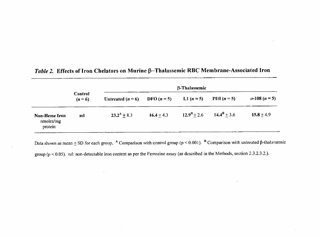
TlIble 2. Effects of Iron Chelators on Murine fJ-Thalassemic RUC Membrane-Associated Iron
Non-Herne Iron nmoles/mg protein
Control (n =6)
nd
Untreated (n = 6) DFO (Il = 5)
23.2A ± 8.3 16.4 ± 4.3
~-Thalassemic
LI (Il = 5) PIH (Il = 5) 0-108 (Il = 5)
12.98 ± 2.6 14.48 ± 3.6 15.8 ± 4.9
Data shown as mean ± SD for each group. A Comparison with control group (p < 0.001). 8 Comparison with untreated p-thalassemic
group (p < 0.05). nd: non-detectable iron content as per the Ferrozine assay (as described in the Methods, section 2.3.2.3.2.).

Table 3. Effects of Iron Chelators on ROC and Reticulocyte Parameters in 13-Thalassemic Mice
RBC Parameters Reticulocyte Parameters
Mice Hct Retie MCV MCHC L M H MCVr % % fL g/dL % mature % interm. % immature g/dL
Control 49.5 ± 4.7 2.38 ± 0.96 56.8 ± 3.3 24.1 ± 2.8 58.8 ± 13.8 35.3 ± 10.2 5.9 ± 5.2 59.5 ± 4.5
B-Thal.
Untreated 21.2 ± 1.3 22.0 ± 3.1 46.8 + 2.45 23.3 ± 0.78 22.6 ± 8.0 23.1 ± 5.1 54.4 ± 12.5 57.8 ± 3.3
+DFO 22.8 ± 1.8 17.2A ± 4.2 A 50.3 + 1.92 21.2A ± 0.87 35.4A ± 9.3 33.4A ± 5.8 31.2A + 14.7 59.8 ± 2.0
+ LI 22.1 ±23 17.6 ± 4.2 49.2 ± 2.32 21.6A ± 1.36 28.6 ± 8.9 28.0 ± 8.5 43.5 ± 17.3 61.6 ± 3.4
+PIH 21.4 ± 2.9 l2.0A ± 6.4 49.2 ± 2.33 A 21.5 ± 1.20 36.8" ± 11.1 32.7A ± 7.4 30.5A ± 14.2 57.7 ± 1.3
+ 0-108 21.6 ± 2.8 14.7 ± 7.3 49.2 ± 1.47 21.3" ± 1.07 28.5 ± 9.7 29.8 ± 8.3 41.7 ± 17.4 60.1 ± 0.9 --- - -~_ .. __ ._._--- --
Data shown as mean ± SD for each group. Control (n = 6), Untreated p-Thal. (n = 6), p-ThaI. + chelator (n = 5/group). Ali
differences between control and llntreated p-thalassemic mice were statistically significant, p < 0.001. A Denotes statistically
significant difference between llntreated and chelator-treated p-thalassemic mice, p < 0.05. Hct: hematocrit, Retic: reticulocyte count,
MeV: meall ccII volume, MClIC: mean cell hemoglobin concentration, MCVr: mean cell volume reticulocyte index. Rcticulocyte
parameters L, M, and 1-1 refer to low, medium, and heavy staining-intensity reticulocytes, respectively.

Table 4. Effects of Iron Chelators on Organ Pathology and Tissue Iron in J3-Thalassemic Mice
Mice SpleenlBody Weight Ratio Non-Herne Iron (Liver) Peris' Iron StainingC
% nmoles/mg wet weight % Liver % Spleen
Control 0.26 ± 0.04 0.78 ± 0.23 nd 1.39 ± 0.38
B-Thal.
Untreated 1.55 ± 0.32 1.65 ± 0.03 0.23 ± 0.21 5.45 ± 1.03
+DFO 1.40 ± 0.21 1.54 ± 0.15 0.11 ± 0.11 4.42 ± 0.70
+L1 1.40 ± 0.14 1.56 ± 0.19 0.07 ± 0.09 2.96A ± 1.83
+PIH 1.37 ± 0.27 1.59 ± 0.15 0.04 ± 0.05 3.82A ± 1.08
+0-108 1.44 ± 0.22 1.19A ± 0.47 0.02A ± 0.02 2.36B ± 0.61
Data shown as mean ± SD for each group. AIl differences between control and untreated J3-thalassemic mice were statistically
significant, p < 0.001. A Denotes statistically significant difference between untreated and chelator-treated p-thalassemic mice,
p < 0.05; B denotes p < 0.001. C Areas staining positive for iron using PerIs' Prussian blue dye expressed as a percentage of total area
examined. nd: non-detectable iron as per PerIs' staining technique (described in the Methods, section 2.3.7.3.).

.",.---~----------,---.~ EXCGs:; gloèir. C:hains""' .. ---- --~ Denaturation --- -----.... Degradation
Melt,nem~lobjn MCHI F.,rrltin F H:.?O.! 02 OH
it ~.ICtiCi Haomcsidorin-··· .... 9· .. ·········-l0~j~~~?~::;. A""ers;bli! ' ........... . • 7
V
• • + :::::<~:::.::. nocmlChromes c './ . '. . . ...... . • . ~iaem .2!:'3000..,j laemin ...... lipià bilayerl
Irro"t!rSlbla 1< 1,.. d.lffiage haemlchromes Ca,' rv' '-"oh,'nCh "ll1co :: .... .......... J 1 o0!"j, '.J... '_:..a.., . " .,. "'
oj,lxlliS;or.bo..y ~:::::.::': ::::::~~ a-t.,' 'Ii·. :;4' in!.i;;bilil}' 1 ~ .. --~~:~-' •••. ,',.';, '·;t : p . .!t.:!II: i: L:.:. 1 _"..../;;;::::;:;:;:.:::;:.; ::: SpPJanu'3~ t ~>4::.::.::::.;c. ~' ~: ... ::: .. :: .. :.:.:: .. ., ............. " , ' .. ::::::.:::.:./.: .. : :'t: '&1;-.0 l''~~:'::::;:::::::··:'::··::······::; ,ofccslil'!king' ,~!::norm;JI-'-'::---~çoc:lirt œficiell':'i"'.::: .; ·'1Cflis,enncJ .................. :;::;.:::: ~~::::: ..... "r;e.:.Tlf. . <::" -Icv . AnIe ,.11 œrll:.er.cy .: .. ~ , •.. :: .:;:::::::::::::: :.::: .:: ......... : .... 'ô: .~ ... : ... : ..... : •. -t·. . .AVI:crrn.;':I:~ ~'i • ',. '1' < .. : '" . ·:::;::::·r::i:::::;F:Ô::Ô:::;.;.;~.:-- ·~~-=~~ir.UCIl 'trr.c;;:g ::AIY.Of'llJ)I5r-!)y. ~ \ ., ..... 3.
. '::.:::.:::.::::::: ....,- 1 -~"""'::bindir.nr
t " 1 . 1 G P!'1Csphatidvl Flngmanlâlion ana inSlab.:iry Auto ogVlJS 9 . l ' . 'd't ~ bi "in" serint1 cxpcSt;la nc~eas:ea flgl ! 'l ..
+ ~omplemant nu ;J Oùc:eased d~fcrmabl!tly A
. + ,----. ~~ne removall
Cllva,lon of prcthrombina.:le
,::olllpll>:<
+ ~
~ar.)o;"f 19r.10Vill
Figure 1. Pathways Involved in Membrane Damage and Shortcncd RBC Lifespan in 13-Thalassemia. sp: spectrin, Pufa: polyunsaturated fatty acid, Pe: phosphatidyl ethanolamine, lev: inside-out vesicles, MCIl: mean cell hernoglobin, MCHC: mean ccli hemoglobin concentration. From Weatherall (1998, Bailliere 's Clinicat Haematotogy Il: 127-46).
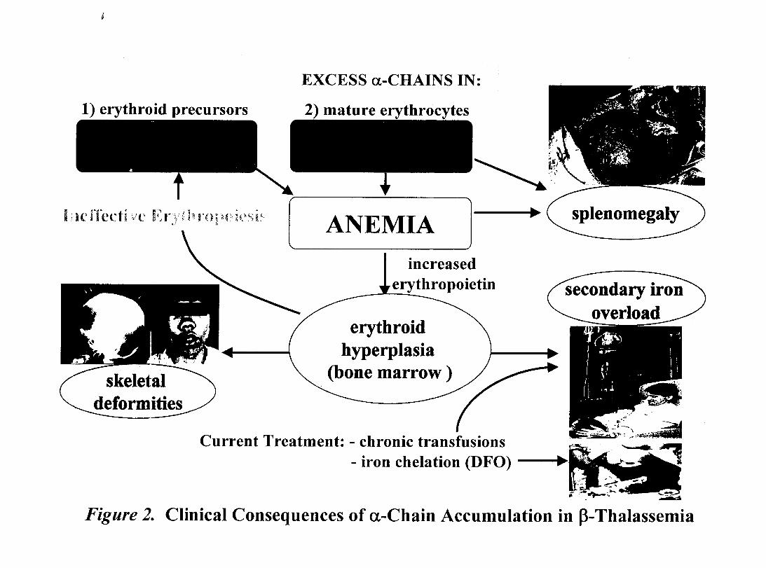
1) erythroid precursors
i·lI(,t"rl\l,ti 'II' H~'I' /h3'f\I)O~JiH,,;( 1 ~ 1 1. '-' ..... 9...... '-. a.J ~, a:'. J ~~, .,1 ~ . '1 '\ •.• } U • ,
EXCESS a-CHAINS IN:
[ANEMIA--J increased
erythropoietin -erythroid
hyperplasia (bone marrow )
Current Treatment: - chronic transfusions - iron chelation (DFO)
...... JIll'"
Figure 2. Clinical Consequences of a-Chain Accumulation in ~-Thalassemia

~:Nofmal Human 7:~';..
... ~., .. c.,
Erythrocyte .. ,,+ ;;~
-......- -a-chains 00 HYPOSMOTIC L YSIS
4°C, 45 minutes
' 00-·~O'::'···
1) Normal erythrocytes mix with exogenous purified a-chains (with heme moieties attached)
i; 5) C ells reseal
2) Cells regurgitate contents into medium
3) Cell contents mix with a-chains
o ISOTONIe
RESEALING
37°C, 45 minutes
4) Cells swell, incorporate regurgitated Hb and added a-chains
Figure 3. Preparation of Human Model ~-Thalassemic Erythrocytes
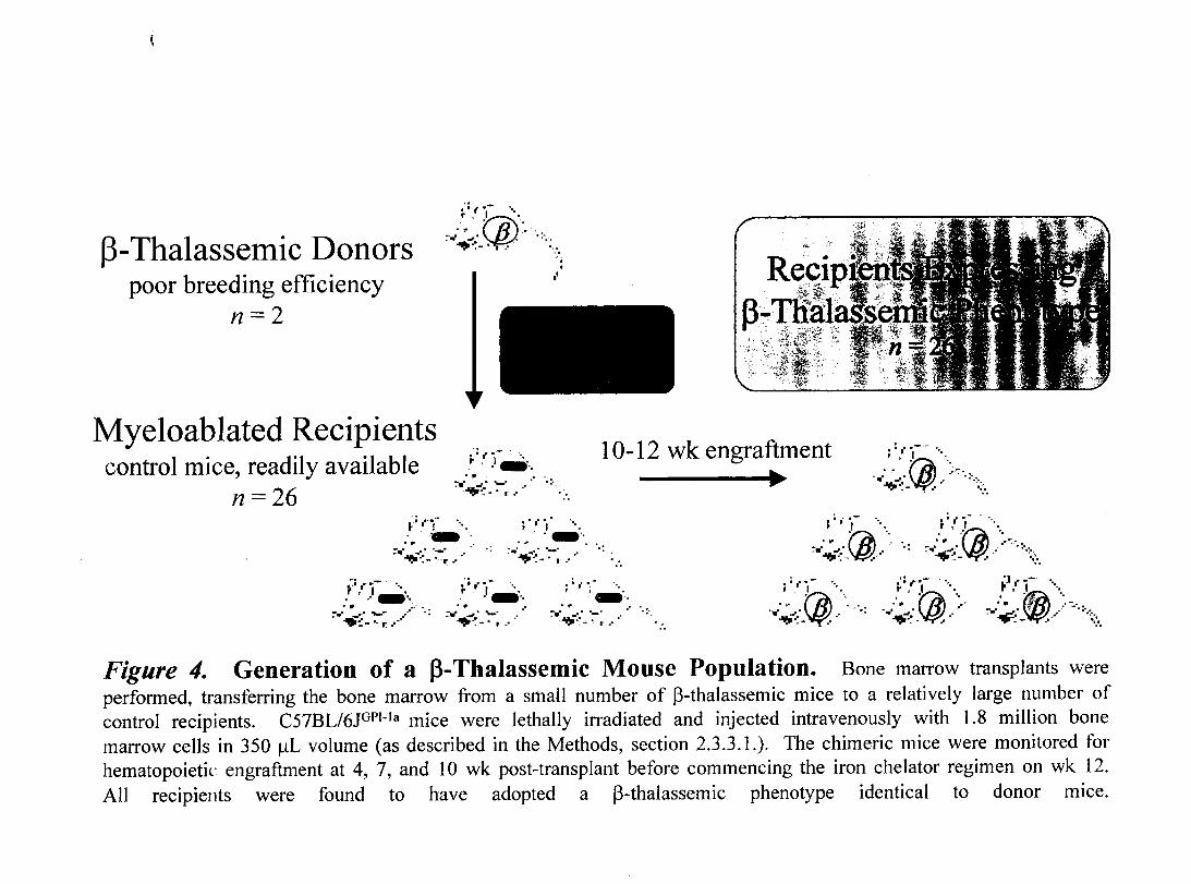
~-Thalassemic Donars poor breeding efficiency
n=2
~:,..·@t ". c' •
• fi' .. _
~" J' . .;.
~-:- ... -
"
. •
Myeloablated Recipients control mice, readily available
n=26
, . ( .... .~ .. r,.I_ .. 10-12 wk engraftment ;:.i0,f··'-" . '", -":' .. -. ." ....... ... ~~:-. ".'
.; .... - ". ,t ~
;"" .. - .... . _'. 'P
.... ,.." "-'* .... ... ~.:-. -: ~ ;.," ..,." --" ~ ~-:-., .... , ... . !o •• ,.-' .~ ,.C ... - ". r, 1· r, 1 .
. "-". "-" ,:",: "JO......- ........ ::./" ~J" __ • 0"
....::- .. -: " .. ' ~-:-' .. ' ~ , .. , ' .. ",
--:"': ~".,;::'. ':-; .' "
-.; "J' • "~":"" ~-=- .~. \'.
..... JIIII""
~;"r -'. • P "fOa
~" ~J-:.' .\I:P.. .'
; ,.- . 'c ., 1 ' • . '0, ' . -.. ~. ..' .' •... :"::" -....... ~- ." ':.
.J,ij- ..... . ~,. . " ."--" . -.. ~. " ...•.. ...... ~- .. '~ ".::' .
. :,..- ~ ;:,..- "
Ic~1 ••.. ,r,0,' .... • Y" • .". .,' .. ".J;.: ." .... : -."....,." ... -.. ~.-.' ...... "
Figure 4. Generation of a P-Thalassemic Mouse Population. Bone marrow transplants were performed, transferring the bone marrow from a small number of ~-thalassemic mice to a relatively large number of control recipients. C57BL/6JGPI-la mice were lethally irradiated and injected intravenously with 1.8 million bone marrow cells in 350 JlL volume (as described in the Methods, section 2.3.3.1.). The chimeric mice were monitored for hematopoietic engraftment at 4, 7, and 10 wk post-transplant before commencing the iron chelator regimen on wk 12. AlI recipients were found to have adopted a ~-thalassemic phenotype identical to donor mice.

\ J
y.j
o
IS 126.4
Nonnal a-chain
a-chain
15000 15~Otl 15400 15600
Nonna! p-chain
15ROO
Glutalhionylated p-chain 16172.7
16000 16200
p-çhain +haem
16482.0. mass (Da) , i
16400 16600
Figure 5 A. Standard deconvoluted ESI mass spectra of blood· from a healthy adult subject diluted 500-fold in ddH20. Normal a-hemoglobin chains display a characteristic peak at 15126.4 Da, while the a-chain + heme group displays a peak at 15741.1 Da. From: Wild BJ, Green B~, Cooper EK. et al. Rapid identification ofhaemoglobin variants by electrospray ionizàtion mass spectrometry. Blood CelIs, Molecules, and Diseases. 27:691-704,2001.
100 i
95 1 90; 85 i 80 j 75 ,
! 70 j 85 ;
§80 1 ~55 1 ~50 j • l ~"5 i .!! ; :.-0 i
35'
301 25 j 20 j
1 15 :
15126.0
i\ ii Normal a.-globin
!\
i\
1 \
i \
Il 1 \ ,\ 1 \ :1
\_i \ a.-globin + herne
10 ! 15741.0 51 ~ .. __ ---.. _ ~-'. __ . __ ". _ .... __ .-----.;\ ........ --....._--' ....... ,.,.2' .• -"\., .... . j" .. _,'"" ... _', ..... -.. __ ~_ a ..:··t·~~! ..... , .. "T'- •• r-"""T"' ... ~·~··r-"', .,....-, ..,-! ...... ,-...-, ...,..., -,-( ..... _. • .. ·~·i , i j 1
14800 15000 i i
18400 15200 15800 115000 15-000 "5200
Figure 5 B. Deconvoluted ESI mass spectra of 0.8 mg/mL purified a-hemoglobin. Subunits \-vere isolated from normal adult \-vhole blood and diluted 1: 150 \-vith ddH20 to obtain characteristic peaks and associated molecular mass values.
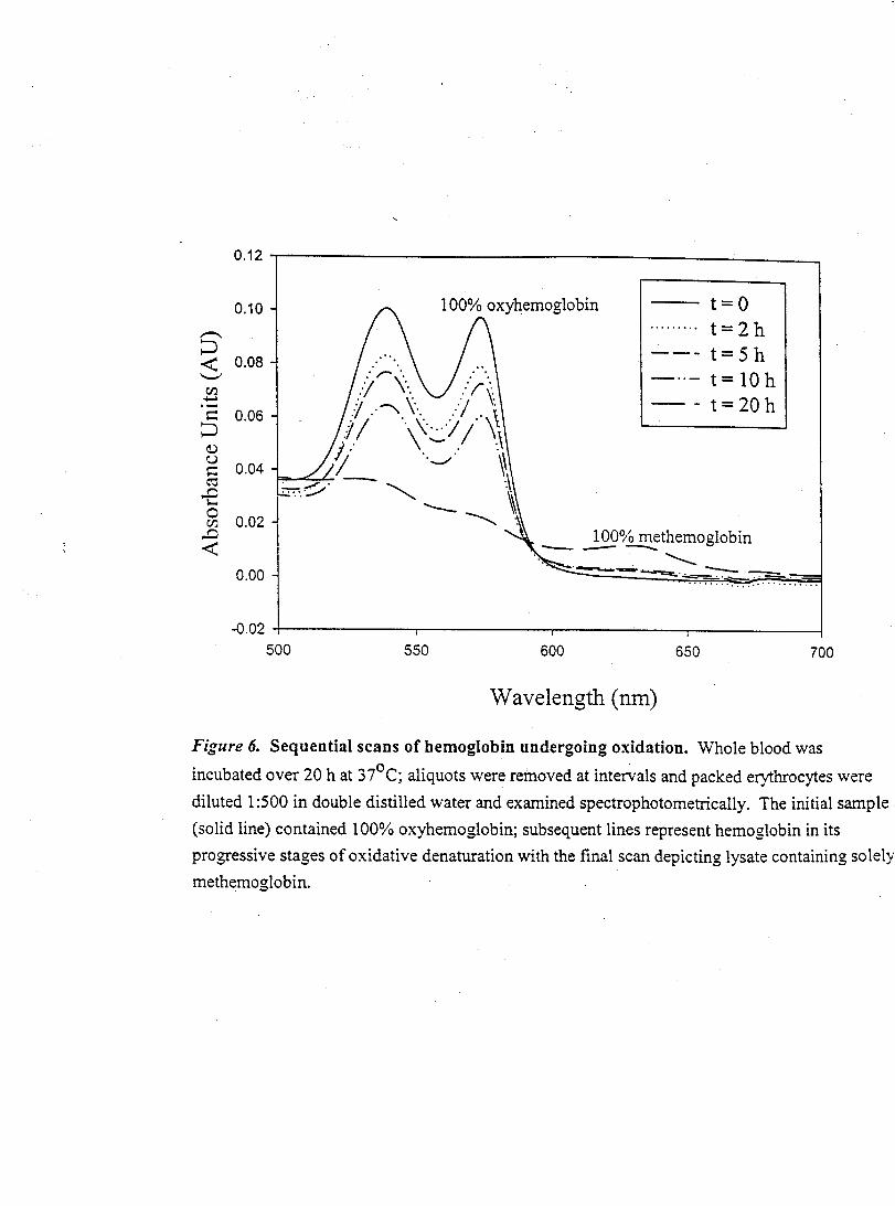
0.12
0.10 t=O
----......... t= 2 h
::J -< 0.08 "-"
--- t=5h - .. - t= 10h
Cf.) ...... C 0.06
- - t=20h ::J ~ u
0.04 ,... -~ .-e 0
0.02 Cf.)
..0 -<
100% methemoglobin ---'-0.00 '~----~'------="":'~ -:-:-=... . ..........................
-0.02 500 550 600 650 700
Wavelength (nm)
Figure 6. Sequential scans of hemoglobin undergoing oxidation. Whole blood was
incubated over 20 h at 37oC; aliquots were removed at inten,als and packed erythrocytes were
diluted 1:500 in double distilled water and examined spectrophotometrically. The initial sample.
(solid line) contained 100% oxyhemoglobin; subsequent lines represent hemoglobin in its
progressive stages of oxidative denaturation with the final scan depicting lysate containing solely
methemoglobin.

0.36 -r---------------------------,
0.34
---~ $ 0.32
00 ..... . -.... -~ ~ 0.30 u C co -€ 0.28 o
00 ..0 ~
0.26
....
~ ~
100% oxyhemoglobin
100% methemoglobin .........
0.24 +-------,--------,...-------r--------l 500 550 600 650 700
Wavelength (nm)
Figure 7. Initial Spectrum of Purified a-Hemoglobin Chains and Spectrum Taken Following 20 h Incubation. Subunits were diluted 1: 1 00 in double distilled water and analyzed spectrophotometrically for functional denaturation response over time. Initial composition of lysate was 100% oxyhemoglobin (solid line); following ovemight incubation at 2SoC, progressive oxidative denaturation of a-chains took place, yielding 100% methemoglobin (dotted line) in finallysate.

A)
C)
~r---------------------~ ~r-----------------------
B)
• i C ......
\
G1-FITC G1-FITC
1 1
'i.J .' 1 1 i 1 t '''II 1 i,. ..... . i t. Li ..
,\
Relative Intensity Relative Intensity
~1 ~i M
1 , D) ,1\
1
J J"
1 \~ l'\l ~, ~'~\ 1 ",,' :J i'\ =~
~I cl
,
J '-'1 lli 1
i 1 ~ 1
\ 1 I~ 1 E
E 1 \ ~ '~ , ~ .J
l 1 l'f , D D t 1 .;J Ql ,1 ~. ,1 c::: 1 , 1 l "j dl! i"l, , .... , '1' , \ t 'r' \., ~h
t il li d' ...... .t ' 1 ••• .1., li l • , .... 1. , o •• "" ' . " ' .
. 1 HlOO .1 Gl-FITC Gl-FITC
1\"HlIl
Relative Intensity Relative Intensity
Figure 8. Encapsulation of purified a-hemoglobin chains into hum an erythrocytes. FACScan data depicting the tluorescence of er:1hrocytes loaded \vith FITC-Dextran alone or admixed with purified a-hemog:lobin chains. Cells exhibitinQ relative fluorescence intensities Qreater than l
~ - -(within region "D"). were considered to have incorporated FITC-Dex while values below l retlected typical low-level inherent cellular fluorescence. Background cellular tluorescence \vas measured in control (A) and control-resealed cells (B), yielding 1.18'% and 0.9% respectively. The fluorescence intensities of cells lysed and resealed in the presence of either 5 mg FITC-Dex/mL pRBC alone or FlTC-Dex in suspension with a-subunits (13.5 mg a-chains/mL pRBC) are seen in panels C and D, respectivel;:. In both cases, when fluorescent RBC \vere expressed as a percentage of the total counted (5000 cells). more than 98.5% of red cells were found to have incorporated the exogenous material. Samples were prepared in triplicate and representative histograrns
for each condition are sho\vTI.
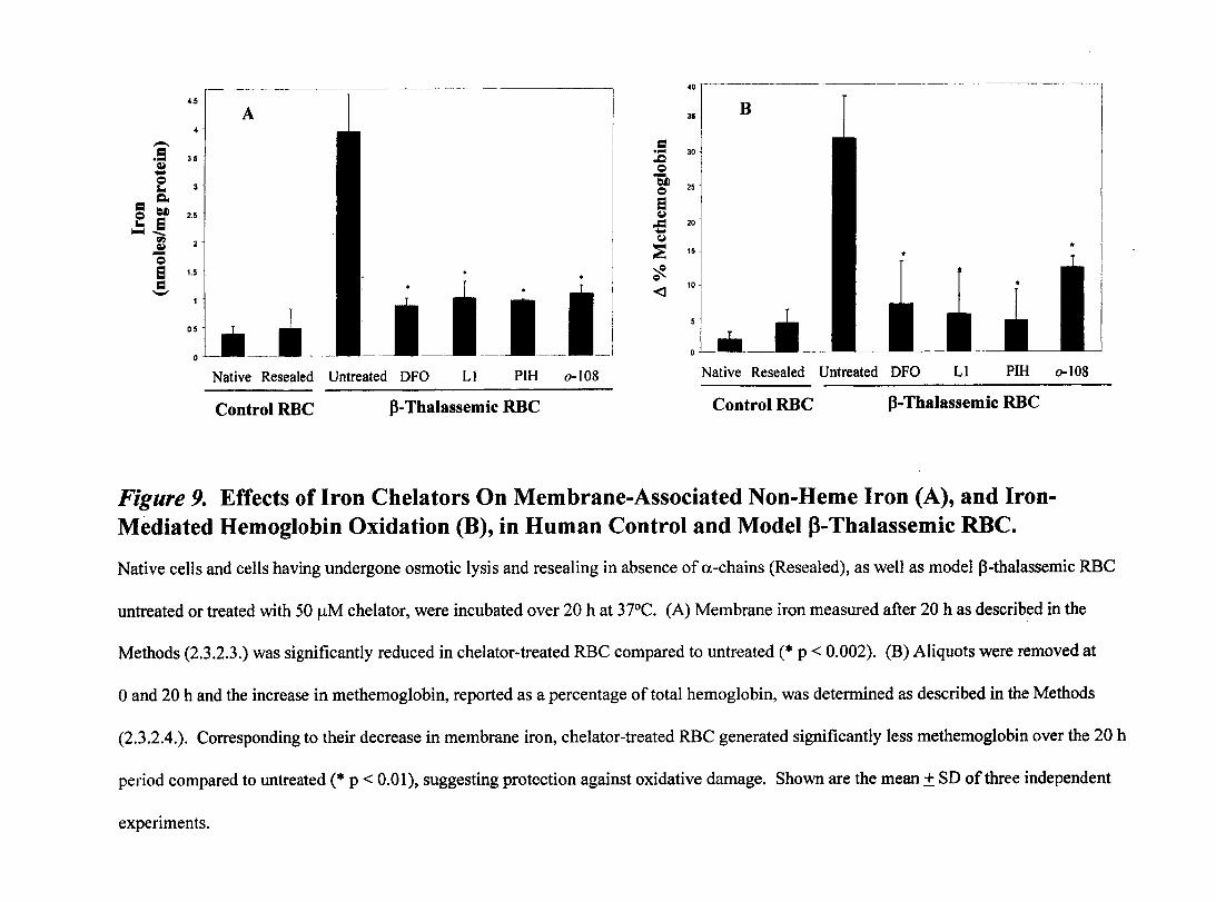
40
451 A 31
- 1:1 1:1
3.5 .... 30 .... ,.Q
~ .... = = "Q ... 25
= Q. = e = bJ) 2.5 ~
.=:.@ .CI 20 .... rIl ~ ~ ~ - 15
= e 1.5 ~ 1:1
---j-j-I-Q -- <1
10
0.5 i IL __ Native Resealed Untreated DFO LI PIH 0-108 Native Resealed Untreated DFO LI PIH 0-108
Control RBC ~-Thalassemic RBC ControlRBC ~-Thalassemic RBC
Figure 9. Effects of Iron Chelators On Membrane-Associated Non-Heme Iron (A), and IronMediated Hemoglobin Oxidation (B), in Human Control and Model 13-Thalassemic RBC.
Native cells and cells having undergone osmotic lysis and resealing in absence of a-chains (Resealed), as well as model ~-thalassemic RBC
untreated or treated with 50 JlM chelator, were incubated over 20 h at 37°C. (A) Membrane iron measured after 20 h as described in the
Methods (2.3.2.3.) was significantly reduced in chelator-treated RBC compared to untreated (* p < 0.002). (B) Aliquots were removed at
o and 20 h and the increase in methemoglobin, reported as a percentage of total hemoglobin, was determined as described in the Methods
(2.3.2.4.). Corresponding to their decrease in membrane iron, chelator-treated RBC generated significantly less methemoglobin over the 20 h
period compared to untreated (* p < 0.01), suggesting protection against oxidative damage. Shown are the mean ± SD ofthree independent
experiments.

Weeks post-transplant:
4wk
Gpi Gpi 1 2 3 4 5 6 7 8 9 10 a b
7wk
Gpi Gpi 1 2 345678910 a b
lOwk
Gpi Gpi
Control Recipient a b
Isoform
1 2 3 4 5
fl-Thalassemic Donor Isoform
Figure 10. Glucose Phosphate Isomerase (Gpi) Phenotyping of Recipient Mice 4, 7, and 10 Weeks Following Murine 13-Thalassemic Bone Marrow Transplant. Peripheral blood of transplanted mice was analyzed at the protein level to determine the proportion of endogenous RBC remaining, and the proportion contributed by the donor (Gpi-lb) at different stages following BMT. Gpi-1 isoforms "a" and "b" were derived from C57BLl6JGPI-la (recipient) and C57BLl6JGPI-lb (donor) mice, respectively. Lanes 1-10 reveal the Gpi-1 isotypes of ten representative transplanted mice. At 4 weeks, the majority ofrecipient RBC were of endogenous origin, as Gpi-1a was the principal form of the enzyme detected. After 7 weeks, however, over 50% of recipient RBC expressed Gpi-lb. By week 10, aIl recipients produced strictly isoform "b", indicating a switch to p-thalassemic RBC production exclusively, and therefore, complete hematopoietic engraftment.
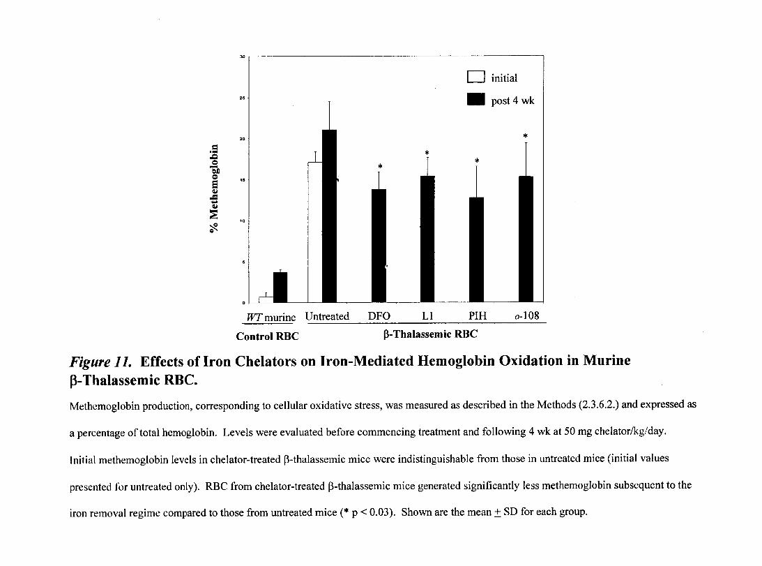
.9 ,.Q
oS bJ) Q
El ~
.CI ... ~
~ ';ft.
30r---------------------------------------------------------------------------------~
20
20
10
10
WT murine Untreated DFO LI
o initial
• post4wk
PIH 0-108
Control RBC P-Thalassemic RBC
Figure 11. Effects of Iron Chelators on Iron-Mediated Hemoglobin Oxidation in Murine p-Thalassemic RBC.
Methemoglobin production, corresponding to cellular oxidative stress, was measured as described in the Methods (2.3.6.2.) and expressed as
a percentage of total hemoglobin. Levels were evaluated before commencing treatment and following 4 wk at 50 mg chelator/kg/day.
Initial methemoglobin levels in chelator-treated p-thalassemic mice were indistinguishable from those in untreated mice (initial values
presented for untreated only). RBC from chelator-treated p-thalassemic mi ce generated significantly less methemoglobin subsequent to the
iron removal regime compared to those from untreated mice (* p < 0.03). Shown are the mean ± SD for each group.
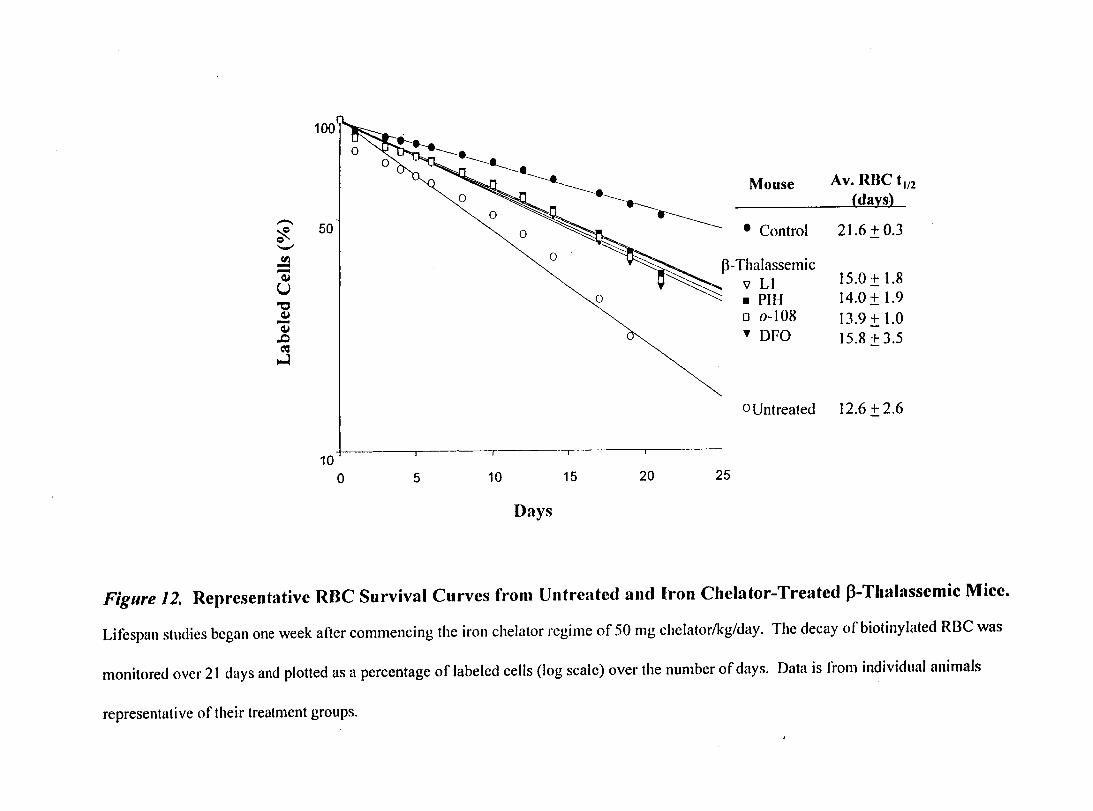
100
Mouse Av. Rue t l12
{days}
O~ ~ • Control 21.6 ± 0.3 0 --.
50 ~ C)
'-"
~P-Thulassemic 15.0±1.8
0
~ :~:I 14.0 ± 1.9
CIl -0
-~ U "0
13.9 ± 1.0 o 0-108 ~ -~ 15.8±3.5 ~ OFO .t:J
C'J ~
0Untreated 12.6 ± 2.6
10 o 5 10 15 20 25
Days
Figure 12. Representative RBC Survival Curves from Untreated and Iron Chelator-Treated f3-Thalassemie Mice.
Lifespan studies begun one week after commencing the iron chelutor rcgime of 50 mg chelator/kg/day. The decay of biotinylated RBC was
monitored over 21 days and plotted as a pereentage of labeled eells (log seale) over the number of days. Data is l'rom individual animaIs
representative oftheir treutment groups.

Control Liver Untreated
LI
(3-Thalassemic Livers ,...................................................................................................... . ................................................................................................... .
Untreated DFO
PIH 0-108 * : 1
1
1
............. , ......... " ............................. " .......................................... , .... , .......... , ........................ , .... " ......... , ............................................................................................................................................... , ................... ..
Figure 13. Iron Chelators Reduce Liver Iron Accumulation in f3-Thalassemic Mice.
Following 4 wk of iron chelator treatment, liver sections were stained for iron using PerIs' Prussian blue dye and a hematoxylin and
eosin counterstain as described in the Methods. The percentage of stainable iron was averaged for each treatment group and
representative tissue sections from individual animaIs are depicted (20 X). * p < 0.05 compared to untreated p-thalassemic group.

(-'
Control Spleen fJ-Thalassemic Spleens Untreated
·· .... ········· .... ····· .. ···· .. ····lJ~~~~~~~d·· .. ·· .. ·· .. ······ .......................... · .... ······ ...... ·· .. ··· .. ··Dp·Ü·· .. ··· .. ·· .. ···· .... ···· .... ···· ........ ····1
tfi~t\Ji~lt!tl~ 1
~:~3C;;~~~;t~~~J:ty ·:~/i,'~1~~~ 1
~t~~i~~~li~'~\}il:,tÎ 1 r···························· .......... ····················..................................................... 1
! LI * PIH * 0-108 ** 1
i~i-IJ~~r~~fllf~~I··· .~~. ::~m~:!i:.::~:;.~··?::~r:&<~;~~;:;~$!::I{~~~~~~~ r1~)~;ï~~~Ç~:~_~I;~ :11 i ~;il.~.';\~f.:~ .~;~~:~:{,i:,:il;. ~~!~d:U;·~r;·:.:· .. ·.:.:jj;;:~:~(:,/~~: .~~~~:~~;W~~fir f:s~~~i~~:·~~I~:I~·~,~ j 1
1
Figure 14. Iron Chelators Reduce Splenic Iron Accumulation in fJ-Thalassemic Mice.
Spleen sections were stained for iron using Peris' Prussian blue dye and a hematoxylin and eosin counterstain, as in Fig. 13 (described in the
Methods). The percentage of stainable iron was averaged for each treatment group and representative tissue sections are depicted (20 X) .
... p < 0.05, .... p < 0.001 compared to untreated B-thalassemic group.