Embed Size (px)
Citation preview
Iranian Journal of Basic Medical Sciences
ijbms.mums.ac.ir
Assessment of expressions of Bcl-XL, b-FGF, Bmp-2, Caspase-3, PDGFR-α, Smad1 and TGF-β1 genes in a rat model of lung ischemia/reperfusion
Hasan Şimşek 1, Şeniz Demiryürek 2*, Tuncer Demir 2, Hüsne Didem Atabay 2, Ali Osman Çeribasi 3, Recep Bayraktar 4, Davut Sinan Kaplan 2, Serdar Öztuzcu 4, Beyhan Cengiz 5
1 Dumlupınar University, Department of Physiology, Faculty of Medicine, Kütahya, Turkey 2 Gaziantep University, Department of Physiology, Faculty of Medicine, Gaziantep, Turkey 3 Fırat University, Department of Pathology, Faculty of Veterinary Science, Elazığ, Turkey 4 Gaziantep University, Department of Medical Biology, Faculty of Medicine, Gaziantep, Turkey 5 Gazi University, Department of Medical Genetic, Faculty of Medicine, Ankara, Turkey
A R T I C L E I N F O A B S T R A C T
Article type: Original article
Objective(s): Ischemia is described as organs and tissues are destitute of oxygen due to decreased arterial or venous blood flow. Many mechanisms play role in cell death happened as a consequence of a new blood flow is needed for both cell regeneration and to clean toxic metabolites during ischemia and later. Lung damage induced by ischemia/reperfusion (I/R) is a frequent problem in lung transplantation. Apoptosis (programmed cell death) is known as cell suicide, and plays a key role in embryonic developmental and in maintain adult tissue’s life. Materials and Methods: It is investigated expressions of Smad1, Bmp-2, Bcl-XL, b-FGF, Caspase-3, TGF-β1, PDGFR-α genes for molecular changes in lung tissues, after I/R is formed, in this study. For this, we included 40 Wistar albino rats to this study and divided 4 groups (n=10). The Groups were determined as Control (C), Group 1= 1 hr ischemia (I), Group 2= 1 hr ischemia+2 hr reperfusion (I+2R), Group 3= 1 hr ischemia+4 hr reperfusion (I+4R). Besides, molecular analysis and histopathologic examinations of tissues were performed, and the results were evaluated by normalization and statistics analysis. Results: We have found a significant increase in expression of Bcl-XL (P=0.046) and Caspase-3 (P=0.026) genes of group 1, and it was not monitored any significant difference in Group 2 and Group 3. In all groups, the changes in b-FGF (P=0.087), Bmp-2 (P=0.457), TGF-β1 (P=0.201) and PDGFR-α (P=0.116) were not significant compared to control group. We did not see any mRNA expression of Smad1 gene in all groups include control. Conclusion: These findings suggest that I/R injury may trigger apoptotic mechanism in lung.
Article history: Received: Jul 6, 2015 Accepted: Dec 24, 2015
Keywords: Apoptosis Growth factors Ischemia/reperfusion Lung
►Please cite this article as: Şimşek H, Demiryürek Ş, Demir T, Didem Atabay H, Osman Çeribasi A, Bayraktar R, Sinan Kaplan D, Öztuzcu S, Cengiz B . Assessment of expressions of Bcl-XL, b-FGF, Bmp-2, Caspase-3, PDGFR-α, Smad1 and TGF-β1 genes in a rat model of lung ischemia/reperfusion. Iran J Basic Med Sci 2016; 19:209-214.
Introduction Ischemia/reperfusion (I/R) is characterized by
histopathological changes due to the fact that it cause lung edema and neutrophil extravasation and some changes in vascular permeability. These changes are mostly formed by reactive oxygen species (ROS) generated during reperfusion (1). Membrane lipids, proteins, nucleic acids and deoxyribonucleic acids are most sensitive ones of cell components to reperfusion damage (2). Pulmonary dysfunction caused by I/R damage is one of the important problems in lung transplantation (3). After I/R damage, intense inflammatory response, endothelial injury, neutrophil infiltration and pulmonary edema are occurred in lung tissue (4). Pulmonary vascular cell injury is formed as a result of I/R damage or
chronic hypoxia (5). The function of reperfusion in ischemic lung tissue is like double-edged sword, so both reperfusion is needed to keep alive the cell and it may triggers a chain of events, like increased microvascular permeability, pulmonary vascular resistance, pulmonary edema, disturbed oxygenation and pulmonary hypertension, may cause serious I/R damage (6).
I/R applications may trigger one of the three programmed cell death, apoptosis, autophagy and necrosis (7). First of these mechanism is apoptosis, the best known. Apoptosis was described by Kerr et al (8). in 1972. This mechanism provides digestion of damaged cells and is characterized by caspase activation (5).
Aim of this study is to investigate whether I/R
*Corresponding author: Şeniz Demiryürek. Gaziantep University, Department of Physiology, Faculty of Medicine, Gaziantep, Turkey.
email: [email protected]

Şimşek et al Ischemia triggers apoptotic mechanism
Iran J Basic Med Sci, Vol. 19, No. 2, Feb 2016
2
induces apoptosis or not in lung, also is to shed light on which pathway is used by apoptosis in lung tissues when it is stimulated by I/R. For this purpose, we have selected some genes; apoptotic gene (Caspase-3), anti-apoptotic gene (Bcl-XL), growth factor genes (b-FGF, TGF-β1), growth factor receptor gene (PDGFR-α), and Bmp-2 signaling pathway genes (Bmp-2, Smad1) are important to understand apoptosis and its pathway.
Caspases are inactive in cells, and when a caspase is activated, it starts a cascade to activates other pro-caspase (9). Caspase-3 is the most important one among the caspases is, it is also known as hangman caspase and is activated by initiator caspases (caspase-8, 9 and 10), and by the other main components, like B-cell lymphoma 2 (Bcl-2) protein family, determine the place of cells between death and life. This family have both proapoptotic and antiapoptotic members (10), and the best known member of this family is Bcl-XL protein (11).
b-FGF is known as premotor gene of angiogenesis and lymph angiogenesis and has role in cell migration, growth and differentiation (12). TGF family has 40 members with the inclusion of bone morphogenetic protein family (BMP), biggest subfamily in this group and have many functions in apoptosis (13), and is responsible for cell proliferation, differentiation, motility and extracellular matrix production (14).
Smad family members have been classified into three subgroups, receptor-regulated Smads (R-Smads), common mediator Smads (Co-Smads) and inhibitory Smad proteins. Smad1 is member of R-Smads, and activated by BMP ligands (15).
Platelet derived growth factor (PDGF) isoforms takes effect by binding -α and –β (PDGFR-α and PDGFR-β) receptors, and the main function is to stimulate growth of mesenchymal cells, also efficient on motility and survival (16).
Materials and Methods The animals
In the study, 40 adult Wistar albino rats with a weight of 250–300 g body mass and used. Before the study, all animals were kept in the laboratory so as to orientate them to the laboratory conditions including 50-60 % humidity, 12 hr light and 12 hr dark cycle and 22-24 °C temperature. All studies were carried out in accordance with the National Institutes of Health guidelines on animal care and with the approval of the Ethics Committee of the Gaziantep University, Gaziantep, Turkey.
I/R model
In order to induce lung ischemia, atraumatic clamps were used (Picture 1). Control group
Anything did to this group. Animals were anaesthetized and tissue samples were got out.
Picture 1. Experimental lung ischemia model. 1) incision was performed from manubrium sterni to xiphoid. 2) left pulmonary artery was isolated and clamped with an atraumatic microvascular clamp during 60 min
Group 1 (I) Left pulmonary artery was clamped with
atraumatic clamp during 1 hr. Then the tissue samples were got out from lung.
Group 2 (I/2R) Left pulmonary artery was clamped with atraumatic
clamp during 1 hr. Then the clamp was opened and reperfusion was allowed during 2 hr. At the end of 3 hr, tissue samples were got out from lung. Group 3 (I/4R)
Left pulmonary artery was clamped with atraumatic clamp during 1 hr. Then the clamp was opened and reperfusion was allowed during 4 hr. At the end of 5 hr, tissue samples were got out from lung.
Before the surgery, the animals were anesthetized with xylazine hydrochloride 5 mg/kg and ketamine hydrochloride 35 mg/kg IP injection, and after the surgery, lung tissues were divided into 2 with a scalpel. One part was kept in 10 % formalin solution for histopathological investigation, and the remaining parts were kept in nitrogen tank for further molecular studies. Tissue homogenization
Qiagen TissueLyser LT machine (Catalog No:85600, Mainz, Germany) and QIAzol Lysis Reagent (Qiagen, Catalog No:79306, Mainz, Germany) were used to disintegrate and to homogenizate the tissue samples.
Total RNA isolation and cDNA conversion
Roche High Pure RNA Tissue Kit’s (Product No. 12033674001, Mannheim, Germany) protocol was used to isolate total RNA from tissue samples. Then, Ipsogen RT Kit (Catalog no: 679913, Qiagen, Hilden, Germany) was used to convert mRNA isolated to cDNA.
cDNA concentration measurement Epoch (BioTek, Winooski VT, USA) measurement
machine was used to measure the concentration of all cDNA samples, and the last concentrations were adjusted 50 ng/ µl.
Ischemia triggers apoptotic mechanism Şimşek et al
Iran J Basic Med Sci, Vol. 19, No. 2, Feb 2016
211
Table 1. Histopathologic evaluation in lung tissue for each group
Reverse transcription-polymerase chain reaction (RT-PCR)
RT-PCR conditions specified for each gene were performed with GeneAmp® PCR System 9700 (Applied Biosystem, Singapore) PCR device. mRNA levels of b-FGF (Sense: 5’-GGCTATGAAGGAA-GATGGAC-3’ Anti-sense: 5’-GTTCGTTTCAGTGCC-ACA-3’), Bcl-XL (Sense: 5’-AGGCTGGCGATGAGTTT-GAA-3’ Anti-sense: 5’-TGAAACGCTCCTGGCCTTTC-3’), Bmp-2 (Sense: 5’-AAGGCACCCTTTGTATGTGG-3’ Anti-sense: 5’-CATGCCTTAGGGATTTTGGA-3’), Caspase-3 (Sense: 5’-GACTGCGGTATTGAGACAGA-3’ Anti-sense: 5’-CGAGTGAGGATGTGCATGAA-3’), PDGFR-α (Sense: 5’-GTCCAGGTGAGGTTAGAGG-3’ Anti-sense: 5’- CACGGAGGAGAACAAAGAC-3’), Smad-1 (Sense: 5’- CCGCCTGCTTACCTGCCTCCTGAA-3’ Anti-sense: 5’-GAACGCTTCGCCCACACGGTTGT-3’), TGF-β1 (Sense: 5′-GCTAATGGTGGACCGCAACAAC-3′ Anti-sense: 5′-CAGCAGCCGGTTACCAAG-3′) were studied by RT-PCR using primer sets. Amounts of PCR products were normalized by using β-actin (Sense: 5’-AGGCCAACCGTGAAAAGATG-3’ Anti-sense: 5’-ACCAGAGGCATACAGGGACAA-3’) as housekeeping gene. cDNA was denatured at 94 °C for 4 min, annealed at 60 °C for 30 sec (bFGF), at 60 °C for 30 sec (Bcl-XL), at 60 °C for 30 sec (Bmp-2), at 59.1 °C for 45 sec (Caspase-3), at 59.1 °C for 30 sec (PDGFR- α), at 71 °C for 45 sec (Smad-1), at 60.9 °C for 30 sec (TGF-β1). RT-PCR mixtures (8 µl) were electrophoresed on a 2% agarose gel, which was subsequently stained with 0.5 ml/ml ethidium bromide. Gels were scanned on an imaging analyzer (Picture 2), and the corresponding band densities were measured with imageJ program. Histopathology
The lung specimens dehydrated and embedded in paraffin, and sectioned at 5 μm thickness. The samples were stained with Hematoxylin-Eosin (H-E) and analyzed under light microscopy. Statistical analysis
All the values were calculated as mean ± standard deviation by SPSS for Windows version 22.0. for all the evaluated genes. Kruskal-wallis test and Dunn’s multiple comparison were used to compare the variables. P<0.05 was considered to be statistically significant.
Results Gene expression results
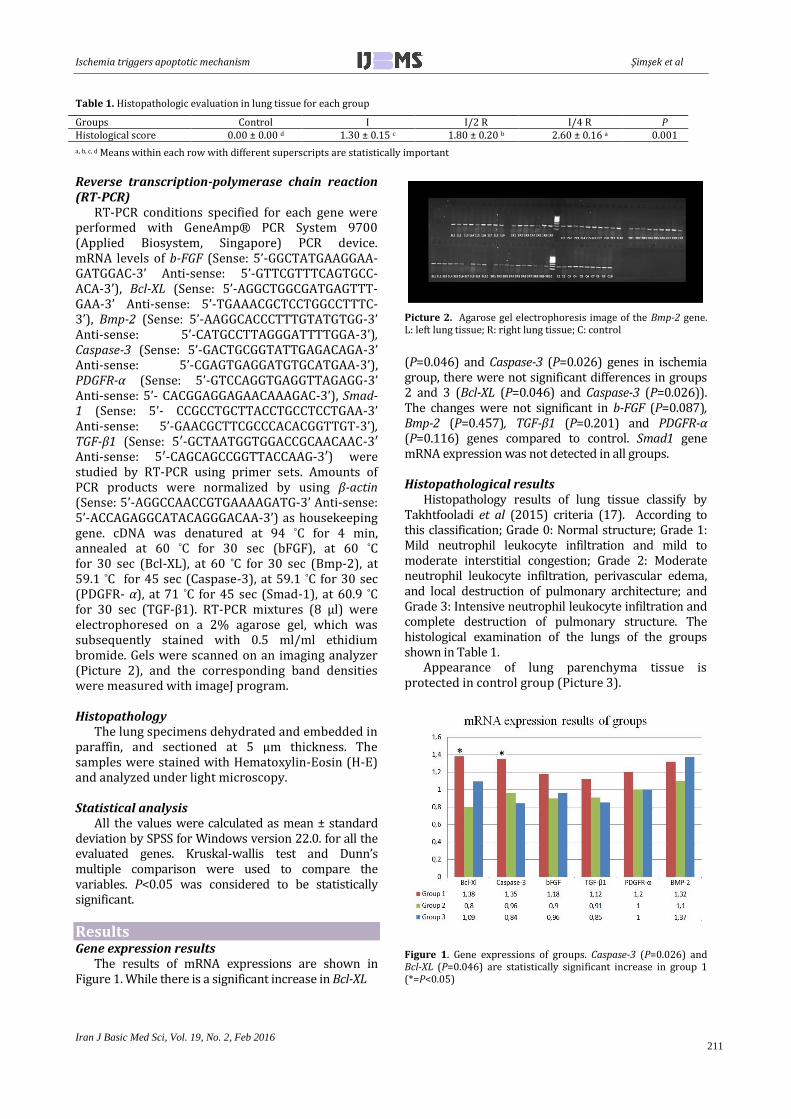
The results of mRNA expressions are shown in Figure 1. While there is a significant increase in Bcl-XL
Picture 2. Agarose gel electrophoresis image of the Bmp-2 gene. L: left lung tissue; R: right lung tissue; C: control
(P=0.046) and Caspase-3 (P=0.026) genes in ischemia group, there were not significant differences in groups 2 and 3 (Bcl-XL (P=0.046) and Caspase-3 (P=0.026)). The changes were not significant in b-FGF (P=0.087), Bmp-2 (P=0.457), TGF-β1 (P=0.201) and PDGFR-α (P=0.116) genes compared to control. Smad1 gene mRNA expression was not detected in all groups. Histopathological results
Histopathology results of lung tissue classify by Takhtfooladi et al (2015) criteria (17). According to this classification; Grade 0: Normal structure; Grade 1: Mild neutrophil leukocyte infiltration and mild to moderate interstitial congestion; Grade 2: Moderate neutrophil leukocyte infiltration, perivascular edema, and local destruction of pulmonary architecture; and Grade 3: Intensive neutrophil leukocyte infiltration and complete destruction of pulmonary structure. The histological examination of the lungs of the groups shown in Table 1.
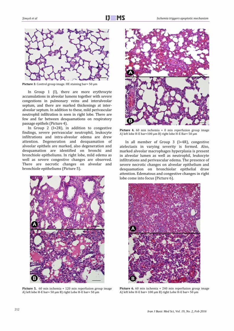
Appearance of lung parenchyma tissue is protected in control group (Picture 3).
Figure 1. Gene expressions of groups. Caspase-3 (P=0.026) and Bcl-XL (P=0.046) are statistically significant increase in group 1 (*=P<0.05)
Groups Control I I/2 R I/4 R P Histological score 0.00 ± 0.00 d 1.30 ± 0.15 c 1.80 ± 0.20 b 2.60 ± 0.16 a 0.001
a, b, c, d Means within each row with different superscripts are statistically important
Şimşek et al Ischemia triggers apoptotic mechanism
Iran J Basic Med Sci, Vol. 19, No. 2, Feb 2016
212
Picture 3. Control group image. HE staining bar= 50 μm
In Group 1 (I), there are more erythrocyte accumulations in alveolar lumens together with severe congestions in pulmonary veins and interalveolar septum, and there are marked thickenings at inter-alveolar septum. In addition to these, mild perivascular neutrophil infiltration is seen in right lobe. There are few and far between desquamations on respiratory passage epithels (Picture 4).
In Group 2 (I+2R), in addition to congestive findings, severe perivascular neutrophil, leukocyte infiltrations and intra-alveolar edema are drew attention. Degeneration and desquamation of alveolar epithels are marked, also degeneration and desquamation are identified on bronchi and bronchiole epitheliums. In right lobe, mild edema as well as severe congestive changes are observed. There are necrotic changes on alveolar and bronchiole epitheliums (Picture 5).
Picture 5. 60 min ischemia + 120 min reperfusion group image A) left lobe H-E bar= 50 μm B) right Lobe H-E bar= 50 μm
Picture 4. 60 min ischemia + 0 min reperfusion group image A) left lobe H-E bar=100 μm B) right lobe H-E Bar= 50 μm
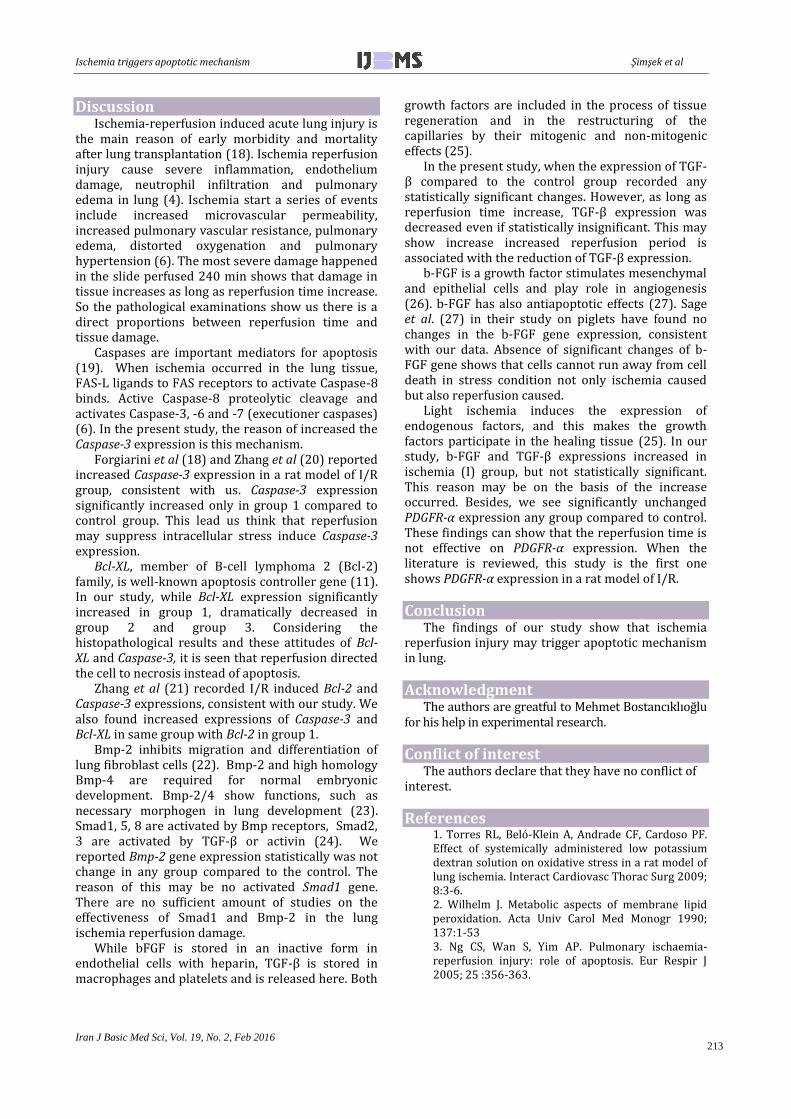
In all member of Group 3 (I+4R), congestive atelectasis in varying severity is formed. Also, marked alveolar macrophages hyperplasia is present in alveolar lumen as well as neutrophil, leukocyte infiltrations and perivascular edema. The presence of severe necrotic changes on alveolar epithelium and desquamation on bronchiolar epithelial draw attention. Edematous and congestive changes in right lobe come into focus (Picture 6).
Picture 6. 60 min ischemia + 240 min reperfusion group image A) left lobe H-E bar= 100 μm B) right Lobe H-E bar= 50 μm
Ischemia triggers apoptotic mechanism Şimşek et al
Iran J Basic Med Sci, Vol. 19, No. 2, Feb 2016
213
Discussion Ischemia-reperfusion induced acute lung injury is
the main reason of early morbidity and mortality after lung transplantation (18). Ischemia reperfusion injury cause severe inflammation, endothelium damage, neutrophil infiltration and pulmonary edema in lung (4). Ischemia start a series of events include increased microvascular permeability, increased pulmonary vascular resistance, pulmonary edema, distorted oxygenation and pulmonary hypertension (6). The most severe damage happened in the slide perfused 240 min shows that damage in tissue increases as long as reperfusion time increase. So the pathological examinations show us there is a direct proportions between reperfusion time and tissue damage.
Caspases are important mediators for apoptosis (19). When ischemia occurred in the lung tissue, FAS-L ligands to FAS receptors to activate Caspase-8 binds. Active Caspase-8 proteolytic cleavage and activates Caspase-3, -6 and -7 (executioner caspases) (6). In the present study, the reason of increased the Caspase-3 expression is this mechanism.
Forgiarini et al (18) and Zhang et al (20) reported increased Caspase-3 expression in a rat model of I/R group, consistent with us. Caspase-3 expression significantly increased only in group 1 compared to control group. This lead us think that reperfusion may suppress intracellular stress induce Caspase-3 expression.
Bcl-XL, member of B-cell lymphoma 2 (Bcl-2) family, is well-known apoptosis controller gene (11). In our study, while Bcl-XL expression significantly increased in group 1, dramatically decreased in group 2 and group 3. Considering the histopathological results and these attitudes of Bcl-XL and Caspase-3, it is seen that reperfusion directed the cell to necrosis instead of apoptosis.
Zhang et al (21) recorded I/R induced Bcl-2 and Caspase-3 expressions, consistent with our study. We also found increased expressions of Caspase-3 and Bcl-XL in same group with Bcl-2 in group 1.
Bmp-2 inhibits migration and differentiation of lung fibroblast cells (22). Bmp-2 and high homology Bmp-4 are required for normal embryonic development. Bmp-2/4 show functions, such as necessary morphogen in lung development (23). Smad1, 5, 8 are activated by Bmp receptors, Smad2, 3 are activated by TGF-β or activin (24). We reported Bmp-2 gene expression statistically was not change in any group compared to the control. The reason of this may be no activated Smad1 gene. There are no sufficient amount of studies on the effectiveness of Smad1 and Bmp-2 in the lung ischemia reperfusion damage.
While bFGF is stored in an inactive form in endothelial cells with heparin, TGF-β is stored in macrophages and platelets and is released here. Both
growth factors are included in the process of tissue regeneration and in the restructuring of the capillaries by their mitogenic and non-mitogenic effects (25).
In the present study, when the expression of TGF-β compared to the control group recorded any statistically significant changes. However, as long as reperfusion time increase, TGF-β expression was decreased even if statistically insignificant. This may show increase increased reperfusion period is associated with the reduction of TGF-β expression.
b-FGF is a growth factor stimulates mesenchymal and epithelial cells and play role in angiogenesis (26). b-FGF has also antiapoptotic effects (27). Sage et al. (27) in their study on piglets have found no changes in the b-FGF gene expression, consistent with our data. Absence of significant changes of b-FGF gene shows that cells cannot run away from cell death in stress condition not only ischemia caused but also reperfusion caused.
Light ischemia induces the expression of endogenous factors, and this makes the growth factors participate in the healing tissue (25). In our study, b-FGF and TGF-β expressions increased in ischemia (I) group, but not statistically significant. This reason may be on the basis of the increase occurred. Besides, we see significantly unchanged PDGFR-α expression any group compared to control. These findings can show that the reperfusion time is not effective on PDGFR-α expression. When the literature is reviewed, this study is the first one shows PDGFR-α expression in a rat model of I/R.
Conclusion The findings of our study show that ischemia
reperfusion injury may trigger apoptotic mechanism in lung.
Acknowledgment The authors are greatful to Mehmet Bostancıklıoğlu
for his help in experimental research.
Conflict of interest The authors declare that they have no conflict of
interest.
References 1. Torres RL, Beló-Klein A, Andrade CF, Cardoso PF. Effect of systemically administered low potassium dextran solution on oxidative stress in a rat model of lung ischemia. Interact Cardiovasc Thorac Surg 2009; 8:3-6. 2. Wilhelm J. Metabolic aspects of membrane lipid peroxidation. Acta Univ Carol Med Monogr 1990; 137:1-53 3. Ng CS, Wan S, Yim AP. Pulmonary ischaemia-reperfusion injury: role of apoptosis. Eur Respir J 2005; 25 :356-363.
Şimşek et al Ischemia triggers apoptotic mechanism
Iran J Basic Med Sci, Vol. 19, No. 2, Feb 2016
214
4. Delbin MA, Antunes E, Zanesco A. Role of exercise training on pulmonary ischemia/reperfusion and inflammatory response. Rev Bras Cir Cardiovasc 2009; 24:552-561. 5. Ryter SW, Choi AM. Autophagy in the lung. Proc Am Thorac Soc 2010; 7:13-21. 6. den Hengst WA, den Hengst WA. Lung ischemia-reperfusion injury: a molecular and clinical view on a complex pathophysiological process. Am J Physiol Heart Circ Physiol 2010; 299:H1283-1299. 7. Hotchkiss RS, Strasser A, McDunn JE, Swanson PE. Cell death. N Engl J Med 2009; 361:1570-1583. 8. Kerr JF, Wyllie AH, Currie AR. Apoptosis: a basic biological phenomenon with wide-ranging implications in tissue kinetics. Br J Cancer 1972; 26:239-257. 9. Connolly PF, Jäger R, Fearnhead HO. New roles for old enzymes: killer caspases as the engine of cell behavior changes. Front Physiol 2014; 5:149. 10. Cheng EH, Wei MC, Weiler S, Flavell RA, Mak TW, Lindsten T, Korsmeyer SJ. BCL-2, BCL-X(L) sequester BH3 domain-only molecules preventing BAX- and BAK-mediated mitochondrial apoptosis. Mol Cell 2001; 8:705-711. 11. Chipuk JE, Moldoveanu T, Llambi F, Parsons MJ, Green DR. The BCL-2 family reunion. Mol Cell 2010; 37:299-310. 12. Gacche RN, Meshram RJ. Angiogenic factors as potential drug target: Efficacy and limitations of anti-angiogenic therapy. Biochim Biophys Acta 2014; 1846:161-179. 13. Li HH, Huo LJ, Gao ZY, Zhao F, Zeng JW. Regulation of scleral fibroblast differentiation by bone morphogenetic protein-2. Int J Ophthalmol 2014; 7:152-156. 14. Herhaus L, Sapkota GP. The emerging roles of deubiquitylating enzymes (DUBs) in the TGFbeta and BMP pathways. Cell Signal 2014; 26:2186-2192. 15. Schul D, Schmitt A, Regneri J, Schartl M, Wagner TU. Burst BMP triggered receptor kinase activity drives Smad1 mediated long-term target gene oscillation in C2C12 cells. PloS One 2013; 8:e59442. 16. Heldin CH. Targeting the PDGF signaling pathway in tumor treatment. Cell Commun Signal 2013; 11:97. 17. Takhtfooladi H, Takhtfooladi M, Moayer F, Mobarakeh S. Melatonin attenuates lung injury in a
hind limb ischemia-reperfusion rat model. Rev Port Pneumol 2015; 21:30-35. 18. Forgiarini LF, Forgiarini LA Jr, da Rosa DP, Silva MB, Mariano R, Paludo Ade O,et al. N-acetylcysteine administration confers lung protection in different phases of lung ischaemia-reperfusion injury. Interact Cardiovasc Thorac Surg 2014; 19:894-899. 19. Porter AG, Janicke RU. Emerging roles of caspase-3 in apoptosis. Cell Death Differ 1999; 6:99-104. 20. Zhang Z, Shen H, Qin HD, Xu Y, Ma Mz, Bao L, Wang H. [Protective effect of N-acetylcysteine against pneumocyte apoptosis during ischemia/reperfusion injury of lung in rats]. Zhongguo wei zhong bing ji jiu yi xue = Chinese critical care medicine = Zhongguo weizhongbing jijiuyixue 2012; 24:111-115. 21. Zhang C, Guo Z, Lio H, Shi Y, Ge S. Influence of levosimendan postconditioning on apoptosis of rat lung cells in a model of ischemia-reperfusion injury. PloS One 2015; 10:e0114963. 22. Gao X, Cao Y, Staloch DA, Gonzales MA, Aronson JF. Bone morphogenetic protein signaling protects against cerulein-induced pancreatic fibrosis. PloS One 2014; 9:e89114. 23. Langenfeld EM, Kong Y, Langenfeld J. Bone morphogenetic protein 2 stimulation of tumor growth involves the activation of Smad-1/5. Oncogene 2006; 25:685-692. 24. Goumans MJ, Mummery C. Functional analysis of the TGFbeta receptor/Smad pathway through gene ablation in mice. Int J Dev Biol 2000; 44:253-265. 25. Fu XB, Yang YH, Sun TZ, Gu XM, Jiang LX, Sun XQ, Sheng ZY. Effect of intestinal ischemia-reperfusion on expressions of endogenous basic fibroblast growth factor and transforming growth factor betain lung and its relation with lung repair. World J Gastroenterol 2000; 6:353-355 26. Florkiewicz RZ, Ahluwalia A, Sandor Z, Szabo S, Tarnawski AS. Gastric mucosal injury activates bFGF gene expression and triggers preferential translation of high molecular weight bFGF isoforms through CUG-initiated, non-canonical codons. Biochem Biophys Res Commun 2011; 409:494-499. 27. Sage E, Mercier O, Van den Eyden F, de Perrot M, Barlier-Mur AM, Dartevelle P, Eddahibi S. Endothelial cell apoptosis in chronically obstructed and reperfused pulmonary artery. Respir Res 2008; 9:19.