Embed Size (px)
Citation preview

Clay Minerals (1981) 16, 375-381.
IR S P E C T R A OF P O W D E R H E M A T I T E : E F F E C T S OF P A R T I C L E SIZE A N D S H A P E
J O S E L. R E N D O N * AND C A R L O S J. S E R N A
Grupo de Fisico-Qu{mica Mineral. C.S.L C., Serrano l lS-dpdo. Madrid6, Spain
(Received 3 August 1981)
ABSTRACT: Hematites obtained by heating goethite gave different IR absorption spectra depending on the temperature of formation. Hematites formed between 250-600~ consisted of lath-like crystals (average size 0-4 x 0-08 #m) and showed, in accordance with theoretical predictions, very similar IR spectra whose absorption bands could all be assigned to surface mode vibrations. However, significantly different IR spectra were given by hematites formed between 700-950~ the differences being correlated with variations in the size and shape of the particles. Differences observed in the IR spectra of powder hematite do not therefore justify new names for the mineral, as have been proposed in the literature.
The IR absorption frequencies of many inorganic powders have frequently been interpreted as lattice vibration modes with no regard to the actual size and shape of the particles. Lattice mode frequencies are independent of particle size and shape only if the dimensions of a polar crystal are very large compared with the phonon wavelength (Ruppin & Englman, 1970). For smaller crystals, e.g. clay minerals, the departure from bulk-mode frequencies may become quite important (Farmer ,~ Russell, 1966).
Crystalline iron oxide and oxyhydroxide products of pedogenesis differ markedly in particle size as well as in extent of isomorphous substitution (Schwertmann & Taylor, 1977). Since both effects greatly influence the IR spectra they need to be thoroughly understood before a species identification is made on the basis of IR evidence. The effect of isomorphous substitutions on the IR spectra of goethite have recently been described by Mendelovici et al. (1979).
During the course of an investigation into the thermal and mechanical transformation of goethite, it was noted that IR spectra of the hematite products differed markedly depending on the temperature at which they were formed and the length of time for which they were ground. Similar observations have been recently reported and explained either in terms ofcrystallinity and crystal defects (Yariv & Mendelovici, 1979) or the presence of OH absorption bands (Wolska, 1981). However, similar differences in the IR spectra of natural hematites had been previously interpreted by Estep (1977) as due to the dependence o f l R optical phonon frequencies on crystallite size and shape. The purpose of this investigation is to demonstrate that particle size and shape strongly affect the IR spectra of powder hematite and that they could be the underlying reason for the reported differences.
E X P E R I M E N T A L P R O C E D U R E
Synthetic goethite (~-FeOOH) was obtained following the procedure described by * On leave from: Centro de Edafologia y Biologia Aplicada del Cuarto. C.S.I.C. Aptdo. 1052. Sevilla, Spain.
0009-8558/81/1200-0375502.00 �9 I98I The Mineralogical Society

376 Josd L. Rend6n and Carlos J. Serna
(a)
(b) FIG. 1. Electron micrographs of hematite products obtained from heating goethite at (a) 250~
and (b) 950~
Atkinson et al. (1967). X-ray diffractometry and IR spectroscopy indicated that the sample was a pure goethite. The BET surface area was 50 m 2 g-1 (Rend6n, 1981).
Heating goethite between 250 and 600~ produced a microcrystalline hematite (~-Fe203) without altering the size and shape of the original goethite microcrystals, i.e. the dehydroxylation reaction occurred within a microcrystal. Electron micrographs showed that the hematite product had a lath-like morphology with an average particle size of about 0.4 x 0.08/~m (Fig. la).
Heating goethite at higher temperatures (700-950~ produced hematites with a range of particle sizes and shapes due to sintering (Fig. 1 b).
The hematite samples used for IR study (Perkin Elmer 580B spectrophotometer) were selected after X-ray diffraction (Philips PW 1010 diffractometer with Cr-Ka radiation) and electron microscopic (Siemens Elmiskop 102 apparatus) observations. The IR
IR spectra of powder hematite 377
spectra were carefully scanned, so that the precision of the frequency determinations was better than 2 cm -~.
R E S U L T S A N D D I S C U S S I O N
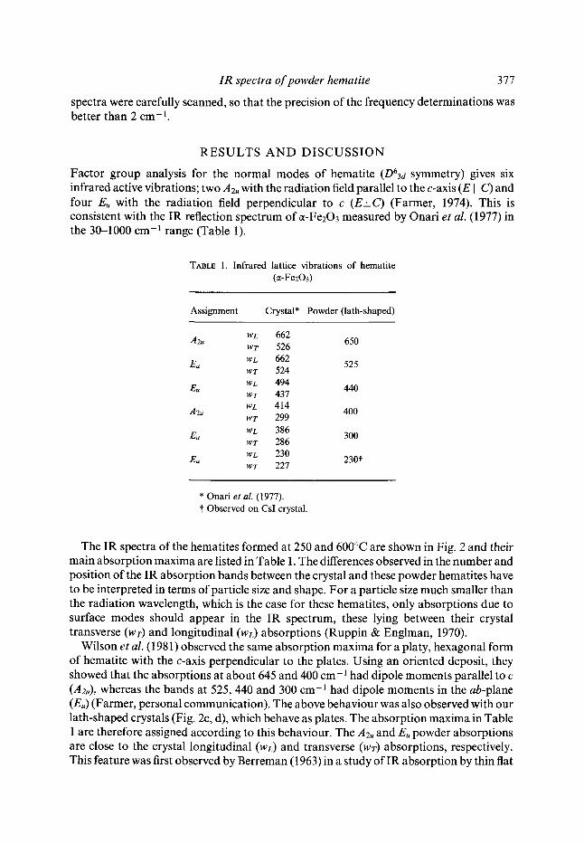
Factor group analysis for the normal modes of hematite (D63d symmetry) gives six infrared active vibrations; two A2, with the radiation field parallel to the c-axis (E II C) and four E, with the radiation field perpendicular to c (E_I_C) (Farmer, 1974). This is consistent with the IR reflection spectrum of ~-Fe203 measured by Onari et al. (1977) in the 30-1000 cm -1 range (Table 1).
TABLE 1. Infrared lattice vibrations of hematite (~-Fe203)
Assignment Crystal* Powder (lath-shaped)
-'/2u WL 662 650 wr 526
Eu WL 662 525 WT 524
Eu WL 494 440 WT 437
A2u WL 414 400 Wr 299
Eu wL 386 300 wr 286
Eu WL 230 w r 227 230"~
* Onari et al. (1977). t Observed on CsI crystal.
The IR spectra of the hematites formed at 250 and 600~ are shown in Fig. 2 and their main absorption maxima are listed in Table 1. The differences observed in the number and position of the IR absorption bands between the crystal and these powder hematites have to be interpreted in terms of particle size and shape. For a particle size much smaller than the radiation wavelength, which is the case for these hematites, only absorptions due to surface modes should appear in the IR spectrum, these lying between their crystal transverse (wr) and longitudinal (wL) absorptions (Ruppin & Englman, 1970).
Wilson et al. (1981) observed the same absorption maxima for a platy, hexagonal form of hematite with the c-axis perpendicular to the plates. Using an oriented deposit, they showed that the absorptions at about 645 and 400 cm- 1 had dipole moments parallel to c (A2u), whereas the bands at 525, 440 and 300 cm-l had dipole moments in the ab-plane (Eu) (Farmer, personal communication). The above behaviour was also observed with our lath-shaped crystals (Fig. 2c, d), which behave as plates. The absorption maxima in Table 1 are therefore assigned according to this behaviour. The A2u and E, powder absorptions are close to the crystal longitudinal (wL) and transverse (wv) absorptions, respectively. This feature was first observed by Berreman (1963) in a study of IR absorption by thin flat

378 Josd L. Renddn and Carlos J. Serna
300 [ 300 -oA/ /J/il /..,, ,4;I/I
410 650 360 565 400
~_~ ) 350
800 600 400 200 600 &00 200
WAVENUMBER CM -1
FIG. 2. IR spectra of hematite products obtained from heating goethite in the range 250-600~ (a) 250~ in KBr pellets (em=2"25); (b) 600~ in KBr pellets (em=2"25); (c) and (d) 250~ deposited in KSR-5 and scanned at 0 ~ and 30 ~ with respect to the beam (era = 1"0); (e) 600~
deposited on KSR-5 and scanned at 0 ~ (era = 1"0).
films of cubic ionic crystals. He concluded that the high frequency surface mode was of longitudinal character and the low frequency surface mode of transverse character. Thus, these surface modes hardly move with changes in the dielectric constant of the surrounding medium (era), as is illustrated in Fig. 2 (spectra a, c; b, e).
In addition to the main absorption maxima discussed above, the IR spectrum of the hematite obtained at 250~ showed three shoulders at about 565,470 and 350 cm- 1; these became more noticeable in the product formed at 600~ These shoulders were affected by the dielectric constant of the medium in which the specimen was dispersed (Fig. 2), and must be attributed to surface mode absorptions of hematite crystals with morphologies other than lath-like.
The IR spectra of powder hematites produced from goethite between 700-950~ are shown in Fig. 3. Interparticle sintering occurred in this temperature interval, altering the particle size and shape (Fig. lb).
While the effect of particle size and dielectric constant of the dispersion medium on IR absorption are well understood, the effect of particle shape is largely unknown. This effect has only been studied for spherical, cylindrical and slab shapes (Ruppin & Englman, 1970). For aggregate samples, shape effects have been only considered for small, well-defined clusters of spherical units (Clippe et al., 1976).
To attempt a detailed interpretation of these IR spectra would thus be unfruitful, due to the wide range in particle size and shape of the products formed during this temperature interval. In general, these variations cause broadening of the IR absorption bands (Fig. 3). As expected, the absorption bands at 400 and 650 cm -l (Fig. 2a) shift to lower

IR spectra of powder hematite 379
o
442 305
A ~ 53O 18
/
Y 1 ; I I I I
800 600 400 200
55O
3L2
d g~
I ] I I I I 800 600 400 200
WAVENUMBER CM -1
FIG. 3. IR spectra of hematite products obtained from heating goethite in the range 700-950~ (a) 700~ (b) 800~ (c) 950~ (d) product obtained by prolonged grinding of goethite in
mechanical mortar. All products pressed in KBr pellets.
wavenumbers (Fig. 3), since the lath-like morphology has disappeared (Fig. la, b). The other bands observed are due to the major contribution of surface modes for powder particle aggregates (Clippe et al., 1976).
From the above considerations, and in agreement with Estep (1977), one would expect important differences in the IR spectrum of a powder hematite depending on its origin.
Yariv & Mendelovici (i979) interpreted these differences in terms of powder crystallinity and proposed the name protohematite for the poorly crystallized material. However, one should be cautious in relating crystallinity to features of the IR spectra, since it can be observed by XRD that crystal quality of hematite improves with temperature of formation over the interval 250-600~ (Fig. 4) while a more or less uniform IR spectrum is obtained for hematites formed throughout this temperature range (Fig. 2). Moreover, a hematite product obtained by prolonged grinding of goethite in a mechanical mortar gave a similar IR spectrum (Fig. 3d) to that obtained from hematite formed from goethite heated at 950~ (Fig. 3c). That both samples differ considerably in degree of crystallinity is obvious from Fig. 4.
On the other hand, Wolska (1981) recently proposed the name hydrohematite for a hematite in which OH- ions substitute for 02 ions in the lattice. In order to counterbalance the resulting decrease in negative charge, the author suggested that some divalent iron (8~o) must be present in the structure. The IR spectrum of hydrohematite contained two absorption bands at 950 and 630 cm- 1, not present in the IR spectrum of hematite, which were attributed to hydroxyl groups. The 950 cm -1 absorption band, although not observed by us, could be due to hydroxyl groups, being the corresponding deformation modes of the OH stretching absorption due to surface hydroxyls reported in hematite by Rochester & Topham (1979). However, the absorption at 630 cm -l can be clearly assigned to an Az, surface mode vibration (Table 1). This absorption band almost

380 Jose L. Renddn and Carlos J. Serna
104
116
204 102
I ~ I I I I I I I I 84 80 76 64 60 56 52 48 36 32
DEGREES 2 e
FIG. 4. X-ray diffraction traces of hematite products obtained from heating goethite at: (a) 250~ (b) 600~ (c) 950~ (d) product obtained by prolonged grinding of goethite.
disappears in the IR spectra of hematite formed at higher temperatures, due to sintering (Fig. 3c). Also, divalent iron ions, presumably present in the hydrohematite structure as claimed by Wolska (1981), were not detected by M6ssbauer spectroscopy in any of the samples studied here. Therefore, the presence of surface OH groups in hematite, as in most powder oxides, does not justify the introduction of a new name for the mineral.
C O N C L U S I O N S
The IR spectrum of a powder sample can vary depending on its particle size and shape.
I R spectra o f powder hemat i te 381
T h e r e f o r e , in o r d e r to i den t i fy a m i n e r a l co r r ec t ly , b o t h effects h a v e to be well u n d e r s t o o d .
D i f f e r ences d u e to these effects in the I R s p e c t r u m d o n o t j u s t i fy n e w m i n e r a l n a m e s .
ACKNOWLEDGME N T S
Thanks are due to Dr R. Gancedo for the M6ssbauer determinations and to Mr L. Puebla and Mrs M. A. Muro for technical assistance. Valuable suggestions from Dr V. C. Farmer are also acknowledged.
REFERENCES
ATKINSON R.J., POSNER A.M. & QUIRK J.P. (1967) Adsorption of potential-determining ions at the ferric oxide-aqueous electrolyte interface. J. Phys. Chem. 71, 550.
BERREMAN D.W. (1963) Infrared absorption at longitudinal optic frequency in cubic crystal films. Phys. Rev. 130, 2193.
CLIPPE P., EVRARD R. & LUCAS A.A. (1976) Aggregation effect on the infrared absorption spectrum of small ionic crystals. Phys. Rev. B 14, 1715.
ESTEP P.A. (1977) Infrared Spectroscopy. Pp. 529-603 in: Physical Methods in Determinative Mineralogy (J. Zussman, editor). Academic Press.
FARMER V.C. (1974) The anhydrous oxide minerals. Pp. 183-204 in: The Infrared Spectra of Minerals (V. C. Farmer, editor). Mineralogical Society, London.
FARMER V.C. 8/. RUSSELL J.n. (1966) Effects of particle size and structure on the vibrational frequencies of layer silicates. Spectrochim. Acta 22, 389.
MENDELOVICI E., YARIV S.H. & VILLALBA R. (1979) Aluminum-bearing goethite in Venezuelan laterites. Clays Clay Min. 27, 368.
ONARI S., ARAI T. • KUDO K. (1977) Infrared lattice vibrations and dielectric dispersion in a-Fe203. Phys. Rev. B 16, 1717.
REND6N J.L. (198 l) Estudio de la transformaci6n de goetita (ct-FeOOH) en hematites (ct-Fe203) pot tratamientos t&micos y mecdnicos. Ph.D. Thesis., Univ. Sevilla, Spain.
ROCHESTER C.H. & TOPHAM S.A. (1979) Infrared study of surface hydroxyl groups on hematite. J.C.S. Faraday I 75, 1073.
RUPPIN R. t~r ENGLMAN R. (1970) Optical phonons of small crystals. Rep. Prog. Phys. 33, 149. SCHWERTMANN U. t~r TAYLOR R.M. (1977) Iron oxides. Pp. 145-180 in: Minerals in Soil Environments (J. B.
Dixon & S. B. Weed, editors). Soil Science Society of America. Madison, U.S.A. WILSON M.J., RUSSELL J.D., TAIT J.M., CLARK D.R., FRASER A.R. & STEPHEN I. (1981) A swelling
hematite/layer-silicate complex in weathered granite. Clay Miner. 16, 261-277. WOLSKA E. (1981) The structure of hydrohematite. Z. Krist. 154, 69. YARIV S.H. & MENDELOVICI E. (1979) The effect of degree of crystallinity on the infrared spectrum of hematite.
Appl. Spectrosc. 33, 410.
RI~SUMI~S: Les spectres d'absorption IR d'h~matite sont function de la temp+rature de formation lorsque ce min6ral est produit par chauffage de goethite. Les h6matites, formbes entre 250 ~ et 600~ pr6sentent des critaux ~t faci6s de lattes (environ 0.4 x 0.08 #m) et des spectres IR tr6s voisins, en accord avec des pr~visions th~oriques; les bandes d'absorption peuvent 6tre attribu6es fi des modes de vibration de surface. Toutefois, des spectres IR diff6rents sont obtenus sur des h6matites form6es entre 700 ~ et 950~ les diff6rences &ant li+es aux variations de taille et de forme des particules. Des diff6rences observ6es dans des spectres IR d'h~matite en poudre ne justifient pas l'attribution de noms nouveaux ~i ces min6raux, contrairement ~ ce qui fut propos6 dans la litt6rature.
KURZREFERAT: H/imatit, gewonnen durch Goethiterhitzung, zeigte in Abh/ingigkeit zur Bildungstemperature unterschiedliche IR-Absorptions-spektren. Die zwischen 250-600~ gebil- deten Hfimatite bestanden aus latten/ihnlichen Kristallen (Durchschnittsgr6ge 0.4 x 0-08 ltm) und zeigten im Einklang mit theoretischen Vorhersagen ganz /ihnliche IR-Spektren, deren Absorptionsbanden insgesamt als Oberflfichenschwingungen charakterisiert werden konnten. Im

382 Josd L. Rend6n and Carlos J. Serna
Gegensatz dazu lieferten die bei 70(~950~ hergestellten H/imatite signifikant unterschiedliche IR-Spektren, korrelierber mit Ver~nderungen ihrer Partikelgr6ge und Gestalt. Deshalb rechtfer- tigen die bei der IR-Spektroskopie von gepulverten H/imatiten beobachteten Unterschiede keine neue Namensgebung ffir das Mineral, wie es in der Literatur vorgeschlagen wurde.
RESUMEN: Muestras de hematita en polvo obtenidas por tratamiento t6rmico de goetita presentan diferentes espectros IR. En el margen de 250-600~ las muestras presentan un espectro muy semejante en el que las absorciones son debidas a modos superficiales, como se predice te6ricamente para este tamafio (0.4 x 0.08 #m) y forma (cintas) de las particulas. Sin embargo, en el margen de 700-950~ los espectros IR muestran diferencias importantes que se relacionan con las variaciones debidas a sinterizacidn en el tamafio y la forma de las particulas. Por tanto, las diferencias encontradas en los espectros IR de la hematita en polvo no justifican el uso de nuevos hombres para el mineral, como se ha propuesto en la bibliografia.