Embed Size (px)
Citation preview
Ionization of cytosine monomer and dimer studied by
VUV photoionization and electronic structure calculationsw
Oleg Kostko,a Ksenia Bravaya,b Anna Krylov*b and Musahid Ahmed*a
Received 14th October 2009, Accepted 11th January 2010
First published as an Advance Article on the web 9th February 2010
DOI: 10.1039/b921498d
We report a combined theoretical and experimental study of ionization of cytosine monomers
and dimers. Gas-phase molecules are generated by thermal vaporization of cytosine followed by
expansion of the vapor in a continuous supersonic jet seeded in Ar. The resulting species are
investigated by single photon ionization with tunable vacuum-ultraviolet (VUV) synchrotron
radiation and mass analyzed using reflectron mass spectrometry. Energy onsets for the measured
photoionization efficiency (PIE) spectra are 8.60 � 0.05 eV and 7.6 � 0.1 eV for the monomer
and the dimer, respectively, and provide an estimate for the adiabatic ionization energies (AIE).
The first AIE and the ten lowest vertical ionization energies (VIEs) for selected isomers of
cytosine dimer computed using equation-of-motion coupled-cluster (EOM-IP-CCSD) method are
reported. The comparison of the computed VIEs with the derivative of the PIE spectra suggests
that multiple isomers of the cytosine dimer are present in the molecular beam. The calculations
reveal that the large red shift (0.7 eV) of the first IE of the lowest-energy cytosine dimer is due to
strong inter-fragment electrostatic interactions, i.e., the hole localized on one of the fragments is
stabilized by the dipole moment of the other. A sharp rise in the protonated cytosine ion (CH+)
signal at 9.20 � 0.05 eV is ascribed to the formation of protonated cytosine by dissociation
of the ionized dimers. The dominant role of this channel is supported by the computed energy
thresholds for the CH+ appearance and the barrierless or nearly barrierless ionization-induced
proton transfer observed for five isomers of the dimer.
I. Introduction
The structures and properties of many biological molecules are
very well characterized. However, despite 50 years of research
that have elapsed since Watson and Crick’s original postulation
for the structure of DNA, the fundamental aspects of the
‘‘nuts and bolts’’ of the molecules, which are the building
blocks of life, are not yet fully understood.1,2 The ionization of
nucleobases is a key step leading to damage and mutation of
DNA.3 The electron hole introduced by ionization or oxidation
migrates along the helix through various hopping mechanisms
coupled with tautomerization through proton transfer and
ultimately leads to distant chemical modifications of the bases,
strand cleavage and dissociation of the helix itself. Apart from
the evolutionary and carcinogenic effects that this damage
induces in living systems, there is also much interest in the
electronic properties of the DNA molecules themselves owing
to their potential use in molecular electronics.4,5 Molecular
shape, conformational dynamics and electronic properties
(i.e., charge distributions, excited states) of DNA play crucial
roles in its selectivity and function. The properties of DNA are
determined by the properties of its individual blocks and their
complex interactions. Hence, the intrinsic properties of the
DNA bases are of fundamental interest.2
While the ionization of isolated DNA bases has been studied
extensively, there is very little experimental information for the
dimers. Moreover, even on the monomer level, the picture is
not yet complete owing to a daunting task of disentangling
contributions of numerous tautomers and conformers that can
be produced under the experimental conditions (especially in
the case of cytosine and guanine). Previous ionization studies
of cytosine consist of photoionization mass spectrometry
(PIMS) measurements of AIEs,6,7 photoelectron spectroscopy
(PES) at the valence8–11 and core level,12 resonance enhanced
multiphoton ionization (REMPI) experiments13,14 and a
number of electronic structure calculations.9,15–18 Trofimov
et al.9 measured a valence-shell PES of cytosine. By analyzing
photoelectron energy and angular distributions, they
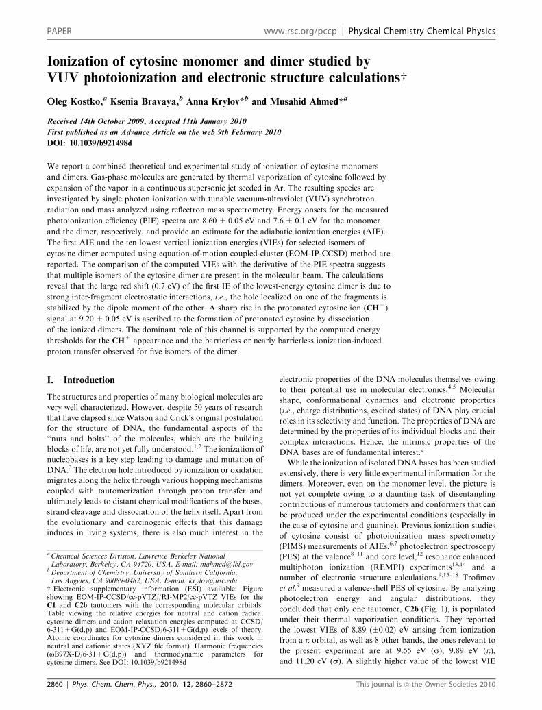
concluded that only one tautomer, C2b (Fig. 1), is populated
under their thermal vaporization conditions. They reported
the lowest VIEs of 8.89 (�0.02) eV arising from ionization
from a p orbital, as well as 8 other bands, the ones relevant to
the present experiment are at 9.55 eV (s), 9.89 eV (p),and 11.20 eV (s). A slightly higher value of the lowest VIE
a Chemical Sciences Division, Lawrence Berkeley NationalLaboratory, Berkeley, CA 94720, USA. E-mail: [email protected]
bDepartment of Chemistry, University of Southern California,Los Angeles, CA 90089-0482, USA. E-mail: [email protected]
w Electronic supplementary information (ESI) available: Figureshowing EOM-IP-CCSD/cc-pVTZ//RI-MP2/cc-pVTZ VIEs for theC1 and C2b tautomers with the corresponding molecular orbitals.Table viewing the relative energies for neutral and cation radicalcytosine dimers and cation relaxation energies computed at CCSD/6-311+G(d,p) and EOM-IP-CCSD/6-311+G(d,p) levels of theory.Atomic coordinates for cytosine dimers considered in this work inneutral and cationic states (XYZ file format). Harmonic frequencies(oB97X-D/6-31+G(d,p)) and thermodynamic parameters forcytosine dimers. See DOI: 10.1039/b921498d
2860 | Phys. Chem. Chem. Phys., 2010, 12, 2860–2872 This journal is �c the Owner Societies 2010
PAPER www.rsc.org/pccp | Physical Chemistry Chemical Physics
(8.94 eV) was reported in an earlier PES study.10 Although the
tautomer ratio of C1, C2a, C2b and C3a in the PES study
(463 K) is expected to be 0.22 : 0.17 : 0.38 : 0.24 (as estimated9
by the authors using ab initio thermodynamical data from
ref. 19), Trofimov et al. concluded that their experimental
results can be explained by population of C2b only. In
contrast, a very recent core-level PES study12 of cytosine
suggested that 3 tautomers of cytosine are populated upon
thermal vaporization at 450 K, with tautomer C2(a+b)
(E60%) being the dominant species based on free energy
calculations of Trygubenko et al.20 and Fogarasi.19
A previous PIMS measurement from our group determined
the AIE of cytosine monomer to be 8.65 (�0.05) eV,6 in
agreement with 8.68 eV reported in early PIMS work,7 and
within the range (8–9 eV) obtained using one and two color
resonant 2 photon ionization spectroscopy by Nir et al.14 The
latter work also reported that two tautomers, one keto (C1)
and one enol (C2b), are prevalent in their laser desorption
jet-cooled molecular beam.
Ab initio calculations of the IE’s of the biologically relevant
tautomer of cytosine have been reviewed by Roca-Sanjuan
et al.16,17 and Cauet et al.17 Other tautomers were considered
by Wolken et al. who estimated the lowest IEs by DFT, MP2
and CCSD(T) calculations.15 More reliable values of valence-
shell VIEs of the five lowest-energy tautomers obtained using
electron propagator methods were reported by Ortiz and
coworkers.21 However, accurate estimates of the AIEs and
valence-shell VIEs obtained at the same level of theory (so that
ionization-induced relaxation effects can be quantified) for the
most stable cytosine tautomers that are likely to be populated
in the experiment are still missing. As discussed in ref. 22
and 23, using computational methods that are capable of
describing multiple interacting states of open-shell character
is crucial for obtaining reliable results for the ionized systems,
and EOM-IP-CC is one such approach.24–29
There are no experimental reports for the IEs of the cytosine
dimer. A number of groups characterized the dimers by using
multiphoton ionization spectroscopy and laser ablation.
Nir et al. reported that while two tautomers were populated
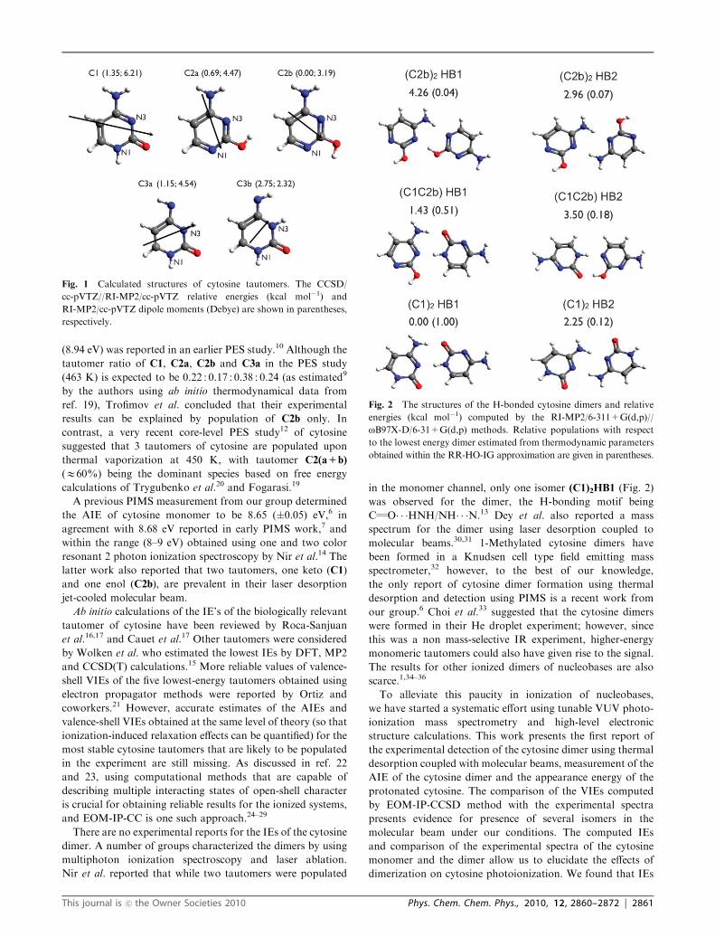
in the monomer channel, only one isomer (C1)2HB1 (Fig. 2)
was observed for the dimer, the H-bonding motif being
CQO� � �HNH/NH� � �N.13 Dey et al. also reported a mass
spectrum for the dimer using laser desorption coupled to
molecular beams.30,31 1-Methylated cytosine dimers have
been formed in a Knudsen cell type field emitting mass
spectrometer,32 however, to the best of our knowledge,
the only report of cytosine dimer formation using thermal
desorption and detection using PIMS is a recent work from
our group.6 Choi et al.33 suggested that the cytosine dimers
were formed in their He droplet experiment; however, since
this was a non mass-selective IR experiment, higher-energy
monomeric tautomers could also have given rise to the signal.
The results for other ionized dimers of nucleobases are also
scarce.1,34–36
To alleviate this paucity in ionization of nucleobases,
we have started a systematic effort using tunable VUV photo-
ionization mass spectrometry and high-level electronic
structure calculations. This work presents the first report of
the experimental detection of the cytosine dimer using thermal
desorption coupled with molecular beams, measurement of the
AIE of the cytosine dimer and the appearance energy of the
protonated cytosine. The comparison of the VIEs computed
by EOM-IP-CCSD method with the experimental spectra
presents evidence for presence of several isomers in the
molecular beam under our conditions. The computed IEs
and comparison of the experimental spectra of the cytosine
monomer and the dimer allow us to elucidate the effects of
dimerization on cytosine photoionization. We found that IEs
Fig. 1 Calculated structures of cytosine tautomers. The CCSD/
cc-pVTZ//RI-MP2/cc-pVTZ relative energies (kcal mol�1) and
RI-MP2/cc-pVTZ dipole moments (Debye) are shown in parentheses,
respectively.
Fig. 2 The structures of the H-bonded cytosine dimers and relative
energies (kcal mol�1) computed by the RI-MP2/6-311+G(d,p)//
oB97X-D/6-31+G(d,p) methods. Relative populations with respect
to the lowest energy dimer estimated from thermodynamic parameters
obtained within the RR-HO-IG approximation are given in parentheses.
This journal is �c the Owner Societies 2010 Phys. Chem. Chem. Phys., 2010, 12, 2860–2872 | 2861
of cytosine are strongly affected by the inter-fragment
interactions in the dimer, i.e., the lowest IE of the most stable
dimer is red-shifted by almost 1 eV. This effect is much larger
than previously reported values for similar systems.22,23,37
We also discuss the ionization-induced dissociation of cytosine
dimers leading to the formation of the protonated cytosine
species. Our results indicate that ionization of the five
H-bonded cytosine dimers considered in this study initiates a
barrierless proton transfer from one base to another. In these
proton-transferred structures, the positive charge is localized
on the closed-shell protonated fragment, whereas the unpaired
electron resides on the deprotonated moiety. By comparison of
the measured dependence of the CH+ signal on the photon
energy to the computed energy thresholds, we demonstrate
that the CH+ formation can be ascribed to the dissociation
of proton-transferred cytosine dimers. Ionization-induced
barrierless proton transfer in hydrogen-bonded dimers might
have important implications for the mechanism of hole
migration through the DNA molecule. The proton transfer
in the H-bonded pairs results in the separation of the unpaired
electron and the positive charge between the strands and may
result in hole trapping.38 In a subsequent paper, we will
address the ionization-induced proton transfer between the
complementary pairs of nucleobases.
The structure of the paper is as follows. The next section
briefly outlines the experimental and theoretical methods
(a complete description is given in ref. 22). The experimental
spectra and computed IEs and energetic parameters are
presented in Results section. Analysis of inter fragment
interactions on cytosine ionization and interpretation of
experimental spectra results are given in the Discussion section.
Our main results and concluding remarks are summarized in
the Conclusions section.
II. Methods
The theoretical and experimental methods have been described
in detail in our paper on photoionization of thymine and
adenine.22 Here we give only the essential parameters for the
cytosine study. The experiments are performed on a molecular
beam apparatus coupled to a 3 meter VUV monochromator
on the Chemical Dynamics Beamline at the ALS. The thermal
vaporization source6 was heated to around 600 K to generate
cytosine monomers and dimers in a supersonic jet expansion.
In the present experiments, the backing pressure was 35 kPa of
Ar through a 100 mm diameter nozzle. The time-of-flight
spectra were recorded for the photoionization energy range
between 7.4 and 11.5 eV. The typical step size for the PIE
scans is 50 meV and a dwell time of 10 s at a repetition rate of
10 kHz.
The equilibrium structures of the neutral monomeric
tautomers were optimized with the RI-MP2 method39–41 using
Dunning’s cc-pVTZ basis set. The geometries of the neutral and
cationic dimers were computed using the long-range corrected
oB97X42 functional with an empirical dispersion correction
(oB97X-D)43–45and the 6-31+G(d,p) basis set. Using these
geometries, binding energies and relative energies of the neutral
dimers were computed using RI-MP2/6-311+G(d,p) and
oB97X-D/6-311++G(2df,2pd) methods. To verify the structures,
the Hessians were computed with the oB97X-D/6-31+G(d,p)
method. The fine EML(75,302) grid consisting of 75 points
in the Euler–Maclaurin radial grid46 and 302 points in the
Lebedev angular grid47 was used in all DFT calculations.
Thermodynamic analysis was performed within the rigid
rotor—harmonic oscillator—ideal gas approximation
(RR-HO-IG) for the laboratory conditions (T = 298.18 K,
p = 1 atm) and for T = 582 K. Thermodynamic
analysis for the cytosine monomers was performed using
RI-MP2/6-311+G(d,p)//RI-MP2/6-311+G(d,p) frequencies
and CCSD/cc-pVTZ//RI-MP2/cc-pVTZ relative energies. RI-MP2/
6-311+G(d,p)//oB97X-D/6-31+G(d,p) relative energies were
used for the dimer thermodynamic analysis along with
frequencies computed with oB97X-D/6-31+G(d,p) method.
Since zero-point energy is included in the enthalpy term in
the Q-Chem code for the RR-HO-IG calculations, the
non-ZPE corrected electronic energies were used in the Gibbs
energy calculations.48
VIEs of the monomers and dimers were computed with the
EOM-IP-CCSD method24–29 and the cc-pVTZ and
6-311+G(d,p) basis sets, respectively. The frozen natural
orbitals (FNO)49 approximation was used for the IE calculations
of the dimers with the virtual space truncated using 99.50%
natural population cut-off criterion. Performance of FNO
approximation for the cases of both vertical and adiabatic
ionization energies was extensively discussed in ref. 49. The
errors in IEs introduced by truncation of active virtual orbital
space according to the 99.5% population criterion relative to
the full virtual space results were found to be less than 0.1 eV
for similar molecular systems, including thymine, guanine
and uracil dimer.22,49 The core electrons were frozen in all
EOM-IP-CCSD calculations. The lowest AIEs were obtained
as the difference between the EOM-IP-CCSD energy of the
first ionized state at the cation geometry and the CCSD energy
of the reference state at the geometry of the neutral species.
Optimized geometries, relevant total energies, and harmonic
frequencies are given in the ESI.w All calculations were
performed using the Q-CHEM electronic structure program.50
III. Results
As mentioned above, the analysis of the experimental spectra
is complicated by the presence of multiple cytosine tautomers
and even larger amount of the dimer isomers. Moreover,
ionization-induced fragmentation of larger clusters
contributes to the signal of the smaller ones giving rise to
‘‘fill-in’’ and ‘‘drop-out’’ effects. Therefore, interpretation of
the spectra requires taking into account contributions from
multiple isomers and ionization-induced dimer dissociation
channels. With this in mind, we organized this section as
follows. The first subsection (Cytosine monomers: structures,
relative energies and populations) addresses the selection of
cytosine tautomers that can be populated under our
experimental conditions and discusses their properties relevant
for the analysis of inter-fragment interactions in the dimers.
We then proceed to describe the structures of the selected
cytosine dimers, their binding and relative energies, and
provide estimates of their relative populations assuming
thermal equilibrium conditions (Cytosine dimers: structures,
2862 | Phys. Chem. Chem. Phys., 2010, 12, 2860–2872 This journal is �c the Owner Societies 2010
binding energies and populations). The third subsection
presents the mass spectra of the ionized species and discusses
the dissociation and fragmentation channels (Experimental
mass spectrum and thermal fragmentation). We then discuss
the measured PIE spectra for the monomer and the dimer, as
well as appearance energy curve for protonated cytosine
(Experimental PIE curves). Subsection E (IEs of cytosine
monomers and dimers) summarizes computational results on
cytosine ionization and includes EOM-IP-CCSD estimates of
1st AIE and lowest VIEs computed for the selected cytosine
tautomers and dimers. In the last subsection (Ionization-
induced proton transfer and dissociation), we describe several
ionization-induced dimer dissociation channels and present
the respective energy thresholds for dissociation products
formation computed with oB97X-D.
A Cytosine monomers: structures, relative energies
and populations
We considered the five lowest energy isomers of the cytosine
monomer, four of them lying within 1.35 kcal mol�1 and the
fifth one being 2.75 kcal mol�1 above the most stable tautomer
(Fig. 1). Tautomerization strongly affects the dipole moments
of cytosine, i.e., the computed dipole moments vary from
2.32 D for the C3b tautomer to 6.21 D for the biologically
relevant C1 tautomer. As discussed below, these changes in
dipole moment reverse the relative stability of the dimers
formed by different tautomeric forms of cytosine (as compared
to the monomers) and also explain the magnitude of the IEs
shifts of the dimers relative to the monomer values.
Populations of different tautomeric forms based on the free
energy calculations were previously reported for several
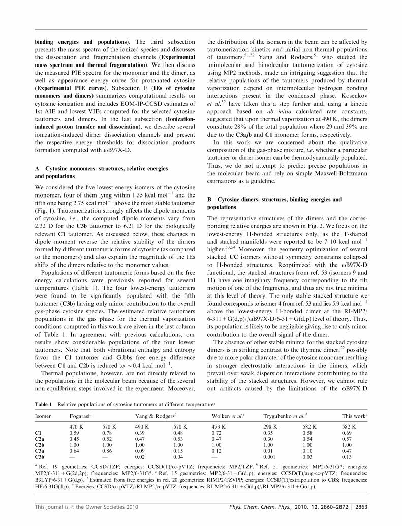
temperatures (Table 1). The four lowest-energy tautomers
were found to be significantly populated with the fifth
tautomer (C3b) having only minor contribution to the overall
gas-phase cytosine species. The estimated relative tautomers
populations in the gas phase for the thermal vaporization
conditions computed in this work are given in the last column
of Table 1. In agreement with previous calculations, our
results show considerable populations of the four lowest
tautomers. Note that both vibrational enthalpy and entropy
favor the C1 tautomer and Gibbs free energy difference
between C1 and C2b is reduced to B0.4 kcal mol�1.
Thermal populations, however, are not directly related to
the populations in the molecular beam because of the several
non-equilibrium steps involved in the experiment. Moreover,
the distribution of the isomers in the beam can be affected by
tautomerization kinetics and initial non-thermal populations
of tautomers.51,52 Yang and Rodgers,51 who studied the
unimolecular and bimolecular tautomerization of cytosine
using MP2 methods, made an intriguing suggestion that the
relative populations of the tautomers produced by thermal
vaporization depend on intermolecular hydrogen bonding
interactions present in the condensed phase. Kosenkov
et al.52 have taken this a step further and, using a kinetic
approach based on ab initio calculated rate constants,
suggested that upon thermal vaporization at 490 K, the dimers
constitute 28% of the total population where 29 and 39% are
due to the C3a/b and C1 monomer forms, respectively.
In this work we are concerned about the qualitative
composition of the gas-phase mixture, i.e. whether a particular
tautomer or dimer isomer can be thermodynamically populated.
Thus, we do not attempt to predict precise populations in
the molecular beam and rely on simple Maxwell-Boltzmann
estimations as a guideline.
B Cytosine dimers: structures, binding energies and
populations
The representative structures of the dimers and the corres-
ponding relative energies are shown in Fig. 2. We focus on the
lowest-energy H-bonded structures only, as the T-shaped
and stacked manifolds were reported to be 7–10 kcal mol�1
higher.53,54 Moreover, the geometry optimization of several
stacked CC isomers without symmetry constrains collapsed
to H-bonded structures. Reoptimized with the oB97X-D
functional, the stacked structures from ref. 53 (isomers 9 and
11) have one imaginary frequency corresponding to the tilt
motion of one of the fragments, and thus are not true minima
at this level of theory. The only stable stacked structure we
found corresponds to isomer 4 from ref. 53 and lies 5.9 kcal mol�1
above the lowest-energy H-bonded dimer at the RI-MP2/
6-311+G(d,p)//oB97X-D/6-31+G(d,p) level of theory. Thus,
its population is likely to be negligible giving rise to only minor
contribution to the overall signal of the dimer.
The absence of other stable minima for the stacked cytosine
dimers is in striking contrast to the thymine dimer,22 possibly
due to more polar character of the cytosine monomer resulting
in stronger electrostatic interactions in the dimers, which
prevail over weak dispersion interactions contributing to the
stability of the stacked structures. However, we cannot rule
out artifacts caused by the limitations of the oB97X-D
Table 1 Relative populations of cytosine tautomers at different temperatures
Isomer Fogarasia Yang & Rodgersb Wolken et al.c Trygubenko et al.d This worke
470 K 570 K 490 K 570 K 473 K 298 K 582 K 582 KC1 0.59 0.78 0.39 0.48 0.72 0.35 0.58 0.69C2a 0.45 0.52 0.47 0.53 0.47 0.30 0.54 0.57C2b 1.00 1.00 1.00 1.00 1.00 1.00 1.00 1.00C3a 0.64 0.86 0.09 0.15 0.12 0.01 0.10 0.47C3b — — 0.02 0.04 — 0.001 0.03 0.13
a Ref. 19 geometries: CCSD/TZP; energies: CCSD(T)/cc-pVTZ; frequencies: MP2/TZP. b Ref. 51 geometries: MP2/6-31G*; energies:
MP2/6-311+G(2d,2p); frequencies: MP2/6-31G*. c Ref. 15 geometries: MP2/6-31+G(d,p); energies: CCSD(T)/aug-cc-pVTZ; frequencies:
B3LYP/6-31+G(d,p). d Estimated from free energies in ref. 20 geometries: RIMP2/TZVPP; energies: CCSD(T)/extrapolation to CBS; frequencies:
HF/6-31G(d,p). e Energies: CCSD/cc-pVTZ//RI-MP2/cc-pVTZ; frequencies: RI-MP2/6-311+G(d,p)//RI-MP2/6-311+G(d,p).
This journal is �c the Owner Societies 2010 Phys. Chem. Chem. Phys., 2010, 12, 2860–2872 | 2863
functional in describing the interplay between the two different
types of interactions in relatively weakly bound systems.
An early MD study of free energy surface of the cytosine
dimer also reported only T-shaped and H-bonded isomers as
stable minima.54
Sampling the full configurational space of the cytosine
dimer is beyond the scope of this work and we have chosen
to focus on a few representative structures with different inter-
fragment arrangements and H-bonding patterns composed of
tautomers C1 and C2b (see Fig. 2). These are: the (C1)2HB1
structure with the O� � �H/N� � �H bonding; (C1C2b)HB2 and
(C1C2b)HB1 that have the N� � �H/O� � �H bonding; (C1)2HB2
with the N� � �H/H� � �N bonding; and (C2b)2HB1 and
(C2b)2HB2 that have the N� � �H/H� � �N bonding. Binding
energies calculated by oB97X-D and RI-MP2 are given in
Table 2. The RI-MP2/6-311+G(d,p)//oB97X-D/6-31+G(d,p)
relative energies are shown in Fig. 2. The (C1)2HB1 dimer is
the most stable followed by five other isomers which lie within
4.5 kcal mol�1. Although DFT and MP2 yield different energy
ordering of the cytosine tautomers,55,56 the DFT errors
cancel out in the calculations of binding energies (which are
dominated by electrostatic interactions), and the DFT values
agree well with those computed with RI-MP2 (Table 2). For
the (C1)2HB1, and (C1)2HB2 H-bonded dimers, our binding
energies of 23.6 and 21.7 kcal mol�1 are close to interaction
energies calculated by Kabelac and Hobza54 who reported the
values of 20.0 and 19.3 kcal mol�1, respectively. A recently
reported BSSE-corrected binding energy of 19.51 kcal mol�1
for the (C1)2HB2 dimer57 is also in agreement with our value.
Interestingly, despite a higher stability of the C2b monomer
relative to C1, the dimers formed by the C1 tautomers are
lower in energy due to favorable dipole–dipole interactions of
the two units (the respective dipole moments are 6.21 D and
3.19 D for C1 and C2b (Fig. 1)).
The relative populations of the cytosine dimers estimated
using the RR-HO-IG approximation based on the RI-MP2
relative energies are shown in Fig. 2. All of the H-bonded
dimers are expected to have notable population under thermal
equilibrium conditions (T = 582 K) with the dominant
contributions from the (C1)2HB1, (C1C2b)HB1 and
(C1C2b)HB2 isomers.
To date only the (C1)2HB1 isomer has been identified in a
molecular beam by de Vries and co-workers, who pioneered
the study of DNA bases and their clusters using multiphoton
ionization spectroscopies in conjunction with supersonic jets
and mass spectrometry.1 They produced cytosine dimers using
laser desorption and presented strong evidence for only one
isomer—(C1)2HB1—being present in their molecular beam.13
According to Kabelac and Hobza,54 this is the most populated
isomer based on their molecular dynamics/quenching calculations
(albeit of a 298 K ensemble). The absence of other isomers
could be explained1 by a number of reasons stemming from
the detection scheme in a REMPI experiment: (1) poor
absorption in the first excited state; (2) ionization energy
higher than accessible by two photons; (3) fragmentation of
the cation; (4) lifetimes of the excited state being too short for
second photon absorption.
Not only does ionization induce significant changes in the
structural parameters of the dimers, but it also changes the
chemical structure of the fragments. Geometry optimization of
dimer cations converged to the structures with the proton
being transferred from one base to another for all the dimers,
except for (C1)2HB2 (Fig. 3). This structure has one very short
hydrogen bond (1.550 A), and a barrier for proton transfer
along this bond is expected to be small (the structure was
verified by frequency calculations and no imaginary frequencies
were found). Proton transfer results in structures in which the
positive charge is localized on a closed-shell protonated
fragment, whereas the unpaired electron resides on the
deprotonated neutral moiety. This process is accompanied by
large relaxation energies, which amounts to B15–20 kcal mol�1
for the dimers considered in this study (see ESI).wDissociation of the proton-tranferred dimers can give rise to
the signal of protonated monomer in the resulting mass
spectra.
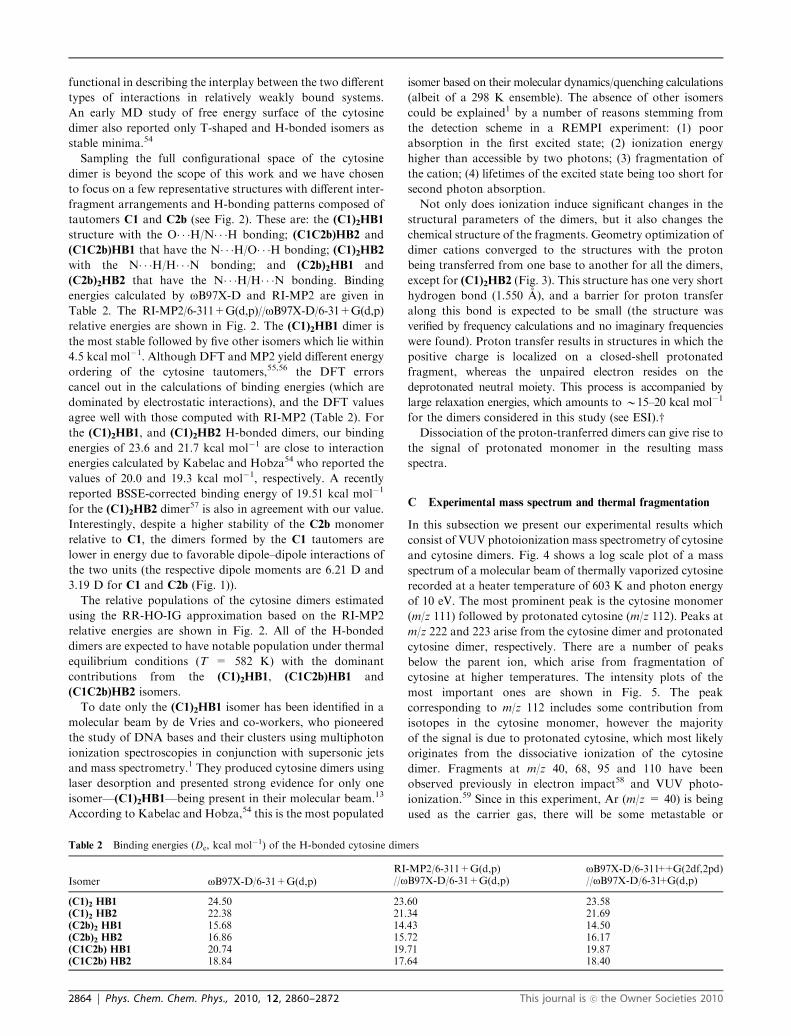
C Experimental mass spectrum and thermal fragmentation
In this subsection we present our experimental results which
consist of VUV photoionization mass spectrometry of cytosine
and cytosine dimers. Fig. 4 shows a log scale plot of a mass
spectrum of a molecular beam of thermally vaporized cytosine
recorded at a heater temperature of 603 K and photon energy
of 10 eV. The most prominent peak is the cytosine monomer
(m/z 111) followed by protonated cytosine (m/z 112). Peaks at
m/z 222 and 223 arise from the cytosine dimer and protonated
cytosine dimer, respectively. There are a number of peaks
below the parent ion, which arise from fragmentation of
cytosine at higher temperatures. The intensity plots of the
most important ones are shown in Fig. 5. The peak
corresponding to m/z 112 includes some contribution from
isotopes in the cytosine monomer, however the majority
of the signal is due to protonated cytosine, which most likely
originates from the dissociative ionization of the cytosine
dimer. Fragments at m/z 40, 68, 95 and 110 have been
observed previously in electron impact58 and VUV photo-
ionization.59 Since in this experiment, Ar (m/z = 40) is being
used as the carrier gas, there will be some metastable or
Table 2 Binding energies (De, kcal mol�1) of the H-bonded cytosine dimers
Isomer oB97X-D/6-31+G(d,p)RI-MP2/6-311+G(d,p) oB97X-D/6-311++G(2df,2pd)//oB97X-D/6-31+G(d,p) //oB97X-D/6-31+G(d,p)
(C1)2 HB1 24.50 23.60 23.58(C1)2 HB2 22.38 21.34 21.69(C2b)2 HB1 15.68 14.43 14.50(C2b)2 HB2 16.86 15.72 16.17(C1C2b) HB1 20.74 19.71 19.87(C1C2b) HB2 18.84 17.64 18.40
2864 | Phys. Chem. Chem. Phys., 2010, 12, 2860–2872 This journal is �c the Owner Societies 2010
Rydberg ionization of Ar and this is confirmed by the slight
dependence of its signal versus temperature shown in Fig. 5.
m/z 68 probably arises from elimination of NCOH from the
enol tautomersC2a/b, and/orC3a/b, since this involves breakage
of two single bonds. Plekan et al.59 discussed the formation of
m/z 68 and 69 in the context of dissociative ionization in the
VUV and electron impact (EI) experiments, however we see
very little evidence of m/z 69. It is also important to note that
the mass spectra arising from fragmentation at 10 eV are
mostly due to thermal energy bond breaking in contrast to
dissociative ionization, hence the results are reflective of
neutral cytosine. m/z 95 originates from NH2 elimination,
and Rice et al.58 suggested that m/z 68 could be due to the
further HCN elimination from this fragment. This channel
can operate at higher temperatures employed in our work.
m/z 109, which arises from H2 elimination from cytosine, is a
very prominent peak at higher temperatures and has been
observed previously upon EI ionization of a hydrated cytosine
beam by Kim et al.60 As mentioned earlier, Kosenkov et al.52
estimated that upon thermal vaporization at 490 K, dimers
constitute 28% of the total population. As clearly seen from
the dimer contribution in Fig. 5, our data disagree with this
prediction. In our experiment we see only 3% dimer contribution
(dimer + protonated monomer) around B550 K, and only
when we reach a temperature of 640 K, the dimer population is
around 41%. This increase in dimer population is due to an
increase in the concentration of monomers at higher temperatures.
In principle, Vant-Hoff type plots could be extracted from the
temperature dependence shown in Fig. 5, however we have
refrained from performing such an analysis here since it is
believed that molecular beams give rise to a highly non-
equilibrium environment. A previous attempt31 at generating
association constants from such experiments have been shown
to be subsequently wrong.61 However, qualitatively our results
suggest that it appears that cytosine, protonated cytosine and
the cytosine dimer are generated with sufficient concentration
and stability for us to extract meaningful photionization
efficiency curves and these are presented next.
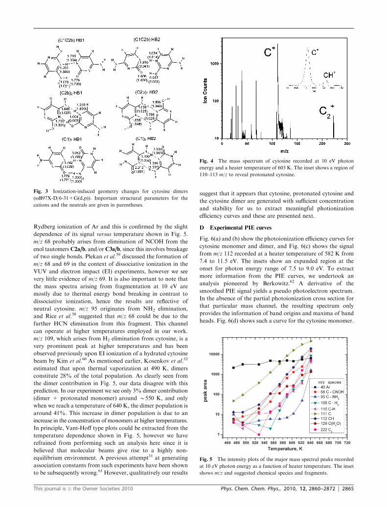
D Experimental PIE curves
Fig. 6(a) and (b) show the photoionization efficiency curves for
cytosine monomer and dimer, and Fig. 6(c) shows the signal
from m/z 112 recorded at a heater temperature of 582 K from
7.4 to 11.5 eV. The insets show an expanded region at the
onset for photon energy range of 7.5 to 9.0 eV. To extract
more information from the PIE curves, we undertook an
analysis pioneered by Berkowitz.62 A derivative of the
smoothed PIE signal yields a pseudo photoelectron spectrum.
In the absence of the partial photoionization cross section for
that particular mass channel, the resulting spectrum only
provides the information of band origins and maxima of band
heads. Fig. 6(d) shows such a curve for the cytosine monomer.
Fig. 3 Ionization-induced geometry changes for cytosine dimers
(oB97X-D/6-31+G(d,p)). Important structural parameters for the
cations and the neutrals are given in parentheses.
Fig. 4 The mass spectrum of cytosine recorded at 10 eV photon
energy and a heater temperature of 603 K. The inset shows a region of
110–113 m/z to reveal protonated cytosine.
Fig. 5 The intensity plots of the major mass spectral peaks recorded
at 10 eV photon energy as a function of heater temperature. The inset
shows m/z and suggested chemical species and fragments.
This journal is �c the Owner Societies 2010 Phys. Chem. Chem. Phys., 2010, 12, 2860–2872 | 2865
Three previously reported photoelectron spectra8,9 are also
shown in that figure and are normalized relative to the first
band head maximum at 8.9 eV. The near perfect fit of our first
peak to the PES ones gives us confidence in using this
technique for interpreting the experimental photoionization
results. Fig. 6(e) shows a similar spectrum for the cytosine
dimer. The signal-to-noise ratio is worse compared to the
monomer spectrum, however a number of bands can be
clearly identified in the spectrum. Finally, Fig. 6(f) shows the
isotopically corrected plot for m/z 112, protonated cytosine.
Energy onsets for the measured photoionization efficiency
(PIE) spectra are 8.60 � 0.05 eV and 7.6 � 0.1 eV for the
monomer and the dimer, respectively, and provide an estimate
for the adiabatic ionization energies.
E IEs of cytosine monomer and dimers
AIE and VIEs computed with EOM-IP-CCSD for several
cytosine tautomers are given in Table 3. As one can see,
tautomerization affects both AIE and VIEs. For example,
AIE for C2a and C3a differ by 0.2 eV, and the fourth ionized
state of C1 is red-shifted by more than 1.3 eV relative to C2a.
The relaxation energy, i.e., the VIE-AIE energy difference,
varies from 0.11 to 0.34 eV for different tautomers. Large
variations in VIEs are due to changes in the nature and the
order of ionized states upon tautomerization. These effects are
discussed in details in a forthcoming paper on ionization of
individual DNA bases.63
The AIE and the ten lowest VIEs for the dimers are
presented in Table 4 and the corresponding molecular orbitals
are shown in Fig. 7–9. Both the AIE and VIEs of the
dimers are red-shifted relative to the monomer. Moreover,
the ionization-induced relaxation is much larger in the dimers
Fig. 6 PIE curves for: (a) cytosine monomer (m/z 111); (b) cytosine dimer (m/z 222); (c) m/z 112. The shaded area is the mean standard deviation
of the results generated from 12 scans. The red line is obtained by applying a 5 point adjacent averaged smoothing routine. The insets in (a), (b),
and (c) show expanded region of the onset between 7.5 and 9.5 eV. (d) Derivative of 5 point smoothed signal from cytosine monomer shown in
(a), red and blue lines are the PES’s from Trofimov et al.,9 green line is the PES from Yu et al.8 (offset by 0.27 eV). (e) Derivative of 5 point
smoothed signal from cytosine dimer shown in (b). (f) Isotope corrected PIE from m/z 111 showing true appearance energy of the protonated
cytosine at m/z 112.
Table 3 The lowest AIE and VIEs for the selected cytosine tautomerscalculated at the EOM-IP-CCSD/cc-pVTZ//IP-CISD/6-31+G(d)and EOM-IP-CCSD/cc-pVTZ//RI-MP2/cc-pVTZ levels, respectively.All energies in eV
State C1 C2a C2b C3a C3b
AIE 8.67 8.52 8.54 8.71 8.68VIE1 8.78 8.86 8.86 8.90 8.88VIE2 9.55 9.58 9.62 9.89 10.01VIE3 9.65 10.12 10.02 10.28 10.19VIE4 10.06 11.38 11.34 10.74 11.06VIE5 12.28 11.91 11.94 12.78 12.75VIE6 13.27 13.52 13.48 13.28 13.31
2866 | Phys. Chem. Chem. Phys., 2010, 12, 2860–2872 This journal is �c the Owner Societies 2010
than in the monomer due to large geometry changes caused by
proton transfer between the bases in the ionized dimers.
Ionization energies of the cytosine dimers depend strongly
on their structures. Even for the dimers formed by the same
monomers, the shifts in VIEs can be as large as 0.45 eV (first
VIE for (C1)2HB1 and (C1)2HB2 dimers).
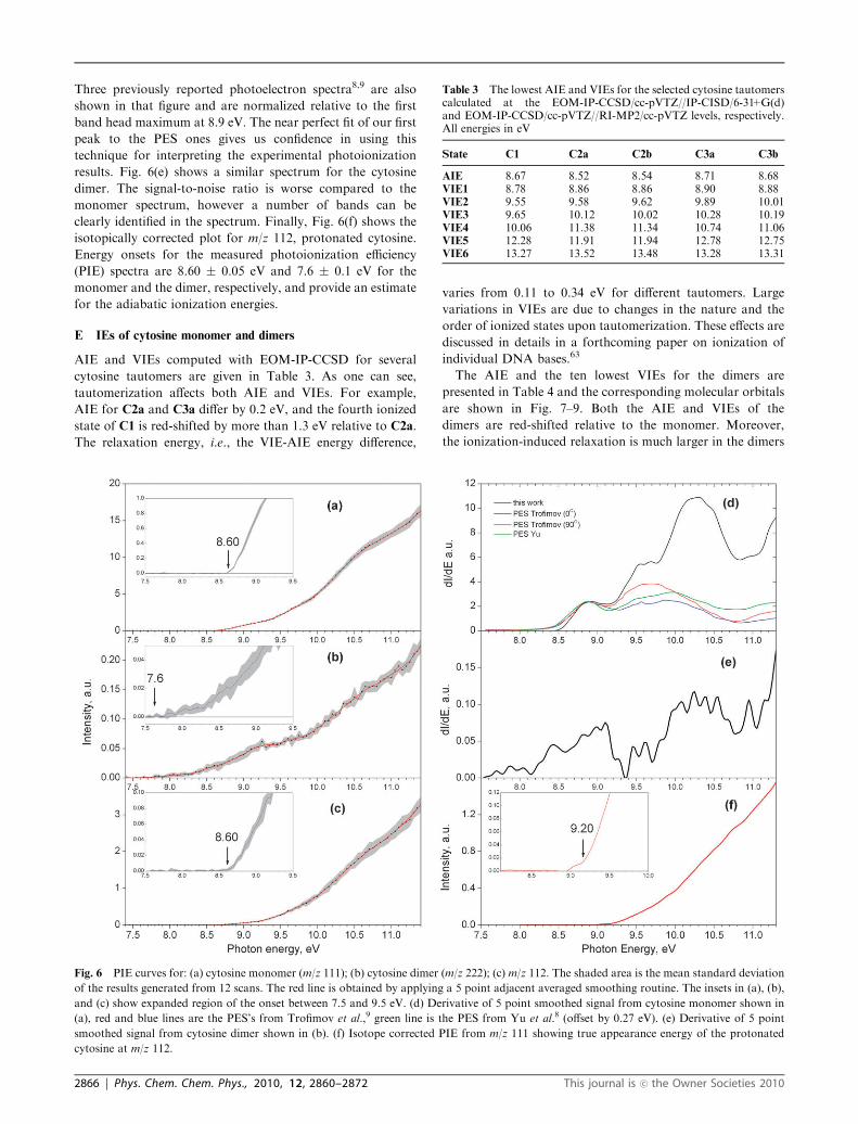
Further analysis of dimerization effects on ionization of
cytosine requires detailed characterization of the electronic
structure of the ionized dimers. The (C1)2HB1 dimer is
composed of two non-equivalent (due to non-symmetric
structure) fragments, which results in the localized ionized
states, i.e., the eight lowest ionized states of the dimer correspond
to the four lowest ionized states of each of the C1 fragments
(see Fig. 7). The (C1)2HB2 dimer has C2h symmetry and is
formed by two equivalent (by symmetry) C1 fragments.
Consequently, the hole is equally delocalized between the
two fragments (Fig. 7). The electronic structure of this type
of dimers can be described in terms of DMO-LCFMO (dimer
molecular orbitals—linear combination of fragment molecular
orbitals) framework.37,64 DMO-LCFMO assumes that the
dimer MOs are in-phase and out-of-phase combination of
the monomer’s MOs, and the states shown in Fig. 7 are of this
type. However, the MOs describing the 5th to 8th ionized state
of this dimer slightly deviate from this model (Fig. 7), i.e., even
though the MOs are the in-phase and out-of-phase combinations
of the fragment molecular orbitals (FMO), the shapes of the
FMOs are slightly different in the pairs of states. Thus, the
corresponding shifts in VIEs relative to the monomer should
be considered with caution. The first and third ionized states of
the (C1)2HB2 dimer are of non-Koopmans character and are
derived from ionization from the orbitals corresponding
to the first and second ionized state of the monomer (see
ESI).w Due to this mixed character, the shift of the dimer VIE
relative to the monomer is not well defined for these multi
configurational states.
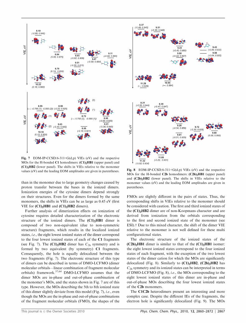
The electronic structure of the ionized states of the
(C2b)2HB1 dimer is similar to that of the (C1)2HB1 isomer:
the eight lowest ionized states correspond to the four ionized
states of each fragment, with the exception of the two lowest
states of the dimer cation for which the MOs are significantly
delocalized (Fig. 8). Similarly to (C1)2HB2, (C2b)2HB2 has
C2h symmetry and its ionized states can be interpreted in terms
of DMO-LCFMO (Fig. 8), i.e., the MOs corresponding to the
eight lowest ionized states of this dimer are in-phase and
out-of-phase MOs describing the four lowest ionized states
of the C2b monomers.
The C1C2b heterodimers present an interesting and more
complex case. Despite the different IEs of the fragments, the
electron hole is significantly delocalized (Fig. 9). The MOs
Fig. 7 EOM-IP-CCSD/6-311+G(d,p) VIEs (eV) and the respective
MOs for the H-bonded C1 homodimers: (C1)2HB1 (upper panel) and
(C1)2HB2 (lower panel). The shifts in VIEs relative to the monomer
values (eV) and the leading EOM amplitudes are given in parentheses. Fig. 8 EOM-IP-CCSD/6-311+G(d,p) VIEs (eV) and the respective
MOs for the H-bonded C2b homodimers: (C2b)2HB1 (upper panel)
and (C2b)2HB2 (lower panel). The shifts in VIEs relative to the
monomer values (eV) and the leading EOM amplitudes are given in
parentheses.
This journal is �c the Owner Societies 2010 Phys. Chem. Chem. Phys., 2010, 12, 2860–2872 | 2867
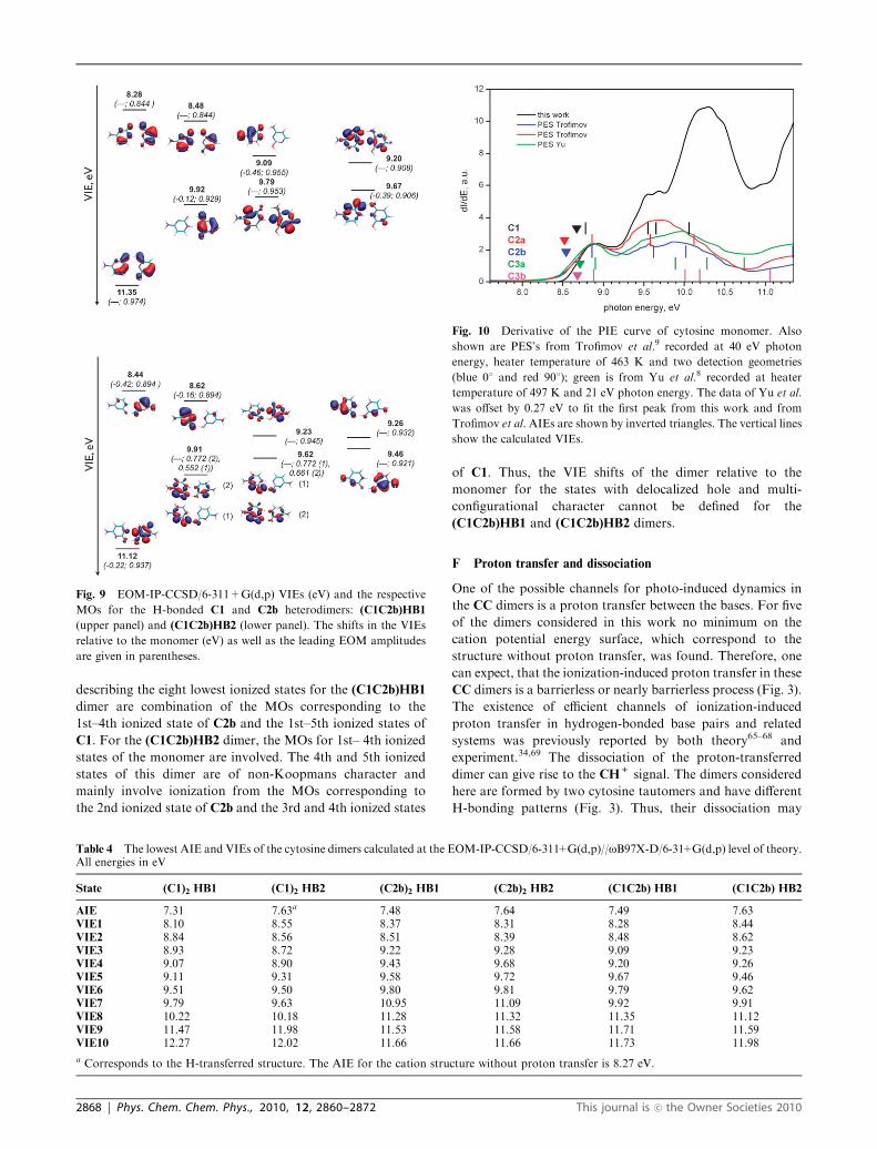
describing the eight lowest ionized states for the (C1C2b)HB1
dimer are combination of the MOs corresponding to the
1st–4th ionized state of C2b and the 1st–5th ionized states of
C1. For the (C1C2b)HB2 dimer, the MOs for 1st– 4th ionized
states of the monomer are involved. The 4th and 5th ionized
states of this dimer are of non-Koopmans character and
mainly involve ionization from the MOs corresponding to
the 2nd ionized state of C2b and the 3rd and 4th ionized states
of C1. Thus, the VIE shifts of the dimer relative to the
monomer for the states with delocalized hole and multi-
configurational character cannot be defined for the
(C1C2b)HB1 and (C1C2b)HB2 dimers.
F Proton transfer and dissociation
One of the possible channels for photo-induced dynamics in
the CC dimers is a proton transfer between the bases. For five
of the dimers considered in this work no minimum on the
cation potential energy surface, which correspond to the
structure without proton transfer, was found. Therefore, one
can expect, that the ionization-induced proton transfer in these
CC dimers is a barrierless or nearly barrierless process (Fig. 3).
The existence of efficient channels of ionization-induced
proton transfer in hydrogen-bonded base pairs and related
systems was previously reported by both theory65–68 and
experiment.34,69 The dissociation of the proton-transferred
dimer can give rise to the CH+ signal. The dimers considered
here are formed by two cytosine tautomers and have different
H-bonding patterns (Fig. 3). Thus, their dissociation may
Fig. 9 EOM-IP-CCSD/6-311+G(d,p) VIEs (eV) and the respective
MOs for the H-bonded C1 and C2b heterodimers: (C1C2b)HB1
(upper panel) and (C1C2b)HB2 (lower panel). The shifts in the VIEs
relative to the monomer (eV) as well as the leading EOM amplitudes
are given in parentheses.
Table 4 The lowest AIE and VIEs of the cytosine dimers calculated at the EOM-IP-CCSD/6-311+G(d,p)//oB97X-D/6-31+G(d,p) level of theory.All energies in eV
State (C1)2 HB1 (C1)2 HB2 (C2b)2 HB1 (C2b)2 HB2 (C1C2b) HB1 (C1C2b) HB2
AIE 7.31 7.63a 7.48 7.64 7.49 7.63VIE1 8.10 8.55 8.37 8.31 8.28 8.44VIE2 8.84 8.56 8.51 8.39 8.48 8.62VIE3 8.93 8.72 9.22 9.28 9.09 9.23VIE4 9.07 8.90 9.43 9.68 9.20 9.26VIE5 9.11 9.31 9.58 9.72 9.67 9.46VIE6 9.51 9.50 9.80 9.81 9.79 9.62VIE7 9.79 9.63 10.95 11.09 9.92 9.91VIE8 10.22 10.18 11.28 11.32 11.35 11.12VIE9 11.47 11.98 11.53 11.58 11.71 11.59VIE10 12.27 12.02 11.66 11.66 11.73 11.98
a Corresponds to the H-transferred structure. The AIE for the cation structure without proton transfer is 8.27 eV.
Fig. 10 Derivative of the PIE curve of cytosine monomer. Also
shown are PES’s from Trofimov et al.9 recorded at 40 eV photon
energy, heater temperature of 463 K and two detection geometries
(blue 01 and red 901); green is from Yu et al.8 recorded at heater
temperature of 497 K and 21 eV photon energy. The data of Yu et al.
was offset by 0.27 eV to fit the first peak from this work and from
Trofimov et al.AIEs are shown by inverted triangles. The vertical lines
show the calculated VIEs.
2868 | Phys. Chem. Chem. Phys., 2010, 12, 2860–2872 This journal is �c the Owner Societies 2010

yield different tautomeric forms of the CH+ and C+ species.
Appearance energies for different isomers of CH+ and
C+ computed as the energy differences between the ground
states of the neutral dimer and the corresponding products
(and corrected for ZPE) are given in Table 5. Although the
threshold energies were computed with DFT, we expect
that the errors in relative energy order of the C1 and C2b
tautomers cancel out in a similar way as for binding energies
(see Section III B).
IV. Discussion
A Effects of dimerization
As shown above by the experimental data and ab initio
calculations, dimerization strongly affects both the IEs and
the character of the ionized states. Here we analyze how the
inter-fragment interactions in the different types of the
H-bonded cytosine dimers affect their ionized states in order
to explain the origin of the strong shifts in VIEs due to
dimerization. The dimers considered here can be classified
into two distinct groups: ones with equivalent fragments
((C1)2HB2 and (C2b)2HB2) and those with structurally
((C1)2HB1 and (C2b)2HB1) or chemically ((C1C2b)HB1 and
(C1C2b)HB2) non-equivalent fragments. Below we show that
the VIE shifts can be qualitatively explained by inter-fragment
electrostatic interactions and the character of correspondingMOs.
The first type are the isomers composed of the same
tautomers that are geometrically non-equivalent ((C1)2HB1
and (C2b)2HB1). The VIEs shifts in these dimers relative to
monomers can be explained by inter-fragment electrostatic
interactions: the dipole moment of one fragment destabilizes
the MOs of another leading to a large drop in IEs. As a model
system, consider the (C1)2HB1 dimer (Fig. 7). Different
relative orientation of the monomers results in different VIEs
shifts for the ionized states localized on one of the two
monomers: the ionized states localized on one of the
monomers are affected more (�0.68 – �0.55 eV) than those
localized on the other (–0.44 – 0.16 eV). The same trend is
observed for the (C2b)2HB1 dimer (Fig. 8). However, the MOs
describing ionized states are partially delocalized in that case
due to a lower value of the dipole moment of C2b relative to
C1. This also explains smaller VIE shifts in the (C2b)2HB2
dimer as compared to the (C1)2HB1 isomer.
The shifts in VIEs of the heterodimers can also be explained
by the inter-fragment electrostatic interactions (Fig. 9). For
example, the first ionized state in both (C1C2b)HB1 and
(C2C2b)HB2 is mostly localized on the C2b fragment,
however, the respective VIE is 0.08 eV higher than that of
isolated C1. This points to a strong destabilization of the C2b
HOMO by the large dipole moment of the C1 fragment. The
analysis of the VIEs shifts for higher ionized states is complicated
by the delocalized character of the corresponding MOs.
The second group is the dimers with equivalent fragments.
Their electronic structure as well as the magnitude of the VIE
shifts in different states can be described by DMO-LCFMO to
a large extent. We illustrate this by considering the (C2b)2HB2
dimer as an example (see Fig. 8). In agreement with
DMO-LCFMO, the ionized states of this dimer described by
the MOs that are out-of-phase combination of the monomer
MOs are stabilized in the dimer cation giving rise to the VIEs
shifts of �0.55 – �0.25 eV, and the magnitude of the shifts
correlate well with the overlap of the respective FMOs.
However, the VIE shifts for the states described by the MOs
that are in-phase combinations are smaller or even positive
(�0.47�+0.06 eV). These negative shifts cannot be explained
by DMO-LCFMO, which predicts symmetric splitting
between the pairs of the ionized states. Same-magnitude shifts
of different sign for ionization from bonding/antibonding
pairs of orbitals were observed in a variety of stacked
dimers.22–23,37 Thus, the deviation is likely to be due to the
stronger perturbation of the orbitals introduced by hydrogen
bonding. The observed red shifts in VIEs for the states
described by the in-phase combination of the FMOs suggest
that the interaction of two or more FMOs for each fragment
needs to be considered.
It is worth noting that the magnitudes of the shifts of VIEs
for second type of systems are comparable to those for the
cytosine dimers with non-equivalent fragments, in contrast to
what was observed for thymine dimer in our earlier work.22
This can be explained by a more delocalized character of the
cytosine MOs and, consequently, larger overlap between the
fragments MOs.
The above analysis demonstrates that simple considerations
accounting for electrostatic inter-fragment interactions and
character of corresponding MOs can be used for a qualitative
explanation and prediction of the dimerization effects on
cytosine VIEs.
B Monomers IEs: theory vs. experiment
In addition to providing the basis for the analysis of the
dimerization effects in the ionized states of cytosine,
the computed IEs of the monomers and the dimers can
also be used as a reference for the interpretation of the
experimental spectra. The onset of the PIE spectrum shown
in Fig. 6(a) represents the AIE and is at 8.60 eV. Previously
our group has reported an IE of 8.65(�0.05) eV,6 and a very
early literature value is 8.68 eV.7 As discussed above, thermal
vaporization of cytosine can populate at least four tautomers
under our conditions. The AIEs and VIEs for the five
tautomers of cytosine computed with EOM-IP-CCSD are
shown in Table 3.
Table 5 oB97X-D/6-311++G(2df,2pd)//oB97X-D/6-31+G(d,p) thresholdenergies for cytosine dimer ionization-induced dissociation includingZPE correction (oB97X-D/6-31+G(d,p)). All energies in eV
Species Parent dimer
(C1)2HB1 (C1)2HB2
C1H+(N3) 9.08 9.33(C2b)2HB1 (C1C2b)HB2
C2bH+(N1) 9.16 9.19(C2b)2HB2 (C1C2b)HB1
C2bH+(N3) 9.23 9.27
(C1)2HB1 (C1)2HB2
C1+ 9.61 9.52(C2b)2HB1 (C2b)2HB2 (C1C2b)HB1 (C1C2b)HB2
C2b+ 9.14 9.16 9.47 9.43
This journal is �c the Owner Societies 2010 Phys. Chem. Chem. Phys., 2010, 12, 2860–2872 | 2869
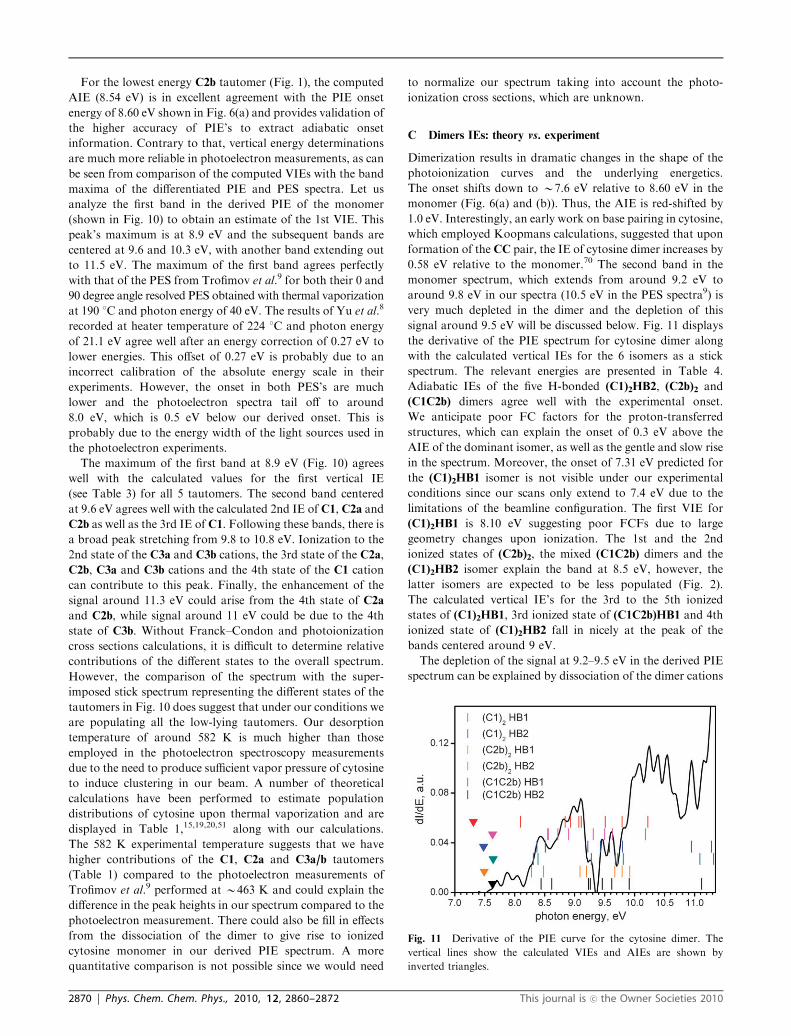
For the lowest energy C2b tautomer (Fig. 1), the computed
AIE (8.54 eV) is in excellent agreement with the PIE onset
energy of 8.60 eV shown in Fig. 6(a) and provides validation of
the higher accuracy of PIE’s to extract adiabatic onset
information. Contrary to that, vertical energy determinations
are much more reliable in photoelectron measurements, as can
be seen from comparison of the computed VIEs with the band
maxima of the differentiated PIE and PES spectra. Let us
analyze the first band in the derived PIE of the monomer
(shown in Fig. 10) to obtain an estimate of the 1st VIE. This
peak’s maximum is at 8.9 eV and the subsequent bands are
centered at 9.6 and 10.3 eV, with another band extending out
to 11.5 eV. The maximum of the first band agrees perfectly
with that of the PES from Trofimov et al.9 for both their 0 and
90 degree angle resolved PES obtained with thermal vaporization
at 190 1C and photon energy of 40 eV. The results of Yu et al.8
recorded at heater temperature of 224 1C and photon energy
of 21.1 eV agree well after an energy correction of 0.27 eV to
lower energies. This offset of 0.27 eV is probably due to an
incorrect calibration of the absolute energy scale in their
experiments. However, the onset in both PES’s are much
lower and the photoelectron spectra tail off to around
8.0 eV, which is 0.5 eV below our derived onset. This is
probably due to the energy width of the light sources used in
the photoelectron experiments.
The maximum of the first band at 8.9 eV (Fig. 10) agrees
well with the calculated values for the first vertical IE
(see Table 3) for all 5 tautomers. The second band centered
at 9.6 eV agrees well with the calculated 2nd IE of C1, C2a and
C2b as well as the 3rd IE of C1. Following these bands, there is
a broad peak stretching from 9.8 to 10.8 eV. Ionization to the
2nd state of the C3a and C3b cations, the 3rd state of the C2a,
C2b, C3a and C3b cations and the 4th state of the C1 cation
can contribute to this peak. Finally, the enhancement of the
signal around 11.3 eV could arise from the 4th state of C2a
and C2b, while signal around 11 eV could be due to the 4th
state of C3b. Without Franck–Condon and photoionization
cross sections calculations, it is difficult to determine relative
contributions of the different states to the overall spectrum.
However, the comparison of the spectrum with the super-
imposed stick spectrum representing the different states of the
tautomers in Fig. 10 does suggest that under our conditions we
are populating all the low-lying tautomers. Our desorption
temperature of around 582 K is much higher than those
employed in the photoelectron spectroscopy measurements
due to the need to produce sufficient vapor pressure of cytosine
to induce clustering in our beam. A number of theoretical
calculations have been performed to estimate population
distributions of cytosine upon thermal vaporization and are
displayed in Table 1,15,19,20,51 along with our calculations.
The 582 K experimental temperature suggests that we have
higher contributions of the C1, C2a and C3a/b tautomers
(Table 1) compared to the photoelectron measurements of
Trofimov et al.9 performed at B463 K and could explain the
difference in the peak heights in our spectrum compared to the
photoelectron measurement. There could also be fill in effects
from the dissociation of the dimer to give rise to ionized
cytosine monomer in our derived PIE spectrum. A more
quantitative comparison is not possible since we would need
to normalize our spectrum taking into account the photo-
ionization cross sections, which are unknown.
C Dimers IEs: theory vs. experiment
Dimerization results in dramatic changes in the shape of the
photoionization curves and the underlying energetics.
The onset shifts down to B7.6 eV relative to 8.60 eV in the
monomer (Fig. 6(a) and (b)). Thus, the AIE is red-shifted by
1.0 eV. Interestingly, an early work on base pairing in cytosine,
which employed Koopmans calculations, suggested that upon
formation of the CC pair, the IE of cytosine dimer increases by
0.58 eV relative to the monomer.70 The second band in the
monomer spectrum, which extends from around 9.2 eV to
around 9.8 eV in our spectra (10.5 eV in the PES spectra9) is
very much depleted in the dimer and the depletion of this
signal around 9.5 eV will be discussed below. Fig. 11 displays
the derivative of the PIE spectrum for cytosine dimer along
with the calculated vertical IEs for the 6 isomers as a stick
spectrum. The relevant energies are presented in Table 4.
Adiabatic IEs of the five H-bonded (C1)2HB2, (C2b)2 and
(C1C2b) dimers agree well with the experimental onset.
We anticipate poor FC factors for the proton-transferred
structures, which can explain the onset of 0.3 eV above the
AIE of the dominant isomer, as well as the gentle and slow rise
in the spectrum. Moreover, the onset of 7.31 eV predicted for
the (C1)2HB1 isomer is not visible under our experimental
conditions since our scans only extend to 7.4 eV due to the
limitations of the beamline configuration. The first VIE for
(C1)2HB1 is 8.10 eV suggesting poor FCFs due to large
geometry changes upon ionization. The 1st and the 2nd
ionized states of (C2b)2, the mixed (C1C2b) dimers and the
(C1)2HB2 isomer explain the band at 8.5 eV, however, the
latter isomers are expected to be less populated (Fig. 2).
The calculated vertical IE’s for the 3rd to the 5th ionized
states of (C1)2HB1, 3rd ionized state of (C1C2b)HB1 and 4th
ionized state of (C1)2HB2 fall in nicely at the peak of the
bands centered around 9 eV.
The depletion of the signal at 9.2–9.5 eV in the derived PIE
spectrum can be explained by dissociation of the dimer cations
Fig. 11 Derivative of the PIE curve for the cytosine dimer. The
vertical lines show the calculated VIEs and AIEs are shown by
inverted triangles.
2870 | Phys. Chem. Chem. Phys., 2010, 12, 2860–2872 This journal is �c the Owner Societies 2010
producing cation radicals (C1+ and C2b+) and protonated
monomers (C1H+(N3), C2bH+(N1) and C2bH+(N3))
(Table 5). The computed thresholds for all considered
channels of the CH+ formation lie within 0.1 eV of the
observed rise at 9.2 eV in the CH+ signal (shown in
Fig. 6(f)). We observed a similar behavior in the photoionization
spectrum and appearance energies of the protonated species
for thymine.
The peaks around 9.5 eV in the derived PIE could arise from
ionization of multiple isomers. A broad feature at 10.25 eV can
be ascribed to the 8th ionized state of the (C1)2HB1 and
(C1)2HB2 isomers. The band at 11 eV could be attributed
to the 7th ionized state (C2b)2HB1. The rise of the signal at
11.25 eV can be explained by ionization to the 8th ionized state
of the (C2b)2 dimers and the (C1C2b)HB1 isomer, as well as by
the 9th ionized state of (C1)2HB1. In summary, there is
evidence for the presence of multiple cytosine dimer isomers,
and the spectra could not be explained by the presence of only
the most stable (C1)2HB1 isomer. The comparison of the
computed dissociation energy thresholds with both the dimer
PIE spectrum and the CH+ signal curve points to the efficient
channel for intradimer proton transfer and dissociation at an
energy above B9.1 eV. This is also supported by the presence
of multiple ionized states in the 9.2–9.5 eV photon energy
region, e.g. the 5th ionized state of (C1)2HB2 and the
4th ionized state of the (C1C2b)HB1 dimer, which are not
observed in the experiment.
V. Conclusions
This work demonstrates strong effects upon dimerization on
cytosine ionization. The interaction between the fragments in
the dimers affects both the character of ionized states and IEs.
By using VUV single photon ionization mass spectrometry we
determined the first experimental AIE for the cytosine dimer
to be 7.6 � 0.1 eV. The onset in the dimer PIE spectra is
red-shifted by B1 eV relative to the monomer. The computed
EOM-IP-CCSD AIEs for the selected cytosine dimers range
between 7.31–7.64 eV. The calculations provide an insight into
the origin of the shifts and the character of ionized states,
aiding the interpretation of the experimentally derived PIE
spectra. The electronic structure analysis reveals that the
origin of this large red shift is in the electrostatic interactions
between the fragments. The largest shift (0.7 eV) was predicted
for the lowest-energy dimer, (C2b)2HB1, in which the hole
localized on one of the fragments is stabilized by a large dipole
moment (6.21 D) of the ‘‘neutral’’ fragment.
Both experimental and theoretical results suggest that a
number of tautomers and H-bonded dimers are present in
the molecular beam, however, more quantitative analysis
would require calculations of FCFs and ionization cross-
sections.
The computed energy thresholds for the ionization-induced
dimer dissociation forming the CH+ species show that this
channel can be efficient at photon energies above B9.1 eV,
which explains the strong rise in the measured CH+ signal at
9.2 eV. The large yield of the protonated species is consistent
with the barrierless (or almost barrierless) proton transfer
observed for the H-bonded cytosine dimers considered in
this study.
Future experiments using sophisticated two color IR-VUV
spectroscopy, ion-electron coincidence spectroscopy and
mass analysed threshold ionization will allow unambiguous
identification of the various species present in our molecular
beam. Nevertheless, the results presented here are a necessary
first step towards an unequivocal molecular-level understanding
of dynamics of photoionization of DNA bases.
Acknowledgements
This work is conducted under auspices of the iOpenShell
Center for Computational Studies of Electronic Structure
and Spectroscopy of Open-Shell and Electronically Excited
Species supported by the National Science Foundation
through the CRIF:CRF CHE-0625419+0624602+0625237
grant. M.R. and O.K. are supported by the Director, Office
of Energy Research, Office of Basic Energy Sciences, Chemical
Sciences Division of the U.S. Department of Energy under
contract No. DE-AC02-05CH11231.
References
1 M. de Vries, in Radiation Induced Molecular Phenomena in NucleicAcids, ed. M. K. Shukla and J. Leszczynski, Springer, 2008, p. 323.
2 M. S. de Vries and P. Hobza, Annu. Rev. Phys. Chem., 2007, 58,585–612.
3 M. K. Shukla and J. Leszczynski, in Radiation Induced MolecularPhenomena in Nucleic Acids, ed. M. K. Shukla and J. Leszczynski,Springer, 2008, p. 1.
4 F. C. Grozema and L. D. A. Siebbeles, Int. Rev. Phys. Chem., 2008,27, 87–138.
5 M. Taniguchi and T. Kawai, Phys. E., 2006, 33, 1–12.6 L. Belau, K. R. Wilson, S. R. Leone and M. Ahmed, J. Phys.Chem. A, 2007, 111, 7562–7568.
7 V. M. Orlov, A. N. Smirnov and Y. M. Varshavsky, TetrahedronLett., 1976, 17, 4377–4378.
8 C. Yu, S. Peng, I. Akiyama, J. Lin and P. R. Lebreton, J. Am.Chem. Soc., 1978, 100, 2303–2307.
9 A. B. Trofimov, J. Schirmer, V. B. Kobychev, A. W. Potts, D. M.P. Holland and L. Karlsson, J. Phys. B: At., Mol. Opt. Phys., 2006,39, 305–329.
10 N. S. Hush and A. S. Cheung, Chem. Phys. Lett., 1975, 34, 11–13.11 D. Dougherty, E. S. Younathan, R. Voll, S. Abdulnur and
S. P. McGlynn, J. Electron Spectrosc. Relat. Phenom., 1978, 13,379–393.
12 V. Feyer, O. Plekan, R. Richter, M. Coreno, G. Vall-Ilosera,K. C. Prince, A. B. Trofimov, I. L. Zaytseva,T. E. Moskovskaya, E. V. Gromov and J. Schirmer, J. Phys.Chem. A, 2009, 113, 5736–5742.
13 E. Nir, I. Hunig, K. Kleinermanns andM. S. de Vries, Phys. Chem.Chem. Phys., 2003, 5, 4780–4785.
14 E. Nir, M. Muller, L. I. Grace and M. S. de Vries, Chem. Phys.Lett., 2002, 355, 59–64.
15 J. K. Wolken, C. Yao, F. Turecek, M. J. Polce andC. Wesdemiotis, Int. J. Mass Spectrom., 2007, 267, 30–42.
16 D. Roca-Sanjuan, M. Rubio, M. Merchan and L. Serrano-Andres,J. Chem. Phys., 2006, 125, 084302.
17 E. Cauet, D. Dehareng and J. Lievin, J. Phys. Chem. A, 2006, 110,9200–9211.
18 E. Cauet and J. Lievin, in Advances in Quantum Chemistry, ElsevierAcademic Press Inc., San Diego, 2007, vol. 52, pp. 121–147.
19 G. Fogarasi, J. Phys. Chem. A, 2002, 106, 1381–1390.20 S. A. Trygubenko, T. V. Bogdan, M. Rueda, M. Orozco,
F. J. Luque, J. Sponer, P. Slavicek and P. Hobza, Phys. Chem.Chem. Phys., 2002, 4, 4192–4203.
21 O. Dolgounitcheva, V. G. Zakrzewski and J. V. Ortiz, J. Phys.Chem. A, 2003, 107, 822–828.
This journal is �c the Owner Societies 2010 Phys. Chem. Chem. Phys., 2010, 12, 2860–2872 | 2871
22 K. Bravaya, O. Kostko, M. Ahmed and A. I. Krylov, Phys. Chem.Chem. Phys., 2010, DOI: 10.1039/b919930f.
23 A. A. Golubeva and A. I. Krylov, Phys. Chem. Chem. Phys., 2009,11, 1303–1311.
24 P. A. Pieniazek, S. E. Bradforth and A. I. Krylov, J. Chem. Phys.,2008, 129, 074104.
25 A. I. Krylov, Annu. Rev. Phys. Chem., 2008, 59, 433–462.26 J. F. Stanton and J. Gauss, J. Chem. Phys., 1994, 101, 8938–8944.27 P. A. Pieniazek, S. A. Arnstein, S. E. Bradforth, A. I. Krylov and
C. D. Sherrill, J. Chem. Phys., 2007, 127, 164110.28 M. Kamiya and S. Hirata, J. Chem. Phys., 2006, 125, 074111.29 S. Pal, M. Rittby, R. J. Bartlett, D. Sinha and D. Mukherjee,
Chem. Phys. Lett., 1987, 137, 273–278.30 M. Dey, J. Grotemeyer and E. W. Schlag, Z. Naturforsch., A:
Phys. Sci., 1994, 49, 776–784.31 M. Dey, F. Moritz, J. Grotemeyer and E. W. Schag, J. Am. Chem.
Soc., 1994, 116, 9211–9215.32 I. K. Yanson, A. B. Teplitsky and L. F. Sukhodub, Biopolymers,
1979, 18, 1149–1170.33 M. Y. Choi, F. Dong and R. E. Miller, Philos. Trans. R. Soc.
London, Ser. A, 2005, 363, 393–412.34 N. Gador, E. Samoylova, V. R. Smith, A. Stolow, D. M. Rayner,
W. G. Radloff, I. V. Hertel and T. Schultz, J. Phys. Chem. A, 2007,111, 11743–11749.
35 E. Nir, K. Kleinermanns and M. S. de Vries, Nature, 2000, 408,949–951.
36 C. Plutzer, I. Hunig, K. Kleinermanns, E. Nir and M. S. de Vries,ChemPhysChem, 2003, 4, 838–842.
37 P. A. Pieniazek, A. I. Krylov and S. E. Bradforth, J. Chem. Phys.,2007, 127, 044317.
38 S. Steenken, Free Radical Res. Commun., 1992, 16, 349–379.39 M. Feyereisen, G. Fitzgerald and A. Komornicki, Chem. Phys.
Lett., 1993, 208, 359–363.40 F. Weigend and M. Haser, Theor. Chem. Acc., 1997, 97,
331–340.41 R. A. Distasio, R. P. Steele, Y. M. Rhee, Y. H. Shao and M.
Head-Gordon, J. Comput. Chem., 2007, 28, 839–856.42 J. D. Chai and M. Head-Gordon, J. Chem. Phys., 2008, 128,
084106.43 S. Grimme, J. Comput. Chem., 2004, 25, 1463–1473.44 S. Grimme, J. Comput. Chem., 2006, 27, 1787–1799.45 J. D. Chai and M. Head-Gordon, Phys. Chem. Chem. Phys., 2008,
10, 6615–6620.46 C. W. Murray, N. C. Handy and G. J. Laming, Mol. Phys., 1993,
78, 997–1014.47 V. I. Lebedev, Zh. Vychisl. Mat. Fiz., 1975, 15, 48.48 Several popular ab initio packages include ZPE in the vibrational
enthalpy term, and using ZPE-corrected energy differencesbetween isomers will result in double-counting, which appears tobe a common pitfall in such calculations.
49 A. Landau, K. Khistyaev, S. Dolgikh and A. I. Krylov, J. Chem.Phys., 2010, 132, 014109.
50 Y. Shao, L. F. Molnar, Y. Jung, J. Kussmann, C. Ochsenfeld,S. T. Brown, A. T. B. Gilbert, L. V. Slipchenko, S. V. Levchenko,D. P. O’Neill, R. A. DiStasio, R. C. Lochan, T. Wang, G. J. O.Beran, N. A. Besley, J. M. Herbert, C. Y. Lin, T. Van Voorhis,S. H. Chien, A. Sodt, R. P. Steele, V. A. Rassolov, P. E. Maslen,P. P. Korambath, R. D. Adamson, B. Austin, J. Baker, E. F. C.Byrd, H. Dachsel, R. J. Doerksen, A. Dreuw, B. D. Dunietz,A. D. Dutoi, T. R. Furlani, S. R. Gwaltney, A. Heyden, S. Hirata,C. P. Hsu, G. Kedziora, R. Z. Khalliulin, P. Klunzinger,A. M. Lee, M. S. Lee, W. Liang, I. Lotan, N. Nair, B. Peters,E. I. Proynov, P. A. Pieniazek, Y. M. Rhee, J. Ritchie, E. Rosta,C. D. Sherrill, A. C. Simmonett, J. E. Subotnik, H. L. Woodcock,W. Zhang, A. T. Bell, A. K. Chakraborty, D. M. Chipman,F. J. Keil, A. Warshel, W. J. Hehre, H. F. Schaefer, J. Kong,A. I. Krylov, P. M. W. Gill and M. Head-Gordon, Phys. Chem.Chem. Phys., 2006, 8, 3172–3191.
51 Z. B. Yang and M. T. Rodgers, Phys. Chem. Chem. Phys., 2004, 6,2749–2757.
52 D. Kosenkov, Y. Kholod, L. Gorb, O. Shishkin, D. M. Hovorun,M.Mons and J. Leszczynski, J. Phys. Chem. B, 2009, 113, 6140–6150.
53 P. Jurecka, J. Sponer and P. Hobza, J. Phys. Chem. B, 2004, 108,5466–5471.
54 M. Kabelac and P. Hobza, J. Phys. Chem. B, 2001, 105, 5804–5817.55 M. Piacenza and S. Grimme, J. Comput. Chem., 2004, 25, 83–98.56 J. R. Sambrano, A. R. de Souza, J. J. Queralt and J. Andres,Chem.
Phys. Lett., 2000, 317, 437–443.57 P. K. Sahu, R. K. Mishra and S. L. Lee, J. Phys. Chem. A, 2005,
109, 2887–2893.58 J. M. Rice and G. O. Dudek, J. Am. Chem. Soc., 1967, 89,
2719–2725.59 O. Plekan, V. Feyer, R. Richter, M. Coreno, M. de Simone and
K. C. Prince, Chem. Phys., 2007, 334, 53–63.60 S. K. Kim, W. Lee and D. R. Herschbach, J. Phys. Chem., 1996,
100, 7933–7937.61 P. Hobza and J. Sponer, Chem. Phys. Lett., 1996, 261, 379–384.62 J. Berkowitz, J. Chem. Phys., 1978, 69, 3044–3054.63 K. Bravaya, O. Kostko, M. Ahmed, S. Dolgikh, A. Landau and
A. I. Krylov, 2009, to be submitted.64 P. A. Pieniazek, J. VandeVondele, P. Jungwirth, A. I. Krylov and
S. E. Bradforth, J. Phys. Chem. A, 2008, 112, 6159–6170.65 H. Y. Chen and I. Chao, ChemPhysChem, 2004, 5, 1855–1863.66 J. Bertran, L. Blancafort and M. Noguera, in Computational
studies of DNA and RNA, ed. J. Sponer and F. Lankas,Springer, 2006, p. 411.
67 J. Rak, J. Makowska and A. A. Voityuk, Chem. Phys., 2006, 325,567–574.
68 H. S. Park, S. H. Nam, J. K. Song, S. M. Park and S. Ryu, J. Phys.Chem. A, 2008, 112, 9023–9030.
69 N. J. Kim, H. M. Kim and S. K. Kim, Int. J. Mass Spectrom.,2007, 261, 32–37.
70 A. O. Colson, B. Besler and M. D. Sevilla, J. Phys. Chem., 1992,96, 9787–9794.
2872 | Phys. Chem. Chem. Phys., 2010, 12, 2860–2872 This journal is �c the Owner Societies 2010