Embed Size (px)
Citation preview
5. Demirkiran M, Jankovic J. Paroxysmal dyskinesias: clinical fea-tures and classification. Ann Neurol 1995;38:571–579.
6. Matsuo H, Kamakura K, Saito M, et al. Familial paroxysmaldystonic choreoathetosis: clinical findings in a large Japanesefamily and genetic linkage to 2q. Arch Neurol 1999;56:721–726.
7. Rainier S, Thomas D, Tokarz D, et al. Myofibrillogenesis regulator1 gene mutations cause paroxysmal dystonic choreoathetosis. ArchNeurol 2004;61:1025–1029.
8. Lee HY, Xu Y, Huang Y, et al. The gene for paroxysmal non-kinesigenic dyskinesia encodes an enzyme in a stress responsepathway. Hum Mol Genet 2004;13:3161–3170.
9. Djarmati A, Svetel M, Momcilovic D, Kostic V, Klein C. Signif-icance of recurrent mutations in the myofibrillogenesis regulator 1gene. Arch Neurol 2005;62:1641.
10. Chen DH, Matsushita M, Rainier S, et al. Presence of alanine-to-valine substitution in myofibrillogenesis regulator 1 in paroxysmalnonkinesigenic dyskinesia: confirmation in 2 kindreds. Arch Neu-rol 2004;61:1025–1029.
11. Przuntek H, Monninger P. Therapeutic aspects of kinesigenicparoxysmal choreoathetosis and familial paroxysmal choreoathe-tosis of the Mount and Reback type. J Neurol 1983;230:163–169.
12. Nordocci N, Lamperti E, Rumi V, Angelini L. Typical and atypicalforms of paroxysman choreoathetosis. Dev Med Child Neurol1989;31:670–681.
13. Byrne E, White O, Cook M. Familial dystonic choreoathetosis withmyokymia: a sleep responsive disorder. J Neurol Neurosurg Psy-chiatry 1991;54:1090–1092.
Involuntary Painful MuscleContractions in Satoyoshi
Syndrome: A SurfaceElectromyographic Study
Gea Drost, MD,1,2* Aad Verrips, MD, PhD,3
Baziel G.M. van Engelen, MD, PhD,2
Dick F. Stegeman, PhD,1,2
and Machiel J. Zwarts, MD, PhD1,2
1Department of Clinical Neurophysiology, Institute ofNeurology, Radboud University Nijmegen Medical Centre,
Nijmegen, The Netherlands; 2Neuromuscular CentreNijmegen, Institute of Neurology, Radboud UniversityNijmegen Medical Centre, Nijmegen, The Netherlands;
3Canisius Wilhelmina Hospital, Nijmegen, The Netherlands
Abstract: We report a child with Satoyoshi syndrome man-ifested by involuntary painful muscle contractions and al-opecia. Although an autoimmune origin of Satoyoshi syn-drome seems likely, its exact etiology remains as yet
unknown, as is the origin of the involuntary contractions.To gain a better understanding of the electrophysiologicalcharacteristics of the involuntary contractions, we per-formed a surface electromyographic (EMG) study. We in-vestigated muscle contractions in the legs using two nonin-vasive techniques: high-density surface EMG (HD-sEMG)recordings on one muscle, and polymyographic surfaceEMG (sEMG) recordings on various muscles. During theinvoluntary contractions, HD-sEMG showed a fourfold in-crease in amplitude compared to maximal voluntary con-tractions. These high potentials were widely distributedacross the whole muscle and showed a pronounced oscilla-tory behavior with a frequency around 45 Hz. Polymyo-graphic sEMG revealed that the involuntary contractionsoften occur simultaneously in various muscles or showed aswitch of activity from one muscle to another. These find-ings point to hyperactivity or a disinhibition at the alphamotor neuron level, originating probably at that level, al-though a central origin cannot be excluded. © 2006 Move-ment Disorder Society
Key words: Satoyoshi syndrome; high-density surfaceEMG; involuntary muscle contractions
Satoyoshi syndrome is a childhood-onset progressivemultisystem disorder characterized by intermittent pain-ful muscle contractions, malabsorption, diarrhea, endo-crinopathy, alopecia, and secondary skeletal abnormali-ties.1 Its etiology is unknown, although an autoimmunebasis is likely through association with other autoim-mune conditions, the presence of antibodies, and theimprovement of symptoms with corticosteroid treatmentand intravenous immunoglobulin.2,3 The painful contrac-tions usually begin in the legs during childhood, andprogress slowly with increasing severity and intensity,also involving limbs, trunk, neck, and masticatory mus-cles. Muscle contractions generally last for a few minutesand usually recur after an interval of less than a minute.These contractions are so intense that they induce abnor-mal posturing of affected limbs. Satoyoshi reported thatthe more severe the muscle contractions are and theearlier the disease onset in these patients is, the moresevere the growth disturbance and the joint deformityare.5
The relation between the presumed autoimmune originand the involuntary muscle contractions currently is stillunclear. The answer to the question whether the painfulmuscle contractions in Satoyoshi syndrome have a pe-ripheral (like muscle cramps) or central origin (like mus-cle spasms) would provide a clue to the pathophysiologyand the cause of this rare syndrome.
Muscle cramp is a clinical diagnosis. It is character-ized by a sudden, uncomfortable squeezing or contrac-tion, lasting for seconds to minutes, often with a palpablehard knot in the affected muscle. Stretching the muscle
*Correspondence to: Dr. Gea Drost, Radboud University NijmegenMedical Centre, Institute of Neurology, P.O. Box 9101, 6500 HB,Nijmegen, the Netherlands. E-mail: [email protected]
Received 10 July 2005; Revised 3 May 2006; Accepted 9 May 2006Published online 13 September 2006 in Wiley InterScience (www.
interscience.wiley.com). DOI: 10.1002/mds.21088
SURFACE EMG AND SATOYOSHI SYNDROME 2015
Movement Disorders, Vol. 21, No. 11, 2006

or contraction of its antagonist muscle speeds relief ofthe cramp.6 Cramp is limited to a single muscle. Theexact site of origin of muscle cramp is still a matter ofdebate. A surface electromyographic (EMG) technique,with multiple electrodes on one muscle, recently hasbeen applied to gain better insights into its pathophysi-ology. It has been demonstrated that muscle crampspresent themselves with a characteristic pattern of aslowly moving fraction of contracting muscle fibers in asingle muscle, indicating that cramp spreads throughneighboring groups of muscle fibers.7 A recently devel-oped surface EMG technique, with multiple surfaceEMG electrodes in a single grid, named high-densitysurface EMG technique (HD-sEMG), allows simulta-neous recordings from various locations across a muscle,providing both temporal and spatial information aboutmotor unit activity.8,9
Muscle spasms are ill-defined. They form part of aspastic syndrome that also includes exaggerated tendonreflexes and muscle tone.10 Zijdewind and Thomasshowed that MU firing rates of the thenar muscles thathave been weakened in patients with chronic spinal cordinjury were low during both submaximal and maximalvoluntary contractions.11 During spasms in patients withchronic cervical spinal cord injury, motor unit firing rateseither increased and then decreased with the spasm in-tensity or were relatively constant. Mean peak spasmfiring rates were 18 � 9 Hz (mean � SD) for therate-modulated units and 11 � 10 Hz for units with littleor no rate modulation.12 Because muscle spasms aretypically not limited to a single muscle, we also recordedpolymyographic surface EMG (sEMG) patterns in vari-ous muscles simultaneously with conventional sEMGtechniques.
Here, we report a child with Satoyoshi syndrome inwhich HD-sEMG and polymyographic sEMG, were per-formed to gain a better understanding of the pathophys-iology of the involuntary muscle contractions in Satoyo-shi syndrome.
CASE REPORT
A 6-year-old girl of Turkish origin suffered from ex-ercise-induced, progressive painful muscle contractionsand thinning hair. Apart from abnormal muscle contrac-tions and a short stature (body length below 3 SD),general and neurological examination revealed no abnor-malities. Laboratory examination revealed a serum cre-atine kinase of 296 U/l (normal value � 200 U/L), thepresence of anti-nuclear antibodies and a transient ane-mia. Endocrine investigation (thyroid gland, pituitaryfunction, urine steroid profile, serum, and urine glucose)was unremarkable. Radiological investigations showed
no skeletal deformities. Quadriceps muscle biopsy wasnormal, as were immunohistochemistry (phosphorylase,phosphofructokinase) and mitochondrial respiratorychain enzyme activities. Hair root analysis showed a highpercentage of catagen follicles. Standard nerve conduc-tion and needle EMG (not during involuntary contrac-tions) showed normal values. Satoyoshi syndrome wasdiagnosed, and therapy was started with prednisone, 1mg/kg per day for 3 months. The painful muscle con-tractions completely disappeared during therapy. Hairgrowth was not restored.
Surface EMG Methods
EMG measurements were performed on the child’sleg, where the involuntary muscle contractions weremost pronounced. First, HD-sEMG recordings of the leftvastus lateralis muscle were recorded during maximalvoluntary contraction and during the painful muscle con-tractions. HD-sEMG allows multichannel recordings of asingle muscle and is a noninvasive tool to record thedistributions of muscle activation over a larger area (6 �4.5 cm) of a muscle.8,9,13 The electrode grid used con-sisted of 130 gold-coated (13 � 10) electrodes with aninterelectrode distance of 5 mm in both directions. Theelectrode grid was carefully placed over the vastus late-ralis muscle with its columns of 13 electrodes parallel tothe muscle fibers. All signals were amplified, band-passfiltered, and simultaneously AD converted (at 2,000 sam-ples/sec) with a multichannel amplifier system.8,13 Thesignals were analyzed off-line and displayed in a bipolarmontage for visual analysis.
In addition, muscle activity of different muscles wererecorded simultaneously with bipolar polymyographicsurface EMG recordings on different muscles of bothlegs, using Ag/AgCl electrode pairs at a 3-cm interelec-trode distance. On both legs, the quadriceps muscles, thetibialis anterior muscles and the gastrocnemius muscleswere investigated.
RESULTS
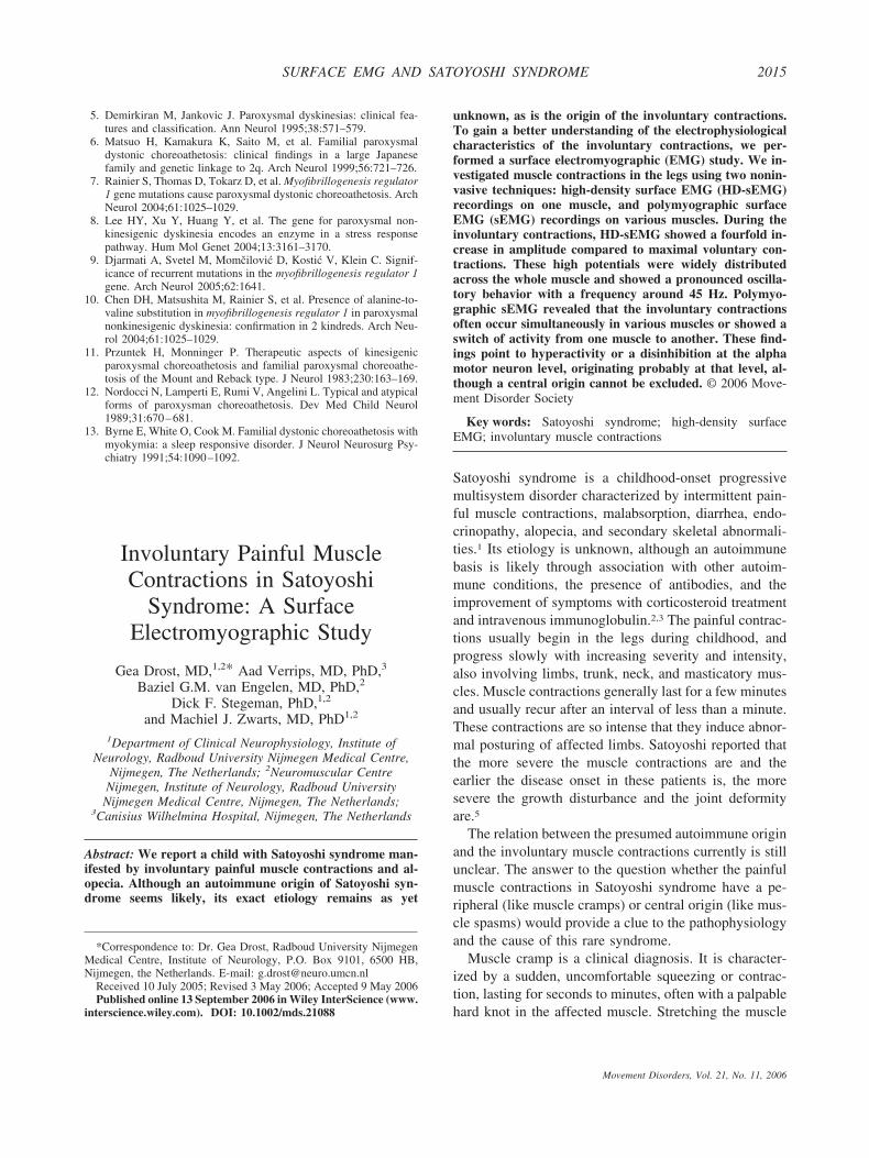
HD-sEMG recordings of the left vastus lateralis mus-cle during maximum voluntary contractions and duringpainful involuntary muscle contractions are shown inFigure 1A and B, respectively. During maximal volun-tary contractions, the HD-sEMG recording showed anormal interference pattern with motor unit action po-tential amplitudes of approximately 50 �V. During theinvoluntary contractions, bipolar HD-sEMG recordingsshowed a regular pattern, most probably of synchronizedmotor unit firings with a frequency of 40 to 50 Hz,resulting in much higher amplitudes (over 200 �V) thanin the maximal voluntary contractions. We observed that
2016 G. DROSF ET AL.
Movement Disorders, Vol. 21, No. 11, 2006
these high-amplitude EMG signals were present with ratherconstant amplitudes over a substantial part of the vastuslateralis muscle. It is important to note that there were nosigns of slow spread of local EMG activity in the muscle asfound with multichannel EMG in muscle cramps.7
Conventional polymyographic sEMG recordingsshowed simultaneous activity both of various muscles ofthe same leg, and of muscles of both legs during theinvoluntary contractions (Fig. 1C and D, respectively). Inthe course of the involuntary contractions, there was aswitch over of EMG activity from one muscle to another,with a “waxing and waning” character (Fig. 1C).
DISCUSSION
The EMG findings of this study strongly support theconcept that the involuntary muscle contractions in Sa-toyoshi syndrome do not fit into the dichotomy between
muscle cramps and spasms in a “classic sense.” We willgive reasons why another mechanism seems likely.
There are several arguments against the mass activityfound in this patient being a muscle cramp. First, ourHD-sEMG recording during the involuntary contractionsshowed an over fourfold increase in amplitudes of theEMG activity compared to the amplitudes recorded dur-ing maximal voluntary contraction and a regular firingfrequency of 40 to 50 Hz. The synchronous regularfirings and the high amplitude during the involuntarycontractions suggest strictly synchronous repetitive ac-tive motor unit discharges, which have not been found incramps.7,14 Second, the motor unit action potentials inthe vastus lateralis muscle during the involuntary con-tractions were present in a large part of the muscle,which is in contrast to what has been found in musclecramps, in which only a (slowly shifting) fraction of the
FIG. 1. A–B: High-density surface electromyographic (EMG) recordings of the left vastus lateralis muscle show high-density EMG recordings ofthe Satoyoshi patient. One column of the grid (12 bipolar montages from 13 monopolar electrodes in a grid column positioned in fiber direction) withthe clearest presentation of the phenomenon of muscle-wide synchronous oscillatory activity (Part B) is displayed. The x axes indicate time in seconds,and the y axes indicate 12 bipolar recordings derived from 13 electrodes. A: High-density surface electromyographic (HD-sEMG) during maximalvoluntary contraction. The sEMG shows a normal motor unit interference pattern. B: Recording from the same area during painful contraction showswidely spread rhythmic EMG activity with a much higher amplitude (note the different gain settings) and an oscillatory frequency of approximately45 Hz. C–D: Conventional bipolar polymyographic sEMG recordings of three different muscles of both legs: the first three traces of the right leg andthe lower three traces of the left leg. C and D are recordings during the involuntary contractions of the Satoyoshi patient at different time points. Thepolymyographic recordings show simultaneous EMG activity in both right and left quadriceps muscles (C), and in different muscles (right quadricepsmuscle and right tibialis anterior muscle) of the same leg (D).
SURFACE EMG AND SATOYOSHI SYNDROME 2017
Movement Disorders, Vol. 21, No. 11, 2006
muscle is involved.7 Third, the discharge frequency ofthe EMG activity during the involuntary contractionswas 40 to 50 Hz. Discharge rates in cramps are typicallyaround 150 Hz.6 Last but not least, our polymyographicrecordings showed that massive electric muscle activitywas not limited to one muscle. It extended over differentleg muscles, even more or less simultaneously in bothlegs. The latter directed us to consider this mass activityas muscle spasms.
As already stated, spasms are ill defined.10 That therewere no clinical signs of pyramidal tract lesion providesan important argument that the involuntary painful con-tractions in this patient are not “conventional” spasms. Inhealthy subjects, motor units usually fire at 6 to 10 Hzwhen first recruited and at 15 to 60 Hz during maximalvoluntary contractions.15 Thomas and Ross showed withneedle EMG studies of motor unit activity in patients afterspinal cord injury that, during spasms, the motor unit firingfrequencies show mean peak firing rates of 18 � 9 Hz.12
Rate coding for many motor units appears to be similarwhether descending motor input is intact or whether it hasbeen reduced severely by spinal cord injury.
In 1978, Satoyoshi already described that, during theinvoluntary contractions in his patients, needle EMG re-cordings revealed synchronized motor unit discharges of 40to 50 Hz and of 4 to 10 mV amplitude. He suggested thatabnormal discharge of anterior horn cells is responsible forthe mass activity.1 The HD-sEMG recordings showed astable spatial extension of such synchronized activitythroughout the vastus lateralis muscle during the contrac-tions. This finding supports and provides additional argu-ments for this hypothesis of massive hyperactivity or dis-inhibition at the alpha motor neuron level. In conclusion,although a more proximal origin cannot be excluded, thesurface EMG findings strongly suggest a deregulation at thealpha motor neuron level leading to involuntary musclecontractions in Satoyoshi syndrome.
Acknowledgment: We thank Henny Janssen, technician, for assist-ing in the EMG measurements and the anonymous reviewers for theiruseful comments.
REFERENCES
1. Satoyoshi E. A syndrome of progressive muscle spasm, alopecia,and diarrhea. Neurology 1978;28:458–471.
2. Satoh A, Tsujihata M, Yoshimura T, Nagataki S. Myastheniagravis associated with Satoyoshi syndrome: muscle cramps, alo-pecia and diarrhea. Neurology 1983;33:1209–1211.
3. Drost G, Verrips A, Hooijkaas H, Zwarts MJ. Glutamic aciddecarboxylase antibodies in Satoyoshi syndrome. Ann Neurol2004;55:450–451.
4. Endo K, Yamamoto T, Nakamura K, et al. Improvement of Sa-toyoshi syndrome with tacrolimus and corticosteroids. Neurology2002;2:2014–1215.
5. Satoyoshi E, Yamada K. Recurrent muscle spasms of centralorigin. Arch Neurol 1967;16:254–264.
6. Miller TM, Layzer RB. Muscle cramps. Muscle Nerve 2005;32:431–422.
7. Roeleveld K, van Engelen BG, Stegeman DF. Possible mecha-nisms of muscle cramp from temporal and spatial surface EMGcharacteristics. J Appl Physiol 2000;88:1698–1706.
8. Blok JH, van Dijk JP, Drost G, Zwarts MJ, Stegeman DF. A high-densitymultichannel surface electromyography system for the characteriza-tion of single motor units. Rev Sci Instrum 2002;73:1887–1897.
9. Zwarts MJ, Stegeman DF. Multichannel surface EMG: basic as-pects and clinical utility. Muscle Nerve 2003;28:1–17.
10. Young RR. Spasticity: a review. Neurology 1994;44(Suppl. 9):S12–S20.
11. Zijdewind I, Thomas CK. Motor unit firing during and after vol-untary contractions of human thenar muscles weakened by spinalcord injury. J Neurophysiol 2003;89:2065–2071.
12. Thomas CK, Ross BH. Distinct patterns of motor unit behaviourduring muscle spasms in spinal cord injured subjects. J Neuro-physiol 1997;77:2847–2850.
13. Drost G, Blok JH, Stegeman DF, van Dijk JP, van Engelen BGM,Zwarts MJ. Propagation disturbance of motor unit action potentialsduring transient paresis in generalized myotonia: a high densitysurface EMG study. Brain 2001;124:352–360.
14. Ross BH, Thomas CK. Human motor unit activity during inducedmuscle cramp. Brain 1995;188:983–993.
15. Monster AW, Chan H. Isometric force production by motor unitsof extensor digitorum communis muscle in man. J Neurophysiol1977;40:1432–1443.
Alzheimer’s Disease Presenting asCorticobasal Syndrome
Pratap Chand, DM, FRCP,1 Jordan Grafman, PhD,2
Dennis Dickson, MD,3 Keisuke Ishizawa, MD,3
and Irene Litvan, MD1*1Department of Neurology, University of Louisville School of
Medicine, Louisville, Kentucky, USA; 2CognitiveNeuroscience Section, National Institute of Neurological
Disorders and Stroke, Bethesda, Maryland, USA;3Department of Pathology, Mayo Clinic Jacksonville,
Jacksonville, Florida, USA
Abstract: A 60-year-old man presented with slowly progres-sive left hemi-Parkinsonism, left hand apraxia, myoclonus,dystonia, visuospatial disturbances, and alien limb phenom-enon, resembling corticobasal syndrome. Eight years later,
This article includes Supplementary Video, available online at http://www.interscience.wiley.com/jpages/0885-3185/suppmat
*Correspondence to: Dr. Irene Litvan, Raymond Lee Lebby Professorof Parkinson Disease Research, Department of Neurology, University ofLouisville School of Medicine, 500, South Preston Street, A Building,HSC#113, Louisville, KY 40202. E-mail: [email protected]
Received 14 February 2005; Revised 7 October 2005; Accepted 12October 2005
Published online 14 September 2006 in Wiley InterScience (www.interscience.wiley.com). DOI: 10.1002/mds.21055
2018 P. CHAND ET AL.
Movement Disorders, Vol. 21, No. 11, 2006