Embed Size (px)
Citation preview

INVITED REVIEW ARTICLE
Nagoya 1. Med. Sci. 62. II - 28. 1999
EFFECTS OF BACTERIAL ENDOTOXIN ON DRUGPHARMACOKINETICS
TAKAAKI HASEGAWA, KENJI TAKAGI and KIYOYUKI KITAICHI
Deparlmen/ of Medical Technology, Nagoya University School (if Heal/h Sciences
ABSTRACT
Bacterial endotoxin (lipopolysaccharide) has a variety of biological and immunological activities. Endotoxin-induced physiological changes in several organs might modify the phannacokinetic behavior, includingthe biliary and urinary excretions and hepatic metabolism, of various drugs. We have conducted a series ofstudies as part of a program for the development of guidelines for the safe use of various drugs in patientswith Gram-negative bacterial infections. We have found that endotoxin isolated from Klebsiella pneul110niaedramatically reduces renal and biliary excretion of organic anionic drugs actively secreted into the urine andbile, respectively. More recently, we found that K. pneumoniae endotoxin decreases the activity of cytochrome P450-mediated drug-metabolizing enzymes in a time-dependent manner.
This aJ1icle reviews recent progress in the description of pharmacokinetic propeI1ies of drugs during conditions of endotoxemia, focusing especially upon the effects of K. pneumoniae endotoxin on the hepatic metabolism and biliary excretion of drugs, and the relationship between phannacokinetic changes and variousendotoxin-induced mediators.
Key Words: Klebsiella pneumoniae endotoxin, drug pharmacokinetics, metabolism, renal and biliary excretion
GENERAL INTRODUCTION OF ENDOTOXIN
Bacterial endotoxin, which is a major component of the outer membrane of Gram-negative
bacteria, is believed to play an important role in the pathogenicity of Gram-negative sepsis,
shock, and the development of multiple organ failure, and is mainly responsible for the high
mortality by Gram-negative bacterial infections. Endotoxin derived from various bacterial fami
lies shares a common architecture. 1.2) The general chemical structure of endotoxin is shown in
Fig. 1. The molecule comprises the O-antigenic polysaccharide, which is linked to the core oli
gosaccharide (R-core), which in turn is linked to the lipid portion, called lipid A. I.3
) The struc
ture of the O-antigenic polysaccharide moiety appears to be variable among species and strain
of bacteria, whereas that of the core oligosaccharide is similar. Endotoxin administered systemi
cally is rapidly distributed into various tissues, such as the liver, lungs, spleen and kidneys, and
is mainly eliminated by the liver. Its metabolites are excreted into the urine with long half-life.
Antibiotics, including those of the ~-lactam and carbapenem familes, are often used in the
treatment of patients suffering Gram-negative bacterial infections and these may enhance the
liberation of endotoxin from bacteria to the body.4.51 It is well known that endotoxin released
from bacteria induces a variety of pathophysiological and immunological changes in the body
Correspondence to: TAKAAKI HASEGAWA; Ph. D., Department of Medical Technology, Nagoya Univ,ersity
School of Health Sciences, 1-1-20 Daikominami, Higashi-ku, Nagoya 46 I-8673, Japan
TEL: 052-719-1558, FAX: 052-7 I9-3009
II
12
Takaaki Hasegawa et al.
L O-specific polysaccharide -l L- R-core -.J 1-Lipid A--.--J
Fig. 1. General chemical structure of endotoxin.
including circulatory shock, disseminated intravascular coagulation (DIC), and damages to numerous organs such as the central nervous system (CNS), liver, kidney, heart, gastrointestinaltract, and lungs. These endotoxin-mediated activities may contribute to the development of tissue injury and consequently lead to shock and death. It is likely that the lipid A moiety is themost responsible for endotoxin's biological activities, because evidence exists suggesting thatboth purified lipid A from endotoxin and synthetic lipid A induce a number of effects similarto those of endotoxin.(')
It is generally difficult to distinguish the biolgical effects mediated by endotoxin from thoseof other contaminations derived from intact bacteria. The animal model appears to be useful forthe prediction of various changes occuring in the human body during Gram-negative bacterialinfections, because purified endotoxin from various Gram-negative bacteria can be prepared andinjected into the animals either intravenously or intraperitoneally avoiding the possible confoundof the various contaminations derived from living bacteria. The choice of animal model must becarefully considered, however, because humans are known to be much more sensitive to endotoxin than animals, and dfferent species and strains of experimental animals have large variations in their susceptibility to the lethal toxicity induced by endotoxin.8
.9
) For example, mice andrats are relatively resistant, but rabbits generally exhibit a relatively higher susceptibility to endotoxin. The lethal toxicity of endotoxin in guinea pigs is reported to be stronger than that inrats and mice since guinea pigs have more Kupffer cells in their livers than do rats and mice.9
)
Moreover, there are a number of factors influencing the effects of endotoxin in the body, suchas the dose, routes and schedules of endotoxin injection. Lastly, endotoxin (055 and 0 Ill)purified from Escherichia coli is widely used in animal experiments since E. coli is the mostfrequent Gram-negative bacterial pathogen in patients with sepsis, although there are strain- andspecies-related differences in potency among Gram-negative bacteria.
Thirty years ago, Nakashima and colleagues in the Department of Bacteriology, Nagoya University School of Medicine discovered that endotoxin isolated from the culture supernatant ofKlebsiella pneumoniae Kasuya strain, which is clinically isolated in their laboratory, possesses amuch stronger ajuvant effect on antibody responses and delayed-type hypersensitivity to proteinantigens in mice than other known adjuvants including endotoxins from E. coli 055, 0 III and0127 and salmonella enteritidis. w.lf,) More recently, Hasegawa and colleagues determined thechemical structure of the O-antigenic polysaccharide isolated from K. pneumoniae endotoxin tobe based on mannan, having a-mannosyl-(l-3)-a-mannosyl-(l-2)-a-mannosyl-(l-2)-a-mannosyl(1-2)-a-mannose units joined through a-mannosyl (l-3)-linkages. 17
,18) This O-antigen polysaccha-
13
ENDOTOXIN MODIFIES DRUG ELIMINATION
ride fraction, isolated by acid hydrolysis, is essentially non-toxic. We have since performedcomparative studies on the antitumor activity of K. pneunwniae endotoxin and its polysaccharide fraction in mice. Endotoxin dramatically suppressed the growth of Sarcoma-ISO, Meth-Aand MM2 tumors in a dose-dependent manner and showed complete regression, whereas theantitumor effect of the polysaccharide fraction, although significant, was much weaker than thatof endotoxin. '9.20) These findings suggest that K. pneumoniae endotoxin and its polysaccharidefraction, which can be obtained by the acid hydrolysis, possess antitumor activity on both allogeneic tumors and syngeneic tumors, and their antitumor activity is probably due to host-mediated actions.
EFFECTS OF ENDOTOXIN ON RENAL EXCRETION OF DRUGS
Drug pharmacokinetics (absorption, distribution, metabolism and excretion; 'ADME') andpharmacodynamics are altered in some disease states, due to functional changes occuring in thebody, especially in the kidney and liver. Endotoxin could modify the pharmacokinetics andpharmacodynamics of drugs since it induces physiological and pathological changes in severalorgans. The kidney is the most important organ for excretion of drugs and their metabolitesfrom the body. Excretion of drugs and their metabolites into the urine involves three processesincluding glomerular filtration, active tubular secretion, and passive tubular reabsorption. Endotoxin induces adverse and toxic effects on the kidneys and causes a number of functionalchanges including decreases in renal plasma flow (RPF), glomerular filtration rate (GFR), andblood pressure. 21 -25 ) The impairment of renal function induced by endotoxin is rapid in onsetand possibly reversible, in which case the renal failure is described as acute. 26)
We have studied the effects of endotoxin on renal excretion using the model drugs ofenprofylline, a xanthine derivative, and tobramycin, an aminoglycoside antibiotic. Enprofylline isprimarily excreted into the urine by an anion tubular secretion mechanism in animals and inhumansy-29) In contrast to enprofylline, aminoglycoside antibiotics, such as tobramycin, gen
tamicin and amikacin, are not bound to plasma proteins and are therefore distributed throughoutthe extracellular fluid, and excreted into the urine by glomerular filtration and tubular reabsorption. That is, aminoglycoside antibiotics are dependent upon the glomeular filtration rate (GFR),making them useful for evaluating renal function. 30
,3I) Given that endotoxin isolated from E. coli
0111 :B4 and Salmonella minnesota R595 distributes rapidly throughout the body organs soonafter an intravenous injection and then slowly disappear from the blood with a half-life of 12h,32) all of our experiments studying the effect of Klebsiella endotoxin on renal excretion ofdrugs were performed 2 h after an intravenous injection of the endotoxin. The results of ourexperiments demonstrated that K. pneumoniae endotoxin decreased the glomerular filtration rate(GFR) in a dose-dependent manner, such that 250 I-lg/kg was enough to reduce renal functionin rats. 33) We therefore chose 250 I-lg/kg of endotoxin as a standard dose for studying the renalexcretion of drugs. This dose significantly decreased the renal clearance (CL
R), the glomerular
filtration rate (GFR) and the clearance ratio (CL/GFR) of tobramycin by 40%, 25% and 20%,respectively. These findings suggest that endotoxin increases the tubular reabsorption oftobramycin in addition to decreasing GFR. The enhanced renal reabsorption of the positivelycharged tobramycin, may be due to its binding to negatively charged phospholipids in the renalbrush border membrane (BBM) surface, by the presence of negatively charged endotoxin orlipid A.
K. pneumoniae endotoxin (250 I-lg/kg) also affected the renal handling of enprofylline, decreasing the apparent maximum capacity of transport (V . ), the Michaelis-Menten constant(K",), and the glomerular filtration rate (GFR) as estimated 'b~ inulin clearance.34) These findings

14
Takaaki Hasegawa el al.
suggest that the endotoxin decreases both the affinity and capacity of the tubular transport system. On the other hand, K. pneumoniae endotoxin modified the pharmacokinetics of the xanthine derivative l-methyl-3-propylxanthine (MPX), which is a highly hydrophobic, bindsstrongly to plasma albumin in a concentration-dependent manner, and is completely metabolizedin the liver. 28.29.35.36) That is, endotoxin significantly increased the systemic clearance and volume
of distribution for total (unbound plus bound) MPX, but these parameters for unbound MPXwere unchanged, indicating that endotoxin may change the protein binding behavior of drugs,particularly those drugs which are highly bound to albumin in plasma. Unbound drug can diffuse across biological membranes and alterations in the degree of protein binding can thereforeinfluence drug distribution and elimination kinetics. Thus, we also studied the contribution ofplasma protein binding to endotoxin-induced changes in renal excretion of drugs, using as amodel compound the ~-lactam antibiotic cefazolin, which is highly bound to plasma proteinsand is mainly excreted into the urine by tubular organic anion transport. 37.38) Endotoxin was
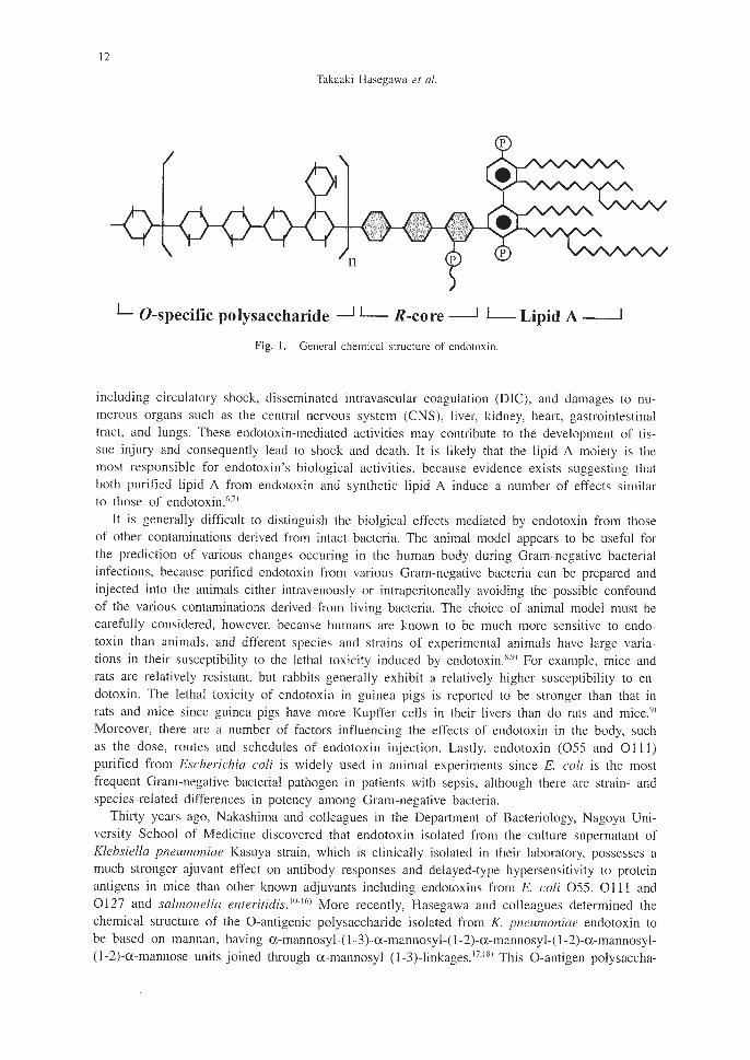
found to decrease the plasma protein binding potency of cefazolin, but did not change the volume of distribution for unbound cefazolin. The estimated protein binding parameters forcefazolin suggested that endotoxin-induced decreases in the protein binding potency may be dueto an altered conformation of the albumin molecule (Fig. 2). Cefazolin is localized in the extra-
80
Endotoxin 214.77 ± 6.26* 50.94 ± 3.43 0.41 ± 0.01*
Protein binding parameters of cefazolin
331.13 ± 22.48 54.61 ± 2.04 0.60 ± 0.05
•
nKd (11 M)nP (11 M)
o
Control
Treatment
o
=o.-.......~
.;: 40=Q)U
=ou
"t:l
§ 20o~
..-..-8-eI.l60::t
o 10 20Unbound concentration (J1. g/ml)
30
Fig. 2. Effect of endotoxin on the protein binding behavior of cefazolin highly bound to fresh plasma obtainedfrom control (0) and endotoxin-treated (.) rats. Solid lines represent computer-fitted curves.

15
ENDOTOXIN MODIFIES DRUG ELIMINATION
cellular water space and binds to plasma protein in the intravascular and interstitial fluids innondisposing organs. 39.40) We have also demonstrated that K. pneumoniae endotoxin does notchange the extracellular fluid volume, which is estimated as the volume of distribution of inulin.33) Based on these observations, it may be suggested that endotoxin decreases the bindingpotency of cefazolin to albumin in the interstitial fluid, as well as in plasma, and that the actual distribution volume is not changed. The systemic and renal clearances for unboundcefazolin and the glomerular filtration rate (GFR) were significantly decreased by endotoxin.Moreover, the clearance ratio of unbound cefazolin (renal clearance divided by glomerular filtration rate) dropped to 70% of that in untreated rats, and the net tubular secretion of cefazolinwas also reduced. Because cefazolin is one of the most widely used ~-Iactam antibiotics forpreoperative and postoperative prevention of Gram-negative bacterial infections, these findingsare especially important determining the appropriate dosage of cefazolin.
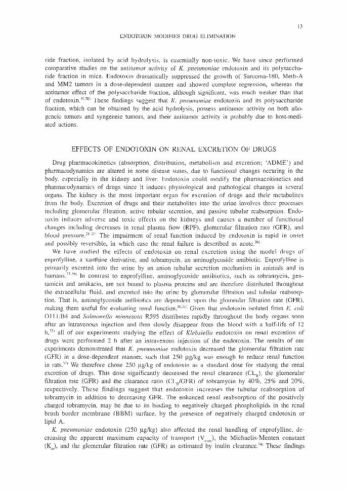
We further investigated the time-dependent changes in renal excretion of enprofylline inendotoxemic rats.41 ) Significant changes in the renal clearance for enprofylline were observed inrats pretreated 2 or 10 h earlier with endotoxin (250 Ilg/ml), but no such changes were observed in rats pretreated 24 h earlier with endotoxin (Fig. 5). It is interesting to note thatendotoxin-induced reductions in both the renal clearance of enprofylline and the glomerular filtration rate (GFR) occun'ed immediately (within 30 min) and proceeded gradually, whereas bothparameters returned to normal levels within 24 h. Moreover, endotoxin did not induce any histopathological changes in the kidney.34) Accordingly, intravenous administration of K.pneumoniae endotoxin at a dose of 250 Ilg/kg induces acute renal failure in a dose-dependentand time-dependent manner as a result of reduced GFR, and it dramatically reduces renal excretion of enprofylline by decreasing both glomerular filtration and tubular secretion (Figs. 3and 4). Such observations suggest that endotoxin-induced decreases in renal functions appear tobe transient events and, at least at the dose used in this experiment, endotoxin has no renal cellcytotoxicity.
It is well known that anionic drugs are actively taken into the tubular cells across thebasolateral membranes (BLM) and pass through the brush border membranes (BBM) into the
0.6
0.4
0.2
oo 50 250 500 Oh 2h 10 h 24 h
Doses of endotoxin (j1. g/kg) Time after endotoxin injection
Fig. 3. Dose- and time-dependent changes in renal function induced by endotoxin. Glomerular filtration rate(GFR) was estimated as inulin clearance.

16
Takaaki Hasegawa er al.
Organic anion drug
Glomemlar filtration
)Ir Tubular
1J 'ttret;o" i
Organic cation drug
Glomemlar filtration
Urine
NormalUrine
EndotoxemiaUrine
NormalUrine
Endotoxemia
Fig. 4. Effect of endotoxin on renal handling of organic anions and cations.
urine by facilitated diffusion. In order to study the possible effect of endotoxin on these processes, we performed renal uptake kinetic experiments in mice using enprofylline as a modeldrug. 42) We demonstrated that this drug appears to share a common transport system with endotoxin or lipid A in the tubular cells and that the endotoxin-induced reduction in tubular secretion is likely to be caused by competition for renal uptake into the tubular cells. We havealso studied the effect of K. pneumoniae endotoxin on renal excretion of organic cations usingas a model compound famotidine, which is secreted into the urine by an active tubular cationsecretory mechanism in the renal proximal tubules.43.44) Endotoxin increased the clearance ratioof famotidine (CL/GFR), but not the net tubular secretion, indicating that the endotoxin haslittle or no effect on the active tubular cation secretory system.4S) Based on these observations,we proposed that endotoxin influences the renal uptake system at the basolateral membrane(BLM) rather than excretion at the brush border membranes (BBM).
More recently, we have studied the contribution of lipid A, isolated from endotoxin by acidhydrolysis, to the endotoxin-induced reduction of renal excretion and the intrarenal accumulationof drugs, using the aminoglycoside antibiotic gentamicin as a model compound.46) No significantdiference was observed in the tubular reabsorption or intrarenal accumulation of gentamicin between endotoxin and lipid A. However, both endotoxin and lipid A induced, to the same degree, significant decreases in the GFR and systemic clearance of gentamicin, and significantincreases in plasma levels of urea nitrogen (BUN) and creatinine. These findings demonstratethat, similar to other effects of endotoxin, lipid A plays an important role in the endotoxin-induced reduction in the renal excretion of drugs.
It is generally accepted that endotoxin does not directly effect renal function, although theprecise mechanism of endotoxin-induced acute renal failure is not yet completely understood.The indirect effects of endotoxin may be mediated by various molecules released by endotoxin,such as certain cytokines, certain arachidonic metabolites, platelet activating factor (PAF).47-S01
For instance, experiments using isolated perfused rat kidney suggested that endotoxin has nodirect effect on renal functions, including GFR, Na+ reabsorption or K+ content.SII The indirectmechanism is most likely due to vasoconstriction, which may be mediated by arachidonic acidmetabolites, thromboxane, prostaglandins, leukotrienes, platelet activating factor (PAF) and

17
ENDOTOXIN MODIFIES DRUG ELIMINATION
endothelin.52l Tumor necrosis factor (TNF-cx) and its related cytokines may also be involved inthe indirect actions of endotoxin since intravenous injection of TNF-cx induces a state similar toendotoxin shock.53) On the contrary, reactive oxygen metabolites seem not to be important mediators of endotoxin-induced acute renal failure because neither the oxygen radical scavengersuperoxide dismutase (SOD) or catalase has a protective effect against endotoxin-induced renalfailure. 54) On the other hand, it has been reported that adenosine antagonists, such as theophylline, 8-phenyltheophylline and 8-cyclopentyl-I,3-dipropylxanthine, can protect against the nephrotoxicity induced by glycerol, indicating that adenosine acts as a modulator in the hemodynamicand pathophysiological changes in the kidney induced by glyceroI.55.56) Adenosine also appearsto be involved in the renal hemodynamic changes induced by renal ischemia in animals. 57)
Similarly, there is in vivo evidence that pretreatment with theophylline, at concentrations highenough to antagonize adenosine, can reverse the endotoxin-induced decrease in glomerular filtration (GFR), thus suggesting that adenosine may also play an important role in endotoxin-induced acute renal failure. 45) The role of adenosine in endotoxin-induced renal failure may thusbe similar to its role in glycerol-induced acute renal failure.
EFFECT OF ENDOTOXIN ON BILIARY EXCRETION OF DRUG
The liver consisting of hepatic parenchymal cells, vascular endothelial cells and Kupffer cells(hepatic macrophage), plays an important role in the specific elimination and detoxification ofendogenous (e.g. bilirubin) and exogenous (e.g. drug, xenobiotics) substances. For example, organic anions widely used for diagnosis of hepatic function such as indocyanine green (leG)and dibromosulfophthalein (DBSP), are eliminated from blood to bile by three processes including uptake into the sinusoidal membrane, intracellular transport, and excretion into the bilecanalicular membrane. The biliary excretion of such anionic drugs might be int1uenced by endotoxin.
Systemically administered endotoxin is rapidly taken into the liver and localized in bothhepatocytes and Kupffer cells. It induces morphological and functional hepatic changes in theearly stages, and may subquently cause severe damage to the liver. 58) The endotoxin-inducedfunctional alterations of the liver include decreases of hepatic blood t10w and protein synthesis,cholestasis, hyperbilirubinemia, and increases of acute phase proteins; the morphological changesinclude Kupffer cell swelling, formation of platelet thrombi, and accumulation of neutrophils(PMNs) in hepatic sinusoids. Endotoxin appears to localize and accumulate PMNs at sites ofGram-negative bacterial infections. PMNs play an important role in the defence of the hostagainst Gram-negative bacterial infections whereas they also contributes to the pathogenesis ofthe associated.58.59) PMN infiltration is observed in the early stages of morphologic changes inthe liver, suggesting that the accumulation of PMNs in the liver may contribute to endotoxininduced liver injury. Indeed, this theory is supported by the demonstration that pretreatmentwith the immunoglobulin fraction from rabbit serum immunized with rat PMNs (anti-PMN Ig)reduces E. coli endotoxin-induced increase in the number of PMNs in the liver and protectsagainst liver injury.60) These PMN-mediated changes generally occur within I h after endotoxininjection and precede degenerative effect in parenchymal cells. The actions endotoxin on hepaticparenchymal cells, on the other hand, which result in cholestasis and hyperbilirubinemia, appearto be direct and independent of PMNs. For example, endotoxin has been reported to causecholestatic jaundice with concomitant elevations in plasma concentrations of alanine aminotransferase (ALT) and aspartate aminotransferase (AST)52.60) in patients with Gram-negative bacterial
infections. There is evidence that endotoxin directly decreases bile secretion by inhibiting Na+,K+-ATPase on the bile canalicular membrane of hepatocytes.61 )

J8
Takaaki Hasegawa et 01.
We have investigated the characteristics of biliary and renal excretions of certain anionicdrugs in a spontaneous hyperbilirubinemic rat with conjugated hyperbilirubinemia (Eisaihyperbilirubinemic mutant rats; EHBRs).62.631 EHBRs exhibit abnormalities in the excretion ofglutathione disulfate, organic anions, bile glucuronide, and sulfate. These abnormalities appear tobe similar to those observed in human with Dubin-Johnson syndrome and in TR- and GY rats.We have demonstrated, in experiments using the ~-lactam antibiotic cefpiramide as a modelcompound, that EHBRs abnormally excrete this drug which usually is almost completely recovered in the urine and bile in the unchanged form (>90%). The biliary clearance of cefpiramidein EHBRs markedly decreased to less than 10% of that in normal rats, while total urinary recovery and renal clearance of cefpiramide increased. Hyperbilirubinemia appears to increase theurinary excretion of cefpiramide while reducing the biliary excretion.621 However, the effects ofhyperbilirubinemia on the protein binding of drug must also be taken into account: it is knownthat bilirubin binds strongly to plasma albumin and that only drug unbound by plasma albuminis capable of diffusing across various bilogical membranes to be distributed in the body andthis subject to metabolism and renal excretion. It is possible that bilirubin competes for thebinding of drugs which are normally highly bound by plasma proteins. To investigate this possibility, we investigated the effects of hyperbilirubinemia, using EHBRs, on the renal handlingof enprofylline which is normally highly bound by albuminYI The pharmacokinetics ofenprofylline in EHBRs were changed due to the altered protein binding behavior induced byhyperbilirubinemia, but the renal handling of this drug, including glomerular filtration and tubular secretion, was unchanged. Biliary excretion of the organic anion dyes indocyanine green(lCG) and dibromosulfophthalein (DBSP) are also remakedly decreased in EHBRs becausee ofthe reduced intracellular transport rate for rCG and the impaired transport rate of DBSP acrossthe canalicular membrane.641 These observations suggest that endotoxemia might also modify thepharmacokinetics of drugs that strongly bind to plasma albumin.
Bile comprises bile salt-dependent and bile salt-independent fractions. 651 Na+, K+-ATPase onthe bile canalicular membrane of hepatocytes contributes to the latter. Endotoxin reduces Na+,K+-ATPase activity on the canalicular membrane and thus decreases bile formation and flow byinhibiting the bile salt-independent fraction, but it does not change the active secretion of bileacids. 611 Because basal bile flow is predominantly driven by the secretion of anions rather thanby bile acids in rats.(6) The endotoxin-induced decrease in the bile acid-independent flow rate isattributable to decreased activity of the ATP-dependent canalicular multiple organic anion transporter (cMOAT). Although the precise mechanism responsible for endotoxin-induced liver injuryremains unknown, drugs primarily excreted into the bile may be influenced by endotoxin-induced impairments of hepatic function and liver elimination. For example, endotoxin decreasesthe biliary excretion of the organic anion dyes sulfobromophthalein (BSP) and ICG, which areprimarily excreted into the bile by an active transport system.(7
) Endotoxin is likely to impairthe excretion process of organic anions at the stage of transport from intracellular storage tobile via the canalicular membrane, rather than at the stage of transport from blood to hepatocytes via the sinusoidal membrane.
Endotoxin stimulates macrophages and Kupffer cells, all of which produce eicosanoids andcytokines including TNF-(X and interleukin-I (IL-I ).68) TNF-(X is likely to contribute to the endotoxin-induced decrease in sodium-dependent bile acid uptake via basolateral membranes sinceanti-TNF-(X prevents against the endotoxin-induced decrease in bile flow rate. 69
.70
) On the otherhand, there is also evidence that anti-TNF-(X can not prevent the endotoxin-induced reduction oftaurocholate uptake in mixed hepatocyte membranes. 71
) We recently found that granulocytecolony-stimulating factor (G-CSF), which suppresses TNF-(X production by endotoxin, can notprotect against the decrease in the bile flow rate induced by K. pneumoniae endotoxin.72J It is

19
ENDOTOXIN MODIFIES DRUG ELIMINATION
thus unlikely that TNF-a alone plays the major role in endotoxin-induced decreases in bile flowand in the biliary transport of organic anions, and the precise mechanism remains to be elucidated.
To establish guidelines for the use of antibiotics in patients with hepatobiliary infections byGram-negative bacteria, we investigated the effects of K. pneul1wniae endotoxin on the hepaticelimination of a ~-lactam antibiotic, cefoperazone. This drug is often used in the treatment ofGram-negative bacterial infections, and is actively secreted from blood to bile, via three processes: active uptake into the hepatocytes through a sinusoidal membrane, intracellular translocation, and excretion by a carrier-mediated transport system through the canalicular membraneinto the bile. K. pneumoniae endotoxin dramatically reduced both biliary excretion of the ~
lactam antibiotic and bile flow rate, due to changes occurring in the biliary secretory system. 721
From these observations, it appears that endotoxin may decrease the ATP-dependent transport oforganic anions, including cefoperazone, through the canalicular membrane as a result of decreased Na+, K+-ATPase activity. A recent report suggests that endotoxin decreases the activityof the ATP-dependent canalicular multiple organic transporter (cMOAT), which is a transporterfor cefoperazone and an important player in the generation of the bile salt-independent bileflow. 7J
) In our study, the amount of biliary excretion of cefoperazone in rats pretreated withendotoxin was significantly correlated with the mean bile flow rate, suggesting that cMOATactivity is an important determinant of both of these changes. 72
) We also found that pretreatmentwith an anti-inflammatory drug, dexamethazone, can protect against the endotoxin-induced decrease in biliary excretion of cefoperazone, suggesting that some inflammatory mediatorsreleased by endotoxin contibute to decreased hepatic functions (unpublished data). Endotoxin-induced decrease in the biliary excretion of organic anions may be caused by inhibiting the aniontransport system across the sinusoidal and/or bile canalicular membrane (Fig. 5). Further studiesare needed to calrify the precise mechanism.
Normal Endotoxemia
Fig. 5. Effect of endotoxin on biliary excretion of cefoperazone (CPZ), an organic anion.

20
Takaaki Hasegawa et al.
EFFECT OF ENDOTOXIN ON HEPATIC DRUG METABOLISM
Bacterial endotoxin plays a key role in decreased hepatic drug-metabolizing enzyme activityin animals and in humans during Gram-negative bacterial infections. Endotoxin reduces both thecontent and activity of hepatic cytochrome P450-mediated drug metabolizing enzymes, and thuscan delay the elimination of drugs, which are almost completely metabolized in the liver. Theactivity of hepatic drug-metabolizing enzymes in rats decreases 2 h after endotoxin administration and the activity of the hepatic type 0 form of xanthine oxidase in mice is decreased byIL-I, a cytokine released by endotoxin stimulation. 74) On the other hand, we previously foundthat 2-h pretreatment with endotoxin has little or no effect on the metabolism of theophyllinecatalyzed by the cytochrome P450 monooxygenase system in rats. 75 ) This discrepancy could bedue to a relatively small contribution of the liver to theophylline metabolism in rats or to atime-dependent effect of endotoxin on drug pharmacokinetic behavior which went undiscoveredafter the 2-h pretreatment. There is little information currently available on the time-dependenteffects of endotoxin on hepatic drug-metabolizing enzyme activity in animals and humans, andthe remainder of this review focusses on this measure in rats.
There is evidence that the turnover time of P450 in rats is 2 to 4 days76) and that endotoxindecreases the amount of mRNA for the hepatic cytochrome P450 isozymes 2C 11, 2E I, and3A2 from 6 to 48 h after its administration in rats. 77
) In recent study using antipyrine as amodel substrate in rats, we determined that K. pneumoniae endotoxin time-dependently reducesthe activity of hepatic cytochrome P450-mediated drug-metabolizing enzymes.78
) Antipyrine isalmost completely metabolized by the hepatic cytochrome P450 isozymes (CYPIA2, CYP2B6,CYP2C8, CYP2CI8 and CYP3A4).79) The systemic clearance of antipyrine represents the entirecapacity of its metabolism, because the protein binding potency of this drug in plasma is negligible and its elimination is independent on hepatic blood flow. We evaluated the time-dependenteffects of endotoxin on the activity of hepatic drug metabolizing enzymes, including anilinehydroxylase (also known as the P450 isozyme CYP2E), aminopyrine N-demethylase (CYP3A),benzphetamine N-demethylase (CYP2B) and p-nitoroanisole-O-demethylase (several isozymes),and on the content of hepatic cytochrome P450 and b5.
8o-S}) Both the systemic clearance of antipyrine and the activity of hepatic cytochrome P450-dependent drug-metabolizing enzymes weresignificantly decreased 24 h after a single intraperitoneal injection of endotoxin (1 mg/kg bodyweight), but these parameters returned to control levels by 96 h. 78) Reductions in antipyrineclearance and enzyme activity after repeated endotoxin treatments (once daily for 4 days) wereto the same degree as those seen 24 h after a single injection, indicating that repeated endotoxin treatments induce tolerance to endotoxin or to inflammatory stimulation. The systemicclearance of antipyrine correlated significantly both with the activity of all hepatic microsomalenzymes tested and with the content of cytochrome P450 and b5, suggesting that changes in thesystemic clearance of antipyrine reflect changes in the activity of hepatic P450-mediated drugmetabolizing enzymes. Only moderate hypertrophy of Kupffer cells was observed histopathologically in rats sacrificed 24 h after either the single and or repeated treatments, suggesting theabsence of severe liver-tissue damage. Based on these histopathological examinations, it is unlikely that the changes in the activity of hepatic cytochrome P450-mediated drug-metabolizingenzymes are caused directly by endotoxin- or cytokine-induced hepatic damage. Whereas it waspreviously thought that endotoxin affected hepatic cytochrome P450 by endotoxin is by increasing the degradation of cytochrome P450 haem proteins,84.85) it is now believed that endotoxininduced depression of hepatic P450-mediated drug metabolism is likely to be caused bysuppression of protein translation and mRNA transcription for cytochrome P450 isozymes, mediated by certain inflammatory cytokines.86-90)

21
ENDOTOXIN MODIFIES DRUG ELIMINATION
Cytokines, polypeptide mediators, are released from Kupffer cells in the liver, and by circulating monocytes and macrophages. TNF-a induces IL-I, and both TNF-a and IL-l induce IL6 production. It is well known that endotoxin increases release of certain inflammatorycytokines, such as IL-I, IL-2, IL-6, TNF-a and y-interferon (lFN-y), which contribute to thephysiological effects and lethal toxicity associated with endotoxemia. The circulating concentration of TNF-a peaks I to 2 h after injection of endotoxin and those of IL-I and IL-6 peak 2to 6 h after endotoxin injection. These cytokines appear to play an important role in endotoxininduced changes in the activity of hepatic drug-metabolizing enzymes. For example, the mainbiological activity of IL-6 is to regulate protein synthesis by hepatocytes. Both TNF-a and ILI increase an acute phase protein, ai-acid glycoprotein (AGP) in plasma, whereas this effect ofIL-6 is very weak. Similarly, TNF-a and IL-I dramatically suppress both the hepatic P450 contents and activity of ethoxycoumarin-O-deethylase (represented as P450 subfamilies CYP 1A and2B) and the activity of aniline hydroxylase (CYP2E), whereas these effects of IL-6 areweak.87.91-93) In addition, IL-I and IL-6 dramatically decrease ethylmorphine-N-demethylase activity (CYP3A) to the same extent as does endotoxin itself.94.95) There is an interesting report thatsystemic clearance of antipyrine correlates significantly with the peak concentrations of TNF-aor lL-6 after endotoxin injection in humans, suggesting the possibility that measurement of peakserum concentrations of TNF-a and lL-6 may yield predictions of the activity of hepatic P450mediated drug-metabolizing enzymes during Gram-negative bacterial infections.96
) Although ithas been demonstrated that G-CSF prevents endotoxin-induced TNF-a release, we recently reported that pretreatment with G-CSF can not block the suppressed hepatic cytochrome P450mediated drug-metabolizing activity induced by K. pneul110niae endotoxin, suggesting that thesynergistic action of other cytokines78) in addition to TNF-a must be involved in this effect.
Although the mechanism mediating suppression of hepatic drug-metabolizing enzyme activityin endotoxemia remains to be fully elucidated, one important factor may be a free radical gas,nitric oxide (NO). NO is synthesized from the enzyme, NO synthase (NOS), which enzymatically converts the amino acid, L-arginine, into L-citrulline and NO. NOS exists as severalisozymes including Ca2+-calmodulin activated, constitutive NOS in neurons (nNOS) and endothelial cells (eNOS), and endotoxin- and cytokine-inducible Ca2+-independent NOS (iNOS) in macrophages, Kupffer cells and PMNs. NO possesses the biological activity of endothelium-derivedrelaxing factor (EDRF) and has an inhibitory activity of platelet aggregation. It has been demonstrated that injection of endotoxin induces iNOS mRNA expression and up-regulated iNOSenzyme activity in several cell types, including macrophages, smooth muscle cells, endothelialcells, hepatocytes and Kupffer ceIls.52.97-103) In the liver, the major organ of NO production, en
dogenous NO appears to play multiple roles in the physiological control of hepatic functions,immune responses, host defenses against bacterial infection, and inflammatory disease. Thephysiological properties of NO allow it to react with various sustances, including heme, iron,superoxide, thiols and oxygen. In particular, NO appears to bind reversibly to the heme-ironmoiety of P450, forming an inactive P450-NO complex.I04.105) Heme oxygenase also plays an
important role in degradation of cytochrome P450 and liberation of heme, since it is inducedimmediately after endotoxin injection. There are also reports suggesting that NO decreases theactivity of various cytochrome P450 isozymes, including CYP2Bl, CYPlAl, and CYPlA2.I06.109)
Our laboratory has recently focused on the role of NO in the suppression of cytochromeP450-mediated drug-metabolizing enzymes activity induced by K. pneumoniae endotoxin in rats,as illustrated by following experiments. Plasma concentrations of nitrite plus nitrate (N0
2'/N03
; NOx) were found to increase, beginning 4 h after a single intraperitoneal injection of endotoxin (I mg/kg), to peak at approximately 400 11M after 12 h, and to returned to undetectablelevels 24 h after the injection of endotoxin, indicating that production of NO is enhanced for a

22
Takaaki Hasegawa er al.
prolonged period by endotoxin. We assessed the contribution of NO to endotoxin-induced suppression of hepatic drug-metabolizing enzyme activity using two tools: an exogenous source ofNO, (±)-(E)-ethyl-2-[(E)-hydroxyimino]-5-nitro-3-hexenamide (FK-409), and a selective iNOS inhibitor, S-methylisothiourea (SMT).IIO-112) There is a evidence that selective iNOS inhibitors, in
cluding SMT, do not increase endotoxin-induced liver injury, but nonselective iNOS inhibitor,,rvG-nitro-L-arginine methyl ester (L-NAME), increases." 3) In fact, SMT suppresses the manifestation of acute lung injury due to endotoxin-induced overproduction of NO, reduced endotoxinrelated tissue damages and lethal toxicity, and blocks endotoxin-induced vascular contractility. II~)
First, we examined in vivo whether SMT can suppress both the endotoxin-induced overproduction of NO and the decrease in the systemic clearance of antipyrine, which is representative ofhepatic cytochrome P450 activity. Indeed, a single intraperitoneal injection of SMT at a dose of5 mg/kg in rats, 2 h after endotoxin injection, completely suppressed the overproduction of NOand protected against the endotoxin-induced decrease in the systemic clearance of antipyrine,suggesting that the selective iNOS inhibitor suppresses the endotoxin-induced decrease in theactivity of cytochrome P450-mediated drug-metabolizing enzymes (Fig. 6). These results indicatethat NO is involved in the effect of endotoxin. Second, we studied in vivo whether the NOdonor FK-409 can suppress the activity of cytochrome P450-mediated drug-metabolizing enzymes. We adjusted the dosing schedule of the NO donor on the basis of the disappearancecurve of NOx after a single intraperitoneal injection of this drug because NO, a potent vasodilator, induces immediate hypotension. For example, a single intraperitoneal injection of FK-409in rats initially decreases the mean arterial blood pressure (MAP), which recovers to baselinelevels after 30 min. Repeated injections of FK-409 (10 mg/kg), administered in a pattern demonstrated to mimic the prolonged overproduction of NO following K. pneul110niae endotoxininjection, dramatically reduced the systemic clearance of antipyrine when the antipyrine was
100
~
I:'C.S:....I: 80I':----0 '0~
l-.....c.- I:I: 0eo: c.-l-.I': ~~
.,60c:; '-'
c.-'S~....'"~
rJ1
40
oControl Endotoxin Endotoxin SMT
+SMT
400
:?:::l.'-' 300I:
.S:....I':l-.....I:
2l 200I:oc.-i><oZI': 100e'"I':
E:
oBasal Endotoxin Endotoxin SMT
+SMT
Fig. 6. Protective effect of a selective iNOS inhibitor against the endotoxin-induced overproduction of NO anddecreased antipyrine clearance, the latter of which reflects hepatic drug-metabolizing enzyme activity.

23
ENDOTOXIN MODIFIES DRUG ELIMINATION
Antibiotics
Gram-negativebacteria NO
C§ dO~ ~r------t-----,N-[Fe2+]-NN- I -N
Complex S Kupffer cellsFormation <1ro~ei:V
Cytochrome P-450 activity .J.Fig. 7. Possible mechanism by which endotoxin decreases cytochrome P450-mediated drug-metabolizing enzyme
activity.
administered 24 h after endotoxin injection 10 h after final dose of FK-409. The decrease wasto the same degree as that seen after endotoxin injection, indicating that in vivo treatment withthe exogenous source of NO reduces the activity of hepatic drug-metabolizing enzymes. Thesefindings suggest that the overproduction of NO, caused by endotoxin itself or by some endotoxin-induced cytokines, mediates the suppression of cytochrome P450-mediated drug metabolizing enzyme activity induced by endotoxin. A possible mechanism for the endotoxin-induceddecrease in hepatic drug-metabolizing enzyme activity is shown in Fig. 7. Our data also suggestthat selective inhibition of iNOS may prevent a variety of pathophysiological changes associatedwith bacterial sepsis and septic shock.
CONCLUSION
This article reviews bacterial endotoxin induced changes in renal and biliary drug excretionmechanisms and in hepatic drug-metabolizing enzyme activity, although the precise mechanismsresponsible for these changes are unclear. Endotoxin may induce clinical complications due tochanges occuning in the pharmacokinetics (renal and biliary excretions and metabolism) of certain dugs, thus increasing the risk for developing serious adverse effects. With respect tohepatic drug-metabolizing enzyme activity during Gram-negative bacterial infections, endotoxininduced overproduction of NO plays a key role in reducing the activty of cytochromeP450-mediated drug-metabolizing enzymes. This report provides further characterization ofendotoxin-induced decreases in hepatic drug-metabolizing enzyme activity and should provideuseful information for designing drug regimens in patients with Gram-negative bacterial infections.

24
Takaaki Hasegawa et al.
ACKNOWLEDGMENTS
The authors are grateful to Dr. Nobuo Kato, President of Aichi Arts Center; Dr. ToshitakaNabeshima, Professor of Department of Hospital Pharmacy, Nagoya University School of Medicine; Dr. Masayuki Nadai, Associate Professor of Gifu Pharmaceutical University; and Dr. LiWang, Associate Professor of West China University School of Medical Sciences for their valuable support.
REFERENCES
I) Mayer, H., Bhat, U.R., Masoud, H., Radziejewska-Lebrecht, J., Widemann, C. and Kraus, J.H.: Bacterial lipopolysaccharides. Pure appl. Chem. 61, 1271-1282 (1989).
2) Rietschel, E.T., Brade, L., Holst, 0., Kulshin, VA., Linder, B., Morgan, A., Schade, U.F., Zahringer, U. andBrade, H.: Molecular structure of bacterial endotoxin in relation to bioactivity. In Cellular and Molecular Aspects of Endotoxin Reactions, Edited by Nowotony, A., Spitzer, 1.1. and Ziegler, E.J., pp. 15-32 (1990),Elsevier, Amsterdam.
3) Westphal, 0., Jann, K. and Himmelspach, K.: Chemistry and immunochemistry of bacterial polysaccharidesas cell wall antigens and endotoxins. Prog. Allergy 33, 9-39 (1983).
4) Anderson, B.M. and Solberg, 0.: The endotoxin-liberating effect of antibiotics on meningococci in vitro. ACIIJPathol. Microbial. Scwld. 88, 23 I-236 (1980).
5) Shenep, J.L. and Morgan, K.A.: Kinetics of endotoxin release during antibiotic therapy for experimentalGram-negative bacterial sepsis. J. Infect. Dis. 150,380-388 (1984).
6) Galanos, c., Luderitz, 0., Rietschel, E.T. and Westphal, 0.: Newer aspects of the chemistry and biology ofbacterial lipopolysaccharides, with special reference to their lipid A component. Int. Rev. Biochem. 14, 239335 (1977).
7) Nowotny, A.: Review of the molecular requirements of endotoxin actions. Rev. Infect. Dis. 9 (Suppl. 5),5503-55 I I (1987).
8) Kuida, H., Gilbart, R.P., Hinshaw, L.B., Brunson, J.G. and Visscher, M.B.: Species differences in effect ofGram-negative endotoxin on circulation. Am. 1. Physiol. 200, I 197-1202 (196 I).
9) McCuskey, R.S., McCuskey, R.A., Urbaschek, R. and Urbaschek, B.: Species differences in Kupffer cells andendotoxin sensitivity. Infect. Immun. 45, 278-280 (1984).
10) Nakashima, I., Kobayashi, T. and Kato, N.: Alterations in the antibody response to bovine serum albumin bycapsular polysaccahride of Klebsiella pneumoniae. J. Imlllunoi. 107, I I 12-1 121 (1971).
I I) Nakashima, I.: Adjuvant action of capsular polysaccharide of Klebsiella pneumoniae on antibody response. I.Intensity of its action. J. Imlllunology 108, 1009-1016 (1972).
12) Nakashima, I. and Kato, N.: Nonspecific stimulation of immunoglobulin synthesis in mice by capsularpolysaccharide of Klebsiella pneumoniae. Immunology 27, 179-193 (1974).
13) Nakashima, I., Nagase, F., Yokochi, T., Ohta, M. and Kato, N.: Adjuvant actions of polyclonal lymphocyteactivators. I. Comparison and characterization of their actions in antibody response to deaggregated bovineserum albumin. Cell. Immunol. 46, 69-76 (1979).
14) Nakashima, I., Nagase, F., Matsuura, A. and Kato, N.: Adjuvant actions of polycional lymphocyte activators.II. Comparison and characterization of their actions in initiation and potentiation of immune responses to Tdependent and T-independent soluble antigens. Cell. Immunol. 49, 360-371 (1980).
15) Ohta, M., Nakashima, I. and Kato, N.: Adjuvant action of bacterial lipopolysaccharide in induction of delayed-type hypersensitivity to protein antigens. I. Action of the 03 antigen of Klebsiella from culture fluid.Cell. Il11munol. 66, I I 1-120 (1982).
16) Ohta, M., Nakashima, I. and Kato, N.: Adjuvant action of bacterial lipopolysaccharide in induction of delayed-type hypersensitivity to protein antigens. II. Relationships of intensity of the action to that of otherimmunological activities. Immunology 163, 460-469 (1982).
17) Hasegawa, T., Ohta, M., Mori, M., Nakashima, I. and Kato, N.: The Klebsiella 03 lipopolysaccharide isolated from culture fluid: structure of the polysacccharide moiety. Microbiol. Iml11unol. 27, 683-694 (1983).
18) Hasegawa, T., Ohta, M., Nakashima, I., Kato, N., Morikawa, K., Hanada, T. and Okuyama, T.: Structure ofpolysaccharide moiety of the Klebsiella 03 lipopolysaccharide isolated from culture supernatant of decapsulated mutant (Klebsiella 03:KI'). Chem. Pharm. Bull. 33, 333-339 (1985).
19) Miyamoto, K., Koshiura, R., Hasegawa, T. and Kato, N.: Antitumor activity of Klebsiella 03 lipopolysaccha-

25
ENDOTOXIN MODIFIES DRUG ELIMINATION
ride in mice. Japall. 1. Pharlllcol. 36, 51-57 (1984).20) Hasegawa, T, Ohta, M., Kido, N., Kato. N., Miyamoto, K. and Koshiura, R.: Comparative studies on antitu
mor activity of Klehsiel/a 03 lipopolysaccharide and its polysaccharide fraction in mice. Japall. 1.Pha17l1acol. 38. 355-360 (1986).
2 I) Gilbert. R.P.: Mechanisms of the hemodynamic effects of bacterial endotoxin. Physiol. ReI'. 40. 245-279(1960).
22) McKay, D.G.• Margaretten. N. and Csavossy. I.: An electron microscope study of the etfects of bacterial endotoxin on the blood vascular system. Lab. Illvesi. 15. 1815-1829 (1966).
23) Cavanagh, D., Rao, P.S .• SUllon, D.M., Bhagat. B. and Bachmann. F: Pathophysiology of endotoxin shock inthe primate. Alii. 1. OIWel. GYllecol. 108.705-722 (1970).
24) Bradley, G.G.: Cellular and molecular mechanism of bacteria endotoxins. AIIII. Rev. Microbial. 33, 67-94(1979).
25) Churchill. P.e.. Bidani. A.K. and Schwartz. M.M.: Renal effects of endotoxin in the male rats. Am. .J.Physiol. 253. F244-F255 (1987).
26) Kikeri, D., Pennell. J.P., Hwang. K.H .• Jacob. A.I.. Richman. A.V. and Bourgoignie, J.J.: Endotoxemic acuterenal failure in awake rats. Alii. 1. Physiol. 250, F1098-1 106 (1986).
27) Nadai, M., Hasegawa. T, Muraoka, I., Takagi, K. and Nabeshima, T: Dose-dependent pharmacokinetics ofenprofylline and its renal handling in rats. 1. Pharm. Sci. 80. 648-652 (1991).
28) Apichartpichean, R., Hasegawa, T, Nadai, M., Kuzuya, T and Nabeshima, T.: Structure-pharmacokinetic relationships among the N'. N"-alkylxanthines in rats . .I. Pharm. PharnUlcol. 43, 262-269 (1991).
29) Tsunekawa. Y. Hasegawa. T. Nadai. M.. Takagi, K. Nabeshima. T.: Interspecies differences and scaling forthe pharmacokinetics of xanthine deivatives. 1. Pharm. Pharnwcol. 44. 594-599 (1992).
30) Pechere, J.e. and Dugal. R.: Clinical pharmacokinetics of aminoglycoside antibiotics. Cli//. Pharlllacokillel. 4,170-199 (1979).
31) Wenk, M., Vozeh, S., Follath, F.: Serum level monitoring of antibacterial drugs. A review. Clill.Phanllacokil1el. 9. 475-492 (1984).
32) Mathison. J.e. and Ulevitch. RJ.: The clearance. tissue distribution. and cellular localization of intravenouslyinjected lipopolysaccharide in rabbits. 1. Immlll1ol. 123. 2133-2143 (1979).
33) Nadai. M.. Hasegawa, T. KalO. K.• Wang. L.. Nabeshima. T and Kato. N.: Intluence of a bacterial lipopolysaccharide on the pharmacokinetics of tobramycin in rats. J. Pharlll. Pharmacol. 45. 971-974 (1993).
34) Nadai. M .. Hasegawa. T. Kato. K.• Wang. L., Nabeshima. T and Kato. N.: The disposition and renal handling of enprofylline in endotoxemic rats by bacterial lipopolysaccharide (LPS). Dl'IIg Mewb. Dispo.1'. 21.611-616 (1993).
35) Hasegawa. T. Apichartpichean. R.• Kuzuya. T, Nadai. M.• Tsunekawa. Y, Horiuchi, T and Miyamoto, K.:Protein binding characteristics of new bronchodilators. l-methyl-3-propylxanthine (MPX) and 3propyl xanthine (enprofylline). 1111. .I. Pharlll. 56. 235-241 (1989).
36) Hasegawa. T. Takagi. K.• Nadai. M. and Miyamoto. K.: Protein binding of xanthine derivatives to guinea pigserum albumin. ./. Pharm. Sci. 80. 349-352 (1991).
37) Brogard. J.M .• Comte. F and Pinget. M.: Pharmacokinetics of cepharosporin antibiotics. AlIlibiolic.Chemother. 25. 123-162 (1978).
38) Yamazaki, I.. Shirakawa, Y and Fugono, T: Comparison of the renal excretory mechanisms of cefmenoximeand other cepharosporines: renal clearance in rats and rabbits. J. AlIlibio(. 34. 1055-1063 (1981).
39) Tsuji, A.. Terasaki. T.. Imaeda. N. and Nakashima. E.: Etfect of extracellular water volume on the distributionkinetics of ~-Iactam antibiotics. J. Pharamcobio-DYI1. 8, 167-174 (1985).
40) Tsuji. A.• Nishide. K., Minami. H.. Nakashima. E.• Terasaki. T and Yamana. T: Physiologically based pharmacokinetic model for cefazolin in rabbits and its preliminary extrapolation to man. Drug Metab. Dispos. 13.729-739 (1985).
41) Nadai, M., Hasegawa. T. Wang. L.. Haghgoo. S.. Nabeshima. T and Kato, N.: Time-dependent changes inthe pharmaokinetics and renal excretion of xanthine derivative enprofylline induced by bacterial endotoxin inrats. BioI. Pharm. BII/I. 18. 1089-1093 (1995).
42) Nadai, M .• Hasegawa, T. Wang. L.. Haghgoo. S .. Okasaka. T.. Nabeshima. T and Kato. N.: Alterations inrenal uptake kinetics of the xanthine derivative enprofylline in endotoxaemic mice. 1. Pharm. Pharl1lacol. 48.744-748 (1996).
43) Rennick. B.R.: Renal tubule transport of'organic cations. Am. .I. Physiol. 240. F83-F89 (1981).44) Lin, J.H .. Los. L.E.• Ulm. E.H. and Duggan. D.E.: Urinary excretion kinetics of famotidine in rats. Drug
Mewb. Dispos. 15.212-216 (1987).45) Hasegawa, T. Nadai. M.• Wang, L.. Takayama. Y. Kato. K., Nabeshima. T and Kato. N.: Renal excretion of
famotidine and role of adenosine in renal failure induced by bacterial lipopolysaccharide in rats. Drllg Mewb.

26
Takaaki Hasegawa el al.
Di~pos. 22, 8-13 (1994).46) Hasegawa, T, Nadai, M., Wang, L., Haghgoo, S., Nabeshima, T and Kato, N.: Influence of endotoxin and
lipid A on the renal handling and accumulation of gentamicin in rats. Bioi. Pharm. Bu/l. 17, 1651-1655(1994).
47) Schlondorff, D., Satriano, J.A., Hagege, L Poret, J. and Baud, L.: Effect of platelet-activating factor and serum treated zymozan on prostaglandin E2 synthesis, arachidonic acid release and contraction of cultured ratmesangial cells. 1. Clill. Illvesl. 73, 1227-1231 (1984).
48) Badr, K.E, Kelly. YE., Reunke. H.G. and Brenner, B.M.: Roles for lhromboxane A, and leukolrienes in en-dotoxin-induced acute renal failure. Kidllev 1111. 30. 474-480 (1986). -
49) Wang, J. and Dunn, M.J.: Platelet-activating factor mediates endotoxin-induced acute renal insufficiency inrats. Am. J. Phvsiol. 253, F 1283-F1289 (1987).
50) Beasley, D., Dinarello, e.A. and Cannon, J.G.: Interleukin-I induces natriuresis in conscious rats: role of renal prostaglandins. Kidlley 1111. 33, 1059-1065 (1988).
51) Cohen, U., Black, A.J. and Wertheim, S.J.: Direct effects of endotoxin on the function of the isolated perfused rat kidney. Kidlley 1111. 37, 1219-1226 (1990).
52) Hewett, LA. and Roth R.A.: Hepatic and extrahepatic pathobiology of bacterial lipopolysaccharides.Pharmacol. Rev. 45, 382-411 (1993).
53) Beutler, B., Milsark, I.W. and Cerami, A.: Passive immunization against cachectin/tumor necrosis hlctor protects mice from lethal effect of endotoxin. Sciellce 229, 869-871 (1985).
54) Walker, P.D. and Shah, S.Y: Reactive oxygen metabolites in endotoxin-induced acute renal failure in rats.Kidlley 1111. 38, 1125-1132 (1990).
55) Bowmer, C.J., Collis, M.G. and Yates, M.S.: Effect of the adenosine antagonist 8-phenyltheophylline on glycerol-induced acute renal failure in the rat. 81: J. Pharmacol. 88, 205-212 (1986).
56) Kellett, R., Bowmer, C.J., Collis, M.G. and Yates, M.S.: Amelioration of glycerol-induced acute renal failurein the rat with 8-cyclopentyl-1 ,3-dipropylxanthine. 81: 1. Pharmacol. 98, 1066-1074 (1989).
57) Spielman, WS. and Thompson, e.1.: A proposed role for adenosine in the regulation of renal hemodynamicsand renin release. Am. 1. Physiol. 242, F423-F435 (1982).
58) Utili, R., Abernathy, C.O. and Zimmerman, H.J.: Minireview. Endotoxin effects on the liver. L!fe Sci. 20,553-568 (1977).
59) Levy, E., Path, Ee. and Ruebner, B.H.: Hepatic changes produced by a single dose of endotoxin in themouse. Light microscopy and histochemistry. Am. J. ParllOl. 51, 269-285 (1967).
60) Hewett, J.A., Schultz, A.E., VanCise, S. and Roth, R.A,: Neutrophil depletion protects against liver injuryfrom bacterial endotoxin. Lab. Illvesl. 55, 347-361 (1992).
61) Utili, R., Abernathy, e.0. and Zimmerman, H.J.: Inhibition of Na+, K+-adenosine triphosphatase by endotoxin: a possible mechanism for endotoxin-induced cholestasis. J. In/ecl. Dis. 136, 583-587 (1977).
62) Muraoka. I., Hasegawa, T, Nadai, M., Wang, L., Haghgoo, S., Tagaya, O. and Nabeshima, T: Biliary andrenal excretions of cefpiramide in Eisai hyperbilirubinemic rats. Alllimicrob. Agellls Chemolher. 39, 70-74(1995).
63) Nadai, M., Hasegawa, T, Wang, L., Tagaya, O. and Nabeshima, T.: Alterations in the pharmacokinetics andprotein binding of enprofylline in Eisai hyperbilirubinemic rats. Drug Melab. Dispos. 22, 561-565 (1994).
64) Sathirakul, K., Suzuki, H., Yasuda, K., Hanano, M., Tagaya, 0., Horie, T and Sugiyama, Y: Kinetic analysisof hepatobiliary transport of organic anions in Eisai hyperbilirubinemic mutant rats. J. Pharmacol. Exp. Ther.265, 1301-1312 (1993).
65) Erlinger, S. and Dhumeaux, D.: Mechanisms and control of secretion of bile water and electrolytes. Gastroemerology 66, 281-304 (1974).
66) Ballatori, N. and Truong, AT.: Glutathion as a primary osmotic driving force in hepatic bile formation. Am.1. Physiol. 263, G617-G624 (1992).
67) Utili, R., Abernathy, e.0. and Zimmerman, H.J.: Cholestatic effects of Escherichia coli endotoxin on the isolated perfused rat liver. Gaslroelllerology 70, 248-253 (1976).
68) Raetz, e.R.H., Ulevilch, R., Wright, S.D., Sibley, e.H., Ding, A. and Nathan, e.E: Gram-negative endotoxin:an extraordinary lipid wih profound effects on eukaryotic signal transduction. FASEB 1. 5, 2652-2660 (1991).
69) Whiting, J.E, Green, R.M., Rosenbluth, A.B. and Gollan, J.L.: Tumor necrosis factor-alpha decreases hepatocyte bile salt uptake and mediates endotoxin-induced cholestasis. Hep(l/ology 22, 1273-1278 (1995).
70) Green, R.M., Beier, D. and Gollan, J.L.: Regulation of hepatocyte bile salt transporters by endotoxin and inflammatory cytokines in rodents. Gaslroel7lerology Ill, 193-198 (1996).
71) Moseley, R.H., Jones, L., Wang, W. and Takeda, H.: FUl1her insights into the role of cytokines in sepsis-associated cholestasis. Heparology 22, A317 (Abstract) (1995).
72) Nadai, M., Matsuda, I., Wang, L., Itoh, A., Naruhashi, K., Nabeshima, T, Asai, M. and Hasegawa, T: Granu-
27
ENDOTOXIN MODIFIES DRUG ELIMINATION
locyte colony-stimulating factor enhances endotoxin-induced decrease in biliary excretion of the antibioticcefoperazone in rats. Anlimicrob. Agel11s Chemolher. 42, 2178-2183 (1998).
73) Bolder, U., Ton-Nu, H.-T, Schteingart, CO., Frick, E. and Hofmann, A.F.: Hepatocyte transport of bile acidsand organic anions in endotoxemic rats: impaired uptake and secretion. Gaslroel11erology 112, 214-225(1997).
74) Golodischer, R., Krasner, J., McDevitt, U., Nolan, J.P and Yaffe, SJ.: Hepatic microsomal drug metabolismafter administration of endotoxin in rats. 13iochem. Pharmacol. 25, 351-353 (1976).
75) Wang, L., Hasegawa, T, Nadai, M., Muraoka, I., Nabeshima, T and Kato, N.: The effect of lipopolysaccharide on the disposition of xanthines in rats. J. Phann. Pharamcol. 45, 34-38 (1993).
76) Arias, I.M. and DeLeon, A.: Estimation of the turnover rate of barbiturate side-chain oxidation enzyme in ratliver. Mol. Pharmacol. 3, 216-218 (1967).
77) Sewer, M.B., Koop, O.R. and Morgan, E.T: Endotoxemia in rats is associated with induction of the P4504Asubfamily and suppression of several other forms of cytochrome P450. Drug Mewb. Dispos. 24, 401-407( 1996).
78) Nadai, M., Sekido, T, Matsuda, I., Wang, L., Kitaichi, K., Itoh, A., Nabeshima, T and Hasegawa, T: Timedependent efTects of Klebsiella pneumoniae endotoxin on hepatic drug-metabolizing enzyme activity in rats.J. Pharm. Plwrmacol. 50, 871-879 (1998).
79) Engel, G., Hofmann, U., Heidemann, H., Cosme, J. and Eichelbaum, M.: Antipyrine as a probe or humanoxidative drug metabolism: Identification of the cytochrome P450 enzymes catalyzing 4-hydroxyantipyrine, 3hydroxymethylantipyrine, and norantipyrine formation. Clin. Pharmacol. Ther. 59, 613-623 (1996).
80) Bauer, C, Corsi, C and Paolini, M.: Stability of microsomal monooxygenases in murine liver S9 fractionsderived from phenobarbital and beta-naphthof!avone induced animals under various long-term conditions ofstorage. Carcinog. Muwgen. 14, 13-22 (1994).
81) Ahmed, S.S., Napoli, K.L. and Strobel, H. W.: Oxygen radical formation during cytochrome P450-catalyzedcyclosporine metabolism in rat and human liver microsomes at varying hydrogen ion concentrations. Mol.Cell. 13iochem. 151, 131-140 (1995).
82) Monostory, K. and Vereczkey, L.: Interaction of theophylline and ipriflavone at the cytochrome P450 level.£lil: J. Drug Mewb. Pharmacokinel. 20, 43-47 (1995).
83) Anderson, K.E., Hammons, GJ .. Kadlubar, FF., Potter, J.D., Kaderlik, K.R., lIett, K.F., Minchin, R.F., Teitel,CH., Chou, H.C, Martin, M.Y., Guengerich, F.P., Barone, G.W., Lang, N.P. and Peterson, L.A.: Metabolicactivation of aromatic amines by human pancreas. Carcinogenesis 18, 1085-1092 (1997).
84) Bissell, D.M. and Hammaker, L.E.: Cytochrome P-450 heme and the regulation of hepatic heme oxygenaseactivity. Arch. 13iochem. Biophys. 176,91-102 (1976).
85) Bissell, D.M. and Hammaker, L.E.: Cytochrome P-450 heme and the regulation of aminolevulinic acid synthetase in the liver. Arch. Biochem. 13iophys. 176, 103-112 (1976).
86) Stanley, L.A., Adams, OJ., Lindsay, R., Meehan, R.R., Liao, W. and Wolf, CR.: Potentiation and suppressionof mouse liver cytochrome P-450 isozymes during the acute-phase response induced by bacterial endotoxin.£111: J. Biochem. 174,31-36 (1988).
87) Bertini, R., Bianchi, M., Erroi, A., Villa, P. and Ghezzi, P: Dexamethasone modulation of in vivo effects ofendotoxin, tumor necrosis factor, and interleukin-I on liver cytochrome P-450, plasma t1brinogen, and serumiron. J. Leukocyle Bioi. 46, 254-262 (1989).
88) Morgan, E.T.: Suppression of constitutive cytochrome P-450 gene expression in livers of rats undergoing anacute phase response to endotoxin. Mol. Pharmacol. 36, 699-707 (1989).
89) Bertini, R., Gervasi, P.G., Longo, Y. and Ghezzi, P.: Depression of hepatic drug metabolism in endotoxintreated and sarcoma-bearing mice. Res. Commun. Chon. P(l/hol. Pharmacol. 76, 223-231 (1992).
90) Cantoni, L., Carelli, M., Ghezzi, P., Delgado, R., Faggioni, R. and Rizzardini, M.: Mechanisms ofinterleukin-2-induced depression of hepatic cytochrome P-450 in mice. £111: J. Pharmacol. 292, 257-263( 1995).
91) Ghezzi, P., Saccardo, B.. Villa, P., Rossi, Y., Bianchi, M. and Dinarello, CA.: Role of interleukin-I in thedepression of liver drug metabolism by endotoxin. Infecl. Immun. 54, 837-840 (1986).
92) Ghezzi, P, Saccardo, B. and Bianchi, M.: Recombinant tumor necrosis factor depresses cytochrome P-450dependent microsomal drug metabolism in mice. 13iochem. Biophys. Res. Commun. 136, 316-321 (1986).
93) Bertini, R., Bianchi, M., Villa, P. and Ghezzi, P.: Depression of liver drug metabolism and increase in plasmajlbrinogen by interleukin I and tumor necrosis factor: a comparision with Iymphotoxin and interferon. 1111. J.Immunopharmacol. 10, 525-530 (1988)..
94) Chen, YL., Florentin, I., Batt, A.M., Ferrari, L., Giround, J.P. and Chauvelot-Moachon, L.: Effects ofinterleukin-6 on cytochrome P450-dependent mixed-function oxidases in the rat. Biochem. Pharmacol. 44,137-148 (1992).

28
Takaaki Hasegawa et al.
95) Morgan, E.T, Thomas, K.B., Swanson, R., Vales, T, Hwang, .I. and Wright, K.: Selective suppression of cytochrome P450 gene expression by interleukin I nd 6 in rat liver. Biochim. Biophys. Acta 1219,475-483( 1994).
96) Sehedlofsky, S.L., Israel, B.C., McClain, C..I., Hill, D.B. and Blouin, R.A.: Endotoxin administration to humans inhibits hepatic cytochrome P450-mediated drug metbolism. 1. Clin. Invest. 94, 2209-2214 (1994).
97) Kilbourn, R.G. and Grifllth, O.W.: Overproductions of nitric oxide in cytokine-mediated and septic shock. .I.Natl. Cancer 1n.>I. 84, 827-877 (1992).
98) Stuehr, 0..1. and Grifllth, O.W.: Mammalian nitric oxide synthases. Adv. Enzymol. Refm. Areas Mol. Bioi. 65,287-346 (1994).
99) Kolls, .I., Xie, .I., LeBlanc, R., Malinski, T, Nelson, S., Summer, W. and Greenberg, S.S.: Rapid induction ofmessenger RNA for nitric oxide synthase II in rat neutrophils in vivo by endotoxin and its suppression byprednisolone. PSEBM 205, 220-225 (1994).
1(0) Bredt, D.S. and Snyder, S.H.: Nitric oxide: a physiologic messenger molecule. Ann. Rei: Biochem. 63, 175195 (1994).
1(1) Morris,.Ir. S.M. and Billiar, TR.: New insights into the regulation of inducible nitric oxide synthesis. Am. .I.Physiol. 266, E829-E839 (1994).
1(2) Ruetten, H. and Thiemermann, C.: Prevention of the expression of inducible nitric oxide synthase byaminoguanidine or aminoethylisothiourea in macrophages and in the rat. Biochem. Biophys. Res. Commun.225, 525-530 (1996).
103) Sewer, M.B., Barclay, TB. and Morgan, E.T.: Down-regulation of cytochrome P450 mRNAs and proteins inmice lacking a functional NOS2 gene. Mol. Pharmacol. 54, 273-279 (1998).
1(4) Hurshman, A.R. and Marietta, M.A.: Nitric oxide complexes of inducible nitric oxide synthase: spectral characterization and effect on catalytic activity. Biochemistry 34, 5627-5634 (1995).
1(5) Minamiyama, Y, Takemura, S., Imaoka, S., Funae, Y, Tanimoto, Y and Inoue, M.: Irreversible inhibition ofcytochrome P450 by nitric oxide. 1. Pharmacol. E,p. Ther. 283, 1479-1485 (1997).
1(6) Stadler, .I., Trockfeld, .I., Schmalix, W.A., Brill, T, Siewert, .I.R., Greim, H. and Doehmer, .I.: Inhibition ofcytochrome P450lA by nitric oxide. Proc. Nat!' Acad. Sci. U.S.A. 91, 3559-3563 (1994).
1(7) Khatsenko, O.G. and Kikkawa, Y: Nitric oxide differentially affects constitutive cytochrome P450 isoforms inrat liver..I. Pharmacol. Exp. Ther. 280, 1463-1470 (1997).
1(8) Donato, M.T., Guillen, M.l., .lover, R., Castell, .IV and Gomez-Lechon, M..I.: Nitric oxide-mediated inhibition of cytochrome P450 by interferon-g in human hepatocytes. 1. Pharmacol. Exp. Ther. 281, 484-490(1997).
1(9) Khatsenko, O.G, Boobis, A.R. and Gross, S.S.: Evidence for nitric oxide participation in down-regulation ofCYP2B 1/2 gene expression at the pretranslational level. Toxicol. Lell. 90, 207-216 (1997).
110) Kita, Y, Fukuyama, S. and Hirasawa, Y: Close correlation between nitric oxide (NO) formation from NOreleasers and the biological activities of these agents in rats. Japal1. J. Pharmacol. 69, 69-74 (1995).
I I I) Fukuyama, S., Azuma, T, Hirasawa, Y, Morokoshi, N., Akama, T, Koda, S. and Kita, Y: Nitric oxide (NO)releasing pathway of FK409 in the presence of sultl1ydryl-bearing compounds. Pharm. Res. 13, 1238-1242(1996).
112) Kita, Y, Hirasawa, Y, Fukuyama, S., Ohkubo, K., Kato, Y, Takamatsu, H., Ohno, M., Nishino, S., Kato, M.and Seki, .I.: Oral biological activities of spontaneous nitric oxide releasers are accounted for by their nitricoxide-releasing rates and oral absorption manners. 1. Pharmacol. Exp. Ther. 276, 421-425 (1996).
113) Vos, TA., Gouw, A.S., Klok, P.A., Havinga, R., van Goor, H., Huitema, S., Roelofsen, H., Kuipers, F.,Jansen, PL. and Moshage, H.: Differential effects of nitric oxide synthase inhibitors on endotoxin-inducedliver damage in rats. Gastroe11lerology 113, 1405-1407 (1997).
114) Numata, M., Suzuki, S., Miyazawa, N., Miyashita, A., Nagashima, Y, Inoue, S.. Kaneko, T and Okubo, T:Inhibition of inducible nitric oxide synthase prevents LPS-induced acute lung injury in dogs. J. 1m111111101.160,3031-3037 (1998).