Embed Size (px)
Citation preview

Hindawi Publishing CorporationEvidence-Based Complementary and Alternative MedicineVolume 2011, Article ID 571721, 8 pagesdoi:10.1155/2011/571721
Research Article
Investigation of Antidiabetic, Antihyperlipidemic, and In VivoAntioxidant Properties of Sphaeranthus indicus Linn. in Type 1Diabetic Rats: An Identification of Possible Biomarkers
S. Ramachandran,1 K. Asokkumar,2, 3 M. Uma Maheswari,2, 3 T. K. Ravi,2, 3
A. T. Sivashanmugam,2, 3 S. Saravanan,4 A. Rajasekaran,5 and J. Dharman6
1 Department of Pharmacology, KMCH College of Pharmacy, Tamil Nadu, Coimbatore 641 048, India2 Department of Pharmacology, College of Pharmacy, SRIPMS, Tamil Nadu, Coimbatore 641 044, India3 Department of Pharmaceutical Analysis, College of Pharmacy, SRIPMS, Tamil Nadu, Coimbatore 641 044, India4 Department of Pharmacology, PSG College of Pharmacy, Tamil Nadu, Coimbatore 641 004, India5 Department of Pharmaceutical Chemistry, KMCH College of Pharmacy, Tamil Nadu, Coimbatore 641 048, India6 Department of Pharmaceutical Analysis, KMCH College of Pharmacy, Tamil Nadu, Coimbatore 641 048, India
Correspondence should be addressed to S. Ramachandran, [email protected]
Received 1 May 2010; Accepted 31 August 2010
Copyright © 2011 S. Ramachandran et al. This is an open access article distributed under the Creative Commons AttributionLicense, which permits unrestricted use, distribution, and reproduction in any medium, provided the original work is properlycited.
The present investigation was aimed to study the antidiabetic, antihyperlipidemic, and in vivo antioxidant properties of the rootof Sphaeranthus indicus Linn. in streptozotocin- (STZ-) induced type 1 diabetic rats. Administration of ethanolic extract ofSphaeranthus indicus root (EESIR) 100 and 200 mg/kg to the STZ-induced diabetic rats showed significant (P < .01) reductionin blood glucose and increase in body weight compared to diabetic control rats. Both the doses of EESIR-treated diabetic ratsshowed significant (P < .01) alteration in elevated lipid profile levels than diabetic control rats. The EESIR treatment in diabeticrats produced significant increase in superoxide dismutase (SOD), catalase (CAT), glutathione peroxidase (GPx) and decrease inthiobarbituric acid reactive substances (TBARS) levels than diabetic control rats. Administration of EESIR 200 mg/kg producedsignificant (P < .01) higher antioxidant activity than EESIR 100 mg/kg. The high performance liquid chromatography (HPLC)analysis of EESIR revealed the presence of biomarkers gallic acid and quercetin. In conclusion, EESIR possess antidiabetic,antihyperlipidemic, and in vivo antioxidant activity in type 1 diabetic rats. Its antioxidant and lipid lowering effect will help toprevent diabetic complications, and these actions are possibly due to presence of above biomarkers.
1. Introduction
Diabetes mellitus (DM) is a group of metabolic diseasescharacterized by hyperglycemia resulting from the defectsin insulin secretion, insulin action, or both. A chronichyperglycemic condition in diabetes is associated with long-term damage, dysfunction, and failure of various organs,such as eyes, kidneys, nerves, heart, and blood vessels. Itis the most common serious metabolic disorder and isconsidered to be one of the five leading causes of death inthe world [1]. DM is classified into two major categories:type 1 and type 2 diabetes. Although both types of diabeteshave distinct pathogenesis, hyperglycemia, and various life-threatening complications are common to both [2]. The
plasma lipids are usually raised in diabetes and such anelevation represents a risk factor for coronary heart disease[3]. Free radicals have significant role in the causation ofseveral diseases like diabetes, parkinson’s disease, cirrhohsis,cancer, and cardiovascular disorders [4]. In DM, oxidativedamage and tissue injury may lead to alterations in theendogenous free radical scavenging defense mechanisms andineffective scavenging of reactive oxygen species [5]. Thecurrent treatment for control of DM includes diet, exercise,oral antidiabetic drugs, and insulin therapy. However, insulinand other oral hypoglycemic drugs have characteristic profileof adverse effects. This has initiated the identification ofnovel drugs which might act in mechanistically distinctway compared to existing drug targets [6]. Hence, research

2 Evidence-Based Complementary and Alternative Medicine
is focused on medicinal plants which are used in thetraditional practices and development of newer drug leadsfrom phytoconstituents with more potential and effectiveagents with lesser side effects than the existing hypoglycemicagents [1]. Traditionally, many medicinal plants are currentlyused in India for the treatment of diabetes and its efficacy hasbeen proved scientifically [7].
Sphaeranthus indicus Linn. (Family-Compositae) is abranched herb with purple flowers that grows abundantly inrice field and distributed throughout India. It is used indige-nously in the Indian system of medicine as an anthelmintic[8]. The plant has a wide range of medicinal value andhas been used in hemicranias, jaundice, leprosy, diabetes,fever, pectoralgia, cough, gastropathy, hernia, hemorrhoids,helminthiasis, dyspepsia, skin diseases and nerve tonic [9,10]. Pharmacological activities such as immunomodulatory[11], antimicrobial [12, 13], antibacterial [14, 15], anxiolytic[10], wound healing action [16] were reported on this plant.Phytoconstituents isolated from this plant are eudesman-olides [17], isoflavonoids [18], 7-hydroxy eudesmanolides[19], sterol glycoside [20], essential oil (cadiene, ocimene,citral, p-methoxycinnamaldehyde, geraniol, eugenol andgeranyl acetate) [21], and eudesmanolides [22]. In India, thetribals of Madhya Pradesh used this plant for the treatmentof diabetes [23]. Globally, type 1 DM affects considerablepercentage of population and it leads to morbidity andmortality of the diabetic patients. Antioxidants play a majorrole in the prevention of diabetes and its complications byscavenging free radicals. Survey of current literature revealedthat there is no scientific data documented for the effect ofSphaeranthus indicus root in the treatment of type 1 diabetesmellitus and its in vivo antioxidant action. Therefore, thepresent study was undertaken to investigate the antidiabetic,antihyperlipidemic and in vivo antioxidant potential ofEESIR in type 1 diabetic rats as well as identification of thepossible biomarkers in EESIR.
2. Methods
2.1. Plant Material. Sphaeranthus indicus Linn. was col-lected from Erode, Tamil Nadu, India and authenticated byDr.P.Venu, Joint Director of Botanical Survey of India, TamilNadu Agriculture University (TNAU), Coimbatore. Theauthentication certificate bearing number BSI/SC/5/21/04-05/Tech-1850 is documented in our laboratory. The rootswere separated, cleaned, air dried under shade and powderedusing mechanical grinder and stored in air-tight container.
2.2. Preparation of EESIR. Dried powdered plant rootswere (85 g) extracted with 70% v/v ethanol using Soxhletapparatus for 72 hours. The extract was concentrated underreduced pressure in rota evaporator and yield of the extractwas found to be 15.24 g. The EESIR was stored at 10◦C in therefrigerator until the completion of pharmacological studies.
2.3. Drugs and Chemicals. Acetonitrile: n-hexane sulphonicacid were of HPLC grade (Himedia, Mumbai). Gallic acidand quercetin reference standards were purchased from
Sigma Aldrich (USA). Streptozotocin was procured fromSisco Research Laboratories Private Ltd., Mumbai, India. Allother chemicals and reagents were of analytical grade, andenzymatic kits used in this study were obtained commer-cially.
2.4. Preparation of Standard and Sample Solutions for HPLC.Stock solutions of gallic acid (GA) and quercetin (QA) wereprepared at concentration of 1000 μg/ml and 2000 μg/mlimmediately before use and used as reference standard.The EESIR was dissolved in mobile phase, to obtain aconcentration of 4 mg/ml and used as sample solution. Allsample solutions were filtered through 0.45 μm membranefilter (Millipore) and injected directly.
2.5. Chromatographic Conditions for HPLC. Chromato-graphic analysis was carried out by Phenomenex-Luna C18reversed-phase column (ø 250 mm × 4.6 mm) packed with5 μm diameter particles; the mobile phase was acetonitrile:n-hexane sulphonic acid (20 : 80 v/v) pH 2.5 adjusted withglacial acetic acid. This mobile phase was filtered througha 0.45 μm membrane filter (Millipore), then deaeratedultrasonically prior to use. The GA and QA were quantifiedby photodiode array detector following HPLC separation at299 nm. The mobile phase flow rate and injection volumewere 1.2 ml/min and 20 μL, respectively. The chromato-graphic peaks of the analytes were confirmed by comparingtheir retention time (Rt) and UV spectra with those ofthe reference standards. The GA (2.5 to 12.5 μg/ml) andQA (10 to 80 μg/ml) standard solutions were injected intothe HPLC and peak area responses obtained. Standardgraphs were prepared by plotting concentration versus area.Quantification was carried out from integrated peak areasof the samples using the corresponding standard graph. Allchromatographic operations were carried out at ambienttemperature.
2.6. Experimental Animals. Wistar albino rats of both sexesweighing 150−200 g were used for the study. All animalswere maintained under standard laboratory conditions [tem-perature (22 ± 2◦C) and humidity (45 ± 5◦C)] with12 hours day: 12 hours night cycle. The animals were fedwith normal laboratory diet and allowed to drink waterad libitum. All the experimental procedures conducted afterthe approval of ethical committee (817/04/ac/CPCSEA) andwere in strict accordance with institutional animal ethicalcommittee guidelines for the care and use of laboratoryanimals.
2.7. Acute Toxicity. Acute oral toxicity study was performedas per Organization for Economic Cooperation and Devel-opment (OECD) guidelines 423 [24]. After the oral adminis-tration of EESIR, animals were observed individually at leastonce during the first 30 minutes and periodically during thefirst 24 hours, with special attention given during the first 4hours, and daily thereafter, for a total of 14 days.
Evidence-Based Complementary and Alternative Medicine 3
2.8. Induction of Diabetes. Diabetes was induced in overnightfasted rats by intraperitoneal injection of STZ at a dose of60 mg/kg body weight [25] in 0.1 M cold citrate buffer (pH4.5). STZ can induce fatal hypoglycemia as a result of massivepancreatic insulin release and to avoid this hypoglycemiceffect, the rats were provided with 5% dextrose solution after6 hours of STZ administration for next 24 hours. Inductionof diabetes was verified after 72 hours and the animals wereallowed 14 days for the stabilization of blood glucose level.At 14th day, animals having a blood glucose level higherthan 210 mg/dl were considered diabetic and used for theexperiments.
2.9. Experimental Design for Antidiabetic Activity. The ratswere divided into five groups of six each randomly: group1 normal untreated rats which received 0.2% carboxy methylcellulose (CMC) served as normal control, group 2 diabeticrats received 0.2% CMC served as diabetic control, group 3diabetic rats received EESIR (100 mg/kg), group 4 diabeticrats received EESIR (200 mg/kg), and group 5 diabetic ratsreceived glibenclamide (5 mg/kg). The EESIR and gliben-clamide was suspended in 0.2% CMC and administeredorally for 28 days once daily to the respective groups. On the28th day, the animals were fed with respective dose of drugand, the body weights of all the animals were determinedand blood samples were collected after 1 hour post drugdose. The serum was separated by centrifugation of bloodat 5000 rpm for 10 minutes and the biochemical parameterswere analyzed.
2.10. Estimation of Glucose and Lipid Profile Levels. Theblood glucose level was determined by glucose oxidase-peroxidase method using glucose enzymatic kit (Agappediagnostic, Kerala, India) and expressed in milligrams perdeciliter (mg/dl). The lipid profiles such as total cholesterol(TC), triglycerides (TG), high density lipoprotein (HDL)were determined using enzymatic kits (Agappe diagnostic,Kerala, India) and low density lipoprotein (LDL), verylow density lipoprotein (VLDL) values were calculated byFriedewalds formula as given below [26]
VLDL = TG/5, (1)
LDL = TC− (HDL + VLDL). (2)
2.11. Determination of Lipid Peroxidation Indices and Antioxi-dant Levels. Liver was dissected out and washed immediatelywith ice cold saline to remove blood. Tissue homogenate(10% w/v) was prepared with 0.025 M Tris−HCl buffer (pH7.5) and was used for the assay of lipid peroxidation [27],superoxide dismutase [28], catalase [29], and glutathioneperoxidase [30].
2.12. Statistical Analysis. All the data are expressed as mean± SEM and evaluated by one way analysis of variance(ANOVA), employing Dunnett’s test for multiple compar-isons and values of P < .05 were considered as statisticallysignificant.
−5
−2.5
0
2.5
5
7.5
10
12.5
15
17.5
20
22.5
25
(mA
U)
0 2.5 5 7.5
(min)
Gal
licac
id/2
.685
Qu
erce
tin
/6.6
64
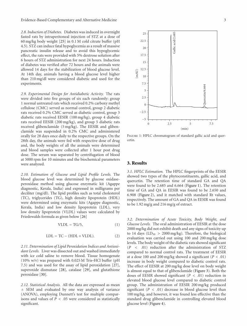
Figure 1: HPLC chromatogram of standard gallic acid and quer-cetin.
3. Results
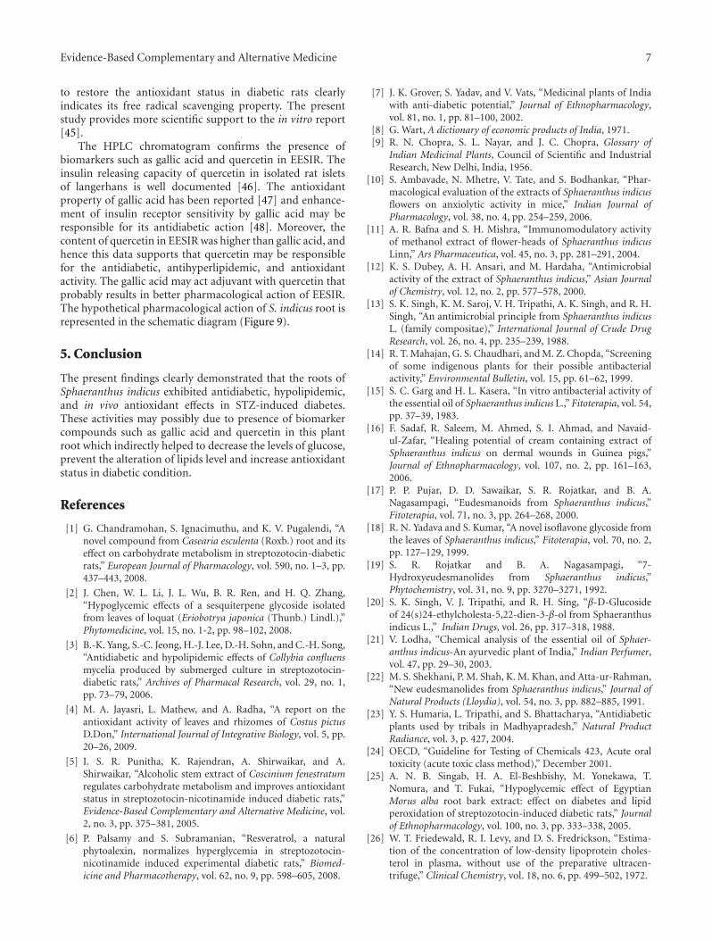
3.1. HPLC Estimation. The HPLC fingerprints of the EESIRshowed two types of the phytoconstituents, gallic acid, andquercetin. The retention time of standard GA and QAwere found to be 2.685 and 6.664 (Figure 1). The retentiontime of GA and QA in EESIR was found to be 2.650 and6.908 (Figure 2), and it matched with standard Rt values,respectively. The amount of GA and QA in EESIR was foundto be 1.92 mg/g and 216 mg/g of extract.
3.2. Determination of Acute Toxicity, Body Weight, andGlucose Levels. The oral administration of EESIR at the dose2000 mg/kg did not exhibit death and any signs of toxicity upto 14 days (LD50 > 2000 mg/kg). Therefore, the biologicalevaluation was carried out using 100 and 200 mg/kg doselevels. The body weight of the diabetic rats showed significant(P < .01) reduction after the administration of STZcompared to normal control rats. The treatment of EESIRat a dose 100 and 200 mg/kg showed a significant (P < .01)increase in body weight compared to diabetic control rats.The effect of EESIR at 200 mg/kg dose level on body weightis almost equal to that of glibenclamide (Figure 3). Both thedoses of EESIR showed significant (P < .01) reduction inelevated blood glucose level compared to diabetic controlgroup. The administration of EESIR 200 mg/kg producedsignificant (P < .01) decrease in blood glucose level than100 mg/kg, and however, it was found less effective than thestandard drug glibenclamide in controlling elevated bloodglucose level (Figure 4).

4 Evidence-Based Complementary and Alternative Medicine
−50
−25
0
25
50
75
100
125
150
175
200
225
250
275
300
325
350
(mA
U)
0 1 2 3 4 5 6 7
(min)
Gal
licac
id/2
.65
Qu
erce
tin
/6.9
08
Figure 2: HPLC chromatogram of EESIR for gallic acid andquercetin.
0
50
100
150
200
Body weight
Val
ues
(g)
a
b
b, c b
ControlDiabetic controlEESIR 100 mg/kg
EESIR 200 mg/kgGlibenclamide 5 mg/kg
Figure 3: Effect of ethanolic extract of Sphaeranthus indicus root onbody weight in STZ-induced diabetic rats. All data are expressed inmean ± SEM (n = 6). aP < .01: diabetic control group comparedwith normal control group. bP < .01: EESIR 100, 200 mg/kg andglibenclamide 5 mg/kg compared with diabetic control group. cP <.01: EESIR 200 mg/kg compared with EESIR 100 mg/kg group.
3.3. Alteration of Lipid Profile by EESIR . In this study, afteradministration of STZ, profound alterations of the lipidprofile were seen in diabetic rats. Both the doses of EESIRshowed significant (P < .01) reduction in elevated TC,TG, and LDL levels and increased HDL level. The dose of200 mg/kg showed significant (P < .05 and P < .01) higher
0
50
100
150
200
250
Blood glucose
Val
ues
(mg/
dL)
a
b
b, cb
ControlDiabetic controlEESIR 100 mg/kg
EESIR 200 mg/kgGlibenclamide 5 mg/kg
Figure 4: Effect of ethanolic extract of Sphaeranthus indicus root onblood glucose in STZ-induced diabetic rats. All data are expressedin mean ± SEM (n = 6). aP < .01: diabetic control group comparedwith normal control group. bP < .01: EESIR 100, 200 mg/kg andglibenclamide 5 mg/kg compared with diabetic control group. cP <.01: EESIR 200 mg/kg compared with EESIR 100 mg/kg group.
0
50
100
150
200
TC TG
Val
ues
(mg/
dL)
a
bb, c
b
a
bb
b, e, f
ControlDiabetic controlEESIR 100 mg/kg
EESIR 200 mg/kgGlibenclamide 5 mg/kg
Figure 5: Effect of ethanolic extract of Sphaeranthus indicus rooton TC and TG in STZ-induced diabetic rats. All data are expressedin mean ± SEM (n = 6). aP < .01: diabetic control comparedwith normal control group. bP < .01: EESIR 100, 200 mg/kg andglibenclamide 5 mg/kg compared with diabetic control group. cP <.05: EESIR 200 mg/kg compared with EESIR 100 mg/kg group. eP <.01: EESIR 200 mg/kg compared with glibenclamide 5 mg/kg group.fP < .01: EESIR 200 mg/kg compared with EESIR 100 mg/kg group.
reduction in elevated TC,TG, and LDL levels when comparedto glibenclamide and 100 mg/kg dose of EESIR. The HDLlevel at the dose of EESIR 200 mg/kg was significantlyincreased compared to glibenclamide and EESIR 100 mg/kg(P < .01). Administration of EESIR and glibenclamide todiabetic rats showed significant (P < .01) reduction in VLDLlevel than diabetic control (Figures 5 and 6).

Evidence-Based Complementary and Alternative Medicine 5
0
20
40
60
80
100
120
140
HDL LDL VLDL
Val
ues
(mg/
dL)
ab
b, e, f
d
b
a
b, e, f
b
ab
b, e, fb
ControlDiabetic controlEESIR 100 mg/kg
EESIR 200 mg/kgGlibenclamide 5 mg/kg
Figure 6: Effect of ethanolic extract of Sphaeranthus indicus rooton HDL, LDL, and VLDL in STZ-induced diabetic rats. All dataare expressed in mean ± SEM (n = 6). aP < .01: diabeticcontrol compared with normal control group. bP < .01: EESIR100, 200 mg/kg and glibenclamide 5 mg/kg compared with diabeticcontrol group. dP < .05: glibenclamide 5 mg/kg compared withdiabetic control group. eP < .01: EESIR 200 mg/kg compared withglibenclamide 5 mg/kg group. fP < .01: EESIR 200 mg/kg comparedwith EESIR 100 mg/kg group.
3.4. Antioxidant Action in Liver Homogenate. There was asignificant reduction of SOD, GPx, and CAT and elevationof TBARS levels were observed in diabetic rats comparedto control animals. The administration of EESIR (P <.01 and P < .05) significantly increased the SOD, GPx,CAT and reduced TBARS level in dose-dependant manner(Figures 7 and 8). However, the dose of 200 mg/kg doseof EESIR significantly (P < .01) increased SOD and CATlevel when compared to 100 mg/kg dose group. Moreover,diabetic rats treated with EESIR 200 mg/kg showed equalefficacy to improve antioxidant level compared to standarddrug glibenclamide.
4. Discussion
The administration of STZ produces selective pancreaticislet beta cell cytotoxicity and has been commonly used toproduce type 1 diabetes in the experimental animal model[31]. The loss of body weight after administration of STZmay be due to the loss of degradation of structural proteins[32]. The administration of EESIR improves body weightcompared to diabetic rats and it indicates preventive effectof EESIR on degradation of structural proteins. The massivedestruction of pancreatic beta cells after STZ injection isdue to alkylation of DNA thereby producing hyperglycaemia[33], and it accounts for drastic reduction in insulin levelwhich in turn alters glucose utilization and metabolism. Themedicinal plants having hypoglycemic activity act throughmultiple mechanisms such as improving insulin sensitivity,augmenting glucose-dependent insulin secretion, and stim-ulating the regeneration of islets of langerhans in pancreas
0
50
100
150
200
250
300
350
SOD CAT
Val
ues
(U/m
gof
prot
ein
)
a bb, d b
ans
b, d b
ControlDiabetic controlEESIR 100 mg/kg
EESIR 200 mg/kgGlibenclamide 5 mg/kg
Figure 7: Effect of ethanolic extract of Sphaeranthus indicus root onSOD and CAT in STZ-induced diabetic rats. All data are expressedin mean ± SEM (n = 6). aP < .01: diabetic control comparedwith normal control group. bP < .01: EESIR 100, 200 mg/kg andglibenclamide 5 mg/kg compared with diabetic control group. dP <.01: EESIR 200 mg/kg compared with EESIR 100 mg/kg group. ns:No significance.
0
0.5
1
1.5
2
2.5
GPx TBARS
GP
x(U
/mg
prot
ein
)an
dT
BA
RS
(nm
olM
DA
/mg
prot
ein
)
a nsc c
a
b bb
ControlDiabetic controlEESIR 100 mg/kg
EESIR 200 mg/kgGlibenclamide 5 mg/kg
Figure 8: Effect of ethanolic extract of Sphaeranthus indicus root onGPx and LPO in STZ-induced diabetic rats. All data are expressedin mean ± SEM (n = 6). aP < .01: diabetic control comparedwith normal control group. bP < .01: EESIR 100, 200 mg/kg andglibenclamide 5 mg/kg compared with diabetic control group. cP <.05: EESIR 200 mg/kg and glibenclamide 5 mg/kg compared withdiabetic control group. ns: No significance.
of STZ-induced diabetic rats [34]. After the administrationof EESIR, significant reduction in elevated glucose level wasobserved in diabetic rats, and it may be due to the actionmediated by EESIR in any one of the above mechanism.

6 Evidence-Based Complementary and Alternative Medicine
Sphaeranthus indicus root powder
Extraction with 70% v/v ethanol
HPLC analysis of S. indicus extract Acute toxicity determination(OECD 423)
Induction of diabetes by STZ(60 mg/kg, i.p.) in rats
Identification of biomarkers in S. indicus extract
Gallic acidQuercetin
Elevation of blood glucose,altered lipid profile and
antioxidant status
Reduction in blood glucose,normalization of altered
lipid profile andantioxidant status
ConclusionGallic acid and quercetin may be
responsible for thesepharmacological action
−500
50100150200250300350
(mA
U)
Gal
licac
id/2
.65
Qu
erce
tin
/6.9
08
0 1 2 3 4 5 6 7(min)
−
Figure 9: Schematic diagram.
The risk of coronary heart disease risk (CHD) isincreased in DM due to profound alterations in the plasmalipids and lipoprotein profile [35, 36]. The elevated level oftotal cholesterol is one of the major factors for occurrenceof CHD. Also, altered lipid profile levels and the incidenceof atherosclerosis are increased in DM [37]. Hence, controlor reduction of lipid profiles level in diabetic conditionwould reduce mortality rate. Many medicinal plants andherbs reported to possess antihyperlipidemic and antidia-betic effect [38, 39]. The blood glucose and lipid loweringefficacy of EESIR could be beneficial in preventing diabetic-related complications and to improve compliance of diabeticpatients.
Oxidative stress plays a major role in generation offree radicals in the pathogenesis of diabetes and its com-plications. In the pathogenesis of diabetic complication,oxidative processes mediated by free radicals have majorrole. Autoxidation of glucose and nonenzymatic proteinglycation occurs during persistent hyperglycemia, and thismay cause disruption of cellular function and oxidativedamage of cell membranes due to increased level of freeradicals. The cell components such as lipid, protein, DNA,and carbohydrates are affected by free radicals. The lipids
are affected by higher level than protein and carbohydrates[40]. In present study, oxidative stress induced by STZ maylead to disturbance of in vivo antioxidant system. The levelof lipid peroxidation (TBARS) and reactive oxygen species(superoxide anion, hydrogen peroxide, and hydroxyl radical)are common markers of oxidative stress in diabetic rats. Anincreased level of TBARS was observed in diabetic rats andthe administration of EESIR significantly reduced TBARSlevels. The SOD catalyses the conversion of superoxide anionto hydrogen peroxide and oxygen [41]. In diabetic rats, liverSOD level was reduced on comparison to normal controland after EESIR treatment there was a significant increasein SOD level. CAT is a hemoprotein enzyme and it catalyzesthe reduction of hydrogen peroxide and protects tissues fromhighly reactive hydroxyl radicals [42]. The level of CATwas reduced in diabetic rats and administration of EESIRimproved CAT level in diabetic rats. In normal condition,GPx detoxifies hydrogen peroxide to water through theoxidation of reduced glutathione [43]. The decreased level ofGPx activity in diabetic control rats indicates an importantadaptive response to increased peroxidative stress [44]. Theadministration of EESIR showed increased GPx activitywhen compared to diabetic control rats. The ability of EESIR
Evidence-Based Complementary and Alternative Medicine 7
to restore the antioxidant status in diabetic rats clearlyindicates its free radical scavenging property. The presentstudy provides more scientific support to the in vitro report[45].
The HPLC chromatogram confirms the presence ofbiomarkers such as gallic acid and quercetin in EESIR. Theinsulin releasing capacity of quercetin in isolated rat isletsof langerhans is well documented [46]. The antioxidantproperty of gallic acid has been reported [47] and enhance-ment of insulin receptor sensitivity by gallic acid may beresponsible for its antidiabetic action [48]. Moreover, thecontent of quercetin in EESIR was higher than gallic acid, andhence this data supports that quercetin may be responsiblefor the antidiabetic, antihyperlipidemic, and antioxidantactivity. The gallic acid may act adjuvant with quercetin thatprobably results in better pharmacological action of EESIR.The hypothetical pharmacological action of S. indicus root isrepresented in the schematic diagram (Figure 9).
5. Conclusion
The present findings clearly demonstrated that the roots ofSphaeranthus indicus exhibited antidiabetic, hypolipidemic,and in vivo antioxidant effects in STZ-induced diabetes.These activities may possibly due to presence of biomarkercompounds such as gallic acid and quercetin in this plantroot which indirectly helped to decrease the levels of glucose,prevent the alteration of lipids level and increase antioxidantstatus in diabetic condition.
References
[1] G. Chandramohan, S. Ignacimuthu, and K. V. Pugalendi, “Anovel compound from Casearia esculenta (Roxb.) root and itseffect on carbohydrate metabolism in streptozotocin-diabeticrats,” European Journal of Pharmacology, vol. 590, no. 1–3, pp.437–443, 2008.
[2] J. Chen, W. L. Li, J. L. Wu, B. R. Ren, and H. Q. Zhang,“Hypoglycemic effects of a sesquiterpene glycoside isolatedfrom leaves of loquat (Eriobotrya japonica (Thunb.) Lindl.),”Phytomedicine, vol. 15, no. 1-2, pp. 98–102, 2008.
[3] B.-K. Yang, S.-C. Jeong, H.-J. Lee, D.-H. Sohn, and C.-H. Song,“Antidiabetic and hypolipidemic effects of Collybia confluensmycelia produced by submerged culture in streptozotocin-diabetic rats,” Archives of Pharmacal Research, vol. 29, no. 1,pp. 73–79, 2006.
[4] M. A. Jayasri, L. Mathew, and A. Radha, “A report on theantioxidant activity of leaves and rhizomes of Costus pictusD.Don,” International Journal of Integrative Biology, vol. 5, pp.20–26, 2009.
[5] I. S. R. Punitha, K. Rajendran, A. Shirwaikar, and A.Shirwaikar, “Alcoholic stem extract of Coscinium fenestratumregulates carbohydrate metabolism and improves antioxidantstatus in streptozotocin-nicotinamide induced diabetic rats,”Evidence-Based Complementary and Alternative Medicine, vol.2, no. 3, pp. 375–381, 2005.
[6] P. Palsamy and S. Subramanian, “Resveratrol, a naturalphytoalexin, normalizes hyperglycemia in streptozotocin-nicotinamide induced experimental diabetic rats,” Biomed-icine and Pharmacotherapy, vol. 62, no. 9, pp. 598–605, 2008.
[7] J. K. Grover, S. Yadav, and V. Vats, “Medicinal plants of Indiawith anti-diabetic potential,” Journal of Ethnopharmacology,vol. 81, no. 1, pp. 81–100, 2002.
[8] G. Wart, A dictionary of economic products of India, 1971.[9] R. N. Chopra, S. L. Nayar, and J. C. Chopra, Glossary of
Indian Medicinal Plants, Council of Scientific and IndustrialResearch, New Delhi, India, 1956.
[10] S. Ambavade, N. Mhetre, V. Tate, and S. Bodhankar, “Phar-macological evaluation of the extracts of Sphaeranthus indicusflowers on anxiolytic activity in mice,” Indian Journal ofPharmacology, vol. 38, no. 4, pp. 254–259, 2006.
[11] A. R. Bafna and S. H. Mishra, “Immunomodulatory activityof methanol extract of flower-heads of Sphaeranthus indicusLinn,” Ars Pharmaceutica, vol. 45, no. 3, pp. 281–291, 2004.
[12] K. S. Dubey, A. H. Ansari, and M. Hardaha, “Antimicrobialactivity of the extract of Sphaeranthus indicus,” Asian Journalof Chemistry, vol. 12, no. 2, pp. 577–578, 2000.
[13] S. K. Singh, K. M. Saroj, V. H. Tripathi, A. K. Singh, and R. H.Singh, “An antimicrobial principle from Sphaeranthus indicusL. (family compositae),” International Journal of Crude DrugResearch, vol. 26, no. 4, pp. 235–239, 1988.
[14] R. T. Mahajan, G. S. Chaudhari, and M. Z. Chopda, “Screeningof some indigenous plants for their possible antibacterialactivity,” Environmental Bulletin, vol. 15, pp. 61–62, 1999.
[15] S. C. Garg and H. L. Kasera, “In vitro antibacterial activity ofthe essential oil of Sphaeranthus indicus L.,” Fitoterapia, vol. 54,pp. 37–39, 1983.
[16] F. Sadaf, R. Saleem, M. Ahmed, S. I. Ahmad, and Navaid-ul-Zafar, “Healing potential of cream containing extract ofSphaeranthus indicus on dermal wounds in Guinea pigs,”Journal of Ethnopharmacology, vol. 107, no. 2, pp. 161–163,2006.
[17] P. P. Pujar, D. D. Sawaikar, S. R. Rojatkar, and B. A.Nagasampagi, “Eudesmanoids from Sphaeranthus indicus,”Fitoterapia, vol. 71, no. 3, pp. 264–268, 2000.
[18] R. N. Yadava and S. Kumar, “A novel isoflavone glycoside fromthe leaves of Sphaeranthus indicus,” Fitoterapia, vol. 70, no. 2,pp. 127–129, 1999.
[19] S. R. Rojatkar and B. A. Nagasampagi, “7-Hydroxyeudesmanolides from Sphaeranthus indicus,”Phytochemistry, vol. 31, no. 9, pp. 3270–3271, 1992.
[20] S. K. Singh, V. J. Tripathi, and R. H. Sing, “β-D-Glucosideof 24(s)24-ethylcholesta-5,22-dien-3-β-ol from Sphaeranthusindicus L.,” Indian Drugs, vol. 26, pp. 317–318, 1988.
[21] V. Lodha, “Chemical analysis of the essential oil of Sphaer-anthus indicus-An ayurvedic plant of India,” Indian Perfumer,vol. 47, pp. 29–30, 2003.
[22] M. S. Shekhani, P. M. Shah, K. M. Khan, and Atta-ur-Rahman,“New eudesmanolides from Sphaeranthus indicus,” Journal ofNatural Products (Lloydia), vol. 54, no. 3, pp. 882–885, 1991.
[23] Y. S. Humaria, L. Tripathi, and S. Bhattacharya, “Antidiabeticplants used by tribals in Madhyapradesh,” Natural ProductRadiance, vol. 3, p. 427, 2004.
[24] OECD, “Guideline for Testing of Chemicals 423, Acute oraltoxicity (acute toxic class method),” December 2001.
[25] A. N. B. Singab, H. A. El-Beshbishy, M. Yonekawa, T.Nomura, and T. Fukai, “Hypoglycemic effect of EgyptianMorus alba root bark extract: effect on diabetes and lipidperoxidation of streptozotocin-induced diabetic rats,” Journalof Ethnopharmacology, vol. 100, no. 3, pp. 333–338, 2005.
[26] W. T. Friedewald, R. I. Levy, and D. S. Fredrickson, “Estima-tion of the concentration of low-density lipoprotein choles-terol in plasma, without use of the preparative ultracen-trifuge,” Clinical Chemistry, vol. 18, no. 6, pp. 499–502, 1972.
8 Evidence-Based Complementary and Alternative Medicine
[27] T. P. A. Devasagayam and U. Tarachand, “Decreased lipidperoxidation in the rat kidney during gestation,” Biochemicaland Biophysical Research Communications, vol. 145, no. 1, pp.134–138, 1987.
[28] S. Marklund and G. Marklund, “Involvement of the super-oxide anion radical in the autoxidation of pyrogallol and aconvenient assay for superoxide dismutase,” European Journalof Biochemistry, vol. 47, no. 3, pp. 469–474, 1974.
[29] A. K. Sinha, “Colorimetric assay of catalase,” AnalyticalBiochemistry, vol. 47, no. 2, pp. 389–394, 1972.
[30] J. T. Rotruck, A. L. Pope, H. E. Ganther, A. B. Swanson, D. G.Hafeman, and W. G. Hoekstra, “Selenium: Biochemical role asa component of glatathione peroxidase,” Science, vol. 179, no.4073, pp. 588–590, 1973.
[31] G. P. S. Kumar, P. Arulselvan, D. S. Kumar, and S. P.Subramanian, “Anti-diabetic activity of fruits of Terminaliachebula on streptozotocin induced diabetic rats,” Journal ofHealth Science, vol. 52, no. 3, pp. 283–291, 2006.
[32] C. Veeramani, G. Pushpavalli, and K. V. Pugalendi, “Anti-hyperglycaemic effect of Cardiospermum halicacabum linn.Leaf extract on STZ-induced diabetic rats,” Journal of AppliedBiomedicine, vol. 6, no. 1, pp. 19–26, 2007.
[33] S. Arumugam, S. Kavimani, B. Kadalmani, A. B. A. Ahmed, M.A. Akbarsha, and M. V. Rao, “Antidiabetic activity of leaf andcallus extracts of Aegle marmelos in rabbit,” ScienceAsia, vol.34, no. 3, pp. 317–321, 2008.
[34] E. Sezik, M. Aslan, E. Yesilada, and S. Ito, “Hypoglycaemicactivity of Gentiana olivieri and isolation of the active con-stituent through bioassay-directed fractionation techniques,”Life Sciences, vol. 76, no. 11, pp. 1223–1238, 2005.
[35] M. Maghrani, A. Lemhadri, N.-A. Zeggwagh et al., “Effects ofan aqueous extract of Triticum repens on lipid metabolism innormal and recent-onset diabetic rats,” Journal of Ethnophar-macology, vol. 90, no. 2-3, pp. 331–337, 2004.
[36] A. Fontbonne, E. Eschwege, F. Cambien et al., “Hypertriglyc-eridaemia as a risk factor of coronary heart disease mortalityin subjects with impaired glucose tolerance or diabetes. Resultsfrom the 11-year follow-up of the Paris Prospective Study,”Diabetologia, vol. 32, no. 5, pp. 300–304, 1989.
[37] B. K. H. Tan, C. H. Tan, and P. N. Pushparaj, “Anti-diabeticactivity of the semi-purified fractions of Averrhoa bilimbi inhigh fat diet fed-streptozotocin-induced diabetic rats,” LifeSciences, vol. 76, no. 24, pp. 2827–2839, 2005.
[38] J. B. Ju, J. S. Kim, C. W. Choi, H. K. Lee, T.-K. Oh, andS. C. Kim, “Comparison between ethanolic and aqueousextracts from Chinese juniper berries for hypoglycaemicand hypolipidemic effects in alloxan-induced diabetic rats,”Journal of Ethnopharmacology, vol. 115, no. 1, pp. 110–115,2008.
[39] V. Sivajothi, A. Dey, B. Jayakar, and B. Rajkapoor, “Anti-hyperglycemic, antihyperlipidemic and antioxidant effect ofPhyllanthus rheedii on streptozotocin induced diabetic rats,”Iranian Journal of Pharmaceutical Research, vol. 7, no. 1, pp.53–59, 2008.
[40] N. Bukan, B. Sancak, O. Yavuz et al., “Lipid peroxidationand scavenging enzyme levels in the liver of streptozotocin-induced diabetic rats,” Indian Journal of Biochemistry andBiophysics, vol. 40, no. 6, pp. 447–450, 2003.
[41] V. T. Selvan, L. Manikandan, G. P. Senthil Kumar et al.,“Antidiabetic and antioxidant effect of methanol extractof Artanema sesamoides in streptozotocin-induced diabeticrats,” International Journal of Applied Research in NaturalProducts, vol. 1, pp. 25–33, 2008.
[42] B. Chance, D. S. Greenstein, and F. J. W. Roughton,“The mechanism of catalase action. I. Steady-state analysis,”Archives of Biochemistry and Biophysics, vol. 37, no. 2, pp. 301–321, 1952.
[43] B. A. Freeman and J. D. Crapo, “Biology of disease. Freeradicals and tissue injury,” Laboratory Investigation, vol. 47, no.5, pp. 412–426, 1982.
[44] M. Kinalski, A. Sledziewski, B. Telejko, W. Zarzycki, and I.Kinalska, “Lipid peroxidation and scavenging enzyme activityin streptozotocin-induced diabetes,” Acta Diabetologica, vol.37, no. 4, pp. 179–183, 2000.
[45] A. Shirwaikar, K. S. Prabhu, and I. S. R. Punitha, “In vitroantioxidant studies of Sphaeranthus indicus (Linn),” IndianJournal of Experimental Biology, vol. 44, no. 12, pp. 993–996,2006.
[46] C. S. T. Hii and S. L. Howell, “Effects of flavonoids on insulinsecretion and 45Ca2+ handling in rat islets of Langerhans,”Journal of Endocrinology, vol. 107, no. 1, pp. 1–8, 1985.
[47] C.-L. Hsu and G.-Y. Yen, “Effect of gallic acid on high fat diet-induced dyslipidaemia, hepatosteatosis and oxidative stress inrats,” British Journal of Nutrition, vol. 98, no. 4, pp. 727–735,2007.
[48] T. H. W. Huang, G. Peng, B. P. Kota et al., “Anti-diabetic actionof Punica granatum flower extract: activation of PPAR-γ andidentification of an active component,” Toxicology and AppliedPharmacology, vol. 207, no. 2, pp. 160–169, 2005.

Submit your manuscripts athttp://www.hindawi.com
Stem CellsInternational
Hindawi Publishing Corporationhttp://www.hindawi.com Volume 2014
Hindawi Publishing Corporationhttp://www.hindawi.com Volume 2014
MEDIATORSINFLAMMATION
of
Hindawi Publishing Corporationhttp://www.hindawi.com Volume 2014
Behavioural Neurology
EndocrinologyInternational Journal of
Hindawi Publishing Corporationhttp://www.hindawi.com Volume 2014
Hindawi Publishing Corporationhttp://www.hindawi.com Volume 2014
Disease Markers
Hindawi Publishing Corporationhttp://www.hindawi.com Volume 2014
BioMed Research International
OncologyJournal of
Hindawi Publishing Corporationhttp://www.hindawi.com Volume 2014
Hindawi Publishing Corporationhttp://www.hindawi.com Volume 2014
Oxidative Medicine and Cellular Longevity
Hindawi Publishing Corporationhttp://www.hindawi.com Volume 2014
PPAR Research
The Scientific World JournalHindawi Publishing Corporation http://www.hindawi.com Volume 2014
Immunology ResearchHindawi Publishing Corporationhttp://www.hindawi.com Volume 2014
Journal of
ObesityJournal of
Hindawi Publishing Corporationhttp://www.hindawi.com Volume 2014
Hindawi Publishing Corporationhttp://www.hindawi.com Volume 2014
Computational and Mathematical Methods in Medicine
OphthalmologyJournal of
Hindawi Publishing Corporationhttp://www.hindawi.com Volume 2014
Diabetes ResearchJournal of
Hindawi Publishing Corporationhttp://www.hindawi.com Volume 2014
Hindawi Publishing Corporationhttp://www.hindawi.com Volume 2014
Research and TreatmentAIDS
Hindawi Publishing Corporationhttp://www.hindawi.com Volume 2014
Gastroenterology Research and Practice
Hindawi Publishing Corporationhttp://www.hindawi.com Volume 2014
Parkinson’s Disease
Evidence-Based Complementary and Alternative Medicine
Volume 2014Hindawi Publishing Corporationhttp://www.hindawi.com