Embed Size (px)
Citation preview
Investigation into the role of monocyte tumour necrosis
factor-‐alpha converting enzyme as a regulator of the
inflammatory response in sepsis
PhD thesis
David John Patrick O’Callaghan
Supervisors:
Doctor Anthony Gordon
Doctor Kieran O’Dea
Professor Masao Takata
Imperial College London
Section of Anaesthetics, Pain Medicine and Intensive Care
Department of Surgery and Cancer

2
The copyright of this thesis rests with the author and is made available under a Creative
Commons Attribution Non-‐Commercial No Derivatives licence. Researchers are free to copy,
distribute or transmit the thesis on the condition that they attribute it, that they do not use
it for commercial purposes and that they do not alter, transform or build upon it. For any
reuse or redistribution, researchers must make clear to others the licence terms of this
work.

3
Abstract
Sepsis consists of both the systemic inflammatory response syndrome (SIRS) and the
compensatory anti-‐inflammatory response syndrome (CARS). How these differential
response states are regulated is yet to be fully elucidated. Tumour necrosis factor-‐alpha
(TNF) is one of the principal cytokines involved in mediating SIRS. TNF is released from cells
by tumour necrosis factor-‐alpha converting enzyme (TACE), this enzyme is responsible for
the ectodomain cleavage of a number of other substrates relevant to inflammation including
both TNF receptors and the adhesion molecule L-‐selectin. How TACE contributes to, and
functions in, SIRS and CARS is not yet known.
My objective was to investigate TACE activity and associated substrate shedding in
monocytes, specifically how the enzyme behaved in the context of in vitro models that I
designed to induce states of priming and tolerance. I then obtained in vivo samples from
critically ill patients to determine whether there were similarities between the TACE activity
profiles found in patient cells, and volunteer cells placed in the in vitro models.
My aims were: 1) Determine how TACE activity profiles were altered when sequential
inflammatory stimuli were utilised in a two-‐hit model of sepsis designed to induce states of
priming and tolerance and 2) To perform a clinical study to investigate TACE behaviour in the
context of critical illness.
I successfully refined a method of isolating primary monocytes from healthy volunteers and
patients that allowed determination of TACE activity profiles. Furthermore, I demonstrated
that the LPS-‐TACE axis was reset in the context of a two-‐hit LPS model and in sepsis. I found
evidence of differential signalling pathway reprogramming in monocytes taken from
patients with infectious and non-‐infectious SIRS. Finally, I demonstrated that the monocyte
TACE response to LPS is dependent on cell contact. These data provide new insights into
monocyte inflammatory function during the immune response.

4
Table of contents
Abstract 3
Table of contents 4
Acknowledgements 7
Declaration 8
1.0 Introduction 9
1.1 Sepsis – the current paradigm 10
1.2 Sepsis and the systemic inflammatory response syndrome 12
1.3 Innate immunity and inflammation 19
1.4 Soluble mediators of inflammation 27
1.5 Leukocyte recruitment and the humoural response 31
1.6 Indicators of immune status in sepsis 38
1.7 Regulation and role of TNF in sepsis 44
1.8 Hypothesis and aims 50
2.0 Materials and methods 52
2.1 Materials 53
2.2 Monocyte isolation 57
2.3 Cell culture 58
2.4 Cell stimulation 58
2.5 Flow cytometry 59
2.6 Fluorescence resonance energy transfer (FRET) assay to determine TACE
activity 62

5
2.7 Enzyme-‐linked immunosorbent assay (ELISA) 65
2.8 Statistics 66
3.0 Development of an optimised in-‐vitro model for the investigation of human
monocyte TACE catalytic activity 67
3.1 Background 69
3.2 Aims 70
3.3 Protocols 70
3.4 Results 72
3.5 Discussion 91
4.0 Modulation of TACE catalytic activity during prolonged exposure to septic
stimuli 96
4.1 Background 98
4.2 Aims 100
4.3 Protocols 100
4.4 Results 103
4.5 Discussion 130
5.0 Determination of monocyte TACE activity and associated shedding profiles in
SIRS and sepsis 137
5.1 Background 139
5.2 Aims 141
5.3 Protocols 141
5.4 Results 144
5.5 Discussion 184

6
6.0 Investigation of the influence of cellular environment on TACE activation 191
6.1 Background 193
6.2 Aims 196
6.3 Protocols 196
6.4 Results 198
6.5 Discussion 219
7.0 Final discussion 226
7.1 An isolation method that enabled the study of monocyte associated enzyme
activity 228
7.2 Resetting of the LPS-‐TACE axis in sepsis 229
7.3 The TACE response is dependent on cell proximity 230
7.4 Monocyte subsets in sepsis and SIRS 231
7.5 Markers of sepsis 232
7.6 The nature of the deactivated monocyte phenotype in sepsis 233
7.7 Concluding remarks 235
References 237
Appendix 1 -‐ publications and presentations 267
Appendix 2 – study documentation 268

7
Acknowledgements
I owe an enormous debt of gratitude to all who have helped me throughout the last three
years. A long, and at times difficult, journey was made easier by the support and
encouragement I received along the way. My thanks to all members of the Section of
Anaesthetics, Pain Medicine and Intensive Care at the Chelsea and Westminster Hospital.
I extend my sincerest thanks to my supervisors without whom I would not have been able to
attempt this PhD project. Doctor Kieran O’Dea has been incredibly generous in sharing his
time, patience and knowledge with me and without his continued input this project would
never have progressed as far as it has. Professor Takata has been a constant source of
support and wisdom; his ceaseless fascination with science is both admirable and
inspirational. Doctor Anthony Gordon has been unstinting in his support of me, offering to
supervise me and providing me with guidance whenever I’ve asked for it and has acted a
mentor, overseeing my academic development.
During my time in the laboratory I received help and support from all the members of the
critical care research group: Alasdair; Alicia; Brij; David; Kate; Kenji; Marianne; Mike – thanks
for all your support and for making the work environment so enjoyable.
Lastly I want to thank both my parents and my fiancée Rebecca, for having the patience that
allowed me to spend so much time at work over the last three years, as well as for their
unending encouragement and reassurance.

8
Declaration
The work contained within the following chapters is original and represents the combined
efforts of David O’Callaghan, Anthony Gordon, Kieran O’Dea and Masao Takata.

9
Chapter 1 Introduction

10
1.1 Sepsis – the current paradigm
The greatest advance in the battle against infection occurred in London at St Mary’s
Hospital, Paddington when Sir Alexander Fleming discovered Penicillin [1]. This discovery
heralded the start of the modern antibiotic era that now is reaching, if not its end, then a
significant juncture. Liberal prescription practices together with partial course completion
have led to the emergence of resistant organisms [2, 3]. These organisms, most famously
methicillin resistant Staphylococcus aureus (MRSA) [4], have become a byword for poor
healthcare practices and have seen renewed public focus on infection control issues and
prescription practices [5].
Although this renewed focus on infection control is welcome, the problems caused by
infection are far from solved. Sepsis accounts for more than 31,000 ICU admissions per year
in the United Kingdom (UK) and results in more than 14,000 deaths [6]. The incidence of
more severe forms of infection (severe sepsis) is increasing and mortality rates, which can
approach 50%, have remained comparatively static. In the UK, the percentage of ICU
admissions with severe sepsis rose from 23.5% in 1994 to 28.7% in 2004 [7], with similar
patterns reported in other areas such as North America [8, 9]. As the population ages, these
conditions will manifest themselves in those who already have organ dysfunction as a result
of chronic illnesses [9], and this is likely to result in a greater demand for ICU services. The
associated costs to healthcare providers as a result of these facts are enormous. In North
America it is estimated that each case of sepsis costs an average of $22,100 and that the
total annual cost of treating patients with sepsis is $16.7 billion [9].
11
The facts and figures outlined above formed the basis on which the surviving sepsis
campaign was founded [10]. This multi-‐national healthcare initiative aimed to improve the
recognition and early management of sepsis, with the aim of reducing morbidity and
mortality. Although early results have been encouraging, and demonstrated some success in
achieving these aims [11], more treatment options are needed.
The issues with resistant organisms outlined above and a reliance on a single treatment
strategy (antibiotics) means that pharmaceutical companies must continue to produce new
antibiotics that work in different ways to stay ahead of emerging resistance patterns.
Unfortunately this is not the case [12] as in comparison to other drugs antibiotics are
difficult to develop, yet even with appropriate antibiotics some patients still die. This has
driven research into the inflammatory cascade generated as a response to infection, in order
that new therapeutic targets can be identified.
There have been some attempts over the years to introduce alternative treatments. As will
be outlined in more detail later, researchers have targeted specific elements of the cytokine
response by seeking to negate the effects of inflammatory cytokines. This approach has
often led to success in animal models but has failed to translate into specific clinical benefit,
often exposing the limitations of animal modelling and lack of caution in interpretation of
their data. Indeed until recently there was only one drug available that was specifically
licenced for the treatment of severe sepsis, activated protein C. The use of this serine
protease was always controversial, with questions about its efficacy and side effect profile
raised throughout the duration of its use. The recent PROWESS-‐SHOCK trial [13] failed to
show a survival benefit and subsequently the manufacturer has withdrawn the drug from
the market after a huge investment. There is therefore no specific therapy licenced for the
treatment of severe sepsis, a condition that is the single most common cause of ICU
admission. There is clearly a need for further treatment strategies in sepsis to help reduce
the significant morbidity and mortality that it causes; hence research in this area in order to
identify potential therapeutic targets is warranted.
In an attempt to better understand the pathophysiology of sepsis and identify novel
therapeutic targets and diagnostic strategies, in this project we studied the inflammatory
pathway responsible for the production of the pro-‐inflammatory cytokine Tumour Necrosis
Factor-‐alpha (TNF), focusing on the regulation and function of TNF-‐alpha converting enzyme
(TACE), which is responsible for the release of soluble TNF, and a number of other proteins,
from the cell surface. In the remainder of the introduction I will expand on the current state

12
of knowledge in sepsis before focusing in on the innate immune response and TNF biology
during the evolution of sepsis. Finally the hypothesis and aims on which this thesis is built
will be presented.
1.2 Sepsis and the systemic inflammatory response syndrome
Part of the difficulty in the early stages of research into sepsis stemmed from a lack of clarity
in definitions. These were agreed at a consensus conference in 1992 and a new concept, the
systemic inflammatory response syndrome (SIRS), was introduced [14]. SIRS is a pro-‐
inflammatory state that was diagnosed when two or more of the following clinical criteria
were present:
1. A temperature of less than 36 or greater than 38 degrees Celsius;
2. A heart rate of greater than 90 beats per minute;
3. A respiratory rate of greater than 20 breaths per minute or an arterial partial
pressure of carbon dioxide of less than 4.3 kilopascals or the need for mechanical
ventilation;
4. A white blood cell count of less than 4x10⁹ or greater than 12 x 109 per litre or >10%
immature (band) forms;
Sepsis was then defined as the co-‐existence of SIRS and the suspicion, or confirmed
presence of infection [14]. Severe sepsis was defined as being present when there was
evidence of acute organ dysfunction or hypo-‐perfusion and septic shock as being
hypotension resulting from sepsis that was refractory to fluid therapy [14]. Each category is
associated with a stepwise increase in mortality [15, 16]. In addition the multiple organ
dysfunction syndrome (MODS) was defined as being impairment of two or more organ
systems, in an acutely unwell patient, where homeostasis cannot be maintained without
therapeutic intervention. Overall the SIRS response describes widespread inflammation and
this reponse is produced and maintained by the release of inflammatory cytokines.
Cytokines are molecules that can be considered the messengers that coordinate the immune
response [17].

13
Lewis Thomas -‐ uncontrolled inflammation
In 1972 in an article entitled Germs [18] the dean of Yale Medical School, Lewis Thomas
wrote the following, “The microorganisms that seem to have it in for us in the worst way -‐
the ones that really appear to wish us ill -‐ turn out on close examination to be rather more
like bystanders, strays, strangers in from the cold. They will invade and replicate if given the
chance, and some of them will get into our deepest tissues and set forth in the blood, but it
is our response to their presence that makes the disease. Our arsenals for fighting off
bacteria are so powerful, and involve so many different defence mechanisms, that we are in
more danger from them than from the invaders. We live in the midst of explosive devices;
we are mined.” This idea of an over stimulated immune system producing organ
dysfunction/failure was widely accepted [19, 20] and was labelled the Lewis Thomas
hypothesis [21]. In this, uncontrolled inflammation manifests itself as organ dysfunction
and/or failure though a combination of endothelial dysfunction [22, 23] and coagulation
activation [24] producing tissue hypo-‐perfusion.
Much scientific interest was initially focused on the inflammatory cytokine TNF as clinical
studies demonstrated elevated circulating levels [25] that were shown to correlate with
mortality [26-‐28]. Animal models revealed that injection of sol-‐TNF produced a syndrome
similar to sepsis, with both hypotension and vascular leak, [29] and demonstrated a survival
benefit if anti-‐TNF treatments were used [30-‐33]. These findings lent sufficient credence to
anti-‐TNF therapy that clinical trials were instigated to investigate its efficacy in the
treatment of sepsis. However these produced only limited success with studies suggesting
that anti-‐inflammatory treatments may be of benefit in only in a small (app. 10%) subgroup
of patients with sepsis [34, 35] and, in some situations, might even lead to harm [36].

14
Roger Bone -‐ the compensatory anti-‐inflammatory response syndrome
It was this lack of clinical benefit provided by anti-‐TNF treatments that led to a reappraisal of
our understanding of sepsis pathophysiology. This resulted in a new concept, the
compensatory anti-‐inflammatory response syndrome (CARS). This idea of a CARS response
was first proposed by Roger Bone in 1996 to denote the mechanism that prevents systemic
dissemination of inflammation [37]. In his article he quoted a maxim from Sir Isaac Newton’s
Philosophiae Naturalis Principia Mathematica [38], “To every action there is always opposed
an equal reaction: or, the mutual action of two bodies upon each other are always equal,
and directed to contrary parts.” By doing so he was outlining his hypothesis that the body
mounts an anti-‐inflammatory response to infection and that, at times, this may be
dominant. Furthermore the importance of the CARS response might explain the failure of
therapeutic strategies aimed solely at attenuating the pro-‐inflammatory response.
Bone’s work was prescient and the idea of a counteractive or balancing anti-‐inflammatory
response in sepsis is now widely accepted, if not yet fully elucidated. His hypothesis explains
the alterations in immune function that are seen in patients exposed to severe
inflammation. Patients who have sepsis display anergy to skin test allergens [39, 40] and
have an increased susceptibility to infection [41, 42]. Their plasma can be seen as an
immunosuppressive milieu [43] as it induces anti-‐inflammatory changes in cells bathed
within it [44-‐46]. It is well documented that leukocytes taken from patients with sepsis
display a reduced release of some pro-‐inflammatory cytokines (such as TNF) on ex vivo
stimulation [47]. There are also non-‐septic inflammatory conditions where an anti-‐
inflammatory response unexpectedly predominates, such as the release of anti-‐
inflammatory cytokines after the aorta is unclamped in bypass surgery [48] and after
resuscitation from cardiac arrest [49].

15
Several putative mechanisms have been proposed as to how this anti-‐inflammatory
response is generated with changes described in both innate and acquired immune cell
function. Monocytes taken from patients with sepsis display alterations in both their
oxidative burst and cytokine production pattern on ex vivo stimulation [50]. Monocytes
taken from patients with both sterile inflammation and sepsis display reduced expression of
the MHC class II antigen-‐presenting molecule HLA-‐DR [51-‐53] and this has been linked to
both outcome and infective complications [54, 55]. This may also result in reduced activation
of lymphocytes, which themselves change from a pro-‐inflammatory Th1 profile to an anti-‐
inflammatory Th2 profile [56].
The dynamics of SIRS and CARS
Although there is agreement that both inflammatory and anti-‐inflammatory response can
dominate in sepsis, the factors governing this and the interplay between them is not yet
clear. In 2003 Hotchkiss and Karl presented their hypothesis as to how these might be
related and termed them the hyper-‐immune and hypo-‐immune response [21]. They
described a temporally separated response in which an initial hyper-‐immune response was
followed by a hypo-‐immune response. They hypothesised that the size and duration of each
response is most likely determined by a combination of factors which include, “the virulence
of the organism, the size of the inoculum, and the patient’s coexisting conditions, nutritional
status, age, and polymorphisms in cytokine genes or other immune-‐effector molecules or
their receptors.” Thus a patient with numerous comorbidities may display a protracted
hypo-‐immune period following their initial presentation with an infective pathology. During
this period of hypo-‐immunity they are vulnerable to nosocomial infection. This is similar to
clinical reality, most patients who succumb to severe sepsis survive their initial infection but
die later in the course of their ICU stay with signs of organ failure and secondary infection
[57]. Roger Bone himself [57] envisaged a situation in which either SIRS or CARS could be
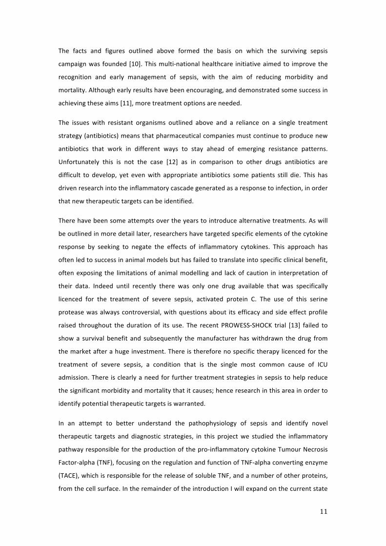
dominant in a similar fashion to that described by Hotchkiss and Karl. An illustration of this
relationship is outlined in figure 1.1.
16
Figure 1.1: Graphical illustration of the possible relationships between SIRS and CARS.
The green line illustrates the inflammatory balance seen in a person with local sepsis -‐ this may oscillate between net pro or anti-‐inflammatory effects. Systemic dissemination of infection produces a cytokine storm and a SIRS response that is characterised by a net pro-‐inflammatory response and early organ dysfunction. This is followed by a period in which there is a net anti-‐inflammatory response, where late organ dysfunction and immune suppression is seen. Here there may be an increased risk of nosocomial infection and viral re-‐activation. A person may recover (as illustrated above) but can die during either the net SIRS or net CARS periods.

17
Other authors have argued that the two processes occur simultaneously but are
compartmentalised; inflammation is spatially limited to the affected extra-‐vascular site or
compartment with a systemic anti-‐inflammatory response occurring to prevent
dissemination [50, 58]. This hypothesis may explain why the anti-‐inflammatory mediators IL-‐
10 and IL-‐1 receptor antagonist are detected as early as one hour after the aorta is
unclamped during cardiac bypass surgery [48]. It also explains why there are higher levels of
pro-‐inflammatory mediators and lower levels of anti-‐inflammatory mediators at the site of
inflammation when compared to those seen systemically [59-‐61].
There is a direct body of evidence to support the compartmentalisation theory of the
immune response in sepsis. It has been reported that, in response to sepsis, gene expression
profiles are organ specific [62] and there is evidence for heterogeneity between different
macrophage populations. In the lung the alveolar macrophage will encounter antigenic
material on a more frequent basis than many other cells. It is of note then that these cells
seem resistant to the induction of LPS tolerance that is seen in other cells of the monocyte-‐
macrophage lineage [63]. Further evidence of organ specific alterations in immunity can be
seen in the liver. Kuppfer cells that are stimulated ex-‐vivo in arginine free media produce
less TNF than those stimulated in media containing arginine [64]. Arginine free conditions
are similar to those found normally within the liver where high levels of hepatic arginase
activity prevent its accumulation [64]. The study authors argue this represents an adaptive
evolution of these cells that prevents them generating excessive cytokine responses despite
their proximity to gut derived LPS. Such alterations in macrophage function are in keeping
with the adaptive nature of cells of the monocyte macrophage lineage.

18
Enhanced levels of pro-‐inflammatory cytokines have been repeatedly demonstrated in
broncho-‐alveolar lavage fluid (BALF) samples taken in the context of chest injury [59] and
ventilator associated pneumonia [65], yet the same patterns were not seen systemically.
Enhanced levels of pro-‐inflammatory BALF cytokines have also been described in the acute
respiratory distress syndrome (ARDS) [66-‐70]. In bacterial pneumonia it has been shown that
there are higher levels of pro-‐inflammatory cytokines in BALF recovered from the involved
lung when compared to the non-‐involved, contralateral lung [71]. Increased levels of pro-‐
inflammatory cytokines have also been shown in fluid retrieved from skin wounds [72], the
cerebrospinal fluid of patients with bacterial meningitis [73], pancreatic ascites [74], urine
from those with urosepsis [75] as well as in peritoneal fluid from patients with appendicitis
[76] and peritonitis [77].
The most developed argument for spatial regulation of the immune response was made by
Pugin and Munford who hypothesised that the body’s normal response to tissue injury is
both systemic and anti-‐inflammatory [58]. They suggested that in some situations this
response could be over zealous in nature resulting in an immune-‐suppressed phenotype that
rendered patients vulnerable to further infection [58]. Although the relationship between
SIRS and CARS is not yet known, it is clear that any immune modulating therapy would need
to be introduced at an appropriate time in order to benefit patients. Thus further work is
required to delineate the regulation of these inflammatory and anti-‐inflammatory
responses.
Compartmentalisation of inflammation in sepsis raises the possibility that circulating
inflammatory cells (e.g. monocytes and neutrophils) would be required to switch from a
relatively passive to more pro-‐inflammatory state while migrating into the inflamed tissue
via the vascular endothelium. Consistent with such environmental triggers, non-‐adherent
versus adherent conditions have been shown to alter the mode of leukocyte response
elicited by stimuli such as complement and IL-‐10 [78, 79]. This raises the possibility that
circulating cells assessed in ex vivo studies might be unrepresentative of cells adherent to
the endothelium.

19
It is clear from the literature that there is an un-‐met need, in both basic science and clinical
medicine, for an improved understanding of inflammation and robust biomarkers of
SIRS/CARS in order that therapies can be guided rather than instigated blindly.
Roger Bone identified four groups of patients [55] who have been exposed to an
inflammatory stimulus:
1. “Patients who show little evidence of a systemic reaction. Although recovery may be
protracted because of the severity of the underlying illness, organ dysfunction rarely
develops.
2. Patients who develop a mild form of SIRS with some evidence of organ dysfunction.
Dysfunction is usually limited to one or two organs and resolves rapidly.
3. Patients in whom a massive systemic inflammatory reaction develops rapidly after
the initial insult. These patients often die of profound shock within a few days.
4. Patients who have a less severe initial course of disease but deteriorate markedly
several days or more after the original insult. Failure of one or more organs is
common, and many of these patients die.”
What we require is the ability to identify a pathological response (groups three and four)
from those having a normal response (groups one and two), as well as a capacity to
determine whether that pathological response is characterised by an excessive
inflammatory or anti-‐inflammatory processes. If we can do this, it may be possible to
instigate future inflammatory/anti-‐inflammatory therapies having appropriately targeted
those who will receive maximum benefit.
1.3 Innate immunity and inflammation
Innate immunity refers to a co-‐ordinated, non-‐specific response that is generated rapidly
after microbial invasion, confers no long lasting immunity to a specific pathogen and is
considered to be the most primitive form of immunity [80]. Whilst the innate immune
response has a number of different constituent parts it is difficult to consider them
individually due to the continuous and complex interplay that occurs between them.

20
Whilst, in strict terms, barriers such as the skin and mucosal defence mechanisms like cilia
form part of the innate immune system, the response element of the system can be broken
down into cellular and humoural components. The cellular response consists of neutrophils,
monocytes/dendritic cells and NK cells [80], whereas the humoural response consists of
complement [81]. Given that the hallmark of the innate immune response is a lack of
specificity, the first, and most fundamental question, is how the system distinguishes self
from non-‐self.
PAMPS, PRRs and self-‐discrimination
The ability to discriminate self from non-‐self prevents the innate immune response from
being inappropriately triggered. This discriminatory capacity is provided through a series of
pattern recognition receptors (PRR) found on monocytes, neutrophils and endothelial cells
that recognise microbial molecules known as pathogen associated molecular patterns
(PAMPS). These PAMPS are conserved across broad groups of microbes [82, 83], the best
characterised being lipopolysaccharide (LPS) or endotoxin. This component of the gram-‐
negative bacterial cell wall has been subjected to considerable scientific interest since it was
first characterised and subsequently synthesised [84]. It is bound by the PRR toll-‐like
receptor (TLR)-‐4 [85, 86] but in order to do so it first must form a complex with LPS binding
protein [87] and requires the presence of the co-‐receptors CD14 [88] (which can be soluble)
and MD2 [89]. TLR-‐4 is one of a series of ten TLRs that are currently described in humans,
each of which binds to a specific ligand/ligands [90]. For example TLR-‐2 binds lipoteichoic
acid, peptidoglycan and bacterial lipopeptides [80], whereas TLR-‐3 binds viral double
stranded RNA [91]. In addition to the TLR family that bind extracellular or endocytosed
PAMPS, there are other PRR which are cytosolic and bind invasive pathogens. These include
the nucleotide oligomerisation domain leucine-‐rich repeat (NOD-‐LRR) receptors (detecting
bacteria) and the cytoplasmic caspase activation and recruiting domain helicases (thought to
be anti-‐viral) that make up the rest of the PRR family [92]. Binding of PAMP to PRR produces
cellular activation through the recruitment of intra-‐cellular proteins that serve to amplify the
inflammatory signal.

21
Sterile inflammation, as may result from trauma, is mediated through similar mechanisms.
However, rather than responding to PAMPs, cells respond to damage associated molecular
patterns (DAMPs) that are released from damaged tissues [93]. These molecules are
released by necrotic (but not apoptotic) cells in response to tissue injury and are bound by
PRRs generating an inflammatory signal as outlined above. Substances identified as DAMPS
include high mobility group box 1 (HMGB-‐1), heparan-‐sulphate (HS) and heat shock proteins
[93, 94]. HMGB-‐1 is a non-‐histone, nuclear DNA binding protein which has roles in in
nucleosome stabilisation and gene transcription [93]. When passively released in large
amounts by necrotic cells, it can become a mediator of severe inflammation [95]. HS is
found on the cell surface and extra-‐cellular matrix, thus is released by tissue injury and
subsequently recognised by TLR-‐4 [94], producing inflammation [96]. An important
difference between PAMPS and DAMPS may be the higher potency of the former, which
could explain why SIRS tends only to develop in the most severely affected trauma patients.
Alternatively, it is argued that a significant number of DAMPS identified in the laboratory,
acquire their activity through trace contamination with potent PAMPs such as endotoxin
during isolation and handling procedures.
Signal amplification and NFκB activation
Activated TLRs induce signalling through a total of five cytoplasmic adapter proteins:
myeloid differentiation protein (MyD) 88; Toll/IL-‐1 receptor homology domain-‐containing
adapter protein (TIRAP); TLR domain containing adaptor inducing interferon β (TRIF);
MyD88-‐4 and MyD88-‐5 [80]. These adapter proteins all serve to amplify the inflammatory
signal, but the core transduction element is the recruitment of interleukin-‐1 receptor
associated kinase (IRAK)-‐4 by MyD88. This induces and recruits IRAK-‐1 and 2, which form a
scaffold with TNF-‐receptor associated factor (TRAF)-‐6, eventually producing an activated
signalosome. This causes phosphorylation and degradation of I-‐kappa B kinase, the negative
regulator of the key inflammatory transcription factor: nuclear factor kappa (NFκ) B [80].
This loss of negative regulation results in nuclear translocation of NFκB and a subsequent
transcription of more than 150 genes of which a significant number have inflammatory
properties [97]. The signal amplification that occurs intra-‐cellularly in response to TLR
activation occurs through two key molecules, reactive oxygen species (ROS) and mitogen
activated protein kinases (MAPK).

22
Signal amplification -‐ ROS
Historically, ROS were thought to be harmful and confined to phagocytic cells where they
were produced by enzymes responsible for generating the oxidative burst [98]. However, it
is now known that enzymes such as NADPH oxidases are widely expressed and provide an
important source of ROS [99, 100]. Mitochondria represent another key source of ROS and
are able to directly produce hydrogen peroxide, potentially as a means of triggering
apoptosis [101]. Although these represent the two major sources of ROS, there are a
number of other enzymes, such as cyclooxygenases and cytochromes, that produce oxidants
[98]. Studies have demonstrated that receptor-‐ligand binding (such as that between LPS and
TLR-‐4) produces ROS [102, 103]. It has become well established that the targets for these
molecules are phosphatase enzymes and that these are activated by oxidative stress [104,
105] establishing a role for ROS in signal transduction. ROS are now known to regulate MAPK
[106-‐109], NFκB [110] and cytoskeletal proteins [111].
Several species of ROS are generated; membrane bound NADPH oxidase utilises cytosolic
NADPH in order that extracellular O2 is reduced to superoxide (O2-‐). There is also a
production of reactive nitrogen species through the action of constitutive and/or inducible
nitric oxide synthase (NOS) that oxidate L-‐arginine producing nitric oxide (NO) [112]. Whilst
neither superoxide nor NO is especially reactive they can combine to produce the
peroxynitrate ion (ONOO-‐) that is highly reactive. Alternatively superoxide can be dismuted
through the action of superoxide dismutase to produce hydrogen peroxide (H2O2), a process
that occurs readily in aqueous environments. This uncharged molecule can readily diffuse
across cell membranes but can be reduced by transition metals in the Fenton reaction to
produce the reactive hydroxyl radical (OH-‐).
ROS are widely generated, highly reactive molecules and must be tightly regulated;
therefore the mechanisms that control this are of considerable scientific interest. Potential
mechanisms include co-‐localisation of target molecules and ROS generating enzymes [113]
and the formation of an intracellular ROS gradient [114]. A further mechanism for conferring
a degree of specificity in signalling exists, and functions by channelling hydrogen peroxide
through membrane expressed aquaporin channels that regulate its entry [115, 116]. A
number of scavenging systems exist to limit the activity of ROS. These include catalase,
which catalyses the decomposition of hydrogen peroxide to water, as well as thioredoxin
and glutathione peroxidase. These latter two enzymes play a key role in maintaining redox
homeostasis through reducing sulphide bridges in target proteins [98].

23
Signal amplification -‐ MAPKs
MAPKs are a series of highly conserved and widely expressed intracellular enzymes that are
responsible for converting external stimuli into cellular responses [117]. In humans there are
five MAPKs: p38; extracellular-‐regulated kinase (ERK); Jun N-‐terminal kinase (JNK); ERK 3/4
and the big mitogen-‐activated protein kinase (BMK1) [107]. Conventional MAPK consists of a
set of three, sequentially acting kinases: a MAPK, a MAPK kinase (MAPKK) and a MAPKK
kinase (MAPKKK). Activation of MAPKKK can occur through phosphorylation or interaction
with a GTP binding protein and results in phosphorylation of a MAPKK at Ser/Thr residues
that in turn results in MAPK activation [117]. MAPK activation requires phosphorylation at
two residues: Thr and Tyr. These sequences are contained within a conserved activation
loop and this dual phosphorylation process was first demonstrated for ERK [118]. Much less
detail is known about the activation of atypical MAPKS such as ERK 3/4 but they are not
thought to function through the same three-‐tiered cascade as conventional MAPKs [117].
MAPKs mediate a broad range of functions through phosphorylation of downstream
molecules, notably members of the MAPK activated protein kinases (MAPKAPKs) [119, 120].
These consist of p90 ribosomal S6 kinases 9 (RSKs) [121], mitogen and stress activated
kinases (MSKs) [122], MAPK-‐interacting kinases (MNKs) [123], MAPK-‐activated protein
kinase 2/3 (MK2/3) [124] and MK5 [124]. These MAPKAPKs serve to amplify the signal and
generate a signalling cascade.
P38MAPK is activated by the MAPKKs MKK3 and MKK6 [125], which are themselves
activated by a stimulus specific MAPKKK. In the case of upstream signalling by ROS this
MAPKKK is thought to be apoptosis signal-‐regulating kinase 1 (ASK-‐1). ASK-‐1 is maintained as
an inactive homodimer through the action of reduced thioredoxin [108, 109]. ROS oxidises
the thioredoxin catalytic disulphide, liberating ASK-‐1, which is subsequently auto-‐
phosphorylated, activating the p38 pathway. As p38MAPK is a stress induced kinase it is
particularly susceptible to activation by ROS and can be activated by superoxide [126],
hydrogen peroxide [127], NO [128] and peroxynitrate [129].

24
P38 is activated through cell stressors such as PAMPs, hyperosmolar states and stretch [130-‐
132]. This activation is rapid but it is also rapidly reduced. In response to an LPS stimulus in
primary human monocytes, we observed increased levels of activated (phosphorylated)
p38MAPK 15 minutes after stimulation that were falling again at 30 minutes post-‐
stimulation [133]. This rapid down regulation of activity occurs through dephosphorylation
at the Tyr or Thr residues through the action of the MAPK phosphatases (MKPs), a family of
dual specificity phosphatases [134]. The p38 pathway is key to modulating the cellular
inflammatory response and has been implicated in the regulation of a number of cytokines
including TNF, IL1-‐β, IL-‐6, IL-‐8 and INF-‐γ [135-‐138]. The main target of p38 is MK2 and these
molecules exist as a pre-‐formed complex that regulates gene transcription [139].

25
Figure 1.2: Schematic illustrating ROS/p38 MAPK activation pathway
ROS are produced in response external signals, such as LPS binding to TLR-‐4, and activate ASK-‐1. In turn this activates MKK3/6 through phosphorylation at Ser and Thr residues. This then phosphorylates p38MAPK at Thr and Tyr resulting in sequential activation of MK2. This process produces an increase in the transcription of a number of mediators that determines the cellular inflammatory response.

26
Evolution of the innate immune response
The sentinel cell in the evolution of the immune response is the resident tissue macrophage.
These cells are thought to be derived from circulating monocytes but also proliferate in situ
[140] and have roles in microbial clearance and the maintenance of local homeostasis [141,
142]. Once activated by PAMPS/DAMPS these cells recognise antigenic material and can
relay this information to neutrophils and other monocytes though the release of cytokines.
Activation of these cells, and also of surrounding endothelial and epithelial cells, results in
the transcription of numerous inflammatory signalling molecules that produce vasodilation
and increased vessel permeability [93, 143], as well as enhanced neutrophil recruitment
[144]. This process can trigger secondary cascades, which augment the cytokine response
and up-‐regulate cell adhesion molecules, further facilitating chemotaxis [145]. The net result
of this process is that cells are recruited to a cytokine rich local environment through the
action of chemokines. Any pathogen is then engulfed (phagocytosis) and subsequently killed
through the action of lysosymes containing reactive oxygen or nitrogen species (ROS/RNS).
Cells responsible for phagocytosis are recruited and tissue macrophages or recruited
neutrophils. As previously discussed, different tissue macrophages may display a modified
response. For example, the alveolar macrophage will regularly be exposed to antigenic
material that passes into the airway. This will not always result in the creation of an
inflammatory focus; often the cell will be capable of disposing of this material by
phagocytosis. However, once the amount of antigenic material exceeds a threshold value
the macrophage will release sufficient chemokines and cytokines to recruit and activate
systemic cells.

27
1.4 Soluble mediators of inflammation -‐ cytokines
These are a series of small, soluble, signalling molecules that effect nearly every biological
process but play a key role in determining the immune response [17]. They are capable of
acting in an autocrine, endocrine and paracrine fashion and can be divided into those
exerting a net inflammatory effect and those exerting a net anti-‐inflammatory effect. Initially
there was some effort made to restrict their nomenclature based on the cells that produced
them but their pleiotropic nature meant that this was rapidly dropped. The term cytokine
now refers to interferons, interleukins, the chemokine family, mesenchymal growth factors,
the tumour necrosis factor family and adipokines [17]. Although cytokines are considered
pro or anti-‐inflammatory in nature, their net effect is often more difficult to quantify and
depends on both local and systemic factors.
Pro-‐inflammatory cytokines
Pro-‐inflammatory cytokines, whose function is to amplify the response to DAMPS/PAMPS,
include TNF, IL-‐1, IL-‐6 and INF-‐γ. The coordinated release of these molecules results in
vasodilation (meaning more leukocytes are diverted to the area), adhesion molecule up-‐
regulation (producing leukocyte arrest that facilitates further recruitment) and enhanced
killing of any pathogenic organism that may be present. The production of these cytokines
occurs concurrently with the acute phase response [143] and serves to further augment it
[80]. The acute phase response is characterised by the generation of fever, a fall in plasma
iron and albumin, and the increased production of a series of proteins, some of which have
well-‐described defensive functions. Increased fibrinogen may help to wall off infected areas,
LBP binds LPS whereas c-‐reactive protein (CRP) binds to streptococci and may signal their
presence [80]. Fever itself enhances immunological functions such as the bacteriocidal
effects of neutrophils, the anti-‐viral effects of interferon and the proliferative responses of
lymphocytes. In addition to the major pro-‐inflammatory cytokines mentioned above there
are a number of other cytokines that may be of importance in sepsis. These include IL-‐4, IL-‐
8, IL-‐9, IL-‐12, IL-‐13, macrophage inhibitory factor and HMGB-‐1 [146].

28
TNF activates and induces margination in neutrophils and monocytes, as well as producing
fever, gluconeogenesis and protein synthesis [146]. The Lewis-‐Thomas hypothesis focused
attention on circulating levels of TNF, as clinical studies had demonstrated these to be
elevated [25] and administration of endotoxin to human volunteers elicited a TNF release
that mimicked the cardiovascular and metabolic changes seen in sepsis [147-‐149].
Unfortunately, treatments based on blocking TNF through the use of either TNF receptor
proteins or anti-‐TNF antibodies had little success except in a small sub-‐group of patients [34,
35], and in some may patients may be harmful [36]. At present the successful use of these
therapies are limited to the treatment of chronic inflammatory conditions such as
rheumatoid arthritis [150] and inflammatory bowel disease [151].
The IL-‐1 superfamily originally consisted of IL-‐1β, IL-‐1α and the IL-‐1 receptor antagonist (IL-‐
1ra) although these have subsequently been added to. Both mononuclear phagocytes and
neutrophils produce IL-‐1β. It enhances leukocyte bacterial killing, enhances the production
of other pro-‐inflammatory cytokines and mediates biological changes similar to those
produced by TNF [152, 153]. IL-‐1α is a related cytokine with a similar spectrum of activity
whereas IL-‐1ra is a soluble inhibitor of IL-‐1β that is produced by mononuclear phagocytes
and neutrophils and binds to cell surface receptors to block the action of IL-‐1β [146]. When
IL-‐1β levels are found to be elevated in the context of septic shock they correlate with an
increased risk of mortality [26].
Activated monocytes, endothelial cells, fibroblasts and lymphocytes release IL-‐6. This
cytokine produces an activation of coagulation and induces the production of hepatic acute
phase proteins [146]. As will be discussed in more detail later it correlates with the severity
of sepsis and may have a role as a clinical marker of such [26].
INF-‐ϒ is released by lymphocytes, specifically activated natural killer cells, helper T-‐cells and
cytotoxic T-‐cells. It induces expression of the major histocompatibility complex antigen and
classically activates macrophages. It is thought to act synergistically with other cytokines in a
manner that may contribute to adverse outcomes in sepsis yet is not consistently elevated in
this state [26]. Recombinant INF-‐ϒ has been used as an immune-‐stimulatory therapy in
patients thought to have sepsis induced immune-‐suppression [154].
29
Anti-‐inflammatory cytokines
The major anti-‐inflammatory cytokines are IL-‐10 and transforming growth factor-‐β (TGF-‐β).
IL-‐10 is released by lymphocytes and mononuclear phagocytes and acts to reduce both the
release of pro-‐inflammatory cytokines and expression of MHC molecules [146]. It reduces
the killing function of mononuclear phagocytes, inhibits coagulation activation and
stimulates lymphocytes [155]. TGF-‐β acts to antagonise the action of both TNF and IL-‐1β
through a reduction in their secretion and a stimulation of their antagonists (soluble TNF
receptors [TNFR] and IL-‐1ra) [156, 157]. In the case of soluble TNFR 1 and 2 these molecules
are released rapidly through the cleavage of membrane-‐expressed proteins in a process
termed ectodomain shedding [158-‐160]. The net effect of this process is variable; soluble
receptors can act to “soak up” cytokines thus limiting their bioactivity [161] yet can also act
as a “sump” resulting in their slow release [162, 163].
Soluble mediators of inflammation – chemokines
These molecules are small (8-‐10kDa) proteins that are able to induce chemotaxis in nearby
cells and hence guide migration of these cells toward an inflammatory focus. They are
divided up into four groups based on the structural homology of cysteine residues:
1. CC chemokines: this group induces the migration of monocytes, NK cells and
dendritic cells. Monocyte chemo-‐attractant protein-‐1 (MCP-‐1) belongs to this group
and induces the recruitment of monocytes from the bloodstream into the tissue
compartment, whereas CC ligand-‐5 (CCL5) induces similar movements in
lymphocytes. Investigators have attempted blockade of receptors to MCP-‐1 in
animal models of sepsis but have found this reduces bacterial clearance and
increases kidney injury [164].
2. CXC chemokines: IL-‐8 belongs to this category and induces migration of neutrophils.
Expression of the CXC receptor 2 (CXCR2) is elevated on neutrophils in septic shock
[165] whereas mice deficient in this receptor are protected from septic shock [166].
3. C chemokines: this group contains the lymphotactins that are partially responsible
for lymphocyte recruitment.
4. CX3C chemokines: this group contains only one molecule, fractalkine which as well
as acting as a chemokine also functions as an adhesion molecule.

30
Prostanoids
This group of signalling molecules consists of prostaglandins and thromboxanes. These are
molecules that are formed when arachidonic acid is metabolised through the action of
prostaglandin synthase or cyclooxygenase enzymes [167]. They sustain homeostatic
functions and yet also mediate pathogenic mechanisms such as the inflammatory response
[167]. There are four principal prostaglandins (PGs) synthesized in vivo: prostaglandin-‐D2
(PGD2), prostaglandin-‐E2 (PGE2), prostaglandin-‐F2 (PGF2) and prostacyclin (PGI2). Together
with thromboxane A2 these molecules are locally produced in inflamed tissues prior to the
recruitment of leukocytes and mediate their actions through a family of prostanoid
receptors. PGD2 has been associated with inflammatory and atopic conditions [167] and is
thought to mediate leukocyte trafficking to the lung, a feature of asthma [168]. PGE2
mediates the general vasodilation and increased microvascular permeability that facilitates
leukocyte recruitment during the inflammatory response [169]. Whilst the exact role of
PGF2 is not precisely defined its administration produces inflammation whereas its
inhibition may have a role in attenuating pulmonary fibrosis [170]. PGI2 is a potent
vasodilator and inhibits platelet aggregation, leukocyte adhesion and vascular smooth
muscle cell proliferation [171]. TXA2 mediates platelet adhesion and aggregation, smooth
muscle contraction and proliferation as well as activation of endothelial inflammatory
responses [172].
There have been a number of studies investigating the clinical utility of targeting
prostaglandin synthesis as a means of improving outcomes from sepsis. Several have
targeted the initial reaction mediated by the cyclooxygenase enzymes in order to reduce
inflammation [173, 174]. Despite success in animal models, these findings are yet to be
reproduced in clinical studies.
Nitric oxide
Nitric oxide (NO) is another mediator of inflammation that has been implicated in sepsis
pathophysiology. It is synthesized from L-‐arginine through the action of the nitric oxide
synthase (NOS) family of enzymes. These consist of a calcium dependent constitutive (cNOS)
isoform, an endothelial (eNOS) isoform, a calcium independent inducible (iNOS) isoform and
a constitutive neuronal (nNOS) isoform [175]. NO derived from nNOS acts as a
neurotransmitter and has a role in mediating the cardiovascular autonomic outflow. NO

31
from eNOS has a key role in determining vascular tone and regional blood flow whereas that
from iNOS is expressed in leukocytes, erythrocytes, vascular smooth muscle, kidney
pancreas liver and lung [176]. The constitutive isoforms are responsible for a constant low
production of NO that can be increased acutely when required (as in inflammation) only for
short periods of time [175]. The inducible isoform however can be increased over a period of
hours thus allowing NO levels to rise from the nanomolar to the micromolar range. NO has a
short half-‐life (8-‐9 seconds) and is degraded to nitrite. It has been of interest to researchers
in sepsis as it mediates the reduction in vascular tone seen in response to LPS, with some
investigators describing the molecule as the final mediator of sepsis [177]. NO is also
thought to play a role in the pathophysiology of myocardial dysfunction in sepsis [178].
Research into this field continues, but at present it would seem that some NO is required for
optimal cardiac function but excessive amounts (as are seen in septic shock) are undesirable
as they result in excessive vasodilation. It would appear that non-‐selective NOS inhibition
produces myocardial depression and does not improve outcomes in sepsis [179], whereas
selective iNOS inhibition may have some beneficial effects on cardiac function [178]. This
difference may be due to issues of dose and timing, but also because targeting a single
molecule may not be sufficient given the complexity of the immune response [175].
1.5 Leukocyte recruitment and the humoural response
Selectins and integrins
The vasodilation that is mediated through cytokines, prostanoids and nitric oxide has two
main functions. It ensures that more blood is diverted to the affected area and slows the
velocity of cells flowing through these vessels [80]. This reduction in leukocyte velocity is an
important step as it facilitates leukocyte recruitment through the mechanisms of rolling,
adhesion and transmigration [180]. Selectins (sugar binding adhesion molecules) are found
on both leukocytes (L-‐selectin) and inflamed endothelial cells (P-‐selectin and E-‐selectin)
[181], the reduction in leukocyte rolling velocity produced by vasodilation allows these
molecules to interact with their glycoprotein counterparts [182], a process that further
slows the cell. Selectin binding induces signalling in both leukocyte and endothelial cell [183-‐
185]; in neutrophils this results in activation of p38MAPK to produce integrin activation
[185].
32
Integrins are a family of ligands that are responsible for mediating leukocyte adhesion [180].
As leukocytes roll along the endothelium they may encounter endothelial molecules that
activate integrins. These include members of the immunoglobulin superfamily, the
intercellular adhesion molecule (ICAM)-‐1 and vascular cell adhesion molecule (VCAM)-‐1
[186, 187]. Integrins are also activated by endothelial surface-‐bound chemokines, produced
as part of the inflammatory response, transported from the abluminal to the luminal surface
of the endothelial cell [188] and supplemented by other chemokines produced by mast cells
and platelets [189, 190]. Integrin activation occurs quickly, almost instantaneously, through
either process [191] and leukocyte arrest then follows. The activation process involves a
change in confirmation as a result of inside-‐out signalling that produces a high affinity
binding confirmation [192] that further strengthens adhesion [180]. Once this process is
complete, transmigration follows and can occur through either the trans or para-‐cellular
routes, meaning that recruited leukocytes can access the inflammatory focus. The net
results are the four signs of inflammation, as recorded in the first century AD by the Roman
encyclopaedist Aulus Cornelius Celsus: calor (heat); dolor (pain); rubor (erythema) and
tumor (swelling).
In health there is a resident population of neutrophils and monocytes that are marginated
(sitting on vessel walls but not invading the organ) within the lung microcirculation [193]. In
sepsis there is an enhanced propensity for these cells to accumulate and it is thought that a
release of inflammatory products by these marginated leukocytes may explain why
pulmonary dysfunction is so common in this setting, potentially contributing to dysfunction
in other organs [193-‐195].
Recruited cells – neutrophils
Neutrophils are the most abundant leukocyte in humans and it is estimated that their
precursor cells constitute 60% of nucleated cells within the bone marrow [196]. As a result
they are found in high numbers in the blood and at any site of inflammation. The primary
neutrophil function is phagocytosis, triggered through the recognition of PAMPs by PRRs
found on the neutrophil surface [197-‐199] or via complement opsonisation [200]. This
process is aided by the fact that neutrophils are “primed” for enhanced phagocytosis and
bactericidal activity by a number of different cytokines and chemokines they encounter.
Phagocytosis triggers both ROS production and fusion of cytoplasmic granules with
pathogen containing vacuoles. ROS are produced through the action of a membrane bound,
33
nicotinamide adenine dinucleotide phosphate (NADPH) oxidase enzyme, that generates high
levels of superoxide in a process known as the respiratory burst [201-‐203]. As cells have such
a high destructive capacity they are tightly regulated and undergo apoptosis (programmed
cell death) rapidly after phagocytosis [204, 205], a process that aids in tissue repair. Both
inflammatory cytokines such as TNF and bacterial toxins delay this apoptosis [206, 207], this
may be biologically useful and facilitate rapid bacterial clearance [208]. Neutrophil granule
proteins, such as secretory vesicles and azurocidin, are left on the endothelium as the cell
migrates, a process thought to aid monocytes chemotaxis [144, 209, 210]. Similarly the
release of substances such as proteinase 3 induces chemokine secretion in surrounding
endothelial cells that causes a further recruitment of cells. The interplay in this situation is
refined; chemokines are rendered up to 1000 fold more potent when they are released into
a milieu containing neutrophil products [211]. Neutrophils recruited to the tissues undergo a
transcriptional burst [212] which may alter the local environment and help recruit/augment
responses in other cells such as monocytes.
Host tissue damage can arise in sepsis through several mechanisms; these include
premature neutrophil activation (seen in the migration stage), an extra-‐cellular “spill over”
of cytotoxic material during the microbial killing phase or through a failure to terminate
inflammatory responses [93, 213]. Neutrophils release elastase, a substance with potent
bacteriocidal activity that also can destroy host tissue [214]. An excessive release of this
enzyme has been implicated in pulmonary diseases that may result from chronic
inflammation such as emphysema [215], pulmonary fibrosis [216] and acute lung injury
[217]. Neutrophil-‐derived heparin binding protein is a potent mediator of increased vascular
permeability and has been implicated in the pathogenesis of oedema resulting from burn
injury [218].
Recruited cells – monocytes
Human monocytes are divided into subsets: classical and non-‐classical (see later for more
detail). The exact role of each subset is not yet clear but there may be different roles in
normal homeostasis when compared to inflammation [144]. Current research [219] supports
the idea that non-‐classical monocytes have a patrolling function, crawling along the luminal
aspect of the vessel wall and monitoring the tissues. In response to inflammation these cells
are then rapidly recruited to the affected tissues where they augment the local cytokine
response and recruit both neutrophils and classical monocytes [144]. The classical

34
monocytes arrive later than neutrophils and then rapidly supersede their non-‐classical
counterparts [144, 210]. On arrival in the tissues monocytes can differentiate to become
macrophages; collectively these cells are referred to as the mononuclear phagocyte system
[220] and can be activated through classical or alternative pathways. Classical activation
occurs through the products of TH1 T-‐cells (mainly interferon-‐γ, IL-‐12 and IL-‐18) resulting in
phagocytosis and cytokine production. Monocytes also present antigens to lymphocytes,
bridging the gap between the innate and the acquired immune systems and thus must be
considered “key players” in the immune response. These mononuclear phagocytes produce
large amounts of TNF as well as other pro-‐inflammatory mediators such as IL-‐1, IL-‐6, IL-‐8,
eucosinoids, ROS, platelet activating factor and nitric oxide. This role means they co-‐ordinate
the immune response ensuring that the correct local milieu has been generated and the
appropriate cells recruited.
In contrast to neutrophils, whose function is limited to the initial pro-‐inflammatory phase,
mononuclear phagocytes display a plasticity of function that allows them to play a role in
tissue repair and regeneration. This is initiated through the alternative activation pathway
triggered via the products of TH2 T-‐cells, IL-‐4 and IL-‐13 [221] and switches the cells function
so that they orchestrate the repair process. A major component of this is efferocytosis, the
process through which they phagocytose dead and dying cells but, in addition, these
alternatively activated macrophages release WNT-‐ligands, secreted glycoproteins that may
play a key role in the regenerative process [222].
In sepsis there is a change in monocyte behaviour. Cells display a reduced capacity to
present antigens as well as reduced production of TNF, IL-‐1, IL-‐6 and IL-‐8 on ex-‐vivo
stimulation, a state that is thought to be induced by IL-‐10 [223] and has been termed
monocyte deactivation [224]. Deactivation has been seen by many as reflecting a state of
systemic immunosuppression [224] and a pathological exaggeration of the CARS response
which may then leave a patient exquisitely vulnerable to secondary infections [225]. Some
investigators have attempted to reverse these changes through the use of immune-‐
stimulatory therapies [154, 224, 226] but this has yet to translate into a direct clinical
benefit.

35
The humoural response – coagulation and complement
In tandem to the activation of the cellular response outlined above, there is also humoural
activation in response to infection or tissue injury. This results in activation of the
coagulation cascade to seal off the site, complement activation to directly kill or opsonize
the pathogen, and release of soluble products that augment leukocyte chemotaxis,
vasodilation and vascular permeability [143]. The coagulation system is mainly activated by
tissue factor [227, 228] a substance expressed on activated monocytes or sub-‐endothelial
cells [229]. The end product of this process is the conversion of fibrinogen to fibrin with the
formation of thrombi that are deposited within the microcirculation and can amplify tissue
injury [230]. In conjunction with this, there is a reduction in fibrinolysis [231] through
alteration of the levels and activity of anticoagulant factors such as protein C, protein S,
antithrombin III and tissue factor pathway inhibitor. In health, protein C is activated through
thrombin-‐α binding to thrombomodulin and subsequently binding to the endothelial protein
C receptor [232]. This activated form of protein C exerts its anticoagulant effects through
inactivating clotting factors Va and VIIIa [233, 234] as well as through inhibiting the synthesis
of plasminogen activator inhibitor [235]. The protein has effects outside of the coagulation
cascade that are relevant to the inflammatory process. It reduces apoptosis [236], leukocyte
adhesion and further cytokine production [237].
The complement cascade is also activated through a number of stimuli that include amongst
others, bacterial components, acute phase proteins and immune complexes [238].
Activation results in construction of the membrane attack complex that subsequently forms
pores in pathogenic organisms resulting in their lysis. The complement product C5 has a
number of pro-‐inflammatory effects [238] such as inducing neutrophil chemotaxis [239] and
superoxide production [240], as well as inducing granular enzyme release from phagocytes
[241]. These latter points serve to illustrate the constant interplay between the humoural
and cellular constituents of innate immunity.
In sepsis there are reduced levels of anticoagulants such as protein C [242]. LPS and TNF
attenuate production of thrombomodulin and the endothelial protein C receptor; hence
protein C activation is impaired [243]. They also increase production of plasminogen
activator inhibitor 1 (PAI-‐1), further inhibiting fibrinolysis [243]. These processes are
compounded by secondary insults such as ischemia and hypoxia (both common in the
context of critical illness) that result in a further release of both tissue factor and PAI-‐1 [244].

36
Similar to these changes in coagulation, a dysregulation of the complement system can also
occur in sepsis [245]. A deficiency of C3 results in a lack of complement effector functions
and increases mortality from sepsis in animal models [246-‐248]. This reinforces the
importance of complement as a component of the immune response. Inhibition of C5
signalling may improve outcomes in animal models [249]. Although this may seem
counterintuitive, given that complement is protective, it may reflect a diversity of
complement functions during the development of sepsis [245]. Some bacterial infections
such as those produced by Haemophilus influenza and Streptococcus pneumonia cause
defects in the opsonisation process that facilitate bacterial killing [250], compromising the
host immune response.
The link to acquired immunity
Both dendritic cells and tissue macrophages perform antigen presentation functions through
the action of the major histocompatibility complex (MHC) class II molecules they express.
These are required in order for a functional adaptive immune response to be induced [251,
252]. However, whilst the MHC molecules themselves are necessary for successful antigen
presentation, alone they are insufficient. An up-‐regulation of other surface markers such as
CD40, CD80 and CD86 are required [80]. Monocytes stimulated by LPS up-‐regulate CD86
expression [253], meaning that antigen presentation is enhanced by TLR stimulation.
Therefore the responsiveness of the TLR system may have an influence on the adaptive
system’s response capability in sepsis. Models of endotoxin tolerance in which the TLR
responsiveness of monocyte is reduced by exposure to large amounts of LPS have
demonstrated reduced antigen presentation capabilities [254]. In sepsis the antigen
presenting capabilities of monocytes are diminished and there is an increased apoptosis of
dendritic cells [224, 255].
Antigen presenting cells acquire and process the antigen in the tissues, but migrate to
secondary lymphoid tissues such as lymph nodes in order to present them to naïve CD4+ T-‐
lymphocytes. This process results in the lymph node swelling that is characteristic of
infection. CD4+ lymphocytes are known as helper T-‐cells and become activated by this
process of antigen presentation. This activation induces differentiation with at least four
pathways available to the naïve T-‐cell [256]. These include TH1, TH2, TH17 and induced
regulatory (iTreg) cells [256, 257]. TH1 cells produce IFN-‐ϒ, IL-‐2 and lymphotoxin-‐α and
favour cell-‐mediated immune responses (although they facilitate the production of
37
opsonising antibodies) by maximising the bacteriocidal phagocytic activity of macrophages
and producing activation and growth of cytotoxic T-‐cells that kill damaged or infected cells.
TH2 cells produce IL-‐4, IL-‐5, IL-‐6, IL-‐10 and IL-‐13 and are primarily involved in optimising the
humoural immune system by stimulating B-‐lymphocytes to produce antibodies directed
against the pathogenic organism. TH1 cells mediate the immune responses against
intracellular pathogens whereas TH2 cells mediate immunity directed against extracellular
parasites [256]. TH17 cells do not produce classical TH1 or TH2 cytokines and are involved in
mediating immunity against extracellular bacteria and fungi as well as in the induction of
autoimmune tissue injury [258]. Similar to other regulatory T-‐lymphocytes the iTreg cells
function to regulate both the immune response itself and self-‐tolerance, the process that
prevents B-‐lymphocytes from producing self-‐directed antibodies [259].
B-‐lymphocytes comprise the other arm of the acquired immune system; their major
functions are antibody production and the formation of memory cells. Activated B-‐cells
differentiate to become either antibody secreting plasma cells or to become memory cells.
Plasma cells produce antibodies that are specific to the pathogenic organism triggering the
immune response and facilitate immunity by binding to the pathogen. This prevents the
pathogen entering or damaging cells and stimulates both their uptake by macrophages and
the complement pathway [260]. Memory B-‐cells remain quiescent, yet are able to produce
antibodies to the original antigen when the same antigenic material is encountered.
Similar to the alterations seen in innate immunity, sepsis also produces alterations in
acquired immunity. These changes may be consistent with an exaggerated CARS response, a
link between the reduced delayed-‐type hypersensitivity reactions seen in ICU patients and
their increased risk of nosocomial infections was described in the 1970’s [39]. Sepsis induces
a lymphopenia that is seen across all lymphocyte subsets which is characterised by increased
levels of apoptosis [261]. It also produces functional alterations in lymphocyte behaviour
such as reduced proliferation and reduced cytokine production in response to stimulation
[262]. Cells display reduced expression of co-‐stimulatory molecules as well as changes in the
circulating regulatory T-‐lymphocyte makeup [263]. These lymphocyte dysfunctions are
accompanied by viral reactivation and deleterious outcome after septic shock [264, 265] and
some authors have described lymphocytes in this context as being exhausted [262].

38
1.5 Indicators of innate immune status in sepsis
Cytokines, surface markers and even leukocyte subsets have all been used in attempting to
determine the nature and severity of the patient’s immune response. In this section I will
focus attention on some of the most pertinent.
Determining the severity of the SIRS response
Over the years a number of cytokines have been investigated as potential markers of
severity. The difficulty of this approach is that circulating levels can be considered to
represent only the tip of the “inflammatory iceberg” and cell associated cytokines can be
demonstrated even in the context of undetectable plasma levels [266] meaning it is unlikely
that any single cytokine will provide us with the perfect marker. Current evidence suggests
that IL-‐6 may be the most effective marker of the severity of both infectious and non-‐
infectious stress [266] but Is not in widespread clinical use. IL-‐6 levels are higher in those
patients who die from SIRS and correlate with illness severity scores [267]. They may help
predict outcomes in severe pancreatitis [268] as well as the onset of infectious
complications in the post-‐operative patient [269]. It is thought that IL-‐6 levels are correlated
with injury severity scores in trauma [270]. Some researchers have explored the circulating
ratio of IL-‐6 to IL-‐10 and have found an increase in this ratio to be correlated with a poor
outcome in SIRS [271] as well as the severity of traumatic injury [272].
Differentiating infectious from non-‐infectious SIRS
The ACCP definitions themselves give an idea of some of the problems facing clinicians in the
ICU. Unsurprisingly a high proportion of critical care patients have SIRS and it can be difficult
to determine whether this has arisen as a result of infectious or non-‐infectious aetiology.
The sepsis definition originally referred to “SIRS in response to infection” [14] but this was
refined in 2001 to SIRS arising in response to “documented or suspected infection” [273].
This change recognised the limitations of the accepted gold standard for demonstrating
infection, positive microbiology. Not all patients with bacterial infection will have positive
cultures and there is a lag time between the sample being obtained and any positive culture
evidence being produced. This means that clinical decisions about starting antibiotics cannot
always be based on positive microbiology. A number of markers suggesting an active

39
immune response to infection have been proposed as being useful in this context: several
interleukins [274]; triggering receptor expressed on myeloid cells 1 (TREM-‐1) [275]; pro-‐
vasopressin [276]; interferon-‐γ [275]; atrial natriuretic peptide (ANP) [277] and resistin [278]
have all been investigated. However, much clinical focus recently has surrounded the use of
procalcitonin (PCT) in this context [279] and some countries have incorporated PCT into
clinical guidelines [280]. PCT is a precursor of calcitonin produced physiologically by thyroid
C cells but also in neuro-‐endocrine tissues in response to bacterial infections. Hence it may
be useful in both diagnosing infection and directing antibiotic therapy [279].
Differentiating between SIRS and CARS
Delineating sepsis from non-‐infectious SIRS has important implications for dictating patient
treatments such as antibiotics. Similarly delineating where a patient lies on the SIRS/CARS
continuum may also help guide therapy. For instance those patients suffering from extreme
pro-‐inflammatory conditions may benefit from therapies to reduce them. It may be that this
category of patient represents those that will derive benefit from anti-‐inflammatory
therapies [35, 281]. In direct contrast, those patients suffering from CARS may benefit from
immune stimulating therapies. Immune stimulating trials that have been performed without
biomarkers have been unsuccessful in demonstrating clinical benefit, this resulted in the
authors of a recent meta-‐analysis concluding that future trials should be biomarker led
[282].
Interest has focused on numerous biomarkers from cytokines to cell surface markers.
Systemic cytokines have been used with investigators focusing on the use of TNF [26-‐28], IL-‐
6 [267] and IL-‐10 [283-‐285]. All of these are elevated in SIRS and correlate with mortality but
as previously discussed the use of systemic cytokine profiles may not tell the whole story
and the systemic circulation may not accurately represent the affected tissue compartment.
Some investigators have argued that the systemic response is by default anti-‐inflammatory
[58]. Prins et al successfully demonstrated that serum from septic patients had the ability to
down regulate ex vivo TNF production by monocytes taken from healthy controls [44]. As
discussed in detail later, a cellular immune-‐responsive phenotype may provide a better
indication of the patient’s net inflammatory status over time than plasma-‐based assays and
measurements. The major cell type used for assessment of sepsis and SIRS patient status has
been monocytes.

40
Monocytes as indicators of immune status: HLA-‐DR
A reduction in circulating monocyte surface expression of HLA-‐DR in sepsis has been
reported by numerous investigators [224, 286]. It has been proposed that measuring surface
markers rather than circulating mediators may be advantageous as it reflects the net result
that the systemic response has on the cell [287]. It is known that expression levels of HLA-‐DR
are regulated both positively and negatively through the action of cytokines such as IL-‐10
and IFN-‐γ, but also through glucocorticoids and catecholamines [288-‐291], therefore
measuring expression levels reflects these processes. There is now an acceptance that
reduced HLA-‐DR expression levels may reliably indicate immune-‐depression [287, 292] and
hence CARS. Reduced expression levels have functional relevance as they reflect a reduced
capacity to present antigens to lymphocytes [293].
One group found persistently low monocyte HLA-‐DR expression to be an independent
marker of mortality in septic shock patients [286], another reported that survivors could be
delineated from non-‐survivors by permanently suppressed or late falling levels [294]. Others
have found it to be a potentially useful marker for the development of secondary infection
[54]. Despite some efforts to standardise practice [295], there is currently no consensus as
to how HLA-‐DR should be measured and what levels constitute pathologically low
expression. Some investigators have termed those patients who have <30% of monocytes
expressing HLA-‐DR (it is normally >80%) as having a state of “immune-‐paralysis” and
suggested these patients are suitable for immune stimulating therapies [224]. One group
used this cut-‐off to guide immune-‐stimulating therapy in the form of granulocyte-‐
macrophage colony-‐stimulating factor (GMCSF) in patients with severe sepsis and septic
shock. They found that patients receiving GMCSF spent less time ventilated and had a
shortened ICU and hospital stay than those who did not [226]. Once again there appears to
be no consensus as to exactly how HLA-‐DR levels should be used in clinical practice. There
are also uncertainties as to how to standardise flow cytometry methodologies including
problems in evaluating positive cells and variability in measured values across different
laboratories.

41
Monocytes as indicators of immune status: subset dynamics
Human monocytes have classically been divided into two distinct subsets on the basis of
their surface expression of the pattern recognition receptor CD14 and the antibody binding
Fc receptor CD16 [296]. These consist of a classical CD14++CD16-‐ population and a non-‐
classical CD14-‐CD16+ population with the vast majority (approx. 90%) of circulating cells
belonging to the former group. Other investigators have argued that there is a third
population, splitting the CD16+ population into both a mature (CD14-‐CD16+) and an
intermediate (CD14+CD16+) population [297-‐299]. Recently it has been proposed that the
classical CD14++CD16-‐ cells represent a pro-‐inflammatory subset whereas the CD14-‐CD16+
cells play a role in patrolling blood vessels to detect virally infected and/or damaged cells
[297]. However a general consensus on this has not been reached with other authors
reporting that the CD14+CD16+ population are the more pro-‐inflammatory subset based on
their increased expression of pro-‐inflammatory cytokines on ex vivo stimulation [300, 301].
Investigators have reported an expansion of the CD16+ (intermediate and mature) cell
population in several conditions including acquired immunodeficiency [302], asthma [303]
and some forms of chronic infections [304]. A substantial expansion of this subset has been
reported by several investigators in sepsis [305-‐307] where the percentage of monocytes
expressing CD16 may increase markedly from 10% to 50% [308]. This expansion occurs with
the onset of bacteraemia and is preceded by cytokine production [306]. Because of this,
such changes have been suggested as a useful monitoring tool in those patients at high risk
of sepsis [309]. The proportion of CD14+CD16+ cells has been shown to correlate with both
body temperature [305] and APACHE II score [310] in the context of surgical intervention. At
present, a lack of consensus in terms of both nomenclature and identification of the various
monocyte populations, combined with the limitations of using flow cytometry in the clinical
setting (cost, access and variation in gating strategies) have precluded their wider use as a
biomarker.

42
Monocytes as indicators of immune status: priming and tolerance
If one can make the argument that cellular expression of markers reflects the effects of a
multitude of mediators, the next logical step is to examine the response of the cell as a
whole. Innate cell immune status can be considered pivotal to both immunity and
inflammation and, whereas neutrophils are capable only of displaying varying degrees of
priming, monocytes can display differential responses to an inflammatory stimulus. These
states are termed priming and tolerance and these may reflect, on a cellular level, the
dominant systemic process occurring in SIRS and CARS. Hence it may be the case that the
monocyte represents an appropriate gauge of the status of the innate immune system.
Priming
Priming refers to an increased response after previous low dose exposure and has been
extensively described in neutrophils in response to numerous stimuli [311, 312]. A
Shwartzman-‐like reaction has been repeatedly reported in which repeated exposure to two
low doses of LPS is then manifest as a shock syndrome [313, 314]. This reaction is mediated
through TNF, and IL-‐1 with monocytes and macrophages implicated as the cell responsible
for their release in response to the second LPS stimulus, although INF-‐ϒ is also involved
[315]. Such “two-‐hit” models seek to reflect the normal or latent inflammatory response
that can become pathophysiological when a second insult is applied. Animal work [316] has
demonstrated that a “two hit” LPS model, when a sub-‐clinical dose of LPS is followed by a
further dose at clinical levels, produces a substantial increase in monocyte membrane
(mem)-‐TNF expression, indicative of priming. In terms of immunology, priming is an adaptive
response to enhance the host response preparedness following a low-‐grade stimulus.
However, it would appear that this protective mechanism could malfunction, reflecting the
unpredictability of sepsis.
In vivo, adherence of cells to the endothelium may represent a form of spatial or cell
interaction dependent priming, whereby cells become more responsive once in contact with
the vascular endothelium. This process, involving sequestration of cells in capillaries, is
probably critical in neutrophil mediated ALI, where they remain in pulmonary capillaries in a
latent activation state but not migrating. In keeping with this picture investigators seeking to
model priming in monocytes have either found adherence to be key or used an adherent
system [317-‐320] with the p38MAPK enzyme found to be a critical determinant of the

43
primed TNF response to LPS. Schaeffer et al found that the complement component C5a
primed both IL-‐6 and TNF production by monocytes through p38MAPK [319]. However they
isolated monocytes by adherence culture and thus only examined cells that may have been
primed by adherence [319]. Cuschieri and co-‐workers found that platelet-‐activating factor
(PAF) primed mononuclear cell production of, amongst others, IL-‐6 and TNF through
enhanced TLR signalling but only under adherent conditions [318]. Another group examined
the LPS priming of alveolar macrophages and again found that the MAPK pathway was
implicated [320] however, these cells may be non-‐representative of other mononuclear
phagocytes due to their altered regulation [63, 321].
Tolerance
Endotoxin tolerance was first described in 1946 [322] and refers to reduced responsiveness
to an LPS challenge following previous exposure to high levels of the molecule [323]. It is
well recognised in innate immune cells such as monocytes and macrophages [324]. The key
readout of endotoxin tolerance is a reduced release of soluble (sol)-‐TNF on secondary LPS
exposure [324]. There are clear parallels between this and clinical reality. Monocytes taken
from patients with a variety of pathologies, in addition to expressing reduced levels of HLA-‐
DR, also display reduced cytokine release on ex vivo LPS stimulation [224, 287, 325]. The
clinical literature often refers to this state of reduced HLA-‐DR expression and LPS
responsiveness as monocyte deactivation [224]. Numerous negative regulators of
inflammatory signalling have been reported as being up-‐regulated in tolerance although
only one, interleukin-‐1-‐receptor associated kinase-‐M, has been confirmed in both murine
and human models [326-‐328]. Interestingly MAPKs have been implicated in the
development of tolerance. Attenuation of MAPK phosphorylation has been demonstrated in
models of tolerance created using both murine macrophages [329] and the pro-‐monocytic
human cell line THP-‐1 [330]. In humans it is thought that this occurs through an up-‐
regulation in the activity of phosphatase enzymes such as MKP-‐1 [331]. Detecting cells that
display this phenotype may help guide the use of immune-‐stimulatory therapies.
We made the argument earlier that the monocyte could be used as a gauge of innate
immune status as it is capable of both priming and tolerance. Therefore detecting these
states is of key importance and could direct immune modulating therapies. Detecting a cell
in a primed state could be predictive of both immune competence and of a potential risk of
excessive inflammation; hence immune suppressant therapies may be warranted. In
44
contrast, detecting a tolerant cell may indicate both immune suppression and a potential
risk of nosocomial infection; hence immune stimulatory therapies may be required. In order
that the primed and tolerant phenotypes can be accurately detected and characterised,
there is a need for in vitro models of these states to be created, a process that may also
identify new targets for immune modulating therapies.
1.6 Regulation and role of TNF in sepsis
Tumour Necrosis Factor and its receptors
Despite the lack of success in sepsis clinical trials, targeting TNF successfully led to
treatments used in other inflammatory conditions [150, 151] and this cytokine is still
considered a primary regulator of systemic inflammation. TNF levels increase before those
of other cytokines in response to systemic or local inflammatory stimuli and it has been the
subject of enormous body of work since it was first identified [332] and subsequently cloned
[333]. Whilst it was initially characterised by its ability to produce cachexia and tumour cell
apoptosis, it is now known to mediate a considerable degree of pleiotropy [334] and signals
through two distinct cell surface receptors, TNF receptor 1 and 2 (TNFR-‐1 [p55] and TNFR-‐2
[p75]) [335]. The trimeric TNF molecule itself exists in two separate forms, a 26kDa non-‐
glycosylated membrane protein (mem-‐TNF) and a 17kDa soluble protein (sol-‐TNF) which is
produced by cleavage of mem-‐TNF [336]. Similar to its soluble counterpart, mem-‐TNF levels
have been implicated in both chronic inflammatory disorders such as hepatitis [337] as well
as infective pathology. It has been suggested that mem-‐TNF plays a role in host protection
from bacterial infection [338] and levels have been shown to correlate with the
development of MODS [339]. Although in recent years attention has been drawn to the
potentially unique functions of memTNF in cell-‐cell signalling, the soluble form remains of
primary importance as the mediator of the majority of TNF functions.

45
Tumour necrosis factor-‐alpha converting enzyme
Tumour necrosis factor-‐alpha converting enzyme (TACE, CD156b) is a widely expressed type
1 trans-‐membrane multi-‐domain enzyme that belongs to the ADAM (a disintegrin and
metalloprotease) family of proteases (ADAM-‐17). It was first discovered in 1997 [340] and is
fundamental to TNF regulation as it cleaves up to 90% of mem-‐TNF to sol-‐TNF, the
remaining 10% being accounted for by the action of other cell surface sheddases [341]. TACE
is responsible for the shedding of nearly 40 proteins [342] rendering it a key enzyme in a
wide array of physiological functions. These can be grouped into those involved in regulating
the immune response and those that influence cell fate, such as cell proliferation and
angiogenesis [343]. Only nine of these substrates have been confirmed in vivo [344]; TNF,
TNFR-‐1 [158], TNFR-‐2 [345], transforming growth factor alpha, L-‐selectin [158], heparin-‐
binding epidermal growth factor, platelet glycoprotein 1b and Fms-‐like tyrosine kinase
ligand 3L [344].
In addition to its function in health, it is important to appreciate the wider importance of
TACE in pathophysiology, as it is vital for normal mammalian development. Mice that are
deficient in the enzyme cannot survive and die shortly before birth [158]; this is thought to
be due to deficient shedding of transforming growth factor alpha. In addition to this, TACE
has been implicated in numerous conditions such as acute lung injury [346, 347],
Alzheimer’s disease [348], cancer [349-‐352], inflammatory bowel disease [353], some skin
diseases [354] and sepsis [355].
In common with other ADAMs, TACE is initially synthesised as a zymogen with a pro-‐domain
(fig 1.2) that serves as an inhibitor during translation by maintaining the zinc ion within the
catalytic domain in an inactive state [356]. This pro-‐domain is subsequently cleaved [357]
and the enzyme is transported to the cell membrane where it is located in micro-‐domains
called lipid rafts [358]. In addition to the pro-‐domain TACE contains multiple other regions
(fig. 1.2). Despite there being 21 other human ADAMs TACE has little sequence homology
with any of them. It most closely resembles ADAM 10, but despite this the sequence
homology between them is only 30% [343].

46
Figure 1.3: The structure of TACE (Adapted from Black et al 2002, Int J Biochem Cell Biol
[341]).
Pro stands for prodomain; Dis for disintegrin; Cys-‐rich for cysteine rich; TM for
transmembrane and Cyto for cytoplasmic. Consistent with other zinc dependent
metalloprotease enzymes there is a Zn ion within the catalytic domain.

47
The potential consequences of TACE shedding are multiple and depend on the nature of the
shed molecule. Shedding of a receptor from the cell may shut down the activity that would
normally be induced by ligand binding. However, the shed molecule could also act as an
antagonist, competing with cells for available ligands. This is of key importance with regard
to TNF biology given that both the molecule and its receptors are shed by TACE. Thus it is
plausible that the enzyme could have both pro-‐inflammatory (shedding sol-‐TNF) and anti-‐
inflammatory (shedding TNFRs) effects. Pellegrini et al examined both monocyte mem-‐TNF
and shed TNFR2 levels in ICU patients. There was a correlation between increased mem-‐TNF
and reduced shedding with increased multiple organ dysfunction score [339]. Although this
initially lacked specificity, if the two parameters were combined as a mem-‐TNF/TNFR ratio
they became a highly specific correlate for the development of organ failure [339],
implicating TACE in sepsis pathophysiology.
TACE regulation
A range of stimuli can induce TACE activation and substrate shedding. These include LPS, the
protein kinase C activator phorbol 12-‐myristate 13-‐acetate (PMA), hydrogen peroxide,
growth factors and cytokines. In addition to stimulation induced shedding, there appears to
be a degree of constitutive shedding with some substrates such as L-‐selectin and TNFRs
being shed in the absence of an inflammatory stimulus. The relevance of this latter process
is unclear.
Previously, work in cell lines demonstrated that TACE was directly activated through reactive
oxygen and nitrogen species, which produced this through nullification of the inhibitory pro-‐
domain [359-‐361]. In contrast, in primary human cells, all cell surface TACE is in the
mature/active form [340] as the pro-‐domain is removed intra-‐cellularly [362, 363], making
nullification or removal of this region an unlikely regulatory mechanism in vivo. In our
laboratory we dissected the mechanism of TACE activation in primary monocytes and found
activation of TACE occurred indirectly, through intracellular ROS acting on p38MAPK, which
in turn produced TACE activation [133]. Furthermore, no role was found for RNS and the
increase in TACE activity was independent of gene expression [133], meaning that the rapid
activity increases seen in response to LPS and INF-‐ϒ [133, 364] are consistent with post-‐
translational modification of the enzyme. We recently suggested that this occurs through
modification of TACE structure (and thereby activity) by disulfide exchange through the
action of an exogenous cell surface oxidoreductase such as protein disulfide isomerase or

48
thioredoxin [133]. A recent study by Xu et al offers an alternative explanation as to how this
structural modification may occur [365]. They demonstrated in a breast cancer cell line that
inactive TACE is present as a dimeric compound, in close association with its known inhibitor
[366], the tissue inhibitor of metalloprotease (TIMP)-‐3 [367]. TACE activation results in a
shift from dimeric to monomeric compounds with a concomitant reduction in TIMP-‐3
association and increased cell surface presentation of TACE [365]. TNF itself has been shown
to increase TACE transcription, expression and catalytic activity [133, 368] and may signal
through the p38MAPK pathway outlined above. This is unlikely to represent the complete
story however, as other investigators have described a reduction in TACE expression in
response to alternate stimuli [369, 370]. Such stimulation induced down regulation has been
suggested as another means of regulating activity levels. In addition, IL-‐10 inhibits TACE
through both TIMP-‐3 dependent and independent pathways [364, 366].
Leukocyte adhesion and migration may have an effect on the enzyme. TACE is known to
interact with both the actin cytoskeleton [371] and the integrin receptors that mediate cell-‐
cell interaction [372]. In addition the integrin activation process has been shown to involve
p38MAPK [373] and TACE expressed by kidney mesangial cells is regulated by β-‐1 integrin
[374]. These factors are suggestive of a common pathway linking both TACE activation and
the integrin mediated adhesion process.
Substrate specificity
Given its numerous substrates it would seem that TACE must have a degree of substrate
specificity, yet how this is regulated is unknown. Some investigators have suggested the
enzyme acts as a “lawn-‐mower sheddase” where it is continuously switched on but held
separately from the substrates it cleaves [375]. Analysis of substrates other than TNF
provide few clues as there are no consensus sequences, and mutations around the cleavage
sites do not always prevent shedding [376]. TNF is unique as it is the only substrate that
TACE will cleave as an isolated peptide [375]. This has enabled investigators to analyse TACE
activity using fluorescence resonance energy transfer assays that are based on TNF cleavage
[133]. It has been suggested that, in a similar manner to the mechanisms outlined above,
post-‐translational modification may play a role in determining substrate specificity [343].
Killock and co-‐workers demonstrated that the cytoplasmic domains of TACE are differentially
regulated by p38 MAPK and PKC to induce shedding [377]. Thus different stimuli could
induce differential regulation of the enzyme that may, in turn, induce changes in substrate

49
specificity and shedding. It is also possible that TACE does not have as many substrates as
are currently attributed to it, but that shedding of one substrate (e.g. TNF) results in a
cascade in which numerous other sheddases are activated.
TACE in priming and tolerance
Given that leukocyte adhesion may impact on TACE there is a possibility that the enzyme
may be spatially regulated through this process. This is intriguing given that the SIRS/CARS
response may be similarly regulated and mononuclear phagocytes are the biggest producers
of TNF. It is also of note that monocytes can display primed or tolerant phenotypes in
response to LPS with evidence for the involvement of p38MAPK and TACE in the
development of both of these states. Cell-‐cell adhesion interactions, such as the β-‐2
integrin-‐ICAM-‐1 interaction, are known to enhance the inflammatory response in monocytes
[378] in a process analogous to priming. Furthermore, it has been demonstrated that
adherent monocytes are primed to produce more TNF than their non-‐adherent counterparts
in response to an LPS challenge [379] meaning that TACE activity/regulation may be altered
in this context. Sol-‐TNF is generally considered to be the best marker of endotoxin tolerance
due to its markedly reduced production in tolerant cells [323], implicating TACE in this
response state. Attenuation of MAPK phosphorylation has been demonstrated in models of
tolerance created using murine macrophages [329] and the pro-‐monocytic human cell line
THP-‐1 [330].
Having established earlier that there is clear benefit in determining whether a monocyte is
primed or tolerant, the data outlined above implicate TACE in both states. TACE responds to
both TNF [133, 368] and IL-‐10 [366], and regulates the release of sol-‐TNF [340] and hence
may determine, or reflect, monocyte inflammatory status. Whilst TACE has previously been
investigated in both cell lines [360, 361, 365, 369, 377, 380] and human cells [133, 340, 364,
366, 370], it is yet to be directly measured in critical illness. The requirement to further
investigate TACE is made even more paramount by the fact that the enzyme appears
sensitive to the adherence state of the monocytes and can have potentially opposing effects
on TNF signalling through differential shedding of TNF and TNFRs. Therefore, any in vitro
model of the primed and tolerant states should include determination of cellular TACE
activity profiles and associated substrate shedding.

50
1.7 Hypothesis and aims
We hypothesised that inducing states of priming and tolerance in monocytes would result in
changes in TACE activity and associated substrate behaviour, meaning that these could be
used as indicators of cellular inflammatory phenotype, potentially reflecting the dominant
systemic SIRS/CARS response. As established earlier in this chapter, there is clear benefit in
knowing where patients lie on this SIRS/CARS continuum as it may help guide immune
modulating therapies. It is possible that such future therapies could be targeted against
TACE if enzyme behaviour in both primed and tolerant monocytes could be determined and
conclusively be shown to mirror clinical reality.
In order to test our hypothesis we attempted to induce priming and tolerance in monocytes
using in vitro models, and then measure both their TACE activity profiles and associated
shedding behaviour. To determine whether these models mirrored clinical reality we
obtained monocytes from critically unwell patients and determined their inflammatory
phenotype using a standard measure of immune status, HLA-‐DR expression.
Our primary aims were as follows:
1. To create in vitro models of priming and tolerance and determine monocyte TACE
activity profiles and associated substrate behaviour therein.
2. To perform a clinical study in which monocytes were obtained from critically unwell
patients and determine their TACE activity profiles and associated shedding
behaviour.
To address these aims:
We first assessed the relative advantages of using a human pro-‐monocytic cell line versus
primary human monocytes for studying TACE biology. This work is detailed in Chapter Three.
Using this information, we then investigated the dynamics of primary monocyte TACE
activation responses in relation to sequential septic stimuli of different intensities under
minimally adherent in vitro conditions as detailed in Chapter Four.

51
Based on the approaches developed in the in vitro models, we carried out a clinical study
using patient peripheral blood monocytes. Standard monocyte-‐related soluble and cellular
markers of sepsis inflammatory status were compared to monocyte TACE expression,
activity and related responses, in patients with sepsis and non-‐infectious SIRS with this work
detailed in Chapter Five.
We concluded the project by extending our previous work on the signalling mechanisms
responsible for TACE catalytic activation, investigating the regulation of such responses
under conditions that aimed to simulate the contrasting systemic non-‐adherent and local
adherent environments that monocytes will encounter during sepsis and the results we
obtained from this make up Chapter Six.

52
Chapter 2
Materials and methods
53
2.1 Materials
Reagents:
General laboratory reagents were purchased from either Invitrogen (Renfrewshire UK) or
Sigma (Dorset, UK). Invitrogen supplied Dulbecco’s Phosphate Buffered Saline (DPBSS),
Foetal Calf Solution (FCS), Hank’s Balanced Salt Solution (HBSS) and distilled water. Sigma
were used to purchase Bovine Serum Albumin (BSA), Dimethylsulfoxide (DMSO),
ethylenediaminetetraacetic acid (EDTA), hydrogen peroxide and sodium azide.
Pharmacologically active agents:
Adenosine tri-‐phosphate (ATP) disodium salt was purchased from TOCRIS bioscience (R&D
systems, Abingdon UK), as were the inhibitors Suramin hexasodium (a purinergic receptor
antagonist) and A74003 (an inhibitor of the P2X7 receptor). Recombinant Human INF-‐ϒ and
monocyte chemotactic protein (MCP)-‐1 were purchased from R&D; LPS E.coli ultra-‐pure was
obtained from InvivoGen (San Diego USA). Super-‐oxide dismutase and catalase were
purchased from Sigma.

54
Antibodies:
These were required for both flow cytometry and for integrin blocking during cell
stimulation experiments.
Flow cytometry:
Antibodies were fluorophore conjugated and anti-‐human. Where used for measurement
purposes (as opposed to gating), the relevant isotype control antibody recommended by the
manufacturer was purchased. Table 2.1 contains details of the antigen, fluorophore and
manufacturer for each antibody.
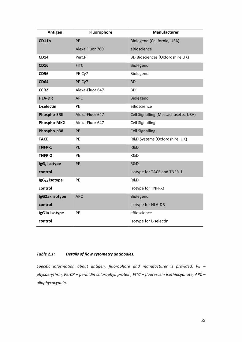
55
Antigen Fluorophore Manufacturer
CD11b
PE
Alexa Fluor 780
Biolegend (California, USA)
eBioscience
CD14 PerCP BD Biosciences (Oxfordshire UK)
CD16 FITC Biolegend
CD56 PE-‐Cy7 Biolegend
CD64 PE-‐Cy7 BD
CCR2 Alexa-‐Fluor 647 BD
HLA-‐DR APC Biolegend
L-‐selectin PE eBioscience
Phospho-‐ERK Alexa-‐Fluor 647 Cell Signalling (Massachusetts, USA)
Phospho-‐MK2 Alexa-‐Fluor 647 Cell Signalling
Phospho-‐p38 PE Cell Signalling
TACE PE R&D Systems (Oxfordshire, UK)
TNFR-‐1 PE R&D
TNFR-‐2 PE R&D
IgG1 isotype
control
PE R&D
Isotype for TACE and TNFR-‐1
IgG2A isotype
control
PE R&D
Isotype for TNFR-‐2
IgG2aκ isotype
control
APC Biolegend
Isotype for HLA-‐DR
IgG1κ isotype
control
PE eBioscience
Isotype for L-‐selectin
Table 2.1: Details of flow cytometry antibodies:
Specific information about antigen, fluorophore and manufacturer is provided. PE –
phycoerythrin, PerCP – perinidin chlorophyll protein, FITC – fluorescein isothiocyanate, APC –
allophycocyanin.
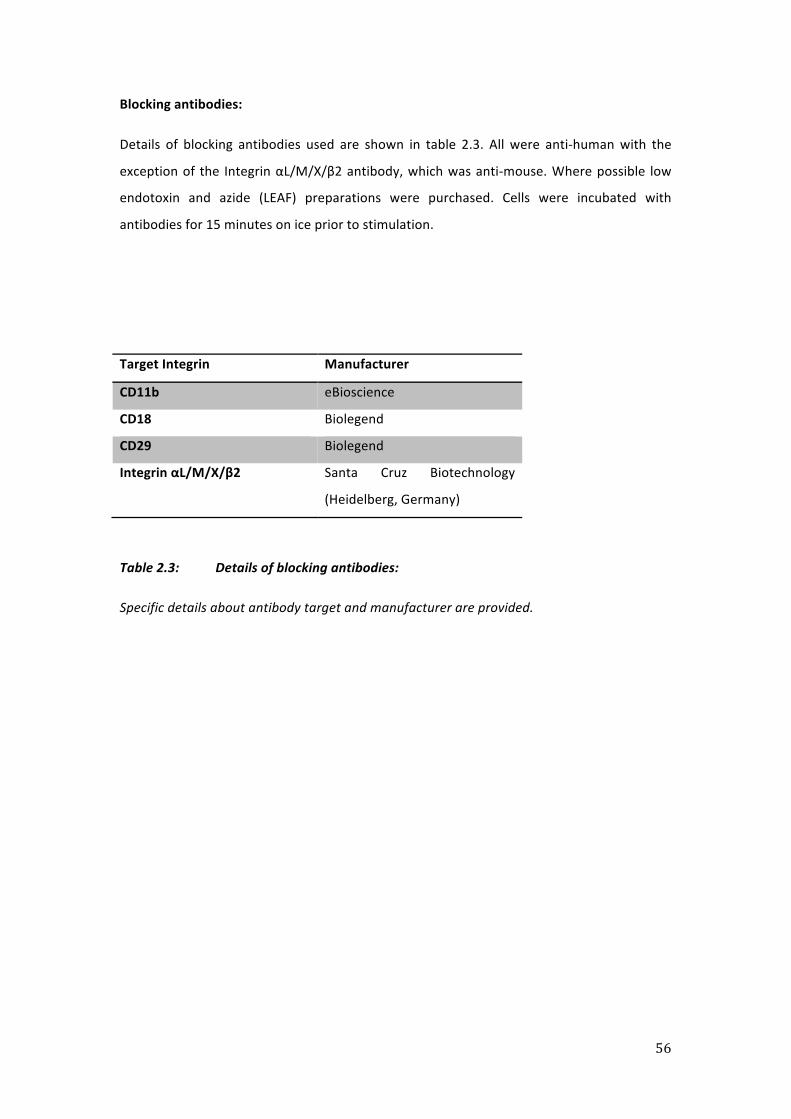
56
Blocking antibodies:
Details of blocking antibodies used are shown in table 2.3. All were anti-‐human with the
exception of the Integrin αL/M/X/β2 antibody, which was anti-‐mouse. Where possible low
endotoxin and azide (LEAF) preparations were purchased. Cells were incubated with
antibodies for 15 minutes on ice prior to stimulation.
Target Integrin Manufacturer
CD11b eBioscience
CD18 Biolegend
CD29 Biolegend
Integrin αL/M/X/β2 Santa Cruz Biotechnology
(Heidelberg, Germany)
Table 2.3: Details of blocking antibodies:
Specific details about antibody target and manufacturer are provided.

57
2.2 Monocyte isolation
PBMCs were isolated from healthy donor blood using Histopaque -‐1077 (Sigma) in
conjunction with Leucosep tubes (Greiner, Stonehouse, UK) and by following the
manufacturers instructions. PBMCs were re-‐suspended in cell separation buffer –0.1% BSA
(Sigma) and 2mM EDTA (Sigma) in HBSS. PBMC numbers were then assessed by flow
cytometry (below).
CD14+ monocytes were purified from PBMCs by positive immune-‐magnetic bead selection
(MACS® – Miltenyi Biotech, Surrey, UK). Cells were incubated for 15 minutes with anti-‐CD14
antibodies that were directly labelled with magnetic beads and an FcR blocking agent (both
Miltenyi Biotech) that blocked unwanted binding of antibodies to human Fc receptor-‐
expressing cells (such as B cells, monocytes, and macrophages). Cells were then applied to a
magnetic separation column (LS, Miltenyi Biotech) to deplete non-‐labelled cells. Purified
monocytes were collected from the column and passed through a second column (MS,
Miltenyi Biotech) to both reduce platelet contamination and increase cell purity.
Where a negative selection protocol was run for comparison, this was done using the
monocyte isolation II kit (Miltenyi). This contained a cocktail of biotinylated antibodies
designed to deplete T-‐lymphocytes (CD3&7), natural killer (NK) cells (CD7, CD16 & CD56), B-‐
lymphocytes (CD19), erythrocytes (CD235a) and a combination of dendritic cells, myeloid
precursors, macrophages, mast cells, basophils and megakaryocytes (all CD123). A separate
biotinylated anti-‐CD41 antibody (Serotec Oxford, UK) was used for platelet depletion. Cells
were incubated with this cocktail before anti-‐biotin micro-‐beads were added and non-‐
monocytic cells were retained in the magnetic column, hence depleted.
58
2.3 Cell culture:
THP-‐1 cells (European Collection of Animal Cells) were cultured in suspension culture 37°C in
95% air/5% CO2 for 16 hours in RPMI-‐1640 containing 10% FCS, 2mM glutamine, 10 µg/ml
streptomycin and 10 U/ml penicillin-‐G (all from Invitrogen, Paisley, UK). Cells were cultured
in 50-‐600ml tissue culture flasks (Becton Dickinson) with media changed every 48 hours by
spinning cells down. As per our laboratory protocols each media change was counted as a
single passage with cells used between passage four and fifteen.
Isolated monocytes were cultured at 37°C in 95% air/5% CO2 for 16 hours in RPMI-‐1640
containing 10% FCS, 2 mM glutamine, 10 µg/ml streptomycin and 10 U/ml penicillin-‐G
(Invitrogen). Cells were cultured in 12 or 24 tissue culture plates (Becton Dickinson, New
Jersey, USA) at a density of 3x10⁶/ml within non-‐adherent polytetrafluoroethylene (PTFE,
Teflon®) cell inserts (Millipore, Carrigtwohill, Ireland). Also added to culture media were
ultra-‐pure E. Coli LPS or recombinant Human INF-‐γ. Retrieval from overnight culture was
performed on ice and by washing inserts with cold HBSS supplemented with 5% FCS. Cells
were then centrifuged at 1400rpm for 10 minutes and suspended in HBSS supplemented
with 5% FCS prior to stimulation.
2.4 Cell stimulation:
After culture or isolation cells were counted by flow cytometry (below) before being
suspended in HBSS containing 5% FCS at a density of 2x10⁶ to 5x10⁶ CD14+ cells/ml. Cells
were then placed in 1.5ml Eppendorf tubes before being stimulated at 37°C. Inhibitors and
blocking antibodies were incubated with cells for 15 minutes prior to stimulation. Where no
inhibitor was used cells and stimulant were added to Eppendorfs simultaneously. The
concentrations of inhibitors and stimulants used were saturating doses based on review of
the literature/previous laboratory experience within our group.
After stimulation cells were washed in HBSS (x4 excess) prior to having TACE activity
determined or flow cytometric analysis performed (below).

59
2.5 Flow cytometry:
Cells were incubated with the relevant antibodies (table 2.1) for 30 minutes at 4°C then
washed in 2ml of wash buffer (WB – 2% FCS, 5 mM EDTA [Sigma] and 0.01% sodium azide in
PBS) before being centrifuged (5 minutes at 2000rpm). Cells were then suspended in WB
and flow cytometric acquisition of samples was performed using a Cyan ADP fluorescence
activated cell sorter (Dako, Denmark) with Summit Version 4.3.02 (Beckman Coulter, High
Wycombe UK). Accucheck fluorescent counting beads (Invitrogen) were used to perform cell
counts before and after magnetic separation. By comparing numbers of cells expressing
CD14 before and after separation both purity and yield could be determined. Representative
flow cytometry plots for monocytes post separation are shown below (fig 2.1)
Viability was determined using propidium iodide (PI). This penetrates the membrane of non-‐
viable cells and binds to DNA, producing a fluorescent signal that is detected via flow
cytometry.
A B
Figure 2.1: Representative dot plots of monocytes isolated using CD14 positive selection
protocol.
A: Plot obtained after PBMCs exposed to monocyte isolation protocol. In keeping with
expectations a single cell population with forward scatter (FSC) and side scatter (SSC)
characteristics consistent with monocytes was obtained. A single gate was placed around
these cells.
B: Gated cells expressed CD14 as expected and consistent with monocytes. These cells
were then used to determine cell surface marker expression.

60
Determination of intracellular MAPK levels (p38, MK-‐2, ERK):
To determine MAPK levels cells were placed in Eppendorfs and incubated with LPS 1μg/ml
for 15 and 30 minutes at 37°C along with un-‐stimulated controls. Post stimulation cells had
Cytofix/Cytoperm™ (Becton Dickinson) x4 excess added (in order to both fix and
permeabilise the cells) and were incubated for a further five minutes. 500μl of wash buffer
(PBS supplemented with 2% FCS, sodium azide and saponin) was then added and cells
centrifuged at 400G. Supernatants were removed and cells suspended in 1ml of wash buffer.
Centrifugation was repeated and cells dissolved in 50μl of PWB before being incubated with
antibodies (table 2.1) for 30 minutes in the dark at room temperature before undergoing
washing and flow cytometric acquisition as outlined above. In order to control for non-‐
specific staining, non-‐activated cells were used as the control population (fig 2.2C). Levels of
MAPK phosphorylation in these non-‐activated cells were subtracted from those in LPS
activated cells to determine the MAPK response.
Representative dot plots and a histogram demonstrating p38 MAPK activation
(phosphorylation) after LPS treatment is shown in figure 2.2.

61
A B
C
Figure 2.2: Flow cytometry protocol for determination of MAPK phosphorylation
A: Representative flow cytometry plot of forward scatter (FSC) and side scatter (SSC)
obtained after PBMC were exposed to the MAPK protocol and LPS LPS 1μg/ml for 15 minutes
at 37°C. Cells were stimulated at 5x10⁶ monocytes/ml in static Eppendorf culture. A gate was
drawn around cells with characteristics consistent with monocytes.
B: A further gate was then made on cells expressing high levels of CD14 and MAPK (p38
MAPK shown in this example).
C: Representative histogram of p38 MAPK phosphorylation in monocytes after LPS
stimulation. Grey curve represents non-‐stimulated controls whereas black represents cells
exposed to LPS in static Eppendorf culture.

62
Flow cytometry data analysis:
Acquisition analysis was performed using Flowjo V.7.5. (Tree Star Inc., Oregon). Expression
levels are given as the geometric mean of fluorescence intensity (MFI) emitted by cells, with
the isotype control value subtracted. Results are reported as mean of repeated experiments
± the SD of the mean.
2.6 Fluorescence resonance energy transfer (FRET) assay to determine TACE activity:
The assay utilises fluorescence resonance energy transfer in order to give a read out of TACE
catalytic activity in live cells. Isolated monocytes were incubated with a peptide consisting of
two donor fluorophores conjugated by a TACE sensitive 13 amino acid mem-‐TNF sequence.
One fluorophore acts as a donor (fluorescein (FAM)) and the other as an acceptor
(tetramethylrhodamine (TAMRA)). Cleavage of the peptide at the alanine-‐valine bond
contained within the peptide by TACE results in an increase in fluorescence due to loss of
internal quenching (fig 2.3). Measurement of this fluorescence allows TACE activity to be
ascertained as described in detail below.
Protocol
Following incubation and stimulation (above), cells were washed and suspended in assay
media–1% bovine serum albumin (Sigma) in HBSS. Cell counts were performed by flow
cytometry and cells were plated at a density of 1x10⁵ cells per well or 2.5x10⁴ per well in
either 96 or 384 well plates (Corning Incorporated, Surrey, UK) in triplicate or greater
wherever possible. Each well totalled 100µL in 96 well plates or 25μl in 384 well plates and
this consisted of 5µM fluorescein-‐tetramethylrhodamine (FAM-‐TAMRA, GlaxoSmithKline,
Stevenage, UK) peptide and an appropriate volume of assay media. TACE activity was
determined via measurement of fluorescence intensity using a microplate fluorimeter (Flx-‐
800, Bio-‐tek Instruments Inc., UK) in conjunction with KC4 data analysis software (Bio-‐tek
Instruments Inc.). Assays were run at 37oC using an excitation wavelength of 485nm and an
emitted wavelength of 535nm. The methodology outlined above is consistent with a
published protocol generated within our laboratory [381].

63
FRET assay data analysis:
Data were exported and subsequently analysed using Microsoft Excel. TACE activity was
calculated by determining the gradient of the curve (fluorescence vs. time) obtained from
the FRET assay over a 10 minute time period. For each experiment activity has been
calculated by determining the mean of experimental replicates and values are reported as
mean of separate experiments ±SD in fluorescence units per minute (FU/min).

64
Figure 2.3: Illustration of FRET assay:
Peptide containing 13 amino acid sequences conjugated to donor and acceptor fluorophore.
TACE cleaves the peptide at the alanine-‐valine (AV) bond, resulting in fluorescence from
donor fluorophore as a result of loss of internal quenching. Fluorescence is proportional to
TACE catalytic activity hence activity can be calculated by the rate of increase in the
fluorescent signal per unit time.

65
2.7 Enzyme-‐linked immunosorbent assay (ELISA):
This was performed using our in-‐house assay with reagents purchased from R&D systems.
ELISA plates (Nunc, Roskilde, Denmark) were coated with capture antibody (R&D) at 2μg/ml
and left at 4°C for 16 hours. Each well was then washed x 4 with PBS supplemented with
0.05% Tween-‐20 (Sigma), this washing protocol was automated as standardised throughout
the protocol. Wells were then blocked for an hour using a PBS supplemented with 1% BSA
(Sigma) and 0.05% Tween-‐20 (Sigma). The washing step was repeated before standards
consisting of rhTNF (R&D) were added. Samples were added and diluted in block buffer
where appropriate. Plates were left for two hours before being washed and having
detection antibody (R&D) added at 100ng/ml and then left for a further two hours. Plates
were washed once again and streptavidin-‐HRP (R&D) was added at a 1/200 dilution and left
for 20 minutes. Substrate solution (R&D) was added and plates incubated for a further 20
minutes before 10M hydrochloric acid (VWR, Leicestershire UK) was added as a stop
solution.
Plates were read using a Dynex MRX II plate reader (Dynex Technologies LTD, Worthing UK)
in conjunction with RevelationTM (version 4.22, Dynex).

66
2.8 Statistics
Results for each experiment are reported as the mean of repeated experiments ±SD. For
non-‐parametric data medians and inter-‐quartile ranges are provided. For parametric data
with two variables a t-‐test was used, for those with three or more a one-‐way analysis of
variance (ANOVA) was used with Bonferroni correction for multiple comparisons. A
corrected p-‐value of <0.05 was considered to be statistically significant.
For categorical data chi-‐squared analysis was used. Mann-‐Whitney U test or Kruskal Wallis
testing was used for non-‐parametric data.

67
Chapter 3 Development of an optimised in vitro model for the investigation of human
monocyte TACE catalytic activity

68
Summary
Sepsis is comprised of both the systemic inflammatory response syndrome and the
compensatory anti-‐inflammatory response syndrome. There are similarities between these
systemic processes and the primed vs. tolerant states that monocytes can display in
response to an in vitro inflammatory stimulus such as LPS. Previously we have shown that
primary human monocytes up-‐regulate their TACE activity in response to LPS. Before
generating primed and tolerant cells it was crucial to reproduce these findings and to
develop a reliable means of providing monocytes. In an attempt to address these two key
areas I performed a series of pilot experiments in the human pro-‐monocytic cell line THP-‐1.
We hypothesised that cells would display a >3 fold increase in their TACE activity when
challenged with LPS in accordance with our previous findings.
Undifferentiated THP-‐1 cells displayed no up-‐regulation of TACE activity in response to LPS.
This hypo-‐responsive state persisted despite efforts to induce differentiation toward a
monocyte phenotype using vitamin-‐D3 (VD3). A partial response to LPS appeared to be
induced by a 16-‐hour IFN-‐ϒ treatment also designed to induce differentiation. However, this
response was small and did not reach statistical significance.
The project was then re-‐appraised and an alternative means of providing monocytes was
found by employing density gradient centrifugation followed by immune-‐magnetic activated
cell sorting together with a CD14 positive selection strategy. The kinetics of the acute TACE
response in cells isolated using this protocol were determined and found to be suitable for
our needs. Finally an optimised flow cytometry protocol to detect activated MAPK levels in
response to LPS was created.
In conclusion, I ascertained the limitations of using cell lines when examining the TACE
response. I then determined a means of rapidly isolating primary human monocytes that
forms one of the core pieces of methodology contained within this thesis.

69
Chapter 3.1 Background
Previously within our group we have demonstrated that, in response to LPS, TACE activity is
rapidly up regulated [133, 381]. This response is sufficiently rapid (x2-‐5 fold, present at 30
minutes) to be consistent with post-‐translational modification of the enzyme and is
mediated indirectly through intra-‐cellular ROS activating and phosphorylating p38MAPK
[133]. Before measuring monocyte TACE activity in models, designed to induce states of
priming and tolerance, it was necessary to reaffirm TACE behaviour in single hit conditions
as this would form the basis for any comparison between cell states. TACE catalytic activity
was measured using a fluorescence resonance energy transfer (FRET) assay that was
developed and refined by our group [381]. This assay represents a significant improvement
on previous methods of quantifying TACE activity where activity was calculated indirectly
through derived indices, differential inhibition or inferred indirectly from expression levels
[355, 364, 369]. However, the FRET assay is incapable of distinguishing between cell types,
and as TACE is widely expressed [340], it is necessary to perform experiments in a single cell
line/isolated cells for accurate interpretation of results.
Previously [133], within our laboratory we had used both primary human monocytes and
human cell lines with both approaches having advantages and disadvantages. Primary cells
have obvious biological relevance but the isolation process [133] is expensive and
incorporates density gradient centrifugation followed by magnetic activated cell isolation
employing a negative selection protocol. In this process non-‐monocyte populations are
labelled with antibodies attached to magnetic beads meaning they are retained in a column
placed in a magnetic field and pure monocyte preparations can be obtained.
When validating the FRET assay both MonoMac-‐6 (an alternate human monocyte cell line)
and THP-‐1 cell lines were used [381] and two populations of MonoMac-‐6 cells were found,
one expressed high levels of TACE and another that expressed negligible amounts. It was
noted that the relative proportions of these groups changed with time, rendering this cell
line sub-‐optimal for use in investigating the acute TACE response to LPS. The THP-‐1 cells
appeared to present no such problem and displayed a significant (x3.3 fold) increase in TACE
activity after a three hour LPS stimulation. Hence, in order try and minimise resources
required related to material (human blood), cost, speed, productivity and reproducibility, we
decided to use the THP-‐1 cell line and investigate the TACE response to a single LPS stimulus.

70
In addition to studying the TACE response we sought to determine a rapid and reliable
means of determining intra-‐cellular levels of activated MAPK markers.
3.2 Aims
The specific aims of this chapter were to:
1. Determine a suitable method of providing monocytes in order that the acute TACE
response could be determined in single hit conditions.
2. Determine a rapid and reproducible method for determining MAPK activation levels.
3.3 Protocols
Cell culture
THP-‐1 cells were cultured as outlined in Chapter Two (methods).
THP-‐1 cell stimulation
On the day that cells were removed from suspension culture conditions they were
centrifuged before being re-‐suspended in HBSS with 5% FCS at a cell density of 5x10⁶/ml.
Stimulation was performed in 1.5ml Eppendorfs using LPS at concentrations of 1pg to
10µg/ml for time periods of 1.5 to 3 hours at 37°C.
Blood collection
30 ml of donor blood was obtained by venesection from healthy volunteers who had given
informed consent. Exclusion criteria were inter-‐current illness or any immune-‐suppression.
Blood was extracted directly into vacutainers containing EDTA.
71
Monocyte stimulation
Monocytes were isolated from whole blood using differential centrifugation and a CD14+
bead selection strategy as outlined in Chapter Two (methods). Cells were then re-‐suspended
at a concentration of 5x10⁶/ml in HBSS with 5% FCS and stimulated using LPS at
concentrations of 1pg to 10µg/ml for time periods of 1.5 to 3 hours at 37°C.
Quantification of MAPK response
This was performed as outlined in Chapter Two (methods). Briefly, cells were fixed and
permeated before being stained with antibodies specific for the activated form of the MAPK
enzyme. Cell stimulation was performed at the same density and conditions as outlined for
the TACE response. An LPS concentration of 1µg/ml was used with PBMCs stimulated for
either 15 or 30 minutes.

72
3.3 Results
The FRET assay is quantitative for TACE activity:
In order to demonstrate the quantitative nature of the FRET assay untreated THP-‐1 cells
were plated at varying densities and introduced into FRET assay conditions. Cells were
incubated with the FAM-‐TAMRA peptide at a concentration of 5µM and their fluorescence
intensity measured every minute for an hour (fig 3.1A & B).
The system was then allowed to equilibrate for a 20-‐minute period after which TACE activity
was calculated by ascertaining the average rate of increase in the fluorescent signal over a
ten-‐minute window. The curves obtained from the assay (fig 3.1A) are linear after the initial
equilibration period indicating a constant rate of substrate hydrolysis. The results
demonstrate that TACE activity was directly proportional to cell density as expected. The
FRET assay has previously been validated for the THP-‐1 cell line by our laboratory [381]. The
data (fig 3.1B) shown is for a single experiment in unstimulated THP-‐1 cells that was in line
with our previous work [381], hence they were not immediately repeated and I moved to
determine the TACE response of these cells to LPS.
Although using high cell densities in the assay (e.g. 1x10⁶, fig 3.1A) produced a high TACE
signal, previous published protocols from within our laboratory (including for THP-‐1 cells)
have been standardised to a cell density of either 1 or 2x10⁵ per well [133, 381]. Such
densities have previously produced a robust TACE signal whilst preventing excessive use of
resources such as would be required at higher cell densities. Hence a concentration of 1x10⁵
cells/well was initially used when cells were placed in FRET assay conditions to determine
their TACE activity.

73
A
B
Fig 3.1: The FRET assay is quantitative for unstimulated THP-‐1 cells.
A Data obtained from FRET assay for unstimulated THP-‐1 cells plated at different
densities and left for 60 minutes. B TACE activity calculated from fluorescence curves
seen in figure 3.1A. Activity is calculated as mean increase in FU over a ten minute time
period. Bars demonstrate that activity is directly proportional to cell density (n=1).

74
Undifferentiated THP-‐1 cells do not acutely up-‐regulate their TACE activity in response to
an LPS stimulus:
In order to determine the kinetics of TACE activity in response to an inflammatory stimulus,
THP-‐1 cells were stimulated with LPS. Previously in THP-‐1 cells [381] an LPS induced increase
in TACE activity has been shown to occur by three hours. TACE activation in primary cells has
been shown to occur as early as 30 minutes [133]. Hence cells were removed from culture
and stimulated for time periods that varied from 15 minutes to three hours using a
saturating concentration of LPS (1µg/ml).
No consistent pattern of TACE activity up-‐regulation was seen in response to LPS (fig 3.2). A
trend toward increased activity was seen only at the 30-‐minute time point (un-‐stimulated
379.9 ±101.68FU/min vs. stimulated 454.4 ±41.58FU/min; non-‐significant). Baseline THP-‐1
cell TACE activity increased from 0.25 hours (un-‐stimulated 577.5 ±34.79FU.min) through to
three hours (un-‐stimulated 1470.3 ±24.18FU/min; p<0.001).

75
Figure 3.2: Undifferentiated THP-‐1 cells did not up-‐regulate their TACE activity in response
to a LPS stimulus applied between 0.25 and three hours.
THP-‐1 cell time course in response to LPS stimulation at a concentration of 1µg/ml (n=3).
No consistent increase in TACE activity in response to LPS seen
* Indicates significant difference to baseline un-‐stimulated value (p<0.001)

76
No acute up-‐regulation of TACE activity was seen in undifferentiated THP-‐1 cells over a
range of LPS concentrations:
In order to further determine whether THP-‐1 cells displayed any alteration in their TACE
response, a range of LPS concentrations were used. In keeping with both previous laboratory
protocols and our focus on cellular acute inflammatory signalling characteristics a three-‐
hour time point was used. LPS concentration ranged from 1pg/ml through to 10µg/ml.
In keeping with the results of the previous time course experiment, no up-‐regulation of THP-‐
1 cell TACE activity was seen over a range of concentrations of LPS (fig 3.3 A). Untreated cell
TACE activity was similar to that obtained with the highest LPS concentration of 10µg/ml
(untreated 121.2 ±35.25FU/min vs. LPS 10µg/ml 118.1 ±39.87FU/min; non-‐significant).
There were also no differences in cell surface TACE expression (untreated MFI 145.61 ±21.33
vs. LPS 10μg/ml MFI 127.31 ±25.6; non-‐significant, fig 3.3B).
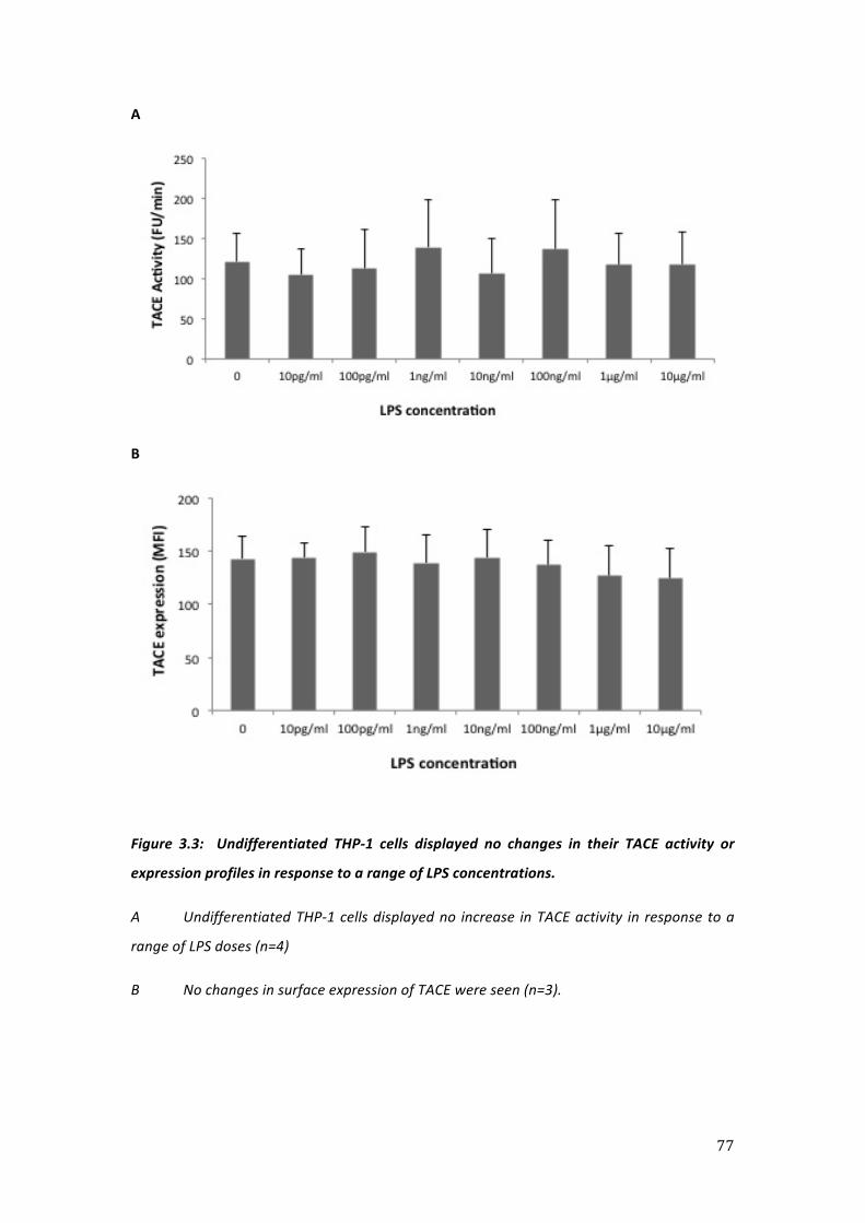
77
A
B
Figure 3.3: Undifferentiated THP-‐1 cells displayed no changes in their TACE activity or
expression profiles in response to a range of LPS concentrations.
A Undifferentiated THP-‐1 cells displayed no increase in TACE activity in response to a
range of LPS doses (n=4)
B No changes in surface expression of TACE were seen (n=3).

78
THP-‐1 cell TACE activity remained refractory to LPS despite the use of 1,25-‐
dihydroxyvitamin D3 (VD3) to induce differentiation:
The THP-‐1 cell line is pro-‐monocytic (i.e. an undifferentiated monocyte precursor). It is
possible that the lack of response seen to LPS described above reflects this [382], and cells
require differentiation to generate full inflammatory signalling. Our group had previously
used the cytokine INF-‐ϒ to induce this process but we were concerned this may have other,
unrecognised, actions that might impact on my results.
Efforts were made to induce differentiation in the THP-‐1 cells toward a monocyte phenotype
using vitamin D3 (VD3). In accordance with a previously published protocol [383] cells were
treated for 72 hours with VD3 at a concentration of 10-‐7M then stimulated with LPS 1μg/ml
for three hours (fig 3.6). There were no significant differences in THP-‐1 cell TACE activity in
response to LPS after 72-‐hour culture (untreated 400.55 ±69.9FU/min vs. LPS 1μg/ml 462.24
±77.18FU/min; non-‐significant). There were also no differences between VD3 treated cells
(VD3 only 457.58 ±69.04FU/min vs. VD3 and LPS μg/ml 583.14 ±100.81FU/min; non-‐
significant). Similar TACE activity results were obtained for 72-‐hour VD3 treatment at a
higher concentration of 10-‐6M (VD3 only 544.04 ±165.18FU/min vs. VD3 and LPS 1μg/ml
695.77 ±122.3FU/min; non-‐significant).

79
Figure 3.6: THP-‐1 cells exposed to 72-‐hour VD3 culture and subsequently stimulated with
LPS displayed no TACE activation.
THP-‐1 cells cultured with VD3 10-‐7M then stimulated with LPS 1μg/ml for 3 hours displayed
no significant increase in their TACE activity levels (n=3).

80
THP-‐1 cells remained relatively refractory to LPS despite culture with interferon-‐ϒ
designed to induce differentiation:
THP-‐1 cells were used when developing the FRET assay and demonstrated a 3.3 fold increase
in activity in response to a three hour LPS stimulus. However, prior to this, they were treated
for 16-‐hours with INF-‐ϒ 100ng/ml [381]. Hence we developed a similar protocol using a
range of INF-‐ϒ doses in an effort to induce a TACE response to LPS.
Cells placed in 16-‐hour culture ±INF-‐ϒ prior to LPS stimulation displayed a non-‐significant
trend toward increased TACE activity in response to a three hour LPS stimulus (fig 3.4A). This
ranged in size from a maximal value of a 1.65 fold increase (untreated 244.8 ±80.3FU/min vs.
LPS 404 ±8.89FU/min; non-‐significant) in those cells exposed to LPS but no INF-‐ϒ (untreated
244.8 ±80.3FU/min vs. LPS 404 ±8.89FU/min; non-‐significant) through to a 1.14 fold increase
to those cells exposed to LPS after 16-‐hour culture with INF-‐ϒ 1μg/ml (INF-‐ϒ 313.87
±14.94FU/min vs. INF-‐ϒ and LPS 358.4 ±57.98FU/min; non-‐significant). At no point was
statistical significance achieved or was an increase close to the 3.3 fold previously reported
seen. This trend was not present in cells exposed to LPS for only 90 minutes after 16-‐hour
INF-‐ϒ culture (fig 3.4B).

81
A
B
Figure 3.4: The effect of 16 hour INF-‐ϒ pre-‐treatment on THP-‐1 cell TACE catalytic activity at baseline and in response to LPS stimulation.
A: Three hour LPS stimulation 1μg/ml (n=3)
B: Ninety minute LPS stimulation 1μg/ml (n=2)
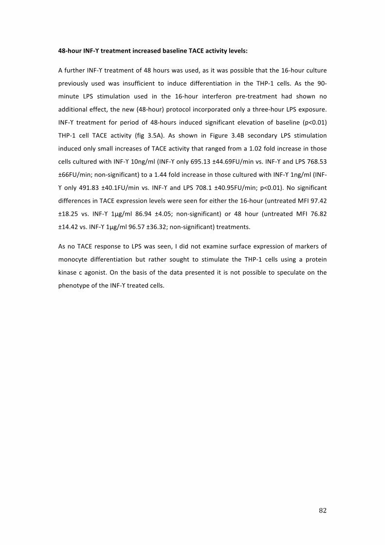
82
48-‐hour INF-‐ϒ treatment increased baseline TACE activity levels:
A further INF-‐ϒ treatment of 48 hours was used, as it was possible that the 16-‐hour culture
previously used was insufficient to induce differentiation in the THP-‐1 cells. As the 90-‐
minute LPS stimulation used in the 16-‐hour interferon pre-‐treatment had shown no
additional effect, the new (48-‐hour) protocol incorporated only a three-‐hour LPS exposure.
INF-‐ϒ treatment for period of 48-‐hours induced significant elevation of baseline (p<0.01)
THP-‐1 cell TACE activity (fig 3.5A). As shown in Figure 3.4B secondary LPS stimulation
induced only small increases of TACE activity that ranged from a 1.02 fold increase in those
cells cultured with INF-‐ϒ 10ng/ml (INF-‐ϒ only 695.13 ±44.69FU/min vs. INF-‐ϒ and LPS 768.53
±66FU/min; non-‐significant) to a 1.44 fold increase in those cultured with INF-‐ϒ 1ng/ml (INF-‐
ϒ only 491.83 ±40.1FU/min vs. INF-‐ϒ and LPS 708.1 ±40.95FU/min; p<0.01). No significant
differences in TACE expression levels were seen for either the 16-‐hour (untreated MFI 97.42
±18.25 vs. INF-‐ϒ 1μg/ml 86.94 ±4.05; non-‐significant) or 48 hour (untreated MFI 76.82
±14.42 vs. INF-‐ϒ 1μg/ml 96.57 ±36.32; non-‐significant) treatments.
As no TACE response to LPS was seen, I did not examine surface expression of markers of
monocyte differentiation but rather sought to stimulate the THP-‐1 cells using a protein
kinase c agonist. On the basis of the data presented it is not possible to speculate on the
phenotype of the INF-‐ϒ treated cells.

83
A
B
Figure 3.5: Effects of 48 hour INF-‐ϒ treatment on THP-‐1 cell TACE catalytic activity.
A: THP-‐1 cells with no LPS stimulation (n=3, * p<0.01 vs. untreated control)
B: THP-‐1 cells after secondary LPS stimulation 1μg/ml for three hours (n=3)

84
THP-‐1 cells were refractory to stimulation with phorbol-‐12-‐myristate-‐13-‐acetate (PMA):
The potent protein kinase c agonist PMA is known to produce TNF release from THP-‐1 cells
[384, 385]. It has also been shown to generate intra-‐cellular ROS and induce TACE substrate
shedding [380]. Doses as low as 30ng/ml of PMA have been shown to induce an increase in
TACE catalytic activity as rapidly as 10-‐15 minutes [381]. Hence cells were directly stimulated
with PMA to determine whether any increase in TACE activity could be elicited.
Undifferentiated THP-‐1 cells were incubated with PMA 1μg/ml for 0.5, 1 and 2 hours, before
being removed from culture conditions and entered into the FRET assay. PMA stimulation
had no effect on cellular TACE activity at the 0.5 hour (untreated 175.95FU/min vs. PMA
186.95FU/min), one hour (untreated 226.43 ±79.94FU/min vs. PMA 251.86 ±74.52FU/min)
or two hour (untreated 148.33 ±15.17FU/min vs. PMA 134.57 ±10.83FU/min) time points
studied (fig 3.7). TACE expression levels were not measured.

85
Figure 3.7: Undifferentiated THP-‐1 cells did not increase TACE activity in response to a PMA
stimulus.
THP-‐1 cells stimulated with PMA 1μg/ml for either 0.5 or 1H displayed no up-‐regulation of
TACE activity (n=1 for 0.5H, n=2 for 1H & 2H).

86
CD14 positive bead selection strategy produced satisfactory cell yields and viability whilst
platelet contamination was minimised by utilising a two-‐column separation strategy:
Purified monocytes were obtained from PBMCs by the use of differential centrifugation and
immune-‐magnetic bead selection (fig 3.8 A&B). The use of a second column with a smaller
surface area was shown to reduce platelet contamination to acceptable levels (platelet:
monocyte ratio reduced from 5:1 to 0.5:1) and increase purity of the eluted fraction without
prohibitively reducing cell retrieval. This is important as platelets express TACE and reduce
the signal to noise ratio of the FRET assay. Purity of the cell preparation was determined by
percentage of CD14+ cells as a proportion of all intact cells and was routinely greater than
90%. Typically cell yield was 60% as calculated by comparing CD14+ cell numbers pre and
post separation. Viability was greater than 90%.
Positively selected monocytes produced an expression independent increase in TACE
activity in response to LPS:
Similar to previous data obtained using a negative selection strategy, positively selected
monocytes produced an acute up-‐regulation of TACE activity when stimulated with LPS
1μg/ml for one hour (untreated 44.02 ±16.58FU/min vs. LPS 103.84 ±42.03FU/min; p<0.01,
fig 3.9 A). TACE activity increases seen on LPS stimulation ranged from a 1.75 -‐ 3.54 fold
increase with a mean value of 2.4. These increases in enzyme activity were accompanied by
a consistent, but non-‐significant, reduction in cell surface expression (untreated MFI 57.34
±24.98 vs. LPS MFI 51.76 ±17.02 stimulated; fig 3.9 B).

87
A B
Figure 3.8: Immune-‐magnetic bead separation using a CD14 positive selection strategy
produced monocyte preparations of high purity.
Representative flow cytometer plot for PBMCs with forward scatter (FSC-‐H) and side scatter
(SSC-‐H) labelled axis:
A -‐ Cells pre separation procedure
B -‐ Cells post separation procedure (two column).
The unlabelled population of cells circled in purple was occasionally present but was not a
consistent feature. These may be artefacts related to the isolation procedure.
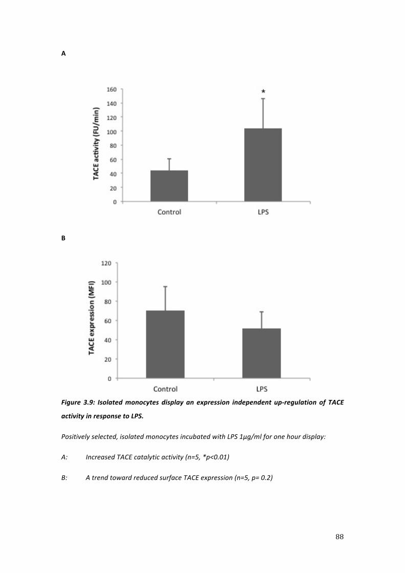
88
A
B
Figure 3.9: Isolated monocytes display an expression independent up-‐regulation of TACE
activity in response to LPS.
Positively selected, isolated monocytes incubated with LPS 1μg/ml for one hour display:
A: Increased TACE catalytic activity (n=5, *p<0.01)
B: A trend toward reduced surface TACE expression (n=5, p= 0.2)

89
Monocytes isolated by a positive selection strategy display increased TACE activity in
response to an LPS stimulus of one hour but not 30 minutes:
In order to compare the kinetics of the TACE activity increase using this new isolation
strategy, negatively selected and positively selected monocytes were stimulated with LPS
and placed in the FRET assay. Positive selection produced an attenuated TACE activity
increase at 30 minutes (untreated 18.4FU/min vs. LPS 22.2FU/min) but not at one hour
(untreated 20.8FU/min vs. LPS 53.4FU/min). This was in contrast to the negatively selected
monocytes where the activity increase was seen at 30 minutes (untreated 67.63FU/min vs.
LPS 109.8FU/min) as well as at one hour (untreated 62.1FU/min vs. LPS 123.6FU/min).
Hence LPS stimulations performed with positively selected monocytes were standardised to
one hour.
LPS induced TACE up-‐regulation is mediated in part through p38MAPK:
The rapid increase in TACE activity that is seen in response to an LPS stimulus has been
shown to be mediated, at least in part, through p38MAPK [133]. To confirm this within the
paradigm of our model, PBMCs were stimulated with LPS (1µg/ml) and subjected to the
intra-‐cellular staining protocol. Markers stained for, and measured using flow cytometry,
were: phosphorylated (phospho)-‐p38MAPK, MK-‐2 and ERK. As can be seen from the results
(fig 3.10) a signal was seen across all three markers. This signal reached significance for
phospho-‐p38MAPK at 15 minutes (451 ±128 vs. 145 ±85 at baseline, p<0.05). This was
repeated with freshly isolated monocytes and similar results obtained reinforcing our
previous data [133] that, in single hit conditions using static Eppendorf tubes, LPS induced
TACE up-‐regulation is mediated, in part, through p38MAPK.

90
A
B C
Figure 3.10: PBMCs stimulated with LPS produced measurable increases in activated MAPK
levels.
Markers of intracellular activation in response to LPS 1μg/ml for 0, 15 and 30-‐minutes.
Figures are for PBMCs gated on CD14+ population. For all three markers LPS 15 minutes
produces measurable increases in activated levels with reduction towards baseline by 30
minutes
N=3 for all, *p<0.05 vs. control
91
3.5 Discussion
For the project to progress it was fundamentally important to be able to generate a reliable
and viable supply of monocytes. Previously in the laboratory both primary human
monocytes and human cell lines have been used to provide this. Due to the expense
associated with previous methods of monocyte isolation [133], and because the FRET assay
had previously been partially validated with them, the pro-‐monocytic human cell line THP-‐1
was initially chosen as the cell source.
Undifferentiated THP-‐1 cells did not produce a TACE response to LPS
In primary human monocytes we have shown that the acute TACE response to LPS is
mediated through the CD14-‐TLR4 complex, intra-‐cellular ROS and p38MAPK [133]. It is
known that the THP-‐1 cell line in their undifferentiated form express low levels of CD14
[386] whilst it has also been noted that relatively high concentrations of LPS (≥1μg/ml) are
required to elicit a sol-‐TNF response in undifferentiated cells [387], possibly as a result of the
lower CD14 levels. However, despite this, some groups using undifferentiated THP-‐1 cells
have reported LPS induced ROS production [388] and phosphorylation of MAPK (p38 and
JNK) [389].
Previously within our lab we have used the cytokine INF-‐ϒ in these cells to induce
differentiation. This cytokine is likely to have multiple effects and can be considered “dirty”.
Encouraged by reports of LPS induced activation in THP-‐1 cells, despite the reports of low
CD14 levels, we decided initially to avoid using INF-‐ϒ and work with undifferentiated THP-‐1
cells. Unfortunately it rapidly became apparent that the acute TACE response to LPS was
lacking in these cells and we saw no response even at very high (10μg/ml) LPS
concentrations. There were however, some time dependent increases in non-‐stimulated
THP-‐1 cell TACE activity that presumably reflects some activation of the cells as a result of
the incubation process. A lack of responsiveness to LPS has previously been attributed to a
relative immaturity of the THP-‐1 cell [387]. Thus the next step was to try and induce
differentiation in the cells.

92
Differentiated THP-‐1 cells did not produce a consistent response to LPS
In a continued effort to avoid using INF-‐ϒ due to the concerns outlined above we considered
alternative methods of inducing differentiation. Previously both PMA and VD3 have been
used [383] to differentiate THP-‐1 cells. PMA activates protein kinase C [390] and has been
shown itself to activate both TACE [381] and induce TNF shedding [392] in cell lines, thus
may not be appropriate as an agent of differentiation in studying TACE biology.
VD3 acts via the vitamin D receptor [393] and has been used to induce differentiation of
THP-‐1 cells. It therefore seemed a rational agent to use in this context. We followed a
published protocol [383] that involved a 72-‐hour incubation with VD3. Cells were then
stimulated with LPS in an effort to induce an acute TACE response. No response was seen
which could reflect either a failure of the differentiation process or cellular hypo-‐
responsiveness due to contamination. Contamination of cells with LPS would produce a
“tolerant” cell phenotype that would be refractory to further LPS stimulation [322]. In
addition Mycoplasma contamination of THP-‐1 cells is known to attenuate TLR responses
[394] and is a plausible explanation for their lack of response to LPS, although the lack of
response to a non-‐TLR stimulus (PMA) is not in keeping with this last point.
We then employed an INF-‐ϒ differentiation protocol that had previously been successful at
inducing an acute TACE response in THP-‐1 cells [381]. This incorporated a 16-‐hour INF-‐ϒ
treatment followed by a secondary LPS stimulation. This protocol resulted in a small but
consistent TACE response to LPS. However this response did not reach statistical significance
nor approach the x3.3 fold activity increase that had been seen previously. It is possible that
the cells were not fully differentiated by the 16-‐hour time point so we extended the INF-‐ϒ
treatment to 48-‐hours. However, this treatment increased basal TACE activity further
reinforcing our concerns about “off target” inflammatory effects of INF-‐ϒ.

93
CD14 positive bead selection allows isolation of monocytes without attenuating their TACE
response to LPS
At this point the project was reappraised and we decided that an alternative source of cells
should be investigated. We had previously discovered limitations when examining TACE
biology in another cell line (MonoMac 6) so decided to identify a viable method of isolating
primary monocytes [381].
Previously within the group we had isolated monocytes using a magnetic activated cell
separation technique employing a negative selection strategy. In this technique non-‐
monocyte populations are labelled with antibodies attached to magnetic beads. The cell
mixture is then placed within a column that is within a magnetic field and labelled (non-‐
monocyte) cells are retained within the column and unlabelled cells (monocytes) pass
through. As the monocytes are unlabelled this technique may not alter their response
characteristics. However, this technique requires a number of different antibody-‐bead
combinations, is expensive and thus was not a plausible option for this project. A further
disadvantage of this technique is the presence of platelets as a contaminant of the eluted
fraction. Platelets express low levels of TACE meaning that the signal: noise ratio in the FRET
assay is reduced. Previously we had modified the isolation protocol to include anti-‐CD61
microbeads [133], further increasing the cost.
An alternative to the negative selection procedure outlined above is to positively select cells.
In this protocol the desired cells (in this case monocytes) are themselves labelled with
magnetic beads. Thus when placed within the column within a magnetic field the monocytes
are retained within a column and can be retrieved. As only one set of antibody labelled
beads is required this technique is cheaper but as the monocytes are labelled their response
characteristics may be affected. Nevertheless we decided to investigate this as an option for
isolating cells using an anti-‐CD14 antibody.

94
We rapidly established that this was an effective way of producing monocyte suspensions of
high purity (fig 3.8) and viability. By modifying the protocol so that two columns were used
in series, platelet contamination was reduced substantially providing optimised conditions
to detect the TACE response. I then demonstrated that cells isolated in this manner
produced an up-‐regulation of TACE activity in response to an LPS stimulus applied for an
hour. To ensure that the use of a CD14 binding antibody did not substantially alter the
response to LPS (mediated through CD14) we compared the TACE response to LPS in
negatively and positively selected cells. Although the activity increase seen in response to
LPS was apparent in both at one hour, it was apparent in only the negatively selected cells at
30 minutes. Hence all stimulations performed with positively isolated monocytes were
standardised to one hour.
Although non-‐significant, this TACE activation in the positively selected cells was
accompanied by a down-‐regulation in cell surface expression of the enzyme. Previously with
the negative selection technique we had not seen this. We hypothesise that this is due to
either shedding or stimulation induced down-‐regulation both of which have been described
in relation to TACE [369]. The fact that this was not seen in our previous work on the acute
TACE response [133] presumably reflects the difference in the duration of the LPS
stimulation (30 minutes vs. one hour), the different methods of monocyte isolation or both.
Thus, despite the importance of CD14 in LPS recognition and TLR-‐4 signalling, we observed
no obvious deficit in LPS-‐induced TACE activation at the one hour time point, compared to
the previously used negative selection method. This apparent lack of interference
presumably reflects the epitope specificity of the CD14 monoclonal antibody and the non-‐
saturating antibody concentrations recommended by the manufacturer (Miltenyi, UK). In
combination these two factors should ensure that any effects of the CD14 antibody on the
CD14-‐LPS signalling complex are minimised.
Isolated monocytes stimulated with LPS contained increased levels of activated MAPKs
The final part of work in this chapter was to provide a method for determining MAPK
responses. MAPKs exist in two isoforms, un-‐phosphorylated and phosphorylated with the
latter representing the activated isoform that is generated in response to LPS [133].
Previously we have used materials to fix and make permeable the cells, antibodies specific
for the phosphorylated MAPK isoform, and flow cytometry, to detect changes in MAPK

95
activation status. We modified and optimised this protocol in which PBMCs were fixed and
made permeable before being incubated with antibodies specific to the phosphorylated
forms of these enzymes. We were successful in identifying a signal across all three markers
(p38, MK-‐2, ERK) with significantly elevated levels of activated p38MAPK 15 minutes after
LPS stimulation. These results were in line with our previous findings [133] on LPS induced
signalling in static Eppendorfs.
Conclusions
In summary, in this chapter I successfully identified and optimised a viable and reproducible
method of providing sufficient quantities of monocytes to proceed with the project. I
outlined the kinetics of the acute TACE response and its upstream signalling in the form of
MAPK activation in these cells, and found them to be broadly in keeping with our previous
work in this area. This work was of fundamental importance to the project as the method
developed here underpins every subsequent piece of work contained within this thesis.

96
Chapter 4
Modulation of TACE catalytic activity during prolonged exposure to septic stimuli

97
Summary
Having established a suitable method of isolating monocytes, I next sought to determine
how the TACE activity profiles were altered by continuous exposure to a septic stimulus such
as LPS in vitro.
Monocytes are known to become tolerant to a second inflammatory stimulus when they are
exposed to a large initial stimulus. They can also be primed to subsequent inflammatory
stimuli by a prior low-‐dose stimulus. Such states may be considered similar to CARS and SIRS
respectively yet TACE activity has not been evaluated in tolerant or primed monocytes.
Changes in TACE activity due to priming or tolerance may impact on the levels and activity of
its membrane substrates and their soluble cleavage products.
I created a two-‐hit model using a culture insert made of polytetrafluoroethylene (PTFE), a
minimally adherent material, in order to prevent any spontaneous in-‐situ differentiation of
monocytes during the extended in vitro incubations. Monocyte TACE activity profiles were
determined over a range of different LPS treatments that were intended to induce
differential states of priming and tolerance. Soluble TNF levels were quantified in order to
definitively determine how these activity profiles related to monocyte inflammatory
balance/output. We found that a state of net tolerance was induced by 16-‐hour PTFE culture
with LPS at a concentration of 1μg/ml. Tolerant cells displayed elevated baseline activity and
an attenuated response to further LPS stimulation. However, we found no evidence of
priming with this in vitro model. We next sought to determine the kinetics of the MAPK
response to LPS within cells rendered tolerant by LPS, but found that untreated 16-‐hour
PTFE culture alone attenuated this response. This meant that the MAPK response was
directly affected by non-‐adherent culture. This prevented us from further analysing MAPK
responses in the two-‐hit model.

98
Finally we examined the behaviour of known TACE substrates within the model and found
that they were differentially affected. Consistent with a tolerant state, TNFR shedding was
reduced by LPS pre-‐exposure. In contrast, L-‐selectin shedding was reduced by PTFE culture
alone. Although we were successful in producing a net state of tolerance in cells, the clinical
relevance of these results is uncertain.
4.1 Background
We hypothesised that TACE plays a key role in determining monocyte inflammatory balance,
and therefore is a central regulator of both the hyper and hypo-‐responsive states that
determine sepsis pathophysiology [37]. Monocytes can become tolerant to an inflammatory
stimulus after large dose exposure and this can be considered hypo-‐responsive. Similarly
monocytes can be primed to an inflammatory stimulus and this can be considered hyper-‐
responsive. We attempted to induce these states (tolerance and priming) in monocytes
before measuring their TACE activity and substrate behaviour to determine whether the
enzyme was acting as an inflammatory regulator.
To try and induce the primed and tolerant phenotypes we decided to use a “two-‐hit”
strategy in which cells were exposed to two inflammatory stimuli. The first of these was
intended as a conditioning stimulus (of varying doses) designed to induce a state of priming
or a state of tolerance. The second was a standardised stimulus designed to reveal the
underlying response phenotype. LPS was used for both hits as it is the classical agent used to
induce tolerance [322] and has also been used to prime cells [316, 395, 396]. Other
investigators had induced priming and tolerance using protocols in which the two LPS hits
were 20-‐24 hours apart [315, 397]. In the first of these papers, Heremans et al
demonstrated priming in a murine model in which a footpad injection of LPS generated a
supra-‐normal response to a secondary LPS stimulus administered 24 hours later. In the
second, Porta et al generated an in-‐vitro model of tolerance in which monocytes were
incubated with LPS for 20 hours before being re-‐challenged with LPS. Given these two
studies we set the duration of the initial LPS culture at 16 hours and, in keeping with the
work detailed in Chapter Three, the duration of the second hit was standardised to one
hour. We also quantified soluble TNF release in response to a second LPS hit to allow us to
relate enzyme activity profiles to monocyte inflammatory output. As addressed in the
introduction, monocyte response states could be modulated through the release of TNFR-‐1
and 2. For example, shedding of TNFRs could bind any locally released sol-‐TNF, potentially

99
resulting in a tolerant state. The reverse process may increase local levels of sol-‐TNF and
result in a primed state. Therefore, I measured both the expression and LPS induced
shedding of TNFR-‐1 and TNFR-‐2. I also chose to measure the expression and LPS induced
shedding of the adhesion molecule L-‐selectin as adherence has been specifically implicated
in priming [378].
Previous methods of modelling tolerance and priming have used an adherence step to
isolate monocytes from PBMC populations [326], cultured bone marrow derived progenitors
[398] or cell lines [399]. Adherence has been demonstrated to induce differential effects of
IL-‐10 in monocytes [400] as well as enhancing TNF production [378, 401]. Therefore we had
significant concerns that placing monocytes in an adherent environment could induce
spontaneous activation/maturation and result in a change in their response phenotype.
In order to prevent any such change occurring and also to facilitate a gentle non-‐activating
cell retrieval process we used a minimally adherent material, polytetrafluoroethylene (PTFE),
a form of Teflon®. Teflon® has been used in the suspension culture of monocytes [402] with
PTFE though to adhere and activate the lowest numbers of monocytes [403].

100
4.2 Aims
The specific aims of this chapter were:
1. To quantify monocyte TACE activity profiles within a two-‐hit LPS model
2. Then determine whether response characteristics were consistent with
differential states of priming and tolerance
4.3 Protocols
Blood collection
30 ml of donor blood was obtained by venesection from healthy volunteers who had given
informed consent. Exclusion criteria were inter-‐current illness or immune-‐suppression.
Blood was extracted directly into vacutainers containing EDTA.
Monocyte stimulation for first LPS hit
Monocytes were isolated from whole blood using differential centrifugation followed by
magnetic activated cell sorting utilising a CD14 positive bead selection strategy as outlined in
detail in Chapter Two. Monocytes were then placed in PTFE inserts within 12 well plates at a
density of 3x10⁶/ml for 16-‐hours with or without LPS (1ρg/ml -‐ 1μg/ml). For determination
of TNFR-‐1 & 2, L-‐selectin and HLA-‐DR expression, PBMC, isolated from whole blood and
plated using the same protocol at 3x10⁶ CD14+ cells per ml, were used.
Where TACE activity was determined this was done as outlined in detail in Chapter Two.

101
Monocyte stimulation for second LPS hit
Cells were retrieved from culture and re-‐suspended at a density of 2x10⁶/ml in HBSS with 5%
FCS. They were subsequently placed in 1.5ml polypropylene Eppendorfs and stimulated
using LPS at a concentration of 1µg/ml for one hour at 37°C. Polypropylene Eppendorfs were
used as monocytes were LPS stimulated in these tubes during the work presented in Chapter
Three. For determination of TNFR-‐1 & 2, L-‐selectin and HLA-‐DR expression the same
protocol was used but with PBMCs at a density of 2x10⁶ CD14+ cells per ml. These densities
were lower than those used previously but were required to mitigate against volume (and
hence cell) losses during the protocol.
Interferon stimulation
Monocytes were placed in 16-‐hour PTFE culture with human INF-‐ϒ at different
concentrations. Cells were retrieved and their TACE activity determined. Where cellular
response to a subsequent LPS stimulation was determined, monocytes were placed in
culture conditions identical to those used for the second LPS hit (outlined above).
Soluble TNF quantification
Monocytes were stimulated for the first hit as above. Cells were subsequently retrieved and
re-‐suspended and left for an hour to allow release of any cell associated soluble mediators
(including TNF) that might have been carried over from the previous culture. They then
received an additional rinse before being re-‐suspended and stimulated with LPS 1μg/ml at
37°C for second LPS hit. Supernatants were collected at one, three and six hours with sol-‐
TNF levels determined by ELISA.

102
Quantification of MAPK response
This was performed as outlined in detail in Chapter Two. Briefly, cells were fixed and
permeabilised before being stained with antibodies specific for the activated
phosphorylated form of the MAPK enzyme. Cell stimulation was performed at the same
density and conditions as outlined for the TACE response. An LPS concentration of 1µg/ml
was used with monocytes stimulated for either 15 or 30 minutes.
103
4.4 Results
Evaluation of cell recovery and viability in 16-‐hour PTFE culture
After 16-‐hour PTFE culture, monocytes were retrieved by performing three rinses with cold
HBSS supplemented with 5% FCS. Gentle pipetting was employed to ensure minimal
activation during recovery of non-‐adherent cells. This process resulted in mean retrieval
rates of 53.12 ±8.95% of the total number of monocytes placed into 16-‐hour culture (both
for untreated and LPS-‐treated). Total retrieved cell viability was 73.68 ±7.08%.
After the second LPS hit in Eppendorfs, total retrieval rates fell further and typically were
between 20-‐30% of those placed in overnight culture. To avoid the requirement for
additional donor monocyte collection, the FRET assay was miniaturised by a factor of four
with cell density reduced four fold, compared to our previously published protocol [381]. A
series of internal validation experiments were performed to ensure that accuracy was
maintained. In these experiments monocytes were stimulated with LPS then TACE activity
values determined by both the old assay protocol (96 well plate with 100μl total volume)
and the new, miniaturised protocol (384 well plate with 25μl total volume). Results were
then compared with a variation of less than 10% seen across the protocols (figure 4.1).

104
Figure 4.1: Monocyte TACE activity as determined by the FRET assay was similar when assessed using the non-‐miniaturised and miniaturised protocols.
Data for monocyte TACE activity assessed using non-‐miniaturised (96 well plate, 1x10⁵ monocytes/well, 100μl final volume) and miniaturised (384 well plate, 2.5x10⁴ monocytes/well, 25μl final volume).
All experiments performed at 37°C
N=5 for all.

105
Monocytes in 16-‐hour PTFE culture had increased baseline TACE activity but no changes in
expression:
Having established that cells could be placed in PTFE culture and retrieved, we next sought
to establish the effect of this culture on their TACE activity. We found that untreated
monocytes after 16-‐hour PTFE culture displayed increased basal TACE activity when
compared to freshly isolated cells (44.02 ±16.58 FU/min freshly isolated vs. 82.64 ±18.82
FU/min after 16-‐hour culture, p<0.01 fig 4.2A). There were no differences in cell surface
expression of the enzyme (MFI 57.34 ±24.97 freshly isolated vs. 44.85 ±11.53 after 16-‐hour
culture, non-‐significant fig 4.2B).

106
A
B
Fig 4.2: Monocytes placed in untreated PTFE culture for 16-‐hours have an expression
independent increase in TACE activity.
A Monocyte TACE activity was higher in monocytes placed in 16-‐hour PTFE culture than
in freshly isolated cells (n=5 freshly isolated n=9 untreated culture, *p<0.01)
B There were no-‐significant changes in TACE expression (n=5)

107
Evidence for monocyte LPS-‐induced tolerance but not priming was seen in the two hit LPS
model
1. TACE response to primary LPS treatment.
We next moved to determine the effect of adding LPS to the PTFE culture. Monocytes were
placed in 16-‐hour PTFE culture with/without LPS at three different concentrations. These
were designed at a range of concentrations in order to explore sensitisation (low
concentrations) and de-‐sensitisation (high concentrations) of the secondary TACE response.
These concentrations, determined based on previous dose response experiments performed
within our laboratory [133], are listed below:
1. LPS 1pg/ml: low concentration
2. LPS 1ng/ml: intermediate concentration
3. LPS 1μg/ml: high (saturating) concentration
TACE activity after 16-‐hour culture with LPS at 1pg/ml was not different to that after
untreated culture (82.64 ±18.82 FU/min untreated vs. 74.62 ±24.29 FU/min LPS treated;
non-‐significant). The intermediate LPS concentration of 1ng/ml also had no effect (82.64
±18.82 FU/min untreated vs. 66.5 ±21.68 FU/min LPS treated; non-‐significant). In contrast
the high concentration of LPS 1μg/ml produced a significant increase in enzyme activity
(82.64 ±18.82 FU/min untreated vs. 137.17 ±62.97 FU/min LPS treated; p<0.05; fig 4.3A).
There were no significant differences in TACE expression between the groups (MFI 44.85
±11.53 untreated, MFI 48.34 ±0.26 LPS 1pg/ml, MFI 52.43 ±6.34 LPS 1ng/ml, MFI 42.16
±19.44 LPS 1μg/ml; non-‐significant, fig 4.3B).

108
A
B
Figure 4.3: TACE activity was increased after 16-‐hour culture with LPS 1μg/ml but
TACE expression was not affected.
A TACE activity after 16-‐hour PTFE culture (n=3-‐9, *p<0.05)
B TACE expression after 16-‐hour PTFE culture (n=3-‐9, non significant)
109
2. TACE response to secondary LPS treatment.
TACE activity profiles after secondary LPS stimulation were altered by 16-‐hour culture
conditions:
After 16-‐hour PTFE culture cells were exposed to a standardised secondary LPS stimulation
of LPS 1μg/ml for one hour. For the purposes of clarity the abbreviations below will be used
to describe culture conditions within the model. The concentration of the first LPS treatment
will be specified each time:
CC= Untreated 16h culture (C) + no secondary 1h treatment (C)
CL =Untreated 16h culture (C) + secondary 1h LPS treatment (L)
LC =LPS 16h culture (L) + no secondary 1h treatment (C)
LL= LPS 16h culture (L) + secondary 1h LPS treatment (L)
As might be expected, untreated monocytes left in PTFE for 16 hours significantly increased
their TACE activity (x1.58 fold) when exposed to secondary LPS stimulation (82.64
±18.82FU/min CC vs. 130.29 ±34.25FU/min CL, p<0.01). Addition of LPS to the 16-‐hour
culture resulted in a slight attenuation of this secondary response at both the low (x1.49 fold
increase in LL cells compared to LC cells) and intermediate (x1.51 fold increase in LL cells
compared with LC cells) concentrations (fig 4.4A). There was however, a marked attenuation
of this response in cells cultured for 16-‐hours with LPS at the high concentration. These cells
displayed no further TACE activity increase on secondary LPS exposure (137.17
±62.97FU/min LC vs. 137.05 ±60.53FU/min LL; non-‐significant, fig 4.4A). These results are
consistent with previous LPS exposure attenuating the TACE response to subsequent LPS
exposure. This could be considered as consistent with a state of tolerance.

110
Monocytes exposed to the two-‐hit LPS model had their membrane TACE expression
measured. There were no significant changes seen between any of the different culture
conditions. However, after the second LPS hit there was a consistent trend toward reduced
expression (untreated, CC MFI 44.85 ±11.53 vs. CL MFI 38.09 ±4.67; LPS 1pg/ml, LC MFI
48.34 ±0.26 vs. LL MFI 40.8 ±3; LPS 1ng/ml, LC MFI 52.43 ±6.34 vs. LL MFI 39.26 ±8.66; LPS
1μg/ml, LC MFI 42.16 ±19.44 vs. LL MFI 29.35 ±9.79; non-‐significant, fig 4.4B).

111
A
B
Figure 4.4: TACE activity and expression profiles in response to a secondary LPS
stimulus were altered by prior 16-‐hour PTFE culture with LPS
Grey bars represent values after 16-‐hour PTFE culture, black bars that after secondary LPS
stimulus applied.
A TACE activity profiles (n=9 unstimulated, n=3 1pg/ml, n=5 1ng/ml, n=7 1μg/ml,
*p=<0.01)
B TACE expression profiles (n=4 unstimulated, n=3 for all other LPS concentrations)
112
Similar activity profiles were exhibited with additional intermediate LPS concentrations
As we saw no evidence of in vitro priming with the low, intermediate and high
concentrations of LPS used in the 16 hour PTFE culture, we expanded them in an effort to
generate conditions that may result in the primed phenotype. Additional intermediate
concentrations of 10pg/ml, 100pg/ml and 10ng/ml were used during 16-‐hour PTFE culture;
there were no significant differences in enzyme activity levels seen in LC monocytes (other
than the previously described increase seen at 1μg/ml).
All LPS culture conditions attenuated the response to the secondary LPS stimulus (table 4.1
and fig 4.5).
Thus within the model we found no evidence for priming of the TACE response by prior LPS
exposure. High concentration (1μg/ml) exposure appeared to exhaust the TACE pathway,
significantly attenuating the response to secondary LPS stimulation in a manner that could
be in keeping with a state of tolerance.

113
Table 4.1: TACE activity profiles from two-‐hit LPS model:
Data displayed as mean ±SD
TACE activity values refer to FU/min
LPS concentration per
ml in 16-‐hour culture
(first hit)
0
1pg
10pg
100pg
1ng
10ng
1μg
TACE activity after first
LPS hit
82.64
±18.82
74.62
±22.49
77.17
±18.82
53.51
±26.74
66.5
±21.68
97.61
±13.21
137.17
±62.97
TACE activity after
second LPS hit
130.29
±34.25
111.42
±29.02
108.91
±8.97
68.12
±11.87
100.3
±30.22
123.63
±18.49
137.05
±60.53
Fold increase in TACE
activity on second hit
X1.58
X1.49
X1.41
X1.27
X1.51
X1.27
X1.0

114
Figure 4.5 TACE activity profiles from 2 hit LPS model
Complete TACE activity profiles for two-‐hit LPS model. Grey bars represent activity after 16-‐
hour PTFE culture, black bars that after secondary LPS stimulus applied.
N=9 un-‐stimulated
N=3 LPS 1pg/ml
N=2 for LPS 10pg/ml, 100pg/ml and 10ng/ml
N=5 LPS 1ng/ml
N=7 LPS 1μg/ml

115
Evaluation of monocyte surface marker alterations after model conditions:
Human monocytes mature from a CD14++CD16-‐ (immature) toward a CD14-‐CD16+ (mature)
phenotype during in vivo and vitro adherence culture [404]. In order to determine whether
monocytes were maturing during the 16-‐hour culture period we stained for both CD14 and
CD16. Results may show some evidence of maturation. Although CD14 levels were similar
between freshly isolated and CC cells (MFI 136±31 and 200±72 respectively; non-‐significant,
n=5) there was a non-‐significant trend toward higher CD16 levels in CC cells (MFI 35±14.6 in
freshly isolated cells vs. MFI 76±40 in CC cells; p=0.09). However, this increase in CD16 levels
did not reach the ten fold difference that has previously been reported when comparing
subsets [405]. There was a tendency toward elevated CD14 levels and reduced CD16 levels
in cells exposed to LPS (both LC and LL).
We also investigated the behaviour of the MHC class II antigen presenting molecule, HLA-‐
DR, which has been suggested as a clinically useful marker of tolerance in patients [224]. Its
expression levels are reduced on deactivated/tolerant cells [224] and have been suggested
as a means of stratifying risk in some patient groups [54, 225, 286, 295]. Freshly isolated
cells expressed detectable amounts of HLA-‐DR (MFI 484.43 ±99.98, fig 4.9D). As shown in
figure 4.6 HLA-‐DR expression was increased dramatically by 16-‐hour culture (baseline MFI
484.43 ±99.98 vs. untreated 16-‐hour culture MFI 5807.33 ±217.89). Expression levels were
reduced only marginally by the addition of LPS to the PTFE culture (LPS 1μg/ml MFI 4122.33
±819.38). These results are in keeping with other in vitro studies that have reported an initial
increase in HLA-‐DR expression levels followed by a fall [254, 406, 407].

116
Figure 4.6 Monocyte HLA-‐DR expression profiles before and after model conditions
N=3, *p<0.0001, **p<0.01
‘Freshly isolated’ refers to cells evaluated prior to PTFE culture Untreated culture to those
cultured in PTFE alone for 16-‐hours and LPS culture to those when LPS 1μg/ml was added to
PTFE inserts.

117
LPS pre-‐treatment, in keeping with a state of tolerance, suppressed sol-‐TNF levels in
response to the secondary LPS stimulation
In order to determine whether the TACE activity changes seen in the two-‐hit model mirrored
the overall changes in monocyte responsiveness, sol-‐TNF production was determined under
the different stimulation conditions. Firstly sol-‐TNF production was determined after 16-‐
hour PTFE culture only. Monocytes placed in untreated 16-‐hour culture (CC) produced less
sol-‐TNF than those that were cultured with LPS (78.0 ±30.27ρg/ml CC; 142.11 ±35.65ρg/ml
LC LPS 1pg/ml; 2610.67 ±406.73ρg/ml LC LPS 1ng/ml; 5041.63 ±2400.6ρg/ml LC LPS 1μg/ml;
fig 4.7A).
CL monocytes produced sol-‐TNF on secondary LPS stimulation (564.36 ±272.71pg/ml at six
hours). Previous exposure to LPS (LL cells) resulted in reduced responsiveness to LPS
stimulation for 6 hours in a dose dependent manner. This was most marked in those cells
exposed to LPS in PTFE culture at the intermediate concentration of 1ng/ml and the high
concentration of 1μg/ml (1ng/ml 70.81 ±24.68; 1μg/ml undetectable all at six hours; fig
4.7B).

118
A
B
Figure 4.7: Monocytes in 16-‐hour PTFE culture with LPS produced sol-‐TNF but not
when exposed to a secondary LPS stimulus
A Sol-‐TNF levels after 16-‐hour PTFE culture (n=3, *p<0.01 vs. control)
B Sol-‐TNF levels after secondary LPS stimulation (n=3, p< 0.05 control vs. LPS 1ng/ml
and 1μg/ml)
119
Monocytes in untreated 16-‐hour PTFE culture have an attenuated MAPK response to LPS:
To further understand the altered TACE activation and TNF production responses in PTFE
culture, and gain insight into the upstream signalling pathways affected, we evaluated the
MAPK pathway activation.
Cells were placed in untreated 16-‐hour culture conditions within PTFE inserts then removed,
centrifuged, re-‐suspended in Eppendorfs and stimulated with LPS 1 μg/ml. Activated
phosphorylated kinase levels were measured at baseline, 15 and 30 minutes by intracellular
flow cytometry. As can be seen (fig 4.8A) the up-‐regulation of phospho-‐p38MAPK expected
at 15 minutes was lost. A similar loss of up-‐regulation at all time points was seen on staining
for both phospho-‐MK2 and phospho-‐ERK (fig 4.8B &C).
These results were in keeping with an overall depression of the LPS inducible pathways
related to TACE activation and TNF expression.
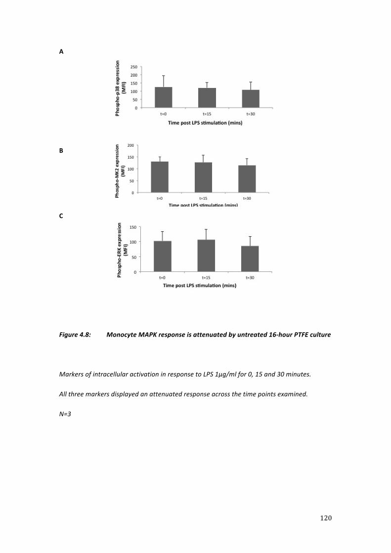
120
A
B
C
Figure 4.8: Monocyte MAPK response is attenuated by untreated 16-‐hour PTFE culture
Markers of intracellular activation in response to LPS 1μg/ml for 0, 15 and 30 minutes.
All three markers displayed an attenuated response across the time points examined.
N=3

121
Using INF-‐ϒ in 16-‐hour culture produced no evidence of a primed phenotype:
Given that we found no evidence of a primed phenotype using the two hit LPS model we
then altered the stimulus used in the 16-‐hour culture period. INF-‐ϒ is an immune-‐stimulatory
cytokine that is known to prime cells and reverse tolerance [408-‐410]. Hence this was added
to PTFE culture in place of LPS to induce the primed phenotype.
As shown in figure 4.9A the addition of INF-‐ϒ to the culture conditions increased monocyte
TACE activity (untreated 88.68 ±7.58FU/min; INF-‐ϒ 1ng/ml 186.54 ±4.12FU/min; INF-‐ϒ
100ng/ml 265.06 ±51.8FU/min). This was in keeping with our findings in the THP-‐1 cell line
(Chapter Three). There was no further increase in activity if monocytes were further
stimulated with LPS 1μg/ml (INF-‐ϒ 1ng/ml 207.45FU/min; INF-‐ϒ 100ng/ml 176.1FU/min)
meaning that, similarly to LPS, INF-‐ϒ did not produce priming.
There were no changes in TACE expression in the INF-‐ϒ treated cells (untreated control MFI
28.05 ±19.33; INF-‐ϒ 1ng/ml MFI 27.61 ±14.74; INF-‐ϒ 100ng/ml MFI 26.66; figure 4.9B).

122
A
B
Figure 4.9: Isolated monocytes cultured with INF-‐ϒ display increased TACE activity in
an expression independent manner
Monocytes placed in PTFE culture for 16-‐hours and stimulated with INF-‐ϒ at the doses shown
developed:
A Increased TACE activity
B No change in enzyme expression
N=2

123
Evaluation of constitutively expressed TACE substrate behaviour in PTFE culture
In order to further determine the TACE behaviour of freshly isolated monocytes as well as
cells exposed to the two-‐hit LPS model, we examined expression levels and shedding
patterns of constitutively expressed TACE substrates relevant to inflammation: TNFR-‐1,
TNFR-‐2 and L-‐selectin.
Freshly isolated cells
Freshly isolated monocytes had baseline substrate expression and shedding characteristics
determined. Results showed that freshly isolated cells express detectable amounts of all
three substrates (TNFR-‐1 MFI 20.93 ±0.3; TNFR-‐2 MFI 72.31 ±3.42; L-‐selectin MFI 72.27
±27.25; fig 4.11 A-‐C).
In response to an LPS stimulus of 1μg/ml for one hour shedding of the three TACE substrates
was seen (representative histograms shown in fig 4.10). In order to quantify this percentage
shedding of each available substrate was calculated for freshly isolated cells stimulated for
an hour with LPS 1μg/ml.
This was done using the equation below:
(Baseline expression – expression after LPS 1μg/ml one hour) X 100 / Baseline expression
Percentage shedding of each substrate was calculated for freshly isolated cells (baseline
shedding values in freshly isolated cells: TNFR-‐1 87.8 ±0.41%; TNFR-‐2 87.7±0.23%; L-‐selectin
94.4 ±0.53%; fig 4.12).
Percentage shedding of these substrates in response to LPS 1μg/ml for one hour was then
determined after untreated and LPS treated 16-‐hour PTFE culture.

124
A B
C
Figure 4.10: Representative histograms demonstrating shedding of TACE substrates in response to LPS stimulation in freshly isolated monocytes.
A L-‐selectin
B TNFR-‐1
C TNFR-‐2
For each histogram black curve represents non-‐stimulated controls whereas grey represents cells stimulated with LPS 1μg/ml for 1 hour at 37°C in Eppendorf culture.

125
Effect of untreated and LPS treated PTFE culture on TACE substrate expression and shedding
Untreated – refers to cells placed in untreated PTFE culture for 16 hours. Cells then had
expression and percentage shedding in response to an LPS stimulus of 1μg/ml for one hour
determined for each TACE substrate.
LPS treated – refers to cells placed in PTFE culture for 16 hours with the high concentration of
LPS (1μg/ml) that produced maximal changes in TACE behaviour previously. Cells then had
expression and percentage shedding in response to an LPS stimulus of 1μg/ml for one hour
determined for each TACE substrate.
TNFR-‐1
Untreated 16-‐hour PTFE culture resulted in a reduction in TNFR-‐1 expression (freshly
isolated MFI 20.93 ±0.3 vs. untreated culture MFI 6.47 ±2.46; fig 4.11A). Shedding rates in
response to LPS 1μg/ml for an hour were preserved at 75.9% from the 87.8% seen in freshly
isolated cells (fig. 4.12A).
The additional of LPS to the 16-‐hour culture conditions resulted in no net change in TNFR-‐1
expression (MFI 6.63 ±1.8; fig 4.11A). Shedding rates on secondary LPS stimulation were
reduced to 21% (fig 4.12A).
TNFR-‐2
Untreated 16-‐hour PTFE culture did not alter TNFR-‐2 expression (freshly isolated MFI 72.31
±3.42 vs. untreated culture MFI 92.3 ±0.58; fig 4.11B). Shedding rates in response to LPS
1μg/ml for an hour were reduced to 67.2% from the 87.7% seen in freshly isolated cells (fig
4.12B).
The addition of LPS to the 16-‐hour culture conditions resulted in increased TNFR-‐2
expression (MFI 254.2 ±26.28; fig 4.11B). Shedding rates on secondary LPS stimulation were
further reduced to 9.7% (fig 4.12B).
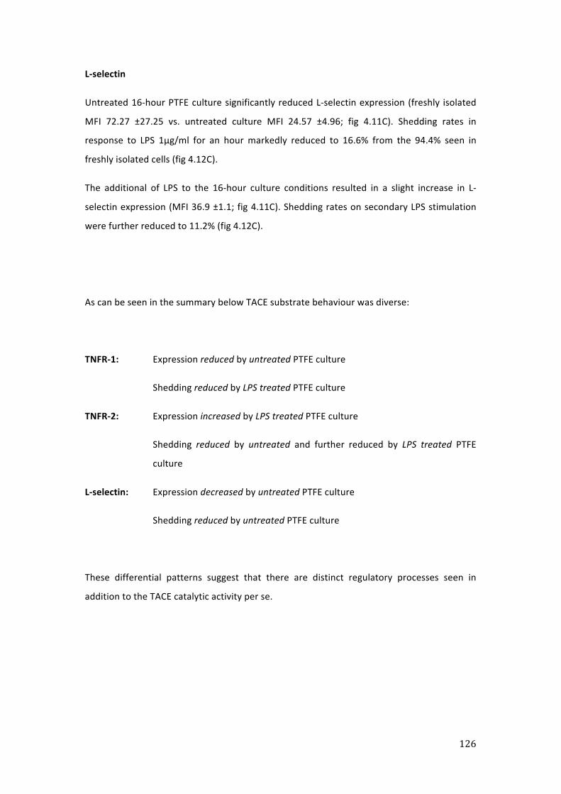
126
L-‐selectin
Untreated 16-‐hour PTFE culture significantly reduced L-‐selectin expression (freshly isolated
MFI 72.27 ±27.25 vs. untreated culture MFI 24.57 ±4.96; fig 4.11C). Shedding rates in
response to LPS 1μg/ml for an hour markedly reduced to 16.6% from the 94.4% seen in
freshly isolated cells (fig 4.12C).
The additional of LPS to the 16-‐hour culture conditions resulted in a slight increase in L-‐
selectin expression (MFI 36.9 ±1.1; fig 4.11C). Shedding rates on secondary LPS stimulation
were further reduced to 11.2% (fig 4.12C).
As can be seen in the summary below TACE substrate behaviour was diverse:
TNFR-‐1: Expression reduced by untreated PTFE culture
Shedding reduced by LPS treated PTFE culture
TNFR-‐2: Expression increased by LPS treated PTFE culture
Shedding reduced by untreated and further reduced by LPS treated PTFE
culture
L-‐selectin: Expression decreased by untreated PTFE culture
Shedding reduced by untreated PTFE culture
These differential patterns suggest that there are distinct regulatory processes seen in
addition to the TACE catalytic activity per se.
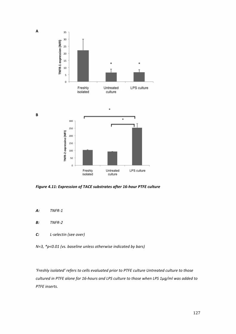
127
A
B
Figure 4.11: Expression of TACE substrates after 16-‐hour PTFE culture
A: TNFR-‐1
B: TNFR-‐2
C: L-‐selectin (see over)
N=3, *p<0.01 (vs. baseline unless otherwise indicated by bars)
‘Freshly isolated’ refers to cells evaluated prior to PTFE culture Untreated culture to those
cultured in PTFE alone for 16-‐hours and LPS culture to those when LPS 1μg/ml was added to
PTFE inserts.
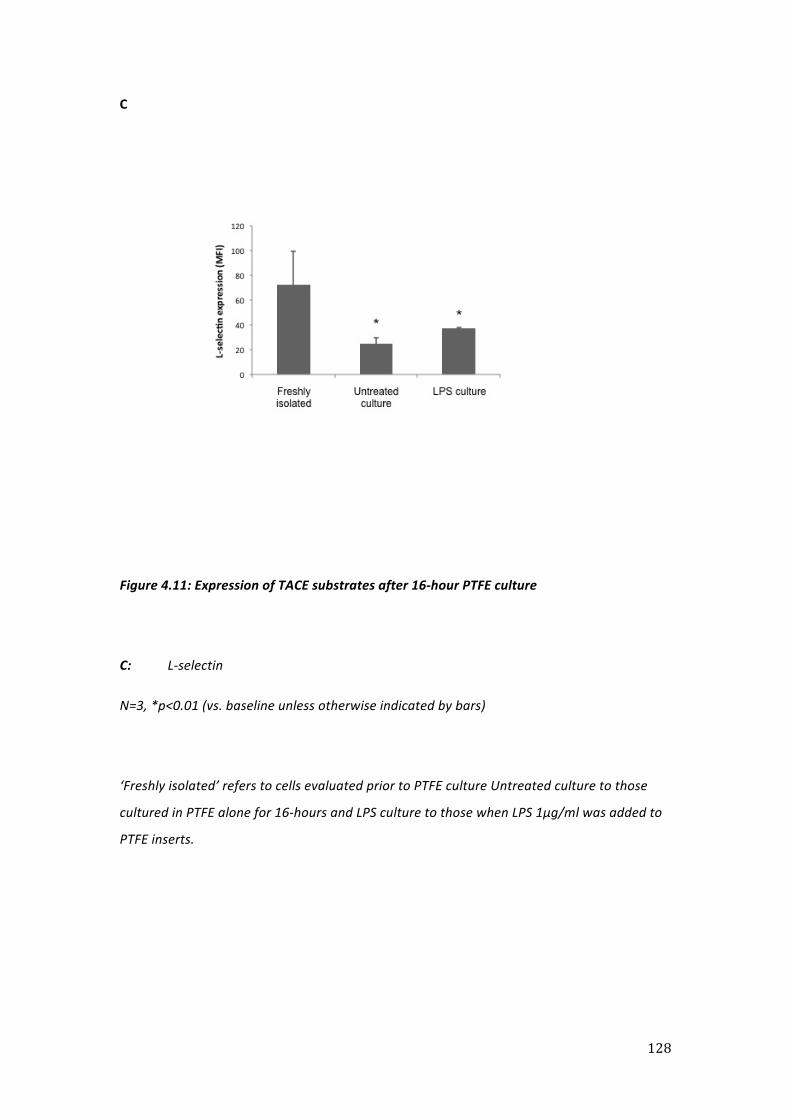
128
C
Figure 4.11: Expression of TACE substrates after 16-‐hour PTFE culture
C: L-‐selectin
N=3, *p<0.01 (vs. baseline unless otherwise indicated by bars)
‘Freshly isolated’ refers to cells evaluated prior to PTFE culture Untreated culture to those
cultured in PTFE alone for 16-‐hours and LPS culture to those when LPS 1μg/ml was added to
PTFE inserts.
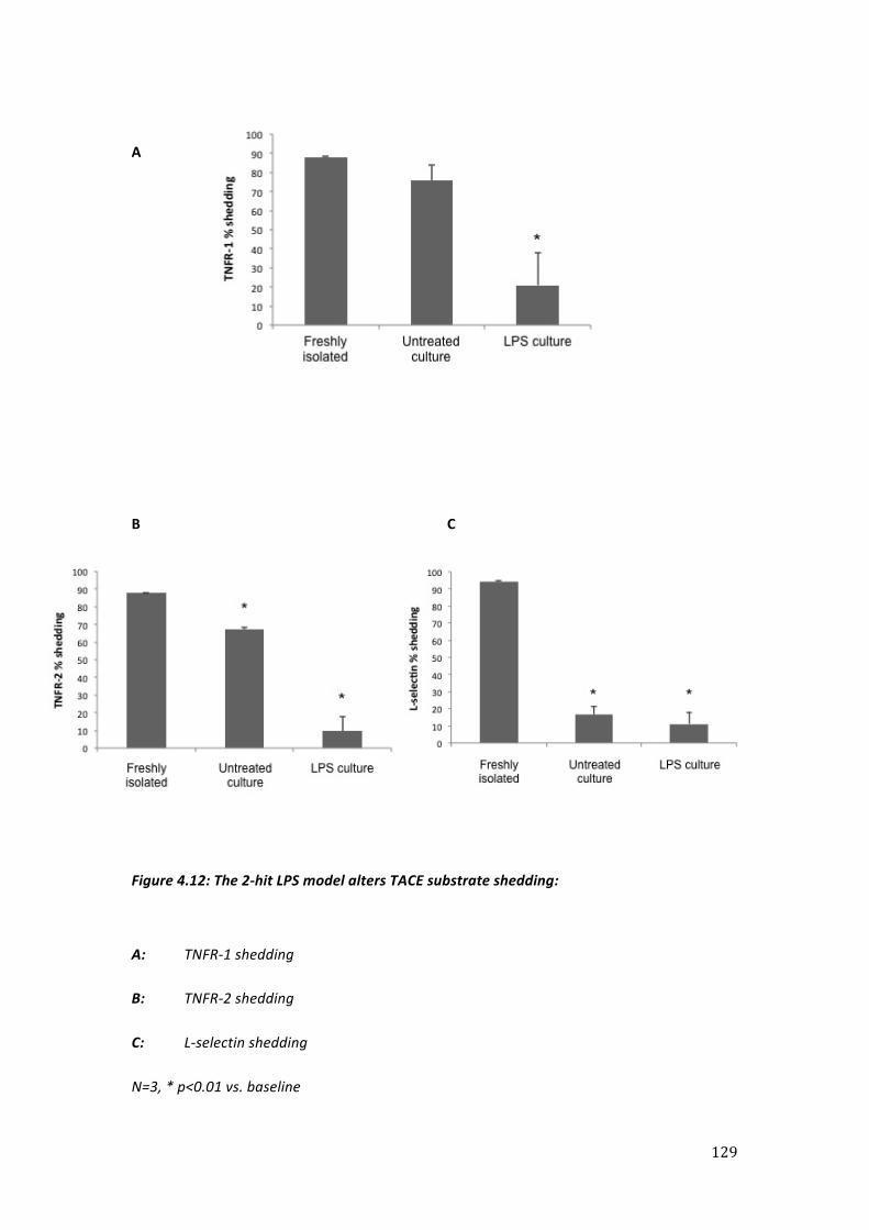
129
A
B C
Figure 4.12: The 2-‐hit LPS model alters TACE substrate shedding:
A: TNFR-‐1 shedding
B: TNFR-‐2 shedding
C: L-‐selectin shedding
N=3, * p<0.01 vs. baseline

130
4.5 Discussion
I used a two-‐hit LPS model in an attempt to induce differential states of tolerance and
priming in monocytes isolated utilising the methods developed in Chapter Three. Being able
to model these response states would provide further insights into the immune response,
potentially identifying mechanisms that could be manipulated or targeted for clinical benefit
in sepsis patients. Therapies aimed at augmenting the immune response may be appropriate
in patients displaying a tolerant phenotype whereas those patients displaying a primed
phenotype may benefit from immune suppression. Whilst signalling alterations seen in
endotoxin tolerance have been investigated both in vitro and in vivo, the role of TACE is yet
to be evaluated.
As outlined in the background to this chapter, the construction of the two-‐hit LPS model was
limited by the need to use isolated monocytes in sufficient numbers to run the FRET assay
[381]. Additionally, we were concerned that monocytes may differentiate over the 16-‐hour
culture period potentially altering their phenotype. To try and prevent this we used PTFE cell
culture inserts during this period. Cells cultured in these inserts formed a thin monolayer
over the PTFE membrane when visualised by microscopy and were relatively easily retrieved
from culture conditions in a viable and functional state.
Increased baseline TACE activity after untreated culture may represent activation or
maturation
We demonstrated that the monocytes placed in untreated 16-‐hour PTFE culture had
significantly elevated baseline TACE activity but no change in membrane expression. Hence
we surmise that this represents a true increase in activity per se. There is more than one
explanation as to what this may represent. Firstly, it is known that cells of the
monocyte/macrophage cell line are able to become activated in vitro and in vivo [411, 412],
and the increased levels of activity seen may represent such a state. Although we used a
minimally adherent medium and a 16-‐hour culture period to try and induce a degree of
quiescence in the cells, there may be some activation. If this is true it is likely that some
degree of “basal” TACE activity is likely to be unavoidable using in vitro systems.
The second explanation for the increase in activity is maturation. Monocytes can
differentiate into dendritic cells or macrophages [413] and it is known that both MAPK
signalling and the cellular response to LPS are altered in macrophages [414, 415]. In our
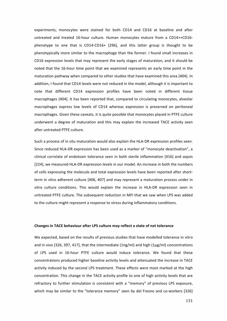
131
experiments, monocytes were stained for both CD14 and CD16 at baseline and after
untreated and treated 16-‐hour culture. Human monocytes mature from a CD14++CD16-‐
phenotype to one that is CD14-‐CD16+ [296], and this latter group is thought to be
phenotypically more similar to the macrophage than the former. I found small increases in
CD16 expression levels that may represent the early stages of maturation, and it should be
noted that the 16-‐hour time point that we examined represents an early time point in the
maturation pathway when compared to other studies that have examined this area [404]. In
addition, I found that CD14 levels were not reduced in the model, although it is important to
note that different CD14 expression profiles have been noted in different tissue
macrophages [404]. It has been reported that, compared to circulating monocytes, alveolar
macrophages express low levels of CD14 whereas expression is preserved on peritoneal
macrophages. Given these caveats, it is quite possible that monocytes placed in PTFE culture
underwent a degree of maturation and this may explain the increased TACE activity seen
after untreated PTFE culture.
Such a process of in situ maturation would also explain the HLA-‐DR expression profiles seen.
Since reduced HLA-‐DR expression has been used as a marker of “monocyte deactivation”, a
clinical correlate of endotoxin tolerance seen in both sterile inflammation [416] and sepsis
[224], we measured HLA-‐DR expression levels in our model. An increase in both the numbers
of cells expressing the molecule and total expression levels have been reported after short-‐
term in vitro adherent culture [406, 407] and may represent a maturation process under in
vitro culture conditions. This would explain the increase in HLA-‐DR expression seen in
untreated PTFE culture. The subsequent reduction in MFI that we saw when LPS was added
to the culture might represent a response to stress during inflammatory conditions.
Changes in TACE behaviour after LPS culture may reflect a state of net tolerance
We expected, based on the results of previous studies that have modelled tolerance in vitro
and in vivo [326, 397, 417], that the intermediate (1ng/ml) and high (1µg/ml) concentrations
of LPS used in 16-‐hour PTFE culture would induce tolerance. We found that these
concentrations produced higher baseline activity levels and attenuated the increase in TACE
activity induced by the second LPS treatment. These effects were most marked at the high
concentration. This change in the TACE activity profile to one of high activity levels that are
refractory to further stimulation is consistent with a “memory” of previous LPS exposure,
which may be similar to the “tolerance memory” seen by del Fresno and co-‐workers [326]

132
and used to describe refractoriness to an ex vivo LPS stimulus within their model of
tolerance. Thus these changes are in keeping with a net state of tolerance. The
refractoriness of the TACE response to secondary LPS stimulation does not seem to
represent the maximal inflammatory output of the cell, as 16-‐hour INF-‐ϒ culture resulted in
TACE activity levels that exceeded those seen with LPS culture.
The definitive evidence that we modelled a net state of tolerance comes from the
attenuated levels of sol-‐TNF levels generated after the second LPS “hit”. It is interesting that,
although this attenuation was significant for LPS pre-‐treatments of 1ng/m and 1μg/ml, there
was a trend toward a similar pattern at a dose of 1pg/ml, a dose that we expected to
generate priming. These patterns were consistent with the attenuated increase in TACE
activity seen on secondary LPS stimulation at all doses. This may be explained by considering
what is happening on a sub-‐cellular level. During the culture period the cells are exposed to
a stimulus (LPS) that, in the case of tolerance, has been demonstrated to up-‐regulate
numerous negative regulators including IRAK-‐M, ST2 and the short version of MyD88 [324].
Interestingly there appears to be some discordance between murine and human models of
tolerance with only IRAK-‐M having been confirmed as being elevated across both species
[208, 326, 327]. In humans this elevation is seen in monocytes taken from patients with both
septic [418] and non-‐septic aetiologies [326].
The signalling pathways involved in priming are not as well elucidated but are thought to
include an up-‐regulation of inflammatory effectors acting through IRAK-‐1 [419]. Any change
elicited in cell signalling within our model is likely to be induced at a maximal level by the
constant presence of LPS throughout the 16-‐hour culture period. Hence a reduced response
to a subsequent LPS stimulus is perhaps to be expected and may explain why the increase in
activity induced by this secondary stimulus is reduced as the concentration of LPS used in
PTFE culture is increased. This represents a general limitation of the model.
Changes in MAPK signalling were induced by PTFE culture
We found that untreated 16-‐hour culture in PTFE produced an attenuation of the MAPK
response to LPS. As we thought that cells might be maturing during this period it is possible
that this process was altering the signalling characteristics of the monocytes. However,
MAPK signalling is similar across monocyte subsets and still present in macrophages [415],
hence these changes are unlikely to be explained by in situ maturation alone. We have

133
previously shown that primary human monocytes mediate their TACE response to LPS
through p38MAPK [133]. Therefore an attenuation of this MAPK signalling is in keeping with
the reduced TACE response to secondary LPS stimulation that we report after both
untreated and treated PTFE culture compared to freshly isolated cells. Attenuation of MAPK
activation has been reported in models of tolerance [329, 330], but to our knowledge, has
not previously been measured in PTFE culture.
Given this attenuation of MAPK and TACE signalling, we had concerns that placing
monocytes in minimally adherent conditions would induce a state of anergy where cells
would become globally unresponsive. However, several factors indicated that this was not
the case. Elevated TACE activity levels, coupled with changes in CD14, CD16 and HLA-‐DR
levels in response to LPS gave us evidence that cells were capable of responding after PTFE
culture. The increase in CD14 in response to 16-‐hour LPS culture is in keeping with previous
findings [420]. CD16 has been suggested as a possible metalloprotease substrate [421] and
the rapid reduction in levels of this cell surface marker after cells were exposed to LPS would
be in keeping with shedding, suggesting that some cellular enzymatic functions are
preserved. Further evidence that cells were capable of responding in PTFE culture is
provided by their retained ability to produce sol-‐TNF during the 16-‐hour LPS treatment
period. It is known that adherence of the monocytic cell line THP-‐1 to collagen results in
p38MAPK activation [422]. Thus, the absent p38MAPK response to LPS after untreated PTFE
culture, may represent signalling alterations as a result of the minimally adhesive conditions
used, another potential limitation of our model.
Alternate TACE substrate behaviour
It is possible that TACE substrate specificity is altered by chronic LPS exposure; hence
increased enzyme activity may not always result in increased sol-‐TNF production. This would
mean that the altered TACE activity profiles (increased baseline & reduced response to LPS)
seen in cells after two LPS hits may represent cleavage of substrates other than mem-‐TNF. It
is difficult to imagine that TACE sheds its range of substrates [342] without some degree of
selectivity as some (e.g. TNF and TNFR-‐1&2) may have opposing effects.
With specific regard to L-‐selectin, shedding can be induced through several different
pathways [377]. It is possible that differential signalling may produce conformational
changes in either TACE [365], or in upstream signalling that result in altered substrate
134
specificity. This latter point is consistent with work suggesting that signalling pathways in
tolerant cells are altered in a manner that renders the cell unresponsive to some stimuli (e.g.
LPS) but responsive to others (e.g. IL-‐10) [324]. In order to investigate possible changes in
substrate selectivity further we determined the shedding kinetics of other TACE substrates
relevant to inflammation.
We found that PTFE untreated and LPS treated culture produced differential effects on TNFR
and L-‐selectin expression. However, all three substrates (TNFR-‐1, TNFR-‐2 & L-‐selectin) had
shedding impaired by both treated and untreated PTFE culture when compared to freshly
isolated cells. Hence we found no convincing evidence of altered substrate
affinity/specificity.
The increase in TNFR-‐2 expression seen after 16-‐hour LPS culture is in keeping with previous
reports suggesting that it is inducible both in vitro [423] and in vivo whereas, again
consistent with our findings, TNFR-‐1 is not [424]. Pedron et al found that TNFR-‐2 shedding in
response to LPS is p38MAPK dependent but did not find the same for TNFR-‐1 [425]. Hence
the increased TNFR-‐2 expression may represent p38MAPK impairment after LPS culture. The
fact that this is not seen for TNFR-‐1 may reflect differential upstream signalling between the
receptors. Both TNF receptors are normally rapidly shed in response to LPS [426] and L-‐
selectin is shed in response to a number of stimuli such as LPS, IL-‐8, antibody cross-‐linking
and changes in tonicity [427-‐429]. The impaired TNF receptor shedding seen after LPS
culture could be considered to be in keeping with a state of tolerance. The impaired L-‐
selectin shedding seen after untreated 16-‐hour culture is anomalous in this context but
perhaps explainable, as both PKC and p38MAPK have been implicated in upstream signalling
to L-‐selectin. Hence shedding could occur despite impaired p38MAPK signalling, such as that
seen in our model.
No evidence for priming was seen in the model
Based on previous studies we hypothesised that a low concentration of LPS (e.g. 1pg/ml) in
the 16-‐hour PTFE culture should prime the cells to a subsequent LPS challenge in a
Shwartzman-‐like reaction [395] [315]. We saw no evidence for priming in either the TACE
activity, expression or sol-‐TNF values. Priming of monocytes by LPS has previously been
described in Teflon® conditions but for enhanced superoxide release rather than TNF [430]
and encouraged by this we persisted, changing the stimulus used in PTFE culture to INF-‐ϒ.
135
However, this produced a similar pattern to that seen when we used THP-‐1 cells (Chapter
Three) and with LPS in PTFE culture: high baseline activity but no inducibility.
It is striking when reviewing the literature that other studies using LPS to prime cells have
either found adherence to be key [318] or have used adherent systems [319, 320]. Other
studies have used in vivo systems [315, 316] hence it is plausible that, in keeping with our
hypothesis that monocytes are acutely responsive to their microenvironment, our system
fails to recapitulate all the necessary environmental cues required for priming. These are
likely to include adherence and cell-‐cell signalling/contact and the absence of these
represents another limitation of our model. These omissions may have been compounded
by our decision to use a purified TLR-‐4 stimulus rather than whole bacteria. Furthermore, it
has been demonstrated that leukocytes produce different amounts of cytokines (including
TNF and IFN-‐Υ) when stimulated in whole blood as compared to PBMC culture [431]. Any
further work attempting to induce the primed state in monocytes should specifically
examine the role of these factors.
Conclusions
We set out to create a model in which we could explore TACE activity profiles for evidence
of both priming and tolerance. Despite using a range of doses, the primary LPS treatment did
not prime in any way to secondary LPS, while only a very high concentration suppressed the
secondary response effectively. This absence of priming may result from the non-‐adherent
culture or the fact that cells spent a prolonged period of time in culture with LPS. Further
work in this area should incorporate different lengths of LPS conditioning and incorporate a
washing step, whereby LPS is removed from the culture conditions, prior to any secondary
LPS exposure. The effect of using whole bacteria rather than a purified TLR stimulus should
also be evaluated.
The environment monocytes were exposed to during the 16-‐hour culture seemed to induce
a state of “pure” tolerance in which almost all aspects of inflammatory signalling examined
were attenuated. The system therefore seems ‘non-‐interactive’ i.e. the only way to change
the dynamic is to treat with a dose of LPS (1ug) that triggers the TACE activating pathway
and exhausts it. It is possible that lower doses may well trigger elements of the TACE
signalling mechanism (such as p38MAPK) but these underlying perturbations (not actual
TACE activation) have no effect, neither enhancing nor suppressing. We were unable to

136
examine these in detail because the PTFE culture conditions themselves blunted this
response to LPS.
In conclusion, although the model produced changes in TACE behaviour that could be
considered in keeping with a net state of tolerance, our data exposed its limitations. These
related to the presence of LPS and the use of a minimally adherent medium throughout the
16-‐hour culture period. These are compounded by the use of a single cell type (monocytes),
and a purified TLR-‐4 stimulus (LPS) meaning that it is unlikely my model correctly
recapitulates the complex interplay that occurs in vivo. However, this is a criticism that can
be levelled at in vitro models in general and it may be better to limit the use of such models
to testing specific hypothesises generated using in vivo systems.
Thus it may be the case that the model has limited application to clinical reality, yet the
techniques optimised whilst constructing it allowed us to refine our protocols for the clinical
study that we performed next.
137
Chapter 5
Determination of monocyte TACE activity and associated shedding profiles in SIRS
and sepsis
138
Summary
There are parallels between in vitro endotoxin tolerance and the in vivo phenomenon of
monocyte deactivation. Monocyte deactivation occurs in response to a number of different
pathologies and is characterised by reduced inflammatory cytokine production together
with reduced HLA-‐DR expression and impaired antigen presentation. However, it is
incompletely understood and, at present is poorly categorised. I sought to determine
whether TACE activity and substrate shedding in critically ill patients with monocyte
deactivation would be altered when compared to healthy controls, and whether similarities
with the tolerance model detailed in Chapter Four could be identified. In addition, we hoped
to enhance understanding of deactivated monocyte functionality.
Blood samples (day 0, 2, 4 and 6) were taken from mechanically ventilated patients who had
the systemic inflammatory response syndrome (SIRS). PBMCs were obtained via density
gradient centrifugation. An optimised flow cytometry protocol was designed in order to
identify all monocytes subpopulations in these preparations. However, samples obtained
from patients displayed a large variation in subset surface markers. It appears that accurate
determination of monocyte subsets may not be possible in the context of critical illness with
the surface markers we used, a limitation that may not have been fully appreciated. Instead
all monocytes were analysed together as one population, and identified as deactivated on
the basis of reduced HLA-‐DR expression and sol-‐TNF production on ex vivo LPS stimulation.
Monocytes were successfully isolated from patient samples using the CD14+ selection
protocol generated in Chapter Three and their TACE activity profiles were quantified using
the FRET assay. Monocyte TACE basal and LPS-‐induced activity profiles were altered
between healthy volunteers and patients, and between patients with septic and non-‐septic
SIRS. By contrast, cell surface TACE expression levels were not different between groups.
Sepsis produced attenuated phosphorylated p38MAPK and TACE responses to LPS as well as
producing increased expression and impaired shedding of TNFR-‐1. Expression and shedding
of TNFR-‐2 and L-‐selectin were similar across the groups.
These results suggest that the systemic inflammatory response may produce a
heterogeneous response (as opposed to a homogeneous anergy) in monocytes exposed to a
single inflammatory stimulus. Thus cells appear to be ‘reprogrammed’ with altered
regulation in specific pathways. Interestingly the preserved L-‐selectin shedding with reduced
TACE activity and TNF responses provide evidence that cell migration may be preserved.
139
5.1 Background
Monocyte deactivation was first identified in the 1990s in septic patients [154, 224]. This
phenomenon of reduced HLA-‐DR expression, impaired antigen presentation and reduced
sol-‐TNF release [224] has since been described in conditions producing sterile inflammation
such as trauma [432], burn injury [433], cardiopulmonary bypass [416] and surgery [51, 53].
Hypo-‐reactivity of immature, developing monocytes released as a result of systemic stress
has been suggested as a mechanism of deactivation [433], however more recent work has
demonstrated that HLA-‐DR is down regulated across cells of differing maturity making this
explanation unlikely [53]. It has also been suggested that phagocytic uptake of apoptotic
cells, found in increased numbers in sepsis [255], down regulates HLA-‐DR expression and sol-‐
TNF release by monocytes, however at present the mechanism through which deactivation
occurs is not known. Soluble mediators suggested as inducing deactivation include LPS, IL-‐
10, catecholamines [154, 434-‐436] and steroids [437].
As well as being incompletely understood monocyte deactivation is poorly categorised.
Work in this area has often focused on sepsis and attempted to correlate the degree of HLA-‐
DR down regulation to clinical outcomes. Some investigators have used the term ‘immune
paralysis’ when referring to patients who have <30% of monocytes expressing HLA-‐DR [224,
438], and suggested that these patients have worse clinical outcomes than others who have
greater numbers of cells expressing the molecule [286]. However, a recent review failed to
find a threshold in HLA-‐DR expression that successfully predicted unfavourable outcomes in
sepsis [225] and there is no accepted consensus around threshold levels or in measurement
techniques. Measurement of monocyte HLA-‐DR expression in these studies is usually based
on limited, rather simplistic flow cytometry gating strategies such as forward and side
scatter properties and expression of single markers (e.g. CD14) [51, 286, 295], both of which
are known to vary considerably between monocyte subsets [297], and may be further
altered during disease. Given the broad range of aetiologies that produce deactivation,
some have argued it represents a more complex reprogramming of the cell. This
reprogramming is generated by the systemic CARS response and may produce differential
effects on specific cellular pathways and functions [416, 439].
Several studies have shown that reduced HLA-‐DR expression can be reversed by granulocyte
macrophage colony stimulating factor (GMCSF) both in vitro and in vivo [440, 441]. In a
study of patients with multi-‐factorial sepsis, who were stratified to receive GMCSF therapy
on the basis of reduced HLA-‐DR expression levels, it was found therapy shortened

140
ventilation and length of ICU stay [226]. However, a recent meta-‐analysis concluded that, at
present, GMCSF did not have sufficient evidence to support its use in sepsis [282], although
only the study mentioned above used HLA-‐DR expression levels to guide treatment.
There is evidence that TACE behaviour is of importance as a determinant of the in vivo
inflammatory response. Neutrophil TACE expression has previously been shown to increase
in septic SIRS and to correlate with both disease severity and mortality [355]. TACE has been
shown to regulate the pulmonary endothelial inflammatory response to LPS [442], and
inflammatory mediators released by this process have been implicated in the generation of
remote organ injury [443]. TACE knockout results in reduced inflammatory cytokine release
and a mortality benefit in a murine peritonitis model [444]. Given these findings and the fact
that previous clinical work has found correlations between morbidity scores and soluble
TNF: TNFR receptor ratios [339] a detailed investigation of the monocyte functional TACE
response is well justified and yet to be performed, making our work novel.
We therefore decided to conduct a clinical study investigating monocytes obtained from
sepsis/SIRS patients and focusing on TACE behaviour and substrate shedding in deactivated
cells. By focusing on a single pathway that is involved in both acute inflammation [133] as
well as leukocyte rolling and tethering (through the effect of TACE on L-‐selectin [158]) we
aimed to determine whether these linked elements were differentially affected or globally
hypo-‐responsive in response to a single, defined microbial stimulus, and if the patterns
observed differed under sterile versus infectious SIRS conditions. Hence monocytes may
display evidence of reprogramming as well as biomarker changes that may be specific for
sepsis-‐induced deactivation. In addition we hoped to generate a more sophisticated flow
cytometry gating strategy than has previously been used in studies examining deactivation
in order to accurately identify monocytes and their subsets.

141
5.2 Aims
The specific aims for this chapter were:
1. To determine the effects of sepsis/SIRS on TACE behaviour and substrate
shedding.
2. To explore these data for evidence of differential effects on inflammatory
signalling and leukocyte rolling/tethering.
3. To develop an optimised flow cytometry protocol to accurately identify
monocytes and their subsets.
5.3 Protocols
Patient selection
An independent research ethics committee approved the study (North London REC 3
reference: 10/H0709/77) and we successfully achieved our aim of recruiting 24 patients. This
sample size was based on the calculation that to have 90% power to detect a 50% difference
in TACE activity between the two groups (p<0.05) 24 patients would be required. Intubated
and ventilated patients admitted to Charing Cross Hospital with SIRS, between May 2010
and April 2012, were screened by myself to see if they were eligible. We recruited patients
within 48 hours of admission to the ICU, and limited recruitment to those who were
expected to remain intubated for a further 48 hours or more. We excluded those with
immunosuppression and those who were receiving granulocyte colony stimulating factor. As
patients were incapacitated at the time of inclusion, consent was obtained from the
patient’s personal (relative/friend) or professional consultee. Retrospective consent was
then obtained from the patient once they had regained capacity. Appendix 2 contains copies
of the information sheets, consent forms and the study protocol.
Enrolled patients were subdivided them into non-‐infectious SIRS (from here on referred to
as SIRS patients) and sepsis on the basis of their clinical picture at the point of recruitment.
Standard definitions for SIRS and sepsis as outlined in detail in section 1.2 of the
introduction were used (see protocol in appendix 2 on page 270 for further details). I
recruited patients within 48 hours of admission to the ICU, and limited recruitment to those
who were expected to remain intubated for a further 48 hours or more. I excluded those
with immunosuppression and those who were receiving granulocyte colony stimulating

142
factor. As patients were incapacitated at the time of inclusion, consent was obtained from
the patient’s personal (relative/friend) or professional consultee. Retrospective consent was
then obtained from the patient once they had regained capacity.
In addition to recruiting patients from the ICU, we recruited healthy volunteers from
laboratory staff at Imperial College London. Fifteen samples were obtained and volunteers
were excluded if they refused to consent, were immunosuppressed, had chronic
pathology/inter-‐current illness and had recently undergone venesection.
Sample collection
Blood (30ml) was obtained from patients at 48-‐hour time points from baseline (D0) to D6 or
unit discharge. Samples were obtained from arterial cannulas wherever possible. Where
these were not present samples were taken from central venous catheters. EDTA
vacutainers were used for sample collection and cells transported on ice to the laboratory
facilities where they underwent immediate processing.
A single blood sample (30ml) was taken from each healthy volunteer. This was obtained by
peripheral venesection into EDTA vacutainers (on ice) and processed immediately.
Monocyte isolation
Monocytes were isolated from whole blood using density gradient centrifugation followed
by magnetic activated cell selection utilising a CD14 positive bead selection strategy as
outlined in detail in Chapter Two. Cells were stimulated with LPS and their TACE catalytic
activity determined by the FRET assay.
PBMC treatment/stimulation
PBMCs were isolated from whole blood by differential centrifugation. HLA-‐DR staining was
performed immediately after isolation to minimise any change in signal. Cells were then re-‐
suspended at 5x10⁶ CD14-‐positive cells/ml and incubated for one hour at 37°C in the
presence or absence of LPS (1μg/ml). Surface expression of TNFR-‐1, TNFR-‐2, TACE and L-‐
selectin was determined by flow cytometry. In some cases CCR-‐2 was also measured.
143
To determine ex vivo sol-‐TNF production, cells were plated and left for four hours at 37°C
with or without LPS (1μg/ml). Supernatants were then collected and sol-‐TNF levels were
measured using ELISA.
Quantification of MAPK response
This was performed as outlined in detail in Chapter Two. Briefly, PBMCs were fixed and
made permeable before being stained with antibodies specific for the activated form of the
MAPK enzyme. An LPS concentration of 1µg/ml was used with monocytes stimulated for
either 15 or 30 minutes.

144
5.4 Results
Patient recruitment and demographic information:
Patient details are given in table 5.1 and consisted of eight SIRS patients and sixteen with
sepsis. As can be seen in the table the groups were well matched apart from a younger
mean age in the SIRS group. Patients were recruited using the documentation provided in
Appendix One, and outcome data (ICU and hospital discharge) was available for all. Further
details about the source of infection within the sepsis group are provided in table 5.2. Of
these patients only eight (50%) generated positive microbiological data. Four (25%) cultured
gram-‐positive organisms, three (18.75%) cultured gram-‐negative organisms and one patient
(6.25%) both.
Although we recruited patients that we expected to remain on ICU for at least 48 hours (and
within 48 hours of intubation) some were discharged before a second sample could be
taken. This, in combination with the fact that some patients improved rapidly and were
discharged from ICU ahead of expectations, meant that serial samples were obtained from
14 patients (58%). Seven patients (29%) provided three and four samples.
A single sample was also obtained from 15 healthy volunteers recruited from Imperial
College staff. This group had a mean age of 33.3 (±3.6) and was 60% (n=9) male.

145
Table 5.1: Demographic and outcome data
Demographic data obtained from SIRS and sepsis groups as well as further detail from SIRS
group detailing admitting team specialty and from sepsis group outlining site of sepsis.
SIRS
N=8
Sepsis
N=16
P-‐value
Age, mean ±SD
51 (17.3)
67 (16.4)
<0.05
Male sex, n (%)
5 (63)
10 (63)
0.73
Medical, n (%)
6 (75)
12 (75)
1.0
APACHE II, median (IQR)
20
(15.5-‐23.75)
19
(16-‐21)
0.62
ICU LOS in days, median (IQR)
8
(4.75-‐13)
11.5
(6.5-‐15.75)
0.37
ICU mortality, n (%)
2 (31%)
5 (31%)
0.75
Hospital mortality, n (%)
2 (31%)
5 (31%)
0.75
Referring team for SIRS
patients
N (%)
Medicine 2 (25)
Neurosciences 6 (75)
Sepsis site N (%)
Pulmonary 9 (56)
Peritoneal 3 (18.8)
Soft tissue 2 (12.6)
Biliary 1 (6.3)
CNS 1 (6.3)

146
Monocytes could be isolated from patient samples using the CD14 bead positive-‐selection
strategy.
Previously, in pilot experiments in our laboratory, isolation of monocytes from septic
patients using the mononuclear cell separation and negative selection method resulted in
low purity levels due to significant neutrophil contamination. In this study, PBMC prepared
from septic patient blood by density gradient centrifugation contained variable levels of
contamination with neutrophils (fig 5.1A). These levels ranged from 0% to 60% of events on
the forward scatter (FSC; size) and side scatter (SSC; granularity) gates. However, using the
positive selection method, these neutrophils could be completely removed during the
magnetic bead isolation process (figure 5.1B). Yields and purity were in line with those
described during validation work with healthy volunteer blood in Chapter Three.

147
A
B
Figure 5.1: Isolation of monocytes from patient samples by positive, CD14 immune-‐
magnetic bead selection
A: Patient sample showing neutrophil infiltration of PBMC preparation
B: Enrichment of monocytes after CD14 bead selection, with counting beads used to quantify
cell numbers.

148
Development of an optimised, flow cytometry gating strategy to identify monocytes in
SIRS and sepsis patients
To accurately describe the monocyte response by flow cytometry, we aimed to analyse all
monocytes rather just those cells conforming to the classical CD14++CD16-‐ phenotype.
However, most well-‐defined flow cytometric gating and analysis methods for monocytes are
standardised for normal blood leukocytes and may be incapable of dealing with
unpredictable phenotypic marker changes occurring during acute or prolonged SIRS. Our
previous protocol for examining monocytes in humans under normal or mild inflammatory
conditions employed FSC and SSC characteristics combined with expression of HLA-‐DR,
which is not present on NK cells and neutrophils [53]. In order to measure HLA-‐DR in
severely ill patients where there is potential for considerable reductions of its expression,
we devised a gating strategy that was independent of this marker. An alternative and more
definitive marker for monocytes (M-‐CSF receptor, CD115), was unsuitable because of its
rapid TACE-‐dependent cleavage in response to LPS and other stimuli [316, 445]. We
therefore considered additional and more stable markers to ensure detection of monocytes.
Density gradient PBMC preparations from healthy individuals normally contain only
lymphocytes and monocytes. However, patient samples contained significant numbers of
neutrophils (fig 5.1A), presumably due either neutrophilia or changes in their physical
properties. Our first step was to exclude granulocytes on the basis of their combined higher
SSC and lower FSC profile relative to monocytes (gate R1 on fig 5.1A). The majority of
lymphocytes were then excluded by gating on a CD11b high population (gate R2 on fig 5.1B).
The major remaining non-‐monocyte population were NK cells, which express moderate
levels of both CD11b and CD16. If not excluded these cells could potentially contaminate the
monocyte CD14-‐CD16+ subset gate. Neural cell adhesion marker (NCAM) or CD56 is an
antigen expressed by natural killer (NK) cells [446] but not monocytes [297]. Thus staining
for CD56 provided us with a means of differentiating between NK cells and monocytes. Cells
from the R1 gate shown in figure 5.1A were gated on both CD11b and CD56; this identified
two groups, a monocyte population that was CD11b+CD56-‐ (R2 on fig 5.1B) and an NK cell
population that was CD11b+CD56+ (R3 on Fig 5.1B). Evaluation of cells within the R2 gate
using CD14 and CD16 expression profiles (fig 5.2C) indicated the majority of CD11b+CD56-‐
events were comparable to the monocyte CD14/CD16 continuum described in the literature.
In addition, CD14/CD16 gating of cells thought to be NK cells and found in R3
(CD11b+CD56+) confirmed that they were CD14-‐CD16+ (fig 5.1D) as expected.

149
A
B
A: Granulocytes were excluded on the basis of their combined higher SSC and lower FSC
profile relative to monocytes by drawing gate around cells consistent with monocytes (gate
R1)
B: Cells within the R1 gate were then gated on CD11b and CD56; this identified two
populations
CD11b+CD56-‐ (R2): monocytes
CD11b+CD56-‐ (R3): NK cells (R2 & R3)
B and T lymphocytes are excluded by these gates and are found within the un-‐gated CD11b-‐
CD56-‐ population.
C: Population R2: monocytes displaying an appropriate CD14 and CD16 expression
continuum (see next page).
D: Population R3: NK cells (CD11b+ CD56+) were confirmed to be CD14-‐CD16+ (see next
page).
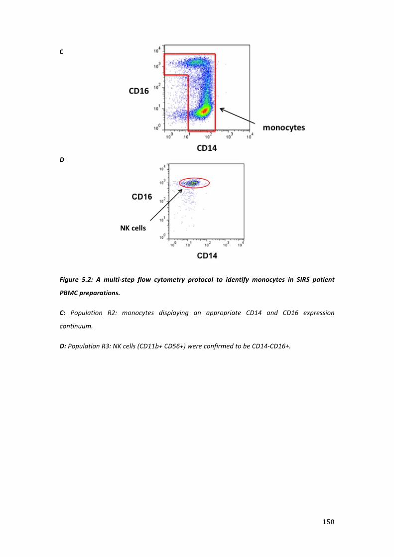
150
C
D
Figure 5.2: A multi-‐step flow cytometry protocol to identify monocytes in SIRS patient
PBMC preparations.
C: Population R2: monocytes displaying an appropriate CD14 and CD16 expression
continuum.
D: Population R3: NK cells (CD11b+ CD56+) were confirmed to be CD14-‐CD16+.
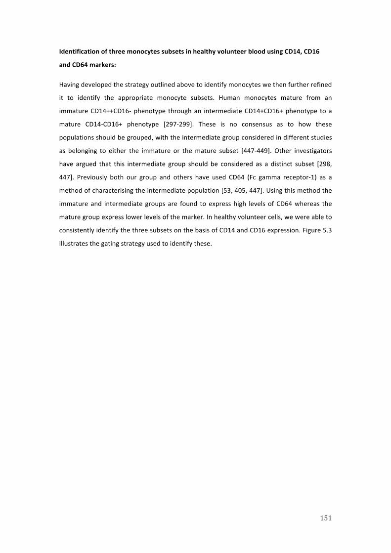
151
Identification of three monocytes subsets in healthy volunteer blood using CD14, CD16
and CD64 markers:
Having developed the strategy outlined above to identify monocytes we then further refined
it to identify the appropriate monocyte subsets. Human monocytes mature from an
immature CD14++CD16-‐ phenotype through an intermediate CD14+CD16+ phenotype to a
mature CD14-‐CD16+ phenotype [297-‐299]. These is no consensus as to how these
populations should be grouped, with the intermediate group considered in different studies
as belonging to either the immature or the mature subset [447-‐449]. Other investigators
have argued that this intermediate group should be considered as a distinct subset [298,
447]. Previously both our group and others have used CD64 (Fc gamma receptor-‐1) as a
method of characterising the intermediate population [53, 405, 447]. Using this method the
immature and intermediate groups are found to express high levels of CD64 whereas the
mature group express lower levels of the marker. In healthy volunteer cells, we were able to
consistently identify the three subsets on the basis of CD14 and CD16 expression. Figure 5.3
illustrates the gating strategy used to identify these.

152
A B
C
Figure 5.3: In healthy volunteer cells CD64 allowed accurate identification of the
intermediate subset
A: Monocytes gated as previously described in figure 5.1 were subdivided into immature
CD14++CD16-‐ subset (R1) and CD16+ (R2) cells (R2 contains both intermediate and mature
subsets).
B: CD14++ cells were gated on CD64 in order to provide a reference point as to where the
intermediate monocyte gate should be drawn.
C: CD16+ monocytes could then be subdivided on the basis of CD64 expression into CD64+
cells (R4), and CD64-‐ cells (R5).
D: The CD64+ cells (R4) consisted of the intermediate monocyte subset that is CD14+CD16+
(see next page).
E: The CD64-‐ cells (R5) contained mature CD14-‐CD16+ monocytes (see next page).
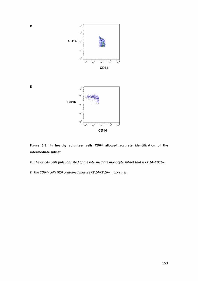
153
D
E
Figure 5.3: In healthy volunteer cells CD64 allowed accurate identification of the
intermediate subset
D: The CD64+ cells (R4) consisted of the intermediate monocyte subset that is CD14+CD16+.
E: The CD64-‐ cells (R5) contained mature CD14-‐CD16+ monocytes.

154
Evaluation of monocyte subset gating in SIRS patients:
We attempted to apply the subset gates to patient samples. Although we had previously
successfully used this strategy in delineating subsets in patients undergoing elective surgery,
we had not previously used it in critically unwell patients. Applying the subset gates in this
context proved problematic, as alterations in expression of individual markers led to
distortion of the subset classification patterns common in healthy individuals.
As shown in figure 5.4 the utility of CD64 in differentiating between the intermediate and
mature subsets was reduced and often completely lost (fig 5.5). In the example shown in
figure 5.4, lower CD64 expression was no longer confined to CD14-‐CD16+ monocytes, which
could be interpreted as decreased CD64 expression on the intermediate CD14+CD16+
subset. An additional uncertainty was apparent in the definition of the classical
CD14++CD16-‐ and intermediate CD14+CD16+ subsets where a continuum of CD16
expression (fig 5.5B) prevented identification of discreet populations. An extreme instance
was observed in one patient where the CD14-‐CD16+ expressing monocytes were effectively
absent during the course of sampling (fig 5.5C). It is likely that some of these patterns can be
attributed to alterations in expression of the Fcγ receptors CD16 and CD64, both of which
are modified during monocyte activation [309, 450-‐453], independently of the subset
differentiation process.
We considered that using monocyte subset gating regions based on the standard pattern in
healthy individuals could lead to segregation of populations according to altered functional
marker (e.g. CD16) expression rather than true subset identity. Hence, rather than attempt
such arbitrary gating and division of phenotype, we decided to perform our analysis on the
monocyte population as whole and exclude this as an analysis variable.
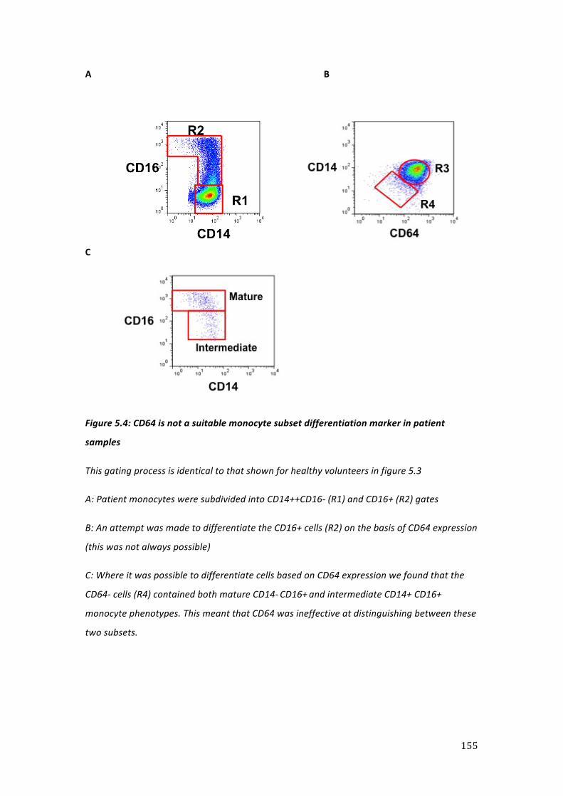
155
A B
C
Figure 5.4: CD64 is not a suitable monocyte subset differentiation marker in patient
samples
This gating process is identical to that shown for healthy volunteers in figure 5.3
A: Patient monocytes were subdivided into CD14++CD16-‐ (R1) and CD16+ (R2) gates
B: An attempt was made to differentiate the CD16+ cells (R2) on the basis of CD64 expression
(this was not always possible)
C: Where it was possible to differentiate cells based on CD64 expression we found that the
CD64-‐ cells (R4) contained both mature CD14-‐ CD16+ and intermediate CD14+ CD16+
monocyte phenotypes. This meant that CD64 was ineffective at distinguishing between these
two subsets.

156
A B
C
Figure 5.5: Different surface marker profiles were seen within the monocyte gate:
A: Typical pattern of surface marker expression seen within the monocyte gate in healthy
volunteers
B: Surface marker expression seen within the monocyte gate in a patient with pneumonia
C: Surface marker expression seen within the monocyte gate in a patient with aspiration
pneumonia

157
Total monocyte and neutrophil count differed between the groups:
Monocyte and neutrophil counts were obtained from the white cell differential that each
patient had measured daily whilst on the ICU. Both SIRS and sepsis patients displayed a
monocytosis that trended toward higher levels in the SIRS group than sepsis. As expected
the neutrophil count increased across both groups, this was more marked in sepsis patients
than in SIRS patients. Total cell counts for each group are shown in figure 5.6.

158
A
B
Figure 5.6: Monocyte counts tended to be higher in the SIRS group whilst neutrophils
tended to be higher in sepsis.
Median neutrophil and monocyte counts for each group.
A: SIRS and sepsis patient’s (all patients within each group) monocyte count (x109/L)
B: SIRS and sepsis patient’s (all patients within each group) neutrophil count (x109/L)
Patient data shown as median and IQR. Normal range shown on x axis (median, 5th to 95th
centile).

159
Monocyte HLA-‐DR expression was reduced in SIRS and sepsis:
To confirm that the patient monocytes were deactivated, we measured membrane
expression of HLA-‐DR. In keeping with our expectations, levels of HLA-‐DR at baseline (D0)
were attenuated across both groups when compared to healthy volunteer subjects (fig 5.7A
& B). The degree of attenuation at baseline between the two patient groups was similar
(MFI 627.28 ±306.33 healthy controls vs. 162.12 ±154.08 SIRS and 192.81 ±184.27 sepsis).
Within the SIRS group the degree to which HLA-‐DR levels were reduced remained relatively
constant between D0 and both D2 (166.86 ±154) and D4 (137.95 ±91.18). At D6 HLA-‐DR
levels had increased to exceed those obtained at baseline (316.09 ± 214.68, fig 5.3A) but did
not approach the levels seen in healthy controls. In contrast, for sepsis patients levels
continued to fall at both D2 (111.25 ±118.71) and D4 (76.89 ±100.62).
There was no convincing evidence of a recovery in HLA-‐DR expression levels across the
groups by the end of the sampling timetable.

160
A
B
Figure 5.7: Monocyte HLA-‐DR expression was reduced across both groups when compared
to healthy volunteers (HV)
A: SIRS group, * p<0.05 vs. HV
B: Sepsis group, * p<0.05 vs. HV
Analysis of transformed data with graphical illustration of backlog data (mean & 95% CI)
For both patient groups D0 is sampling baseline, D2 is day two, D4 is day four and D6 is day 6

161
Sol-‐TNF production with ex vivo LPS stimulation was reduced in SIRS and sepsis:
Deactivated monocytes, in addition to expressing reduced levels of HLA-‐DR, have been
shown to produce reduced levels of sol-‐TNF upon ex vivo stimulation with LPS [224]. In
order to further confirm that cells obtained from these patients were deactivated, we
measured sol-‐TNF production after four-‐hour LPS stimulation.
As expected, healthy volunteer PBMCs produced high levels of sol-‐TNF during the
stimulation period (4102.81 ±3397ρg/ml). In contrast, cells from patients of both the SIRS
and sepsis groups produced markedly lower levels of the cytokine when exposed to the
same culture conditions (501.38 ±594.9ρg/ml SIRS D0 vs. 548.22 ±1011.6ρg/ml sepsis D0, fig
5.8). A similar pattern of reduced sol-‐TNF production was seen across the groups at D2
(936.65 ±730.9ρg/ml SIRS vs. 826.97 ±944.9ρg/ml sepsis), D4 (444.07 ±159ρg/ml SIRS vs.
837.53 ±811.4ρg/ml sepsis) and D6 (618.14 ±245.58ρg/ml SIRS vs. 474.2 ±450.1ρg/ml
sepsis).
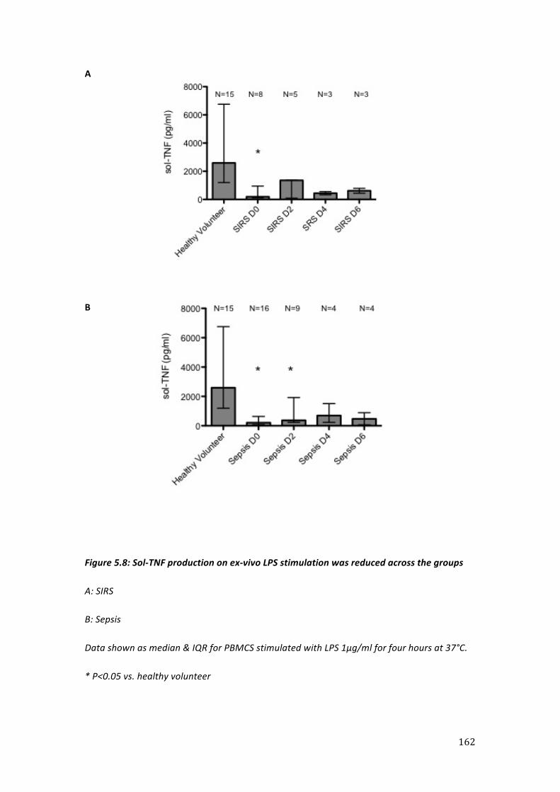
162
A
B
Figure 5.8: Sol-‐TNF production on ex-‐vivo LPS stimulation was reduced across the groups
A: SIRS
B: Sepsis
Data shown as median & IQR for PBMCS stimulated with LPS 1μg/ml for four hours at 37°C.
* P<0.05 vs. healthy volunteer

163
Higher baseline TACE activity was seen at D6 in the SIRS group:
Having confirmed that cells were deactivated, we then went on to determine TACE activity
on isolated monocytes. In SIRS patients, monocytes had similar un-‐stimulated TACE activity
(Fig 5.9A) at baseline (D0) to those taken from healthy volunteers (32.74 ±14.13 HV vs. 45.74
±27.35 FU/min). Activity was not significantly different at either D2 (24 ±7.86) or D4 (47.02
±11.9). By D6, in keeping with the results obtained for HLA-‐DR expression in this group,
baseline TACE activity levels were significantly increased (86.26 ±31.9) as compared to
healthy volunteers (fig 5.9A, p<0.01). There were no significant differences in TACE
expression (table 5.2).
Similar to previous results, healthy volunteers increased their TACE activity in response to
LPS stimulation (fig 5.9B) by between x1.73 to x4.4 fold (un-‐stimulated 32.74 ±14.13FU/min
vs. stimulated 89.85 ±42.31FU/min; p<0.001). This increase, whilst attenuated (Fig 5.9) was
not significantly different in SIRS compared to healthy volunteers.

164
Healthy
volunteer
SIRS patients
D0
SIRS patients
D2
SIRS patients
D4
SIRS patients
D6
Un-‐stimulated
monocyte TACE activity,
mean (±SD)
32.74
(14.13)
45.74
(27.35)
24 (7.86)
47.02 (11.9)
86.26 (31.9)
Stimulated monocyte
TACE activity, mean
(±SD)
89.85
(42.32)
77.05
(22.66)
69.14
(35.84)
89.8 (28.82) 129.88
(27.66)
Monocyte TACE
expression, MFI (±SD)
55.45
(24.84)
52.41
(34.24)
40.92
(29.68)
55.3 (37.22) 40.35
Table 5.2: TACE activity profiles for SIRS patients’ monocytes.
Values are given for both un-‐stimulated cells and those stimulated with LPS. Un-‐stimulated
monocyte TACE expression for each day is also shown.

165
A
B
Figure 5.9: Monocytes from patients with SIRS displayed increased un-‐stimulated TACE
activity but LPS induced activity was not significantly affected.
A: Un-‐stimulated monocyte TACE activity (mean + 95%CI), data shown is reverse log from
transformed raw data
B: Fold increase in activity on stimulation with LPS 1μg/ml for one hour (mean +SD)
*P<0.01 compared to healthy volunteer

166
Higher baseline TACE activity and reduced LPS-‐induced up-‐regulation in sepsis patients:
In sepsis patients, monocytes displayed significantly elevated baseline TACE activity on D0,
D2 and D4 (Fig 5.10A). This was accompanied by a reduced TACE activity response to LPS
when compared to healthy volunteer cells. This attenuation in LPS-‐induced activity persisted
until the sampling timetable was complete (Fig 5.10B).
There were no significant differences in TACE expression (Table 5.3).

167
Healthy
volunteer
Sepsis
patients D0
Sepsis
patients D2
Sepsis
patients D4
Sepsis
patients D6
Un-‐stimulated
monocyte TACE activity,
mean (±SD)
32.74
(14.13)
69.7 (36.88) 112.66
(45.48)
89.84
(62.67)
66.61
(26.27)
Stimulated monocyte
TACE activity, mean
(±SD)
89.85
(42.32)
115.36
(42.21)
162.15
(77.37)
130.84
(110.1)
83.55
(10.43)
Monocyte TACE
expression MFI (±SD)
55.45
(24.84)
63.77
(21.81)
59.27
(25.32)
57.41
(31.76)
67.23
(15.87)
Table 5.3: TACE activity profiles for sepsis patients’ monocytes.
Values are given for both un-‐stimulated cells and those stimulated with LPS. Un-‐stimulated
monocyte TACE expression for each day is also shown.

168
A
B
Figure 5.10: Monocytes from patients with sepsis displayed increased un-‐stimulated TACE
activity at D0, D2 & D4, but LPS induced activity was attenuated throughout.
A: Un-‐stimulated monocyte TACE activity (mean + 95%CI), data shown is reverse log from
transformed raw data
B: Fold increase in activity on stimulation with LPS 1μg/ml for one hour
*P<0.01 compared to healthy volunteer
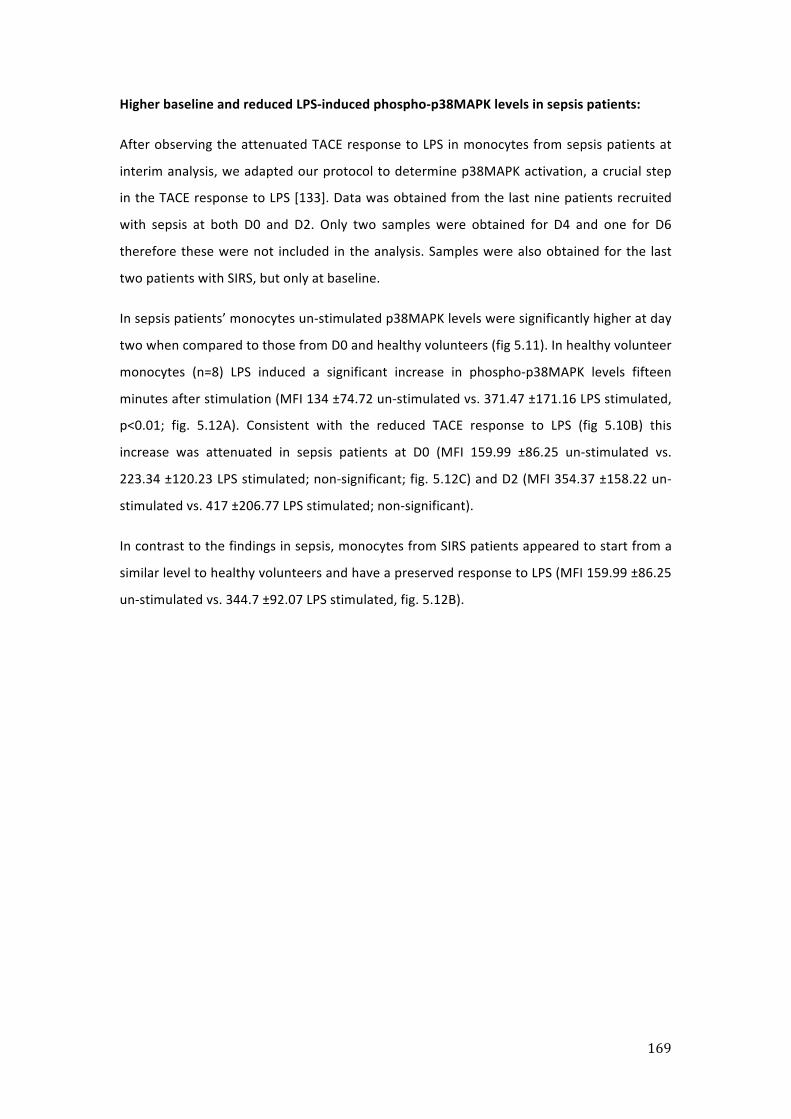
169
Higher baseline and reduced LPS-‐induced phospho-‐p38MAPK levels in sepsis patients:
After observing the attenuated TACE response to LPS in monocytes from sepsis patients at
interim analysis, we adapted our protocol to determine p38MAPK activation, a crucial step
in the TACE response to LPS [133]. Data was obtained from the last nine patients recruited
with sepsis at both D0 and D2. Only two samples were obtained for D4 and one for D6
therefore these were not included in the analysis. Samples were also obtained for the last
two patients with SIRS, but only at baseline.
In sepsis patients’ monocytes un-‐stimulated p38MAPK levels were significantly higher at day
two when compared to those from D0 and healthy volunteers (fig 5.11). In healthy volunteer
monocytes (n=8) LPS induced a significant increase in phospho-‐p38MAPK levels fifteen
minutes after stimulation (MFI 134 ±74.72 un-‐stimulated vs. 371.47 ±171.16 LPS stimulated,
p<0.01; fig. 5.12A). Consistent with the reduced TACE response to LPS (fig 5.10B) this
increase was attenuated in sepsis patients at D0 (MFI 159.99 ±86.25 un-‐stimulated vs.
223.34 ±120.23 LPS stimulated; non-‐significant; fig. 5.12C) and D2 (MFI 354.37 ±158.22 un-‐
stimulated vs. 417 ±206.77 LPS stimulated; non-‐significant).
In contrast to the findings in sepsis, monocytes from SIRS patients appeared to start from a
similar level to healthy volunteers and have a preserved response to LPS (MFI 159.99 ±86.25
un-‐stimulated vs. 344.7 ±92.07 LPS stimulated, fig. 5.12B).

170
Figure 5.11: Baseline phospho-‐p38 MAPK levels were elevated in sepsis patient monocytes
at day two
Un-‐stimulated phospho-‐p38 MAPK levels for healthy volunteers (n=8) and sepsis patients
(n=9).
* P<0.05 vs. healthy volunteer and sepsis D0

171
A
B
C
Figure 5.12: The p38MAPK response to LPS was present in healthy volunteers and SIRS
patients, but attenuated in sepsis.
P38 MAPK phosphorylation in response to LPS 1μg/ml for 0, 15 and 30-‐minutes.
Figures are for PBMCs gated on monocytes.
A: Healthy volunteers, n=8
B: SIRS patients, n=2
C: Sepsis patients, n=9
*P<0.05 vs. un-‐stimulated (HV t=0)

172
Higher baseline TNFR-‐1 expression and reduced LPS-‐induced shedding in sepsis patients:
Within the SIRS group monocyte TNFR-‐1 expression on D0 was not significantly different to
that seen on healthy volunteer cells (MFI: 19.46 ±8.18 HV vs. 28 ±19.88 SIRS D0, fig 5.13).
There were no significant differences in these values at D2 (MFI: 17.8 ±6.37), D4 (MFI: 8.05
±5.1) or D6 (MFI: 14.92 ±12.49). Although higher on all sampling days, TNFR-‐1 expression in
the sepsis group was not significantly different from healthy volunteers at D0 (MFI 27.1
±10.92), D2 (MFI 35.54 ±27.9) or D4 (37.21 ±21.69). However, expression levels were
elevated at D6 (MFI 70.79 ±29.2, p<0.01 vs. HV and sepsis D0, fig 5.13).
In response to LPS the percentage shedding of TNFR-‐1 was calculated in the same manner as
outlined in Chapter Four. Healthy volunteer monocytes shed TNFR-‐1 as expected in
response to LPS (76.1%) as did SIRS patients (69.5% D0, 73.5% D2, 87.6% D4 and 59.1% D6).
There were no significant differences in this percentage shedding between these groups (fig
5.14). In addition to the elevated monocyte TNFR-‐1 expression seen in sepsis patients there
was significantly reduced shedding in response to LPS at D0 (43.2%, fig 5.14B). Whilst
shedding was not significantly different at the other sampling time points (MFI 59.2% D2,
33.7% D4 and 40.1% D6, fig 5.14), this may show a trend toward reduced shedding in the
sepsis group.

173
A
B
Figure 5.13: TNFR-‐1 expression in SIRS was not affected but in sepsis it was elevated
MFI of TNFR-‐1 on monocytes taken from:
A: SIRS patients (mean + SD)
B: Sepsis patients (mean + 95% CI), data shown is reverse log from transformed raw data
*P<0.01 vs. HV and sepsis D0
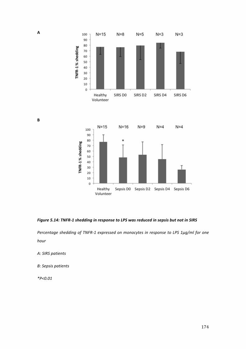
174
A
B
Figure 5.14: TNFR-‐1 shedding in response to LPS was reduced in sepsis but not in SIRS
Percentage shedding of TNFR-‐1 expressed on monocytes in response to LPS 1μg/ml for one
hour
A: SIRS patients
B: Sepsis patients
*P<0.01

175
TNFR-‐2 expression and LPS-‐induced shedding were unchanged in SIRS and sepsis patients:
As shown in figure 5.15, expression of TNFR-‐2 at D0 was not significantly different between
healthy volunteers (MFI 17.26 ±8.07), SIRS patients (MFI 23.48 ±9.32) or sepsis patients
(26.94 ±19.07). Expression levels were not significantly different at D2 (MFI 27.9 ±14.04 SIRS
& 13.29 ±5.05 sepsis), D4 (MFI 14.28 ±9.51 SIRS & 12.78 ±4.96 sepsis) or D6 (MFI 17.0 ±8.88
SIRS & 11.58 ±5.6 sepsis).
In response to LPS, healthy volunteer monocytes shed the majority of surface expressed
TNFR-‐2 (86.0%) and this response was maintained in SIRS patients (78.1% D0, 79.5% D2,
71.1% D4 & 79.0% D6, fig 5.16A). In the sepsis group TNFR2 shedding was apparent (65.6%
D0, 65.8% D2, 55% D4 & 54.3% D6, fig 5.16B), but the levels were reduced as compared
healthy volunteers and SIRS patients. Whilst non-‐significant this may show a trend toward
reduced shedding in the sepsis group and therefore similar to the difference found between
the patients groups with TNFR1 shedding.
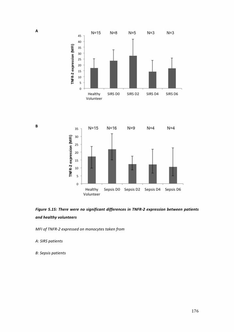
176
A
B
Figure 5.15: There were no significant differences in TNFR-‐2 expression between patients
and healthy volunteers
MFI of TNFR-‐2 expressed on monocytes taken from
A: SIRS patients
B: Sepsis patients
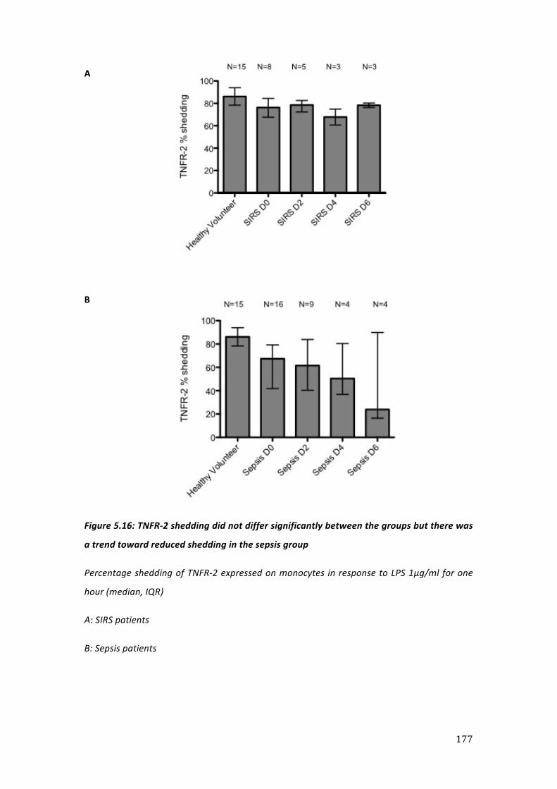
177
A
B
Figure 5.16: TNFR-‐2 shedding did not differ significantly between the groups but there was
a trend toward reduced shedding in the sepsis group
Percentage shedding of TNFR-‐2 expressed on monocytes in response to LPS 1μg/ml for one
hour (median, IQR)
A: SIRS patients
B: Sepsis patients

178
L-‐selectin expression and LPS-‐induced shedding were unchanged in both SIRS and sepsis:
As shown in figure 5.17, monocyte L-‐selectin expression levels at D0 were similar between
healthy volunteers (MFI 92.27 ±60.75), SIRS patients (MFI 84.88 ±63.96) and sepsis patients
(MFI 111.57 ±71.19). Similarly there were no significant differences in expression at D2 (MFI
139.35 ±81.94 SIRS, 52.23 ±22.53 sepsis), D4 (MFI 38.76 ±33.78 SIRS, 62.18 ±21.64 sepsis)
and D6 (MFI 78.08 ±8.81 SIRS, 63.63 ±27.56 sepsis).
In response to LPS, monocytes from healthy volunteers shed the majority of L-‐selectin
(85.2%, fig. 5.18). Near complete shedding also occurred on monocytes taken from both
SIRS patients (89.4%) and sepsis patients (71%) at D0. This pattern was also seen at D2, D4
and D6 although there was some variation (fig. 5.18). These results of unchanged L-‐selectin
shedding in the septic group were in marked contrast to the reductions of LPS-‐induced TACE
activation and TNFR1 shedding, suggesting differential pathway modulation with potential
functional significance.

179
A
B
Figure 5.17: L-‐selectin expression levels were not affected by SIRS or sepsis
L-‐selectin expressed on monocytes (median, IQR) taken from
A: SIRS patients
B: Sepsis patients

180
A
B
Figure 5.18: L-‐selectin shedding in response to LPS was preserved in both SIRS and sepsis
Percentage shedding of L-‐selectin (median, IQR) expressed on monocytes in response to LPS
1μg/ml for one hour
A: SIRS patients
B: Sepsis patients
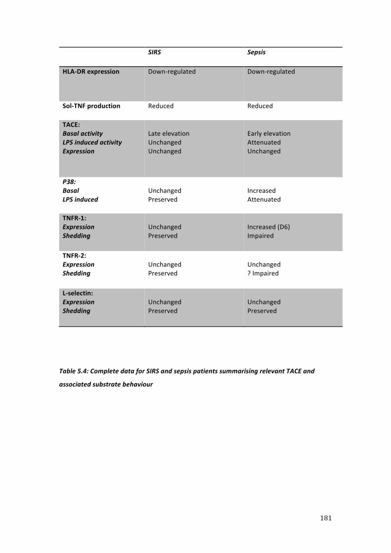
181
SIRS Sepsis
HLA-‐DR expression Down-‐regulated Down-‐regulated
Sol-‐TNF production Reduced Reduced
TACE: Basal activity LPS induced activity Expression
Late elevation Unchanged Unchanged
Early elevation Attenuated Unchanged
P38: Basal LPS induced
Unchanged Preserved
Increased Attenuated
TNFR-‐1: Expression Shedding
Unchanged Preserved
Increased (D6) Impaired
TNFR-‐2: Expression Shedding
Unchanged Preserved
Unchanged ? Impaired
L-‐selectin: Expression Shedding
Unchanged Preserved
Unchanged Preserved
Table 5.4: Complete data for SIRS and sepsis patients summarising relevant TACE and
associated substrate behaviour

182
Expression of chemokine receptor two (CCR2) on monocytes from SIRS and sepsis patients:
To identify an inflammatory process that might be linked to the preserved L–selectin
shedding phenotype in SIRS and sepsis patients’ monocytes, we investigated their migration
capability. As a critical mediator of monocyte chemotaxis and migration we determined
whether patient monocytes expressed CCR-‐2, the receptor for monocyte chemotactic
protein-‐1 (MCP-‐1) [454] [455].
For the final six patients (two with SIRS and four with sepsis) recruited we stained for CCR-‐2
at D0 and compared this to levels obtained from the final four healthy volunteers. Although
patient monocytes expressed the receptor there was a non-‐significant trend toward this
being at lower levels than in healthy volunteer cells (MFI 358.83 ±210.25 healthy volunteer
vs. 155.93 ±81.23 patients; p=0.06; fig 5.19).

183
Figure 5.19: Monocytes taken from patients expressed CCR2 but displayed a trend toward
reduced levels when compared to healthy volunteers
Monocyte CCR2 expression, mean +SD
N=4 healthy volunteers
N=6 patients

184
5.5 Discussion
In this study I successfully developed and refined a flow cytometry protocol to identify all
human monocyte subpopulations. I found that monocytes expressed characteristics
previously described as the ‘deactivation’ phenotype. However, with respect to TACE activity
and substrate shedding, there appeared to be a more complex pattern that is indicative of
‘reprogramming’ of the normal response. These patterns appeared different according to
different SIRS aetiologies, i.e. sepsis, but not non-‐septic SIRS, produced a down regulation of
HLA-‐DR and sol-‐TNF release combined with increased baselines (yet reduced inducibility) of
p38MAPK and TACE activity (table 5.4). It was also apparent that the TACE activity profiles in
isolated monocyte were altered in a manner that was broadly similar to those induced by
the model of in vitro tolerance developed in Chapter Four.
Demographics
The patient groups were well matched apart from a younger age group within the SIRS
group. This is likely to reflect the fact that sepsis occurs with increasing incidence as age
increases [456]. A high number of these SIRS patients are from neurosciences as Charing
Cross Hospital is a tertiary referral centre for this specialty. The fact that the healthy
volunteer group has a younger age than either of the patient groups was unavoidable as
these were recruited from staff at Imperial College London. Indeed with specific regard to
PBMC HLA-‐DR, increasing age has been found to increase expression [457], potentially
enhancing the significance of the fall we saw with this marker in sepsis.

185
Monocyte subset markers
Our first aim was to successfully construct a flow cytometry protocol that would allow us to
identify monocytes in PBMC preparations that were often contaminated with neutrophils.
We found that using FSC, SSC, CD11b and CD56 allowed us to do this in an HLA-‐DR
independent manner, meaning that we could then accurately quantify expression of the
MHC class II molecule. This represented an advance on our previous protocol that included
HLA-‐DR in the gating strategy [53], and on numerous HLA-‐DR SIRS related studies, which had
used more simplistic methods of identifying monocytes, and had not attempted to examine
subsets [458-‐461].
As mentioned in the introduction to this chapter, whilst it has been recognised for some
time that human monocytes are heterogeneous, there is no consensus on how these
subsets should be classified. We had aimed to use expression of CD14, CD16 and CD64 to
categorise subsets and determine how these behaved individually in SIRS. We found that
significant distortion of these markers occurred in patient samples. This should not be
considered a surprising finding given that levels of CD64 [462], CD16 [307] and CD14 [463]
change in the context of critical illness and that the CD14-‐CD16+ subset expands [305, 310].
What was striking was the degree to which these alterations dramatically altered the
appearance of the cells contained within the monocyte gate and obscured the cut-‐off points
between the subsets. This made distinguishing the classical CD14++CD16-‐ from the
intermediate CD14+CD16+ group difficult and, in conjunction with the reduced ability of
CD64 expression to differentiate between the overlapping intermediate (CD14+CD16+) and
mature (CD14-‐CD16+) groups, made it difficult to draw the gates required to identify the
monocyte subsets. Any subset gates that were drawn would be arbitrary in nature with
potential implications on any results obtained, calling into question the legitimacy of
categorising monocytes as discrete subpopulations in critical illness based on the markers
we used. While recognising the potential exclusion of CD14-‐CD16+ and perhaps some
CD14+CD16+ monocytes from our TACE activity studies by our use of a CD14+ positive bead
selection protocol, for flow cytometric analysis we decided it was necessary to avoid any
arbitrary or imprecise sub-‐division of monocytes and to evaluate the population in its
entirety.

186
Cells from both groups were deactivated and could not be differentiated using HLA-‐DR
expression levels alone
Having developed our flow cytometry protocol, I then established that cells from both
patient groups showed changes consistent with a state of deactivation, namely reduced
HLA-‐DR expression and sol-‐TNF production on ex vivo LPS stimulation. In Chapter Four we
demonstrated that even normal monocytes, once cultured for a prolonged period of time
(16 hours) showed changes in p38MAPK activation status and activity profiles. In order to
minimise any phenotypic change in the cells, the duration of the ex vivo stimulation was
deliberately short (four hours). The fact that sol-‐TNF levels were significantly reduced across
all four samples in sepsis whereas in SIRS this was only significant at D0 is likely due to the
lower numbers of patients within this latter group. Consistent with a previous study [54],
levels of HLA-‐DR did not differ between SIRS and septic patients and therefore would lack
any diagnostic value for the presence of infection.
TACE activity profiles
Isolation of monocytes allowed the kinetics of the acute TACE response to be elicited. This is
the first time its catalytic activity has been directly measured in critical illness. Within the
sepsis group, baseline TACE activity was enhanced and LPS-‐induced activity decreased, in a
manner similar to that generated by LPS exposure in the tolerance model I developed.
Exposure to LPS has been suggested as an initiator of monocyte deactivation [436], this may
occur directly or indirectly through the induction of other mediators such as IL-‐10 to
produce the changes in TACE behaviour seen. It has been shown that IL-‐10 inhibits TACE in a
TIMP-‐3 dependent and independent manner [366]. Alternatively, TACE activity may have
reached a ceiling in these patients and further induction ex vivo was not possible. Although
LPS-‐induced enzyme activity values were attenuated from D0, the peak increase in basal
TACE activity was seen at D2 in the sepsis group. This corresponded to the point at which
un-‐stimulated phosphorylated levels of p38MAPK were elevated in monocytes and a
reduction in response to LPS was seen. Given that p38MAPK activates TACE [133], this may
offer some explanation as to how the changes in TACE behaviour are occurring. Our findings
may be in line with those of Rosengart and colleagues who performed western blotting of
mixed cells obtained from BAL fluid and found that a sub-‐group of trauma patients had
elevated levels of phospho-‐p38MAPK that were refractory to ex vivo stimulation [464].

187
Within the SIRS group no significant increases in basal TACE activity were seen except on D6.
As patients improved or died they were removed from the study, meaning those remaining
in the study at D6 were likely to be making unsatisfactory/slow clinical progress. Therefore
these changes in TACE behaviour seen at D6 in the SIRS group may represent
nosocomial/secondary infection. We did not see any increase in monocyte TACE cell surface
expression as has previously been reported on neutrophils in sepsis [355], meaning that the
activity changes we report reflect a true change in activity as opposed to more enzyme.
TACE substrates -‐ TNFRs
By examining both expression levels and shedding profiles of the TNF receptors and L-‐
selectin, we aimed to develop a more complete understanding of how cells were behaving
with respect to TACE-‐mediated functions. We found no differences in either expression or
shedding for either of the TNF receptors in SIRS patients’ monocytes. In contrast within the
sepsis group there was attenuated shedding of TNFR-‐1 at D0 and significantly increased
expression by D6. This increased expression of monocyte TNFR-‐1 has previously been
reported [50]. TNFR-‐1 signalling forms part of the “extrinsic” death receptor pathway that
induces apoptosis in leukocytes [465] and has been implicated in neutrophil apoptosis [466].
It has also been shown that recruited, exudative macrophages accumulate in early acute
lung injury but then decline as a result of apoptosis [467]. It can be speculated that this
increase in monocyte TNFR-‐1 expression in sepsis represents a limit (brake) on classically
activated inflammatory monocytes, such that they are removed as appropriate to orientate
monocyte function towards an alternatively activated tissue repair phenotype. Indeed
monocyte apoptosis has been demonstrated in sepsis and has been suggested to be
protective [468], which may be in keeping with this hypothesis. Other potential reasons for
this increase in TNFR-‐1 may include the mitochondrial dysfunction seen in sepsis [469], given
that recent work has suggested mitochondria may mediate TNFR-‐1 ectodomain shedding via
a calcium dependent process [470].
Although we saw no significant differences in TNFR-‐2 expression and shedding within the
sepsis group, there was a trend toward impaired shedding over time that was not present in
the SIRS group. Further work, with greater patient numbers and thus increased power may
be needed to demonstrate this conclusively. These patterns of TNF receptor expression
were not comparable with those seen in the tolerance model we constructed in vitro, and

188
may suggest, as might be expected, that the in vivo situation is more complicated than it is
feasible to model using a single cell type (monocytes) and stimulus (LPS).
TACE substrates – L-‐selectin
In contrast to TNFR-‐1, in both SIRS and sepsis the expression and shedding of the adhesion
molecule L-‐selectin were no different to that seen in healthy volunteers. Our data
demonstrates perturbation of MAPK signalling in the sepsis group, which would appear to
contradict the findings of Killock et al who found L-‐selectin shedding to be p38MAPK
dependent [377] through the use of a MAPK inhibitor. However, some investigators have
questioned the selectivity of these inhibitors [471] and moreover, unpublished data from
our group found that p38MAPK inhibition did not inhibit L-‐selectin shedding [472].
Kermarrec and colleagues found increased soluble L-‐selectin in patients with peritoneal
sepsis and surmised that this was due to increased neutrophil TACE expression [355]. Our
results of unaltered, or partially reduced, L-‐selectin shedding and no changes in TACE
expression in sepsis patients’ monocytes suggest that marked differences may exist in the
regulation of neutrophil and monocyte TACE related shedding. The unchanged baseline
expression, higher TACE activity but maintained substrate expression in sepsis patients
highlights the lack of a simple relationship between TACE expression, catalytic activity and
substrate shedding. In general, when comparing the relationship between TACE activity and
substrate shedding, it may be helpful to think of TNF separately given the high affinity that
TACE displays for this substrate when compared to non-‐TNF substrates [375]. For TNF there
appears to be a direct relationship between TACE activity and cleavage rates. In contrast, the
lower affinity to non-‐TNF substrates [375] means additional steps are required for cleavage
that limit their shedding even in the presence of catalytically active TACE (as observed in the
sepsis group).
The impact of L-‐selectin shedding on monocyte function is still unclear. Some investigators
have demonstrated that neutrophil rolling was impaired if L-‐selectin shedding was inhibited
and produced leukocyte accumulation [473]. Others have reported that inhibiting L-‐selectin
shedding had no effect on neutrophil rolling or migration [474]. The idea of some cellular
functions (such as L-‐selectin shedding) being preserved whilst others are suppressed is in
keeping with recent findings suggesting that bacterial reprogramming of PBMCs impairs
their phagocytic capacity, but preserves their ability to stimulate naïve T-‐lymphocytes [439].
It is also in keeping with compartmentalisation hypothesis of SIRS/CARS regulation, i.e. cells
189
are suppressed systemically but are capable of entering the tissues, once there they may be
capable of mounting an inflammatory response. This is similar to Munford and Pugin’s
argument that the systemic response to injury/inflammation is anti-‐inflammatory [58].
Limitations
There are some limitations of this study that should be considered. The definition for sepsis
used is of suspected infection in combination with two out of the four SIRS criteria. Whilst
this is generally the accepted clinical definition it is broad. Hence, it is entirely possible that
some patients in the sepsis group have sterile SIRS and vice versa. There is also considerable
heterogeneity within the sepsis group with patients presenting with infection in different
tissues and with different organisms. In future it may be more appropriate to try and study a
more homogenous population by concentrating on specific pathologies (e.g. pneumonia) or
organisms.
Although the FRET assay used isolated monocyte preparations when measuring TCAE
activity profiles there remains a possibility that these contained some low level
contamination with other circulating leukocytes/apoptotic cells. These may have
contributed, in part, to the elevated unstimulated levels of TACE activity seen within the
sepsis group. However, the elevated basal p38MAPK levels in monocytes taken from sepsis
patients would seem to indicate that there are mechanistic data, taken from intact
monocytes, which support these changes. It is possible that the changes in TACE activity
profiles within the sepsis group (increased baseline and loss of induced LPS response), rather
than reflecting immune suppression, are just reflective of maximal immune stimulation.
Further work is clearly required to accurately discriminate between these two scenarios.
In the introduction to this thesis I discussed the possibility that the immune response is
compartmentalised. Some investigators have argued that the systemic circulation is, by
default, anti-‐inflammatory [58]. Thus, the systemic changes we describe may not be
representative of the processes occurring within infected tissue beds. In the future, when
possible, it would be of benefit to compare and contrast the immune response in cells taken
from the systemic circulation to that seen in cells taken from infected tissues. I have
expanded on this theme in the final discussion contained in Chapter Seven.

190
Conclusions
I have contributed to the knowledge base in this field in several areas. Firstly, through the
construction of a flow cytometry protocol which allows us to identify monocytes. Secondly, I
have identified that monocyte surface markers change in critical illness, in a manner that
may preclude accurate identification of subsets. Although it may be possible to identify
subsets using recently identified differential markers [475], my findings mean that previous
patient subset data may be misleading. Thirdly, I have shown that the acute TACE response
is differentially affected in SIRS arising from non-‐infectious and septic aetiologies. In sepsis a
rapid decrease in LPS induced activity and increases in baseline activity, MAPK signalling
changes and alterations in TNFR-‐1 expression and shedding profiles are seen (table 5.4) and
could plausibly be used as diagnostic biomarkers.
Finally, I have shown that monocyte-‐signalling pathways appear to be differentially affected
in sepsis. These cells appear reprogrammed rather than deactivated. In sepsis the cellular
response phenotype may change depending on the stimulus, the cell and its environment
[476], but to our knowledge, this is the first time that differential responses (preserved L-‐
selectin shedding, impaired p38 activation and TNF release) to a single stimulus (LPS) have
been demonstrated and serves to illustrate the complexity of the situation. What allowed us
to demonstrate these novel findings was our capacity to focus on several aspects of a
pathway: p38MAPK (the signal), TACE (a regulatory protein) and TNFR/L-‐selectin (the target
effector molecules). Further investigation of these pathways will enhance our understanding
of the changes seen in monocyte signalling pathways during sepsis.

191
Chapter 6
Investigation of the influence of cellular environment on TACE activation

192
Summary
In Chapter Four I obtained evidence for LPS-‐mediated inactivation of the monocyte TACE
response, but not for priming. These experiments were performed in blood-‐derived
monocytes cultured under minimally adherent conditions to reduce any spontaneous ex vivo
activation. However, during inflammation in vivo, monocytes come into prolonged contact
with the microvascular endothelium through margination and then with extravascular
parenchymal cells following migration. In the case of systemic inflammation, widespread
margination of leukocytes is seen and therefore cell contact or proximity may be a central
process related to the priming of blood monocyte responses. On this basis, we hypothesised
that cell contact-‐related processes are essential for optimal TACE activation in monocytes
under inflammatory conditions.
In order to test this hypothesis, I compared the responses of cells stimulated both in static
culture conditions (to produce a cell-‐cell contact environment) and in a rotating wheel (to
minimise cell-‐cell interactions, simulating the circulating cell condition). These models
demonstrated that the TACE response to LPS was ablated in low cell-‐cell proximity states,
indicating that a contact or proximity-‐dependent signal was required for optimal TACE
activation. We next sought to determine where the proximity-‐dependent signal acted
upstream or downstream of p38MAPK signalling. LPS-‐induced monocyte phospho-‐p38MAPK
levels were similarly elevated in static and rotation culture conditions, indicating that the
contact signal does not directly influence p38-‐MAPK activation, but rather acts downstream
to p38MAPK in promoting TACE activation.
I then attempted to determine the nature of the contact-‐related signal. We focused on the
two principle processes, which we considered would operate under close cell proximity
conditions: secreted soluble mediators and cell adhesion molecule-‐related signalling. Based
on the previous literature on TACE activation and substrate cleavage, we investigated ROS
and adenosine triphosphate (ATP) for soluble mediators, and β1 and β2 integrins for cell
adhesion molecules. Although hydrogen peroxide (H2O2) as a source of ROS effectively
induced TACE responses in rotation culture, blocking extracellular ROS (whilst reducing the
response to H2O2) did not block the response to LPS, leading us to conclude that
extracellular ROS did not mediate the contact-‐related signal. In contrast to previous reports,
ATP alone had no effect on TACE activity. Using a combination of antibodies to block or
stimulate monocyte integrins, we found some evidence of integrin-‐mediated signalling
impacting on the TACE pathway. We conclude on the basis of this data that cell proximity-‐

193
dependent intra-‐cellular ROS signalling promotes TACE activation by acting downstream of
p38MAPK.
6.1 Background
The inability to discern a primed phenotype from in vitro experimentation and in vivo
observations thus far is striking. In the patient samples, this was not particularly surprising,
as priming by its very nature is an early event during the course of sepsis/SIRS and may
precede the development of the clinical condition. The in vitro findings however, led us to
reappraise our thinking and hypothesise that the TACE response to LPS seen when
monocytes were stimulated in Eppendorfs represented maximal inflammatory signalling in
response to this stimulus. In this situation cells will be in close proximity at the bottom of the
Eppendorf. We therefore hypothesised that a signal indicating cell proximity, or even cell-‐
cell contact, may be required for maximal inflammatory signalling. It could be this proximity
signal which represents “priming” of the monocyte to the LPS stimulus. Indeed, in the
concluding remarks to Chapter Four I wrote, “It is striking when reviewing the literature that
other studies using LPS to prime cells have either found adherence to be key or used
adherent systems.”
In order for us to further develop this concept, we first stimulated monocytes with LPS in the
minimally adhesive PTFE medium that we used to create the two hit model in Chapter Four.
We then directly compared responses under conditions that promoted cell-‐cell proximity,
using the standard static culture set-‐up, to those in rotation culture to simulate normal, non-‐
adherent resting conditions. The rotating culture consisted of monocytes placed in
Eppendorfs at normal densities and mixed continuously by rotation on a wheel. During this
period the Eppendorfs are held in the horizontal plane, whilst the wheel rotates in the
vertical plane. This method allowed a reduction of cell-‐cell or cell-‐plastic interactions while
maintaining all other conditions present during static Eppendorf stimulation constant. We
deliberately created a single cell model system for several reasons; firstly, because the FRET
assay requires a single cell type and secondly, to limit cell-‐cell interactions to those occurring
between monocytes.

194
From the literature and our previous investigation [133], we identified several candidate
mediators and mechanisms likely to be involved in transducing any contact-‐related signal.
We identified three key groups: ROS, ATP and integrins.
Reactive oxygen species
Molecules mediating any proximity signal would need to be rapidly released, labile, and
tightly regulated. ROS fit this profile and are produced by leukocytes in response to
stimulation resulting in both intra-‐cellular [202] and extra-‐cellular release [477]. Extra-‐
cellular release would result in a proximity-‐dependent concentration and activity gradient.
Indeed extracellular neutrophil ROS production has been shown to induce changes in
adjacent endothelial cell adhesion molecule expression and inflammatory signalling [477],
demonstrating that ROS act as inter-‐cellular messengers.
Our previous investigation found TLR-‐4 stimulation by LPS resulted in ROS mediated p38
MAPK induction to produce TACE activation [133]. However, the anti-‐oxidant N-‐acetyl
cysteine, whilst producing complete inhibition of TACE activation, produced only partial
inhibition of p38MAPK phosphorylation [133]. This partial discrepancy could be in keeping
with a separate ROS signalling event downstream of p38MAPK to activate TACE, potentially
through extracellular ROS, as part of a proximity-‐dependent pathway to facilitate full
inflammatory signalling.
ATP
ATP is released extra-‐cellularly from monocytes in response to microbial components. It
signals through the purinergic P2X7 receptor that is expressed on monocytes and signalling
induces an autocrine release of IL-‐1β and IL-‐18 [478]. Similar to ROS, this extra-‐cellular
release would result in a concentration/activity gradient of ATP that could mediate any cell
proximity signal. ATP has already been implicated in signalling pathways that may intersect
on monocyte inflammatory signalling including reactive oxygen species [479], p38-‐MAPK
[480] and TACE mediated shedding of heparin binding epithelial growth factor-‐like growth
factor [481]. Furthermore, ATP-‐mediated activation of the P2X7 receptor on monocytes has
been shown to result in shedding of L-‐selectin [482].

195
Integrins
Previous in vivo work from our group demonstrated that low-‐dose intravenous LPS
produced priming in lung-‐marginated monocytes evidenced by their enhanced TNF
expression in response to a subsequent LPS stimulus [316]. It is possible that, rather than
LPS as a direct agent of priming, its effect is through LPS-‐induced prolonged margination of
monocytes in contact with the pulmonary endothelium. Such monocytes are in the vicinity
of other marginated leukocytes as it is thought that marginated monocytes facilitate
neutrophil extravasation [193]. Monocytes in co-‐culture with endothelial cells have been
shown to synthesise more prostanoids, cytokines and chemokines than those in
monoculture conditions [317, 483-‐485] and it is known that cell-‐cell adhesion interactions,
such as the β-‐2 integrin-‐ICAM-‐1 interaction, enhance the inflammatory response in
monocytes [378]. Monocytes lacking the membrane protein CD11b (a sub-‐unit of the
integrin receptor) display a reduced inflammatory response to LPS and the integrin
activation process has been shown to involve p38MAPK [373]. Furthermore, it has been
demonstrated that adherent monocytes are primed to produce more TNF than their non-‐
adherent counterparts in response to an LPS challenge [379, 486]. As previously mentioned
it is now known that increased LPS induced TACE activity is mediated via p38MAPK [133].
Together these factors are supportive of a common pathway linking both TACE activation
and the integrin receptors that mediate cell-‐cell interaction [372].

196
6.2 Aims
The specific aims for this chapter were to:
1. Evaluate LPS-‐induced TACE and p38MAPK activation under cell contact and non-‐
contact promoting conditions in vitro.
2. Investigate the proximity-‐ or contact-‐related mechanisms responsible for any
difference found in TACE and p38MAPK activation responses.
6.3 Protocols
Blood collection
30 ml of donor blood was obtained by venesection from healthy volunteers who had given
informed consent. Exclusion criteria were inter-‐current illness or immune-‐suppression.
Blood was extracted directly into vacutainers containing EDTA.
Monocyte stimulation and model conditions
Monocytes were isolated from whole blood using differential centrifugation followed by
magnetic activated cell sorting utilising a CD14 positive bead selection strategy as outlined in
detail in Chapter Two. Cells were then placed in Eppendorfs at a density of 5x10⁶/ml and
stimulated with LPS 1μg/ml for one hour at 37°C. Wherever H2O2 and ATP were used the
concentration they were used at is specified in the text. Where antibodies and inhibitors
were used, cells were treated with these and left on ice for 15 minutes before being
stimulated. Antibodies used were low endotoxin and azide preparation wherever possible.
For the stimulation period some cells were left in static conditions but others were placed in
a wheel that continuously rotated (x20 per min) throughout. Cells were then retrieved and
placed in the FRET assay as previously described.
197
Quantification of the MAPK response
This was performed as outlined in detail in Chapter Two. Briefly, cells were fixed and made
permeable before being stained with antibodies specific for the activated form of the MAPK
enzyme. Cell stimulation was performed at the same density and conditions as outlined for
the TACE response. An LPS concentration of 1µg/ml was used with monocytes stimulated for
15 minutes.

198
6.4 Results
The kinetics of the acute TACE response to LPS are altered when monocytes are stimulated
directly in PTFE:
In order to determine whether the acute TACE response was affected by minimally adhesive
conditions, isolated monocytes were stimulated with 1µg/ml of LPS directly in PTFE wells (fig
6.1). Activity was then determined using the FRET assay.
LPS stimulation in PTFE produced no increase in TACE activity at either the one hour or two
hours time point (12.7 ±0.53 FU/min and 18.6 ±1.69 FU/min respectively) when compared to
un-‐stimulated controls (14 ±3.74 FU/min). This was in marked contrast to stimulation in
polypropylene Eppendorfs (fig 3.9, below), and provided evidence for our hypothesis that
monocyte inflammatory signalling is critically affected by cell proximity and/or contact.
Figure 3.9: Isolated monocytes display an expression independent up-‐regulation of TACE
activity in response to LPS.
Positively selected, isolated monocytes incubated with LPS 1μg/ml for one hour display
increased TACE catalytic activity (n=5, *p<0.01)

199
Figure 6.1 The kinetics of the acute TACE response to LPS are altered when monocytes
are stimulated directly in PTFE:
N=2

200
TACE activity up-‐regulation response to LPS is reduced when monocytes are stimulated in
rotation culture
Purified monocytes were stimulated with LPS (1µg/ml) in Eppendorf tubes at 37°C under
static or rotation conditions for one hour and then processed for measurement of TACE
activity under standard conditions. Monocytes in static culture up-‐regulated their TACE
activity by x2.3 fold (23.1 ±4.81FU/min to 46.37 ±0.71FU/min) (fig 6.2). Rotation culture
reduced un-‐stimulated monocyte TACE activity (fig 6.2) and markedly reduced the up-‐
regulation seen in response to LPS (Static LPS 46.37 ±0.71FU/min vs. rotation LPS 15.21
±2.2FU/min, p<0.05; fig 6.2). The increase in TACE activity with LPS treatment in rotation
culture was small (rotation 3.25FU/min vs. static 23.27FU/min) and was not statistically
significantly different from baseline. TACE expression levels were unchanged across the
groups at baseline (MFI 23.81 ±13.75 static vs. 22.0 ±9.14 rotation; non-‐significant) and after
LPS stimulation (MFI 19.18 ±10.77 static vs. 18.98 ±5.86 rotation; non-‐significant).

201
Figure 6.2: The acute TACE response was attenuated in rotation culture
TACE activity for isolated monocytes placed in suspension and rotation culture then
stimulated with LPS 1μg/ml for one hour
Dark bars represent un-‐stimulated cells, light bars LPS stimulated
N=4
*P<0.05

202
Increasing cell density could inhibit the acute TACE response seen in static culture:
In order to confirm that cell-‐cell proximity had an effect on the acute TACE response, the
effect of varying cell density in static culture was determined. Cells were stimulated for one
hour with LPS and TACE activity determined. As shown in figure 6.3, the magnitude of the
TACE response to LPS was reduced by a factor of two when cell density was reduced from
the standard 5x10⁶ cells/ml to 5x10⁵ cells/ml. A reduction in activity was observed at the
highest cell density; a likely reflection of ‘overcrowding’ phenomenon such as reduced
availability of LPS. Some decrease in the baseline activity was apparent at the lowest
density. Repeat experiments will be needed to confirm these findings.
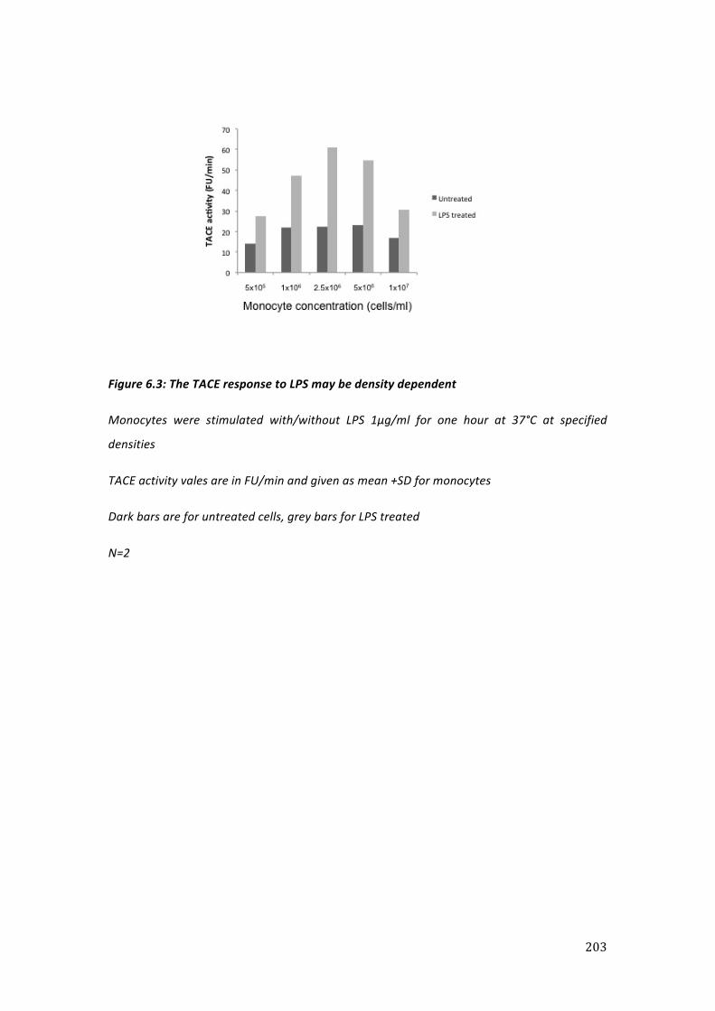
203
Figure 6.3: The TACE response to LPS may be density dependent
Monocytes were stimulated with/without LPS 1μg/ml for one hour at 37°C at specified
densities
TACE activity vales are in FU/min and given as mean +SD for monocytes
Dark bars are for untreated cells, grey bars for LPS treated
N=2

204
LPS-‐induced p38MAPK activation is independent of cell proximity
Monocytes were then stimulated in static and rotation culture to determine whether, similar
to the reduced TACE response, the p38MAPK response to LPS was diminished by rotation
culture. As can be seen in fig 6.4, phospho-‐p38MAPK levels were similar both at baseline
(MFI 59.43 ±8.12 static vs. 58.82 ±11.83 rotation) and after LPS stimulation (MFI 226.31
±36.36 static vs. 211.84 ±34.36 rotation).
This finding was unexpected and led us to reappraise our concept of the LPS-‐induced
p38MAPK-‐TACE signalling pathway. As illustrated by the schematic in figure 6.5, we now
hypothesized that, because the phospho-‐p38MAPK response was maintained and is
essential for TACE activation [133], any contact-‐related signal would need to be acting
downstream. This may either intersect directly on the p38MAPK-‐TACE pathway or act in
parallel to it. Alternatively, although the p38MAPK pathway is required for the TACE
activation, an alternative contact-‐dependent pathway or process is also essential.

205
Figure 6.4: The p38MAPK response was maintained across culture conditions
Phospho-‐p38MAPK response after LPS stimulation 1μg/ml for 15 minutes
Dark bars represent un-‐stimulated cells, light bars LPS stimulated
N=4
* P<0.01

206
Figure 6.5: Schematic illustrating contact/proximity signal

207
H2O2 treatment produced an increase in monocyte TACE activity independent of culture
conditions:
To investigate the likely cell proximity and/or contact-‐dependent mechanisms of TACE
activation, we first considered the role of extracellular ROS signalling. Because of their
extremely short half-‐life, extra-‐cellular ROS act over only very short distances between
adjacent cells under physiological conditions. Addition of H2O2 to monocytes had previously
been shown to activate TACE in a p38MAPK-‐dependent fashion, but only at a high, non-‐
physiological range, of concentrations (e.g. 0.5-‐5mM) [133]. To first assess whether H2O2 at
these high concentrations could bypass the need for cell proximity in TACE activation, it was
added to cells in both static and rotating culture conditions at a concentration of 0.5mM,
alone or in combination with LPS (1µg/ml). As can be seen (fig 6.6), the addition of H2O2
produced an increase in TACE activity that was independent of culture conditions.
To explore in more depth whether H2O2-‐induced TACE activation (proximity-‐independent)
could substitute for the lack of LPS-‐induced TACE activation in rotation conditions, additional
dose response experiments were performed. More specifically, whether H2O2 added at
limiting concentrations could substitute for the missing p38MAPK downstream signal and
thereby enhance LPS-‐induced TACE up-‐regulation in suspension culture. This produced a
dose dependent increase in TACE activity (0.2mM 21.82 ±2.68FU/min, 0.5mM 36.63
±2.95FU/min, 2mM 76.05 ±7.07FU/min and 5mM 55.7 ±1.83FU/min, fig 6.7). There was no
added effect if these doses of H2O2 were used in combination with LPS 1μg/ml (0.2mM+LPS
26.3 ±3.33FU/min, 0.5mM+LPS 49.39 ±2.39FU/min, 2mM+LPS 77.35 ±21.58FU/min and
5mM+LPS 43.2 ±3.38 5FU/min). These results indicated that added H2O2 did not synergise
with LPS to enhance TACE activation in rotation culture, but did not rule out a role for
release and signalling of endogenous (intracellular) ROS in LPS-‐induced TACE activation in
static culture.

208
Figure 6.6: The addition of H2O2 to rotation culture produced an increase in TACE activity
Graphical illustration of data contained in table 6.2
TACE activity values (mean +SD) for isolated monocytes stimulated for one hour with LPS
1μg/ml, H2O2 0.5mM or both.
N=2

209
Figure 6.7: H2O2 in rotation culture conditions produced a dose dependent increase in
activity with no additional effect when added with LPS
TACE activity values (mean +SD) for isolated monocytes stimulated for one hour with H2O2 at
a range of doses either in isolation or in conjunction with LPS 1μg/ml.
Dark bars represent monocytes stimulated with H2O2 only, light bars stimulated with H2O2
and LPS.
N=2
210
Inhibition of extracellular ROS activity did not attenuate the TACE response to LPS in static
culture:
Having shown the potential for extracellular ROS to potentially bypass or substitute for any
contact-‐related signal, we used a combination of non-‐cell permeable agents to accelerate
depletion of the extracellular environment of any ROS generated during LPS stimulation.
Previously the broad spectrum cell permeable ROS inhibitor was used to demonstrate their
role in TACE activation, but in order to assess the role of extracellular ROS, cell impermeable
inhibitors were required [133]. Cell-‐impermeable superoxide dismutase (SOD) and catalase
have previously been used to demonstrate extracellular ROS activity in vitro [487], as
illustrated schematically in fig 6.8, with catalase effectively degrading H2O2 derived from SOD
conversion of the O2-‐ free radical. As shown in figure 6.8A the addition of either catalase or
SOD attenuated the TACE response to H2O2 (48.21% inhibition with catalase, 56.53% with
SOD and 51.15% both) in static culture conditions, confirming their efficacy in this assay.
However, these results were not reproduced when used in combination with LPS (<1%
inhibition with catalase, SOD or in combination, fig 6.8B). It is not possible from these
limited data to ascertain whether extra-‐cellular ROS were completely inhibited by SOD and
catalase, either given individually or in combination.
On the basis of the limited experiments outlined above we concluded that, whilst H2O2 was
able to induce a TACE response in suspension culture, extracellular ROS could be excluded as
the p38MAPK-‐downstream, contact-‐related signal elicited by LPS in static culture. Rather
than continuing these experiments, we moved to consider other potential mediators of the
contact signal.

211
Figure 6.8: Schematic illustrating ROS metabolic pathways
Reactive oxygen species may be produced through the action of flavoprotein oxoreductase,
resulting in the production of superoxide (O2-‐). This reactive molecule is dismutated by
superoxide dismutase (SOD) to H2O2. This is subsequently reduced by the Fenton reaction to a
reactive hydroxyl radical (OH-‐). Catalase then catalyses the breakdown of H2O2 to H2O and
O2, preventing OH-‐ accumulation.

212
A
B
Figure 6.9: The TACE response to H2O2 could be attenuated by the use of catalase and SOD
but that to LPS could not
A: Monocytes in static culture stimulated with H2O2 2mM in the presence/absence of
catalase (1000u/ml), SOD (1000u/ml) or both
B: Monocytes in static culture stimulated with LPS 1μg/ml in the presence/absence of
catalase (1000u/ml), SOD (1000u/ml) or both
N=2

213
ATP does not induce TACE activity up-‐regulation
ATP was added to monocytes in static culture at a range of concentrations designed to
stimulate either low or high affinity purinergic receptors. ATP at a concentration of 3mM is
sufficient to stimulate the P2x7 receptor expressed on monocytes [488, 489]. Contrary to
our expectation, cells stimulated with ATP at this concentration in the presence of LPS
displayed an inhibited response to LPS (LPS 46.85 ±1.91 FU/min vs. LPS & ATP 11.83
±6.69FU/min; fig 6.10A). The addition of ATP to rotation culture conditions at a dose of 3mM
appeared to have no effect on either baseline (unstimulated 16.4 FU/min vs. ATP 10.78
FU/min) or LPS induced activity levels (LPS 17.8 FU/min vs. LPS & ATP 11.88 FU/min).
Lower concentrations of ATP stimulate different purinergic receptors such as the P2Z
receptor [490]. Hence we examined the effect that these lower concentrations had on basal
TACE activity. There were no differences in activity seen across the range of concentrations
used (fig 6.10B). In view of previous studies that had demonstrated ATP induced shedding of
TACE substrates, this was surprising but may reflect the fact that these studies did not
examine TNF cleavage [481, 482].

214
A
B
Figure 6.10: The effect of ATP on baseline and LPS induced TACE activity in static culture
conditions
A: Isolated monocytes stimulated in static culture with LPS 1μg/ml, ATP 3mM or both for one
hour (n=2)
B: ATP dose response (one hour stimulation) in static culture using isolated monocytes (n=2)

215
Pharmacological ATP inhibition did not affect the TACE response to LPS:
To further exclude a stimulatory effect of ATP we used suramin (a general, non-‐specific
inhibitor of ATP) as well as A740003 (an inhibitor of the P2X7 receptor) in static culture
conditions. Inhibition of ATP with either suramin or A740003 had no effect on the TACE
response to LPS (fig 6.11). This was consistent with our previous results and would seem to
exclude a stimulatory effect of ATP. Suramin alone appeared to produce a reduction in un-‐
stimulated TACE activity (un-‐stimulated 14.46 ±6.2FU/min vs. suramin 7.15 ±1FU/min; fig
6.11).

216
Figure 6.11: Inhibition of ATP appeared to restore the LPS response in cells stimulated ATP
& LPS In static culture conditions
Isolated monocytes cultured with LPS 1μg/ml, suramin (sur) 230μM, A740003 (A74) 10μM
separately or in combination for one hour in static conditions (n=3)
217
CD18 blockade may reduce basal and LPS-‐inducible TACE activity in static culture:
To try and ascertain whether the contact-‐related signal was mediated by integrins a panel of
antibodies were used. These were as follows:
1. Anti-‐CD18 blocking antibody in order to block macrophage-‐1 antigen (Mac-‐1, CD11b
CD18), lymphocyte function-‐associated antigen-‐1 (LFA-‐1, CD11aCD18) in addition to
alphaXbeta2 (CD11cCD18) and alphaDbeta2 (CD11dCD18)
2. Anti-‐CD11b blocking antibody to block Mac-‐1
3. Anti-‐CD29 stimulating antibody to determine the role of very late antigen-‐4 (VLA-‐4
(CD49dCD29)
Blocking antibodies were used in static culture to try and block the contact-‐related signal (fig
6.12). The anti-‐CD29 stimulating antibody was used in rotation culture conditions to try and
replicate the contact-‐related signal and generate a TACE response to LPS. LPS induced TACE
activity appeared to be diminished by CD18 blockade (26.57 ±8.68 vs. 18.3 ±4.1, LPS-‐only vs.
LPS + CD18 mAb), with a similar relative reduction in baseline TACE activity (15.24 ±5.1 vs.
7.68 ±5.9 Fu/min, no antibody vs. CD18 mAb). Effects were less clear with CD11b blockade,
possibly implicating the LFA-‐1 integrin, which consists of the CD11a and CD18 heterodimers.
The addition of the stimulating antibody to rotation culture conditions did not generate an
increase in TACE activity either alone or in combination with LPS (8.81 ±0.44 antibody alone
vs. 10.28 ±2.47 with LPS), making VLA-‐4 an unlikely mediator for the contact-‐related signal.

218
Figure 6.12: The CD18 blocking antibody may reduce basal TACE activity and the response
to LPS
Isolated monocytes were pre-‐treated with anti-‐CD18 and CD11b antibodies before being
exposed to LPS 1μg/ml for one hour in static culture conditions.
N=2
Similarly cells were pre-‐treated with an anti-‐CD29 antibody then exposed to LPS in rotation
culture conditions.

219
6.5 Discussion
The primary aim in this chapter was to elicit any differences in monocyte inflammatory
signalling that may be present in culture conditions where cells were in proximity when
compared to conditions where contact was minimised.
Static vs. rotation culture – TACE activity
In order to do this we manipulated the conditions in which monocytes were stimulated with
LPS. We first stimulated them in a minimally adherent medium (PTFE) in which cells are in
proximity but only in a single plane, as monocytes form a thin monolayer over the PTFE
insert. These conditions produced an attenuation of the TACE response to LPS. We next
placed monocytes in rotation culture to completely minimise cell proximity, and compared
this to the static conditions we used to elicit the TACE response in previous chapters. Our
results show that full TACE activation required cells to be in proximity with one another, and
that the TACE response to LPS was almost completely lost when cells were stimulated in
rotation culture. In addition the TACE response in the static culture was density dependent
lending further weight to the argument that cell proximity impacts on inflammatory
signalling. Adherent monocytes have been shown to produce more sol-‐TNF in response to
an LPS stimulus than their non-‐adherent counterparts [379], indicating a potential TACE
effect in this context.
The fact that TACE activity may be modified by adhesion or proximity should not be
considered surprising. Several members of the metalloprotease family have been shown to
interact with integrins via their disintegrin domain and TACE itself has been shown to
interact with both the actin cytoskeleton [371] and the α5β1 integrin VLA-‐5 [372]. Although
ultimately it may not be integrins that deliver the signal, it is plausible that the process of
leukocyte recruitment and leukocyte-‐endothelial interaction facilitates TNF release, as this
would help focus the inflammatory response, prevent wider systemic release or help
exclude some of the systemic “noise” that may be present in sepsis. As well as preventing
monocytes from substrate shedding (e.g. of TNF & TNFR) before they reach the target site
this process may allow cells to “switch off” after responding in one environment (e.g.
adherent) by shedding their receptors (TNFR1/2) in order to prevent further, inappropriate,
activation.

220
This situation may be considered analogous to that of marginated or rolling monocytes,
which are adherent within the microcirculation and produce an inflammatory signal.
Adherent monocytes have been shown to produce more sol-‐TNF in response to an LPS
stimulus than their non-‐adherent counterparts [379], but the potential role of TACE cleavage
as a limiting step was not examined. Leukocytes are often present in close proximity in
sepsis where they cluster in the peripheral vasculature, meaning that there is a real in vivo
correlate for our in vitro model.
Static vs. rotation culture – p38MAPK
Having elicited these differences in TACE signalling within the model, we next moved to
determine any effect on p38MAPK phosphorylation. We found that p38MAPK was
phosphorylated in response to LPS across all culture conditions, which led us to conclude
that any proximity signal must be acting downstream of p38MAPK (fig 6.3). This is in keeping
with the results of Aplin and co-‐workers [491] who found p38MAPK phosphorylation and
nuclear accumulation to be adhesion independent, but in opposition to the findings of
Detmers et al who demonstrated that, in neutrophils, p38MAPK functions to signal
leukocyte dependent integrin adhesion [492]. Our results indicate that the p38MAPK
response, although essential, is not sufficient to produce TACE activation and that an
additional cell proximity signal, downstream of p38MAPK, is required.

221
ROS
We then sought to determine the nature of this signal and focused our attention on three
areas: ROS, ATP and integrins. The addition of H2O2 (to provide a source of ROS) to both
static and rotating culture produced a TACE response. In rotation culture conditions this was
dose dependent with no synergistic effect when added with LPS. It was unclear from these
data whether the response generated by H2O2 was reproducing the same response as LPS in
static culture conditions. We further tested this by using SOD and catalase to deplete extra-‐
cellular ROS in static culture conditions. Despite being able to attenuate the TACE response
to ROS the addition of SOD, catalase or both had no effect on the response to LPS.
The effect of SOD may at first appear paradoxical given that it produces H2O2 rather than
catalyses its breakdown. However, it is possible that the alteration in the extra-‐cellular redox
environment directly modulates TACE, this has previously been described as the mechanism
through which p38MAPK modulates TACE [133]. There is also a possibility that intra-‐cellular
OH-‐ is produced [493] and then released in to the extra cellular environment and potentially
would not be removed through the action of SOD or catalase. However, monocytes do not
produce OH-‐ in vitro in the absence of exogenous iron [494], making this risk small in our
system. Given that the SOD and catalase we used are cell impermeable, our data led us to
conclude that extra-‐cellular ROS do not mediate the contact signal. However, it is possible
that intra-‐cellular ROS are released as a result of contact signalling. Hence these are
bypassed by using an external source of ROS (H2O2), but when LPS is used as the stimulus
ROS would be intracellular and contact dependent. This contention, which is not excluded by
our data, was reinforced by the findings of other investigators who have demonstrated that
integrin activation induces intracellular ROS release [495-‐497].
222
Although extracellular ROS may not directly mediate any proximity signal, the significance of
the TACE response generated by their addition must be considered. It is important for us to
highlight that H2O2 penetrates the monocyte cell membrane [498] hence will result in the
production of both extracellular and intracellular ROS with actions in both of these areas. It
is possible that these highly reactive molecules, rather than acting in a selective manner,
flood the cell and bypass the normal mechanisms that exist to regulate them. This would
result in the activation of numerous signalling cascades in addition to upstream signalling to
TACE though p38MAPK as previously demonstrated [133], producing an increase in TACE
activity. It is plausible that the synergism seen when LPS and H2O2 were both added to static
culture conditions has a degree of biological relevance given extracellular ROS are released
from neutrophils and can modulate adjacent cells [477]. As neutrophils and monocytes are
in close proximity at inflammatory sites, it may be that extracellular ROS, released from
neutrophils, augment monocyte inflammatory signalling.
ATP
We next examined the role of ATP as a potential signal mediator. We hypothesised that ATP
would reproduce the proximity signal and hence produce a TACE response to LPS in rotation
culture. In fact this was not the case, ATP and LPS added to rotation culture conditions had
no effect on TACE activity. Furthermore, to our surprise, ATP inhibited the TACE response to
LPS in static culture. This finding was reinforced by data generated using both the general
inhibitor of ATP suramin and A740003 an inhibitor of the P2X7 receptor. These two
inhibitors had no effect on the TACE response to LPS. These findings led us to reappraise our
original hypothesis. ATP is broken down by the cell surface enzyme CD39 (nucleoside
triphosphate diphosphohydrolase-‐1) an ectoenzyme that degrades ATP to AMP [499]. Cell
proximity states result in breakdown of ATP through this mechanism and may result in a loss
of the negative regulation that ATP would normally provide. Hence it is possible that ATP has
a role in the TACE pathway, however, its lack of effect in rotation culture means it does not
recapitulate the proximity signal that is missing therein.

223
Integrins
Our final candidate group was integrins. Monocytes express (amongst others) Mac-‐1, VLA-‐4,
LFA-‐1, alphaXbeta2 and alphaDbeta2. Hence we used a series of blocking/stimulating
antibodies to try and delineate which, if any, of these integrins mediated the proximity
signal required for full activation of the TACE pathway. The CD11b blocking antibodies did
not prevent the TACE response to LPS in static culture meaning it was unlikely that Mac-‐1
was responsible for mediating the proximity signal in our system. It should not be
completely ruled out on the basis of our data however, as ligation of the monocyte Mac-‐1
integrin has been shown to enhance TNF mRNA expression in response to LPS [378] and
similarly is required for full LPS induced gene expression in macrophages [373]. The
stimulating CD29 antibody did not reconstitute the response in rotation culture leading us to
conclude that VLA-‐4 is unlikely to mediate proximity in our system.
The CD18 blocking antibody appeared to reduce both basal TACE activity and the response
to LPS. This may mean that CD18 activation is required for full TACE signalling in response to
LPS. This would be in keeping with other work that has shown that CD18 cross-‐linking in
human neutrophils induced degranulation, L-‐selectin shedding and actin polymerisation
[500]. Any interaction between integrins and TACE is likely to occur through the disintegrin
domain of the enzyme that is thought to function as an integrin binding molecule [343]. This
process may result in “backwards” signalling in which the integrin mediated contact
influences TACE activity as the disintegrin domain has a regulatory role on the catalytic
domain of the enzyme [501, 502].
Further investigation in this area is clearly warranted, as we did not block every component
of each integrin (only those we deemed most likely) and there are other integrins expressed
on monocytes, which we did not investigate. The role of CD18 should be assessed in more
detail and other candidate integrins should be considered. In particular the integrin VLA-‐5,
that has been shown to co-‐localise with TACE on the cell membrane of a human cell line,
[372] should be examined.
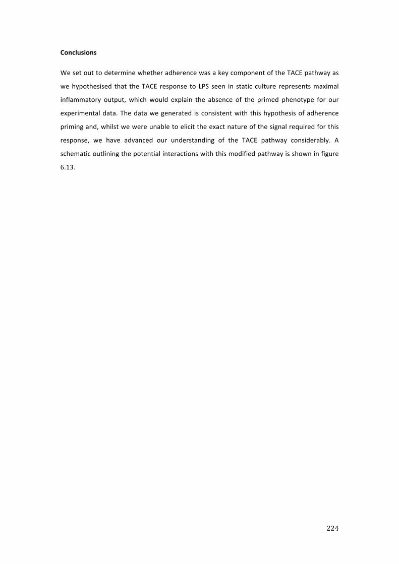
224
Conclusions
We set out to determine whether adherence was a key component of the TACE pathway as
we hypothesised that the TACE response to LPS seen in static culture represents maximal
inflammatory output, which would explain the absence of the primed phenotype for our
experimental data. The data we generated is consistent with this hypothesis of adherence
priming and, whilst we were unable to elicit the exact nature of the signal required for this
response, we have advanced our understanding of the TACE pathway considerably. A
schematic outlining the potential interactions with this modified pathway is shown in figure
6.13.
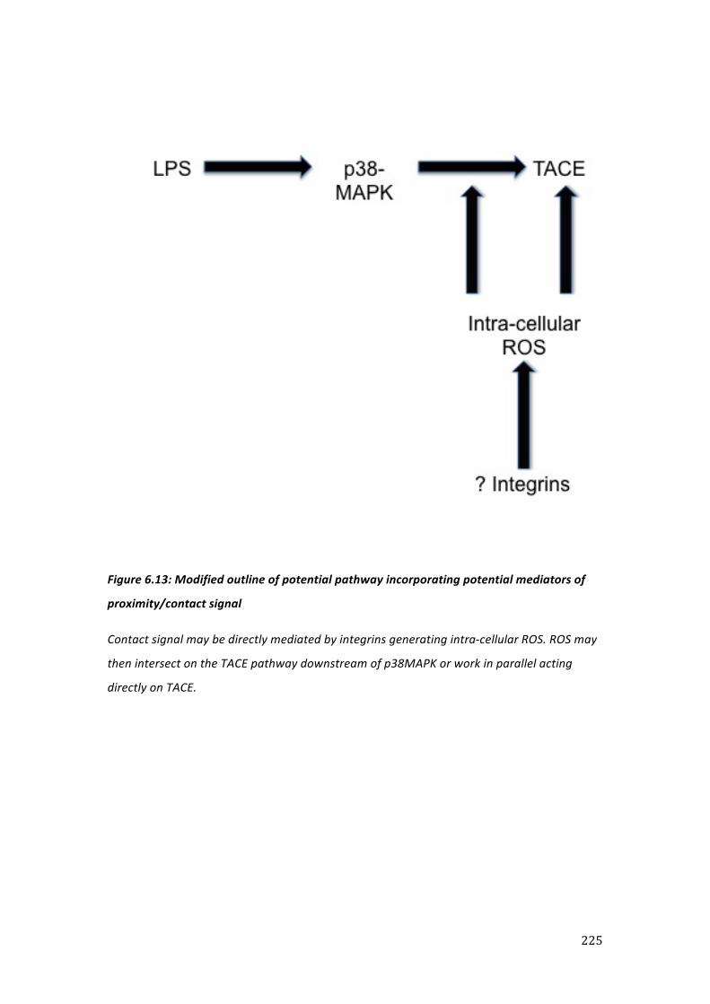
225
Figure 6.13: Modified outline of potential pathway incorporating potential mediators of
proximity/contact signal
Contact signal may be directly mediated by integrins generating intra-‐cellular ROS. ROS may
then intersect on the TACE pathway downstream of p38MAPK or work in parallel acting
directly on TACE.

226
Chapter 7
Final discussion

227
In the introduction I discussed the urgent need for further treatments to help supplement
our armamentarium when battling sepsis. Whilst it is certainly the case that the widespread
and uncontrolled use of antibiotics has promoted the emergence of resistance, this is not
the complete story. In fact, only a year after the discovery of penicillin some strains of
Staphylococcus aureus were already resistant [503]. In addition, analysis of permafrost
samples from 30,000 years ago has identified genes encoding resistance to beta-‐lactam,
tetracycline and glycopeptide antibiotics [504], meaning that antibiotic resistance was
inevitable. Thus the need to fully understand the processes of immunity (good) and
inflammation (bad) have become ever more pressing in order to avoid a continued over-‐
reliance on antibiotics. I would contend that the US Surgeon Generals 1967 proclamation,
“the war on infectious diseases has been won” [505], was at best premature and at worst
incorrect. Hence the need for basic scientific and clinical research into this area is warranted.
The principal aim of this project was to determine how monocyte TACE activity is affected by
sepsis and during an inflammatory response. Over the last four chapters I have presented
the data I obtained whilst attempting to elucidate the mechanisms involved. Having
demonstrated that TACE biology in cell lines is different to that in primary cells, I refined a
method of isolating primary cells from both volunteers and patients for the remainder of the
project. We then attempted to model states of tolerance and priming in vitro and found that
TACE activity profiles within cells placed in model conditions were different to those seen in
freshly isolated cells, they displayed both higher baseline activity and a reduced response to
LPS. We carried out a clinical project in which we successfully determined TACE activity and
substrate-‐shedding profiles in monocytes obtained from patients with sepsis and SIRS, and
found evidence of differential reprogramming in sepsis compared to non-‐infectious SIRS.
Lastly we examined how TACE signalling is affected by cell proximity and found that cell-‐to-‐
cell contact was critical for activation of TACE. Whilst the discussions in the results chapters
were limited to the scope of that chapter, here in this final discussion I will attempt to draw
the findings together and make some conclusions, highlighting our key achievements and
their novelty. These are mainly with regard to the plasticity of monocytes: specifically the
alterations they display in sepsis and in response to changes in their microenvironment.
228
7.1 An isolation method that enabled the study of monocyte associated enzyme activity
We began our work by investigating the utility of the immortalised human cell line THP-‐1 in
investigating TACE behaviour. We previously used this cell line to establish a FRET-‐peptide
based TACE enzyme activity assay, because it shared similar characteristics to the primary
cells in terms of surface expression of the mature TACE isoform and its response to LPS
[133]. However, it became apparent that these cells did not produce a TACE response
comparable to that seen in primary cells in a reliable and reproducible fashion. This
difference may be explained as, in common with many lines, the THP-‐1 cell is an
immortalised (leukaemic) cell line [382]. Previously when used by our group the THP-‐1 cell
line did produce a TACE response to LPS [381], although their passages and lots had to be
carefully matched. However, the cells that I purchased appeared not to have retained their
TACE responsiveness. Cells in culture are prone to phenotypic and genotypic drift [506], and
can vary considerably in their response to LPS [507], potentially explaining why I was unable
to reproduce our previous findings with the THP-‐1 cells. Previously such phenotype
instability was encountered with MonoMac6 cell line that displayed temporal alterations in
their TACE response [133, 381]. Thus my findings led us to conclude that the mechanism of
TACE activity regulation in vivo can only realistically be evaluated through the use of primary
cells.
We then successfully validated a method of isolating monocytes using a CD14 positive
immune-‐magnetic bead selection protocol, which was faster and more economical than our
previous protocol of negative selection, and did not appear to impact on the LPS-‐TLR4
signalling axis. Our previous attempts at isolating monocytes from sepsis patients using a
negative bead selection protocol had been unsuccessful, primarily due to contamination by
neutrophils, whose phenotype tends to exhibit significant changes with sepsis. However, we
were able to isolate monocytes with a reasonable purity by positive selection from septic
patients blood for use in the FRET assay, which represents a significant advance. This
facilitated for the first time the direct measurement of TACE catalytic activity in patients
with sepsis and could be widely adopted for other clinical conditions. The speed of the
monocyte positive selection method and its avoidance of issues related to leukocyte
phenotype change during inflammation are clearly advantages for clinical studies. A recent
review of leukocyte isolation methods concluded that “positive selection strategies… be
adopted as the procedure of choice” [508].
229
7.2 Resetting of the LPS-‐TACE axis in sepsis
We next found evidence that the LPS-‐TACE axis appears to be reset in sepsis. Monocytes
isolated from patients with sepsis displayed altered TACE characteristics. These were
increased baseline activity and refractoriness to further stimulation with LPS, with reduced
TNFR but retained L-‐selectin shedding. Similar findings were seen in the PTFE culture model
used in Chapter Four, although L-‐selectin shedding was also impaired in these cells. Whilst
these changes could be in keeping with a state of tolerance, we found no evidence of
priming in either PTFE or sepsis patients’ monocytes. Our use of primary monocytes and a
minimally adherent medium in the PTFE model removed the variables of cell lines and
minimised the influence of cell contact, thus enabling dissection of TACE refractoriness as a
direct function of LPS exposure. The PTFE system always tended toward this state of
increased basal TACE activity and reduced response to secondary LPS stimulation, no matter
what concentration of LPS was used in the 16-‐hour culture period. Given the broad
substrate base of TACE [342], it is difficult to draw firm conclusions as to the functional
significance of these changes in TACE behaviour yet changes in activity may represent
altered substrate affinity/specificity. However, within the PTFE model we found no evidence
of altered TACE specificity for the substrates TNFR-‐1, TNFR-‐2 and L-‐selectin as the shedding
of all three was impaired. These findings raise questions as to how sepsis simulation should
be carried out in vitro.
Our work highlights the differences in monocyte TACE response in situations where cells are
in close proximity. This means that techniques using adherence as a means of isolating
monocytes should be avoided as they could induce a change in the cellular response
phenotype and hence impact on results [509]. Care should be taken to control cell contact
status during the stimulation period with an emphasis placed on reproducing the in vivo
situation. This may mean that different systems are used to represent the circulating cell
and the marginated cell, perhaps using co-‐culture systems where possible and appropriate.
It is possible that the absence of contact during the PTFE culture period may have affected
monocyte behaviour in our model although the consistencies between our in vitro and in
vivo TACE activity data are striking.

230
7.3 The TACE response is dependent on cell proximity
We were surprised by the lack of a primed response in the PTFE model but surmised that
this may be due to a lack of cell contact and created a model to explore this further. We
found that the monocyte TACE response is dependent on cell contact. In neutrophils it is
thought that the two-‐hit priming process acts as a brake to ensure that damaging mediators
such as ROS are not released inappropriately [311]. Thus it may be that this reliance on cell
proximity to allow full inflammatory signalling represents a brake to ensure monocyte
inflammatory products are spatially regulated, and released by cells only in close proximity
with the endothelium and/or other cells. Monocytes are inherently adherent cells; more so
than neutrophils, thus contact may represent the predominant regulatory process in
monocytes whereas neutrophils respond to chemical mediators. It can be argued that this
state represents the primed phenotype and it would appear that the monocytes placed in
LPS PTFE culture, as well as those obtained from sepsis patients, have this regulatory
mechanism disturbed. This would explain the absence of priming in the PTFE culture model.
Our results would be in keeping with the primed response representing monocytes in close
proximity that are capable of full inflammatory signalling. Thus it would appear that this may
provide a means of spatially regulating the SIRS/CARS response using compartmentalisation:
circulating cells are different from those marginated in the microcirculation which may
themselves be different to those in the tissues. Such a situation would help to prevent
circulating cells from releasing their inflammatory products and generating disseminated
systemic inflammation.
231
7.4 Monocyte subsets in sepsis and SIRS
One of the achievements of our clinical study was to create a flow cytometry protocol in
which we could accurately identify monocytes without gating on HLA-‐DR. This allowed
accurate measurement of the expression of this antigen-‐presenting molecule without
excluding cells expressing only low levels, such as are seen in sepsis [292] and that would be
excluded using an HLA-‐DR+ gate. As part of this flow cytometry protocol we intended to
identify the previously characterised monocyte subsets: immature (CD14++CD16-‐),
intermediate (CD14+CD16+) and mature (CD14-‐CD16+) [297-‐299]. However, it became
apparent that both sepsis and SIRS distorted the expression of the monocyte surface
markers that we were using to identify the subsets. This occurred to such a degree in some
patients that we decided against drawing arbitrary gates, which may have impacted on our
results, and evaluated the monocytes as an entirety.
Whilst it is recognised that there is an expansion of the CD16+ monocyte population in
sepsis [305-‐307], to our knowledge, no other investigators have reported this inability to
accurately distinguish between the subsets. Whilst it may be possible to identify subsets
based on the expression of recently identified alternative surface markers [475] our results
would caution against drawing conclusions on subset functionality [510] based on standard
monocyte surface markers.

232
7.5 Markers of sepsis
Having identified this monocyte population we found that cells from both patients with
sepsis and SIRS expressed features consistent with monocyte deactivation – reduced HLA-‐DR
expression and reduced sol-‐TNF release on ex vivo LPS stimulation [224]. We could not
identify any differences between the groups based on these two markers meaning that they
lack the capacity to distinguish between septic and non-‐septic aetiologies. However, on
examination of p38MAPK signalling, TACE activity profiles and TACE substrate shedding we
identified several potential differences. In comparison to SIRS patients, monocytes from
sepsis patients had elevated baseline levels of phosphorylated p38MAPK and TACE, as well
as a reduced response to LPS. They also displayed increased expression and impaired
shedding of TNFR-‐1.
The changes in p38MAPK and TACE activity were both present from baseline in the sepsis
group and this raises the possibility that these could be used as markers to differentiate
sepsis from SIRS. Although further work is clearly required, these data highlight possible
differences in the aetiology of, and signalling changes induced by, monocyte deactivation.
233
7.6 The nature of the deactivated monocyte phenotype in sepsis
All monocytes obtained from patients with SIRS and sepsis displayed characteristics
consistent with a state of deactivation [224] – reduced HLA-‐DR expression and reduced sol-‐
TNF release on ex vivo LPS stimulation. This has been considered representative of a state of
immune suppression and similar to the tolerant monocyte phenotype [50, 323, 511].
However, by focusing on the TACE pathway, we were able to identify differences between
the groups. Sepsis patients’ monocytes displayed increased un-‐stimulated TACE activity and
a reduced response to LPS from D0, whereas these changes were not seen in SIRS until the
end of the study period. In addition, there was evidence of a differential responsiveness of
the cellular signalling pathways.
In sepsis patients we found preliminary evidence to suggest that the p38MAPK pathway was
“turned off” whereas the (unknown) L-‐selectin pathway was, for the most part, “turned on”
explaining the retained ability of their monocytes to shed L-‐selectin. It can be speculated
that sepsis produces a situation in which a number of pathways are “turned off” whereas
others are left “turned on”, a situation that could be considered a state of reprogramming; a
term that others have also suggested [50, 323, 439]. This appears to be different to non-‐
infectious SIRS patients where both of these pathways appeared to be “turned on”.
Such reprogramming of the monocyte in sepsis may involve differential effects on circulating
cells as opposed to tissue macrophages. Hence there may be a circulating monocyte
phenotype that displays reduced inflammatory signalling (as a result of the p38MAPK
pathway being turned off) and a tissue macrophage phenotype. The function of this
reprogrammed monocyte may be to prevent an inappropriate and continued release of
inflammatory products. As we speculated in the discussion to Chapter Five the increased
expression of TNFR-‐1 on these cells is of interest in this regard as TNFR-‐1 signalling forms
part of the “extrinsic” death receptor pathway that induces apoptosis in leukocytes. Thus it
may be that this represents a method of removing classically activated monocytes in order
that monocyte function is reoriented toward a tissue repair phenotype.
There is an alternative explanation; as we said in the introduction, “circulating inflammatory
cells would be required to switch from a relatively passive to more pro-‐inflammatory state
while migrating into the inflamed tissue via the vascular endothelium”. Our work suggests
that cells in close proximity to one another, such as may be seen on the vascular
endothelium have enhanced inflammatory signalling capacity when compared to those not
in proximity. Thus circulating septic, reprogrammed, cells with reduced inflammatory

234
signalling (through “turned off” p38MAPK) may have this partially restored (“turned on”) by
proximity, and produce an inflammatory focus. The fact that monocytes from septic patients
did not demonstrate this when stimulated in the cell dense conditions, such as were present
in Eppendorfs used for the LPS stimulation, may mean that other cues are required. Co-‐
culture experiments using monocytes and endothelial cells may provide further information
in this regard.
The retained (“turned on”) L-‐selectin shedding capacity may mean that these reprogrammed
cells are capable of migrating across the endothelium into the inflamed tissue in a normal
manner. Our preliminary results using a chemotaxis assay suggested that this might be the
case. Once in the tissue further phenotypic changes may occur as a result of the local
cytokine environment and continued contact with the extra-‐cellular matrix. Further work,
comparing tissue macrophages to circulating monocytes, will reveal further details and may
provide further evidence of a spatial regulation of the SIRS/CARS response.
In non-‐infectious SIRS the situation was different, as the p38MAPK pathway remained
“turned on”. Hence, despite outwardly appearing deactivated, these cells are still capable of
mounting a normal TACE response on ex vivo stimulation and it may be that these
circulating cells are “primed” for a secondary inflammatory stimulus. Certainly the situation
seems more complicated than a simple homogenous deactivation response to
heterogeneous stimuli (SIRS and sepsis). Although in SIRS patients these cells retain a TACE
response to LPS they produce lower levels of sol-‐TNF on ex vivo stimulation than freshly
isolated cells. Once again, the effect of cell proximity is likely to be of key importance and
should be examined closely in any future work in this area.

235
7.7 Concluding remarks
The current paradigm of sepsis is based on the body’s attempt to restore homeostasis, in
doing so it becomes immune-‐compromised leading to a vicious cycle of recurrent infection
and organ injury. Part of this theory has its origin in changes to the monocyte phenotype,
which can be classified at two levels:
1. Refractoriness to further stimulation in ex vivo assays (e.g. LPS-‐induced TNF expression)
and
2. The presence of an in vivo phenotype implying reduced (immune) function (e.g. HLA-‐DR).
This study revealed that ex vivo analysis of TACE, and related substrate shedding, may be a
valid approach to discriminating between different aetiologies of SIRS, contrasting the lack
of clear differences found using the standard measures described above. By varying the ex
vivo conditions, we were also able to obtain some insight into factors affecting changes to
TACE behaviour during sepsis, indicating the multifactorial nature of this response and in
particular the previously unappreciated role of cell proximity as a limiting step in TACE
catalytic activation. The physiological consequences of these changes in TACE related
biology are not clear as yet, but they do highlight the need to perform monocyte ex vivo
assays of more functional relevance to innate immune system response to infection and
injury.
With specific regard to our initial aims, the limiting role of cell proximity and our use of a
non-‐adherent medium in our in vitro model meant that, whilst we may have modelled a
tolerant phenotype, we were not able to model priming. However, later results suggested
that the primed phenotype is proximity dependent and that this state may be signalled
through integrins and intra-‐cellular ROS. TACE signalling did appear to be differentially
affected across these phenotypes with our tolerance model producing increased un-‐
stimulated TACE activity and a reduced response to LPS. These findings were also seen in
monocytes taken from patients with sepsis, where evidence of reprogramming was seen
within the p38MAPK/TACE pathway, which appeared to be “turned off”.

236
Our results fit more with a compartmentalised immune response with an anti-‐inflammatory
response dominating systemically. This highlights the limitations of using plasma as the
window through which to view the immune response. Future work should focus on the
processes occurring within both the microcirculation and the tissues. Comparing these to
the systemic circulation may provide a more accurate representation of the nature of the
innate immune response. This more rigorous and technically demanding approach may be
essential to unravel the complexities of infectious and non-‐infectious SIRS and in the design
of effective immunotherapeutic strategies.

237
References

238
1. Fleming, A., On the antibacterial action of cultures of a penicillium, with special reference to their use in the isolation of B. influenzae. 1929. Bull World Health Organ, 2001. 79(8): p. 780-‐90.
2. Walsh, C., Molecular mechanisms that confer antibacterial drug resistance. Nature, 2000. 406(6797): p. 775-‐81.
3. Barbosa, T.M. and S.B. Levy, The impact of antibiotic use on resistance development and persistence. Drug Resist Updat, 2000. 3(5): p. 303-‐311.
4. Jevons, M.P., A.W. Coe, and M.T. Parker, Methicillin resistance in staphylococci. Lancet, 1963. 1(7287): p. 904-‐7.
5. Shlaes, D.M., et al., Society for Healthcare Epidemiology of America and Infectious Diseases Society of America Joint Committee on the Prevention of Antimicrobial Resistance: guidelines for the prevention of antimicrobial resistance in hospitals. Clin Infect Dis, 1997. 25(3): p. 584-‐99.
6. O'Callaghan, D.J. and A.C. Gordon, Sepsis: an update for physicians. Clin Med, 2011. 11(6): p. 619-‐22.
7. Harrison, D.A., C.A. Welch, and J.M. Eddleston, The epidemiology of severe sepsis in England, Wales and Northern Ireland, 1996 to 2004: secondary analysis of a high quality clinical database, the ICNARC Case Mix Programme Database. Crit Care, 2006. 10(2): p. R42.
8. Martin, G.S., et al., The epidemiology of sepsis in the United States from 1979 through 2000. N Engl J Med, 2003. 348(16): p. 1546-‐54.
9. Angus, D.C., et al., Epidemiology of severe sepsis in the United States: analysis of incidence, outcome, and associated costs of care. Crit Care Med, 2001. 29(7): p. 1303-‐10.
10. Dellinger, R.P., et al., Surviving Sepsis Campaign guidelines for management of severe sepsis and septic shock. Intensive Care Med, 2004. 30(4): p. 536-‐55.
11. Levy, M.M., et al., The Surviving Sepsis Campaign: results of an international guideline-‐based performance improvement program targeting severe sepsis. Intensive Care Med, 2010. 36(2): p. 222-‐31.
12. Charles, P.G. and M.L. Grayson, The dearth of new antibiotic development: why we should be worried and what we can do about it. Med J Aust, 2004. 181(10): p. 549-‐53.
13. Ranieri, V.M., et al., Drotrecogin alfa (activated) in adults with septic shock. N Engl J Med, 2012. 366(22): p. 2055-‐64.
14. American College of Chest Physicians/Society of Critical Care Medicine Consensus Conference: definitions for sepsis and organ failure and guidelines for the use of innovative therapies in sepsis. Crit Care Med, 1992. 20(6): p. 864-‐74.
15. Rivers, E., et al., Early goal-‐directed therapy in the treatment of severe sepsis and septic shock. N Engl J Med, 2001. 345(19): p. 1368-‐77.
16. Rangel-‐Frausto, M.S., et al., The natural history of the systemic inflammatory response syndrome (SIRS). A prospective study. JAMA, 1995. 273(2): p. 117-‐23.
17. Dinarello, C.A., Historical insights into cytokines. Eur J Immunol, 2007. 37 Suppl 1: p. S34-‐45.
18. Thomas, L., Germs. N Engl J Med, 1972. 287(11): p. 553-‐5. 19. Goris, R.J., et al., Multiple-‐organ failure. Generalized autodestructive
inflammation? Archives of surgery, 1985. 120(10): p. 1109-‐15.

239
20. Goris, R.J., Multiple organ failure: whole body inflammation? Schweiz Med Wochenschr, 1989. 119(11): p. 347-‐53.
21. Hotchkiss, R.S. and I.E. Karl, The pathophysiology and treatment of sepsis. N Engl J Med, 2003. 348(2): p. 138-‐50.
22. Miyauchi, T., et al., Involvement of endothelin in the regulation of human vascular tonus. Potent vasoconstrictor effect and existence in endothelial cells. Circulation, 1990. 81(6): p. 1874-‐80.
23. Gomez-‐Jimenez, J., et al., L-‐arginine: nitric oxide pathway in endotoxemia and human septic shock. Crit Care Med, 1995. 23(2): p. 253-‐8.
24. Levi, M., et al., Pathogenesis of disseminated intravascular coagulation in sepsis. JAMA, 1993. 270(8): p. 975-‐9.
25. Riche, F.C., et al., Inflammatory cytokine response in patients with septic shock secondary to generalized peritonitis. Crit Care Med, 2000. 28(2): p. 433-‐7.
26. Casey, L.C., R.A. Balk, and R.C. Bone, Plasma cytokine and endotoxin levels correlate with survival in patients with the sepsis syndrome. Ann Intern Med, 1993. 119(8): p. 771-‐8.
27. Cannon, J.G., et al., Circulating interleukin-‐1 and tumor necrosis factor in septic shock and experimental endotoxin fever. J Infect Dis, 1990. 161(1): p. 79-‐84.
28. Martin, C., et al., Prognostic values of serum cytokines in septic shock. Intensive Care Med, 1994. 20(4): p. 272-‐7.
29. Tracey, K.J., et al., Shock and tissue injury induced by recombinant human cachectin. Science, 1986. 234(4775): p. 470-‐4.
30. Tracey, K.J., et al., Anti-‐cachectin/TNF monoclonal antibodies prevent septic shock during lethal bacteraemia. Nature, 1987. 330(6149): p. 662-‐4.
31. Beutler, B., I.W. Milsark, and A.C. Cerami, Passive immunization against cachectin/tumor necrosis factor protects mice from lethal effect of endotoxin. Science, 1985. 229(4716): p. 869-‐71.
32. Silva, A.T., K.F. Bayston, and J. Cohen, Prophylactic and therapeutic effects of a monoclonal antibody to tumor necrosis factor-‐alpha in experimental gram-‐negative shock. J Infect Dis, 1990. 162(2): p. 421-‐7.
33. Lorente, J.A. and J.C. Marshall, Neutralization of tumor necrosis factor in preclinical models of sepsis. Shock, 2005. 24 Suppl 1: p. 107-‐19.
34. Reinhart, K. and W. Karzai, Anti-‐tumor necrosis factor therapy in sepsis: update on clinical trials and lessons learned. Crit Care Med, 2001. 29(7 Suppl): p. S121-‐5.
35. Zeni, F., B. Freeman, and C. Natanson, Anti-‐inflammatory therapies to treat sepsis and septic shock: a reassessment. Crit Care Med, 1997. 25(7): p. 1095-‐100.
36. Fisher, C.J., Jr., et al., Treatment of septic shock with the tumor necrosis factor receptor:Fc fusion protein. The Soluble TNF Receptor Sepsis Study Group. N Engl J Med, 1996. 334(26): p. 1697-‐702.
37. Bone, R.C., Sir Isaac Newton, sepsis, SIRS, and CARS. Crit Care Med, 1996. 24(7): p. 1125-‐8.
38. Newton, I., Philosophiæ naturalis principia mathematica1687, Londini,: Jussu Societatis Regiæ ac Typis Josephi Streater. Prostat apud plures Bibliopolas. 4 p.l., 383, 400-‐510 p., 1 l.

240
39. Meakins, J.L., et al., Delayed hypersensitivity: indicator of acquired failure of host defenses in sepsis and trauma. Ann Surg, 1977. 186(3): p. 241-‐50.
40. Christou, N.V., Host-‐defence mechanisms in surgical patients: a correlative study of the delayed hypersensitivity skin-‐test response, granulocyte function and sepsis. Can J Surg, 1985. 28(1): p. 39-‐46, 49.
41. Lederer, J.A., M.L. Rodrick, and J.A. Mannick, The effects of injury on the adaptive immune response. Shock, 1999. 11(3): p. 153-‐9.
42. Oberholzer, A., C. Oberholzer, and L.L. Moldawer, Sepsis syndromes: understanding the role of innate and acquired immunity. Shock, 2001. 16(2): p. 83-‐96.
43. Cavaillon, J.M., "Septic Plasma": an immunosuppressive milieu. Am J Respir Crit Care Med, 2002. 166(11): p. 1417-‐8.
44. Prins, J.M., et al., Release of tumor necrosis factor alpha and interleukin 6 during antibiotic killing of Escherichia coli in whole blood: influence of antibiotic class, antibiotic concentration, and presence of septic serum. Infect Immun, 1995. 63(6): p. 2236-‐42.
45. Majetschak, M., et al., Regulation of whole blood tumor necrosis factor production upon endotoxin stimulation after severe blunt trauma. J Trauma, 1997. 43(6): p. 880-‐7.
46. Brandtzaeg, P., et al., Net inflammatory capacity of human septic shock plasma evaluated by a monocyte-‐based target cell assay: identification of interleukin-‐10 as a major functional deactivator of human monocytes. J Exp Med, 1996. 184(1): p. 51-‐60.
47. Ertel, W., et al., Downregulation of proinflammatory cytokine release in whole blood from septic patients. Blood, 1995. 85(5): p. 1341-‐7.
48. Kawamura, T., R. Wakusawa, and K. Inada, Interleukin-‐10 and interleukin-‐1 receptor antagonists increase during cardiac surgery. Can J Anaesth, 1997. 44(1): p. 38-‐42.
49. Adrie, C., et al., Successful cardiopulmonary resuscitation after cardiac arrest as a "sepsis-‐like" syndrome. Circulation, 2002. 106(5): p. 562-‐8.
50. Adib-‐Conquy, M. and J.M. Cavaillon, Compensatory anti-‐inflammatory response syndrome. Thromb Haemost, 2009. 101(1): p. 36-‐47.
51. Allen, M.L., et al., Early postoperative monocyte deactivation predicts systemic inflammation and prolonged stay in pediatric cardiac intensive care. Crit Care Med, 2002. 30(5): p. 1140-‐5.
52. Flohe, S., et al., Influence of surgical intervention in the immune response of severely injured patients. Intensive Care Med, 2004. 30(1): p. 96-‐102.
53. Handy, J.M., et al., HLA-‐DR expression and differential trafficking of monocyte subsets following low to intermediate risk surgery. Anaesthesia, 2010. 65(1): p. 27-‐35.
54. Lukaszewicz, A.C., et al., Monocytic HLA-‐DR expression in intensive care patients: interest for prognosis and secondary infection prediction. Crit Care Med, 2009. 37(10): p. 2746-‐52.
55. Monneret, G., et al., The anti-‐inflammatory response dominates after septic shock: association of low monocyte HLA-‐DR expression and high interleukin-‐10 concentration. Immunol Lett, 2004. 95(2): p. 193-‐8.
56. O'Sullivan, S.T., et al., Major injury leads to predominance of the T helper-‐2 lymphocyte phenotype and diminished interleukin-‐12 production associated

241
with decreased resistance to infection. Ann Surg, 1995. 222(4): p. 482-‐90; discussion 490-‐2.
57. Bone, R.C., Immunologic dissonance: a continuing evolution in our understanding of the systemic inflammatory response syndrome (SIRS) and the multiple organ dysfunction syndrome (MODS). Ann Intern Med, 1996. 125(8): p. 680-‐7.
58. Munford, R.S. and J. Pugin, Normal responses to injury prevent systemic inflammation and can be immunosuppressive. Am J Respir Crit Care Med, 2001. 163(2): p. 316-‐21.
59. Keel, M., et al., Different pattern of local and systemic release of proinflammatory and anti-‐inflammatory mediators in severely injured patients with chest trauma. J Trauma, 1996. 40(6): p. 907-‐12; discussion 912-‐4.
60. Bastian, D., et al., Systemic and local cytokine kinetics after total hip replacement surgery. Eur Surg Res, 2008. 41(4): p. 334-‐40.
61. Marie, C., et al., Cytokines and soluble cytokine receptors in pleural effusions from septic and nonseptic patients. Am J Respir Crit Care Med, 1997. 156(5): p. 1515-‐22.
62. Chinnaiyan, A.M., et al., Molecular signatures of sepsis: multiorgan gene expression profiles of systemic inflammation. Am J Pathol, 2001. 159(4): p. 1199-‐209.
63. Philippart, F., C. Fitting, and J.M. Cavaillon, Lung microenvironment contributes to the resistance of alveolar macrophages to develop tolerance to endotoxin. Crit Care Med, 2012.
64. Callery, M.P., M.J. Mangino, and M.W. Flye, A biologic basis for limited Kupffer cell reactivity to portal-‐derived endotoxin. Surgery, 1991. 110(2): p. 221-‐30.
65. Millo, J.L., et al., Compartmentalisation of cytokines and cytokine inhibitors in ventilator-‐associated pneumonia. Intensive Care Med, 2004. 30(1): p. 68-‐74.
66. Millar, A.B., et al., Tumour necrosis factor in bronchopulmonary secretions of patients with adult respiratory distress syndrome. Lancet, 1989. 2(8665): p. 712-‐4.
67. Hyers, T.M., et al., Tumor necrosis factor levels in serum and bronchoalveolar lavage fluid of patients with the adult respiratory distress syndrome. Am Rev Respir Dis, 1991. 144(2): p. 268-‐71.
68. Jorens, P.G., et al., Interleukin 8 (IL-‐8) in the bronchoalveolar lavage fluid from patients with the adult respiratory distress syndrome (ARDS) and patients at risk for ARDS. Cytokine, 1992. 4(6): p. 592-‐7.
69. Tran Van Nhieu, J., et al., Expression of tumor necrosis factor-‐alpha gene in alveolar macrophages from patients with the adult respiratory distress syndrome. Am Rev Respir Dis, 1993. 147(6 Pt 1): p. 1585-‐9.
70. Pugin, J., et al., Proinflammatory activity in bronchoalveolar lavage fluids from patients with ARDS, a prominent role for interleukin-‐1. Am J Respir Crit Care Med, 1996. 153(6 Pt 1): p. 1850-‐6.
71. Dehoux, M.S., et al., Compartmentalized cytokine production within the human lung in unilateral pneumonia. Am J Respir Crit Care Med, 1994. 150(3): p. 710-‐6.

242
72. Mateo, R.B., J.S. Reichner, and J.E. Albina, Interleukin-‐6 activity in wounds. Am J Physiol, 1994. 266(6 Pt 2): p. R1840-‐4.
73. Waage, A., et al., Local production of tumor necrosis factor alpha, interleukin 1, and interleukin 6 in meningococcal meningitis. Relation to the inflammatory response. J Exp Med, 1989. 170(6): p. 1859-‐67.
74. Dugernier, T.L., et al., Compartmentalization of the inflammatory response during acute pancreatitis: correlation with local and systemic complications. Am J Respir Crit Care Med, 2003. 168(2): p. 148-‐57.
75. Olszyna, D.P., et al., Chemotactic activity of CXC chemokines interleukin-‐8, growth-‐related oncogene-‐alpha, and epithelial cell-‐derived neutrophil-‐activating protein-‐78 in urine of patients with urosepsis. J Infect Dis, 2000. 182(6): p. 1731-‐7.
76. Rivera-‐Chavez, F.A., et al., Regional and systemic cytokine responses to acute inflammation of the vermiform appendix. Ann Surg, 2003. 237(3): p. 408-‐16.
77. Holzheimer, R.G., M. Schein, and D.H. Wittmann, Inflammatory response in peritoneal exudate and plasma of patients undergoing planned relaparotomy for severe secondary peritonitis. Archives of surgery, 1995. 130(12): p. 1314-‐9; discussion 1319-‐20.
78. Adib-‐Conquy, M., et al., Paradoxical priming effects of IL-‐10 on cytokine production. Int Immunol, 1999. 11(5): p. 689-‐98.
79. Takabayashi, T., et al., A new biologic role for C3a and C3a desArg: regulation of TNF-‐alpha and IL-‐1 beta synthesis. J Immunol, 1996. 156(9): p. 3455-‐60.
80. Beutler, B., Innate immunity: an overview. Mol Immunol, 2004. 40(12): p. 845-‐59.
81. Carroll, M.C. and D.E. Isenman, Regulation of humoral immunity by complement. Immunity, 2012. 37(2): p. 199-‐207.
82. Kimbrell, D.A. and B. Beutler, The evolution and genetics of innate immunity. Nat Rev Genet, 2001. 2(4): p. 256-‐67.
83. Janeway, C.A., Jr., Approaching the asymptote? Evolution and revolution in immunology. Cold Spring Harb Symp Quant Biol, 1989. 54 Pt 1: p. 1-‐13.
84. Galanos, C., et al., Synthetic and natural Escherichia coli free lipid A express identical endotoxic activities. Eur J Biochem, 1985. 148(1): p. 1-‐5.
85. Poltorak, A., et al., Defective LPS signaling in C3H/HeJ and C57BL/10ScCr mice: mutations in Tlr4 gene. Science, 1998. 282(5396): p. 2085-‐8.
86. Poltorak, O.M., E.S. Chukhray, and I.Y. Torshin, Dissociative thermal inactivation, stability, and activity of oligomeric enzymes. Biochemistry (Mosc), 1998. 63(3): p. 303-‐11.
87. Wright, S.D., et al., CD14, a receptor for complexes of lipopolysaccharide (LPS) and LPS binding protein. Science, 1990. 249(4975): p. 1431-‐3.
88. Muta, T. and K. Takeshige, Essential roles of CD14 and lipopolysaccharide-‐binding protein for activation of toll-‐like receptor (TLR)2 as well as TLR4 Reconstitution of TLR2-‐ and TLR4-‐activation by distinguishable ligands in LPS preparations. Eur J Biochem, 2001. 268(16): p. 4580-‐9.
89. Nagai, Y., et al., Essential role of MD-‐2 in LPS responsiveness and TLR4 distribution. Nat Immunol, 2002. 3(7): p. 667-‐72.
90. Aderem, A. and R.J. Ulevitch, Toll-‐like receptors in the induction of the innate immune response. Nature, 2000. 406(6797): p. 782-‐7.

243
91. Alexopoulou, L., et al., Recognition of double-‐stranded RNA and activation of NF-‐kappaB by Toll-‐like receptor 3. Nature, 2001. 413(6857): p. 732-‐8.
92. Creagh, E.M. and L.A. O'Neill, TLRs, NLRs and RLRs: a trinity of pathogen sensors that co-‐operate in innate immunity. Trends Immunol, 2006. 27(8): p. 352-‐7.
93. Cinel, I. and S.M. Opal, Molecular biology of inflammation and sepsis: a primer. Crit Care Med, 2009. 37(1): p. 291-‐304.
94. Johnson, G.B., G.J. Brunn, and J.L. Platt, Activation of mammalian Toll-‐like receptors by endogenous agonists. Crit Rev Immunol, 2003. 23(1-‐2): p. 15-‐44.
95. Ulloa, L. and K.J. Tracey, The "cytokine profile": a code for sepsis. Trends Mol Med, 2005. 11(2): p. 56-‐63.
96. Johnson, G.B., G.J. Brunn, and J.L. Platt, Cutting edge: an endogenous pathway to systemic inflammatory response syndrome (SIRS)-‐like reactions through Toll-‐like receptor 4. J Immunol, 2004. 172(1): p. 20-‐4.
97. Pahl, H.L., Activators and target genes of Rel/NF-‐kappaB transcription factors. Oncogene, 1999. 18(49): p. 6853-‐66.
98. Finkel, T., Signal transduction by reactive oxygen species. J Cell Biol, 2011. 194(1): p. 7-‐15.
99. Brown, D.I. and K.K. Griendling, Nox proteins in signal transduction. Free Radic Biol Med, 2009. 47(9): p. 1239-‐53.
100. Aguirre, J. and J.D. Lambeth, Nox enzymes from fungus to fly to fish and what they tell us about Nox function in mammals. Free Radic Biol Med, 2010. 49(9): p. 1342-‐53.
101. Giorgio, M., et al., Electron transfer between cytochrome c and p66Shc generates reactive oxygen species that trigger mitochondrial apoptosis. Cell, 2005. 122(2): p. 221-‐33.
102. Hsu, H.Y. and M.H. Wen, Lipopolysaccharide-‐mediated reactive oxygen species and signal transduction in the regulation of interleukin-‐1 gene expression. J Biol Chem, 2002. 277(25): p. 22131-‐9.
103. Bae, Y.S., et al., Epidermal growth factor (EGF)-‐induced generation of hydrogen peroxide. Role in EGF receptor-‐mediated tyrosine phosphorylation. J Biol Chem, 1997. 272(1): p. 217-‐21.
104. Lee, S.R., et al., Reversible inactivation of protein-‐tyrosine phosphatase 1B in A431 cells stimulated with epidermal growth factor. J Biol Chem, 1998. 273(25): p. 15366-‐72.
105. Salmeen, A., et al., Redox regulation of protein tyrosine phosphatase 1B involves a sulphenyl-‐amide intermediate. Nature, 2003. 423(6941): p. 769-‐73.
106. Matsuzawa, A., et al., ROS-‐dependent activation of the TRAF6-‐ASK1-‐p38 pathway is selectively required for TLR4-‐mediated innate immunity. Nat Immunol, 2005. 6(6): p. 587-‐92.
107. McCubrey, J.A., M.M. Lahair, and R.A. Franklin, Reactive oxygen species-‐induced activation of the MAP kinase signaling pathways. Antioxid Redox Signal, 2006. 8(9-‐10): p. 1775-‐89.
108. Tobiume, K., M. Saitoh, and H. Ichijo, Activation of apoptosis signal-‐regulating kinase 1 by the stress-‐induced activating phosphorylation of pre-‐formed oligomer. J Cell Physiol, 2002. 191(1): p. 95-‐104.

244
109. Saitoh, M., et al., Mammalian thioredoxin is a direct inhibitor of apoptosis signal-‐regulating kinase (ASK) 1. EMBO J, 1998. 17(9): p. 2596-‐606.
110. Schreck, R., P. Rieber, and P.A. Baeuerle, Reactive oxygen intermediates as apparently widely used messengers in the activation of the NF-‐kappa B transcription factor and HIV-‐1. EMBO J, 1991. 10(8): p. 2247-‐58.
111. Ushio-‐Fukai, M. and R.W. Alexander, Reactive oxygen species as mediators of angiogenesis signaling: role of NAD(P)H oxidase. Mol Cell Biochem, 2004. 264(1-‐2): p. 85-‐97.
112. Forstermann, U., et al., Isoforms of nitric oxide synthase. Properties, cellular distribution and expressional control. Biochemical pharmacology, 1995. 50(9): p. 1321-‐32.
113. Woo, H.A., et al., Inactivation of peroxiredoxin I by phosphorylation allows localized H(2)O(2) accumulation for cell signaling. Cell, 2010. 140(4): p. 517-‐28.
114. Niethammer, P., et al., A tissue-‐scale gradient of hydrogen peroxide mediates rapid wound detection in zebrafish. Nature, 2009. 459(7249): p. 996-‐9.
115. Bienert, G.P., et al., Specific aquaporins facilitate the diffusion of hydrogen peroxide across membranes. J Biol Chem, 2007. 282(2): p. 1183-‐92.
116. Miller, E.W., B.C. Dickinson, and C.J. Chang, Aquaporin-‐3 mediates hydrogen peroxide uptake to regulate downstream intracellular signaling. Proc Natl Acad Sci U S A, 2010. 107(36): p. 15681-‐6.
117. Cargnello, M. and P.P. Roux, Activation and function of the MAPKs and their substrates, the MAPK-‐activated protein kinases. Microbiol Mol Biol Rev, 2011. 75(1): p. 50-‐83.
118. Robbins, D.J., et al., Regulation and properties of extracellular signal-‐regulated protein kinases 1 and 2 in vitro. J Biol Chem, 1993. 268(7): p. 5097-‐106.
119. Gaestel, M., Specificity of signaling from MAPKs to MAPKAPKs: kinases' tango nuevo. Front Biosci, 2008. 13: p. 6050-‐9.
120. Roux, P.P. and J. Blenis, ERK and p38 MAPK-‐activated protein kinases: a family of protein kinases with diverse biological functions. Microbiol Mol Biol Rev, 2004. 68(2): p. 320-‐44.
121. Carriere, A., et al., The RSK factors of activating the Ras/MAPK signaling cascade. Front Biosci, 2008. 13: p. 4258-‐75.
122. Asp, E. and P. Sunnerhagen, Mkp1 and Mkp2, two MAPKAP-‐kinase homologues in Schizosaccharomyces pombe, interact with the MAP kinase Sty1. Mol Genet Genomics, 2003. 268(5): p. 585-‐97.
123. Buxade, M., J.L. Parra-‐Palau, and C.G. Proud, The Mnks: MAP kinase-‐interacting kinases (MAP kinase signal-‐integrating kinases). Front Biosci, 2008. 13: p. 5359-‐73.
124. Ronkina, N., A. Kotlyarov, and M. Gaestel, MK2 and MK3-‐-‐a pair of isoenzymes? Front Biosci, 2008. 13: p. 5511-‐21.
125. Cuenda, A., et al., Activation of stress-‐activated protein kinase-‐3 (SAPK3) by cytokines and cellular stresses is mediated via SAPKK3 (MKK6); comparison of the specificities of SAPK3 and SAPK2 (RK/p38). EMBO J, 1997. 16(2): p. 295-‐305.

245
126. Klotz, L.O., et al., Mitogen-‐activated protein kinase (p38-‐, JNK-‐, ERK-‐) activation pattern induced by extracellular and intracellular singlet oxygen and UVA. Eur J Biochem, 1999. 260(3): p. 917-‐22.
127. Guyton, K.Z., et al., Activation of mitogen-‐activated protein kinase by H2O2. Role in cell survival following oxidant injury. J Biol Chem, 1996. 271(8): p. 4138-‐42.
128. Lander, H.M., et al., Redox regulation of cell signalling. Nature, 1996. 381(6581): p. 380-‐1.
129. Schieke, S.M., et al., Activation pattern of mitogen-‐activated protein kinases elicited by peroxynitrite: attenuation by selenite supplementation. FEBS Lett, 1999. 448(2-‐3): p. 301-‐3.
130. Han, J., et al., A MAP kinase targeted by endotoxin and hyperosmolarity in mammalian cells. Science, 1994. 265(5173): p. 808-‐11.
131. Rouse, J., et al., A novel kinase cascade triggered by stress and heat shock that stimulates MAPKAP kinase-‐2 and phosphorylation of the small heat shock proteins. Cell, 1994. 78(6): p. 1027-‐37.
132. Freshney, N.W., et al., Interleukin-‐1 activates a novel protein kinase cascade that results in the phosphorylation of Hsp27. Cell, 1994. 78(6): p. 1039-‐49.
133. Scott, A.J., et al., Reactive oxygen species and p38 MAPK mediate TNF-‐alpha converting enzyme (TACE/ADAM17) activation in primary human monocytes. The Journal of biological chemistry, 2011.
134. Keyse, S.M., Protein phosphatases and the regulation of mitogen-‐activated protein kinase signalling. Curr Opin Cell Biol, 2000. 12(2): p. 186-‐92.
135. Lee, J.C., et al., A protein kinase involved in the regulation of inflammatory cytokine biosynthesis. Nature, 1994. 372(6508): p. 739-‐46.
136. Shapiro, L. and C.A. Dinarello, Osmotic regulation of cytokine synthesis in vitro. Proc Natl Acad Sci U S A, 1995. 92(26): p. 12230-‐4.
137. Beyaert, R., et al., The p38/RK mitogen-‐activated protein kinase pathway regulates interleukin-‐6 synthesis response to tumor necrosis factor. EMBO J, 1996. 15(8): p. 1914-‐23.
138. Berenson, L.S., et al., Selective requirement of p38alpha MAPK in cytokine-‐dependent, but not antigen receptor-‐dependent, Th1 responses. J Immunol, 2006. 176(8): p. 4616-‐21.
139. Gaestel, M., MAPKAP kinases -‐ MKs -‐ two's company, three's a crowd. Nature reviews. Molecular cell biology, 2006. 7(2): p. 120-‐30.
140. Gordon, S. and P.R. Taylor, Monocyte and macrophage heterogeneity. Nat Rev Immunol, 2005. 5(12): p. 953-‐64.
141. Gordon, S., Biology of the macrophage. J Cell Sci Suppl, 1986. 4: p. 267-‐86. 142. Gordon, S., The role of the macrophage in immune regulation. Res
Immunol, 1998. 149(7-‐8): p. 685-‐8. 143. Riedemann, N.C., R.F. Guo, and P.A. Ward, Novel strategies for the
treatment of sepsis. Nat Med, 2003. 9(5): p. 517-‐24. 144. Soehnlein, O. and L. Lindbom, Phagocyte partnership during the onset and
resolution of inflammation. Nat Rev Immunol, 2010. 10(6): p. 427-‐39. 145. Zhao, Q., et al., MAP kinase phosphatase 1 controls innate immune
responses and suppresses endotoxic shock. J Exp Med, 2006. 203(1): p. 131-‐40.
146. Zanotti, S., A. Kumar, and A. Kumar, Cytokine modulation in sepsis and septic shock. Expert Opin Investig Drugs, 2002. 11(8): p. 1061-‐75.
246
147. Michie, H.R., et al., Detection of circulating tumor necrosis factor after endotoxin administration. N Engl J Med, 1988. 318(23): p. 1481-‐6.
148. Hesse, D.G., et al., Cytokine appearance in human endotoxemia and primate bacteremia. Surg Gynecol Obstet, 1988. 166(2): p. 147-‐53.
149. Michie, H.R., et al., Tumor necrosis factor and endotoxin induce similar metabolic responses in human beings. Surgery, 1988. 104(2): p. 280-‐6.
150. Navarro-‐Sarabia, F., et al., Adalimumab for treating rheumatoid arthritis. J Rheumatol, 2006. 33(6): p. 1075-‐81.
151. Lawson, M.M., A.G. Thomas, and A.K. Akobeng, Tumour necrosis factor alpha blocking agents for induction of remission in ulcerative colitis. Cochrane Database Syst Rev, 2006. 3: p. CD005112.
152. Dinarello, C.A., Biology of interleukin 1. FASEB J, 1988. 2(2): p. 108-‐15. 153. Last-‐Barney, K., et al., Synergistic and overlapping activities of tumor
necrosis factor-‐alpha and IL-‐1. J Immunol, 1988. 141(2): p. 527-‐30. 154. Docke, W.D., et al., Monocyte deactivation in septic patients: restoration by
IFN-‐gamma treatment. Nat Med, 1997. 3(6): p. 678-‐81. 155. Moore, K.W., et al., Interleukin-‐10. Annu Rev Immunol, 1993. 11: p. 165-‐
90. 156. Bogdan, C. and C. Nathan, Modulation of macrophage function by
transforming growth factor beta, interleukin-‐4, and interleukin-‐10. Ann N Y Acad Sci, 1993. 685: p. 713-‐39.
157. Turner, M., et al., Induction of the interleukin 1 receptor antagonist protein by transforming growth factor-‐beta. Eur J Immunol, 1991. 21(7): p. 1635-‐9.
158. Peschon, J.J., et al., An essential role for ectodomain shedding in mammalian development. Science, 1998. 282(5392): p. 1281-‐4.
159. Hooper, N.M., E.H. Karran, and A.J. Turner, Membrane protein secretases. Biochem J, 1997. 321 ( Pt 2): p. 265-‐79.
160. Kiessling, L.L. and E.J. Gordon, Transforming the cell surface through proteolysis. Chem Biol, 1998. 5(3): p. R49-‐62.
161. Hehlgans, T. and D.N. Mannel, The TNF-‐TNF receptor system. Biol Chem, 2002. 383(10): p. 1581-‐5.
162. Aderka, D., The potential biological and clinical significance of the soluble tumor necrosis factor receptors. Cytokine Growth Factor Rev, 1996. 7(3): p. 231-‐40.
163. Aderka, D., et al., Shedding kinetics of soluble tumor necrosis factor (TNF) receptors after systemic TNF leaking during isolated limb perfusion. Relevance to the pathophysiology of septic shock. J Clin Invest, 1998. 101(3): p. 650-‐9.
164. Feterowski, C., et al., CC chemokine receptor 2 regulates leukocyte recruitment and IL-‐10 production during acute polymicrobial sepsis. Eur J Immunol, 2004. 34(12): p. 3664-‐73.
165. Chishti, A.D., et al., Neutrophil chemotaxis and receptor expression in clinical septic shock. Intensive Care Med, 2004. 30(4): p. 605-‐11.
166. Ness, T.L., et al., Immunomodulatory role of CXCR2 during experimental septic peritonitis. J Immunol, 2003. 171(7): p. 3775-‐84.
167. Ricciotti, E. and G.A. FitzGerald, Prostaglandins and inflammation. Arterioscler Thromb Vasc Biol, 2011. 31(5): p. 986-‐1000.

247
168. Kabashima, K. and S. Narumiya, The DP receptor, allergic inflammation and asthma. Prostaglandins Leukot Essent Fatty Acids, 2003. 69(2-‐3): p. 187-‐94.
169. Funk, C.D., Prostaglandins and leukotrienes: advances in eicosanoid biology. Science, 2001. 294(5548): p. 1871-‐5.
170. Oga, T., et al., Prostaglandin F(2alpha) receptor signaling facilitates bleomycin-‐induced pulmonary fibrosis independently of transforming growth factor-‐beta. Nat Med, 2009. 15(12): p. 1426-‐30.
171. Noda, M., et al., Neuroprotective role of bradykinin because of the attenuation of pro-‐inflammatory cytokine release from activated microglia. J Neurochem, 2007. 101(2): p. 397-‐410.
172. Nakahata, N., Thromboxane A2: physiology/pathophysiology, cellular signal transduction and pharmacology. Pharmacol Ther, 2008. 118(1): p. 18-‐35.
173. Hinshaw, L.B., et al., Effects of acetylsalicylic acid on the canine response to endotoxin. J Pharmacol Exp Ther, 1967. 157(3): p. 665-‐71.
174. Northover, B.J. and G. Subramanian, Analgesic-‐antipyretic drugs as antagonists of endotoxin shock in dogs. J Pathol Bacteriol, 1962. 83: p. 463-‐8.
175. Hauser, B., et al., Nitric oxide synthase inhibition in sepsis? Lessons learned from large-‐animal studies. Anesth Analg, 2005. 101(2): p. 488-‐98.
176. Hauser, W., et al., Expression of nitric oxide synthase isoforms in hypothalamo-‐pituitary-‐adrenal axis during the development of spontaneous hypertension in rats. Brain Res Mol Brain Res, 2005. 138(2): p. 198-‐204.
177. Kilbourn, R.G., C. Szabo, and D.L. Traber, Beneficial versus detrimental effects of nitric oxide synthase inhibitors in circulatory shock: lessons learned from experimental and clinical studies. Shock, 1997. 7(4): p. 235-‐46.
178. Belcher, E., J. Mitchell, and T. Evans, Myocardial dysfunction in sepsis: no role for NO? Heart, 2002. 87(6): p. 507-‐9.
179. Lopez, A., et al., Multiple-‐center, randomized, placebo-‐controlled, double-‐blind study of the nitric oxide synthase inhibitor 546C88: effect on survival in patients with septic shock. Crit Care Med, 2004. 32(1): p. 21-‐30.
180. Ley, K., et al., Getting to the site of inflammation: the leukocyte adhesion cascade updated. Nat Rev Immunol, 2007. 7(9): p. 678-‐89.
181. Kansas, G.S., Selectins and their ligands: current concepts and controversies. Blood, 1996. 88(9): p. 3259-‐87.
182. McEver, R.P. and R.D. Cummings, Role of PSGL-‐1 binding to selectins in leukocyte recruitment. J Clin Invest, 1997. 100(11 Suppl): p. S97-‐103.
183. Yoshida, M., et al., Leukocyte adhesion to vascular endothelium induces E-‐selectin linkage to the actin cytoskeleton. J Cell Biol, 1996. 133(2): p. 445-‐55.
184. Abbal, C., et al., Lipid raft adhesion receptors and Syk regulate selectin-‐dependent rolling under flow conditions. Blood, 2006. 108(10): p. 3352-‐9.
185. Simon, S.I., et al., Neutrophil tethering on E-‐selectin activates beta 2 integrin binding to ICAM-‐1 through a mitogen-‐activated protein kinase signal transduction pathway. J Immunol, 2000. 164(8): p. 4348-‐58.
186. Campbell, J.J., et al., Biology of chemokine and classical chemoattractant receptors: differential requirements for adhesion-‐triggering versus

248
chemotactic responses in lymphoid cells. J Cell Biol, 1996. 134(1): p. 255-‐66.
187. Campbell, J.J., et al., Chemokines and the arrest of lymphocytes rolling under flow conditions. Science, 1998. 279(5349): p. 381-‐4.
188. Middleton, J., et al., Transcytosis and surface presentation of IL-‐8 by venular endothelial cells. Cell, 1997. 91(3): p. 385-‐95.
189. von Hundelshausen, P., et al., RANTES deposition by platelets triggers monocyte arrest on inflamed and atherosclerotic endothelium. Circulation, 2001. 103(13): p. 1772-‐7.
190. Huo, Y., et al., Circulating activated platelets exacerbate atherosclerosis in mice deficient in apolipoprotein E. Nat Med, 2003. 9(1): p. 61-‐7.
191. Shamri, R., et al., Lymphocyte arrest requires instantaneous induction of an extended LFA-‐1 conformation mediated by endothelium-‐bound chemokines. Nat Immunol, 2005. 6(5): p. 497-‐506.
192. Arnaout, M.A., B. Mahalingam, and J.P. Xiong, Integrin structure, allostery, and bidirectional signaling. Annu Rev Cell Dev Biol, 2005. 21: p. 381-‐410.
193. Kreisel, D., et al., In vivo two-‐photon imaging reveals monocyte-‐dependent neutrophil extravasation during pulmonary inflammation. Proc Natl Acad Sci U S A, 2010. 107(42): p. 18073-‐8.
194. Holman, J.M., Jr. and T.M. Saba, Hepatocyte injury during post-‐operative sepsis: activated neutrophils as potential mediators. J Leukoc Biol, 1988. 43(3): p. 193-‐203.
195. Astiz, M.E., et al., Microvascular function and rheologic changes in hyperdynamic sepsis. Crit Care Med, 1995. 23(2): p. 265-‐71.
196. Bainton, D.F., J.L. Ullyot, and M.G. Farquhar, The development of neutrophilic polymorphonuclear leukocytes in human bone marrow. J Exp Med, 1971. 134(4): p. 907-‐34.
197. Weingarten, R., et al., Interactions of lipopolysaccharide with neutrophils in blood via CD14. J Leukoc Biol, 1993. 53(5): p. 518-‐24.
198. Wright, S.D., et al., Activation of the adhesive capacity of CR3 on neutrophils by endotoxin: dependence on lipopolysaccharide binding protein and CD14. J Exp Med, 1991. 173(5): p. 1281-‐6.
199. Akira, S. and K. Takeda, Toll-‐like receptor signalling. Nat Rev Immunol, 2004. 4(7): p. 499-‐511.
200. Sarma, J.V. and P.A. Ward, The complement system. Cell Tissue Res, 2011. 343(1): p. 227-‐35.
201. Babior, B.M., NADPH oxidase. Curr Opin Immunol, 2004. 16(1): p. 42-‐7. 202. Babior, B.M., The leukocyte NADPH oxidase. Isr Med Assoc J, 2002. 4(11):
p. 1023-‐4. 203. Babior, B.M., J.D. Lambeth, and W. Nauseef, The neutrophil NADPH oxidase.
Arch Biochem Biophys, 2002. 397(2): p. 342-‐4. 204. Coxon, A., et al., A novel role for the beta 2 integrin CD11b/CD18 in
neutrophil apoptosis: a homeostatic mechanism in inflammation. Immunity, 1996. 5(6): p. 653-‐66.
205. Gamberale, R., et al., Modulation of human neutrophil apoptosis by immune complexes. J Immunol, 1998. 161(7): p. 3666-‐74.
206. Colotta, F., et al., Modulation of granulocyte survival and programmed cell death by cytokines and bacterial products. Blood, 1992. 80(8): p. 2012-‐20.

249
207. DeLeo, F.R., Modulation of phagocyte apoptosis by bacterial pathogens. Apoptosis, 2004. 9(4): p. 399-‐413.
208. Kobayashi, S.D., et al., Neutrophils in the innate immune response. Archivum immunologiae et therapiae experimentalis, 2005. 53(6): p. 505-‐17.
209. Soehnlein, O., A. Zernecke, and C. Weber, Neutrophils launch monocyte extravasation by release of granule proteins. Thromb Haemost, 2009. 102(2): p. 198-‐205.
210. Soehnlein, O., L. Lindbom, and C. Weber, Mechanisms underlying neutrophil-‐mediated monocyte recruitment. Blood, 2009. 114(21): p. 4613-‐23.
211. Berahovich, R.D., et al., Proteolytic activation of alternative CCR1 ligands in inflammation. J Immunol, 2005. 174(11): p. 7341-‐51.
212. Borregaard, N., O.E. Sorensen, and K. Theilgaard-‐Monch, Neutrophil granules: a library of innate immunity proteins. Trends Immunol, 2007. 28(8): p. 340-‐5.
213. Marshall, J.C., The pathogenesis and molecular biology of sepsis. Crit Care Resusc, 2006. 8(3): p. 227-‐9.
214. Chua, F. and G.J. Laurent, Neutrophil elastase: mediator of extracellular matrix destruction and accumulation. Proc Am Thorac Soc, 2006. 3(5): p. 424-‐7.
215. Janoff, A., et al., Experimental emphysema induced with purified human neutrophil elastase: tissue localization of the instilled protease. Am Rev Respir Dis, 1977. 115(3): p. 461-‐78.
216. Taooka, Y., et al., Effects of neutrophil elastase inhibitor on bleomycin-‐induced pulmonary fibrosis in mice. Am J Respir Crit Care Med, 1997. 156(1): p. 260-‐5.
217. Tamakuma, S., et al., Relationship between neutrophil elastase and acute lung injury in humans. Pulm Pharmacol Ther, 2004. 17(5): p. 271-‐9.
218. Johansson, J., et al., Neutrophil-‐derived heparin binding protein-‐-‐a mediator of increased vascular permeability after burns? Burns, 2009. 35(8): p. 1185-‐7.
219. Auffray, C., et al., Monitoring of blood vessels and tissues by a population of monocytes with patrolling behavior. Science, 2007. 317(5838): p. 666-‐70.
220. Hume, D.A., et al., The mononuclear phagocyte system revisited. J Leukoc Biol, 2002. 72(4): p. 621-‐7.
221. Gordon, S., Alternative activation of macrophages. Nat Rev Immunol, 2003. 3(1): p. 23-‐35.
222. Stefater, J.A., 3rd, et al., Metchnikoff's policemen: macrophages in development, homeostasis and regeneration. Trends Mol Med, 2011. 17(12): p. 743-‐52.
223. Geng, Y., et al., Monocyte deactivation by interleukin 10 via inhibition of tyrosine kinase activity and the Ras signaling pathway. Proc Natl Acad Sci U S A, 1994. 91(18): p. 8602-‐6.
224. Volk, H.D., et al., Monocyte deactivation-‐-‐rationale for a new therapeutic strategy in sepsis. Intensive care medicine, 1996. 22 Suppl 4: p. S474-‐81.
225. Cheron, A., et al., [Low monocytic HLA-‐DR expression and risk of secondary infection]. Ann Fr Anesth Reanim, 2010. 29(5): p. 368-‐76.
250
226. Meisel, C., et al., Granulocyte-‐macrophage colony-‐stimulating factor to reverse sepsis-‐associated immunosuppression: a double-‐blind, randomized, placebo-‐controlled multicenter trial. Am J Respir Crit Care Med, 2009. 180(7): p. 640-‐8.
227. Versteeg, H.H., M.P. Peppelenbosch, and C.A. Spek, The pleiotropic effects of tissue factor: a possible role for factor VIIa-‐induced intracellular signalling? Thromb Haemost, 2001. 86(6): p. 1353-‐9.
228. Gando, S., et al., Significant correlations between tissue factor and thrombin markers in trauma and septic patients with disseminated intravascular coagulation. Thromb Haemost, 1998. 79(6): p. 1111-‐5.
229. Vickers, J., et al., Monocyte tissue factor (CD142) and Mac-‐1 (CD11b) are increased in septic patients. Thromb Haemost, 1998. 79(6): p. 1219-‐20.
230. Russell, J.A., Management of sepsis. N Engl J Med, 2006. 355(16): p. 1699-‐713.
231. Raaphorst, J., et al., Early inhibition of activated fibrinolysis predicts microbial infection, shock and mortality in febrile medical patients. Thromb Haemost, 2001. 86(2): p. 543-‐9.
232. Esmon, C.T., Structure and functions of the endothelial cell protein C receptor. Crit Care Med, 2004. 32(5 Suppl): p. S298-‐301.
233. Walker, F.J., P.W. Sexton, and C.T. Esmon, The inhibition of blood coagulation by activated Protein C through the selective inactivation of activated Factor V. Biochim Biophys Acta, 1979. 571(2): p. 333-‐42.
234. Fulcher, C.A., et al., Proteolytic inactivation of human factor VIII procoagulant protein by activated human protein C and its analogy with factor V. Blood, 1984. 63(2): p. 486-‐9.
235. van Hinsbergh, V.W., et al., Activated protein C decreases plasminogen activator-‐inhibitor activity in endothelial cell-‐conditioned medium. Blood, 1985. 65(2): p. 444-‐51.
236. Joyce, D.E., et al., Gene expression profile of antithrombotic protein c defines new mechanisms modulating inflammation and apoptosis. J Biol Chem, 2001. 276(14): p. 11199-‐203.
237. Murakami, K., et al., Activated protein C prevents LPS-‐induced pulmonary vascular injury by inhibiting cytokine production. Am J Physiol, 1997. 272(2 Pt 1): p. L197-‐202.
238. Riedemann, N.C., R.F. Guo, and P.A. Ward, The enigma of sepsis. J Clin Invest, 2003. 112(4): p. 460-‐7.
239. Shin, H.S., et al., Chemotactic and anaphylatoxic fragment cleaved from the fifth component of guinea pig complement. Science, 1968. 162(3851): p. 361-‐3.
240. Sacks, T., et al., Oxygen radicals mediate endothelial cell damage by complement-‐stimulated granulocytes. An in vitro model of immune vascular damage. J Clin Invest, 1978. 61(5): p. 1161-‐7.
241. Goldstein, I.M. and G. Weissmann, Generation of C5-‐derived lysosomal enzyme-‐releasing activity (C5a) by lysates of leukocyte lysosomes. J Immunol, 1974. 113(5): p. 1583-‐8.
242. Creasey, A.A. and K. Reinhart, Tissue factor pathway inhibitor activity in severe sepsis. Crit Care Med, 2001. 29(7 Suppl): p. S126-‐9.
243. Liaw, P.C., et al., Patients with severe sepsis vary markedly in their ability to generate activated protein C. Blood, 2004. 104(13): p. 3958-‐64.

251
244. Lawson, C.A., et al., Monocytes and tissue factor promote thrombosis in a murine model of oxygen deprivation. J Clin Invest, 1997. 99(7): p. 1729-‐38.
245. Markiewski, M.M., R.A. DeAngelis, and J.D. Lambris, Complexity of complement activation in sepsis. J Cell Mol Med, 2008. 12(6A): p. 2245-‐54.
246. Quezado, Z.M., et al., The third component of complement protects against Escherichia coli endotoxin-‐induced shock and multiple organ failure. J Exp Med, 1994. 179(2): p. 569-‐78.
247. Wessels, M.R., et al., Studies of group B streptococcal infection in mice deficient in complement component C3 or C4 demonstrate an essential role for complement in both innate and acquired immunity. Proc Natl Acad Sci U S A, 1995. 92(25): p. 11490-‐4.
248. Fischer, M.B., et al., Increased susceptibility to endotoxin shock in complement C3-‐ and C4-‐deficient mice is corrected by C1 inhibitor replacement. J Immunol, 1997. 159(2): p. 976-‐82.
249. Ward, P.A., Role of the complement in experimental sepsis. J Leukoc Biol, 2008. 83(3): p. 467-‐70.
250. Walport, M.J., Complement. First of two parts. N Engl J Med, 2001. 344(14): p. 1058-‐66.
251. Hoffmann, M. and R.W. Dutton, Immune response restoration with macrophage culture supernatants. Science, 1971. 172(3987): p. 1047-‐8.
252. Dutton, R.W., et al., Cell components in the immune response. IV. Relationships and possible interactions. Cell Immunol, 1970. 1(2): p. 196-‐206.
253. Mahanonda, R., et al., Monocyte activation by Porphyromonas gingivalis LPS in aggressive periodontitis with the use of whole-‐blood cultures. J Dent Res, 2004. 83(7): p. 540-‐5.
254. Wolk, K., et al., Impaired antigen presentation by human monocytes during endotoxin tolerance. Blood, 2000. 96(1): p. 218-‐23.
255. Hotchkiss, R.S. and D.W. Nicholson, Apoptosis and caspases regulate death and inflammation in sepsis. Nat Rev Immunol, 2006. 6(11): p. 813-‐22.
256. Zhu, J. and W.E. Paul, CD4 T cells: fates, functions, and faults. Blood, 2008. 112(5): p. 1557-‐69.
257. Murphy, K.M. and S.L. Reiner, The lineage decisions of helper T cells. Nat Rev Immunol, 2002. 2(12): p. 933-‐44.
258. Afzali, B., et al., The role of T helper 17 (Th17) and regulatory T cells (Treg) in human organ transplantation and autoimmune disease. Clin Exp Immunol, 2007. 148(1): p. 32-‐46.
259. Sakaguchi, S., Naturally arising CD4+ regulatory t cells for immunologic self-‐tolerance and negative control of immune responses. Annu Rev Immunol, 2004. 22: p. 531-‐62.
260. Kurosaki, T., B-‐lymphocyte biology. Immunol Rev, 2010. 237(1): p. 5-‐9. 261. Weber, S.U., et al., Induction of Bim and Bid gene expression during
accelerated apoptosis in severe sepsis. Crit Care, 2008. 12(5): p. R128. 262. Boomer, J.S., et al., Immunosuppression in patients who die of sepsis and
multiple organ failure. JAMA, 2011. 306(23): p. 2594-‐605. 263. Venet, F., et al., Increased circulating regulatory T cells (CD4(+)CD25
(+)CD127 (-‐)) contribute to lymphocyte anergy in septic shock patients. Intensive Care Med, 2009. 35(4): p. 678-‐86.
252
264. Heininger, A., et al., Cytomegalovirus reactivation and associated outcome of critically ill patients with severe sepsis. Crit Care, 2011. 15(2): p. R77.
265. Kalil, A.C. and D.F. Florescu, Prevalence and mortality associated with cytomegalovirus infection in nonimmunosuppressed patients in the intensive care unit. Crit Care Med, 2009. 37(8): p. 2350-‐8.
266. Adib-‐Conquy, M. and J.M. Cavaillon, Stress molecules in sepsis and systemic inflammatory response syndrome. FEBS Lett, 2007. 581(19): p. 3723-‐33.
267. Oda, S., et al., Sequential measurement of IL-‐6 blood levels in patients with systemic inflammatory response syndrome (SIRS)/sepsis. Cytokine, 2005. 29(4): p. 169-‐75.
268. Gregoric, P., et al., SIRS score on admission and initial concentration of IL-‐6 as severe acute pancreatitis outcome predictors. Hepatogastroenterology, 2010. 57(98): p. 349-‐53.
269. Mokart, D., et al., Procalcitonin, interleukin 6 and systemic inflammatory response syndrome (SIRS): early markers of postoperative sepsis after major surgery. Br J Anaesth, 2005. 94(6): p. 767-‐73.
270. Gebhard, F., et al., Is interleukin 6 an early marker of injury severity following major trauma in humans? Archives of surgery, 2000. 135(3): p. 291-‐5.
271. Taniguchi, T., et al., Change in the ratio of interleukin-‐6 to interleukin-‐10 predicts a poor outcome in patients with systemic inflammatory response syndrome. Crit Care Med, 1999. 27(7): p. 1262-‐4.
272. Taniguchi, T., et al., The ratio of interleukin-‐6 to interleukin-‐10 correlates with severity in patients with chest and abdominal trauma. Am J Emerg Med, 1999. 17(6): p. 548-‐51.
273. Levy, M.M., et al., 2001 SCCM/ESICM/ACCP/ATS/SIS International Sepsis Definitions Conference. Crit Care Med, 2003. 31(4): p. 1250-‐6.
274. Harbarth, S., et al., Diagnostic value of procalcitonin, interleukin-‐6, and interleukin-‐8 in critically ill patients admitted with suspected sepsis. Am J Respir Crit Care Med, 2001. 164(3): p. 396-‐402.
275. Wu, H.P., et al., Plasma transforming growth factor-‐beta1 level in patients with severe community-‐acquired pneumonia and association with disease severity. J Formos Med Assoc, 2009. 108(1): p. 20-‐7.
276. Guignant, C., et al., Assessment of pro-‐vasopressin and pro-‐adrenomedullin as predictors of 28-‐day mortality in septic shock patients. Intensive Care Med, 2009. 35(11): p. 1859-‐67.
277. Seligman, R., et al., Prognostic value of midregional pro-‐atrial natriuretic peptide in ventilator-‐associated pneumonia. Intensive Care Med, 2008. 34(11): p. 2084-‐91.
278. Koch, A., et al., Serum resistin levels in critically ill patients are associated with inflammation, organ dysfunction and metabolism and may predict survival of non-‐septic patients. Crit Care, 2009. 13(3): p. R95.
279. Kibe, S., K. Adams, and G. Barlow, Diagnostic and prognostic biomarkers of sepsis in critical care. J Antimicrob Chemother, 2011. 66 Suppl 2: p. ii33-‐40.
280. O'Grady, N.P., et al., Guidelines for evaluation of new fever in critically ill adult patients: 2008 update from the American College of Critical Care Medicine and the Infectious Diseases Society of America. Crit Care Med, 2008. 36(4): p. 1330-‐49.

253
281. Reinhart, K., et al., Randomized, placebo-‐controlled trial of the anti-‐tumor necrosis factor antibody fragment afelimomab in hyperinflammatory response during severe sepsis: The RAMSES Study. Crit Care Med, 2001. 29(4): p. 765-‐9.
282. Bo, L., et al., Granulocyte-‐colony stimulating factor (G-‐CSF) and granulocyte-‐macrophage colony stimulating factor (GM-‐CSF) for sepsis: a meta-‐analysis. Crit Care, 2011. 15(1): p. R58.
283. Hynninen, M., et al., Predictive value of monocyte histocompatibility leukocyte antigen-‐DR expression and plasma interleukin-‐4 and -‐10 levels in critically ill patients with sepsis. Shock, 2003. 20(1): p. 1-‐4.
284. Opal, S.M. and V.A. DePalo, Anti-‐inflammatory cytokines. Chest, 2000. 117(4): p. 1162-‐72.
285. Gogos, C.A., et al., Pro-‐ versus anti-‐inflammatory cytokine profile in patients with severe sepsis: a marker for prognosis and future therapeutic options. J Infect Dis, 2000. 181(1): p. 176-‐80.
286. Monneret, G., et al., Persisting low monocyte human leukocyte antigen-‐DR expression predicts mortality in septic shock. Intensive Care Med, 2006. 32(8): p. 1175-‐83.
287. Monneret, G., et al., Monitoring immune dysfunctions in the septic patient: a new skin for the old ceremony. Mol Med, 2008. 14(1-‐2): p. 64-‐78.
288. Koppelman, B., et al., Interleukin-‐10 down-‐regulates MHC class II alphabeta peptide complexes at the plasma membrane of monocytes by affecting arrival and recycling. Immunity, 1997. 7(6): p. 861-‐71.
289. Krakauer, T. and J.J. Oppenheim, IL-‐1 and tumor necrosis factor-‐alpha each up-‐regulate both the expression of IFN-‐gamma receptors and enhance IFN-‐gamma-‐induced HLA-‐DR expression on human monocytes and a human monocytic cell line (THP-‐1). J Immunol, 1993. 150(4): p. 1205-‐11.
290. Basta, P.V., et al., A beta-‐adrenergic agonist modulates DR alpha gene transcription via enhanced cAMP levels in a glioblastoma multiforme line. J Immunol, 1989. 142(8): p. 2895-‐901.
291. Schwiebert, L.M., et al., Modulation of MHC class II expression in human cells by dexamethasone. Cell Immunol, 1995. 165(1): p. 12-‐9.
292. Tschoeke, S.K. and L.L. Moldawer, Human leukocyte antigen expression in sepsis: what have we learned? Critical care medicine, 2005. 33(1): p. 236-‐7.
293. Ayala, A., W. Ertel, and I.H. Chaudry, Trauma-‐induced suppression of antigen presentation and expression of major histocompatibility class II antigen complex in leukocytes. Shock, 1996. 5(2): p. 79-‐90.
294. Tschaikowsky, K., et al., Coincidence of pro-‐ and anti-‐inflammatory responses in the early phase of severe sepsis: Longitudinal study of mononuclear histocompatibility leukocyte antigen-‐DR expression, procalcitonin, C-‐reactive protein, and changes in T-‐cell subsets in septic and postoperative patients. Crit Care Med, 2002. 30(5): p. 1015-‐23.
295. Docke, W.D., et al., Monitoring temporary immunodepression by flow cytometric measurement of monocytic HLA-‐DR expression: a multicenter standardized study. Clin Chem, 2005. 51(12): p. 2341-‐7.
296. Ziegler-‐Heitbrock, L., The CD14+ CD16+ blood monocytes: their role in infection and inflammation. J Leukoc Biol, 2007. 81(3): p. 584-‐92.

254
297. Cros, J., et al., Human CD14dim monocytes patrol and sense nucleic acids and viruses via TLR7 and TLR8 receptors. Immunity, 2010. 33(3): p. 375-‐86.
298. Skrzeczynska-‐Moncznik, J., et al., Peripheral blood CD14high CD16+ monocytes are main producers of IL-‐10. Scand J Immunol, 2008. 67(2): p. 152-‐9.
299. Wong, K.L., et al., Gene expression profiling reveals the defining features of the classical, intermediate, and nonclassical human monocyte subsets. Blood, 2011. 118(5): p. e16-‐31.
300. Belge, K.U., et al., The proinflammatory CD14+CD16+DR++ monocytes are a major source of TNF. J Immunol, 2002. 168(7): p. 3536-‐42.
301. Szaflarska, A., et al., Antitumor response of CD14+/CD16+ monocyte subpopulation. Exp Hematol, 2004. 32(8): p. 748-‐55.
302. Nockher, W.A., L. Bergmann, and J.E. Scherberich, Increased soluble CD14 serum levels and altered CD14 expression of peripheral blood monocytes in HIV-‐infected patients. Clin Exp Immunol, 1994. 98(3): p. 369-‐74.
303. Rivier, A., et al., Blood monocytes of untreated asthmatics exhibit some features of tissue macrophages. Clin Exp Immunol, 1995. 100(2): p. 314-‐8.
304. Nockher, W.A. and J.E. Scherberich, Expanded CD14+ CD16+ monocyte subpopulation in patients with acute and chronic infections undergoing hemodialysis. Infect Immun, 1998. 66(6): p. 2782-‐90.
305. Fingerle, G., et al., The novel subset of CD14+/CD16+ blood monocytes is expanded in sepsis patients. Blood, 1993. 82(10): p. 3170-‐6.
306. Blumenstein, M., et al., Cytokine production precedes the expansion of CD14+CD16+ monocytes in human sepsis: a case report of a patient with self-‐induced septicemia. Shock, 1997. 8(1): p. 73-‐5.
307. Shalova, I.N., et al., CD16 regulates TRIF-‐dependent TLR4 response in human monocytes and their subsets. J Immunol, 2012. 188(8): p. 3584-‐93.
308. Skrzeczynska, J., et al., CD14+CD16+ monocytes in the course of sepsis in neonates and small children: monitoring and functional studies. Scand J Immunol, 2002. 55(6): p. 629-‐38.
309. Herra, C.M., C.T. Keane, and A. Whelan, Increased expression of Fc gamma receptors on neutrophils and monocytes may reflect ongoing bacterial infection. J Med Microbiol, 1996. 44(2): p. 135-‐40.
310. Fingerle-‐Rowson, G., et al., Expansion of CD14+CD16+ monocytes in critically ill cardiac surgery patients. Inflammation, 1998. 22(4): p. 367-‐79.
311. Condliffe, A.M., E. Kitchen, and E.R. Chilvers, Neutrophil priming: pathophysiological consequences and underlying mechanisms. Clin Sci (Lond), 1998. 94(5): p. 461-‐71.
312. Swain, S.D., T.T. Rohn, and M.T. Quinn, Neutrophil priming in host defense: role of oxidants as priming agents. Antioxid Redox Signal, 2002. 4(1): p. 69-‐83.
313. Thomas, L. and R.A. Good, Studies on the generalized Shwartzman reaction: I. General observations concerning the phenomenon. J Exp Med, 1952. 96(6): p. 605-‐24.
314. Billiau, A., et al., Anti-‐interferon-‐gamma antibody protects mice against the generalized Shwartzman reaction. Eur J Immunol, 1987. 17(12): p. 1851-‐4.

255
315. Heremans, H., et al., Interferon gamma, a mediator of lethal lipopolysaccharide-‐induced Shwartzman-‐like shock reactions in mice. J Exp Med, 1990. 171(6): p. 1853-‐69.
316. O'Dea, K.P., et al., Mobilization and margination of bone marrow Gr-‐1high monocytes during subclinical endotoxemia predisposes the lungs toward acute injury. J Immunol, 2009. 182(2): p. 1155-‐66.
317. O'Dea, K.P., et al., Regulation of monocyte subset proinflammatory responses within the lung microvasculature by the p38 MAPK/MK2 pathway. Am J Physiol Lung Cell Mol Physiol, 2011. 301(5): p. L812-‐21.
318. Cuschieri, J., et al., Platelet-‐activating factor priming of inflammatory cell activity requires cellular adherence. Surgery, 2002. 132(2): p. 157-‐66.
319. Schaeffer, V., et al., The priming effect of C5a on monocytes is predominantly mediated by the p38 MAPK pathway. Shock, 2007. 27(6): p. 623-‐30.
320. Hoogerwerf, J.J., et al., Priming of alveolar macrophages upon instillation of lipopolysaccharide in the human lung. Am J Respir Cell Mol Biol, 2010. 42(3): p. 349-‐56.
321. Snelgrove, R.J., et al., A critical function for CD200 in lung immune homeostasis and the severity of influenza infection. Nat Immunol, 2008. 9(9): p. 1074-‐83.
322. Beeson, P.B., Development of tolerance to typhoid bacterial pyrogen and its abolition by reticulo-‐endothelial blockade. Proc Soc Exp Biol Med, 1946. 61: p. 248-‐50.
323. Cavaillon, J.M. and M. Adib-‐Conquy, Bench-‐to-‐bedside review: endotoxin tolerance as a model of leukocyte reprogramming in sepsis. Crit Care, 2006. 10(5): p. 233.
324. Biswas, S.K. and E. Lopez-‐Collazo, Endotoxin tolerance: new mechanisms, molecules and clinical significance. Trends Immunol, 2009. 30(10): p. 475-‐87.
325. Cavaillon, J.M., et al., Endotoxin tolerance: is there a clinical relevance? J Endotoxin Res, 2003. 9(2): p. 101-‐7.
326. del Fresno, C., et al., Potent phagocytic activity with impaired antigen presentation identifying lipopolysaccharide-‐tolerant human monocytes: demonstration in isolated monocytes from cystic fibrosis patients. J Immunol, 2009. 182(10): p. 6494-‐507.
327. van 't Veer, C., et al., Induction of IRAK-‐M is associated with lipopolysaccharide tolerance in a human endotoxemia model. J Immunol, 2007. 179(10): p. 7110-‐20.
328. Kobayashi, K., et al., IRAK-‐M is a negative regulator of Toll-‐like receptor signaling. Cell, 2002. 110(2): p. 191-‐202.
329. Kraatz, J., et al., Macrophage TNF secretion in endotoxin tolerance: role of SAPK, p38, and MAPK. J Surg Res, 1999. 83(2): p. 158-‐64.
330. Durando, M.M., K.E. Meier, and J.A. Cook, Endotoxin activation of mitogen-‐activated protein kinase in THP-‐1 cells; diminished activation following endotoxin desensitization. J Leukoc Biol, 1998. 64(2): p. 259-‐64.
331. Nimah, M., et al., Contribution of MKP-‐1 regulation of p38 to endotoxin tolerance. Shock, 2005. 23(1): p. 80-‐7.
332. Carswell, E.A., et al., An endotoxin-‐induced serum factor that causes necrosis of tumors. Proc Natl Acad Sci U S A, 1975. 72(9): p. 3666-‐70.

256
333. Aggarwal, B.B., et al., Human tumor necrosis factor. Production, purification, and characterization. J Biol Chem, 1985. 260(4): p. 2345-‐54.
334. Pfeffer, K., Biological functions of tumor necrosis factor cytokines and their receptors. Cytokine Growth Factor Rev, 2003. 14(3-‐4): p. 185-‐91.
335. Locksley, R.M., N. Killeen, and M.J. Lenardo, The TNF and TNF receptor superfamilies: integrating mammalian biology. Cell, 2001. 104(4): p. 487-‐501.
336. Tang, P., M.C. Hung, and J. Klostergaard, Human pro-‐tumor necrosis factor is a homotrimer. Biochemistry, 1996. 35(25): p. 8216-‐25.
337. Kusters, S., et al., In vivo evidence for a functional role of both tumor necrosis factor (TNF) receptors and transmembrane TNF in experimental hepatitis. Eur J Immunol, 1997. 27(11): p. 2870-‐5.
338. Alexopoulou, L., et al., Transmembrane TNF protects mutant mice against intracellular bacterial infections, chronic inflammation and autoimmunity. Eur J Immunol, 2006. 36(10): p. 2768-‐80.
339. Pellegrini, J.D., et al., A membrane TNF-‐alpha/TNFR ratio correlates to MODS score and mortality. Shock, 1996. 6(6): p. 389-‐96.
340. Black, R.A., et al., A metalloproteinase disintegrin that releases tumour-‐necrosis factor-‐alpha from cells. Nature, 1997. 385(6618): p. 729-‐33.
341. Black, R.A., Tumor necrosis factor-‐alpha converting enzyme. Int J Biochem Cell Biol, 2002. 34(1): p. 1-‐5.
342. Edwards, D.R., M.M. Handsley, and C.J. Pennington, The ADAM metalloproteinases. Mol Aspects Med, 2008. 29(5): p. 258-‐89.
343. Gooz, M., ADAM-‐17: the enzyme that does it all. Crit Rev Biochem Mol Biol, 2010. 45(2): p. 146-‐69.
344. Scheller, J., et al., ADAM17: a molecular switch to control inflammation and tissue regeneration. Trends Immunol, 2011. 32(8): p. 380-‐7.
345. Solomon, K.A., et al., Cutting edge: a dominant negative form of TNF-‐alpha converting enzyme inhibits proTNF and TNFRII secretion. J Immunol, 1999. 163(8): p. 4105-‐8.
346. Shimizu, M., et al., Effects of TNF-‐alpha-‐converting enzyme inhibition on acute lung injury induced by endotoxin in the rat. Shock, 2009. 32(5): p. 535-‐40.
347. Arndt, P.G., et al., Leukocyte ADAM17 regulates acute pulmonary inflammation. PLoS One, 2011. 6(5): p. e19938.
348. Allinson, T.M., et al., ADAMs family members as amyloid precursor protein alpha-‐secretases. J Neurosci Res, 2003. 74(3): p. 342-‐52.
349. Borrell-‐Pages, M., et al., TACE is required for the activation of the EGFR by TGF-‐alpha in tumors. EMBO J, 2003. 22(5): p. 1114-‐24.
350. McGowan, P.M., et al., ADAM-‐17 predicts adverse outcome in patients with breast cancer. Annals of oncology : official journal of the European Society for Medical Oncology / ESMO, 2008. 19(6): p. 1075-‐81.
351. McGowan, P.M., et al., ADAM-‐17 expression in breast cancer correlates with variables of tumor progression. Clin Cancer Res, 2007. 13(8): p. 2335-‐43.
352. McGowan, P.M., et al., ADAM-‐17: a novel therapeutic target for triple negative breast cancer. Annals of oncology : official journal of the European Society for Medical Oncology / ESMO, 2012.

257
353. Cesaro, A., et al., Differential expression and regulation of ADAM17 and TIMP3 in acute inflamed intestinal epithelia. Am J Physiol Gastrointest Liver Physiol, 2009. 296(6): p. G1332-‐43.
354. Blaydon, D.C., et al., Inflammatory skin and bowel disease linked to ADAM17 deletion. N Engl J Med, 2011. 365(16): p. 1502-‐8.
355. Kermarrec, N., et al., Regulation of peritoneal and systemic neutrophil-‐derived tumor necrosis factor-‐alpha release in patients with severe peritonitis: role of tumor necrosis factor-‐alpha converting enzyme cleavage. Crit Care Med, 2005. 33(6): p. 1359-‐64.
356. Gonzales, P.E., et al., Inhibition of the tumor necrosis factor-‐alpha-‐converting enzyme by its pro domain. J Biol Chem, 2004. 279(30): p. 31638-‐45.
357. Schlondorff, J., J.D. Becherer, and C.P. Blobel, Intracellular maturation and localization of the tumour necrosis factor alpha convertase (TACE). Biochem J, 2000. 347 Pt 1: p. 131-‐8.
358. Tellier, E., et al., The shedding activity of ADAM17 is sequestered in lipid rafts. Exp Cell Res, 2006. 312(20): p. 3969-‐80.
359. Zhang, Z., et al., Reactive oxygen species mediate tumor necrosis factor alpha-‐converting, enzyme-‐dependent ectodomain shedding induced by phorbol myristate acetate. FASEB J, 2001. 15(2): p. 303-‐5.
360. Zhang, Z., et al., Activation of tumor necrosis factor-‐alpha-‐converting enzyme-‐mediated ectodomain shedding by nitric oxide. J Biol Chem, 2000. 275(21): p. 15839-‐44.
361. Myers, T.J., et al., Mitochondrial reactive oxygen species mediate GPCR-‐induced TACE/ADAM17-‐dependent transforming growth factor-‐alpha shedding. Mol Biol Cell, 2009. 20(24): p. 5236-‐49.
362. Peiretti, F., et al., Intracellular maturation and transport of tumor necrosis factor alpha converting enzyme. Exp Cell Res, 2003. 285(2): p. 278-‐85.
363. Srour, N., et al., TACE/ADAM-‐17 maturation and activation of sheddase activity require proprotein convertase activity. FEBS Lett, 2003. 554(3): p. 275-‐83.
364. Armstrong, L., et al., Contribution of TNF-‐alpha converting enzyme and proteinase-‐3 to TNF-‐alpha processing in human alveolar macrophages. Am J Respir Cell Mol Biol, 2006. 34(2): p. 219-‐25.
365. Xu, P., et al., TACE activation by MAPK-‐mediated regulation of cell surface dimerization and TIMP3 association. Science signaling, 2012. 5(222): p. ra34.
366. Brennan, F.M., et al., Interleukin-‐10 regulates TNF-‐alpha-‐converting enzyme (TACE/ADAM-‐17) involving a TIMP-‐3 dependent and independent mechanism. Eur J Immunol, 2008. 38(4): p. 1106-‐17.
367. Xu, P. and R. Derynck, Direct activation of TACE-‐mediated ectodomain shedding by p38 MAP kinase regulates EGF receptor-‐dependent cell proliferation. Mol Cell, 2010. 37(4): p. 551-‐66.
368. Bzowska, M., et al., Tumour necrosis factor-‐alpha stimulates expression of TNF-‐alpha converting enzyme in endothelial cells. Eur J Biochem, 2004. 271(13): p. 2808-‐20.
369. Doedens, J.R. and R.A. Black, Stimulation-‐induced down-‐regulation of tumor necrosis factor-‐alpha converting enzyme. J Biol Chem, 2000. 275(19): p. 14598-‐607.

258
370. Robertshaw, H.J. and F.M. Brennan, Release of tumour necrosis factor alpha (TNFalpha) by TNFalpha cleaving enzyme (TACE) in response to septic stimuli in vitro. Br J Anaesth, 2005. 94(2): p. 222-‐8.
371. Canault, M., et al., FHL2 interacts with both ADAM-‐17 and the cytoskeleton and regulates ADAM-‐17 localization and activity. J Cell Physiol, 2006. 208(2): p. 363-‐72.
372. Bax, D.V., et al., Integrin alpha5beta1 and ADAM-‐17 interact in vitro and co-‐localize in migrating HeLa cells. J Biol Chem, 2004. 279(21): p. 22377-‐86.
373. Perera, P.Y., et al., CD11b/CD18 acts in concert with CD14 and Toll-‐like receptor (TLR) 4 to elicit full lipopolysaccharide and taxol-‐inducible gene expression. J Immunol, 2001. 166(1): p. 574-‐81.
374. Gooz, P., et al., A disintegrin and metalloenzyme (ADAM) 17 activation is regulated by alpha5beta1 integrin in kidney mesangial cells. PLoS One, 2012. 7(3): p. e33350.
375. Mohan, M.J., et al., The tumor necrosis factor-‐alpha converting enzyme (TACE): a unique metalloproteinase with highly defined substrate selectivity. Biochemistry, 2002. 41(30): p. 9462-‐9.
376. Althoff, K., et al., Recognition sequences and structural elements contribute to shedding susceptibility of membrane proteins. Biochem J, 2001. 353(Pt 3): p. 663-‐72.
377. Killock, D.J. and A. Ivetic, The cytoplasmic domains of TNFalpha-‐converting enzyme (TACE/ADAM17) and L-‐selectin are regulated differently by p38 MAPK and PKC to promote ectodomain shedding. Biochem J, 2010. 428(2): p. 293-‐304.
378. Fan, S.T. and T.S. Edgington, Integrin regulation of leukocyte inflammatory functions. CD11b/CD18 enhancement of the tumor necrosis factor-‐alpha responses of monocytes. J Immunol, 1993. 150(7): p. 2972-‐80.
379. Rosengart, M.R., et al., The actin cytoskeleton: an essential component for enhanced TNFalpha production by adherent monocytes. Shock, 2002. 17(2): p. 109-‐13.
380. Zhang, Z., et al., Reactive oxygen species mediate tumor necrosis factor alpha-‐converting, enzyme-‐dependent ectodomain shedding induced by phorbol myristate acetate. FASEB journal : official publication of the Federation of American Societies for Experimental Biology, 2001. 15(2): p. 303-‐5.
381. Alvarez-‐Iglesias, M., et al., Continuous real-‐time measurement of tumor necrosis factor-‐alpha converting enzyme activity on live cells. Lab Invest, 2005. 85(11): p. 1440-‐8.
382. Tsuchiya, S., et al., Establishment and characterization of a human acute monocytic leukemia cell line (THP-‐1). Int J Cancer, 1980. 26(2): p. 171-‐6.
383. Schwende, H., et al., Differences in the state of differentiation of THP-‐1 cells induced by phorbol ester and 1,25-‐dihydroxyvitamin D3. J Leukoc Biol, 1996. 59(4): p. 555-‐61.
384. Naitoh, T., M. Kitahara, and N. Tsuruzoe, Tumor necrosis factor-‐alpha is induced through phorbol ester-‐-‐and glycated human albumin-‐dependent pathway in THP-‐1 cells. Cell Signal, 2001. 13(5): p. 331-‐4.
385. O'Sullivan, A.W., J.H. Wang, and H.P. Redmond, The role of P38 MAPK and PKC in BLP induced TNF-‐alpha release, apoptosis, and NFkappaB activation in THP-‐1 monocyte cells. J Surg Res, 2009. 151(1): p. 138-‐44.

259
386. Tamai, R., et al., Synergistic effects of lipopolysaccharide and interferon-‐gamma in inducing interleukin-‐8 production in human monocytic THP-‐1 cells is accompanied by up-‐regulation of CD14, Toll-‐like receptor 4, MD-‐2 and MyD88 expression. Journal of endotoxin research, 2003. 9(3): p. 145-‐53.
387. Cauwels, A., et al., Coexistence of CD14-‐dependent and independent pathways for stimulation of human monocytes by gram-‐positive bacteria. Infection and immunity, 1997. 65(8): p. 3255-‐60.
388. Zhang, X., et al., Suppressive effects of hydroxytyrosol on oxidative stress and nuclear Factor-‐kappaB activation in THP-‐1 cells. Biological & pharmaceutical bulletin, 2009. 32(4): p. 578-‐82.
389. Li, S. and J. Zhang, Lipopolysaccharide induces autotaxin expression in human monocytic THP-‐1 cells. Biochemical and biophysical research communications, 2009. 378(2): p. 264-‐8.
390. Tahara, E., et al., Activation of protein kinase C by phorbol 12-‐myristate 13-‐acetate suppresses the growth of lung cancer cells through KLF6 induction. Cancer Biol Ther, 2009. 8(9): p. 801-‐7.
391. Hurtado, O., et al., Up-‐regulation of TNF-‐alpha convertase (TACE/ADAM17) after oxygen-‐glucose deprivation in rat forebrain slices. Neuropharmacology, 2001. 40(8): p. 1094-‐102.
392. Pradines-‐Figueres, A. and C.R. Raetz, Processing and secretion of tumor necrosis factor alpha in endotoxin-‐treated Mono Mac 6 cells are dependent on phorbol myristate acetate. J Biol Chem, 1992. 267(32): p. 23261-‐8.
393. Hughes, P.J., et al., The vitamin D receptor-‐mediated activation of phosphatidylinositol 3-‐kinase (PI3Kalpha) plays a role in the 1alpha,25-‐dihydroxyvitamin D3-‐stimulated increase in steroid sulphatase activity in myeloid leukaemic cell lines. J Cell Biochem, 2008. 103(5): p. 1551-‐72.
394. Zakharova, E., et al., Mycoplasma suppression of THP-‐1 Cell TLR responses is corrected with antibiotics. PLoS One, 2010. 5(3): p. e9900.
395. Morris, M. and L. Li, Molecular mechanisms and pathological consequences of endotoxin tolerance and priming. Archivum immunologiae et therapiae experimentalis, 2012. 60(1): p. 13-‐8.
396. van Leeuwen, H.J., et al., The role of tumour necrosis factor in the kinetics of lipopolysaccharide-‐mediated neutrophil priming in whole blood. Clin Exp Immunol, 2005. 140(1): p. 65-‐72.
397. Porta, C., et al., Tolerance and M2 (alternative) macrophage polarization are related processes orchestrated by p50 nuclear factor kappaB. Proc Natl Acad Sci U S A, 2009. 106(35): p. 14978-‐83.
398. Foster, S.L., D.C. Hargreaves, and R. Medzhitov, Gene-‐specific control of inflammation by TLR-‐induced chromatin modifications. Nature, 2007. 447(7147): p. 972-‐8.
399. Li, L., et al., Characterization of interleukin-‐1 receptor-‐associated kinase in normal and endotoxin-‐tolerant cells. J Biol Chem, 2000. 275(30): p. 23340-‐5.
400. Petit-‐Bertron, A.F., et al., Adherence influences monocyte responsiveness to interleukin-‐10. J Leukoc Biol, 2003. 73(1): p. 145-‐54.
401. Krause, S.W., M. Kreutz, and R. Andreesen, Differential effects of cell adherence on LPS-‐stimulated cytokine production by human monocytes and macrophages. Immunobiology, 1996. 196(5): p. 522-‐34.

260
402. van der Meer, J.W., et al., Suspension cultures of mononuclear phagocytes in the teflon culture bag. Cell Immunol, 1979. 42(1): p. 208-‐12.
403. Yun, J.K., et al., Human monocyte/macrophage adhesion and cytokine production on surface-‐modified poly(tetrafluoroethylene/hexafluoropropylene) polymers with and without protein preadsorption. J Biomed Mater Res, 1995. 29(2): p. 257-‐68.
404. Andreesen, R., et al., Surface phenotype analysis of human monocyte to macrophage maturation. J Leukoc Biol, 1990. 47(6): p. 490-‐7.
405. Geissmann, F., S. Jung, and D.R. Littman, Blood monocytes consist of two principal subsets with distinct migratory properties. Immunity, 2003. 19(1): p. 71-‐82.
406. McPeek, M., et al., The expression of HLA-‐DR by monocytes from black and from white donors: different requirements for protein synthesis. Clin Exp Immunol, 1992. 87(1): p. 163-‐8.
407. Smith, B.R. and K.A. Ault, Increase of surface Ia-‐like antigen expression on human monocytes independent of antigenic stimuli. J Immunol, 1981. 127(5): p. 2020-‐7.
408. Adib-‐Conquy, M. and J.M. Cavaillon, Gamma interferon and granulocyte/monocyte colony-‐stimulating factor prevent endotoxin tolerance in human monocytes by promoting interleukin-‐1 receptor-‐associated kinase expression and its association to MyD88 and not by modulating TLR4 expression. J Biol Chem, 2002. 277(31): p. 27927-‐34.
409. Haas, J.G., et al., Inhibition of lipopolysaccharide-‐induced in vitro desensitization by interferon-‐gamma. Eur J Immunol, 1990. 20(5): p. 1181-‐4.
410. Randow, F., et al., In vitro prevention and reversal of lipopolysaccharide desensitization by IFN-‐gamma, IL-‐12, and granulocyte-‐macrophage colony-‐stimulating factor. J Immunol, 1997. 158(6): p. 2911-‐8.
411. Poupot, M., et al., Design of phosphorylated dendritic architectures to promote human monocyte activation. FASEB J, 2006. 20(13): p. 2339-‐51.
412. Kelley, J.L., et al., Activation of human blood monocytes by adherence to tissue culture plastic surfaces. Exp Mol Pathol, 1987. 46(3): p. 266-‐78.
413. Geissmann, F., et al., Development of monocytes, macrophages, and dendritic cells. Science, 2010. 327(5966): p. 656-‐61.
414. Rozenova, K.A., et al., Studies on the role of acid sphingomyelinase and ceramide in the regulation of tumor necrosis factor alpha (TNFalpha)-‐converting enzyme activity and TNFalpha secretion in macrophages. J Biol Chem, 2010. 285(27): p. 21103-‐13.
415. Tudhope, S.J., et al., Different mitogen-‐activated protein kinase-‐dependent cytokine responses in cells of the monocyte lineage. J Pharmacol Exp Ther, 2008. 324(1): p. 306-‐12.
416. Wilhelm, W., et al., Monocyte deactivation in severe human sepsis or following cardiopulmonary bypass. Shock, 2002. 17(5): p. 354-‐60.
417. Draisma, A., et al., Development of endotoxin tolerance in humans in vivo. Crit Care Med, 2009. 37(4): p. 1261-‐7.
418. Escoll, P., et al., Rapid up-‐regulation of IRAK-‐M expression following a second endotoxin challenge in human monocytes and in monocytes isolated from septic patients. Biochem Biophys Res Commun, 2003. 311(2): p. 465-‐72.

261
419. Maitra, U., et al., Low-‐dose endotoxin induces inflammation by selectively removing nuclear receptors and activating CCAAT/enhancer-‐binding protein delta. J Immunol, 2011. 186(7): p. 4467-‐73.
420. Hopkins, H.A., M.M. Monick, and G.W. Hunninghake, Lipopolysaccharide upregulates surface expression of CD14 on human alveolar macrophages. Am J Physiol, 1995. 269(6 Pt 1): p. L849-‐54.
421. Dekkers, P.E., et al., The metalloproteinase inhibitor GI5402 inhibits endotoxin-‐induced soluble CD27 and CD16 release in healthy humans. Infect Immun, 2000. 68(5): p. 3036-‐9.
422. Sondag, C.M. and C.K. Combs, Adhesion of monocytes to type I collagen stimulates an APP-‐dependent proinflammatory signaling response and release of Abeta1-‐40. J Neuroinflammation, 2010. 7: p. 22.
423. Hart, P.H., et al., Regulation of surface and soluble TNF receptor expression on human monocytes and synovial fluid macrophages by IL-‐4 and IL-‐10. J Immunol, 1996. 157(8): p. 3672-‐80.
424. Bertok, S., et al., Characterization of TNF receptor subtype expression and signaling on pulmonary endothelial cells in mice. Am J Physiol Lung Cell Mol Physiol, 2011. 300(5): p. L781-‐9.
425. Pedron, T., R. Girard, and R. Chaby, TLR4-‐dependent lipopolysaccharide-‐induced shedding of tumor necrosis factor receptors in mouse bone marrow granulocytes. The Journal of biological chemistry, 2003. 278(23): p. 20555-‐64.
426. van der Poll, T., et al., Down-‐regulation of surface receptors for TNF and IL-‐1 on circulating monocytes and granulocytes during human endotoxemia: effect of neutralization of endotoxin-‐induced TNF activity by infusion of a recombinant dimeric TNF receptor. J Immunol, 1997. 158(3): p. 1490-‐7.
427. Rizoli, S.B., O.D. Rotstein, and A. Kapus, Cell volume-‐dependent regulation of L-‐selectin shedding in neutrophils. A role for p38 mitogen-‐activated protein kinase. J Biol Chem, 1999. 274(31): p. 22072-‐80.
428. Palecanda, A., et al., Rapid activation-‐independent shedding of leukocyte L-‐selectin induced by cross-‐linking of the surface antigen. Eur J Immunol, 1992. 22(5): p. 1279-‐86.
429. Kishimoto, T.K., et al., Neutrophil Mac-‐1 and MEL-‐14 adhesion proteins inversely regulated by chemotactic factors. Science, 1989. 245(4923): p. 1238-‐41.
430. Megyeri, P., K.M. Pabst, and M.J. Pabst, Serine protease inhibitors block priming of monocytes for enhanced release of superoxide. Immunology, 1995. 86(4): p. 629-‐35.
431. De Groote, D., et al., Direct stimulation of cytokines (IL-‐1 beta, TNF-‐alpha, IL-‐6, IL-‐2, IFN-‐gamma and GM-‐CSF) in whole blood. I. Comparison with isolated PBMC stimulation. Cytokine, 1992. 4(3): p. 239-‐48.
432. West, S.D. and C. Mold, Monocyte deactivation correlates with injury severity score, but not with heme oxygenase-‐1 levels in trauma patients. J Surg Res, 2012. 172(1): p. 5-‐10.
433. Muthu, K., et al., Perturbed bone marrow monocyte development following burn injury and sepsis promote hyporesponsive monocytes. J Burn Care Res, 2008. 29(1): p. 12-‐21.

262
434. Wang, P., et al., Interleukin (IL)-‐10 inhibits nuclear factor kappa B (NF kappa B) activation in human monocytes. IL-‐10 and IL-‐4 suppress cytokine synthesis by different mechanisms. J Biol Chem, 1995. 270(16): p. 9558-‐63.
435. Reddy, R.C., et al., Alveolar macrophage deactivation in murine septic peritonitis: role of interleukin 10. Infect Immun, 2001. 69(3): p. 1394-‐401.
436. Wolk, K., et al., Multiple mechanisms of reduced major histocompatibility complex class II expression in endotoxin tolerance{Wolk, 2003 #3365}. J Biol Chem, 2003. 278(20): p. 18030-‐6.
437. Le Tulzo, Y., et al., Monocyte human leukocyte antigen-‐DR transcriptional downregulation by cortisol during septic shock. Am J Respir Crit Care Med, 2004. 169(10): p. 1144-‐51.
438. Volk, H.D., P. Reinke, and W.D. Docke, Clinical aspects: from systemic inflammation to 'immunoparalysis'. Chem Immunol, 2000. 74: p. 162-‐77.
439. Andre, M.C., et al., Bacterial reprogramming of PBMCs impairs monocyte phagocytosis and modulates adaptive T cell responses. J Leukoc Biol, 2012. 91(6): p. 977-‐89.
440. Williams, M.A., et al., Monocyte anergy in septic shock is associated with a predilection to apoptosis and is reversed by granulocyte-‐macrophage colony-‐stimulating factor ex vivo. J Infect Dis, 1998. 178(5): p. 1421-‐33.
441. Nierhaus, A., et al., Reversal of immunoparalysis by recombinant human granulocyte-‐macrophage colony-‐stimulating factor in patients with severe sepsis. Intensive Care Med, 2003. 29(4): p. 646-‐51.
442. Dreymueller, D., et al., Lung endothelial ADAM17 regulates the acute inflammatory response to lipopolysaccharide. EMBO Mol Med, 2012. 4(5): p. 412-‐23.
443. Markovic, N., et al., Mediators released from LPS-‐challenged lungs induce inflammatory responses in liver vascular endothelial cells and neutrophilic leukocytes. Am J Physiol Gastrointest Liver Physiol, 2009. 297(6): p. G1066-‐76.
444. Long, C., et al., In vivo role of leukocyte ADAM17 in the inflammatory and host responses during E. coli-‐mediated peritonitis. J Leukoc Biol, 2010. 87(6): p. 1097-‐101.
445. Rovida, E., et al., TNF-‐alpha-‐converting enzyme cleaves the macrophage colony-‐stimulating factor receptor in macrophages undergoing activation. J Immunol, 2001. 166(3): p. 1583-‐9.
446. Robertson, M.J. and J. Ritz, Biology and clinical relevance of human natural killer cells. Blood, 1990. 76(12): p. 2421-‐38.
447. Grage-‐Griebenow, E., et al., Identification of a novel dendritic cell-‐like subset of CD64(+) / CD16(+) blood monocytes. Eur J Immunol, 2001. 31(1): p. 48-‐56.
448. Grage-‐Griebenow, E., H.D. Flad, and M. Ernst, Heterogeneity of human peripheral blood monocyte subsets. J Leukoc Biol, 2001. 69(1): p. 11-‐20.
449. Grage-‐Griebenow, E., et al., Human MO subsets as defined by expression of CD64 and CD16 differ in phagocytic activity and generation of oxygen intermediates. Immunobiology, 2000. 202(1): p. 42-‐50.
450. Welch, G.R., H.L. Wong, and S.M. Wahl, Selective induction of Fc gamma RIII on human monocytes by transforming growth factor-‐beta. J Immunol, 1990. 144(9): p. 3444-‐8.

263
451. Phillips, J.H., C.W. Chang, and L.L. Lanier, Platelet-‐induced expression of Fc gamma RIII (CD16) on human monocytes. Eur J Immunol, 1991. 21(4): p. 895-‐9.
452. Hirsh, M., et al., Overexpression of the high-‐affinity Fcgamma receptor (CD64) is associated with leukocyte dysfunction in sepsis. Shock, 2001. 16(2): p. 102-‐8.
453. Barth, E., et al., Differences in the expression of CD64 and mCD14 on polymorphonuclear cells and on monocytes in patients with septic shock. Cytokine, 2001. 14(5): p. 299-‐302.
454. Furutani, Y., et al., Cloning and sequencing of the cDNA for human monocyte chemotactic and activating factor (MCAF). Biochem Biophys Res Commun, 1989. 159(1): p. 249-‐55.
455. Carr, M.W., et al., Monocyte chemoattractant protein 1 acts as a T-‐lymphocyte chemoattractant. Proc Natl Acad Sci U S A, 1994. 91(9): p. 3652-‐6.
456. Angus, D.C., et al., Epidemiology of severe sepsis in the United States: analysis of incidence, outcome, and associated costs of care. Critical care medicine, 2001. 29(7): p. 1303-‐10.
457. Wu, J., et al., Age-‐dependent alterations of HLA-‐DR expression and effect of lipopolysaccharide on cytokine secretion of peripheral blood mononuclear cells in the elderly population. Scand J Immunol, 2011. 74(6): p. 603-‐8.
458. Richter, A., et al., HLA-‐DR expression in acute pancreatitis. Eur J Surg, 1999. 165(10): p. 947-‐51.
459. Haveman, J.W., et al., HLA-‐DR expression on monocytes and systemic inflammation in patients with ruptured abdominal aortic aneurysms. Crit Care, 2006. 10(4): p. R119.
460. van den Berk, J.M., et al., Low HLA-‐DR expression on monocytes as a prognostic marker for bacterial sepsis after liver transplantation. Transplantation, 1997. 63(12): p. 1846-‐8.
461. Muehlstedt, S.G., M. Lyte, and J.L. Rodriguez, Increased IL-‐10 production and HLA-‐DR suppression in the lungs of injured patients precede the development of nosocomial pneumonia. Shock, 2002. 17(6): p. 443-‐50.
462. Danikas, D.D., et al., Prognostic value of phagocytic activity of neutrophils and monocytes in sepsis. Correlation to CD64 and CD14 antigen expression. Clin Exp Immunol, 2008. 154(1): p. 87-‐97.
463. de Werra, I., et al., CD14 expression on monocytes and TNF alpha production in patients with septic shock, cardiogenic shock or bacterial pneumonia. Swiss Med Wkly, 2001. 131(3-‐4): p. 35-‐40.
464. Rosengart, M.R., et al., Mitogen-‐activated protein kinases in the intensive care unit: prognostic potential. Ann Surg, 2003. 237(1): p. 94-‐100.
465. Wesche, D.E., et al., Leukocyte apoptosis and its significance in sepsis and shock. J Leukoc Biol, 2005. 78(2): p. 325-‐37.
466. Murray, J., et al., Regulation of neutrophil apoptosis by tumor necrosis factor-‐alpha: requirement for TNFR55 and TNFR75 for induction of apoptosis in vitro. Blood, 1997. 90(7): p. 2772-‐83.
467. Janssen, W.J., et al., Fas determines differential fates of resident and recruited macrophages during resolution of acute lung injury. Am J Respir Crit Care Med, 2011. 184(5): p. 547-‐60.

264
468. Giamarellos-‐Bourboulis, E.J., et al., Early apoptosis of blood monocytes in the septic host: is it a mechanism of protection in the event of septic shock? Crit Care, 2006. 10(3): p. R76.
469. Brealey, D., et al., Mitochondrial dysfunction in a long-‐term rodent model of sepsis and organ failure. Am J Physiol Regul Integr Comp Physiol, 2004. 286(3): p. R491-‐7.
470. Rowlands, D.J., et al., Activation of TNFR1 ectodomain shedding by mitochondrial Ca2+ determines the severity of inflammation in mouse lung microvessels. The Journal of clinical investigation, 2011. 121(5): p. 1986-‐99.
471. Bain, J., et al., The selectivity of protein kinase inhibitors: a further update. Biochem J, 2007. 408(3): p. 297-‐315.
472. Scott, A.J., The regulation of tumour necrosis factor-‐alpha converting enzyme during acute inflammation. PhD thesis, 2011.
473. Walcheck, B., et al., Neutrophil rolling altered by inhibition of L-‐selectin shedding in vitro. Nature, 1996. 380(6576): p. 720-‐3.
474. Allport, J.R., et al., L-‐selectin shedding does not regulate human neutrophil attachment, rolling, or transmigration across human vascular endothelium in vitro. J Immunol, 1997. 158(9): p. 4365-‐72.
475. Ingersoll, M.A., et al., Comparison of gene expression profiles between human and mouse monocyte subsets. Blood, 2010. 115(3): p. e10-‐9.
476. Cavaillon, J.M. and M. Adib-‐Conquy, Immune status in sepsis: the bug, the site of infection and the severity can make the difference. Critical care, 2010. 14(3): p. 167.
477. Fan, J., et al., Role of neutrophil NADPH oxidase in the mechanism of tumor necrosis factor-‐alpha -‐induced NF-‐kappa B activation and intercellular adhesion molecule-‐1 expression in endothelial cells. J Biol Chem, 2002. 277(5): p. 3404-‐11.
478. Piccini, A., et al., ATP is released by monocytes stimulated with pathogen-‐sensing receptor ligands and induces IL-‐1beta and IL-‐18 secretion in an autocrine way. Proc Natl Acad Sci U S A, 2008. 105(23): p. 8067-‐72.
479. Cruz, C.M., et al., ATP activates a reactive oxygen species-‐dependent oxidative stress response and secretion of proinflammatory cytokines in macrophages. J Biol Chem, 2007. 282(5): p. 2871-‐9.
480. Noguchi, T., et al., Requirement of reactive oxygen species-‐dependent activation of ASK1-‐p38 MAPK pathway for extracellular ATP-‐induced apoptosis in macrophage. J Biol Chem, 2008. 283(12): p. 7657-‐65.
481. Yin, J., et al., Wound-‐induced ATP release and EGF receptor activation in epithelial cells. J Cell Sci, 2007. 120(Pt 5): p. 815-‐25.
482. Labasi, J.M., et al., Absence of the P2X7 receptor alters leukocyte function and attenuates an inflammatory response. J Immunol, 2002. 168(12): p. 6436-‐45.
483. Koll, S., et al., Monocytic-‐endothelial cell interaction: regulation of prostanoid synthesis in human coculture. J Leukoc Biol, 1997. 61(6): p. 679-‐88.
484. Lo, S.K., et al., Induction of tissue factor on monocytes by adhesion to endothelial cells. J Immunol, 1995. 154(9): p. 4768-‐77.

265
485. Lukacs, N.W., et al., Production of chemokines, interleukin-‐8 and monocyte chemoattractant protein-‐1, during monocyte: endothelial cell interactions. Blood, 1995. 86(7): p. 2767-‐73.
486. Bauer, G.J., et al., Adherence regulates macrophage signal transduction and primes tumor necrosis factor production. Shock, 2000. 14(4): p. 435-‐40.
487. Rajecky, M., A. Lojek, and M. Ciz, Differentiating between intra-‐ and extracellular chemiluminescence in diluted whole-‐blood samples. Int J Lab Hematol, 2012. 34(2): p. 136-‐42.
488. Biswas, D., et al., ATP-‐induced autophagy is associated with rapid killing of intracellular mycobacteria within human monocytes/macrophages. BMC Immunol, 2008. 9: p. 35.
489. Hanley, P.J., et al., Transient P2X7 receptor activation triggers macrophage death independent of Toll-‐like receptors 2 and 4, caspase-‐1, and pannexin-‐1 proteins. J Biol Chem, 2012. 287(13): p. 10650-‐63.
490. Khakh, B.S. and R.A. North, P2X receptors as cell-‐surface ATP sensors in health and disease. Nature, 2006. 442(7102): p. 527-‐32.
491. Aplin, A.E., et al., Cell adhesion differentially regulates the nucleocytoplasmic distribution of active MAP kinases. J Cell Sci, 2002. 115(Pt 13): p. 2781-‐90.
492. Detmers, P.A., et al., Role of stress-‐activated mitogen-‐activated protein kinase (p38) in beta 2-‐integrin-‐dependent neutrophil adhesion and the adhesion-‐dependent oxidative burst. J Immunol, 1998. 161(4): p. 1921-‐9.
493. Weiss, S.J., G.W. King, and A.F. LoBuglio, Evidence for hydroxyl radical generation by human Monocytes. J Clin Invest, 1977. 60(2): p. 370-‐3.
494. Britigan, B.E., et al., Mononuclear phagocytes have the potential for sustained hydroxyl radical production. Use of spin-‐trapping techniques to investigate mononuclear phagocyte free radical production. J Exp Med, 1988. 168(6): p. 2367-‐72.
495. Werner, E. and Z. Werb, Integrins engage mitochondrial function for signal transduction by a mechanism dependent on Rho GTPases. J Cell Biol, 2002. 158(2): p. 357-‐68.
496. Husemann, J., et al., CD11b/CD18 mediates production of reactive oxygen species by mouse and human macrophages adherent to matrixes containing oxidized LDL. Arterioscler Thromb Vasc Biol, 2001. 21(8): p. 1301-‐5.
497. Chiarugi, P., et al., Reactive oxygen species as essential mediators of cell adhesion: the oxidative inhibition of a FAK tyrosine phosphatase is required for cell adhesion. J Cell Biol, 2003. 161(5): p. 933-‐44.
498. Lorne, E., et al., Role of extracellular superoxide in neutrophil activation: interactions between xanthine oxidase and TLR4 induce proinflammatory cytokine production. Am J Physiol Cell Physiol, 2008. 294(4): p. C985-‐93.
499. Borsellino, G., et al., Expression of ectonucleotidase CD39 by Foxp3+ Treg cells: hydrolysis of extracellular ATP and immune suppression. Blood, 2007. 110(4): p. 1225-‐32.
500. Walzog, B., et al., Cross-‐linking of CD18 in human neutrophils induces an increase of intracellular free Ca2+, exocytosis of azurophilic granules, quantitative up-‐regulation of CD18, shedding of L-‐selectin, and actin polymerization. J Leukoc Biol, 1994. 56(5): p. 625-‐35.
501. Li, X. and H. Fan, Loss of ectodomain shedding due to mutations in the metalloprotease and cysteine-‐rich/disintegrin domains of the tumor

266
necrosis factor-‐alpha converting enzyme (TACE). J Biol Chem, 2004. 279(26): p. 27365-‐75.
502. Smith, K.M., et al., The cysteine-‐rich domain regulates ADAM protease function in vivo. J Cell Biol, 2002. 159(5): p. 893-‐902.
503. Rammelkamp CH, M.T., Resistance of staphylococcus aureus to the action of penecillin Proc Royal Soc Exper Biol Med, 1942. 51: p. 386-‐389.
504. D'Costa, V.M., et al., Antibiotic resistance is ancient. Nature, 2011. 477(7365): p. 457-‐61.
505. Fauci, A.S., Infectious diseases: considerations for the 21st century. Clin Infect Dis, 2001. 32(5): p. 675-‐85.
506. Pan, C., et al., Comparative proteomic phenotyping of cell lines and primary cells to assess preservation of cell type-‐specific functions. Mol Cell Proteomics, 2009. 8(3): p. 443-‐50.
507. Eperon, S. and T.W. Jungi, The use of human monocytoid lines as indicators of endotoxin. J Immunol Methods, 1996. 194(2): p. 121-‐9.
508. Zhou, L., et al., Impact of human granulocyte and monocyte isolation procedures on functional studies. Clin Vaccine Immunol, 2012. 19(7): p. 1065-‐74.
509. Elkord, E., et al., Human monocyte isolation methods influence cytokine production from in vitro generated dendritic cells. Immunology, 2005. 114(2): p. 204-‐12.
510. Schinkel, C., et al., Functional analysis of monocyte subsets in surgical sepsis. J Trauma, 1998. 44(5): p. 743-‐8; discussion 748-‐9.
511. Adib-‐Conquy, M., et al., NF-‐kappaB expression in mononuclear cells of patients with sepsis resembles that observed in lipopolysaccharide tolerance. Am J Respir Crit Care Med, 2000. 162(5): p. 1877-‐83.

267
Appendix 1
Publications and presentations
The data contained in Chapters 4, 5 and 6 have been partially published in abstract form:
O’Callaghan DJP, O’Dea KP, Takata M, Gordon AC. Deactivated monocytes: not hypo-‐responsive but reprogrammed. Journal of the Intensive Care Society 2012, 13:1; 69 O’Callaghan DJ, O’Dea KP, Gordon AC, Takata M. Monocyte TACE activity profiles are altered by inflammatory stimuli. Intensive Care Medicine 2011, 37 S1: A1010 O’Callaghan DJ, O’Dea KP, Gordon AC, Takata M. Monocyte TACE activity may respond to environmental stimuli. Intensive Care Medicine 2011, 37 S1: A1012 O’Callaghan DJP, O’Dea KP, Scott AJ, Takata M, Gordon AC. TACE activity -‐ a potential indicator of monocyte inflammatory status during sepsis. Journal of the Intensive Care Society 2011, 12:1; 73
The data within Chapter 5 has been presented in oral format and has won the following
prizes:
1st place, London Perioperative Research Meeting, 2012
1st place, International Sepsis Forum, ICS State of the Art Meeting, 2011

268
Appendix 2
Study documentation

269
MEMOTACE
“Mechanisms of monocyte priming and tolerance in vitro and in vivo involving tumour necrosis factor-‐alpha
converting enzyme”
Version 1.6, Nov. 2010

270
MAIN SPONSOR: Imperial College London
FUNDER: The Intensive Care Society
STUDY COORDINATION CENTRE: Charing Cross Hospital
Protocol authorised by:
Name & Role Date Signature
Dr D. O’Callaghan
Investigator
Dr A. Gordon
Chief Investigator

271
Study Management Group
Chief Investigator: Dr Anthony Gordon
Co-‐investigator: Dr David O’Callaghan
Study Coordination Centre
For general queries, supply of study documentation, and collection of data, please contact:
Study Coordinator: Dr David O’Callaghan
Address: Dept. of Critical Care, Charing Cross Hospital
E-‐mail: [email protected]
Tel: 07968 349928
Fax: 0203 3111975
Clinical Queries
Clinical queries should be directed to Dr David O’Callaghan who will direct the query to the appropriate person
Sponsor

272
Imperial College London is the main research sponsor for this study. For further information regarding the sponsorship conditions, please contact the Research Governance Manager at:
Joint Research Office
Sir Alexander Fleming Building
Imperial College
Exhibition Road
London SW7 2AZ
Tel: 020 7594 1188
Fax: 020 7594 1792
http://www3.imperial.ac.uk/clinicalresearchgovernanceoffice
Funder
This study is funded by the Intensive Care Society.
This protocol describes the MEMOTACE study and provides information about procedures for entering participants. Every care was taken in its drafting, but corrections or amendments may be necessary. These will be circulated to investigators in the study. Problems relating to this study should be referred, in the first instance, to the Chief Investigator.
This study will adhere to the principles outlined in the NHS Research Governance Framework for Health and Social Care (2nd edition). It will be conducted in compliance with the protocol, the Data Protection Act and other regulatory requirements as appropriate.
273
Table of Contents
1. INTRODUCTION 278
1.1 BACKGROUND 278
2. STUDY OBJECTIVES 283
3. STUDY DESIGN 283
3.1 STUDY OUTCOME MEASURES 285
4. Participant Entry 286
4.1 PRE-‐REGISTRATION EVALUATIONS 286
4.2 INCLUSION CRITERIA 286
4.3 EXCLUSION CRITERIA 287
4.4 WITHDRAWAL CRITERIA 287
5. adverse events 288
5.1 DEFINITIONS 288
5.3 REPORTING PROCEDURES 288
6. ASSESSMENT AND FOLLOW-‐UP 289
7. STATISTICs and data analysis 290
8. regulatory issues 291
8.1 ETHICS APPROVAL 291
8.2 CONSENT 291
8.3 CONFIDENTIALITY 292
8.4 INDEMNITY 293
8.5 SPONSOR 293
8.6 FUNDING 293
9. Study Management 293
10. Publication Policy 293
11. References 294

274
GLOSSARY OF ABBREVIATIONS
ALI Acute lung injury
APACHE II Acute Physiology and Chronic Health Evaluation II score
ARDS Acute respiratory distress syndrome
BALF Broncho-‐alveolar lavage fluid
CAP Community acquired pneumonia
GCSF Granulocyte colony stimulating factor
ICP Intra-‐cranial pressure
ICU Intensive care unit
LPS Lipopolysaccharide
LTA Lipoteichoic acid
memTNF Membrane bound TNF
PAMPs Pathogen associated molecular patterns
PEEP Positive end expiratory pressure
PRR’s Pattern recognition receptors
SIRS Systemic Inflammatory Response Syndrome
SOFA Sequential Organ Failure Assessment score
solTNF Soluble TNF
TACE Tumour necrosis factor converting enzyme
TNF Tumour necrosis factor

275
KEYWORDS
Acute lung injury
Acute respiratory distress syndrome
Monocytes
Tumour necrosis factor-‐alpha converting enzyme
Tumour necrosis factor-‐alpha

276
STUDY SUMMARY
TITLE Mechanisms of monocyte priming and tolerance
DESIGN Observational study
AIMS To ascertain TACE activity in patients with direct & indirect ALI
OUTCOME MEASURES TACE activity (monocytes)
POPULATION Adult ICU patients and healthy volunteers
ELIGIBILITY Patients with or at risk of ALI
DURATION 36 months

277
REFERENCE DIAGRAM
Bronchoscopy and BALF Collection:
If clinically indicated
Sampling:
Baseline, days 2, 4 and 6
Patient at Risk of ALI develops ALI
Sampling schedule reset to t=0
Adult ICU patient with (or at risk of) ALI:
Patient identification (Clinical Medical / Nursing staff)
Data Collection:
Daily whilst on ICU & on hospital discharge
(Physiological, laboratory and mortality data, length of ICU & hospital

278
1. INTRODUCTION
1.1 BACKGROUND
Sepsis, the systemic inflammatory response syndrome, acute lung injury, the acute respiratory distress syndrome and the compensatory anti-‐inflammatory response syndrome.
Sepsis is the systemic inflammatory response (SIRS) due to infection. It remains a serious problem placing a significant burden on healthcare resources despite efforts to reduce morbidity and mortality. It accounts for 9% of national mortality in the USA (Cavaiilon 2009) representing 215,000 deaths per year (Osuchowski et al 2006). Outcome is related to illness severity with the highest mortality occurring in severe sepsis (organ dysfunction secondary to infection) and septic shock (severe sepsis plus hypotension refractory to fluids) (Dellinger R.P., et al 2008).
During sepsis pathogen associated molecular patterns (PAMPs), such as lipopolysaccharide (LPS) or lipoteichoic acid (LTA), are recognised by pattern recognition receptors (PRR’s) primarily the Toll-‐like receptors. Receptor activation triggers an intracellular signalling cascade which incorporates mitogen activated protein kinases (MAPK: p38; Extracellular Signal-‐Related Kinase and Jun N-‐terminal Kinase) and produces the release of pro-‐inflammatory cytokines: tumour necrosis factor (TNF); Interleukin-‐1 (IL-‐1), Interleukin-‐6 (IL-‐6) and Interferon γ (INFγ). The net result is tissue inflammation, destruction and loss of function. This inflammatory cascade may be beneficial and produce destruction of the pathogenic organism but, if uncontrolled, can be deleterious and result in the multiple organ dysfunction syndrome (MODS).
Within the lung organ dysfunction is manifest as acute lung injury (ALI) or, in its more severe form, the acute respiratory distress syndrome (ARDS). These conditions can be caused by pathologies/insults within the lung tissue (direct, eg pneumonia) or elsewhere (indirect). In order to diagnose either condition the patient must have bilateral infiltrates on a chest radiograph, in the absence of left atrial hypertension. In the presence of these prerequisites a PaO2:FiO2 of between 27 and 40KPa represents ALI and, if less than 27, ARDS. The incidence of ARDS is between 15-‐34 cases per 100,000 inhabitants per year (Frutos-‐Vivar et al 2004) and the combined prevalence of ALI/ARDS amongst ventilated ICU patients is 36.9% (Roupie et al.1999). Both conditions place a significant burden on ICU resources, producing an increase in ventilator dependence (Davidson et al. 1999) and carrying a mortality of up to 40-‐50% (Petrucci and Iacovelli 2006). Current treatment is based around supportive therapy

279
and employing a “lung protective” ventilation strategy (ARDSNET 2000) that focuses on minimising both atelectasis and plateau airway pressures by employing small tidal volumes and high PEEP. A recent Cochrane review concluded that “effective pharmacotherapy for ALI and ARDS is extremely limited, with insufficient evidence to support any specific intervention” (Adhikari et al. 2004). The identification of new therapeutic targets, such as the enzyme whose regulation and activity we are investigating in this study (Tumour necrosis factor converting enzyme: TACE), may result in the introduction of novel treatments to reduce the mortality and morbidity associated with ALI/ARDS.
The concept of a compensatory anti-‐inflammatory response syndrome (CARS) was introduced to explain the mechanism that prevents dissemination of the inflammatory process (Bone 1996). During CARS there is production of anti-‐inflammatory mediators: soluble tumour necrosis factor receptor (sTNFR); interleukin 10 (IL-‐10); IL-‐1 receptor antagonist (IL-‐1Ra) and transforming growth factor β. There are two potential elements to CARS, a temporal element where a pro-‐inflammatory phase is followed by an anti-‐inflammatory one (Hotchkiss 2003) and a localised element (Cavaillon 2006). The latter suggests a compartmentalised syndrome in which a pro-‐inflammatory process occurs within the affected tissue but an anti-‐inflammatory process dominates within the plasma to avoid promoting excessive systemic inflammation.
There is evidence of further compartmentalisation of the SIRS/CARS response, this time between the central circulation and the microcirculation. A key element in the progression of ALI is margination of leukocytes to lung endothelium. Leukocytes roll along endothelium until they bind to adhesion molecules (β-‐2 integrins, ICAM-‐1, L-‐selectin), stimulating chemotaxis. Animal work (O’Dea 2005) has demonstrated increased TNF production by lung marginated monocytes compared to circulating monocytes in response to LPS challenge. This may result in cell associated microvascular signalling via membrane bound TNF (memTNF) or via locally released soluble TNF (solTNF).
Priming/immunosuppression in monocytes
Endotoxin tolerance was first described in 1946 (Beeson 1946) and refers to reduced responsiveness to LPS challenge following a first encounter with the molecule (Cavaillon and Adib-‐Conquy 2006). It is well recognised in monocytes and can be seen as a cytological example of the immunosuppressive processes occurring during CARS.

280
Priming refers to increased response to LPS challenge after previous low dose exposure. Animal work (O’Dea 2009) has demonstrated that that a “two hit” LPS model, where a sub-‐clinical dose of LPS is followed by a further dose at clinical levels, produces a substantial increase in monocyte memTNF expression indicating priming. Priming can be seen as a cytological example of the pro-‐inflammatory state occurring within the affected tissue beds in sepsis/SIRS.

281
Tumour necrosis factor-‐alpha converting enzyme
TACE is a membrane bound enzyme and belongs to the ADAM (a disintegrin and metalloprotease) family of proteases. It is fundamental to TNF regulation and cleaves up to 90% of memTNF to solTNF (Black 2002). TACE is also responsible for the shedding of other cell surface proteins including L-‐selectin, a key adhesion molecule for leukocyte-‐endothelium interaction, TNF type I and II receptors, IL-‐1 type II receptor and the IL-‐6 receptor. Its regulation remains incompletely understood but three key pathways appear to converge on the enzyme: reactive oxygen species (ROS); the pro-‐inflammatory cascade involving TACE and its receptors and leukocyte adhesion/migration mediated via L-‐selectin. Work within our laboratory has demonstrated that rapid up regulation of TACE activity in LPS stimulated monocytes is ROS dependent (Scott 2007).
Due to the potentially opposing effects of TACE activation, the enzyme could play a role in determining the inflammatory/anti-‐inflammatory balance of the cell. For example, release of solTNF promotes an inflammatory process whereas the shedding of cell surface TNF receptors reduces the number available to bind TNF thus reducing inflammation.
1.2 RATIONALE FOR CURRENT STUDY
Research Question
Is there evidence for compartmentalisation of the inflammatory response between the lung tissue and central circulation in ALI? Does this compartmentalised process differ between direct and indirect ALI?
Hypothesis
“TACE regulation is a dynamic process, responding rapidly to environmental cues that place it as a central orchestrator/regulator of the hyper and hypo-‐responsive states that determine sepsis pathophysiology.”
If proved this may have implications for the treatment of sepsis. Increased TACE activity within the tissue focus of sepsis/SIRS could be selectively targeted by inhibitory enzymes in
282
order to reduce the inflammatory process. Those patients (e.g. meningococcemia) who have high circulating pro-‐inflammatory cytokines which correlate with mortality (Girardin 1988) may prove suitable for systemic therapy.
In contrast it is known that septic plasma is an immunosuppressive milieu (Cavaillon 2002) and septic patients display both evidence of altered immune function and increased susceptibility to nosocomial infections. In these patients therapy targeted at stimulating TACE in an effort at countering their tolerant state may be appropriate.
Additional data, generated by NMRS and GC-‐MS analysis of gas and fluid samples, may provide metabolic signatures to aid in the diagnosis of pneumonia and ALI.

283
2. STUDY OBJECTIVES
This study will investigate the physiological mechanisms that contribute to monocyte priming and tolerance in relation to the activation of tumour necrosis factor converting enzyme (TACE) and shedding of its substrates in acute inflammatory conditions in vivo. We will utilise NMRS and GC-‐MS to determine whether novel markers can be found to aid in the diagnosis of pneumonia and ALI.
Specifically we propose to answer the following questions:
i. Do alveolar macrophages (AM’s) and circulating monocytes taken from patients with ALI differ in terms of their TACE activity?
ii. Are there differences in AM and plasma monocyte TACE activity between direct and indirect lung injury?
iii. How do these TACE activity levels compare to those obtained from in-‐vitro models of priming and tolerance constructed from healthy volunteer monocytes?
3. STUDY DESIGN
This is an observational study in which serial blood samples and BALF will be taken from patients who have established, or who are at risk of developing, ALI. The study will run for 36 months and will recruit up to 96 patients in total, including 12 patients with direct ALI and 12 with indirect ALI. The samples will be obtained from patients admitted to ICU within Imperial College Healthcare NHS Trust. Patients will be recruited where possible within 48 hours of intubation and ventilation.
Samples will be taken from patients at the following time points:
• Study entry (baseline) • Day 2 • Day 4 • Day 6

284
If an “at risk” patient develops ALI after study entry, the sampling schedule will be reset to “zero” at the time of ALI diagnosis and subsequent samples collected at the new time points.
BALF will be collected for research purposes when bronchoscopy is being performed for clinical purposes within 48 hours of onset of established acute lung injury/within 48 hours of intubation and ventilation in those at risk of developing acute lung injury. During bronchoscopy 20-‐120ml of saline will be instilled into the lungs and suctioned via the bronchoscope.
Blood (30ml EDTA) will be taken from 15 healthy volunteers recruited from Imperial College London staff and used to construct in-‐vitro models of priming and tolerance. Values obtained from these models will be compared to those obtained from the adult ICU population. Patients will have 35ml of blood collected.

285
3.1 STUDY OUTCOME MEASURES
Patients will be followed up daily whilst in ICU where demographic, physiological (to calculate APACHE II and SOFA scores) and outcome data (length of ICU stay, duration of organ support, nosocomial infection rates, outcome at ICU/hospital discharge) will be recorded.
All of this data is routinely collected for patients on the ICU at Imperial College Healthcare NHS Trust.

286
4. Participant Entry 4.1 PRE-‐REGISTRATION EVALUATIONS Patients will need to have a full blood count, coagulation screen, chest X-‐ray and arterial blood gas analysis before they can meet the study inclusion criteria. These will be part of the routine clinical management while in ICU.
4.2 INCLUSION CRITERIA
Patients
Adult patients (≥ 16 years) who fulfil the diagnostic criteria for ALI or judged to be at high risk of developing ALI
ALI criteria
1. Patient intubated and ventilated
2. Within 48 hours of onset of ALI
3. ALI according to American/European consensus conference definition (Bernard et al)
a) Acute onset
b) Severe hypoxaemic respiratory failure (PaO2/FiO2 ratio ≤ 40 kPa)
c) Bilateral infiltrates on the chest radiograph in the absence of clinical evidence of left atrial hypertension.
High risk of developing ALI
1. Patient intubated and ventilated and expected to be ventilated for >48 hours
2. Within 48 hours of intubation
3. Fulfil 2/4 of the criteria of the systemic inflammatory response syndrome (SIRS). The SIRS criteria are:
(1) Fever (>380 C) or hypothermia (< 360 C), (2) Tachycardia (heart rate > 90 beats per minute), (3) Tachypnoea (respiratory rate > 20 breaths per minute or PaCO2 < 4.3 kPa) or
need for mechanical ventilation,

287
(4) Abnormal leukocyte count (> 12,000 cells/mm3, < 4000 cells/mm3, or > 10% immature [band] forms).
Healthy volunteers
Adult (>16 years)
Free from chronic pathology and inter-‐current illness
4.3 EXCLUSION CRITERIA
Patients
Refusal to consent/gain assent
Immunosuppression (Congenital or acquired)
Use of granulocyte colony stimulating factor
Bronchoscopy not clinically indicated
Healthy Volunteers
Refusal to consent
Venesection within last 14 days
Presence of chronic pathology/inter-‐current illness
4.4 WITHDRAWAL CRITERIA (PATIENTS AND HEALTHY VOLUNTEERS)
Withdrawal of consent/assent
288
5. Adverse events
5.1 DEFINITIONS Adverse Event (AE): any untoward medical occurrence in a patient or clinical study subject. Serious Adverse Event (SAE): any untoward and unexpected medical occurrence or effect that:
• Results in death • Is life-‐threatening – refers to an event in which the subject was at risk of death
at the time of the event; it does not refer to an event which hypothetically might have caused death if it were more severe
• Requires hospitalisation, or prolongation of existing inpatients’ hospitalisation
• Results in persistent or significant disability or incapacity • Is a congenital anomaly or birth defect
Medical judgement will be exercised in deciding whether an AE is serious in other situations. Important AEs that are not immediately life-‐threatening or do not result in death or hospitalisation but may jeopardise the subject or may require intervention to prevent one of the other outcomes listed in the definition above, will also be considered serious. Clinical outcomes. Clinical outcomes normally associated with ALI are exempt from adverse event reporting, unless the investigator deems the event to be related to the research procedure. The following events will be considered clinical outcomes:
• Death related to ALI • Cardiovascular failure, including the need vasopressors / inotropes • Worsening respiratory failure • Hepatic failure • Renal failure, including the need for renal replacement therapy • Haematological / Coagulation failure, including thrombocytopaenia
Clinical details about these clinical outcomes will be routinely collected in the case report form. 5.3 REPORTING PROCEDURES All adverse events must be reported. Depending on the nature of the event the reporting procedures below must be followed. Any questions concerning adverse event reporting must be directed to the Chief Investigator in the first instance. 5.3.1 Non serious AEs All such events, whether expected or not, must be recorded. 5.3.2 Serious AEs An SAE form must be completed and faxed to the Chief Investigator within 24 hours. However, relapse and death due to acute lung injury and its precipitating cause, and

289
hospitalisations for elective treatment of a pre-‐existing condition do not need reporting as SAEs. All SAEs must be reported to the North London REC 3 where in the opinion of the Chief Investigator, the event was:
• ‘related’, i.e. resulted from the administration of any of the research procedures; and
• ‘unexpected’, i.e. an event that is not listed in the protocol as an expected occurrence
Reports of related and unexpected SAEs must be submitted within 15 days of the Chief Investigator becoming aware of the event, using the NRES SAE form for non-‐IMP studies. Local investigators must report any SAEs as required by their Local Research Ethics Committee and/or Research & Development Office.
Contact details for reporting SAEs
Fax: 020 3311 1975, attention Dr A Gordon, Dr D O’Callaghan or Ms M Templeton
Please send SAE forms to: Dr A. Gordon
Tel: 020 3311 7920 (Mon to Fri 09.00 – 17.00)
6. ASSESSMENT AND FOLLOW-‐UP
As no intervention is being made there will be no formal follow up arranged after discharge from the ICU.
The sampling period will end after the last blood sample is taken (6 days post enrolment/diagnosis of ALI/ARDS) but mortality data will be recorded so patients will be followed up until the end of their hospital admission.

290
7. STATISTICs and data analysis
In order to detect a difference of 50% in TACE activity, 12 patients with direct and 12 patients with indirect ALI will be recruited to provide more than 90% power at p=0.05. Assuming a 25% incidence of ALI amongst the at risk population (Rubenfeld 2005) a maximum of 96 “at risk” patients will be recruited.
Using non-‐parametric statistical analysis, data from patient samples will be compared against acute physiology and chronic health evaluation II scores (APACHE II) as well as SOFA and outcome markers (length of ICU stay, duration of organ support, mortality) in order to determine the existence and degree of any correlation present.
Data and all appropriate documentation will be stored for 10 years after the completion of the study, including the follow-‐up period (as per Imperial College London standard policy). Data generated from this study will also be kept for 10 years and may be used in future, ethically approved, studies.

291
8. Regulatory issues
8.1 ETHICS APPROVAL
The Chief Investigator has obtained approval from the North London REC 3 Research Ethics Committee. The study must be submitted for Site Specific Assessment (SSA) at each participating NHS Trust. The Chief Investigator will require a copy of the SSA approval letter before accepting participants into the study. The study will be conducted in accordance with the recommendations for physicians involved in research on human subjects adopted by the 18th World Medical Assembly, Helsinki 1964 and later revisions.
8.2 CONSENT
In most cases it will not be possible to obtain prospective consent from the patient at the time of enrolment. This is due to the fact that many patients will have a reduced level of consciousness due to their illness or due to sedative medication used as part of their treatment. In these cases advice will be sought from a Personal Consultee (PerCon) or if no PerCon can be located then advice will be sought from a Professional Consultee (ProLR). Patients/consultees will be given up to 24 hours to give consent/ advice.
Patient Consent
If possible, informed consent will be obtained from the patient. The patient will be informed about the trial by the responsible clinician or a member of the research team and given a copy of the Patient Information Sheet (PIS). Informed patients will be given an adequate amount of time to consider their participation in the trial. If the patient decides to participate in the trial they will be asked to sign the Patient Consent Form which will then be counter signed by the responsible clinician. The patient will retain one copy of the signed Consent Form. Another copy will be placed in the patient’s medical records whilst the original will be retained in the Trial Site File.
Personal Consultee Advice (PerCon)
If the patient is unable to give consent, advice will be sought from a nominated Personal Consultee (PerCon) who may be a relative, partner or close friend. The PerCon will be informed about the trial by the responsible clinician or a member of the research team and provided with a copy of the Covering Statement for the Personal Consultee with an attached

292
Personal Consultee Information Sheet and asked to give advice as to whether the patient wish to participate in such medical research. If the PerCon advises that the patient would wish to participate they will be asked to sign the PerCon Assent Form which will then be counter signed by the responsible clinician. The PerCon will retain a copy of the signed form. A second copy will be placed in the patients’ medical records whilst the original will be retained in the Trial Site File.
Professional Consultee Advice (ProCon)
If the patient is unable to give informed consent and no PerCon is available, a doctor who is not connected with the conduct of the trial may act as a Professional Consultee (ProCon). The doctor will be informed about the trial by the responsible clinician or a member of the research team and given a copy of the Professional Consultee Information Sheet. If the doctor advises that the patient would wish to participate then they will be asked to sign the ProCon Assent Aorm. The doctor will retain one copy of the signed form. A second copy will be placed in the patient’s medical records; the original will be retained in the Trial Site File.
Retrospective Patient Information
Patients for whom assent is given by a PerCon or ProCon will be informed of their participation in the trial by the responsible clinician or a member of the research team once they regain capacity to understand the details of the study. The responsible clinician will discuss the study with the patient and the patient will be given a copy of the Patient Regaining Capacity Information Sheet to keep. The patient will be asked for retrospective consent to remain in the study and to sign the Patient Regaining Capacity Consent Form. The patient will retain one copy of the signed form. Another copy will be placed in the patient’s medical records whilst the original will be retained in the Study Site File.
The right of the participant to refuse to participate without giving reasons must be respected. All participants are free to withdraw at any time from the study without giving reasons and without prejudicing further treatment.
8.3 CONFIDENTIALITY The Chief Investigator will preserve the confidentiality of participants taking part in the study and is registered under the Data Protection Act.
293
8.4 INDEMNITY Imperial College London holds negligent harm and non-‐negligent harm insurance policies which apply to this study.
8.5 SPONSOR Imperial College London will act as the main sponsor for this study. Delegated responsibilities will be assigned to the NHS trusts taking part in this study.
8.6 FUNDING The Intensive Care Society is funding this study. There will be no participant or investigator payments. The work is being supported by the department of surgery and cancer at Imperial College London who are allowing us to use their laboratory facilities.
8.7 AUDITS The study may be subject to inspection and audit by Imperial College London under their remit as sponsor and other regulatory bodies to ensure adherence to GCP and the NHS Research Governance Framework for Health and Social Care (2nd edition).
9. Study Management
The day-‐to-‐day management of the study will be co-‐ordinated through Dr David O’Callaghan.
10. Publication Policy
On completion the work will form part of a thesis which will be submitted for a higher degree by Dr David O’Callaghan. The work will be submitted for peer review publication in medical journals and presented at national and international medical and scientific meetings in order that any findings can be fully disseminated.
294
11. REFERENCES
Adhikari N, Burns KE, Meade MO. Pharmacologic treatments for acute respiratory distress syndrome and acute lung injury: systematic review and meta-‐analysis. Treat Respir Med. 2004;3(5):307-‐28
Adrie C, Monchi M, Dinh-‐Xuan AT, Dall'Ava-‐Santucci J, Dhainaut JF, Pinsky MR. Exhaled and nasal nitric oxide as a marker of pneumonia in ventilated patients. Am J Respir Crit Care Med 2001;163:1143-‐9
Allardyce RA, Hill AL, Murdoch DR. The rapid evaluation of bacterial growth and antibiotic susceptibility in blood cultures by selected ion flow tube mass spectrometry. Diagn Microbiol Infect Dis 2006;55:255-‐61
Beeson P.B: Development of tolerance to thyphoid bacterial pyrogen and its abolition by reticulo-‐endothelial blockade. Proc Soc Exp Biol Med 1946, 61: 248-‐250
Beckonert O, Keun HC, Ebbels TM, Bundy J, Holmes E, Lindon JC, Nicholson JK. Metabolic profiling, metabolomic and metabonomic procedures for NMR spectroscopy of urine, plasma, serum and tissue extracts. Nat Protoc 2007;2:2692-‐703.
Beger RD, Holland RD, Sun J, Schnackenberg LK, Moore PC, Dent CL, Devarajan P, Portilla D. Metabonomics of acute kidney injury in children after cardiac surgery. Pediatr Nephrol 2008;23 :977-‐84.
Bernard G.R., et al. The American-‐European Consensus Conference on ARDS. Definitions, mechanisms, relevant outcomes, and clinical trial coordination. Am. J. Respir. Crit. Care Med. 1994 149(3): 818-‐824
Black R.A. Tumour necrosis factor converting enzyme. Int J Biochem Cell Biol 2002, 34;1: 1-‐5
Bone R. Sir Isaac Newton, sepsis, SIRS, and CARS. Crit Care Med 1996, 24;7: 1125-‐1128
Boshier PR. The metabolic inflammatory response to major upper gastrointestinal surgery. PhD thesis in Department of Surgery and Cancer. 2010, Imperial College London, London. p 312
British Thoracic Society Guidelines on Diagnostic Flexible Bronchoscopy The British Thoracic Society Bronchoscopy Guideline Committee: a sub-‐Committee of the Standards of Care Committee of the British Thoracic Society -‐ Thorax 2001. 56: (Suppl I); i1-‐i
Carraro S, Rezzi S, Reniero F, Héberger K, Giordano G, Zanconato S, Guillou C, Baraldi E. Metabolomics applied to exhaled breath condensate in childhood asthma. Am J Respir Crit Care Med. 2007;175:986-‐90
Cavaillon J.M., Adib-‐Conquy M. Compensatory anti-‐inflammatory response syndrome. Thromb Haemost 2009, 101:36-‐47
Cavaillon J., Annane D. Compartmentalization of the inflammatory response in sepsis and SIRS. J Endotox Res 2006, 12: 151-‐170

295
Cavaillon J.M, Adib-‐Conquy M. Bench to bedside review: Endotoxin tolerance as a model of leukocyte reprogramming in sepsis. Crit Care 2006, 10: 233-‐240
Cavaillon J.M. “Septic plasma”: an immunosuppressive milieu. Am J Respir Crit Care Med. 2002; 166: 1417-‐1418
Davidson TA, Rubenfeld GD, et al. The effect of acute respiratory distress syndrome on long-‐term
survival. Am J Respir Crit Care Med. 1999 Dec;160(6):1838-‐42
Dellinger R.P., et al., Surviving Sepsis Campaign: international guidelines for management of severe sepsis and septic shock: 2008. Intensive Care Med 2008; 34(1): 17-‐60
Enderby B, Smith D, Carroll W, Lenney W. Hydrogen cyanide as a biomarker for Pseudomonas aeruginosa in the breath of children with cystic fibrosis. Pediatr Pulmonol 2009;44:142-‐7
Frutos-‐Vivar F, Nin N, Esteban A. Epidemiology of acute lung injury and acute respiratory distress syndrome . Curr Opin Crit Care. 2004 Feb;10(1):1-‐6
Girardin E., at al., Tumour necrosis factor and interleukin-‐1 in the serum of children with severe infectious purpura. NEJM 1988, 319: 397-‐400
Hotchkiss R., Karl I. The pathophysiology and treatment of sepsis. NEJM 2003, 348;2: 138-‐150
Mao H, Wang H, Wang B, Liu X, Gao H, Xu M, Zhao H, Deng X, Lin D. Systemic metabolic changes of traumatic critically ill patients revealed by an NMR-‐based metabonomic approach. J Proteome Res 2009;8:5423-‐30.
Nicholson JK, J.C. Lindon. Systems biology: Metabonomics. Nature 2008;455:1054-‐6
Nicholson JK, Holmes E, Wilson ID. Gut microorganisms, mammalian metabolism and personalized health care. Nat Rev Microbiol 2005;3:431-‐8
O’Dea K. et al., Mobilization and margination of bone marrow Gr-‐1 high monocytes during subclinical endotoxaemia predisposes the lungs toward acute injury. J Immunol 2009, 182;2: 1155-‐1166
O’Dea K. et al., Lung marginated monocytes modulate pulmonary microvascular injury during early endotoxemia. Am J Respir Crit Care Med 2005, 172: 1119-‐1127
Osuchowski M., et al., Circulating cytokine/inhibitor profiles reshape the understanding of the SIRS/CARS continuum in sepsis and predict mortality. J Immunol 2006, 177: 1967-‐1974
Petrucci N, Iacovelli W. Lung protective ventilation strategy for the acute respiratory distress syndrome. Cochrane Database Syst Rev. 2007 Jul 18;(3):CD003844
Roupie E, Lepage E, et al., Fagon JY. Prevalence, etiologies and outcome of the acute respiratory distress syndrome among hypoxemic ventilated patients. SRLF Collaborative Group on Mechanical Ventilation. Société de Réanimation de Langue Française. Intensive Care Med. 1999 Sep;25(9):920-‐9
Rubenfeld G.D, et al. Incidence and outcomes of acute lung injury. NEJM 2005; 353: 1685-‐1693
Scott A., et al., Lipopolysaccharide-‐induced upregulation of tumour necrosis factor-‐alpha converting enzyme activity on primary human monocytes. Thorax 2007, 62: A114
296
Smith D, Spanel P. The challenge of breath analysis for clinical diagnosis and therapeutic monitoring. Analyst 2007;132:390-‐6.
Smith D, Spanel P. Selected ion flow tube mass spectrometry (SIFT-‐MS) for on-‐line trace gas analysis. Mass Spectrom Rev 2005;24:661-‐700
Ventilation with lower tidal volumes as compared with traditional tidal volumes for acute lung injury and the acute respiratory distress syndrome. The Acute Respiratory Distress Syndrome Network. N Engl J Med. 2000 May 4;342(18):1301-‐8
Wishart DS. Metabolomics: a complementary tool in renal transplantation. Contrib Nephrol 2008; 160:76-‐87
Zegdi R, Perrin D, Burdin M, Boiteau R, Tenaillon A. Increased endogenous carbon monoxide production in severe sepsis. Intensive Care Med 2002;28:793-‐6
297
Mechanisms of Monocyte Priming & Tolerance
Name of Principal Investigator: Dr Anthony Gordon
Ethics committee: North London REC 3
REC reference: 10/H0709/77
PATIENT REGAINING CAPACITY CONSENT FORM
Please initial each box
1. I confirm that I have read and understand the Patient Regaining Capacity Information Sheet dated November 2010 version 1.6 for the above study and have had the opportunity to
ask questions which have been answered fully.
�
2. I understand that my continued participation is voluntary and I am free to withdraw at any time, without giving any reason, without my medical care or legal rights being affected.
�
3. I understand that sections of any of my medical notes may have been looked at by responsible individuals from Imperial College London/Imperial College Healthcare NHS Trust or from regulatory authorities where it is relevant to my taking part in this research.
I give permission for these individuals to access/continue to access records that are relevant
to this research. �
4. I agree to the use of my samples for future ethically approved research projects and for my blood to be used for DNA testing in inflammation research. I understand that this information will be kept confidential at all times.
�
298
5. The compensation arrangements have been discussed with me. �
6. I agree to my continued participation in the above study. �
7. I agree that data can be stored in anonymous form for up to 10 years & used in future, ethically approved projects.
�
____________________ ________________________ ________________
Name of Subject Signature Date
____________________ _________________________ ________________
Name of Person Signature Date
taking consent
1 copy for subject; 1 copy for Principal Investigator; 1copy to be kept with hospital notes

299
Mechanisms of Monocyte Priming & Tolerance
Ethics committee: North London REC 3
REC reference: 10/H0709/77
Patient Regaining Capacity Information Sheet
Whilst you have been on the Intensive Care Unit you have been unwell with or were at risk of a condition called acute lung injury (ALI) and have received life support from a breathing machine. In order to allow this to happen you have spent time sedated.
We are currently studying patients with or at risk of ALI and whilst you were sedated we wished to recruit you into a study that we have organised in order to learn more about ALI. Because you were sedated we were unable to take consent from you so we approached someone to advise us on your behalf. Such a person is called a consultee and wherever possible it will have been someone who is interested in your well being or is taking part in unpaid care of you and was willing to act as a consultee on your behalf. Sometimes we are not able to identify such a person and in these cases we will have approached the doctor responsible for your care (who is unconnected with the research) to ask them to act as a nominated professional consultee.
This consultee advised us on what they thought your wishes and feelings would be if you were able to consent, and on whether you should take part.
Now that you are off sedation and able to consent for yourself we must ensure that you agree to continue in the study. Before you decide it is important for you to understand why the research is being done and what it will involve/has already involved. Please take time to read the following information carefully and discuss it with others if you wish.
This sheet is designed to tell you the purpose of this study and give you detailed information about its conduct. Ask us if there is anything that is not clear or if you would like more information. Take time to decide whether or not you wish to give consent for your continued participation. Thank you for reading this.
What is the purpose of the study?
White blood cells are a type of cell found throughout the body that help fight infection. Monocytes, the cells we are studying, are a specific type of white blood cell. In response to infection and inflammation these cells release chemicals that help determine the body’s immune response. In some circumstances it is thought that the immune response can be overactive, and in others that it can be underactive. We are studying monocytes to see if a protein inside them called TACE determines how they respond to infection.
If we can find the answer to this question it would help our understanding of the way infection affects the body and perhaps help us identify new ways of treating it.

300
Why have I been chosen?
You have been asked to take part in this study as you developed, or were at risk of developing, a condition called acute lung injury (severely inflamed lungs). We are planning to study 96 patients in total admitted to Imperial College Healthcare NHS Trust.
Do I have to take part?
No. Whilst you were sedated a consultee was approached and was asked for their advice about your wishes. Now that you are not sedated it is up to you to decide whether or not you give consent.
If you do decide to take part you will be given this information sheet to keep and be asked to sign a consent form. If you decide to take part you are still free to withdraw at any time and without giving a reason. A decision to withdraw at any time, or a decision not to take part, will not affect the standard of care you receive.
What has the study involved and what will happen to me if I continue take part?
Samples of lung secretions (phlegm) and blood that were taken from you as part of your routine ICU care have been analysed and the activity levels of this protein (TACE) measured. The lung secretions were taken from within the tubes that lead to your lungs and we used a camera to help guide us to the right area before we took the samples. This procedure is called a bronchoscopy and the samples that were taken are called bronchoscopic washings. Whilst the procedure was being performed you were given extra sedation and painkillers so you were not aware of what we were doing.
This procedure is performed as part of the routine care of patients placed on a breathing machine whilst on the Intensive Care Unit and will only have been performed if you needed it to help you. Therefore you were not subjected to any extra procedure. If done it will have been to help clear the lungs of excess secretions and/or identify bacteria that may be causing infection there.
We also needed to collect blood samples (30mls, around two tablespoons on four occasions) from you during your first six days on the Intensive Care Unit. These samples are normally collected from drips, lines and catheters already in place as part of your routine care. If you were at patient who was at risk of developing acute lung injury who went on to develop the condition we will have taken another four blood samples from you.
The blood and lung secretion samples will be used to measure activity levels of the TACE protein found within the white blood cells that we think is important in producing inflammation. Once we have the samples we will process them and test how active the TACE protein is to help prove our theory.
All samples will be coded and will not contain any personal identifying information. Samples will be stored in anonymous form and may be used in future, ethically approved, studies related to inflammation.

301
What do I have to do?
You will not have to do anything different if you decide to continue to take part. Your care on the ICU will continue as normal apart from having extra blood samples taken.
Once you have left the ICU the only further information we will collect is related to your well-‐being at the time you are discharged from hospital.
What is the drug or intervention that is being tested?
There is no drug or intervention being tested. We are just collecting samples so that we can test these to see how the body responds to infection. We will extract DNA from these samples and, whilst this will not be used in this study, it will be stored in anonymous form and may be used in future, ethically approved, studies looking at inflammation.
What are the possible benefits of taking part?
As we are not testing a new drug or intervention there will be no direct benefit to you. However the information we get might help improve the treatment of people with severe infection and inflammation in the future.
What are the possible disadvantages and risks of taking part?
The bronchoscopy can cause some bleeding in some people and can cause oxygen levels to drop temporarily. Every step will have been taken to prevent this and the procedure will only have been performed if it was required for non-‐research purposes.
Only very small quantities of extra blood samples have been collected from you, usually from existing lines, but it might have been necessary to collect a sample from a new needle which might have resulted in some minor discomfort during collection and possibly a small bruise.
What if something goes wrong?
Imperial College London holds insurance policies which apply to this study. If you experience serious and enduring harm or injury as a result of taking part in this study, you may be eligible to claim compensation without having to prove that Imperial College is at fault. This does not affect your legal rights to seek compensation.
If you are harmed due to someone’s negligence, then you may have grounds for a legal action. Regardless of this, if you wish to complain, or have any concerns about any aspect of the way you have been treated during the course of this study then you should immediately inform the Investigator (Dr Anthony Gordon, contact details at end). The normal National Health Service complaint complaints mechanisms are also available to you. If you are still not satisfied with the response, you may contact the Imperial AHSC Joint Research Office.

302
Will my taking part in this study be kept confidential?
All information which is collected about you during the course of the research will be kept strictly confidential. Any information about you which leaves the hospital will have your name, address and date of birth removed so that you cannot be recognised from it. Other doctors in this hospital treating you will be told of your participation in this study. Information will be stored in anonymous form for up to 3 years after the study ends in order that further analysis can be performed if required. As you are not receiving any new treatments or anything other than normal ICU care your GP will not be informed of your participation.
What will happen to the results of the research study?
The results of this study will be presented at medical meetings and published in scientific journals. Only group information and no personal information will be presented. If you wish a summary of the results of the study can be sent to you.
Who is organising the research?
This study is being organised by doctors and scientists from Imperial College London.
Who has reviewed the study?
All research in the NHS is looked at by an independent group of people called a Research Ethics Committee. This study has been reviewed and given a favourable opinion by the North London REC 3 committee.
Who can I contact for independent research information?
If you have any questions about being in a research study, you can contact the Trust’s Patient Advice Liaison Service (PALS). They will give you advice about who you can talk to for independent advice. Thank you in advance for considering your continued participation in this study. If you have any questions about this research, the study staff will be more than happy to answer them. Their contact details are given below:
Contact Name: Dr Anthony Gordon (Clinical Senior Lecturer, Consultant)
Dr David O’Callaghan (Clinical Research Fellow)
Address: Intensive Care Unit Charing Cross Hospital Fulham Palace Road London, W6 8RF
Telephone Number: 020 3311 1964
303
Mechanisms of Monocyte Priming & Tolerance
Ethics committee: North London REC 3
REC reference: 10/H0709/77
Name of Principal Investigator: Dr Anthony Gordon
ASSENT FORM FOR PERSONAL CONSULTEES
Please initial box
1. I confirm that I have read and understood the Personal Consultee Information Sheet dated November 2010 version 1.6 for the above study and have had the opportunity to ask questions which have been answered fully.
�
2. I understand that I am giving this assent based on what I believe would be my
relative/friend/partner’s wishes. In my opinion they would wish to participate. �
3. I understand that my relative/friend/partner’s participation is voluntary and I am free to change my advice about their wish to participate and to withdraw assent at any time, without
giving any reason and without their medical care or legal rights being affected.
�
4. I understand that sections of any of my relative/friend/partner’s medical notes may be looked at by responsible individuals from Imperial College/Imperial College Healthcare NHS Trust or from regulatory authorities where it is relevant to my taking part in this research.
I assent to these individuals accessing my relative/friend/partner’s records that are relevant to
this research. �
5. I assent to the use of my relative/friend/partner’s samples for future ethically approved research projects and for their blood to be used for DNA testing in inflammation research. I

304
understand that this information will be kept confidential at all times.
�
6. The compensation arrangements have been discussed with me. �
7. I assent to my relative/friend/partner taking part in the above study. � 8. I realise that my relative/friend/partner’s consent will override my assent when they are able to give informed consent.
�
9. I assent to data being stored in anonymous form for up to 10 years & used in future,
ethically approved, projects �
________________________ I am the patient’s ________________________________
Name of patient (please write your relationship to the patient e.g. wife / brother etc.)
_______________________ ________________ ________________
Name of personal consultee Signature Date
________________________ ________________ ________________
Name of researcher taking assent Signature Date
1 copy for subject; 1 copy for Principal Investigator; 1copy to be kept with hospital notes

305
Mechanisms of Monoyte Priming & Tolerance
Ethics committee: North London REC 3
REC reference: 10/H0709/77
Covering statement for Personal Consultee
(to be given with the personal consultee information sheet)
We would like to ask your relative / friend / partner whether they would be prepared to
take part in a research study while they are a patient in this intensive care unit.
Unfortunately, your relative / friend / partner is not well enough to be able to decide for
themselves whether or not to participate. Therefore we ask if you would read the
Personal Consultee Information Sheet carefully and give your advice as to your relative
/ friend / partners wishes about their participation in this medical research.
When your relative / friend / partner has regained consciousness and has the ability to
understand the purpose of this study, we will explain the study to them and seek their
permission to continue to be a part of it. Your relative / friend / partner’s decision to
continue in the study or withdraw will override the advice you have given.
If you have any further questions now or any time in the future please feel free to
contact the Principal Investigator.
Contact Name: Dr Anthony Gordon
Address: Intensive Care Unit Charing Cross Hospital Fulham Palace Road London, W6 8RF
Telephone Number: 020 3311 1964
Thank you in advance for considering this request

306
Mechanisms of Monocyte Priming & Tolerance
Ethics committee: North London REC 3
REC reference: 10/H0709/77
Personal Consultee Information Sheet
We would like to ask your relative/friend/partner to take part in a research study but we are unable to as they are sedated. Because of this we would like to take your advice on what you feel your relative/friend/partners wishes would be if they were able to consent for themselves. You have been approached as a nominated consultee as you are someone who is interested in the welfare of your friend/relative/partner and have indicated that you are prepared to be consulted.
As a nominated consultee you cannot give consent, only advice and the responsibility to decide whether your friend/relative/partner is included in the research lies ultimately with the research team.
Before you give your advice it is important for you to understand why the research is being done and what it will involve. Please take time to read the following information carefully.
This sheet tells you the purpose of this study and what will happen to your friend/relative/partner if they are entered into the study. It also contains more detailed information about the conduct of the research. Ask us if there is anything that is not clear or if you would like more information. Thank you for reading this.
What is the purpose of the study?
White blood cells are a type of cell found throughout the body that help fight infection. Monocytes, the cells we are studying, are a specific type of white blood cell. In response to infection and inflammation these cells release chemicals that help determine the body’s immune response. In some circumstances it is thought that the immune response can be overactive, and in others that it can be underactive. We are studying monocytes to see if a protein inside them called TACE determines how they respond to infection.
If we can find the answer to this question it would help our understanding of the way infection affects the body and perhaps help us identify new ways of treating it.
Why has my friend/relative/partner been chosen?
We would like to ask them to take part in this study as their lungs have become, or are at risk of becoming, severely inflamed. We are planning to study 96 patients in total admitted to Imperial College Healthcare NHS Trust.

307
Do they have to take part?
No. We would like to take your advice about whether they would wish to participate. If you advise that they would want to take part you will be given this information sheet to keep and be asked to sign an assent form on behalf on your friend/relative/partner. You are free to change your advice at any time without giving a reason. Changing your advice, or if you advise that your friend/relative/partner would not wish to participate, will not affect the standard of care they receive.
What will happen to them if they do take part?
If you advise that they would want to be included in the study then samples of sputum and blood that are taken from your friend/relative/partner as part of their routine ICU care will be analysed and the activity levels of the TACE protein measured. The sputum is taken from within the tubes that lead to the lungs and we use a camera to help guide us to the right area before we take the samples. This procedure is called a bronchoscopy and the samples that are taken are called bronchoscopic washings.
This bronchoscopy will only be done if your friend/relative/partner is sufficiently unwell that they need life support to do the work of breathing for them (which will mean that they are asleep). Whilst the procedure is being performed they will be given extra sedation and painkillers so they are not aware of what we are doing. This procedure forms part of the routine care of patients placed on a breathing machine whilst on the Intensive Care Unit so they will not be subjected to any extra procedure. The bronchoscopy is done to help clear the lungs of excess sputum/secretions and to try and identify bacteria that may be causing infection there. This bronchoscopy will be performed once during their first 48 hours on the ICU if they develop, or are at risk of developing, a condition called acute lung injury and it is decided that it is necessary.
We will also need to collect blood samples (30mls, around two tablespoons on four occasions in total) from your friend/relative/partner during their first six days on the Intensive Care Unit. These samples will normally be collected from drips, lines and catheters already in place as part of their routine care. If your friend/relative/partner is at risk of developing severely inflamed lungs and develops a condition called acute lung injury we will collect another four blood samples from them.
The blood and sputum samples will be used to measure activity levels of the TACE protein found within the white blood cells that we think is important in producing inflammation. Once we have the samples we will process them and test how active the TACE protein is to help prove our theory.
All samples will be coded and will not contain any personal identifying information. Samples will be stored in anonymous form and may be used in future, ethically approved, studies related to inflammation.

308
What do I have to do?
We are asking you to advise us whether your friend/relative/partner/would want to be involved in this study. If you advise that they would, you will be given a copy of this sheet to keep and be asked to sign an assent form on their behalf.
Once your friend/relative/partner leaves the Intensive Care Unit the only further information we will collect is related to their well-‐being at hospital discharge. As soon as they are conscious and able to consent for themselves we will approach them directly to ask for consent. If they say no they will be withdrawn.
What is the drug or intervention that is being tested?
There is no drug or intervention being tested. We are just collecting samples so that we can test these to see how the body responds to infection. We will extract DNA from these samples and, whilst this will not be used in this study, it will be stored in anonymous form and may be used in future, ethically approved, studies looking at inflammation.
What are the possible benefits of taking part?
As we are not testing a new drug or intervention there will be no direct benefit to your friend/relative/partner. However the information we get might help improve the treatment of people with severe infection and inflammation in the future.
What are the possible disadvantages and risks of taking part?
There is a risk that your friend/relatives/partners oxygen levels could drop during the bronchoscopy. If this happens the procedure will be stopped until these levels recover. They will be given 100% oxygen before the bronchoscopy to minimise the chances of this happening. There is a chance of the procedure causing some bleeding within the lungs but the bronchoscopy will only be done if it is clinically indicated (i.e. part of routine ICU care) and as safe as possible.
Only small quantities of extra blood samples will be collected from your friend/relative/partner, usually from existing lines, but it might be necessary to collect a sample from a new needle which might result in some minor discomfort during collection and possibly a small bruise.
What if something goes wrong?
Imperial College London holds insurance policies which apply to this study. If your friend/relative/partner experiences serious and enduring harm or injury as a result of taking part in this study, they may be eligible to claim compensation without having to prove that Imperial College is at fault. This does not affect their legal rights to seek compensation.
If your friend/relative/partner is harmed due to someone’s negligence, then they may have grounds for a legal action. Regardless of this, if you wish to complain, or have any concerns about any aspect of the way your friend/relative/partner has been treated during the course of this study then you should immediately inform the Chief Investigator (Dr Anthony Gordon, contact details at end). The normal National Health

309
Service complaint complaints mechanisms are also available to you. If you are still not satisfied with the response, you may contact the Imperial AHSC Joint Research Office.
Will my friend/relative/partners participation in this study be kept confidential?
All information which is collected about your friend/relative/partner during the course of the research will be kept strictly confidential. Any information about them which leaves the hospital will have their name, address and date of birth removed so that they cannot be recognised from it. Other doctors involved in your friend/relatives/partners treatment within this hospital will be told of their participation in this study. Information will be stored in anonymous form for up to three years after the study ends in order that further analysis can be performed if required. As your friend/relative/partner are not receiving any new treatments or anything other than normal ICU care their GP will not be informed of their participation.
What will happen to the results of the research study?
The results of this study will be presented at medical meetings and published in scientific journals. Only group information and no personal information will be presented. If you wish a summary of the results of the study can be sent to you and your friend/relative/partner.
Who is organising the research?
This study is being organised by doctors and scientists from Imperial College, London.
Who has reviewed the study?
All research in the NHS is looked at by an independent group of people called a Research Ethics Committee. This study has been reviewed and given a favourable opinion by the North London REC 3 ethics committee.
Who can I contact for independent research information?
If you have any questions about being in a research study, you can contact the Trust’s Patient Advice Liaison Service (PALS). They will give you advice about who you can talk to for independent advice. Thank you in advance for considering your friend/relative/partners participation in this study. If you have any questions about this research, the study staff will be more than happy to answer them. Their contact details are given below:
Contact Names: Dr Anthony Gordon (Clinical Senior Lecturer & Consultant)
Dr David O’Callaghan (Clinical Research Fellow)
Address: Intensive Care Unit Charing Cross Hospital Fulham Palace Road London, W6 8RF
Telephone Number: 020 3311 1964
310
Mechanisms of Monocyte Priming & Tolerance
Name of Principal Investigator: Dr Anthony Gordon
Ethics committee: North London REC 3
REC reference: 10/H0709/77
PROFESSIONAL CONSULTEE ASSENT FORM
Regarding patient _____________________________________________
This form should be completed by a doctor who is unconnected with the research study only
in situations where the patient is temporarily unable to provide informed consent for
themselves and if there is no relative / friend / partner willing and capable to act as the
nominated personal consultee. The doctor primarily responsible for the medical treatment of
the patient, or a person nominated by the relevant health care provider, can act as a
professional consultee for the patient provided that they are not connected with the conduct of
this study.
I, Dr / Mr / Ms / Prof______________________________ as the clinician treating this patient
declare by signing this form that I have read the Professional Consultee Information Sheet
version -___ dated ___________ and have no objection for this patient to be entered into this
research study. I also understand that should the patient regain consciousness they will be
fully informed of the decision to enter them into this research study and consent will be sought
from them for their continued participation. I agree that the patient’s consent will override my
assent when the patient is able to give consent.
_______________________ ________________ ________________
Name of Professional Consultee Signature Date
________________________ ________________ ________________
Name of researcher taking assent Signature Date
1 copy for ProCon; 1 copy for Principal Investigator; 1copy to be kept with hospital notes

311
Mechanisms of Monocyte Priming & Tolerance
Ethics committee: North London REC 3
REC reference: 10/H0709/77
Professional Consultee Information Sheet
We would like to consent your patient to recruit them into a study but we are unable to as they are sedated and ventilated and therefore lack capacity. We have been unable to identify a suitable personal consultee and are approaching you as a nominated professional consultee because you are independent of the research team and are the doctor primarily responsible for the medical treatment of the patient.
In your role as a consultee you must consider how the wishes and interests of your patient would incline them to decide if they had the capacity to make the decision. Before you give your advice it is important for you to understand the purpose of the research and its components.
Please take time to read the following information carefully and ask us if there is anything that is not clear or if you would like more information. Thank you for reading this.
What is the purpose of the research?
We are interested in the immune response that monocytes mount to inflammatory stimuli. Specifically we are measuring the activity of tumour necrosis factor-‐alpha converting enzyme (TACE) which is responsible for cleaving the pro-‐inflammatory cytokine TNF from its membrane bound to its soluble form. Because it also cleaves TNF receptors its activity can result in net pro or anti inflammatory effects. We hypothesise that TACE is key in dictating the inflammatory balance of the monocyte and that activity levels will differ in macrophages taken from infected tissue (inflammatory) when compared to circulating monocytes (anti-‐inflammatory).
The data generated will aid understanding of the relationship between the systemic inflammatory and compensatory anti-‐inflammatory response syndromes and may help identify potential therapeutic targets.
Why has my patient been chosen?
We are recruiting patients who have established, or are at risk of developing, acute lung injury. We are planning to study 96 patients in total admitted to Imperial College Healthcare NHS Trust.
312
What will happen to them if they do take part?
If you assent to their participation then alveolar macrophages and circulating monocytes obtained from samples that are taken from your patient as part of their routine ICU care will be analysed and cellular TACE activity levels measured. The alveolar macrophages will be obtained from broncho-‐alveolar lavage fluid (BALF) obtained during bronchoscopy and the monocytes from blood samples.
This bronchoscopy will only be done if it is clinically indicated (e.g. to aid sputum clearance or obtain microbiological data) so patients will not be subjected to any extra procedure as part of the study.. The bronchoscopy will be performed once during their first 48 hours on the ICU if they develop, or are at risk of developing, acute lung injury, and it is clinically indicated. Whilst it’s performed their sedation and analgesia will be optimized.
We will also collect blood samples (30mls on four occasions) from your patient during their first six days on the Intensive Care Unit. These samples will normally be collected from invasive lines placed as part of their routine care. If your patient is recruited because they are at risk of developing acute lung injury and then develop established acute lung injury the venesection timetable is reset, and we will collect another four blood samples from them.
The blood and BALF samples will be processed and purified to obtain isolated monocytes and alveolar macrophages which will then have their TACE activity levels measured. We will also extract DNA from these samples and, whilst this will not be used in this study, it will be stored in anonymous form and may be used in future, ethically approved, studies looking at inflammation. The samples themselves will also be stored in anonymous form and again may be used in future, ethically approved, research.
What do I have to do?
We are asking you to assent for your patient to be entered into the study. If you are happy to do this you will be given a copy of this sheet to keep and asked to sign an assent form on their behalf.
Once your patient leaves the Intensive Care Unit the only further information we will collect is related to their status at hospital discharge. As soon as they are conscious and able to give informed consent for themselves we will approach them directly to ask for this. If they say no they will be withdrawn and their data will not be included in the analysis.
What are the compensation arrangements?
Imperial College London holds insurance policies which apply to this study. If your patient experiences serious and enduring harm or injury as a result of taking part in this study, they may be eligible to claim compensation without having to prove that Imperial College is at fault. This does not affect their legal rights to seek compensation.
313
If your patient is harmed due to someone’s negligence, then they may have grounds for a legal action. Regardless of this, if you wish to complain, or have any concerns about any aspect of the way your patient has been treated during the course of this study then you should immediately inform the Chief Investigator (Dr Anthony Gordon, contact details at end). If you are still not satisfied with the response, you may contact the Imperial AHSC Joint Research Office.
Will my patient’s participation in this study be kept confidential?
All information which is collected about your patient during the course of the research will be kept strictly confidential. Any information about them which leaves the hospital will have their name, address and date of birth removed.
Information will be stored in anonymous form for up to three years after the study ends in order that further analysis can be performed if required. As your patient is not receiving any novel treatment or anything other than normal ICU care their GP will not be informed of their participation.
Who is sponsoring the research?
This study is sponsored by Imperial College London.
Who has reviewed the study?
This study has been reviewed and given a favourable opinion by the North London REC 3 ethics committee.
If you have any questions about this research, the study staff will be more than happy to answer them. Their contact details are given below:
Contact Names: Dr Anthony Gordon (Clinical Senior Lecturer & Consultant)
Dr David O’Callaghan (Clinical Research Fellow)
Address: Intensive Care Unit Charing Cross Hospital Fulham Palace Road London, W6 8RF
Telephone Number: 020 3311 1964

314
Mechanisms of Monocyte Priming & Tolerance
Ethics committee: North London REC 3
REC reference: 10/H0709/77
Name of Principal Investigator: Dr Anthony Gordon
HEALTHY VOLUNTEER CONSENT FORM
Please initial each box
1. I confirm that I have read and understand the Healthy Volunteer Information Sheet dated November 2010 version 1.6 for the above study and have had the opportunity to ask questions which have been answered fully.
� 2. I understand that my participation is voluntary and I am free to withdraw at any time, without giving any reason.
� 3. I understand that sections of any of my research records may be looked at by responsible individuals from Imperial College London or from regulatory authorities where it is relevant to my taking part in this research.
I give permission for these individuals to access records that are relevant to this research.�
4. The compensation arrangements have been discussed with me. �
5. I agree to take part in the above study. � 6. I agree that data can be stored in anonymous form for up to 10 years & used in future, ethically approved projects
�

315
____________________ ________________________ ________________
Name of volunteer Signature Date
____________________ _________________________ ________________
Name of person Signature Date
taking consent
1 copy for subject; 1 copy for Principal Investigator; 1copy to be kept in trial master file
316
Mechanisms of Monocyte Priming & Tolerance
Ethics committee: North London REC 3
REC reference: 10/H0709/77
Information Sheet: Healthy Volunteers
You are being invited to take part in a research study. Before you decide it is important for you to understand why the research is being done and what it will involve. Please take time to read the following information carefully and discuss it with others if you wish.
This sheet tells you the purpose of this study and what will happen to you if you take part. It also gives you more detailed information about the conduct of the study. Ask us if there is anything that is not clear or if you would like more information. Take time to decide whether or not you wish to take part. Thank you for reading this.
What is the purpose of the study?
White blood cells are a type of cell found throughout the body that help fight infection. Monocytes, the cells we are studying, are a specific type of white blood cell. In response to infection and inflammation these cells release chemicals that help determine the body’s immune response. In some circumstances it is thought that the immune response can be overactive, and in others that it can be underactive. We are studying monocytes to see if a protein inside them called TACE determines how they respond to infection.
If we can find the answer to this question it would help our understanding of the way infection affects the body and perhaps help us identify new ways of treating it.
Why have I been chosen?
You have been asked to take part in this study as we need healthy volunteer blood. We are going to expose white blood cells from these samples to substances which cause infection/inflammation and compare these results to those we get from patient samples. We are planning to use blood from up to 15 healthy volunteers and 96 patients.
Do I have to take part?
It is up to you to decide whether or not to take part. If you do decide to take part you will be given this information sheet to keep and be asked to sign a consent form. If you decide to take part you are still free to withdraw at any time and without giving a reason.
What will happen to me if I take part?
If you decide to take part we will take blood from you (30mls, around 2 tablespoons full). This blood will be used in the laboratory where it will be exposed to substances which cause infection/inflammation and we will measure how active the TACE protein is. If you agree to it, we may take more than one blood sample but this will not happen within two weeks of your previous blood sample being taken.

317
What are the possible benefits of taking part?
As we are not testing a new drug or intervention there will be no direct benefit to you. However the information we get might help improve the treatment of people with severe infection and inflammation in the future.
What are the possible disadvantages and risks of taking part?
Only small quantities of blood samples will be collected from you but it will be necessary to use a needle which will result in some minor discomfort during collection and possibly a small bruise.
What if something goes wrong?
Imperial College London holds insurance policies which apply to this study. If you experience serious and enduring harm or injury as a result of taking part in this study, you may be eligible to claim compensation without having to prove that Imperial College is at fault. This does not affect your legal rights to seek compensation.
If you are harmed due to someone’s negligence, then you may have grounds for a legal action. Regardless of this, if you wish to complain, or have any concerns about any aspect of the way you have been treated during the course of this study then you should immediately inform the Investigator (Dr David O’Callaghan), contact details at end). The normal National Health Service complaint complaints mechanisms are also available to you. If you are still not satisfied with the response, you may contact the Imperial AHSC Joint Research Office.
Will my taking part in this study be kept confidential?
All information which is collected about you during the course of the research will be kept strictly confidential. Any information about you which leaves the hospital will have your name, address and date of birth removed so that you cannot be recognised from it. Information will be stored in anonymous form for up to three years after the study ends in order that further analysis can be performed if required. As you are not receiving any new treatment your GP will not be informed of your participation.

318
What will happen to the results of the research study?
The results of this study will be presented at medical meetings and published in scientific journals. Only group information and no personal information will be presented. If you wish a summary of the results of the study can be sent to you.
Who is organising the research?
This study is being organised by doctors and scientists from Imperial College, London.
Who has reviewed the study?
All research in the NHS is looked at by an independent group of people called a Research Ethics Committee. This study has been reviewed and given a favourable opinion by the North London REC 3 committee.
Thank you in advance for considering participation in this study. If you have any questions about this research, the study staff will be more than happy to answer them. Their contact details are given below:
Contact Name: Dr Anthony Gordon (Consultant & Clinical Senior Lecturer)
Dr David O’Callaghan (Clinical Research Fellow)
Address: Intensive Care Unit Charing Cross Hospital Fulham Palace Road London, W6 8RF
Telephone Number: 020 3311 1964

319