Embed Size (px)
Citation preview

Investigation Of Antimicrobial Potential Of Nisin Against Bacillus thuringiensis
Thesis submitted to
National Institute Of Technology Rourkela
For The Partial Fulfilment Of The Master Degree in Life Science
SUPERVISED BY: SUBMITTED BY: DR. MOHAMMED SALEEM SANDIP TALAPATRA ASSISTANT PROFESSOR ROLL NO:413LS2053

DEDICATED TO
MY
BELOVED PARENTS

DECLARATION
I do hereby declare that the Project report entitled “Nisin induced morphological changes
& disruption of growth in Bacillus thuringiensis” submitted to theDepartment of Life
Science, National Institute of Technology, Rourkela for the partial fulfilment of the Master
Degree in Life Science is a faithful record of bona fide and original research work carried out
by me under the guidance and supervision of Dr. Mohammed Saleem, Assistant Professor,
Department of life Science, NIT, Rourkela.
SANDIP TALAPATRA


ACKNOWLEDGEMENT
I express my deep sense of gratitude and reverence to my major advisor, Dr.Mohammed Saleem, Assistant Professor, Department of Life Science, NIT-Rourkela, for his excellent guidance, constant and untiring supervision, help and encouragement throughout
the investigation and preparation of this manuscript.
I am extremely grateful and indebted to Dr.Sujit Kumar Bhutia, HOD, Department
of Life Science, NIT-Rourkela, Dr.SumanJha, Dr.Surajit Das, Dr.RasuJaybalan,
Dr.BismitaNayak,Dr.Bibekananda Mallick,Dr.Samir Kumar Patra, Dr.Binod Bihari Sahu, Dr.Monalisa Mishra, Dr.RohanDhiman and Dr.Vidya Devi Negi for their inspiring
suggestionsand valuable advice not only for this investigation but also in many other fronts
without whom it would have been difficult to carry out this work.
I am highly obliged Ashutosh Prince, ManoranjanArka, ParthsarthiNayak,
Shreyasi Astana, DebasisnayakPh.D Scholars, Department of Life Science, NIT-Rourkela,
for theirconstant help and encouragement during the period of my project. I am solely
impressed by their great personalities.
My heartfelt thanks to my friend Chandra Sweta, Eva Dash and all other
classmates for their moral support, help and encouragement throughout the course of this
work. I take the pleasure to acknowledge the constant help and support of my friends has
always been cherished.
My sincere obligations are to the Staffs of Department of Life Science, NIT-Rourkela for their help during this period. Lastly, I acknowledge with highest sense of regards to my parents, my brother, and other members for always supporting me. SANDIP TALAPATRA
CONTENTS
SI PARTICULARS PAGE NO.
1 ABSTRACT 1
2 INTRODUCTION 2-8
3 METHODOLOGOGY 9-12
4 EXPERIMENTAL
PROCEDURE 13-19
5 RESULTS 20-28
6 DISCUSSION 29
7 CONCLUSION 30
8 REFERENCES 31-33

ABSTRACT
Antibiotics have major roles in improvement of human and animal health against bacterial diseases. But now a days many bacterial pathogens show resistance to antibiotics due to high amount of use of these antibiotics. It is necessary to overcome this problem by think about new strategies to kill or inhibit the bacteria. Here we introduced nisin an antimicrobial peptide 34 amino acid sized produced by lactococcus lactis which is highly active against Gram positive bacteria. Discoveries of Lipid II which is a target for nisin and in particular, the important role played by the pyrophosphate group and brought nisin to the forward in the battle with resistant human infections as a model case for the production of new antibiotics. Recent study on Bacillus thuringiensis concluded that high amount of Bt toxin is toxic to human as well as animal.Here we evaluated the effect of antimicrobial activity ofNisin on Bacillus thuringiensisby determining the MIC (Minimum inhibitory concentration), Zeta potential (electrokinetic potential), electron microscopy and bacterial cell viability by Baclightassay in the presence and absence of Ampicillin. We observed that with the increasing concentration of nisin prolonged lag phase and in combination with ampicillin skip the lag phase of Bacillus thuringiensis. The electron micrograph structure showed clumping and blebs of bacteria with increasing concentration of nisin.
1

CHAPTER: 1
INTRODUCTION
2
22

1.INTRODUCTION:
Antibiotics have major roles in improvement of human and animal health against bacterial diseases. But now a days many bacterial pathogens show resistance to antibiotics due to high amount of use of these antibiotics. Hundreds deaths among thousand people occur in a year due to antibiotic dose failures[1]. Presently, the regular approach to overcome this problem is to develop novel antibiotics, however the pathogens will gradually evolve resistance to these novel antibiotics. So it is necessary to think about new strategies for killing and eliminating bacterial pathogens[2].
1.1 ANTIMICROBIAL RESISTANCE:
The microorganisms developed mechanism of resistance to fight against them. Recently antibacterial resistance in bacteria which causes disease is a major threat to disease management in worldwide. Discovery of antibiotics during the mid nineteenth century and brought down the threat of communicable diseases that devastated the race of human. With the discovery of penicillin in 1940, multiple number of unsuccessful treatment and existance of few bacteria like as staphylococci became resistance to penicillin has noticed by the researchers. This gives the first indication of failures of antibiotic treatment [3]. Some drug-resistant bacteria were like Staphylococcus aureus showing resistance against methicillin (MRSA), S.aureus showing resistance against vancomycin (VRSA), extended spectrum beta-lactamase (ESBL), Enterococcus vancomycin-resistant (VRE) and multidrug-resistant A. baumannii (MRAB)[4]. Resistance is often considered from the biological point of view as an interesting evolutionary model, one that we must thank as an incontrovertible proof of evolution by natural – or not-so-natural – selection, in this age of growing superstition. But the biological consequences of antibiotic deployment have seldom been explored outside the obvious field of clinical microbiology, despite clear indications that antibiotic usage is selecting a variety of traits other than resistance, which could have important biological impact[5].WHO, along with partners across many sectors, is developing a global action plan to mitigate AMR. Strengthening global AMR surveillance will be a critical aspect of such planning as it is the basis for informing global strategies, monitoring the effectiveness of public health interventions and detecting new trends and threats[6].
1.2 ACTIVITIES OF ANTIMICROBIAL PEPTIDES:
Antimicrobial peptides that also called host defense peptides were seen in all the classes of life and it is a component of the innate immune response. Differences subsist between eukaryotic and prokaryotic cells that may depicts targets for antimicrobial peptides.
. Among these, significant distinctions include membrane composition and architecture, energetics such as transmembrane potential and polarization, and structural features including sterols, lipopolysaccharide and peptidoglycan.Antimicrobial peptides also
3
called host defense peptides which are potent, broad spectrum antibiotics which act as novel therapeutic agents[7].there are more than hundreds of antimicrobial peptides as having widely diverse sequences have now been characterized, these peptides are classified into comparatively few conformational paradigms. Therefore, it may be argued that a high degree of degeneracy exists within the conformation code governing structure-activity relationships among antimicrobial peptides. MPs have procure vigorous in their structure and topologies when interact with microbial cell membranes. The outer surface of prokaryotic cell is negatively charged due to the presence of lipopolysaccharides or teichoic acid, whereas the outer leaflet of eukaryotic cell is composed of zwitterionic phosphatidylcholine and sphingomyelin phospholipids. The electrostatic interaction of peptides with the negatively charged molecules on the membrane seems to be the primary mechanism for antimicrobial activity. In other cases, AMPs exert antimicrobial activity in target cells by translocating across the cell membrane and inhibit essential cellular processes such as protein synthesis, nucleic acid synthesis, enzymatic activities, and cell wall synthesis[8].
1.3 DIVERSITY IN ANTIMICROBIAL PEPTIDES:
More than 200 antimicrobial peptides have been discovered from animals and plants so far. However, the distribution is so vast that no similar peptide sequence is recovered from two different even if closely related species.
They are listed below:
Group- I
Linear peptides with an α-helical structure : One of the larger and most studied groups of antimicrobial peptides. It forms cationic amphipathic helices, e.g. magainin, cecropin-A. These peptides adopt disordered structures in aqueous solution while fold into a α-helical conformation upon interaction with hydrophobic solvents or lipid surfaces.
Group- II
Conformationally more restrained peptides, predominantly consisting of β-strands connected by intramolecular disulfide bridges: β-sheet peptides are cyclic peptides constrained either by disulfide bonds, as in the case of human β-defensin-2, or by twisting of the peptide backbone. They largely exist in the β-sheet conformation in aqueous solution that may be further stabilized upon interactions with lipid surfaces.
Group- III
Linear peptides with an extended structure, characterized by over representation of one or more amino acids:
4
Certain antimicrobial peptides have an unusual amino acid composition, having a sequence that is rich in one or more specific amino acids. For example, the peptide histatin, which is produced in saliva, is highly rich in His residues. This peptide translocates across the yeast cell membrane and targets mitochondria by an unusual antifungal mechanism.
Group- IV
Peptides containing a looped structure:
Lantibiotics are a class of peptide antibiotics that contain the characteristic polycyclic thioether amino acids, lanthionine or methyllanthionine, as well as the unsaturated amino acids dehydroalanine. These are used as food preservative. One of the peptidenisin hasrelatively high activity against Gram-positive bacteria due to its specific high affinitywith Lipid II, a precursor in the bacterial cell wall synthesis[9].
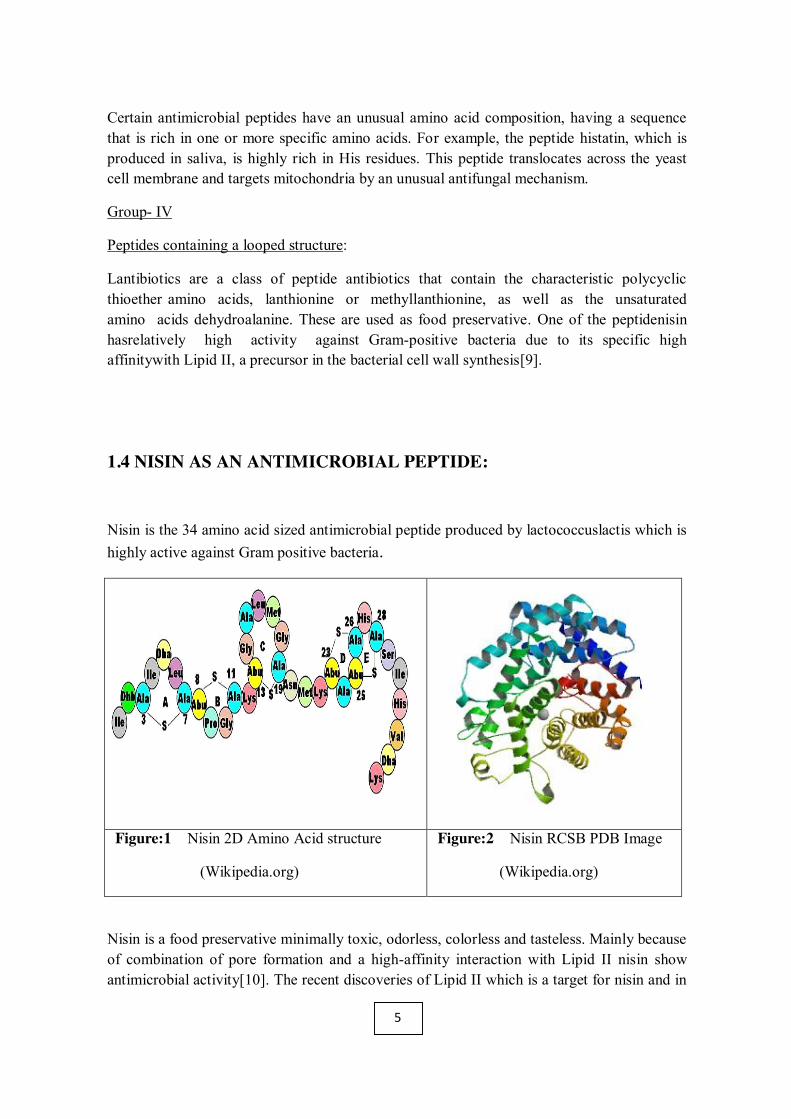
1.4 NISIN AS AN ANTIMICROBIAL PEPTIDE:
Nisin is the 34 amino acid sized antimicrobial peptide produced by lactococcuslactis which is highly active against Gram positive bacteria.
Figure:1 Nisin 2D Amino Acid structure
(Wikipedia.org)
Figure:2 Nisin RCSB PDB Image
(Wikipedia.org)
Nisin is a food preservative minimally toxic, odorless, colorless and tasteless. Mainly because of combination of pore formation and a high-affinity interaction with Lipid II nisin show antimicrobial activity[10]. The recent discoveries of Lipid II which is a target for nisin and in
5
particular, the important role played by the pyrophosphate group and brought nisin to the forward in the battle with resistant human infections as a model case for the production of new antibiotics[11].
Nisin is a positively charged peptide which is able to bind with negatively charged plasma membrane through non specific electrostatic interactions. Permeabilization of the cytoplasmic membrane and interference with the cell-wall biosynthesis of sensitive bacteria is the reason for antimicrobial activity of nisin[12]. Nisin can show good antibacterial activity either alone or in combination with antibiotics and also it has no side effects.
1.4 BACTERIA:
Bacteria constitute a large domain of prokaryotic microorganisms. Typically a few micrometres in length, bacteria have a number of shapes, ranging from spheres to rods and spirals. Bacteria were among the first life forms to appear on Earth, and are present in most of its habitats. Bacteria inhabit soil, water, acidic hot springs, radioactive waste and the deep portions of Earth's crust.
On the basis of cell wall composition and gram staining there are two types of Bacteria.
Gram positive Bacteria: This type of bacteria remains stained by with Gram staining even after washing alcohol or acetone. Here outer cell membrane is absent and cell wall is 20-30 nm thick. The cell wall contains teichoic acid.
Example :Staphylocococcus spp., Enterococcus spp., Bacillus spp.
Gram negative Bacteria: These bacteria do not retain the colour of the stain after washing with alcohol. Here outer membrane is present.Most of the pathogenic bacteria belong to this group.
Example: Escherichia spp, Pseudomonas spp., Salmonella spp.
1.4.1 Bacillus thuringiensis :
Bacillus thuringiensis is a Gram-positive ,soil-dwelling bacterium, which used as a biological pesticide. Naturally Bacillus thuringiensis found in the gut of caterpillars of many types of butterflies and moths, also on leaf surfaces, animal feces, aquatic environments, insect-rich environments, and grain-storage facilities[13].
At the time of sporulation, most of the Bt strains produce crystal proteins which is known as δ-endotoxins, they have insecticidal action. These are generally used as insecticides[14].
Bt proteins,these are naturally occurring insecticides which are produced by the B. thuringiensis,a soil bacterium. It is used to restrict crop pests since the 1920s [15], generally as. The multiple strains of B.thuringiensis are there which produce different Bt proteins according to target,like larvae of moths and beetles, butterflies, and mosquitoes. Inside the
6

bacterium Bt proteins form crystalline protein bodies, thus the name Cry proteins. Full-sized Cry proteins are inactive before they are eaten by object insect larva, and they are cleaved and become active inside the midgut. By the binding interaction of smaller, active peptides with specialized receptors, they create holes in the membrane of gut which cause contents to drip and kill the larvae[16,17].
For last 40 years products of Bacillus thuringiensis are considered as safe to use. But there is still argue is going on whether it is safe or not. two reports prior to 1995 of possible adverse human effects. In the year of 1991 a study which was focused on disclosure through inhalation of Bt sprays outcome showed skin sensitization and immune responses to Bt in 2 out of 123 farm workers [18,19]. In the year of 2006 an another article, the Organic Consumers Association address this attention to viable impacts of Bt in GE foods, warning that ‘Bt crops scaring to public health’[20].
Nowadays a recent study on Bacillus thuringiensis concluded that high amount of Bt toxin is toxic to mammalian blood.It damaged the red blood cell[21]. The size and quantity of the erythrocytes (RBCs) were both significantly reduced, through inhalation of Bt toxin. Here below these are two images which showed the harmful effects of Bacillus thuringiensis.
Figure:3 Bt cotton linked to human allergies Figure: 4 buffalo died after grazing on BT cotton. (Wikipedia.org)
1.5 Antibiotics :
Antibiotics or antibacterials are a type of antimicrobial used in the treatment and prevention of bacterial infection. They may either kill or inhibit the growth of bacteria. Several antibiotics are also effective against fungi and protozoans, and some are toxic to humans and animals, even when given in therapeutic dosage. Previously Bacillus thurigiensis was ampicillin sensitive. But nowadays Bacillus thuringiensis became resistant to ampicillin because of its regular and rapid use. To overcome this problem it is necessary to think about new strategies to inhibit the growth of Bacillus thurigiensis. Here our speculative insight is towards to see the effect of nisin alone or in combination with ampicillin on Bacillus thuringiensis.
7
1.5.1 Ampicillin:
Ampicillin is one of the important antibiotic for the therapy of most infections caused by bacteria. Ampicillin is equivalent to amoxicillin in relation to activity, it belongs to amino penicillin family that is a beta-lactam antibiotic. It have some side effects like diarrhea, nausea, vomiting, rash.
Mechanism of action: Ampicillin have the ability penetrate the membrane of bacteria both Gram-positive as well as Gram negetive. Due to the presence of an amino group it has some difference from penicillin G, or benzylpenicillin, amino group helps to initiate the drug to probing the outer membrane of Gram-negative bacteria. Ampicillin behaves like a irepairable obstacle of the enzyme transpeptidase, which is a major need of bacteria for their cell walls construction. Ampicillin leads to cell lysis of bacteria by inhibiting third and final stage cell wall synthesis. That’s why ampicillin play role like bactericidal.
8

CHAPTER : 2
METHODOLOGY
9

2.1 Minimum inhibitory concentration (MIC) is the minimum concentration of an antimicrobial agent that will inhibit the discernible growth of a microorganism after 6-8 hours of incubation. Minimum inhibitory concentrations are important in diagnostic laboratories to ensure resistance of microorganisms to an antimicrobial agent and also to overseer the activity of recent antimicrobial agents[22].
2.2 Zeta Potential is a framework characterizing electrochemical equilibrium on interfaces. It depends on the properties of both liquid as well as on surfaces. zeta potential plays a major role in theory of aggregative stability. On the value of zeta potential the electrostatic repulsion between particles depends. Zeta potential is proportionally equals to repulsion, the higher the zeta potential, the stronger the repulsion, the system becomes more stable[23]. For instance, high zeta potential of the fat droplets in milk prevents them against coalescence.
Figure:5 Schematic representation of Zeta potential (Wikipedia.org)
10

2.5FE-SEM (Field Emission Scanning electron microscope) is a microscope that works with electrons (the particles which are negetively charged) inplace of light. These electrons are extricate by a field emission source. The object is scanned by electrons according to a crooked pattern.Through FESEM we can visualize very small structure details on the surface or entire or splited objects. Researchers in biology, chemistry and physics apply this microscopy to observe topography of sample that may be as small as 1 nanometer[25]. Here we did FESEM to observe the structural details of bacteria in general and also after giving treatment of antibiotics and nisin (antimicrobial peptide) to that bacteria.
Figure:7 Schematic diagram of Field Emission Scanning Electron Microscope (Wikipedia.org)
11

2.6 BacLight cell viability assay
LIVE/DEAD BacLight Bacterial Viability assay was used to check the bacterial cell viability. Conventional direct-count assays of bacteria viability are based on metabolic characteristics or membrane integrity.Cells with a compromised membrane that are considered to be dead or dying will stain red, whereas cells with an intact membrane will stain green[26]. BacLight is a fluorescent bacterial viability stain that isbecoming more widespread in its use. Manufactured by Molecular Probes (Eugene, OR, USA), it consists of two stains, propidium iodide (PI) and SYTO9, that stain nucleic acid. According to the manufacturer, PI is a red intercalating stain that is membrane impermeant and is therefore excluded by healthy cells. SYTO9 is a green intercalating membrane permeant stain and will stain all cells, provided they contain nucleic acid[27]. The manufacturers argue that PI has a stronger affinity for nucleic acid and that, when the two stains are present within a cell, SYTO9will be displaced from nucleic acid and the cells will fluoresce in red. Live cells, being exclusive of PI, will stain SYTO9 positive and fluoresce in green[28].
12

CHAPTER : 3
EXPERIMENTAL PROCEDURE
13

3.1 Preparation of HEPES Buffer:
HEPES-10mM (molecular weight- 238.30g/mol)
NaCl-150Mm (molecular weight- 58g/mol)
pH- 7.4
Volume-500ml
HEPES: W= m.w×M×V÷1000
=238.30×10×10-3×500÷1000
=1.1915g
NaCl:
W=58.5×150×10-3×500÷1000
=4.38g
Hence 1.1915g of HEPES buffer and 4.38g of NaCl were dissolved in milli Q(deionised) water. The volume was overall made upto 500ml. The pH is fixed at 7.4.
3.2 Preparation of bacterial culture:
Nutrient Broth is used for the cultivation of a large variety of microorganisms. We prepared NB media because it is a liquid media and to check the Minimum Inhibitory Concentration(MIC) of Bacillus thuringiensis we need liquid culture of these strain. Nutrient Broth contains,
Enzymatic Digest of Gelatin -5 g
Beef Extract -3 g
Final pH: 6.8
For preparing the NB media, normally 13 gram medium is dissolve in 1000 ml purified water.
• We took 0.325 gram NB medium which was dissolve in 25 ml of purified water.
14

• Then it was mixed thoroughly.
• This 25 ml of NB media was autoclaved at 121 degree celsius and 15lb pressure for 20 minutes.
• After autoclave when the media becomes cooled, a very small amount of BT culture from old agar slant culture was taken with the help of inoculation loop and was poured into the fresh NB media.
• Then this media was incubated into a shaker incubator for 6 hours at 37 degree Celsius, 150 rpm.
• All above steps were done in Laminar hood to avoid contamination.
• Finally after 6 hours the fresh Bt culture was prepared. Then it was stored into refrigerator for MIC purpose.
3.3 Preparation of Nisin stock solution:
Antimicrobial peptide Nisin stock solution was prepared in a falcon tube by dissolving 3mg Nisin powder in 1 ml of purified water which indicates the 3000U/ml Nisin concentration(1000U=1mg) and it was stored in -20 degree C till before MIC experiment.
During the MIC experiment 5 different concentration 50U/ml, 200U/ml, 500U/ml, 1000U/ml, 1500U/ml was taken from the Nisin stock solution.
3.4 Preparation of antibiotics:
The stock solution of ampicillin was prepared in a 15 ml falcon tube of 1mg/ml concentration, and it was stored in -20 degree C.
15

3.5 Preparation of media:
We prepared 50 ml of Nutrient Broth (NB) media by dissolving 0.625 gram of medium into 50 ml of purified water. This media is needed to fill the volume during MIC experiment.
3.6 Minimum Inhibitory Concentration:
To check the MIC we use 96 well microliter plate method. In the 96 well microtiter plate the 1st column of first three rows are used for positive control. In these wells only Bacillus thuringensis culture and media were added. And the 7th column of first three rows are used as negative control. In these wells the culture of Bacillus thuringensis , ampicillin and media were added.
For MIC we used total 12 columns and 3 rows of 96 well microtiter plate and we did these experiment in triplicate. We treated Bacillus thuringensis culture with Nisin alone on 2-6 columns of first three rows applying different concentration of Nisin in different wells, and we treated Bacillus thuringensis culture in combination with Nisin and ampicillin on 8-12 columns of these rows by increasing the Nisin concentration keeping the fixed amount ampicillin in every wells. The table listed below shows the amount of culture, antibiotic, Nisin solution, and media which were added to the wells. The overall volume in 96 well microliter plate is maintained to 300 microliter.
16
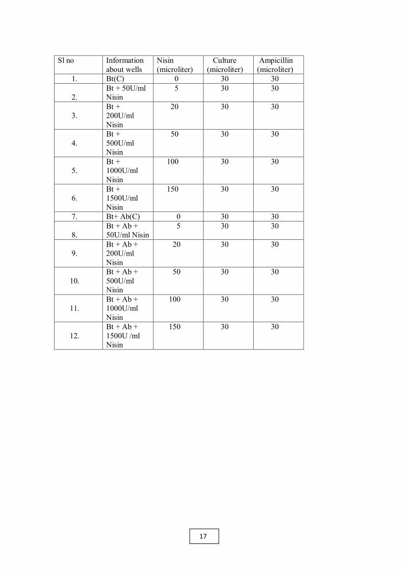
Sl no Information about wells
Nisin (microliter)
Culture (microliter)
Ampicillin (microliter)
1. Bt(C) 0 30 30 2.
Bt + 50U/ml Nisin
5 30 30
3.
Bt + 200U/ml Nisin
20 30 30
4.
Bt + 500U/ml Nisin
50 30 30
5.
Bt + 1000U/ml Nisin
100 30 30
6.
Bt + 1500U/ml Nisin
150 30 30
7. Bt+ Ab(C) 0 30 30 8.
Bt + Ab + 50U/ml Nisin
5 30 30
9.
Bt + Ab + 200U/ml Nisin
20 30 30
10.
Bt + Ab + 500U/ml Nisin
50 30 30
11.
Bt + Ab + 1000U/ml Nisin
100 30 30
12.
Bt + Ab + 1500U /ml Nisin
150 30 30
17

3.7 ZETA POTENTIAL: • Bacillus thuringiensis were allowed to grow overnight at 37 degree celsius and 150
rpm in 5 ml MH.
• 100 microliter of culture was inoculated in 5 ml of muller-hinton broth.
• The suspension was then allowed to grow at 37 degree celsius for OD to be 1.0.
• From the above culture 1.5 ml was taken and centrifuged at 13000 rpm for 8 minutes.
• By using HEPES buffer pellet washed two times.
• 100 microliter of nisin treated suspension was added to 900 microliter of Bacillus thuringiensis cells.
• After 1 hour zeta potential was measured.
3.8 FIELD EMISSION SCANNING ELECTRON MICROSCOPY: • 1.5 ml of overnight culture was taken.
• Centrifuged at 5000 rpm for 5 minutes.
• Pallets were collected and washed twice with PBS by centrifugation at 5000 rpm for 5 minutes.
• Pallets were suspended in PBS.
• One drop of suspended pallet was taken on glass slide.
• The glass slide were flooded with glutaraldehyde 2.5 % prepared in PBS.
• The slides were kept for overnight in incubation at 4 degree celcius.
• Then it was washed with water.
• 1% of tannic acid was flooded over slides.
• It was kept for 5 minutes.
• Then washed with distilled water.
• Using ethanol the slides were dehydrated sequentially as 30%, 50%, 70%, 90%, 100% respectively.
• Finally slides were kept for drying.
18

3.9 BacLight cell viability assay:
x 50mg/ml of nisin stock solution was prepared. x For control 4.9ml broth and 100µl culture were taken in a falcon tube. x For preparation of treated solution, 4.75ml of broth, 100µl culture and 150µl of
nisin.from the stock solution were taken in another falcon tube. x Tubes were marked according to the concentration of nisin. x Both the tubes were incubated overnight. x Centrifugation at 7000rpm at 25 degree Celsius for 15 minutes. x Pellets were collected. x 500µl of HEPES buffer was added and mixed. x 5ml HEPES buffer were taken in 2 separate falcon tubes each. x The pellets with 500µl buffer in the previous tubes were poured in the new falcon
tubes. x Centrifugation at 7000rpm at 25 degree Celsius for 15 minutes. x Pellets were collected. x They are suspended in 5ml of HEPES buffer. x Centrifugation at 7000rpm at 25 degree Celsius for 15 minutes.. x Pellets were collected. x 2.5ml of HEPES buffer was added. x 333 µl culture from each falcon tubes were taken in separate eppendorf tubes. x 1µl BacLight dye was added in each tube. x Incubation in dark for 15 minutes. x 5µl was taken on glass slide and covered with a cover slip. x Image was seen in the Fluoroscent microscope.
19

CHAPTER: 4
RESULTS
20

4.1 MIC
MIC (For 6 hours):
Figure:8 (a)
Figure: 8(b)
Figure 8 : The effect of different concentrations of (a)Nisin on growth kinetics of Bacillus thuringiensis. (b) Nisin supplemeted with ampicillin on growth kinetics of Bacillus thuringiensis.
21

MIC (For 20 hours):
Figure:9 (a)
Figure:9 (b)
Figure: 9 The effect of different concentrations of (a) Nisin on growth kinetics of Bacillus thuringiensis (b) Nisin supplemeted with ampicillin on growth kinetics of Bacillus thuringiensis.
22

Above figure 8(a) and 9(a) showed that with increasing the concentration of Nisin the lag phase of Bacillus thuringiensis gets prolonged.
Figure 8(b) and 9(b) indicated that when bacteria was treated with combination of Nisin and ampicillin the bacteria goes faster in stationary phase.
The MIC of nisin against Bacillus thuringiensis showed that lag phase gets prolonged when the bacteria was treated with 200 U/ml Nisin. The MIC of Nisin in combination with ampicillin showed that they move faster in stationary phase when ampicillin was treated with 50 U/ml of nisin. So most effective concentration of nisin alone is 200 U/ml and in combination with antibiotic is 50 U/ml to inhibit the growth of Bacillus thuringiensis.
23
4.2 ZETA POTENTIAL:
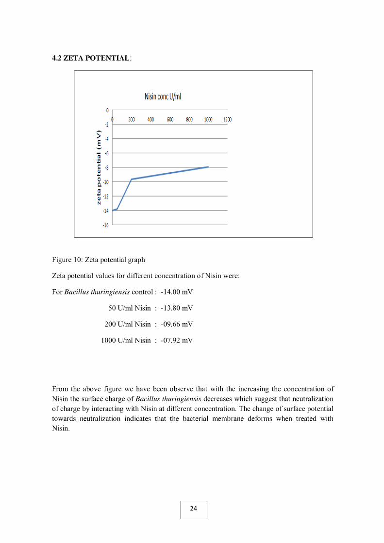
Figure 10: Zeta potential graph
Zeta potential values for different concentration of Nisin were:
For Bacillus thuringiensis control : -14.00 mV
50 U/ml Nisin : -13.80 mV
200 U/ml Nisin : -09.66 mV
1000 U/ml Nisin : -07.92 mV
From the above figure we have been observe that with the increasing the concentration of Nisin the surface charge of Bacillus thuringiensis decreases which suggest that neutralization of charge by interacting with Nisin at different concentration. The change of surface potential towards neutralization indicates that the bacterial membrane deforms when treated with Nisin.
24

4.3 Field emission scanning electron microscopy:
Figure 11: FESEM images of Bacillus thuringiensis control at 5000X, 10000X, 20000X magnification.
25
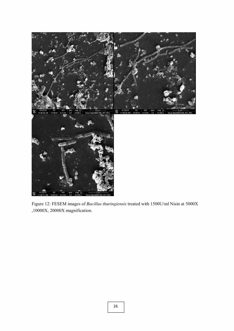
Figure 12: FESEM images of Bacillus thuringiensis treated with 1500U/ml Nisin at 5000X ,10000X, 20000X magnification.
26

Fig 13: FESEM images of Bacillus thuringiensis treated with 1500U/ml nisin in combination with ampicillin at 5000X, 10000X, 20000X magnification.
Breadth: µm(w)
Bacillus thuringiensis (control) : 0.85±0.12
Bacillus thuringiensis1500 U/ml Nisin : 0.56±0.9
Bacillus thuringiensis1500 U/ml Nisin+ Ab : 0.55±0.7
From the FESEM figure it has been observed that with when Bacillus thuringiensis is treated with nisin alone or in combination with ampicillin the membrane of Bacillus thuringiensis got damage. The clumping and blebs are clearly indicates the deformation of membrane.The FE-SEM images of Bacillus thuringiensis at different conditions control, nisin alone or in combination with ampicillin shows that bacteria could not able to maintain their original
27

shape when treated with nisin(1500U/ml). The membrane gets totally ruptured. Its breadth decreases.
4.4 BacLight cell viability assay :
Figure:14 (a) Bacillus thuringiensis (control)
Figure:14 (b) Bacillus thuringiensis (1500 U/ml Nisin)
The kit consists of two stains, propidium iodide (PI) and SYTO9, which both stain nucleic acids. Green fluorescing SYTO9 is able to enter all cells and is used for assessing total cell counts, whereas red fluorescing PI enters only cells with damaged cytoplasmic membranes. The green colour in the figure:14(a) shows that control, the cells are alive, and in figure:14(b), the red colour indicates most of the cells are dead due to the interaction of Bt cells with Nisin (1500U/ml).
28

5. DISCUSSION:
The lantibiotic Nisin is an antimicrobial petide(AMP) whose antimicrobial capacity relies on specific binding to bacterial Lipid II. Here we analysed the effect of nisin alone or in combination with ampicillin to the growth cycle Bacillus thuringiensis. It has resistance against ampicillin and some study reveals that Bacillus thuringiensis have pathogenicity on human and animal and methods to kill or inhibit their growth have become key goals of microbiologists and drug development researchers. In our MIC study we found nisin alone inhibit the growth of bacteria at 200 U/ml concentration. Nisin prolonged the lag phase of bacteria. In combination with ampicillin Nisin can show good results at low concentration(50 U/ml). From figure we observe that Bacteria were very quickly moves to their stationary phase its may be due to nisin & ampicillin both together may directly attack their cell wall when they were in lag phase which results low rising of log phase.
Many studies have indicated that by forming pores and inhibiting cell wall synthesis with a specific molecule, Lipid II, a principal component of the membranes of gram-positive bacteria, Nisin exerts its bactericidal activity. Nisin uses Lipid II as a ‘‘docking molecule’’ to form pores on the cell membrane surface in a targeted manner; at a nanomolar level, then, Nisin is able to effectively kill bacteria[29].
In zeta analysis study, with the increasing the concentration of nisin the cell surface potential moves from negative to positive and indicates a clear relationship between positive charge on bacterial cells and results in death. Damage to the membrane by leakage cause the aggregation of membrane outside the Bacteria.Although it is quite well known that cationic molecules in solution are able to kill bacteria only recently positive charges attached to surfaces, particles, polymers, liposomes or bilayers have really been used to kill bacteria upon contact.
In FE-SEM study ,Bacillus thuringiensis when treated with nisin or in combination with ampicillin a majority of the cells were severely damaged and lost their original cell wall integrity.This is because cholesterol was absent in Bacterial membrane therefore antimicrobial peptide Nisin can easily interact with Lipid-II layers present on the membrane of Bacillus thuringiensis which create pores on the surface of the membrane and causes leakage. The cells may be try to dividing but due to membrane aggregation when treated with nisin they stop dividing and exists as a long chain.
In Baclight cell viability assay the emission properties of the stain mixture bound to DNA change due to the displacement of one stain by the other and quenching by fluorescence resonance energy transfer[30]. When bacteria was treated with Nisin that can damaged the cytoplasmic membrane of that bacteria for that red fluorescing PI enters the cells with damaged cytoplasmic membranes and gives red colour.
29

6. CONCLUSION:
Nisin is in fact an exceptional case, as it uses a blend of a high partiality connection with the membrane anchored Lipid II and pore development to eliminate bacterial cells. Cells of during the late stationary growth phase produce both bio insecticidal crystal protein delta-endotoxin. When treated with Nisin they may be able to produce crystal protein delta-endotoxin but got damaged and leaks out of membrane forming blebs. The findings of the study that Nisin can interact with lipid II of bacterial membrane causes damage of it. So it can be concluded that Nisin is effective to kill or inhibit the growth of bacteria and can be alter the place of antibiotics.
30
8. REFERENCES:
1. Adam,M.,Murali,B.,Glenn,N.O.,andPotter,S.S.(2008).Epigeneticinher- itancebased evolution ofantibiotic resistanceinbacteria. BMCEvol.Biol. 8:52. doi:10.1186/1471-2148-8-52
2. Aminov,R.I.(2009).The role of antibiotics and antibiotic resistance in nature. Environ.Microbiol. 11, 2970–2988.
3. Aarestrup, F. M., Seyfarth, A. M., Emborg, H. D., Pedersen, K., Hendriksen, R. S., and Bager, F. 2001. Effect of abolishment of the use of antimicrobial agents for growth promotion on occurrence of antimicrobial resistance in fecal enterococci from food animals in Denmark. Antimicrob. Agents Chemother. 45: 2054–2059.
4. Byarugaba, D. K. 2004. A view on antmicrobial resistance in developing countries and responsible risk factors. Int. J. Antimicrob. Agents 24: 105–110.
5. Springer Anı´bal de J. Sosa Alliance for the Prudent Use of Antibiotics (APUA) Tufts University Boston, MA USA.
6. ANTIMICROBIAL RESISTANCE Global Report on Surveillance, WHO,2014.
7.Tong Z, Zhang Y, Ling J, Ma J, Huang L, et al. (2014) An In Vitro Study on the Effects of Nisin on the Antibacterial Activities of 18 Antibiotics against Enterococcus faecalis. PLoS ONE 9(2): e89209. doi:10.1371/journal.pone.0089209
8. R. E. W. Hancock and A. Patrzykat, “Clinical development of cationic antimicrobial peptides: from natural to novel antibiotics,” Current Drug Targets, vol. 2, no. 1, pp. 79–83, 2002.
9. Breukink, E., van Kraaij, C., van Dalen, A., Demel, R. A., Siezen, R. J., de Kruijff, B., andKuipers, O. P. (1998) The orientation of nisin in membranes. Biochemistry 37, 8153-8162
10. Hadley EB, Hancock RE (2010) Strategies for the discovery and advancement of novel cationic antimicrobial peptides. Curr Top Med Chem 10: 1872–1881.
11.FDA (1988) Food and Drug Administration. Nisin preparation: Affirmation of GRAS status as a direct human food ingredient. 11251 ed.
12. Naghmouchi K, Le Lay C, Baah J, Drider D (2012) Antibiotic and antimicrobial peptide combinations: synergistic inhibition of Pseudomonas fluorescens and antibiotic-resistant variants. Res Microbiol 163: 101–108.
31

13. Ali, A., Weaver, M. S. &Cotsenmayer, E. (1989). Effectiveness of Bacillus thuringiensis serovar. israelensis(Vectobac 12 AS) and Bacillus sphaericus 2362 (ABG-6232) against Culex spp. mosquitoes in a dairy lagoon in central Florida. Florida Entomologist 72, 585-591.
14. Akhurst, R.J., James, W., Bird, L.J. and Beard, C. (2003) Resistance to the Cry1Ac d-endotoxin of Bacillus thuringiensis in the cotton bollworm, Helicoverpaarmigera (Lepidoptera: Noctuidae). J. Econ. Entomol. 96, 1290–1299.
15. Jenkins, J.N., J.C. McCarty, Jr., and R.E. Buehler. 1997. Resistance of cotton with endotoxin genes from Bacillus thuringiensis var. kurstaki on selected lepidopteran insects. Agronomy Journal 89:768–780.
16. Schnepf E, Crickmore N, Van Rie J, Lereclus D, Baum J, Feitelson J, Zeigler D R, Dean D H(1998) MicrobiolMolBiol Rev 62:775–806.
17. de Maagd R A, Bravo A, CrickmoreN(2001) Trends Genet 17:193–199.
18. McClintock JT, Schaffer CR, Sjoblad RD. 1995. A comparative review of the mammalian toxicity of Bacillus thuringiensis-based pesticides. Pestic. Sci. 45:95–105.
19. Bernstein L, Bernstein JA, Miller M, Tierzieva S, Bernstein DI, et al. 1999. Immune responses in farm workers after exposure to Bacillus thuringiensis pesticides. Environ. Health Perspect. 107:575–82.
20. Carman NJ. 2006. Gene-altered Bt crops threaten public health: Immune responses and skin sensitization to Bt in farm workers and presence of Bt in many genetically engineered foods.
21. New Study Proves Bt Toxins in GMOs Toxic to Mammalian Bloodhttp://www.abovetopsecret.com/forum/thread946024/pg1.
22. Minimum Inhibitory Concentration is the lowest drug concentration that prevents visible microorganism growth after overnight incubation.www.boundless.com/microbiology/textbooks/boundless-microbiology-textbook/antimicrobial-drugs-13.
23. N. Mozes, P. S. Handley, H. J. Busscher, P. G. Rouxhet. Microbial cell surface analysis. s.l. : VCH Publishers, 2001.
24. Allan-Wojtas, P. &Kaláb, M. (1984a) A simple procedure for the preparation of stirred yoghurt for scanning electron microscopy. Food Microstructure 3(2), 197-198.
25. Braten T High resolution scanning electron microscopy in biology: Arti- facts caused by the nature and mode of application of the coating material. JMicrosc I1 3,53-59 (1978)
26. Endo H, Nakayama J, Hayashi T, Watanabe E. Application of flow cytometry for rapid determination of cell number of viable bacteria. Fisheries Sci 1997;63:1024 –1029.
32

27. Duffy G, Sheridan JJ. Viability staining in a direct count rapid method for the determination of total viable counts on processed meats. J Microbiol Methods 1998;31:167–174.
28. Sebastine IM, Stocks SM, Cox PW, Thomas CR. Characterisation of percentage viability. Biotechniques 1996;13:419 – 423.
29. Breukink E, de Kruijff B (2006) Lipid II as a target for antibiotics. Nat Rev Drug Discov 5: 321–332.
30. Stocks, S. M. 2004. Mechanism and use of the commercially available viability stain, BacLight. Cytom. A 61:189-195.
33