Embed Size (px)
Citation preview
I N V E S T I G AT I O N O F A NA C C I D E N TA L E X P O S U R EO F R A D I OT H E R A P YPAT I E N T S I N PA N A M A
Report of a Team of Experts, 26 May–1 June 2001
I N T E R N A T I O N A L A T O M I C E N E R G Y A G E N C Y

INVESTIGATION OF ANACCIDENTAL EXPOSURE OFRADIOTHERAPY PATIENTS
IN PANAMA
Report of a Team of Experts26 May–1 June 2001

The Agency’s Statute was approved on 23 October 1956 by the Conference on the Statute of theIAEA held at United Nations Headquarters, New York; it entered into force on 29 July 1957. TheHeadquarters of the Agency are situated in Vienna. Its principal objective is “to accelerate and enlarge thecontribution of atomic energy to peace, health and prosperity throughout the world’’.
© IAEA, 2001
Permission to reproduce or translate the information contained in this publication may beobtained by writing to the International Atomic Energy Agency, Wagramer Strasse 5, P.O. Box 100,A-1400 Vienna, Austria.
Printed by the IAEA in AustriaAugust 2001
STI/PUB/1114
The following States are Members of the International Atomic Energy Agency:
AFGHANISTANALBANIAALGERIAANGOLAARGENTINAARMENIAAUSTRALIAAUSTRIAAZERBAIJAN, REPUBLIC OFBANGLADESHBELARUSBELGIUMBENINBOLIVIABOSNIA AND HERZEGOVINABRAZILBULGARIABURKINA FASOCAMBODIACAMEROONCANADACENTRAL AFRICAN
REPUBLICCHILECHINACOLOMBIACOSTA RICACOTE D’IVOIRECROATIACUBACYPRUSCZECH REPUBLICDEMOCRATIC REPUBLIC
OF THE CONGODENMARKDOMINICAN REPUBLICECUADOREGYPTEL SALVADORESTONIAETHIOPIAFINLANDFRANCEGABONGEORGIAGERMANY
GHANAGREECEGUATEMALAHAITIHOLY SEEHUNGARYICELANDINDIAINDONESIAIRAN, ISLAMIC REPUBLIC OF IRAQIRELANDISRAELITALYJAMAICAJAPANJORDANKAZAKHSTANKENYAKOREA, REPUBLIC OFKUWAITLATVIALEBANONLIBERIALIBYAN ARAB JAMAHIRIYALIECHTENSTEINLITHUANIALUXEMBOURGMADAGASCARMALAYSIAMALIMALTAMARSHALL ISLANDSMAURITIUSMEXICOMONACOMONGOLIAMOROCCOMYANMARNAMIBIANETHERLANDSNEW ZEALANDNICARAGUANIGERNIGERIANORWAY
PAKISTANPANAMAPARAGUAYPERUPHILIPPINESPOLANDPORTUGALQATARREPUBLIC OF MOLDOVAROMANIARUSSIAN FEDERATIONSAUDI ARABIASENEGALSIERRA LEONESINGAPORESLOVAKIASLOVENIASOUTH AFRICASPAINSRI LANKASUDANSWEDENSWITZERLANDSYRIAN ARAB REPUBLICTHAILANDTHE FORMER YUGOSLAV
REPUBLIC OF MACEDONIATUNISIATURKEYUGANDAUKRAINEUNITED ARAB EMIRATESUNITED KINGDOM OF
GREAT BRITAIN AND NORTHERN IRELAND
UNITED REPUBLICOF TANZANIA
UNITED STATES OF AMERICAURUGUAYUZBEKISTANVENEZUELAVIET NAMYEMENYUGOSLAVIAZAMBIAZIMBABWE
INVESTIGATION OF ANACCIDENTAL EXPOSURE OFRADIOTHERAPY PATIENTS
IN PANAMA
Report of a Team of Experts26 May–1 June 2001
INTERNATIONAL ATOMIC ENERGY AGENCYVIENNA, 2001

VIC Library Cataloguing in Publication Data
Investigation of an accidental exposure of radiotherapy patients in Panama /report of a Team of Experts, 26 May–1 June 2001. — Vienna : InternationalAtomic Energy Agency, 2001.
p. ; 24 cm.STI/PUB/1114ISBN 92–0–101701–4Includes bibliographical references.
1. Radiotherapy — Accidents — Panama. Radiation — Physiologicaleffect. I. International Atomic Energy Agency.
VICL 01–00270

FOREWORD
Early in 2001, serious accidental exposures involving patients undergoingradiotherapeutic procedures were discovered in Panama. The Government of Panamarequested assistance from the IAEA under the terms of the Convention on Assistancein the Case of a Nuclear Accident or Radiological Emergency. The IAEAimmediately notified the World Health Organization (WHO) and assembled and sentto Panama a team of senior experts from France, Japan, the USA and the IAEA, aswell as a senior expert from the Russian Federation nominated by WHO. The expertteam was requested to ensure that the “radiation source(s) involved in the accidentwas (were) in a safe and secure condition”; to “evaluate the doses incurred by theaffected patients”; undertake a medical evaluation of the affected patients’ prognosisand treatment; and to “identify issues on which the IAEA could offer to provideand/or co-ordinate assistance with a view to minimizing the consequences of theaccident”. In addition, the Minister of Health of Panama had requested assistancefrom the Pan American Health Organization (PAHO/WHO), and a PAHO officersupported the expert team. This report contains the expert team’s assessment of theaccidental exposure.
The IAEA is very grateful to the Government of Panama for giving it theopportunity to assist in the aftermath of the accidental exposure described in thisreport and, as a consequence, to draw valuable lessons that can be shared with theinternational community worldwide.
In particular, the IAEA wishes to express its thanks to the Panamanian Ministerof Health and the Director General of Public Health, to the Director of the InstitutoOncológico Nacional (ION), to the Department of Radiation Health of the SocialSecurity Complex Hospital and to the staff of all the Panamanian organizations whichcollaborated with the expert team.
The IAEA is also very grateful to the members of the expert team for theirdedication in carrying out their task and for their contribution to the development andreview of this report. The IAEA wishes to express its thanks to: the Department ofRadiology, School of Medicine, of the University of New Mexico, Albuquerque,USA; to the Département de Radiothérapie of the Institut Curie, Paris, and to theInstitut de Protection et de Sûreté Nucléaire, Fontenay-aux-Roses, France; theResearch Centre for Radiation Emergency Medicine of the National Institute ofRadiological Sciences, Chiba, Japan; and to the Hematologic Department of theRussian Scientific Research Centre, Moscow, (a collaborating centre of WHO) formaking their staff available for this mission. The IAEA is also grateful to PAHO forits readiness to collaborate fully with the expert team.
The members of the team, as well as the names of the supporting experts, aregiven below:
TEAM OF EXPERTS
Akashi, M. Research Center for Radiation Emergency Medicine ofthe National Institute of Radiological Sciences, Chiba,Japan
Cosset, J.-M. Section Médicale et Hospitalière,Département de Radiotherapie, Service B,Institut Curie, Paris, France; Member of Committee 3 ofthe International Commission on Radiological Protection(ICRP)
Gourmelon, P. Commissariat á l’Energie Atomique,Centre d’Etudes Nucléaires de Fontenay-aux-Roses,Institut de Protection et de Sûreté Nucléaire,Fontenay-aux-Roses, France
Konchalovsky, M.V. Hematologic Department of the Scientific Research(representing WHO) Center of the Russian Federation, Moscow, Russian
Federation
Mettler, F.A., Jr., Department of Radiology,(Chairman) The University of New Mexico, School of Medicine,
Albuquerque, New Mexico, United States of America;Delegate of USA to UNSCEAR; Member of the MainCommission and Chairman of Committee 3 of the ICRP
Ortiz López, P. Radiation Safety Section,(Team co-ordinator) Division of Radiation and Waste Safety,
International Atomic Energy Agency; Member ofCommittee 3 of the ICRP
Vatnitsky, S. Dosimetry and Medical Radiation Physics Section,Division of Human Health, International Atomic EnergyAgency
SUPPORTING EXPERTS
Pan American Health Organization
Borrás, C. Program of Essential Drugs and Technology,Division of Health Systems and Services Development,Pan American Health Organization,Washington, D.C., United States of America
Panamanian Government
Arenas, E. Médica Epidemióloga, Ministerio de Salud (MINSA)
Barés, J.P. Director General, Instituto Oncológico Nacional (ION)
Gibbs, E. Jefe del Departamento de Salud Radiológica, Caja deSeguro Social (CSS)
López, R.I. Médico Oncólogo del ION
Morales, E. Director General de Salud, MINSA
Pinzón, N. Subdirectora del ION
Panamanian experts
Bedoya, R. Técnico de Salud Radiológica CSS
Bolaños, V. Técnico de Salud Radiológica CSS
De Infante, M. Técnico de Salud Radiológica CSS
Douglas, A. Técnico de Salud Radiológica CSS
Fernández, M. Técnico de Salud Radiológica CSS
Fisher, V. Técnico de Salud Radiológica CSS
Scotland, E. Técnico de Salud Radiológica CSS
Terán, L.A. Técnico de Salud Radiológica CSS
Tuñón, E. Técnico de Salud Radiológica CSS

EDITORIAL NOTE
This report is based on information made available by or through the Panamanianauthorities.
The report does not address questions of responsibility, legal or otherwise, for acts oromissions on the part of any person.
Although great care has been taken to maintain the accuracy of information containedin this report, neither the IAEA nor its Member States assume any responsibility forconsequences which may arise from its use.
The use of particular designations or countries or territories does not imply anyjudgement by the publisher, the IAEA, as to the legal status of such countries or territories, oftheir authorities and institutions or of the limitation of their boundaries.
The mention of names of specific companies or products (whether or not indicated asregistered) does not imply any intention to infringe proprietary rights, nor should it beconstrued as an endorsement or recommendation on the part of the IAEA.
Material made available by persons who are in contractual relation with governmentsis copyrighted by the IAEA, as publisher, only to the extent permitted by the appropriatenational regulations.

CONTENTS
EXECUTIVE SUMMARY . . . . . . . . . . . . . . . . . . . . . . . . . . . . . . . . . . . . . . . 1
The accidental exposure . . . . . . . . . . . . . . . . . . . . . . . . . . . . . . . . . . . . . 1Lessons and recommendations . . . . . . . . . . . . . . . . . . . . . . . . . . . . . . . . 2
1. INTRODUCTION . . . . . . . . . . . . . . . . . . . . . . . . . . . . . . . . . . . . . . . . . 7
1.1. Background . . . . . . . . . . . . . . . . . . . . . . . . . . . . . . . . . . . . . . . . . . 71.2. Objectives . . . . . . . . . . . . . . . . . . . . . . . . . . . . . . . . . . . . . . . . . . . 91.3. Scope . . . . . . . . . . . . . . . . . . . . . . . . . . . . . . . . . . . . . . . . . . . . . . 91.4. Structure . . . . . . . . . . . . . . . . . . . . . . . . . . . . . . . . . . . . . . . . . . . . 9
2. BACKGROUND INFORMATION . . . . . . . . . . . . . . . . . . . . . . . . . . . . . 10
2.1. Radiotherapy departments in Panama . . . . . . . . . . . . . . . . . . . . . . 102.2. The Radiotherapy Department at the Instituto Oncológico
Nacional . . . . . . . . . . . . . . . . . . . . . . . . . . . . . . . . . . . . . . . . . . . . 102.2.1. External beam radiotherapy . . . . . . . . . . . . . . . . . . . . . . . . 112.2.2. Brachytherapy . . . . . . . . . . . . . . . . . . . . . . . . . . . . . . . . . . 13
2.3. Treatment planning system . . . . . . . . . . . . . . . . . . . . . . . . . . . . . . 142.4. Radiation protection infrastructure and regulatory control . . . . . . . 15
2.4.1. Authorization of the Radiotherapy Department . . . . . . . . . . 162.5. History of recent audits of the Radiotherapy Department . . . . . . . . 16
2.5.1. Audit in February 1999 . . . . . . . . . . . . . . . . . . . . . . . . . . . . 162.5.2. Audit in February 2001 . . . . . . . . . . . . . . . . . . . . . . . . . . . . 182.5.3. Results of IAEA/WHO TLD postal dose quality audits
performed at the ION . . . . . . . . . . . . . . . . . . . . . . . . . . . . . 19
3. THE ACCIDENTAL EXPOSURE . . . . . . . . . . . . . . . . . . . . . . . . . . . . . 19
3.1. Initiating event . . . . . . . . . . . . . . . . . . . . . . . . . . . . . . . . . . . . . . . 193.2. Discovery of the problem . . . . . . . . . . . . . . . . . . . . . . . . . . . . . . . 23
4.. RESPONSE TO THE ACCIDENTAL EXPOSURE . . . . . . . . . . . . . . . . 24
4.1. Actions taken upon discovery of the error . . . . . . . . . . . . . . . . . . . 244.2. Response from the IAEA . . . . . . . . . . . . . . . . . . . . . . . . . . . . . . . 25
5. DOSE ASSESSMENT . . . . . . . . . . . . . . . . . . . . . . . . . . . . . . . . . . . . . . 28
5.1. Introduction . . . . . . . . . . . . . . . . . . . . . . . . . . . . . . . . . . . . . . . . . 285.2. Scope of the dosimetric evaluation . . . . . . . . . . . . . . . . . . . . . . . . 295.3. Comparison of the dosimetry systems and beam calibration . . . . . 31
5.3.1. Dosimetry system intercomparison . . . . . . . . . . . . . . . . . . . 315.3.2. 60Co beam calibration . . . . . . . . . . . . . . . . . . . . . . . . . . . . . 325.3.3. Variation of absorbed dose with field size . . . . . . . . . . . . . . 325.3.4. Depth dose data . . . . . . . . . . . . . . . . . . . . . . . . . . . . . . . . . 335.3.5. Wedge transmission . . . . . . . . . . . . . . . . . . . . . . . . . . . . . . 335.3.6. Verification of dose delivery . . . . . . . . . . . . . . . . . . . . . . . . 33
5.4. Patient doses . . . . . . . . . . . . . . . . . . . . . . . . . . . . . . . . . . . . . . . . . 345.4.1. Assessment of doses from irradiation with external beams . 345.4.2. Consideration of brachytherapy treatments . . . . . . . . . . . . . 355.4.3. Doses equivalent to treatments of 2 Gy per fraction . . . . . . 355.4.4. Other patients treated in the same period . . . . . . . . . . . . . . 46
6. TEST OF THE COMPUTER SOFTWARE USING DIFFERENTAPPROACHES FOR DATA ENTRY . . . . . . . . . . . . . . . . . . . . . . . . . . . 47
6.1. Instructions and warnings . . . . . . . . . . . . . . . . . . . . . . . . . . . . . . . 476.2. Tests performed . . . . . . . . . . . . . . . . . . . . . . . . . . . . . . . . . . . . . . 48
7. MEDICAL ASSESSMENT . . . . . . . . . . . . . . . . . . . . . . . . . . . . . . . . . . 60
7.1. Background information on radiation effects in humans . . . . . . . . 607.2. Basic radiotherapy principles relevant to this accidental exposure . 617.3. Fractionation of radiation exposure . . . . . . . . . . . . . . . . . . . . . . . . 627.4. Adverse effects . . . . . . . . . . . . . . . . . . . . . . . . . . . . . . . . . . . . . . . 627.5. Spectrum of overexposures in this event . . . . . . . . . . . . . . . . . . . . 637.6. Complicating factors in the evaluation of this event . . . . . . . . . . . . 657.7. Results of the medical team’s investigation . . . . . . . . . . . . . . . . . . 65
7.7.1. Surviving patients . . . . . . . . . . . . . . . . . . . . . . . . . . . . . . . . 727.7.2. Deceased patients . . . . . . . . . . . . . . . . . . . . . . . . . . . . . . . . 757.7.3. Sequence of events leading to death . . . . . . . . . . . . . . . . . . 78
8. FINDINGS AND CONCLUSIONS OF THE TEAM,AND LESSONS TO BE LEARNED . . . . . . . . . . . . . . . . . . . . . . . . . . . 79
8.1. Operating organization: Radiotherapy departments . . . . . . . . . . . . 808.1.1. Quality assurance and radiotherapy . . . . . . . . . . . . . . . . . . . 80

8.1.2. Treatment planning systems as a safety issue . . . . . . . . . . . . 818.1.3. Manual calculation check of the computer calculated plan . . 818.1.4. Changes in procedures . . . . . . . . . . . . . . . . . . . . . . . . . . . . . 818.1.5. Workload and team integration . . . . . . . . . . . . . . . . . . . . . . . 828.1.6. Observation of unusual reactions of patients . . . . . . . . . . . . 828.1.7. In vivo dosimetry . . . . . . . . . . . . . . . . . . . . . . . . . . . . . . . . 838.1.8. Local advice from the manufacturer/supplier. . . . . . . . . . . . 83
8.2. National authorities . . . . . . . . . . . . . . . . . . . . . . . . . . . . . . . . . . . . 838.2.1. Quality assurance . . . . . . . . . . . . . . . . . . . . . . . . . . . . . . . . 838.2.2. Communication between regulators and users of radiation . 84
8.3. Equipment manufacturers and suppliers . . . . . . . . . . . . . . . . . . . . 848.3.1 Software in treatment planning . . . . . . . . . . . . . . . . . . . . . . 84
8.4. The medical community . . . . . . . . . . . . . . . . . . . . . . . . . . . . . . . . 858.4.1. Findings . . . . . . . . . . . . . . . . . . . . . . . . . . . . . . . . . . . . . . . 858.4.2. Recommendation on patient care and follow-up . . . . . . . . . 85
ANNEX I. TERMINATION REPORT TO THE CONTACT POINTS . . . . . 87
ANNEX II. LITERATURE REVIEW OF RADIATION EFFECTS IN PERTINENT TISSUES IN THIS ACCIDENTAL EXPOSURE . . 89
Skin . . . . . . . . . . . . . . . . . . . . . . . . . . . . . . . . . . . . . . . . . . . . . . . . . . . . . . . . 89Small intestine . . . . . . . . . . . . . . . . . . . . . . . . . . . . . . . . . . . . . . . . . . . . . . . . . 90Colon, sigmoid and rectum . . . . . . . . . . . . . . . . . . . . . . . . . . . . . . . . . . . . . . . 92Bladder . . . . . . . . . . . . . . . . . . . . . . . . . . . . . . . . . . . . . . . . . . . . . . . . . . . . . . 93Uterus . . . . . . . . . . . . . . . . . . . . . . . . . . . . . . . . . . . . . . . . . . . . . . . . . . . . . . 95Vagina . . . . . . . . . . . . . . . . . . . . . . . . . . . . . . . . . . . . . . . . . . . . . . . . . . . . . . 96Prostate and seminal vesicles . . . . . . . . . . . . . . . . . . . . . . . . . . . . . . . . . . . . . . 97Penis, urethra and scrotum . . . . . . . . . . . . . . . . . . . . . . . . . . . . . . . . . . . . . . . . 97
ANNEX III. DATA ON INDIVIDUAL PATIENTS INVOLVED IN THISACCIDENTAL EXPOSURE . . . . . . . . . . . . . . . . . . . . . . . . . . . 99
REFERENCES . . . . . . . . . . . . . . . . . . . . . . . . . . . . . . . . . . . . . . . . . . . . . . 111CONTRIBUTORS TO DRAFTING AND REVIEW . . . . . . . . . . . . . . . . . . . . 115

EXECUTIVE SUMMARY
THE ACCIDENTAL EXPOSURE
The Instituto Oncológico Nacional (ION) in Panama provides treatment forcancer patients using radiotherapy. As is common practice in most radiotherapydepartments, the ION uses blocks of shielding material to modify the shapes of theradiation beams to protect normal tissue, including critical structures, during treatment.
A computerized treatment planning system (TPS) was used by the ION tocalculate the resulting dose distributions and determine treatment times. The data foreach shielding block should be entered into the TPS separately. The TPS allows amaximum of four shielding blocks per field to be taken into account when calculatingtreatment times and dose distributions.
According to information provided to the IAEA Team, in order to satisfy therequest of a radiation oncologist to include five blocks in the field, in August 2000 themethod of digitizing1 shielding blocks was changed. It was found that it was possibleto enter data into the TPS for multiple shielding blocks together as if they were asingle block2, thereby apparently overcoming the limitation of four blocks per field.
As was found later, although the TPS accepted entry of the data for multipleshielding blocks as if they were a single block, at least one of the ways in which thedata were entered the computer output indicated a treatment time substantially longerthan it should have been. The result was that patients received a proportionately higherdose than that prescribed. The modified treatment protocol was used for 28 patients,who were treated between August 2000 and March 2001 for prostate cancer and cancerof the cervix.
Several characteristics of the TPS made it relatively easy for the error to occur:
— It is questionable whether the information in the instructions is sufficiently clearto guide the user in detail on the way in which the blocks should be digitized;
1
1 ‘Digitizing the blocks’ is a common expression for the process of entering the co-ordinates of the relevant points of the contours of the blocks’ cross-sections into thecomputer, by means of a device called digitizer, which is part of the TPS.
2 The phrase “enter data into the TPS for multiple shielding blocks together as if theywere a single block” means in this report that the block co-ordinates were digitized byfollowing the inner boundaries of the blocks, describing a loop and then following the outerboundaries describing another loop (as explained in Sections 3 and 6). At the end, thetransmission factor is entered once for all blocks.
— Several different ways of digitizing the blocks were accepted by the computer;— There was no warning on the computer screen when blocks were digitized in an
unacceptable way, i.e. any way that is different from the one prescribed in themanual;
— When blocks were digitized incorrectly, the TPS produced a diagram whichwas the same as that produced when data were entered correctly, thereby givingthe impression that the calculational results were correct.
The modified protocol was used without a verification test, i.e. a manualcalculation of the treatment time for comparison with the computer calculatedtreatment time, or a simulation of treatment by irradiating a water phantom andmeasuring the dose delivered. In spite of the treatment times being about twice thoserequired for correct treatment, the error went unnoticed. Some early symptoms ofexcessive exposure were noted in some of the irradiated patients. The seriousness,however, was not realized, with the consequence that the accidental exposure wentunnoticed for a number of months. The continued emergence of these symptoms,however, eventually led to the accidental exposure being detected. This was in March2001.
In May 2001, the Government of Panama requested assistance under theterms of the Convention on Assistance in the Case of a Nuclear Accident orRadiological Emergency. In its response, the IAEA sent a team of five medicaldoctors and two physicists to Panama to perform a dosimetric and medicalassessment of the accidental exposure and a medical evaluation of the affectedpatients’ prognosis and treatment. The team was complemented by a physicist fromthe Pan American Health Organization (PAHO), also at the request of theGovernment of Panama.
By the time of the mission eight patients had already died. At least five of thedeaths were probably radiation related. One death was assumed to be cancer relatedand in two cases there was not enough information to decide the cause of death. All20 surviving patients were examined by the medical team. Most of the injuries ofthese patients were related to the bowel, with a number of patients suffering persistentbloody diarrhoea, necrosis, ulceration and anaemia. About three quarters of them areexpected to develop serious complications, which in some cases may ultimately provefatal.
LESSONS AND RECOMMENDATIONS
A number of lessons and recommendations have been drawn from thisaccidental exposure and are given in summary form here.
2

The operating organization: Radiotherapy departments
Quality assurance
In radiotherapy, a single error or equipment fault can have very severe or evenfatal consequences if not discovered before the radiation dose is incorrectly deliveredto patients. A system that ensures detection and correction of errors before they resultin incorrect dose delivery needs to be in place, i.e. a quality assurance (QA) system.Hospital managers responsible for the radiotherapy department need to put the QAsystem in place and ensure that it works.
Treatment planning systems
Treatment planning systems are a critical component in radiotherapy andtherefore it is important to include them in the quality control procedures atradiotherapy departments. They should include verification by manual calculation ofthe treatment time and dose to the selected point.
Written procedures and testing of new procedures
Every step in the radiotherapy process should be reflected in the writtenprocedures. New procedures or changes in procedures should require formal testing,approval and documentation, as part of the QA programme.
Workload and team integration
Individual and team awareness of each patient are essential to ensure thatabnormal situations are noticed immediately.
Pressure due to a heavy workload, if not properly managed, can result in areduction in quality and safety. The workload should not result in a lowering ofquality and safety standards. Staff should conform to the guidance provided bystandards of good practice (usually given by professional bodies) and their workshould be kept under review and re-examined with regard to the workload (number ofpatients) and with regard to any issue that places an extra burden on them, such as theintroduction of a new technique.
An integrated team approach to radiotherapy, combined with well definedindividual functions and responsibilities, should be part of the design andimplementation of a radiotherapy department.
3

Observation of unusual reactions of patients
Careful and frequent patient observation, followed up by a comprehensiveexamination of the possible causes of unexpected symptoms, is indispensable for theearly discovery of errors and the mitigation of their consequences. Observation ofunexpected symptoms should be prompt.
In vivo dosimetry
Errors in dose delivery can be detected by in vivo dosimetry, by the use of solidstate detectors placed on the patients. This provides evidence that the correct dose hasbeen delivered to a patient, and is a desirable additional level of defence in depth.3
Implementation of in vivo dosimetry requires the allocation of resources interms of equipment, calibration of detectors, QA and, what is most important,adequate training. These requirements are difficult to meet in some countries.Nevertheless, with appropriate planning and allocation of resources, in vivodosimetry can be implemented even in small radiotherapy departments and this is adesirable feature.
In vivo dosimetry should be promoted as far as practicable in radiotherapydepartments, but proper preparation for such a programme is necessary. It requires theallocation of resources in terms of equipment, calibration of detectors, QA and properstaff training.
Request for advice from the manufacturer
For proper use of equipment co-operation between the user and the supplier isessential and should be provided for at the time the equipment is purchased.
Recommendations to national authorities
Quality assurance
A QA programme for radiotherapy should be a mandatory requirement in theregulations, and the requirement should be enforced. The protocols used should be inaccordance with well proven programmes developed either at the national or regionallevels.
4
3 Defence in depth means the application of more than a single protective measure for agiven safety objective such that the objective is achieved even if one of the protective measuresfails.
National authorities should promote external audits; recommendations arisingfrom the audits should be evaluated by the QA and radiation protection committeeand implementation should be closely followed up.
Communication between regulators and users of radiation
Users of radiation should understand that they share a common objective withthe regulatory authority, which is safe operation, and that monitoring compliance withregulatory requirements is oriented to that objective.
Equipment manufacturers and suppliers
Software in treatment planning
Instructions and explanations which do not make clear exactly what is and whatis not allowed leave open the possibility of users choosing an approach that was nottested by the manufacturer.
Software should be tested to ensure that it is as foolproof as possible.Instructions should guide the user explicitly and fully through the process, followingoptions that are allowed and have been tested, so as to avoid users trying any othermethod that may not have been tested by the manufacturer. Deviation from the stepsgiven in the instructions should be prevented by a warning inserted both in theinstructions and on the computer screen display.
The medical community
Findings
Additional radiation effects will become apparent in the affected patients overthe next months and years and, given the radiation doses received, the morbidity andmortality can be expected to increase. Most of the surviving patients already haveserious medical problems related mainly to bowel and bladder overexposure. Most ofthe untoward bowel and bladder effects cannot be remedied.
Recommendation on patient care and follow-up
The following recommendations applicable to this case are also generallyapplicable to other accidental exposures of radiotherapy patients. With regard to theevaluation of the event leading to the overexposure:
5

— There should be a clinical–pathological conference between the medicalexaminer and the clinicians caring for the surviving patients.
— Given the internal nature of the injury, examinations that allow inspection ofinternal organs, such as endoscopy, should be carried out.
Patients should be made aware of the fact that:
— Appropriate nutrition is extremely important. They should be helped andinformed on how to arrange for a low residue, high protein, high calorie, ironrich diet. In some cases hyperalimentation may be necessary.
— Psychological support may provide significant benefits.
The medical follow-up of the patients should take into consideration that:
— Medical care and surveillance should continue to be provided for the survivingpatients. The approach should be interdisciplinary.
— Home care (rather than hospital care) programmes should be favouredwhenever possible.
— Medical care should be supportive and conservative. — Surgery of highly radiation exposed tissue is very risky and should only be
performed when there are extremely strong indications.— An autopsy is strongly recommended when, unfortunately, a patient dies.
6
1. INTRODUCTION
1.1. BACKGROUND
The Instituto Oncológico Nacional (ION) in Panama provides treatment forcancer patients using radiotherapy. As is common practice in most radiotherapydepartments, the ION uses blocks of shielding material to modify the shapes of theradiation beams to protect normal tissue, including critical structure, duringtreatment. Such shielding blocks may be standard rectangular shapes or may befabricated in the required shape and size for a particular treatment or patient. They areused, as needed, at the direction of the radiation oncologist, mainly in treatments ofthe head and neck region, of Hodgkin’s disease and of certain diseases of the pelvicregion (cancers of the prostate, cervix and colon).
A computerized treatment planning system (TPS) was used by the ION tocalculate the resulting dose distributions and determine treatment times. The TPSallows a maximum of four shielding blocks per field4 to be taken into account whencalculating treatment times and the resulting dose distributions.
According to the information provided to the IAEA Team, in order to satisfy therequest of a radiation oncologist to include five blocks in the field, in August 2000 themedical physicists changed the method of entering shielding blocks in order toovercome this limitation for treatments that require more than four shielding blocks.They found that it was possible to enter data into the TPS for several shielding blockstogether as if they were a single block, instead of entering them separately as was thepractice for other treatments.
As was found later, the TPS accepts grouped entry of multiple shielding blocksin various ways, but at least one of these alternative ways results in an incorrect valuefor the calculated treatment time, a time substantially longer than it should be. Whenthis treatment time is used, a patient receives a dose that is about twice the prescribedvalue.
The International Basic Safety Standards for Protection against IonizingRadiation and for the Safety of Radiation Sources (BSS) [1] lays down the basicrequirements for protection and safety, including those related to the protection ofpatients. These requirements include the following:
7
4 ‘Treatment field’ is the term used in radiotherapy to denote the direction of the beamand the size and shape of its cross-section. Treatments in the pelvic region often requiremultiple treatment fields (that is, irradiation from different directions).

“INVESTIGATION OF ACCIDENTAL MEDICAL EXPOSURES
II.29. Registrants and licensees shall promptly investigate any of the followingincidents:
(a) any therapeutic treatment delivered to either the wrong patient or the wrongtissue, or using the wrong pharmaceutical, or with a dose or dose fractionationdiffering substantially from the values prescribed by the medical practitioner orwhich may lead to undue acute secondary effects;
(b) any diagnostic exposure substantially greater than intended or resulting in doses repeatedly and substantially exceeding the established guidance levels;and
(c) any equipment failure, accident, error, mishap or other unusual occurrence withthe potential for causing a patient exposure significantly different from thatintended.
II.30. Registrants and licensees shall, with respect to any investigation required underpara. II.29:
(a) calculate or estimate the doses received and their distribution within the patient;
(b) indicate the corrective measures required to prevent recurrence of such anincident;
(c) implement all the corrective measures that are under their own responsibility; (d) submit to the Regulatory Authority, as soon as possible after the investigation
or as otherwise specified by the Regulatory Authority, a written report whichstates the cause of the incident and includes the information specified in (a) to(c), as relevant, and any other information required by the RegulatoryAuthority; and
(e) inform the patient and his or her doctor about the incident.”
The Government of Panama requested assistance from the IAEA under theterms of the Convention on Assistance in the Case of a Nuclear Accident orRadiological Emergency. The terms of reference of the assistance requested includedthe provision of medical advice on the affected patients and performing an estimationof patient doses. In parallel, the Minister of Health also requested assistance fromPAHO for one of its experts to join a team of international experts selected by theIAEA. Such a team undertook this task and, in addition, performed an investigationof the accidental exposure in the terms described in the BSS. This report contains theresults of that investigation.
8

1.2. OBJECTIVES
For a number of years, the IAEA has, upon request, provided support andassistance and conducted follow-up investigations in the event of serious accidentsinvolving radiation sources. Reports have been published on these investigations,which have covered radiological accidents involving workers, the public, and patientsreceiving radiotherapy. An example of the latter was the accidental exposure in SanJosé, Costa Rica [2]. A report on lessons to be learned from a review of a number ofaccidental exposures in radiotherapy has also been published [3].
The objectives of this report are to compile information about the causes andconsequences of the accidental exposure at the ION, and to make recommendationsabout how such accidental exposures can be avoided in the future.
The information is intended for the use of national authorities such asregulatory and health institutions, health administrators and a broad range ofspecialists, including radiation oncologists, radiotherapy technologists, medicalphysicists, manufacturers, maintenance engineers and radiation protection specialists.
1.3. SCOPE
The present report describes the circumstances and events related to theaccidental exposure. It describes the health effects and provides conclusions relevant tonational authorities, radiotherapy departments and manufacturers of radiotherapy TPS.
1.4. STRUCTURE
Background information about the radiation protection regulations andinfrastructure in Panama, the description of the radiotherapy department in which theevents occurred and the TPS involved is provided in Section 2. An account of thecircumstances of the event is given in Section 3. The actions taken in response to theevent are described in Section 4. The evaluation of the doses received by the affectedpatients is given in Section 5, TPS tests are given in Section 6 and the medicalevaluation of these patients is provided in Section 7. The overall findings andconclusions are given in Section 8.
Annex I contains the official Termination Report to the Contact Pointsidentified under the Convention on Early Notification of a Nuclear Accident and theConvention on Assistance in the Case of a Nuclear Accident or RadiologicalEmergency. Annex II presents a literature review of the effects of high radiation doseson the tissues concerned. Annex III contains data on the individual patients involvedin this accidental exposure.
9
2. BACKGROUND INFORMATION
2.1. RADIOTHERAPY DEPARTMENTS IN PANAMA
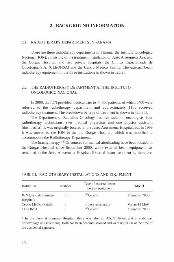
There are three radiotherapy departments in Panama: the Instituto OncológicoNacional (ION), consisting of the treatment installation on Justo Arosemena Ave. andthe Gorgas Hospital, and two private hospitals, the Clínica Especializada deOncología, S.A. (CLEONSA) and the Centro Médico Paitilla. The external beamradiotherapy equipment in the three institutions is shown in Table I.
2.2. THE RADIOTHERAPY DEPARTMENT AT THE INSTITUTOONCOLÓGICO NACIONAL
In 2000, the ION provided medical care to 44 000 patients, of which 6400 werereferred to the radiotherapy department and approximately 1100 receivedradiotherapy treatment. The breakdown by type of treatment is shown in Table II.
The Department of Radiation Oncology has five radiation oncologists, fourradiotherapy technicians, two medical physicists and one physics assistant(dosimetrist). It was originally located in the Justo Arosemena Hospital, but in 1999it was moved to the ION in the old Gorgas Hospital, which was modified toaccommodate the Radiotherapy Department.
The brachytherapy 137Cs sources for manual afterloading have been located inthe Gorgas Hospital since September 2000, while external beam equipment hasremained in the Justo Arosemena Hospital. External beam treatment is, therefore,
10
TABLE I. RADIOTHERAPY INSTALLATIONS AND EQUIPMENT
Institution Number Type of external beam
Modeltherapy equipment
ION (Justo Arosemena 1a 60Co unit Theratron 780C Hospital)Ceutio Médico Paitilla 1 Linear accelerator Varian 18 MeVCLEONSA 1 60Co unit Theratron 780C
a In the Justo Arosemena Hospital, there was also an ATC/9 Picker and a Stabilipanorthovoltage unit (Siemens). Both had been decommissioned and were not in use at the time ofthe accidental exposure.

given at the Justo Arosemena Hospital, while brachytherapy treatments, patienthospitalization and clinical follow-up of patients take place in the Gorgas Hospital.External beam therapy treatments are given from 06:00 to 21:00 using a Theratron780C 60Co unit.
The five radiation oncologists rotate between the Gorgas and Justo ArosemenaHospitals. Each month two of them are assigned to the installation on JustoArosemena Ave., working in two shifts to cover all of the time that the radiotherapyunit is in use. The intention is that a radiation oncologist should always be presentwhile patients are being treated, but in practice there is often no radiation oncologistavailable during the evening shift.
Treatment planning and dose prescription are done at the installation on JustoArosemena Ave. The data are entered in a physics chart, which remains at thehospital. The management of the patient’s treatment is recorded in a clinical chart,which is kept in the Gorgas Hospital. Patients are seen by the radiation oncologist atthe installation on Justo Arosemena Ave. at the beginning of their treatment. They arefollowed up clinically at the Gorgas Hospital in mid-treatment and at the end oftreatment, although usually not by the same radiation oncologist who prescribed thetreatment. The Radiotherapy Department does not have written protocols, but the stafffollow manuals by Fletcher [4] and Pérez and Brady [5], copies of which are kept atthe Justo Arosemena Hospital, and these appeared to be frequently consulted.
2.2.1. External beam radiotherapy
According to the information provided to the IAEA Team, the treatmentplanning process at the ION is the following. For each patient it starts with a radiationoncologist prescribing an appropriate radiation dose to control the malignant tumour.
11
TABLE II. BREAKDOWN OF CANCER TREATMENT AT THE ION
Cancers treated Percentage of treatments
Breast 16.8Cervix 15.5Endometrium 1.5Head and neck 12.1Prostate 9.3Brain 4.3Lung 7.9Colon and rectum 3.9Others 28.7

The prescribed radiation dose is delivered to the tumour by irradiating it with a beamof gamma rays from the cobalt radiotherapy unit. The dose is divided into dailyfractions given on different days over a period of several weeks. Each fraction mayinvolve several different ‘fields’, in each of which the radiation beam is pointed at thetumour from a different direction. The prescribed radiation dose is recorded in thepatient’s clinical chart by the radiation oncologist. This chart also has anthropometricinformation collected during the simulation process5. A source-to-skin distance(SSD) technique6 is always employed at the ION, even for multiple field treatments,and the dose prescriptions require all fields to be applied on each treatment day,except for treatments with eight fields. In these cases four fields were used every day.If shielding blocks are to be used to prevent giving too high a radiation dose to normaltissue, the radiation oncologist draws the cross-sectional shapes of the blocks andtheir positions in the treatment field on the X ray film obtained during the simulationprocess. A physicist enters the necessary data into the computerized TPS, a 2-DMultidata Radiation Therapy Treatment Planning System, RTP/2 Software Version2.11 (see Section 2.3 for a full description), which has options for external beam andbrachytherapy computations. The information to be entered includes, among otherdata:
— The total radiation dose prescribed;— The number of treatment days;— The SSD;— Details of each field;— An outline of the cross-section of each shielding block, drawn on a digitizing
tablet;— The attenuation of the radiation beam by each shielding block.
The TPS calculates treatment times and dose distributions. These are copied bythe physicist from the computer printout into the patient’s physics chart and they arechecked and signed by another physicist.
The patient’s clinical and physics charts show that the transfer of data from theoncologist’s prescription and the TPS to the patient’s physics chart is double checked
12
5 Treatment simulation is an essential step in radiotherapy, in which the treatmentgeometry is reproduced by using X ray equipment, and the image obtained is used to visuallycontrol the tissues and organs that will be later included in the treatment beam.
6 SSD is an external beam treatment technique in which a fixed distance between thesource and the skin is used.
and signed by the two physicists. However, there are no manual checks of whether thecomputer calculated treatment times are correct, and the technologists do notparticipate in the dose calculation process. The ION has protocols for quality controlof the radiation therapy equipment, which were found to be complete from 1999 todate, but no quality control procedures regarding checking of the treatment planningcalculations.
2.2.2. Brachytherapy
Some cases of cancer of the cervix are treated at the ION, as elsewhere, byradiation, both from an external beam and by brachytherapy, in which small radiationsources are placed inside the patient close to, or inside, the tumour. Brachytherapy hasthe advantage that high doses can be delivered to a tumour while minimizing damageto surrounding normal tissue because of the rapid fall-off of the dose at a distancefrom the source. It is supplemented by external beam therapy to deal with parts of thetumour or subclinical disease which do not receive a high enough dose frombrachytherapy.
Sources in patients undergoing brachytherapy emit radiation that may exposethe hospital staff and other patients. One way to optimize radiation protection is toplace the patients in rooms with additional structural shielding. There are four ofthese rooms in the Gorgas Hospital, and they are shared with patients undergoingnuclear medicine treatments. The consequence of this dual use of the rooms is thatbrachytherapy treatments cannot be given, as desired, either in mid-treatment orimmediately after treatment with external beam therapy. Often brachytherapy isscheduled months afterwards, when insertion of the source into the uterine canal isdifficult. To compensate for this, the dose delivered during external beam therapy ishigher than that recommended by Pérez and Brady [5], whose techniques the staff ofthe ION try to follow. (Many of these patients have had a hysterectomy prior toradiation therapy treatment; the scar left during surgery was often shielded with acentral block.)
Cancer of the cervix is treated both by external beam and brachytherapy, thelatter using Suit-Delclos applicators and 137Cs sources and a manual afterloadingtechnique. Until December 2000, brachytherapy treatment times were based ontabulated data for radium sources, and calculated using milligram-hours of radium(mgh Ra) equivalent values of the caesium sources.
Since January 2001, some of the treatment plans for brachytherapy, includingdose distributions, have been calculated using the TPS instead. At the GorgasHospital, the applicators, loaded with dummy sources, are inserted manually in aminor surgery room by a radiation oncologist. The positions are checked at the sametime with a portable X ray machine in the presence of a medical physicist, who thenfills out the appropriate data forms. The insertion geometry must always be approved
13

by the radiation oncologist. The films are taken by the medical physicist to the JustoArosemena Hospital and the treatment plans are calculated using the TPS.
2.3. TREATMENT PLANNING SYSTEM
According to the information provided to the IAEA Team, the TPS used at the ION was the RTP/2 Multidata System, Version 2.11 by International Corp., license Americal Megatrends Inc., 40-0103-016155-00011111-111192-SYMP-F. The manual in use has the title ‘User & Reference Guide Level II, Release2.1 & Up’.
Gamma ray beam data (depth doses and beam profiles) for both 60Co units, theTheratron 780C and the now decommissioned Picker ATC C/9, were entered andverified when the system was first installed in 1993. Output (dose rate for a standardfield in reference conditions), field factors, wedge and tray factors are entered in thesystem when measured, which is usually when the 60Co source is replaced. Theactivities of the 137Cs sources used in brachytherapy were entered in units of mg Raequivalent until the beginning of 2001.
The TPS has several computing options:
— ‘Dose Chart Calculator’ is used to calculate the treatment times needed todeliver a given dose to a prescription point, including the use of blocks.
— ‘Irreg’ is an option to calculate the treatment times needed to deliver a givendose to selected points, specifically for complicated, irregular shaped fields, forexample the so-called mantle field.
— ‘External Beam’ is used when it is intended to generate isodose distributionstogether with the calculation of the treatment time to deliver a given dose to aprescription point. It was this option that was in use when excessive treatmenttimes were calculated.
— ‘Brachytherapy’ computes isodose distributions when using brachytherapysources.
Physicists at the ION used the ‘Dose Chart Calculator’ option of the TPS tocalculate dose to the prescription point, except for irregular shaped fields for whichthe ‘Irreg’ option was used. The ‘External Beam’ option was used only when dosedistributions were requested by the radiation oncologist (dose distributions were notrequested for all patients).
The physicists compute the required dose distributions following the TPS User& Reference Guide. This manual indicates that is it permitted to enter up to fourblocks; however, as described in Section 6.2 of this report, no instructions on how todigitize the block contours are given.
14
2.4. RADIATION PROTECTION INFRASTRUCTURE AND REGULATORY CONTROL
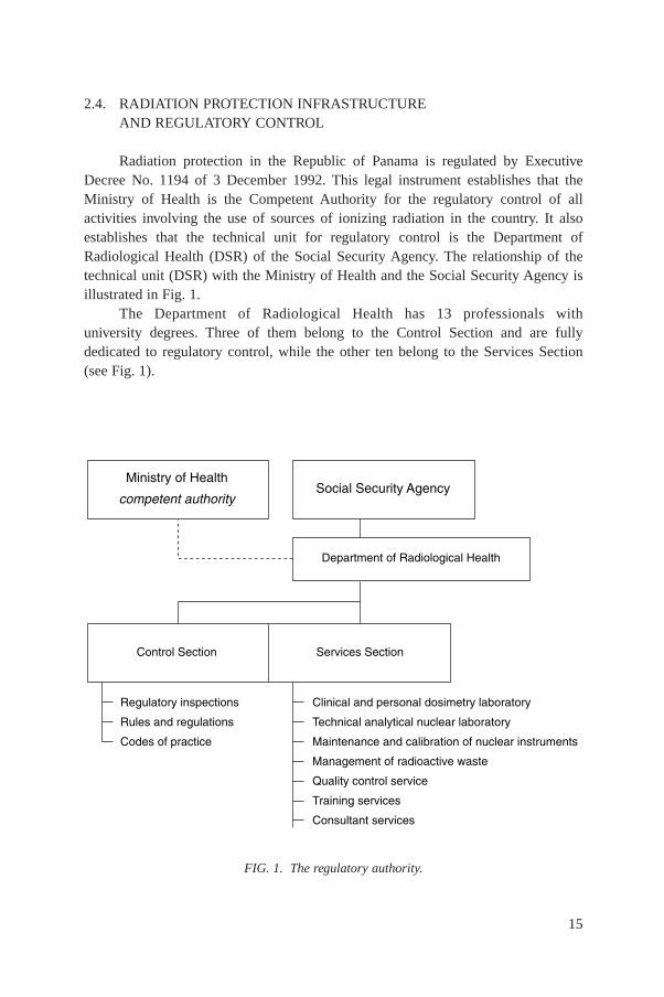
Radiation protection in the Republic of Panama is regulated by ExecutiveDecree No. 1194 of 3 December 1992. This legal instrument establishes that theMinistry of Health is the Competent Authority for the regulatory control of allactivities involving the use of sources of ionizing radiation in the country. It alsoestablishes that the technical unit for regulatory control is the Department ofRadiological Health (DSR) of the Social Security Agency. The relationship of thetechnical unit (DSR) with the Ministry of Health and the Social Security Agency isillustrated in Fig. 1.
The Department of Radiological Health has 13 professionals with university degrees. Three of them belong to the Control Section and are fullydedicated to regulatory control, while the other ten belong to the Services Section (see Fig. 1).
15
������������ ���
�������������� ����� �����������������
��� ��������� ������ ���� ���
���������������������������
������ �����������
�� ��������������
�� ������������������
� � ��������� �� ������� ���
�������� ����� � ����� ���� ���� ���� ���������� �������������
������ �������� ���� ������� �� � ����� ������� ��� �� ���
����� �������������� ������ �� �������� ������������ �� ���
FIG. 1. The regulatory authority.
2.4.1. Authorization of the Radiotherapy Department
The application for authorization of the Radiotherapy Department of the IONwas submitted in 1997, but the authorization had not been granted by the time of theaccidental exposure, pending the resolution of several issues. Only those mostrelevant to this report, taken from the records of the regulatory authority (DSR), arelisted below:
— A number of reminders sent out by the regulatory authority (DSR), from 1997to date, indicate that the Radiotherapy Department was not able to provide allthe information requested by DSR, principally the manuals of procedures forradiation protection and quality assurance.
— In 1997, an IAEA expert mission discovered that a number of brachytherapysources were missing from the ION and were found to be in use at the privateCLEONSA Clinic, without authorization.
— An incident occurred in which a cobalt therapy radiation source did not returnto the ‘OFF’ (shielded) position, risking excessive radiation exposure of staffand patients, there being no radiation oncologist present at the hospital at thetime.
— A nuclear medicine incident with a therapeutic amount of aradiopharmaceutical occurred in a brachytherapy room when, instead of theprescribed amount of 5.6 GBq of 131I, the patient received 11.5 GBq (althoughthe administration of radiopharmaceuticals is usually a nuclear medicineprodure, this incident appears in the records of this facility).
— A letter sent by the regulatory authority reminding the hospital of its obligationto have at least one radiation oncologist always present when patients are beingtreated.
— In October 2000, the regulatory authority initiated disciplinary sanctionsagainst the ION, because of non-compliance with its reiterated instructions.
2.5. HISTORY OF RECENT AUDITS OF THE RADIOTHERAPYDEPARTMENT
2.5.1. Audit in February 1999
The 1999 IAEA audit involved a review of the quality control activities of theION, an intercomparison of the dosimetry equipment of the ION and the DSR, acalibration and a quality control check of the treatment units and a test of the TPS.
The auditors noted the following information with regard to the acceptance testsof the Multidata TPS:
16
— The system was installed in 1993 by a specialist from Multidata, who alsoentered the basic data (the isodoses for single fields of the 60Co units);
— The medical physicists of the ION compared treatment times obtained from theMultidata TPS for certain typical treatments with those calculated manually forthe same treatments;
— Dose distributions (isodoses) for typical treatments were obtained from theMultidata TPS and compared with the isodoses from another TPS in a privateclinic in Panama (Theraplan V) for the same typical treatments;
— No records of these tests had been kept.
With regard to the quality control checks of the equipment and accuracy of thedoses delivered to patients, the auditors reported:
— Calibrations of the beams for the two 60Co units were regularly performed bythe ION. Results were recorded in a logbook, but there was no documentspecifying the frequency of the measurements.
— Daily checks of the items specified in Ref. [6] were performed by theradiotherapy technologists, reviewed by the medical physicists and recorded.Monthly mechanical checks were performed by the medical physicists but werenot recorded.
— An intercomparison was performed by the IAEA during the audit using theavailable dosimetry equipment from the ION and the DSR. The differenceswere within ±1.8%.
— The auditors carried out quality control tests of all three external beam units,and compared their values with the values in use in the hospital. The differenceswere acceptable, with the only significant discrepancy of 10% in the timer ofthe orthovoltage unit.
— The auditors also tested the TPS for different conditions: open field, wedgedfield and a field with two shielding blocks. The tests consisted of:
(1) Manual checks of the treatment time for some randomly selected patientcharts, including box technique treatments, irregular fields and wedges.The TPS results were within a 1% difference between the treatment timesfrom the patients’ charts and the ones calculated by the auditors based onthe data and factors in use.
(2) Prescribing a dose to a point in a water phantom, calculating the irradiationtime with the TPS, irradiating the water phantom using the irradiation timecalculated by the TPS and measuring the actual dose delivered to theprescription point in the water phantom. The difference between theprescribed dose and the measured dose was within ±1.8% for the Theratron780C unit.
17
(3) For brachytherapy, the auditors compared doses calculated by the TPS to aselected point with values obtained from tables. The differences werewithin ±3%.
The auditors recommended that the ION should establish a quality assuranceprogramme based on IAEA-TECDOC-1151 [6].
2.5.2. Audit in February 2001
By the time of this audit, both the Picker ATC C/9 60Co unit and the Stabilipanorthovoltage unit had been decommissioned. This audit noted that quality controlchecks of the Theratron 780C 60Co unit were being regularly carried out. Theyincluded daily, monthly and annual checks, following IAEA-TECDOC-1151 [6], andthe results were well documented.
Other statements in the audit report that are relevant to this report are asfollows:
— An intercomparison was performed of the available dosimetry equipment fromthe three radiotherapy departments in Panama (ION and two private hospitals).The differences were within ±1.2%.
— The auditors verified the values of the parameters included in the protocols ofIAEA-TECDOC-1151 [6]. The differences between the ION and auditors’measurements were: within ±0.4% for the dose rate in reference conditions(5 cm depth in water at 80 cm SSD); within 0.3% for field size factors; within±1% for the tray factor; within ±4.9% for the wedge factors. The auditors foundthat the transit time of the source (–0.016 min) was not being corrected for bythe medical physicists. This implies an error in dose of 1.6% for an irradiationtime of 1 min.
— The auditors also tested the TPS for different conditions: open field of differentfield sizes, wedged field, and a field with one shielding block. The testingconsisted of prescribing a dose to a point in a water phantom, calculating theirradiation time with the TPS, irradiating the water phantom using theirradiation time calculated by the TPS and measuring the actual dose deliveredto the prescription point. The difference between the prescribed dose and themeasured dose was within ±2.1%.
— Localization in gynaecological brachytherapy was done by orthogonal X rayfilms. Conventional X ray equipment was used for simulation/localization forexternal beams.
— New 137Cs sources had been acquired since the previous audit, and there was acomplete inventory of all sources. All source certificates were available and allthese sources were regularly checked by the medical physicists. Spot checks of
18
the source calibrations were performed during the audit and the differencesbetween the values obtained and the values in use at the ION were within±2.6%, in terms of the dose rate at one metre.
It is important to note that the auditors were not made aware that the method ofentering data for several blocks had been changed. Since there were no writtenprocedures, the auditors did not realize that this change had been made, and no testswere done following the new procedure for digitizing several blocks together as ifthey were a single block.
2.5.3. Results of IAEA/WHO TLD postal dose quality audits performed atthe ION
The ION has participated in the IAEA/WHO TLD postal programme forradiotherapy hospitals on a regular basis since 1987. The check of calibration of thetwo 60Co machines was performed during these years according to the proceduredeveloped by the IAEA [7]. The TLDs were sent to the ION along with instructionsto irradiate them in the same way as a patient is irradiated in normal clinical practice.To reflect the clinical situation, the calculation of irradiation time to deliver the doseof 2 Gy was requested to be performed in the same way as for patient treatments.When the irradiated TLDs arrived at the IAEA, they were analysed and the doseswere computed for each dosimeter. The acceptance limit of the IAEA/WHO TLDaudits for hospitals is ±5% and this defines the maximum discrepancy between statedand measured doses which does not require any further investigation. When the resultof a hospital falls outside the acceptance limit of ±5%, follow-up actions areperformed.
The results of the IAEA/WHO TLD postal dose quality audit for the ION(16 checks), including the last audit performed at the ION in August 2000, show thatthe mean ratio of the dose measured by the IAEA to the dose stated by the institute is1.016 ± 0.025; only once (in 1995) was the deviation outside the acceptance limitof ±5%.
3. THE ACCIDENTAL EXPOSURE
3.1. INITIATING EVENT
Treatments in the pelvic region (prostate, cervix, colon) were performed at theION using four fields, an anterior–posterior pair and two opposed laterals (the box
19
technique). Sometimes the lateral fields had 30° wedge filters. Most fields includedshielding blocks to protect normal tissue. Up to four blocks per treatment field wereoften used for the pelvic region. All four fields were treated every day during thetreatment period. For some patients, four additional oblique fields were employed. Inthis case, the two treatments (of each set of four fields) were given on alternate days.The treatments were performed using the SSD technique. Following this regimen,patients received booster doses consisting usually of a so-called skip (arc) exposuredelivered to a smaller volume of tissue.
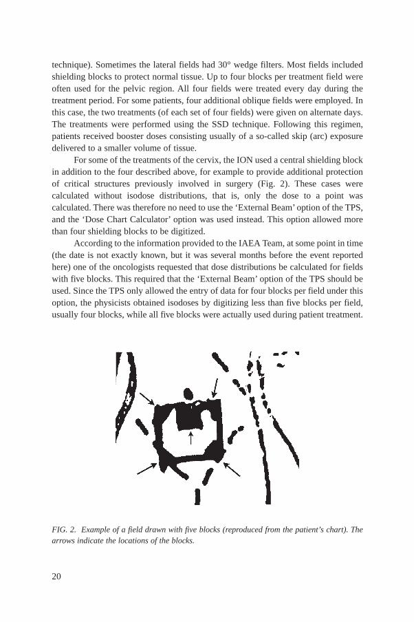
For some of the treatments of the cervix, the ION used a central shielding blockin addition to the four described above, for example to provide additional protectionof critical structures previously involved in surgery (Fig. 2). These cases werecalculated without isodose distributions, that is, only the dose to a point wascalculated. There was therefore no need to use the ‘External Beam’ option of the TPS,and the ‘Dose Chart Calculator’ option was used instead. This option allowed morethan four shielding blocks to be digitized.
According to the information provided to the IAEA Team, at some point in time(the date is not exactly known, but it was several months before the event reportedhere) one of the oncologists requested that dose distributions be calculated for fieldswith five blocks. This required that the ‘External Beam’ option of the TPS should beused. Since the TPS only allowed the entry of data for four blocks per field under thisoption, the physicists obtained isodoses by digitizing less than five blocks per field,usually four blocks, while all five blocks were actually used during patient treatment.
20
FIG. 2. Example of a field drawn with five blocks (reproduced from the patient’s chart). Thearrows indicate the locations of the blocks.
Data for one of the blocks were not entered and therefore not considered in thecalculation.
Digitizing only four of the five blocks causes an error in calculating the dose tothe prescription point because the computer takes into account the scatter componentof the radiation coming from all irradiated parts of the patient. In this case theirradiated parts assumed by the computer include those parts that in fact would becovered by the fifth shielding block, which was not entered into the computer. Theactual contribution to scatter radiation coming from the shielded region is, therefore,lower than the one assumed by the computer calculation. If treatment is given for theduration calculated by the computer, the patient will receive a dose of the order of afew per cent less than that prescribed. This effect was understood and accepted as partof the temporary solution. The team was informed that the physicists omitted the datafor the smallest block from the TPS so as to further minimize the error.
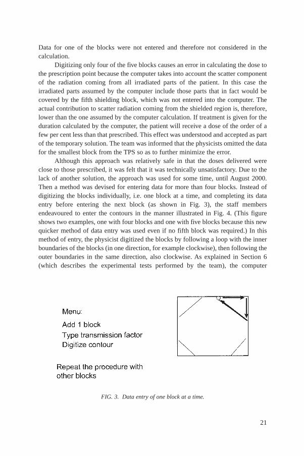
Although this approach was relatively safe in that the doses delivered wereclose to those prescribed, it was felt that it was technically unsatisfactory. Due to thelack of another solution, the approach was used for some time, until August 2000.Then a method was devised for entering data for more than four blocks. Instead ofdigitizing the blocks individually, i.e. one block at a time, and completing its dataentry before entering the next block (as shown in Fig. 3), the staff membersendeavoured to enter the contours in the manner illustrated in Fig. 4. (This figureshows two examples, one with four blocks and one with five blocks because this newquicker method of data entry was used even if no fifth block was required.) In thismethod of entry, the physicist digitized the blocks by following a loop with the innerboundaries of the blocks (in one direction, for example clockwise), then following theouter boundaries in the same direction, also clockwise. As explained in Section 6(which describes the experimental tests performed by the team), the computer
21
FIG. 3. Data entry of one block at a time.

accepted the data entered in this way but the calculated treatment times were laterfound to be substantially larger than they should have been.
From August 2000 onwards, data for multiple blocks were entered for a numberof cases using the new method when calculating exposures of the pelvic region, even
22
!"
#
$
�� ����������
�� ����������
FIG. 4. Circumventing the limitation of the number of blocks by entering the co-ordinates ofmultiple blocks as a single block with two loops in the same direction. The shaded areas denotethe blocks. The computer accepts this way of digitizing multiple blocks describing two loops inthe same direction, but calculates the wrong treatment time by about +100% (referred to as 5%block transmission).
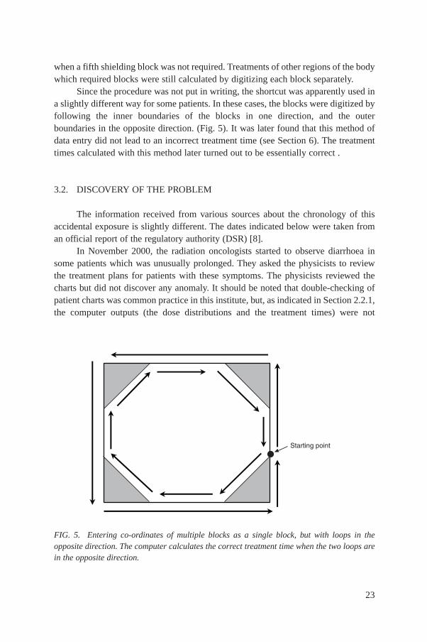
when a fifth shielding block was not required. Treatments of other regions of the bodywhich required blocks were still calculated by digitizing each block separately.
Since the procedure was not put in writing, the shortcut was apparently used ina slightly different way for some patients. In these cases, the blocks were digitized byfollowing the inner boundaries of the blocks in one direction, and the outerboundaries in the opposite direction. (Fig. 5). It was later found that this method ofdata entry did not lead to an incorrect treatment time (see Section 6). The treatmenttimes calculated with this method later turned out to be essentially correct .
3.2. DISCOVERY OF THE PROBLEM
The information received from various sources about the chronology of thisaccidental exposure is slightly different. The dates indicated below were taken froman official report of the regulatory authority (DSR) [8].
In November 2000, the radiation oncologists started to observe diarrhoea insome patients which was unusually prolonged. They asked the physicists to reviewthe treatment plans for patients with these symptoms. The physicists reviewed thecharts but did not discover any anomaly. It should be noted that double-checking ofpatient charts was common practice in this institute, but, as indicated in Section 2.2.1,the computer outputs (the dose distributions and the treatment times) were not
23
�� ����������
FIG. 5. Entering co-ordinates of multiple blocks as a single block, but with loops in theopposite direction. The computer calculates the correct treatment time when the two loops arein the opposite direction.

included in these checks, the implicit assumption being that the computer calculationswere correct.
In December 2000, similar abnormal symptoms were observed in otherpatients. In February 2001, the physicists initiated a search for the possible cause ofthese effects, but it was only in March 2001 that the physicists identified that therewas a problem with the calculation of treatment times and informed the radiationoncologist on duty. The treatment of patients presenting abnormal symptoms wassuspended. Patients treated with the modified method of data entry were identifiedand their patient charts were reviewed.
It then became apparent that the patients who had their treatment timescalculated using the ‘shortcut’ method of data entry had received doses larger thanprescribed, and in mid-March the Director General of the ION was informed.Measurements were then performed using a water phantom simulating the conditionsunder which a patient was treated, and the error was confirmed.
It was also discovered at this time that the sequence in which the co-ordinatesof the blocks were digitized affected the calculated treatment times and hence thedoses delivered. Patients for whom the co-ordinates had been digitized in thesequence shown in Fig. 4 had isodoses and treatment times calculated that weresimilar to the correct ones (the ones from blocks entered individually).
4. RESPONSE TO THE ACCIDENTAL EXPOSURE
4.1. ACTIONS TAKEN UPON DISCOVERY OF THE ERROR
24 March 2001The Panamanian Ministry of Health contacted the representative of PAHO in
Panama with a request for assistance in verifying patient doses. This representativepassed on the request to the PAHO Regional Office. The Ministry of Health suppliedPAHO with an example of the treatment calculations and physics data, provided bythe ION [9], for one of the affected patients. PAHO estimated the absorbed dose tothe patient received by this patient to be about 94 Gy, and informed the Ministry ofHealth through its representative in Panama.
April 2001The affected patients were identified and the absorbed doses and dose
distributions were recalculated, using the procedure of introducing the blocksseparately into the RTP/2 treatment planning computer from Multidata, by thephysicists at the ION.
24

Independently, PAHO performed the calculation of patient doses manually andconfirmed the exposure to its representative in Panama and provided a copy of itsreport to the Director General of the ION on 16 April 2001 [10, 11].
On 19 April 2001, the group of consultants appointed at the request of the IONarrived. The group was composed of one radiation oncologist and two medicalphysicists from the MD Anderson Hospital. The group replicated the error and triedto determine the possible problem with the algorithm. It concluded that, “although nofull explanation could be found it was suspected that the algorithm fails to account forthe scatter component when the data is entered in a certain way”.
The report further states that “Upon return to Houston, calculations for twocases were repeated using a different treatment planning system. The resultsconfirmed the findings found during the visit.” The report also states that “MultidataSystems International Corp. was contacted and the problem was reported to them.Arrangements were made to have Multidata contact ION directly and it wasimpressed upon Multidata to send someone to Panama as soon as possible to resolvethis problem.”
14 May 2001The General Subdirector of the ION informed the regulatory authority DSR
about the accidental exposure.
16 May 2001The ION provided the DSR with documents related to the accidental exposure.
17 May 2001The DSR provided a copy of the report to the General Director of Health, with
the recommendation that medical assistance in treating the overexposed patients besought.
21 May 2001The DSR began a technical evaluation of the accidental exposure for the
Minister of Health.
25 May 2001The DSR issued its report [8].
4.2. RESPONSE FROM THE IAEA
On Saturday 19 May 2001, the IAEA Duty Emergency Response Manager(ERM) was informed of the accidental exposures in Panama by an IAEA staff
25

member in the Department of Technical Co-operation. This staff member conveyed to him information about the incident provided by the Panamanian counterpart7 to the technical co-operation project ‘Development of Technical Capabilities for Sustainable Radiation and Waste Infrastructure’ — RLA/9/044. The counterpart indicated that the IAEA would be receiving a request for assistance in afew days.
On Monday 21 May 2001, the IAEA Duty ERM contacted the National Competent Authority in Panama by telephone to obtain more detailedinformation about the incident. The Panamanian Competent Authority confirmed theaccuracy of the information provided by the counterpart and confirmed that Panamawould be requesting the IAEA to provide assistance under the terms of theConvention on Assistance in the Case of a Nuclear Accident or RadiologicalEmergency.
On the same day, a message was received from the competent authorityrequesting the IAEA to provide a technical expert team to evaluate the incident. The IAEA’s Emergency Response Centre (ERC) then contacted the appropriateexperts to inquire about their willingness and availability to take part in this IAEAmission.
On Tuesday 22 May 2001, the Permanent Mission of Panama to the IAEA senta facsimile message addressed to the IAEA’s Director General requesting assistanceto Panama in connection with the emergency situation.
The IAEA ERC sent out an advisory information message to all National Warning Points (NWPs), all National Competent Authorities and allPermanent Missions to the IAEA. This informed them of the emergency situation inPanama and informed them that the Agency was sending an expert team there. Anumber of countries then requested more information and some offered medicalassistance.
The terms of reference of the IAEA’s mission to Panama were established andapproved by the Panamanian authorities. The terms of reference required the missionto, inter alia:
— Ensure that the radiation source(s) involved in the accident was (were) in a safeand secure condition;
26
7 Coincidentally, the Panamanian counterpart is also the National Competent Authorityfor the Convention on Early Notification of a Nuclear Accident (Early Notification Convention)and the Convention on Assistance in the Case of a Nuclear Accident or RadiologicalEmergency (Assistance Convention).
— Evaluate the doses incurred by the affected patients, inter alia by analysing thetreatment records and physical measurements;
— Undertake a medical evaluation of the affected patients’ prognosis andtreatment, taking into account, inter alia, the autopsy findings for those whodied; and
— Identify issues on which the IAEA could offer to provide and/or co-ordinateassistance to minimize the consequences of the accident.
The team was established and arrived in Panama on Saturday 26 May 2001. Theteam was composed of experts in radiopathology, radiotherapy, radiology, radiationprotection, and medical physics from France, Japan, the USA and the IAEA. Twodays later, the team was joined by an expert from the Russian Federation representingthe World Health Organization (WHO) and by M. Akashi from Japan. The membersof the international team were:
— M. Akashi, Research Center for Radiation Emergency Medicine of the NationalInstitute of Radiological Sciences, Chiba, Japan;
— J.M. Cosset, Département de Radiothérapie, Service B, Institut Curie, Paris,France;
— P. Gourmelon, Centre d’Etudes Nucléaires, Institut de Protection et de SûretéNucléaire, Fontenay-aux-Roses, France;
— M. Konchalovsky (representing WHO), Hematologic Department of theScientific Research Center of Russia, Moscow, Russian Federation;
— F. Mettler, Chairman, Department of Radiology, University of New Mexico,School of Medicine, Albuquerque, USA;
— P. Ortiz López, IAEA;— S. Vatnitsky, IAEA.
At the request of the Government of Panama, expressed during the mission, a staffmember of PAHO, C. Borrás, joined the international team.
The mission was concluded on Friday 1 June 2001.On Saturday 2 June 2001, another advisory information was sent from
the IAEA’s ERC to all National Warning Points, National Competent Authorities and Permanent Missions reporting the preliminary findings of the expert team. On Saturday 9 June 2001, a termination report was sent out to the same contact points confirming the findings of the preliminary report and notifying them that theERC had terminated its activation level in response to the emergency situation. Theaim of these communications was to provide enough information and advice to Statesto help to avoid a similar accidental exposure elsewhere in the coming months,pending the publication of a final report. The termination report is included inAnnex I.
27

5. DOSE ASSESSMENT
5.1. INTRODUCTION
The purpose of dose assessment was to obtain an evaluation of the dosesreceived by the patients that were affected by the use of incorrect treatment times. Theassessment of external beam doses to patients was carried out by manual calculation.Manual calculations are normally done in radiotherapy departments when acomputerized TPS is not used, or to verify computer calculated values. Thecalculation of patient doses was based on the dose rate, the treatment time and allrelevant parameters taken from the patients’ charts, as indicated below. Since thecalculation of patient doses requires the use of a number of dosimetric factors, allthese factors needed to be evaluated and verified by the team.
Standardized quality audit procedures for dosimetry on-site visits toradiotherapy hospitals, developed by the IAEA [12], were used as a guide whenperforming the dosimetry evaluation. The standard procedures are limited to those carried out under the IAEA/WHO TLD postal dose quality audit programme, and focus on the calibration of radiotherapy machines. For the purposesof this assessment, they were modified to cover the evaluation of the treatmentplanning process and to investigate the accidental exposure related to the use of theTPS.
In addition to this assessment, an evaluation of the biologically effective doseand the equivalent dose for 2 Gy per fraction was carried out. This was necessary totake account of the fact that in this accidental exposure doses were given in largerfractions than the typical 2 Gy per fraction.
For this assessment, dosimetry measurements were carried out using equipmentwhich was brought by the IAEA members of the team. A standard instrumentation kitwas used, which contained the following main items of equipment:
— Electrometer PTW UNIDOS, Serial No. 20334;— Ionization chambers PTW W30010 Serial Nos 52 and 53, along with
calibration certificates from the IAEA Dosimetry Laboratory;— Barometer AIR-HB-1A;— Two calibrated mercury thermometers;— Box water phantom (PTW T41014);— Two TLD sets and a TLD holder, along with the instruction and data sheets.
28
5.2. SCOPE OF THE DOSIMETRIC EVALUATION
The dosimetric evaluation performed by the team at the ION focused on thefollowing areas:
— Comparison of IAEA and ION dosimetry systems and 60Co beam calibration.
— Verification of the delivery of prescribed doses to a selected point in the water phantom for different beam arrangements. The verification was carried out by asking ION physicists to calculate exposure times manually and by computer and using the calculated exposure times to irradiate a water phantom, and measuring the corresponding doses to the prescription point in the water phantom using an ionization chamber.
— Assessment of the doses received by the 28 patients with potentialoverexposure.
The ION dosimetry equipment consists of:
— Electrometer PTW UNIDOS Serial No. 20226;— Electrometer Keithley E 35614, Serial No. 18097 (calibration certificate from
ADCL MD Anderson of 24 October 1984);— Ionization chamber PTW W30001, Serial No. 1496 (calibration certificate from
PTW of 11 September 1997);— Well type chamber HDR 1000, Serial No. A970931;— Barometer AIR-HB-1A;— Thermometer Digital Fluke 52.
The comparison of the IAEA and ION dosimetry systems and the verificationof the delivery of prescribed doses were performed by the team at the ION in twosessions on different days. In one session the ION physicists were interviewed on thedosimetry data and treatment techniques used at the ION. The team then reviewed thepatient treatment charts in order to evaluate the radiotherapy techniques used at theION and to ensure that the necessary dosimetry data were available. They also neededto ensure that test dose calculations performed with the TPS corresponded to typicaltreatments actually performed at the ION. Attention was paid to the data used in dosecalculations for pelvic fields.
Safety and mechanical checks of the Theratron 780C treatment unit werecarried out following the interview. The requirements of the BSS were considered, aswell as those of IAEA-TECDOC-1040 [13] for safety, mechanical and other QAaspects. The results were as follows:
29
— Door interlocks, radiation warning lights and emergency switches wereoperational.
— The agreement of the mechanical indicator of gantry rotation with the digitaldisplay was within 1°.
— The agreement of the digital indicator of field size with the measured field sizewas within 2 mm for 5 cm × 5 cm, 10 cm × 10 cm, 15 cm × 15 cm and 20 cm× 20 cm fields.
— The distance from the bottom of the accessory holder to the isocentre was32 cm.
— The tip of the distance stick was in agreement with the lasers within 1 mm forgantry positions of 90° and 270°.
— The lateral lasers were aligned within 1 mm at ±20 cm distance from theisocentre.
— The timer agreed with a manual stopwatch within 1 s for irradiation times of1–5 min.
Following safety and mechanical checks, the ION physicists were asked toperform calibration of the 60Co beam in the same way as they do during their regularquality control checks, and to irradiate TLDs following the standard procedurerecommended by the IAEA for the TLD postal dose audit. The team performed thebeam calibration using the IAEA equipment they had brought with them, and theresults were compared with the results of the ION calibration.
The team evaluated the ION’s method of calculating the irradiation times forpatient treatments. The ION physicists were requested to determine time settings forthe clinical dosimetry tests, including a rectangular field, a wedged field and ablocked field, as described below. The clinical tests were performed in a waterphantom (PTW T41014) using a single field. Time settings were calculated to deliver2 Gy to the prescription point located on the beam at a depth of 5 cm. The followingset-ups were entered into the TPS:
— Field size 6 cm × 16 cm. Prescription point on the central axis. — Field size 8 cm × 8 cm, with 30° wedge. Prescription point on the central axis. — Field size 15 cm × 15 cm, with shielding blocks. Prescription point 3.5 cm
off-axis.
The calculated time settings were verified by manual calculation and were usedon the Theratron 780C treatment unit. The team verified that the prescribed doseswere delivered to the specified points in the phantom using ionization chambermeasurements.
During the second session a direct intercomparison of ION and IAEAdosimetry systems was performed. The aim of this intercomparison was to verify the
30

constancy of response of the ION dosimetry system, with reference to the calibrationcertificate.
5.3. COMPARISON OF THE DOSIMETRY SYSTEMS AND BEAM CALIBRATION
5.3.1. Dosimetry system intercomparison
As a part of the evaluation of the ION dosimetry system, an intercomparison ofbarometers and thermometers was performed. The results are listed in Table III.
A comparison of the ION’s dosimetry system with the IAEA dosimetry systemwas performed by sequential irradiation of ionization chambers in referenceconditions in the 60Co beam of the Theratron 780C treatment unit using thePTWT41014 water phantom. The depth of measurement was 5.0 g/cm2, field size10 cm × 10 cm, SSD = 80 cm. The IAEA chambers, PTW W30010, SerialNos 52 and 53, were calibrated in terms of air kerma and absorbed dose to water atthe IAEA Dosimetry Laboratory and their calibration factors are traceable to theBureau International des Poids et Mesures (BIPM). The calibration factor of the IONionization chamber8 was derived from this comparison and was compared with thevalue listed in the certificate issued on 27 April 1997 by PTW Freiburg:Nk = 47.59 mGy/C. The reference temperature and pressure are 20°C and 101.3 kPA.The results are listed in Table IV.
31
8 The chamber was calibrated in air in terms of air kerma (factor Nk).
TABLE IV. COMPARISON OF CHAMBER FACTORS
Factor of IAEA chamber Factor of ION chamberCertificate/derived
Nk (mGy/C) Nk (mGy/C), derived
49.0 (PTW W30010 No. 52) 47.73 (PTW W30001 No. 1496) 1.00349.4 (PTW W30010 No. 53) 47.58 (PTW W30001 No. 1496) 1.000
TABLE III. BAROMETER AND THERMOMETER INTERCOMPARISON
IAEA ION kTP ratio IAEA/ION
Pressure (kPA) 101.15 101.13Temperature (°C) 26.0 26.4kTP 1.022 1.024 0.998
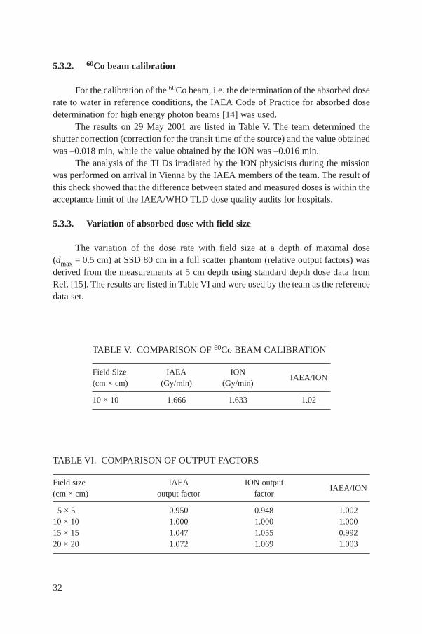
5.3.2. 60Co beam calibration
For the calibration of the 60Co beam, i.e. the determination of the absorbed doserate to water in reference conditions, the IAEA Code of Practice for absorbed dosedetermination for high energy photon beams [14] was used.
The results on 29 May 2001 are listed in Table V. The team determined theshutter correction (correction for the transit time of the source) and the value obtainedwas –0.018 min, while the value obtained by the ION was –0.016 min.
The analysis of the TLDs irradiated by the ION physicists during the missionwas performed on arrival in Vienna by the IAEA members of the team. The result ofthis check showed that the difference between stated and measured doses is within theacceptance limit of the IAEA/WHO TLD dose quality audits for hospitals.
5.3.3. Variation of absorbed dose with field size
The variation of the dose rate with field size at a depth of maximal dose(dmax = 0.5 cm) at SSD 80 cm in a full scatter phantom (relative output factors) wasderived from the measurements at 5 cm depth using standard depth dose data fromRef. [15]. The results are listed in Table VI and were used by the team as the referencedata set.
32
TABLE V. COMPARISON OF 60Co BEAM CALIBRATION
Field Size IAEA IONIAEA/ION
(cm × cm) (Gy/min) (Gy/min)
10 × 10 1.666 1.633 1.02
TABLE VI. COMPARISON OF OUTPUT FACTORS
Field size IAEA ION output IAEA/ION
(cm × cm) output factor factor
5 × 5 0.950 0.948 1.00210 × 10 1.000 1.000 1.00015 × 15 1.047 1.055 0.99220 × 20 1.072 1.069 1.003
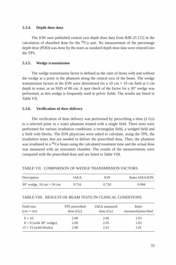
5.3.4. Depth dose data
The ION uses published central axis depth dose data from BJR-25 [15] in thecalculation of absorbed dose for the 60Co unit. No measurement of the percentagedepth dose (PDD) was done by the team as standard depth dose data were entered intothe TPS.
5.3.5. Wedge transmission
The wedge transmission factor is defined as the ratio of doses with and withoutthe wedge at a point in the phantom along the central axis of the beam. The wedgetransmission factors at the ION were determined for a 10 cm × 10 cm field at 5 cmdepth in water, at an SSD of 80 cm. A spot check of the factor for a 30° wedge wasperformed, as this wedge is frequently used in pelvic fields. The results are listed inTable VII.
5.3.6. Verification of dose delivery
The verification of dose delivery was performed by prescribing a dose (2 Gy)to a selected point in a water phantom treated with a single field. Three tests wereperformed for various irradiation conditions: a rectangular field, a wedged field anda field with blocks. The ION physicists were asked to calculate, using the TPS, theirradiation times that are needed to deliver the prescribed dose. Then, the phantomwas irradiated in a 60Co beam using the calculated treatment time and the actual dosewas measured with an ionization chamber. The results of the measurements werecompared with the prescribed dose and are listed in Table VIII.
33
TABLE VII. COMPARISON OF WEDGE TRANSMISSION FACTORS
Description IAEA ION Ratio IAEA/ION
30° wedge, 10 cm × 10 cm 0.716 0.720 0.994
TABLE VIII. RESULTS OF BEAM TESTS IN CLINICAL CONDITIONS
Field size TPS prescribed IAEA measured Ratio(cm × cm) dose (Gy) dose (Gy) measured/prescribed
6 × 16 2.00 2.06 1.038 × 8 (with 30° wedge) 2.00 2.05 1.03
15 × 15 (with blocks) 2.00 2.01 1.01

5.4. PATIENT DOSES
5.4.1. Assessment of doses from irradiation with external beams
Before evaluating the doses to patients, the team verified and reproduced theincorrect calculation of treatment times when several shielding blocks are digitized asa single block, describing two loops in the same direction. Manual calculation ofactual doses delivered to each individual patient was done using treatment timesindicated in the patient charts, since these are the times that were used for the patientirradiation.
The doses were calculated for the point of intersection of beam axes formultiple fields (the prescription point). The calculations employed the followingparameters and information:
— Treatment time (from patient charts);— Absorbed dose rate to water at the reference conditions (based on IAEA
measured values, 29 May 2001);— Field size, equivalent field size (from patient charts);— Depth (from printout of treatment plans);— Output factors (from clinical dosimetry data);— Wedge factors (from clinical dosimetry data);— Depth dose data (from BJR-25 [15]).
A table of calculated doses at the prescription point for the 28 affectedpatients9 was provided to the medical team of the mission for comparison with theclinical findings. This information was complemented with the TPS calculated dosedistributions (printouts of treatment plans) produced by the ION physicists for eachof the 28 patients, using the procedure to digitize the co-ordinates of the shieldingblocks in the TPS separately. These treatment plans were compared with thetreatment plans affected by the error when the data for several blocks were enteredinto the TPS as if they were a single block, describing two loops in the samedirection.
34
9 The report by the DSR [8] indicates that an additional patient might have been affectedby a dose that was 43% higher than intended. The team checked that, according to this patient’schart, the patient was treated with the incorrect time for only one fraction, and the rest of thetreatment was delivered with the time corrected. This patient was treated in March 2001, aboutthe time when the error was discovered.

The results are given in Table IX, where doses to the prescription point arelisted. The dose to critical structures, such as bowel and rectum, are slightly differentas can be inferred from the dose distributions. More accurate calculations of doses tothese structures were not performed because of the lack of anatomical information foreach particular patient.
The following abbreviations are used in the table:
— Field dir: antero/posterior (AP), postero/anterior (PA), left lateral (LLT), rightlateral (RLT) and oblique;
— Equivalent square (BF) = equivalent square for blocked field;— OF = output factor for open field;— dmax = absorbed dose rate to water at a depth of dose maximum (0.5 cm);— Depth dose = the quotient of the absorbed dose at any depth to the absorbed
dose at a depth of dmax;— N = number of fractions;— Equiv. D(2) = the dose that would need to be given if the treatment were in
fractions of 2 Gy each to achieve the same biological effect.
5.4.2. Consideration of brachytherapy treatments
Cancer of the cervix was treated by combining external beams therapy withbrachytherapy for 11 of the patients affected by this event. As mentioned in Section2, brachytherapy doses are prescribed and recorded in milligram hours radium (mghRa) equivalent. The doses to the uterus can be converted to absorbed dose in gray tothe uterus. However, since anatomical information was available for only 2 of the 11patients, the doses to the rectum (1 cm away from the ovoids) are very uncertainbecause of the steep dose gradient in brachytherapy treatments. The doses to therectum and bladder might be of the order of 15–30 Gy, in addition to the dosesdelivered by external beam therapy.
5.4.3. Doses equivalent to treatments of 2 Gy per fraction
Since the event resulted in fractions with high doses (up to 4 or even 6 Gy perfraction), there was a need to compare the total doses with those of normal therapy,i.e. therapy given in fractions of about 2 Gy each, in order to evaluate the clinicaleffects. For this purpose, the 2 Gy per fraction equivalent dose has been calculatedand included in the table.
It should be noted that the equivalent to the 2 Gy per fraction dose wascalculated using an a/b ratio 3 for the intestine. This ratio is appropriate for lateeffects. The resulting Equiv. D(2) values in Table IX should not, therefore, be used forthe evaluation of early effects.
35

36 TABLE IX. CALCULATION OF DOSES TO PATIENTS FROM EXTERNAL BEAM TREATMENTS
Patient Field FieldEquiv.
FSF DepthDepth
Time Dose rate
Wedge Tray Dose/ Total Total Equiv.
number dir. size square
(OF) (cm)dose
(min)Date at dmax factor factor fraction
N dose/ dose D(2) (BF) (BF) (Gy/min) field (Gy) (Gy) (Gy)
1 AP 16 14.5 1.06 10 0.59 1.51 Dec-00 1.77 0.982 1.64 8 13.10PA 16 14.5 1.06 12.5 0.50 1.82 Dec-00 1.77 0.982 1.67 8 13.39RLT 10.5 10 1.01 20 0.27 1.31 Dec-00 1.77 0.982 0.63 8 5.01LLT 10.5 10 1.01 18 0.32 1.4 Dec-00 1.77 0.982 0.77 8 6.19
4.71 37.69 58.14AP 16 14.5 1.06 10 0.59 1.25 Feb-01 1.73 0.982 1.33 7 9.28PA 16 14.5 1.06 12.5 0.50 1.25 Feb-01 1.73 0.982 1.12 7 7.87RLT 10.5 10 1.01 20 0.27 1.37 Feb-01 1.73 0.982 0.64 7 4.48LLT 10.5 10 1.01 18 0.32 1.25 Feb-01 1.73 0.982 0.68 7 4.73
3.77 26.37 35.68Open oblique Dec-00 11.00 11.00Open oblique Jan-01 10.00 10.00ARC 26.00 26.00
111.06 140.822 AP 16 15 1.06 11.5 0.53 1.51 Dec-00 1.77 0.982 1.46 15 21.86
PA 16 15 1.06 12.5 0.50 1.51 Dec-00 1.77 0.982 1.39 15 20.82RLT 10.5 9.5 1.01 19 0.29 1.53 Dec-00 1.77 0.982 0.77 15 11.61LLT 10.5 9.5 1.01 19 0.29 1.53 Dec-00 1.77 0.982 0.77 15 11.61
4.39 65.91 97.47AP 16 15 1.06 11 0.55 1.45 Jan-00 1.75 0.982 1.45 5 7.25PA 16 15 1.06 11 0.55 1.4 Jan-00 1.75 0.982 1.40 5 7.00

37
TABLE IX. (cont.)
Patient Field FieldEquiv.
FSF DepthDepth
Time Dose rate
Wedge Tray Dose/ Total Total Equiv.
number dir. size square
(OF) (cm)dose
(min)Date at dmax factor factor fraction
N dose/ dose D(2) (BF) (BF) (Gy/min) field (Gy) (Gy) (Gy)
RLT 10.5 9.5 1.01 17 0.34 1.47 Jan-00 1.75 0.982 0.86 5 4.31LLT 10.5 9.5 1.01 17 0.34 1.46 Jan-00 1.75 0.982 0.86 5 4.28
4.57 22.84 34.57ARC 30.00 30.00 30.00
118.75 162.043 AP 16 14 1.06 11.5 0.53 1.56 Dec-00 1.77 0.982 1.52 20 30.40
PA 16 14 1.06 11.5 0.53 1.63 Dec-00 1.77 0.982 1.59 20 31.77RLT 10.5 10 1.01 18.5 0.31 1.62 Dec-00 1.77 0.982 0.86 20 17.24LLT 10.5 10 1.01 18.5 0.31 1.62 Dec-00 1.77 0.982 0.86 20 17.24
4.83 96.66 151.42ARC 20.00 20.00 20.00
116.66 171.424 AP 17 16 1.07 11.5 0.53 0.98 Jan-01 1.75 0.982 0.95 28 26.61
PA 17 16 1.07 9 0.63 1.25 Jan-01 1.75 0.982 1.44 28 40.35RLT 13 9 1.03 14 0.42 0.88 Jan-01 1.75 0.982 0.65 28 18.12LLT 13 9 1.03 17 0.33 0.91 Jan-01 1.75 0.982 0.54 28 15.00
3.57 100.08 131.605 AP 15 13 1.05 11 0.54 1.19 Feb-01 1.73 0.982 1.15 25 28.64
PA 15 13 1.05 10 0.58 1.31 Feb-01 1.73 0.982 1.35 25 33.86RLT 11.5 10 1.02 18 0.32 1.23 Feb-01 1.73 0.982 0.67 25 16.80LLT 11.5 10 1.02 19.5 0.28 1.24 Feb-01 1.73 0.982 0.60 25 14.96
3.77 94.26 127.64
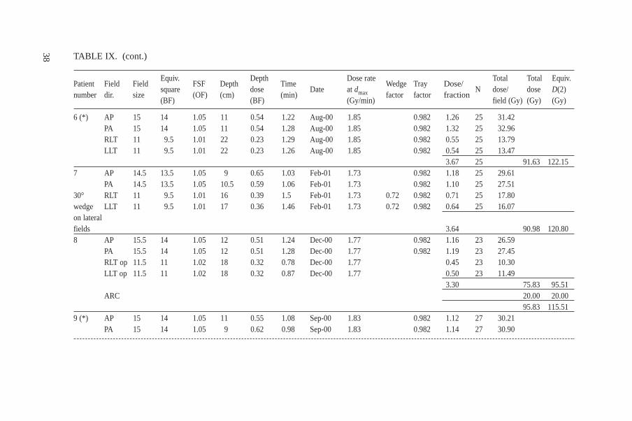
38 TABLE IX. (cont.)
Patient Field FieldEquiv.
FSF DepthDepth
Time Dose rate
Wedge Tray Dose/ Total Total Equiv.
number dir. size square
(OF) (cm)dose
(min)Date at dmax factor factor fraction
N dose/ dose D(2) (BF) (BF) (Gy/min) field (Gy) (Gy) (Gy)
6 (*) AP 15 14 1.05 11 0.54 1.22 Aug-00 1.85 0.982 1.26 25 31.42PA 15 14 1.05 11 0.54 1.28 Aug-00 1.85 0.982 1.32 25 32.96RLT 11 9.5 1.01 22 0.23 1.29 Aug-00 1.85 0.982 0.55 25 13.79LLT 11 9.5 1.01 22 0.23 1.26 Aug-00 1.85 0.982 0.54 25 13.47
3.67 25 91.63 122.157 AP 14.5 13.5 1.05 9 0.65 1.03 Feb-01 1.73 0.982 1.18 25 29.61
PA 14.5 13.5 1.05 10.5 0.59 1.06 Feb-01 1.73 0.982 1.10 25 27.5130° RLT 11 9.5 1.01 16 0.39 1.5 Feb-01 1.73 0.72 0.982 0.71 25 17.80wedge LLT 11 9.5 1.01 17 0.36 1.46 Feb-01 1.73 0.72 0.982 0.64 25 16.07on lateral fields 3.64 90.98 120.808 AP 15.5 14 1.05 12 0.51 1.24 Dec-00 1.77 0.982 1.16 23 26.59
PA 15.5 14 1.05 12 0.51 1.28 Dec-00 1.77 0.982 1.19 23 27.45RLT op 11.5 11 1.02 18 0.32 0.78 Dec-00 1.77 0.45 23 10.30LLT op 11.5 11 1.02 18 0.32 0.87 Dec-00 1.77 0.50 23 11.49
3.30 75.83 95.51ARC 20.00 20.00
95.83 115.519 (*) AP 15 14 1.05 11 0.55 1.08 Sep-00 1.83 0.982 1.12 27 30.21
PA 15 14 1.05 9 0.62 0.98 Sep-00 1.83 0.982 1.14 27 30.90

39
TABLE IX. (cont.)
Patient Field FieldEquiv.
FSF DepthDepth
Time Dose rate
Wedge Tray Dose/ Total Total Equiv.
number dir. size square
(OF) (cm)dose
(min)Date at dmax factor factor fraction
N dose/ dose D(2) (BF) (BF) (Gy/min) field (Gy) (Gy) (Gy)
RLT 11 9.5 1.01 20 0.27 1.12 Sep-00 1.83 0.982 0.55 27 14.80LLT 11 9.5 1.01 20 0.27 0.97 Sep-00 1.83 0.982 0.47 27 12.81
3.29 88.73 111.5510 (*) AP 15 13.5 1.05 15.5 0.43 1.47 Sep-00 1.83 0.982 1.18 17 20.05
PA 15 13.5 1.05 11.5 0.55 1.2 Sep-00 1.83 0.982 1.24 17 21.14RLT 11 9.5 1.01 21.5 0.25 1.37 Sep-00 1.83 0.982 0.61 17 10.34LLT 11 9.5 1.01 21.5 0.25 1.35 Sep-00 1.83 0.982 0.60 17 10.19
3.63 61.72 81.85RPO 10.5 10.5 1.01 20.5 0.29 1.23 Sep-00 1.83 0.64 8 5.15RAO 10.5 10.5 1.01 16.5 0.38 1.23 Sep-00 1.83 0.85 8 6.78LAO 10.5 10.5 1.01 17 0.37 1.23 Sep-00 1.83 0.82 8 6.59LPO 10.5 10.5 1.01 20.5 0.29 1.23 Sep-00 1.83 0.64 8 5.15
2.96 23.67 28.2185.39 110.05
11 AP 15 13.5 1.05 14 0.45 1.53 Sep-00 1.83 0.982 1.28 15 19.24PA 15 13.5 1.05 12 0.52 1.38 Sep-00 1.83 0.982 1.35 15 20.28RLT 11 9.5 1.01 20 0.27 1.43 Sep-00 1.83 0.982 0.70 15 10.49LLT 11 9.5 1.01 20 0.27 1.5 Sep-00 1.83 0.982 0.73 15 11.01
4.07 61.02 86.26RPO 10.5 10.5 1.01 23 0.22 1.31 Sep-00 1.83 0.53 8 4.27RAO 10.5 10.5 1.01 18 0.32 1.13 Sep-00 1.83 0.66 8 5.31

40 TABLE IX. (cont.)
Patient Field FieldEquiv.
FSF DepthDepth
Time Dose rate
Wedge Tray Dose/ Total Total Equiv.
number dir. size square
(OF) (cm)dose
(min)Date at dmax factor factor fraction
N dose/ dose D(2) (BF) (BF) (Gy/min) field (Gy) (Gy) (Gy)
LAO 10.5 10.5 1.01 18 0.32 1.13 Sep-00 1.83 0.66 8 5.31LPO 10.5 10.5 1.01 22 0.24 1.44 Sep-00 1.83 0.63 8 5.08
2.50 19.97 21.9580.99 108.21
12 (*) AP 14.5 13.5 1.05 10 0.58 1.06 Sep-00 1.83 0.982 1.15 20 23.05PA 14.5 13.5 1.05 10 0.58 1.06 Sep-00 1.83 0.982 1.15 20 23.05RLT 11 8.5 1.01 18.5 0.30 1.06 Sep-00 1.83 0.982 0.57 20 11.33LLT 11 8.5 1.01 17.5 0.30 0.98 Sep-00 1.83 0.982 0.52 20 10.48
3.40 67.92 86.88AP 14.5 13.5 1.05 10 0.58 0.41 Dec-00 1.83 0.982 0.45 10 4.46PA 14.5 13.5 1.05 10 0.58 0.41 Dec-00 1.83 0.982 0.45 10 4.46RLT 11 8.5 1.01 18.5 0.30 0.77 Dec-00 1.83 0.982 0.41 10 4.12LLT 11 8.5 1.01 17.5 0.30 0.77 Dec-00 1.83 0.982 0.41 10 4.12
1.71 17.15 16.1785.07 103.05
13 (*) AP 16 13 1.06 10.5 0.56 1.1 Nov-00 1.79 0.982 1.15 25 28.66PA 16 13 1.06 11 0.55 1.25 Nov-00 1.79 0.982 1.30 25 32.58RLT 12 12 1.02 20.5 0.28 0.81 Nov-00 1.79 0.41 25 10.34LLT 12 12 1.02 19 0.31 0.72 Nov-00 1.79 0.40 25 10.11
3.27 81.69 102.4014 (*) AP 15 14.5 1.05 16.5 0.38 1.47 Sep-00 1.83 0.982 1.05 17 17.89
PA 15 14.5 1.05 12.5 0.50 1.22 Sep-00 1.83 0.982 1.15 17 19.61

41
TABLE IX. (cont.)
Patient Field FieldEquiv.
FSF DepthDepth
Time Dose rate
Wedge Tray Dose/ Total Total Equiv.
number dir. size square
(OF) (cm)dose
(min)Date at dmax factor factor fraction
N dose/ dose D(2) (BF) (BF) (Gy/min) field (Gy) (Gy) (Gy)
RLT 11 9 1.01 21 0.25 1.42 Sep-00 1.83 0.982 0.63 17 10.72LLT 11 9 1.01 20.5 0.26 1.32 Sep-00 1.83 0.982 0.62 17 10.57
3.46 58.79 75.94RPO 10.5 10.5 1.01 23 0.22 1.5 Sep-00 1.83 0.61 8 4.85RAO 10.5 10.5 1.01 20 0.28 1.5 Sep-00 1.83 0.77 8 6.13LAO 10.5 10.5 1.01 20 0.28 1.5 Sep-00 1.83 0.77 8 6.13LPO 10.5 10.5 1.01 21 0.25 1.23 Sep-00 1.83 0.56 8 4.52
2.70 21.61 24.6580.41 100.59
15 (*) AP 14.5 12.5 1.05 13.5 0.46 1.47 Sep-00 1.83 0.982 1.25 16 20.06PA 14.5 12.5 1.05 13 0.48 1.33 Sep-00 1.83 0.982 1.18 16 18.95RLT 11 9 1.01 21 0.25 1.11 Sep-00 1.83 0.982 0.49 16 7.88LLT 11 9 1.01 20.5 0.26 1.11 Sep-00 1.83 0.982 0.52 16 8.27
3.45 55.17 71.15RPO 10.5 10.5 1.01 17 0.35 1.14 Sep-00 1.83 0.72 9 6.50RAO 9.5 9.5 1.00 16.5 0.35 1.15 Sep-00 1.83 0.73 9 6.59LAO 9.5 9.5 1.00 17 0.34 0.97 Sep-00 1.83 0.60 9 5.36LPO 10.5 10.5 1.01 18 0.32 1.14 Sep-00 1.83 0.67 9 6.03
2.72 24.48 28.0079.65 99.15
16 (*) AP 15 14 1.05 14.5 0.43 1.25 Sep-00 1.83 0.982 1.00 18 18.01PA 15 14 1.05 13 0.48 1.25 Sep-00 1.83 0.982 1.13 18 20.35

42 TABLE IX. (cont.)
Patient Field FieldEquiv.
FSF DepthDepth
Time Dose rate
Wedge Tray Dose/ Total Total Equiv.
number dir. size square
(OF) (cm)dose
(min)Date at dmax factor factor fraction
N dose/ dose D(2) (BF) (BF) (Gy/min) field (Gy) (Gy) (Gy)
RLT 11 10.5 1.01 22.5 0.24 1.33 Sep-00 1.83 0.982 0.57 18 10.19LLT 11 10.5 1.01 18.5 0.31 0.93 Sep-00 1.83 0.982 0.51 18 9.25
3.21 57.81 71.81RPO 11 11 1.01 22.5 0.24 1.02 Sep-00 1.83 0.44 7 3.10RAO 11 11 1.01 16 0.37 1.54 Sep-00 1.83 1.05 7 7.36LAO 11 11 1.01 16.5 0.35 1.23 Sep-00 1.83 0.79 7 5.56LPO 11 11 1.01 20.5 0.27 1.02 Sep-00 1.83 0.51 7 3.56
2.80 19.57 22.6977.38 94.50
17 AP 15.5 13 1.05 11.5 0.53 1.14 Dec-00 1.77 0.982 1.10 25 27.61PA 15.5 13 1.05 12 0.51 1.15 Dec-00 1.77 0.982 1.07 25 26.81RLT 12 10.5 1.02 17 0.35 0.79 Dec-00 1.77 0.50 25 12.47LLT 12 10.5 1.02 16 0.37 0.75 Dec-00 1.77 0.50 25 12.51
3.18 79.40 98.0718 AP 16.5 14 1.06 8 0.67 0.8 Sep-00 1.83 0.982 1.01 30 30.41
PA 16.5 14 1.06 12.5 0.49 0.94 Sep-00 1.83 0.982 0.88 30 26.33RLT 13.5 12.5 1.04 18 0.34 0.56 Sep-00 1.83 0.982 0.35 30 10.45LLT 13.5 12.5 1.04 16 0.38 0.67 Sep-00 1.83 0.982 0.48 30 14.30
2.72 81.48 93.1519 (*) AP 14 13 1.04 9.5 0.61 0.97 Sep-00 1.83 0.982 1.06 23 24.42
PA 14 13 1.04 9 0.63 0.97 Sep-00 1.83 0.982 1.09 23 25.02
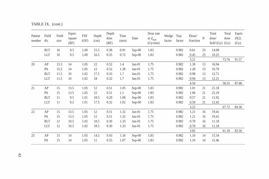
43
TABLE IX. (cont.)
Patient Field FieldEquiv.
FSF DepthDepth
Time Dose rate
Wedge Tray Dose/ Total Total Equiv.
number dir. size square
(OF) (cm)dose
(min)Date at dmax factor factor fraction
N dose/ dose D(2) (BF) (BF) (Gy/min) field (Gy) (Gy) (Gy)
RLT 10 9.5 1.00 15.5 0.38 0.91 Sep-00 1.83 0.982 0.61 23 14.08LLT 10 9.5 1.00 16.5 0.35 0.72 Sep-00 1.83 0.982 0.45 23 10.25
3.21 73.76 91.5720 AP 15.5 14 1.05 12 0.52 1.4 Jan-01 1.75 0.982 1.30 13 16.94
PA 15.5 14 1.05 12 0.52 1.38 Jan-01 1.75 0.982 1.28 13 16.70RLT 11.5 10 1.02 17.5 0.33 1.7 Jan-01 1.75 0.982 0.98 13 12.71LLT 11.5 10 1.02 18 0.32 1.7 Jan-01 1.75 0.982 0.94 13 12.21
4.50 58.55 87.8821 AP 15 13.5 1.05 12 0.51 1.05 Sep-00 1.83 0.982 1.01 21 21.18
PA 15 13.5 1.05 12 0.51 1.1 Sep-00 1.83 0.982 1.06 21 22.19RLT 11 9.5 1.01 18.5 0.29 1.08 Sep-00 1.83 0.982 0.57 21 11.92LLT 11 9.5 1.01 17.5 0.32 1.02 Sep-00 1.83 0.982 0.59 21 12.42
3.22 67.72 84.3022 AP 15 13.5 1.05 12 0.51 1.32 Jan-01 1.75 0.982 1.21 16 19.41
PA 15 13.5 1.05 12 0.51 1.32 Jan-01 1.75 0.982 1.21 16 19.41RLT 12 10.5 1.02 18.5 0.30 1.33 Jan-01 1.75 0.982 0.70 16 11.18LLT 12 10.5 1.02 18.5 0.30 1.33 Jan-01 1.75 0.982 0.70 16 11.18
3.82 61.18 83.5023 AP 15 14 1.05 14.5 0.43 1.34 Sep-00 1.83 0.982 1.10 14 15.34
PA 15 14 1.05 11 0.55 1.07 Sep-00 1.83 0.982 1.10 14 15.46

44 TABLE IX. (cont.)
Patient Field FieldEquiv.
FSF DepthDepth
Time Dose rate
Wedge Tray Dose/ Total Total Equiv.
number dir. size square
(OF) (cm)dose
(min)Date at dmax factor factor fraction
N dose/ dose D(2) (BF) (BF) (Gy/min) field (Gy) (Gy) (Gy)
RLT 11 10 1.01 20 0.27 0.81 Sep-00 1.83 0.982 0.40 14 5.63LLT 11 10 1.01 20 0.27 0.81 Sep-00 1.83 0.982 0.40 14 5.63
3.00 42.06 50.51RPO 10 10 1.00 15.5 0.38 1.33 Sep-00 1.83 0.91 11 10.02RAO 10 10 1.00 17.5 0.33 0.61 Sep-00 1.83 0.36 11 3.98LAO 10 10 1.00 17.5 0.33 1.34 Sep-00 1.83 0.80 11 8.75LPO 10 10 1.00 15.5 0.38 0.61 Sep-00 1.83 0.42 11 4.60
2.49 27.36 30.0269.42 80.53
24 AP 15 13.5 1.05 11.5 0.52 1.32 Sep-00 1.83 0.982 1.29 11 14.22PA 15 13.5 1.05 13 0.48 1.61 Sep-00 1.83 0.982 1.44 11 15.85RLT 11 9.5 1.01 22.5 0.23 1.18 Sep-00 1.83 0.982 0.49 11 5.41LLT 11 9.5 1.01 23 0.22 1.2 Sep-00 1.83 0.982 0.48 11 5.26
3.70 40.74 54.63RPO 10.5 10.5 1.01 23 0.23 1.38 Sep-00 1.83 0.57 7 3.99RAO 10.5 10.5 1.01 16.5 0.35 1 Sep-00 1.83 0.65 7 4.55LAO 10.5 10.5 1.01 17 0.34 1 Sep-00 1.83 0.63 7 4.40LPO 10.5 10.5 1.01 23 0.23 1.38 Sep-00 1.83 0.57 7 3.99
2.42 16.93 18.3457.67 72.97

45
TABLE IX. (cont.)
Patient Field FieldEquiv.
FSF DepthDepth
Time Dose rate
Wedge Tray Dose/ Total Total Equiv.
number dir. size square
(OF) (cm)dose
(min)Date at dmax factor factor fraction
N dose/ dose D(2) (BF) (BF) (Gy/min) field (Gy) (Gy) (Gy)
25 (*) AP 16 13 1.06 11.5 0.52 0.82 Dec-00 1.77 0.982 0.78 18 14.03(Off-axis PA 16 13 1.06 12.5 0.49 1.03 Dec-00 1.77 0.982 0.92 18 16.60contour RLT 10.5 10.5 1.01 17.5 0.33 0.78 Dec-00 1.77 0.46 18 8.23–3.5 cm) LLT 10.5 10.5 1.01 17 0.34 0.78 Dec-00 1.77 0.48 18 8.56
2.63 47.42 53.4426 AP 15.5 13 1.05 10.5 0.57 1.19 Feb-01 1.73 0.982 1.21 15 18.19
PA 15.5 13 1.05 10.5 0.57 1.19 Feb-01 1.73 0.982 1.21 15 18.1930° RLT 11 8.5 1.01 18.5 0.30 1.68 Feb-01 1.73 0.72 0.982 0.61 15 9.17wedge LLT 11 8.5 1.01 19 0.28 1.73 Feb-01 1.73 0.72 0.982 0.61 15 9.09on lateralfields 3.64 54.63 72.5827 AP 15 13.5 1.05 8.5 0.64 0.96 Feb-01 1.73 0.982 1.10 17 18.74
PA 15 13.5 1.05 9.5 0.60 0.96 Feb-01 1.73 0.982 1.03 17 17.46RLT 12 9 1.02 16 0.36 1.22 Feb-01 1.73 0.72 0.982 0.55 17 9.31LLT 12 9 1.02 16.5 0.35 1.22 Feb-01 1.73 0.72 0.982 0.53 17 9.05
3.21 54.56 67.7528 AP 16.5 15 1.06 11 0.55 1.11 Nov-00 1.79 0.982 1.14 6 6.87
PA 16.5 15 1.06 10.5 0.58 1.11 Nov-00 1.79 0.982 1.19 6 7.14RLT 13 12 1.03 19.5 0.29 0.84 Nov-00 1.79 0.45 6 2.69LLT 13 12 1.03 17.5 0.34 0.84 Nov-00 1.79 0.53 5 2.65
3.31 19.35 24.44
Note: An asterisk indicates that the patient received an additional brachytherapy treatment, and therefore the doses to the bladder and rectum are higher than the valuesin this table.

5.4.4. Other patients treated in the same period
The patients affected by this event are assumed to be the 28 identified as havingbeen treated with the entry of multiple block data as if they were a single block,describing two loops in the same direction. For other patients whose treatment planswere not obtained using this method, the treatment times were assumed to be correct,i.e. the time needed to deliver the prescribed dose was not affected by the incorrectcalculations.
During the mission it was not possible to evaluate all patients treated in thesame period (about 500 patients) and only a spot check was performed on ten patientstreated for prostate cancer and cancers of the cervix. A full dose determination wasnot done but only the treatment times were checked, These are listed in the lastcolumn in Table X, which shows that the treatment times for these patients were abouthalf of the treatment times used for the 28 overexposed patients, as can be seen fromTable IX.
46
TABLE X. REVIEW OF CHARTS AND TREATMENT TIMES FOR SOMEOTHER SELECTED PATIENTS WHO WERE TREATED USING THE TPS ATTHE ION BETWEEN JULY 2000 AND FEBRUARY 2001
Patient No. Cancer DateTreatment times (min)
for fields treated
A Prostate 20 Jul.–30 Aug. 2000 0.7, 0.7, 0.3, 0.25B Prostate 11 Jul.–11 Aug. 2000 0.6, 0.6, 0.6, 0.6C Prostate, 31 Jul.–4 Sep. 2000 0.6, 0.7, 0.5, 0.5
4 blocks usedD Prostate 24 Jul.–25 Aug. 2000 0.6, 0.6, 0.6, 0.9E Prostate 10 Oct.–15 Nov. 2000 0.5, 0.6, 0.6, 0.5F Prostate 23 Oct.–29 Nov. 2000 0.6, 0.7, 0.5, 0.5G Prostate 27 Oct.–6 Dec. 2000 0.6, 0.7, 0.6, 0.5H Cervix 30 Aug.–6 Oct. 2000 0.8, 0.8, 0.8, 0.8I Cervix, 14 Aug.–19 Sep. 2000 0.6, 0.8, 0.5, 0.6
4 blocks used J Cervix 6 Feb.–14 March 2000 0.8, 0.6, 0.8, 0.8
Note: These values are related to the individual fields.
6. TEST OF THE COMPUTER SOFTWARE USINGDIFFERENT APPROACHES FOR DATA ENTRY
The evaluation in Section 5 confirmed that the input data used by IONphysicists for computer calculations were valid in the cases tested. It also showed thatthe TPS gave accurate output in these cases, which included open fields, wedgedfields and fields with shielding blocks. The purpose of the tests described below wasto further investigate the performance of the TPS in dose calculations for blockedfields, in particular for the entry of multiple blocks as a single block with a doubleloop.
6.1. INSTRUCTIONS AND WARNINGS
The manufacturer’s disclaimer in the TPS manual [16] states that the TPSRTP/2 is a “DECISION SUPPORT SYSTEM, designed to evaluate possible dosecalculations based on the user supplied radiation beam and patient geometric data aswell as user selected algorithms and control parameters”. It further states that “it isthe responsibility of the user to validate any RESULTS obtained with the system andCAREFULLY check if data, algorithms and settings are meaningful, correct orapplicable, PRIOR to using the results as a part of the decision making process todevelop, define or document a course or treatment. In particular, a USER SHOULDVERIFY THE RESULTS OBTAINED THROUGH INDEPENDENT MEANS ANDEVALUATE ANY DISCREPANCIES CAREFULLY until the USER’SPROFESSIONAL CRITERIA HAS BEEN SATISFIED.”
The team examined the instructions in the manual relating to the blocked fieldsin order to understand the method of entering block data into the system. The manualstates, in the section entitled ‘Beam Handling and Field Set up’, subsection ‘Block’,this function allows for entry of up to 4 blocks of 32 points per beam” and “once theblock is nearly finished, strike enter to close the block contour”; however, noinstructions on how to digitize the block contours are given.
Later in the manual, the section on computational methods describes thealgorithm for block calculation. The manual states in this section, instead of in thesection giving instructions for data entry, that “each block is considered to be aseparate field outline and the outline entered into the system is automatically clippedto the rectangle defined by the collimator jaws”. In the section ‘Block Calculations’,it states that “the calculation is symmetric with respect to whether the outline goesclockwise or counter clockwise”. This, in summary, is the information available to theuser, placed in different sections of the manual, from which he/she can infer how toenter the blocks.
47
6.2. TESTS PERFORMED
Following the instructions described above, the user may conclude that blockco-ordinates should be digitized separately for each individual block, as shown inFig. 6. However, as shown in Section 3 of this report, it is also possible to enter theco-ordinates of several blocks as a single block, as shown in Fig. 7, completing theinner loop with a loop over the contour of the initial open field. In this case, the userreceives no warning that he/she is violating the instructions.
In order to investigate the effect of the procedure of entering block co-ordinatesinto the TPS, the following study was carried out using the ‘External Beams’ option.The first calculation was done for an open 15 cm × 15 cm field, with the central axisof the beam perpendicular to the phantom surface. The TPS printout of the dosedistribution for the central plane of the field is given in Fig. 8. An arrow shows theposition of the 50% isodose.
The next calculation was performed for a 15 cm × 15 cm field beam with fourshielding blocks with 5% transmission, i.e. the dose from radiation passing the blockis 5% of the dose without the block (this transmission value is typically used inradiotherapy and was employed at the ION). The co-ordinates of each block weredigitized separately, as shown in Fig. 6. The transmission factor was also entered foreach block separately. The resulting dose distribution for the central plane of the fieldis given in Fig. 9. This shows the calculated dose distribution for the blocked field,with a diagram indicated by an arrow showing the blocks and their positions in thefield. Figure 10 shows the dose distributions for open and blocked fields: it is clearthat the inclusion of blocks in the corners of the field has not significantly influencedthe dose distribution in the central plane of the beam. The 50% isodose line is atapproximately the same depth in both cases.
The same arrangement of blocks with the same transmission was used in thethird calculation, but this time the co-ordinates of the blocks were digitized as a singleblock describing two loops, as shown in Fig. 6. The calculated dose distribution isshown in Fig. 11. The diagram on the right of the figure again shows the blocks andtheir positions in the field. Note that the diagram shows the shielding blocks correctlypositioned. A comparison of the dose distributions for the two methods of enteringblock data into the TPS is given in Fig. 12. This shows a dramatic change in dosedistribution for the case when block contours were entered as if it were a single block,describing two loops in the same direction. In this case the 50% isodose is muchnearer the surface of the phantom, demonstrating a steeper decrease of the depth doseas compared with the previous calculation.
It is possible to enter the co-ordinates of the multiple blocks as for a singleblock, but to make the second loop in the opposite direction to the first one, as shownin Fig. 13. In this case the TPS produces the dose distribution shown in Fig. 14, againwith the diagram of the block positions to the right of the figure. This dose
48

49
FIG. 6. Digitizing the co-ordinates of each block separately. The shaded areas denote theblocks.
FIG. 7. Digitizing co-ordinates of multiple blocks as a single block describing two loops inthe same direction. The shaded areas denote the blocks.

50
FIG. 8. Isodose distribution in the central plane for an open field (arrow shows position of the 50% isodose).

51
FIG. 9. Isodose distribution in the central plane for a blocked field when the co-ordinates of each block were digitized separately (the arrow at thecentre shows the position of the 50% isodose; the arrow to the right shows the diagram of the block’s position).

52
FIG. 10. Comparison of isodose distributions in the central plane for open field (A) and for a blocked field when the co-ordinates of each block weredigitized separately (B) (the arrow at the centre shows the position of the 50% isodose).
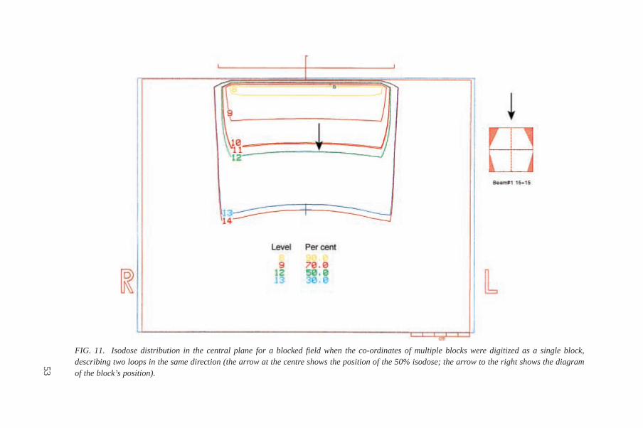
53
FIG. 11. Isodose distribution in the central plane for a blocked field when the co-ordinates of multiple blocks were digitized as a single block,describing two loops in the same direction (the arrow at the centre shows the position of the 50% isodose; the arrow to the right shows the diagramof the block’s position).

54
FIG. 12. Comparison of isodose distributions in the central plane for a blocked field when the co-ordinates of each block were digitized separately(A), and for a blocked field when the co-ordinates of multiple blocks were digitized as a single block, describing two loops in the same direction (B)(the arrows in the centre show the position of the 50% isodose for each case).

distribution is very similar to the normal ones, i.e. the dose distribution for an openfield (Fig. 8), and for the case when the block data were entered separately for eachblock (Fig. 10). There is only a slight distortion of the penumbra region on the rightside of the phantom, as indicated by the arrow.
These results show that the TPS permits the entry of the co-ordinates in theblocks in several different ways. One specific way of entering the block co-ordinates,shown in Fig. 7, gives very different results than the others.
The next test was intended to find out whether it is the use of a double loop inthe same direction, irrespective of the number and size of the blocks, that causes theerror in the calculation. If only a small part of one corner of an open field is blocked,the block should have no significant influence on the dose distribution in the centralplane. Such a field is shown in Fig. 15, where a double loop entry of one block’s co-ordinates is outlined. The calculated dose distribution is presented in Fig. 16, againwith a diagram on the right showing the block and its position in the field. Theresulting dose distribution is clearly incorrect and very much similar to the resultspresented in Fig. 9. This demonstrates that a double loop entry causes an incorrectcalculation even when only co-ordinates of one block are digitized.
55
FIG. 13. Digitizing co-ordinates of multiple blocks as a single block, but with the loops inopposite directions (one loop clockwise and the other one counterclockwise).

56
FIG. 14. Isodose distribution in the central plane for a blocked field with the co-ordinates of multiple blocks digitized as a single block with the loopsin the opposite directions (one loop clockwise and the other one counterclockwise) (the arrow on the left shows the distortion of the penumbra; thearrow in the centre shows the position of the 50% isodose; the arrow on the right shows the diagram of the block’s position).
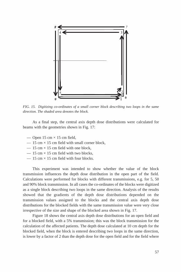
As a final step, the central axis depth dose distributions were calculated forbeams with the geometries shown in Fig. 17:
— Open 15 cm × 15 cm field,— 15 cm × 15 cm field with small corner block,— 15 cm × 15 cm field with one block,— 15 cm × 15 cm field with two blocks,— 15 cm × 15 cm field with four blocks.
This experiment was intended to show whether the value of the blocktransmission influences the depth dose distribution in the open part of the field.Calculations were performed for blocks with different transmissions, e.g. for 5, 50and 90% block transmission. In all cases the co-ordinates of the blocks were digitizedas a single block describing two loops in the same direction. Analysis of the resultsshowed that the gradients of the depth dose distributions depended on thetransmission values assigned to the blocks and the central axis depth dosedistributions for the blocked fields with the same transmission value were very closeirrespective of the size and shape of the blocked area shown in Fig. 17.
Figure 18 shows the central axis depth dose distributions for an open field andfor a blocked field, with a 5% transmission; this was the block transmission for thecalculation of the affected patients. The depth dose calculated at 10 cm depth for theblocked field, when the block is entered describing two loops in the same direction,is lower by a factor of 2 than the depth dose for the open field and for the field where
57
FIG. 15. Digitizing co-ordinates of a small corner block describing two loops in the samedirection. The shaded area denotes the block.
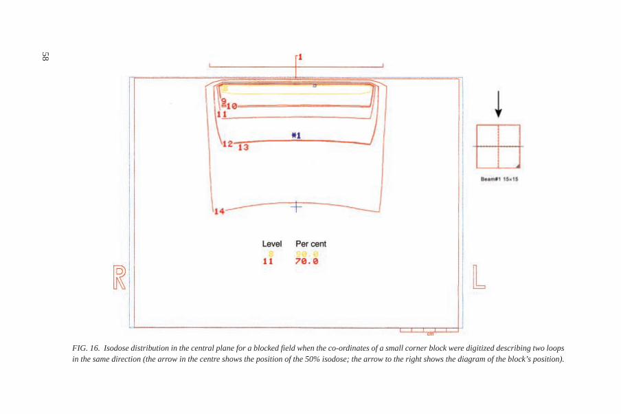
58
FIG. 16. Isodose distribution in the central plane for a blocked field when the co-ordinates of a small corner block were digitized describing two loopsin the same direction (the arrow in the centre shows the position of the 50% isodose; the arrow to the right shows the diagram of the block’s position).
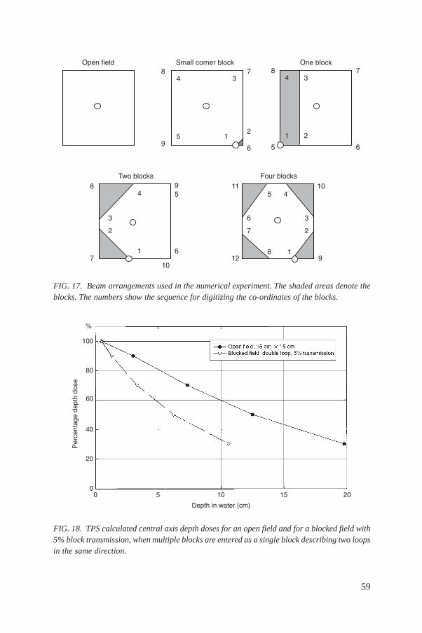
59
%�������� �� ������������& %������&
������&� '������&�
(
)
*
+
(
"
*
+
(
*
)
$,
$$
$
$,
)
#
"
!
$
!
#
$
"
(
#
$
"
+
!
!
+
*
#
$
FIG. 17. Beam arrangements used in the numerical experiment. The shaded areas denote theblocks. The numbers show the sequence for digitizing the co-ordinates of the blocks.
$,,
(,
+,
#,
,
,
-������ �
����
�����
��
.
, " $, $" ,
���������� ����/��0
FIG. 18. TPS calculated central axis depth doses for an open field and for a blocked field with5% block transmission, when multiple blocks are entered as a single block describing two loopsin the same direction.

the co-ordinates of the blocks are digitized separately. This effect causes the TPS tocalculate a doubled treatment time for the delivery of a prescribed dose when data areentered with a double loop, both loops being in the same direction.
The results of the tests of data entry into the TPS indicate that the use of doubleloop entry, with the two loops in the same direction, produces incorrect results, witha steeper gradient in the calculated depth dose distribution than the gradient in thedose distribution when the co-ordinates of the blocks are entered separately for eachblock.
7. MEDICAL ASSESSMENT
7.1. BACKGROUND INFORMATION ON RADIATION EFFECTS INHUMANS
Radiation effects in humans are caused by the deposition of energy in tissue.Sufficiently high doses of radiation cause the death of cells. Not all cells are of equalradiosensitivity. In general, cells with a high DNA content and rapidly dividing cellssuch as those in the testis, skin, epithelium of the pharynx and larynx, intestinal liningand bone marrow are the most sensitive. Blood vessels are of mediumradiosensitivity, and bone and brain are less sensitive.
Clearly, the pathological changes identified after irradiation of a given organdepend not only upon the physical parameters of the exposure, but also upon theradiosensitivity of the various organ components. In cases where the parenchymal(functional) cells of the organ are radiosensitive, loss of function of these cells will bethe initial critical factor, although later vascular compromise may become important.After substantial radiation exposure, failure of the organ system may result relativelyquickly. An example of an organ system with very sensitive parenchymal cells is thegastrointestinal system. Marked abnormalities can be seen within several days after arelatively moderate dose of radiation.
When the irradiated organ has parenchymal cells that are part of a slow renewalsystem, the controlling factor is the more sensitive connective tissue cells, such as themicrocirculation supplying blood to the parenchymal cells. An example of such anorgan is the brain.
One can divide the clinical manifestations of radiation injury into periods ofarbitrary length. The acute clinical period is the first six months; the subacute periodis 6–12 months; the chronic clinical period is 12 months to 5 years; with the lateclinical period being greater than five years after irradiation.
60
The acute clinical period includes the initial destructive processes and variousrepair processes of the organ system. Whether the organ actually survives depends onthe total dose, the volume of the organ irradiated, radiosensitivity and physical andchemical parameters. If the radiation dose is high enough, the tolerance of the organparenchyma will be exceeded, and failure of that system during the acute period willresult. With lesser exposures, there may be some parenchymal damage, and the organmay recover either full or partial function. If the parenchymal cells of the organ arerelatively radioresistant, no changes may be observed during the acute period.However, this should not be construed as indicating that no clinical radiation damagewill result. Vascular changes as a result of arteriolar narrowing may occur later withassociated complications. Vascular changes are often responsible for the limiting dosethat may be given to an otherwise radioresistant organ.
In the subacute period, underlying radiation damage in parenchymal cells maybecome manifest in terms of clinically significant problems. These may also becomplicated by vascular deterioration due to fibrosis, myointimal proliferation, andhyaline sclerosis of the subintimal and medial regions of small arteries and arterioles.
During the chronic clinical period, an organ system may demonstrate furtherdeterioration of vascularity and secondary degeneration of the parenchyma that canlead to decreased resistance to various sorts of stress.
In the late clinical period, there is a slow progression of residual radiationdamage and formation of dense fibrous tissue due to hypoperfusion. A major problemidentified in this late period is radiation carcinogenesis.
At the time of the team’s initial examination of the patients, they had alreadyexperienced the acute clinical period and part of the subacute period, but additionalchanges are to be expected.
7.2. BASIC RADIOTHERAPY PRINCIPLES RELEVANT TO THISACCIDENTAL EXPOSURE
Radiotherapy is the use of ionizing radiation to kill tumour cells in the body.Usually a radiation source outside the body is used to direct a beam of radiation to thearea of the tumour. The beam of radiation deposits energy in the tissues and kills cellsin the area of the beam but not elsewhere. The effects of radiation therefore will belimited to tissues or organs in the beam. As an example, a patient who wasoverexposed to the pelvis will not have headaches or arm pain as a result of radiation.
When the energy from an external radiation beam is deposited in tissues, cellsmay be damaged or killed, but the radiation exposure does not make the tissue or thepatient radioactive. It is completely safe for patients overexposed in this event to bearound other persons and even to hold children. An analogy would be someone whowas burned in a fire. Such patients do not burn other people when they touch them.
61
Sometimes patients and families of patients who were accidentally overexposed donot understand this principle and wonder whether it is safe for them to be near familymembers or children.
7.3. FRACTIONATION OF RADIATION EXPOSURE
Fractionation of radiation exposure, or protraction of the dose over a period oftime, almost always reduces the effect of the dose. Spreading a radiation dose out overdays or weeks allows time for the cells to repair radiation damage. In radiotherapy itis hoped that the normal tissue will repair faster than the tumour tissues, thus allowingthe tumour to be killed while normal tissues are less affected. If a radiation dose isvery protracted there is even less effect because not only is there repair, there is alsocellular repopulation due to continued cellular division. In this accidental exposure,the doses of each treatment were too high, and this resulted in less repair and cellularrepopulation (and more complications and adverse effects) than would have beenexpected from the doses prescribed by the radiation oncologist. The situation issimilar to a physician writing a prescription for two small pills a day of a somewhattoxic drug, but the pharmacy actually gives the patient two large pills per day.
Doses given in different fractionations can be compared in terms of the level oftissue effects they cause. In order to compare the effects of a treatment regimen (withan unusual dose per fraction) with that which would occur from a typical treatmentregimen of 2 Gy per fraction, it is necessary to calculate the 2 Gy per fractionequivalent dose, i.e. the total dose that would lead to the same biological effect ifgiven in fractions of 2 Gy each instead of the actual fractions delivered [17].
7.4. ADVERSE EFFECTS
It should be noted that even excellent radiotherapy practice cannot kill a tumourwithout causing some damage to normal tissues. There is a very narrow range ofdoses and numbers of treatments within which the radiation oncologist must work. Ifthe treating physician does not damage normal tissue to some extent, then there willbe very few tumours cured. In order to be able to cure tumours, radiation oncologistsuse doses that will result in some complications in about 5% of the patients (theacceptability depends on the type of complication). This is regarded as acceptablepractice in order to be able to cure cancers. However, a small increase in dose abovestandard protocols will result in a high complication rate.
The incidence of adverse radiation effects or complications at differentradiation therapy dose levels is well known for most tissues. This is expressed as a‘tolerance dose’ (TD), and the complications are expressed as the percentage of
62
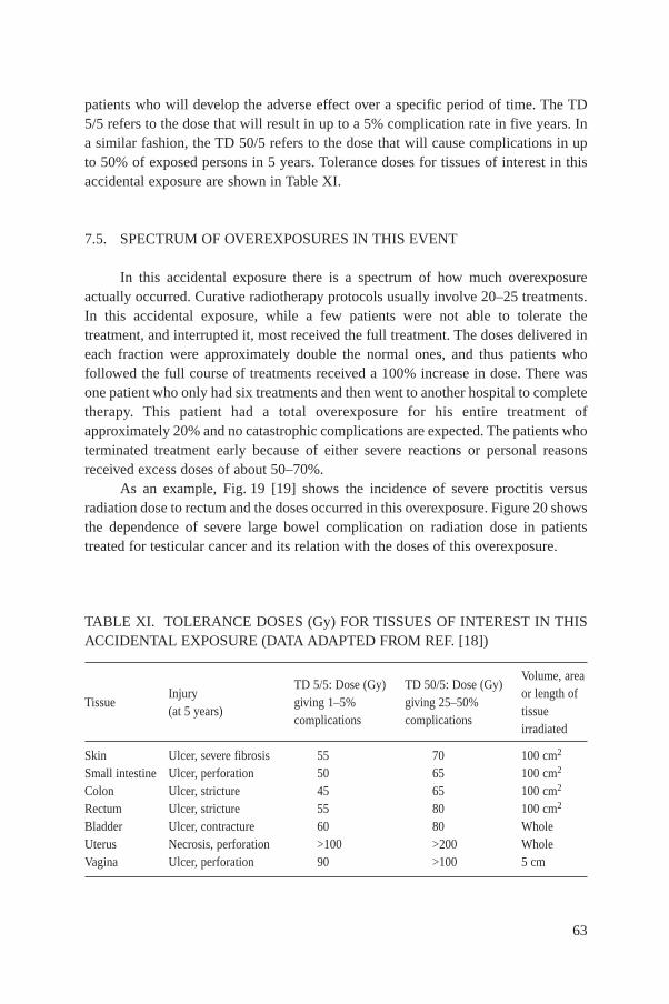
patients who will develop the adverse effect over a specific period of time. The TD5/5 refers to the dose that will result in up to a 5% complication rate in five years. Ina similar fashion, the TD 50/5 refers to the dose that will cause complications in upto 50% of exposed persons in 5 years. Tolerance doses for tissues of interest in thisaccidental exposure are shown in Table XI.
7.5. SPECTRUM OF OVEREXPOSURES IN THIS EVENT
In this accidental exposure there is a spectrum of how much overexposureactually occurred. Curative radiotherapy protocols usually involve 20–25 treatments.In this accidental exposure, while a few patients were not able to tolerate thetreatment, and interrupted it, most received the full treatment. The doses delivered ineach fraction were approximately double the normal ones, and thus patients whofollowed the full course of treatments received a 100% increase in dose. There wasone patient who only had six treatments and then went to another hospital to completetherapy. This patient had a total overexposure for his entire treatment ofapproximately 20% and no catastrophic complications are expected. The patients whoterminated treatment early because of either severe reactions or personal reasonsreceived excess doses of about 50–70%.
As an example, Fig. 19 [19] shows the incidence of severe proctitis versusradiation dose to rectum and the doses occurred in this overexposure. Figure 20 showsthe dependence of severe large bowel complication on radiation dose in patientstreated for testicular cancer and its relation with the doses of this overexposure.
63
TABLE XI. TOLERANCE DOSES (Gy) FOR TISSUES OF INTEREST IN THISACCIDENTAL EXPOSURE (DATA ADAPTED FROM REF. [18])
TD 5/5: Dose (Gy) TD 50/5: Dose (Gy) Volume, area
TissueInjury
giving 1–5% giving 25–50%or length of
(at 5 years)complications complications
tissueirradiated
Skin Ulcer, severe fibrosis 55 70 100 cm2
Small intestine Ulcer, perforation 50 65 100 cm2
Colon Ulcer, stricture 45 65 100 cm2
Rectum Ulcer, stricture 55 80 100 cm2
Bladder Ulcer, contracture 60 80 WholeUterus Necrosis, perforation >100 >200 WholeVagina Ulcer, perforation 90 >100 5 cm

64
FIG. 19. Incidence of severe proctitis versus radiation dose to the rectum (adapted from Shererand Streffer [17]) and its relation to the doses of this accidental exposure.
FIG. 20. Dependence of severe large bowel complications on radiation dose in patients treatedfor testicular cancer [19, 20] and its relation to the doses of this accidental exposure.
7.6. COMPLICATING FACTORS IN THE EVALUATION OF THIS EVENT
There are at least two complicating factors in the medical evaluation of both thesurviving and expired patients. In most cases, no portal films are available to show thelocation of the radiation treatment beam on the body, although pretreatmentradiographs are available for most patients. For patients who are still alive, the fieldlocation can almost always be ascertained by clinical examination of thesubcutaneous fibrosis, skin pigmentation and loss of hair.
A second complicating factor in the evaluation of some of the patients was thepresence of other conditions that increase radiation risk such as prior or subsequenttreatment either with surgery or chemotherapy. Usually the complications and sideeffects could be distinguished by knowledge of the effects of the specific drugs, thetime the effect occurred and knowledge of the type and timing of radiation effectsrelative to exposure. There were at least two patients with diabetes mellitus, which isknown to increase the incidence and severity of adverse radiation effects.
7.7. RESULTS OF THE MEDICAL TEAM’S INVESTIGATION
In this accidental exposure, methods of entering data were used with the TPSthat caused the computer to produce unusual isodose curves and calculate treatmenttimes that were longer than needed to give the prescribed dose to 28 patients.Therefore, longer times than appropriate were used in the daily treatments. Thisresulted in too much energy being deposited in both the normal tissues and thetumour. Overexposure resulted in excessive cell killing and also scarring and damageof the tissues and underlying blood vessels.
It should be noted that most of the approximately 500 patients treated betweenAugust 2000 and February 2001 appear to have received the correct doses. In additionto the incorrect data entry method, the technologists apparently did not notice that inpatients with similar diseases and treatment fields, the treatment times of somepatients were approximately twice those of others. Finally, some of the 28 patientshad inordinately severe early reactions, causing the treatment to be paused or evenhalted. It was only after this had happened a number of times, and there had beensome early deaths, that the cause was determined.
The summary data for both surviving and deceased patients are shown inTable XII. The table is organized by absorbed dose in descending order. Severecomplications are often seen when the 2 Gy per fraction equivalent dose exceeds65–70 Gy (Table XIII). Also, note that treatment times for most other patients withsimilar treatment at the same time was about 0.6 min per AP/PA fields (see TableA–III in Annex III).
65
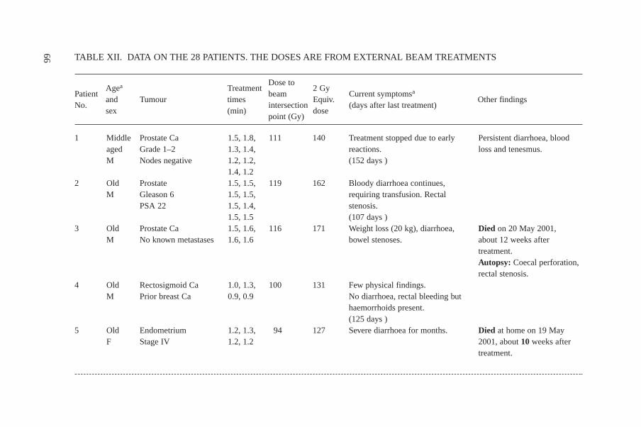
66
Agea TreatmentDose to
2 GyPatient
and Tumour timesbeam
Equiv.Current symptomsa
Other findingsNo.
sex (min)intersection
dose(days after last treatment)
point (Gy)
1
2
3
4
5
MiddleagedM
OldM
OldM
OldM
OldF
Prostate CaGrade 1–2Nodes negative
ProstateGleason 6PSA 22
Prostate Ca No known metastases
Rectosigmoid CaPrior breast Ca
EndometriumStage IV
1.5, 1.8,1.3, 1.4,1.2, 1.2,1.4, 1.21.5, 1.5,1.5, 1.5,1.5, 1.4,1.5, 1.51.5, 1.6,1.6, 1.6
1.0, 1.3,0.9, 0.9
1.2, 1.3,1.2, 1.2
111
119
116
100
94
140
162
171
131
127
Treatment stopped due to earlyreactions.(152 days )
Bloody diarrhoea continues,requiring transfusion. Rectalstenosis.(107 days )Weight loss (20 kg), diarrhoea,bowel stenoses.
Few physical findings. No diarrhoea, rectal bleeding buthaemorrhoids present.(125 days )Severe diarrhoea for months.
Persistent diarrhoea, bloodloss and tenesmus.
Died on 20 May 2001,about 12 weeks aftertreatment.Autopsy: Coecal perforation,rectal stenosis.
Died at home on 19 May2001, about 10 weeks aftertreatment.
TABLE XII. DATA ON THE 28 PATIENTS. THE DOSES ARE FROM EXTERNAL BEAM TREATMENTS
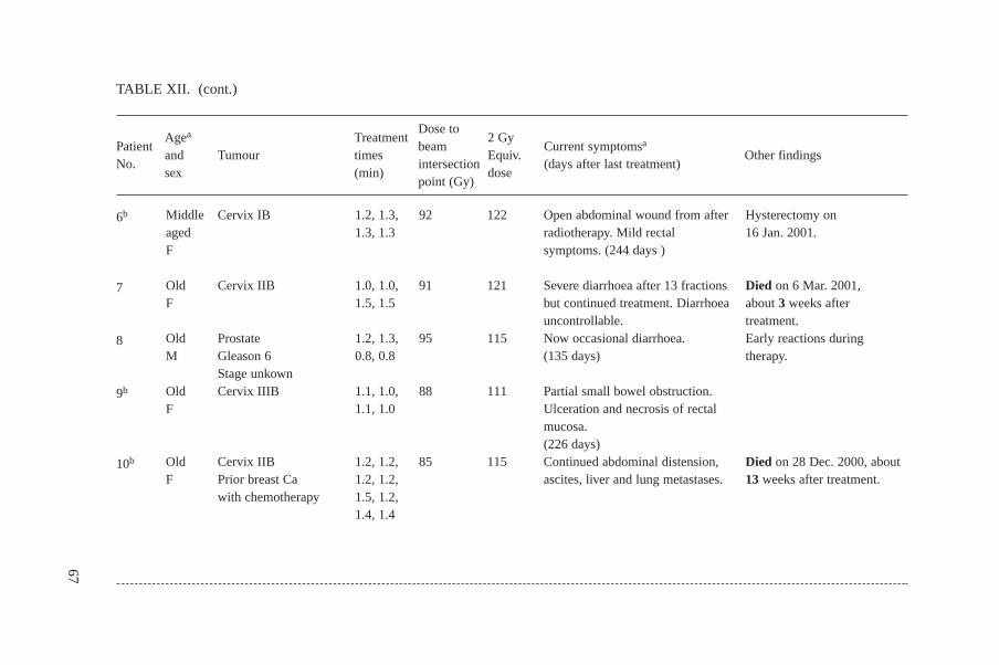
67
Agea TreatmentDose to
2 GyPatient
and Tumour timesbeam
Equiv.Current symptomsa
Other findingsNo.
sex (min)intersection
dose(days after last treatment)
point (Gy)
6b
7
8
9b
10b
MiddleagedF
OldF
OldM
OldF
OldF
Cervix IB
Cervix IIB
ProstateGleason 6Stage unkownCervix IIIB
Cervix IIBPrior breast Cawith chemotherapy
1.2, 1.3,1.3, 1.3
1.0, 1.0,1.5, 1.5
1.2, 1.3,0.8, 0.8
1.1, 1.0,1.1, 1.0
1.2, 1.2,1.2, 1.2,1.5, 1.2,1.4, 1.4
92
91
95
88
85
122
121
115
111
115
Open abdominal wound from afterradiotherapy. Mild rectalsymptoms. (244 days )
Severe diarrhoea after 13 fractionsbut continued treatment. Diarrhoeauncontrollable.Now occasional diarrhoea.(135 days)
Partial small bowel obstruction.Ulceration and necrosis of rectalmucosa.(226 days)Continued abdominal distension,ascites, liver and lung metastases.
Hysterectomy on 16 Jan. 2001.
Died on 6 Mar. 2001,about 3 weeks after treatment.Early reactions duringtherapy.
Died on 28 Dec. 2000, about13 weeks after treatment.
TABLE XII. (cont.)

68
Agea TreatmentDose to
2 GyPatient
and Tumour timesbeam
Equiv.Current symptomsa
Other findingsNo.
sex (min)intersection
dose(days after last treatment)
point (Gy)
11
12b
13b
14b
15b
MiddleagedF
MiddleagedF
MiddleagedF
MiddleagedF
OldF
Cervix IB
Cervix IIB
EndometriumStage IV
Cervix IIA
Endometrial CaStage IC
1.5, 1.4,1.4, 1.5,1.2, 1.3,1.3, 1.31.1, 1.1,1.1, 1.0,0.4, 0.4,0.8, 0.81.1, 1.2,0.8, 0.7
1.5, 1.5,1.2, 1.3,1.5, 1.5,1.5, 1.21.5, 1.3,1.1, 1.1,1.1, 1.2,1.0, 1.1
81
85
82
80
79
108
103
102
100
99
Perineal pain, constant diarrhoea,dyurea.(230 days)
Anaemia, constant bloodydiarrhoea. 15 kg weight loss.(233 days)
Recent onset of one episode ofbloody diarrhoea. No othersymptoms.(156 days )Diarrhoea since Jan. 2001. Colonic pain.(229 days)
Rectal bleeding, constipation,dysuria.(229 days)
Currently hospitalized.
Dysuria. Operation of bowel stenosis.
TABLE XII. (cont.)

69
Agea TreatmentDose to
2 GyPatient
and Tumour timesbeam
Equiv.Current symptomsa
Other findingsNo.
sex (min)intersection
dose(days after last treatment)
point (Gy)
16b
17b
18
19b
20
MiddleagedF
OldM
OldM
MiddleagedF
MiddleagedF
Endometrial CaStage I
Mulleriansarcoma
Rectal adeno Ca
Vaginal CaStage II
Cervix IB
1.3, 1.3,1.3, 0.9,1.0, 1.5,1.2, 1.01.1, 1.1,0.8, 0.7
0.8, 0.9,0.6, 0.7
1.0, 1.0,0.9, 0.7
1.4, 1.4,1.7, 1.7
77
79
81
74
59
94
98
93
91
88
Dysuria with frequency and lowvolume. Rectal mucous but nodiarrhoea, lower abdominal pain.(234 days)Persistent bloody diarrhoea severaltimes/day. Bladder and vaginalprolapse.(150 days)Constant diarrhoea that requiresdiapers.(233 days)Bloody diarrhoea since October.Anaemia requiring transfusions.Urinary frequency.(229 days)
Hysterectomy on 16 Nov.2000.
One episode of bleeding.
Several hospitalizations forenteritis and bloody diarrhoea.Died on 7 May 2001, about13 weeks after treatment.
TABLE XII. (cont.)
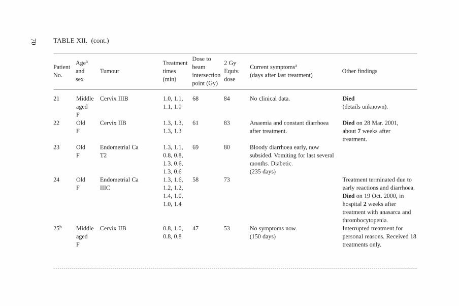
70
Agea TreatmentDose to
2 GyPatient
and Tumour timesbeam
Equiv.Current symptomsa
Other findingsNo.
sex (min)intersection
dose(days after last treatment)
point (Gy)
21
22
23
24
25b
MiddleagedFOldF
OldF
OldF
MiddleagedF
Cervix IIIB
Cervix IIB
Endometrial CaT2
Endometrial CaIIIC
Cervix IIB
1.0, 1.1,1.1, 1.0
1.3, 1.3,1.3, 1.3
1.3, 1.1,0.8, 0.8,1.3, 0.6,1.3, 0.61.3, 1.6,1.2, 1.2,1.4, 1.0,1.0, 1.4
0.8, 1.0,0.8, 0.8
68
61
69
58
47
84
83
80
73
53
No clinical data.
Anaemia and constant diarrhoeaafter treatment.
Bloody diarrhoea early, nowsubsided. Vomiting for last severalmonths. Diabetic.(235 days)
No symptoms now.(150 days)
Died(details unknown).
Died on 28 Mar. 2001,about 7 weeks after treatment.
Treatment terminated due toearly reactions and diarrhoea.Died on 19 Oct. 2000, inhospital 2 weeks aftertreatment with anasarca andthrombocytopenia.Interrupted treatment forpersonal reasons. Received 18treatments only.
TABLE XII. (cont.)
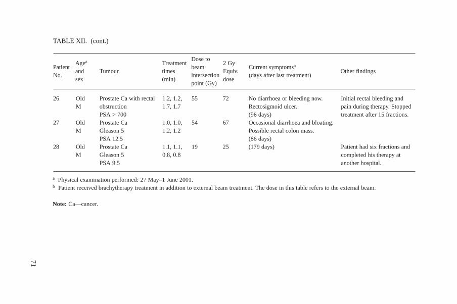
71
Agea TreatmentDose to
2 GyPatient
and Tumour timesbeam
Equiv.Current symptomsa
Other findingsNo.
sex (min)intersection
dose(days after last treatment)
point (Gy)
26
27
28
OldM
OldM
OldM
Prostate Ca with rectalobstructionPSA > 700Prostate CaGleason 5 PSA 12.5Prostate CaGleason 5 PSA 9.5
1.2, 1.2,1.7, 1.7
1.0, 1.0,1.2, 1.2
1.1, 1.1,0.8, 0.8
55
54
19
72
67
25
No diarrhoea or bleeding now.Rectosigmoid ulcer.(96 days)Occasional diarrhoea and bloating.Possible rectal colon mass.(86 days)(179 days)
Initial rectal bleeding andpain during therapy. Stoppedtreatment after 15 fractions.
Patient had six fractions andcompleted his therapy atanother hospital.
TABLE XII. (cont.)
a Physical examination performed: 27 May–1 June 2001.b Patient received brachytherapy treatment in addition to external beam treatment. The dose in this table refers to the external beam.
Note: Ca—cancer.

7.7.1. Surviving patients
The medical team examined all 20 surviving patients. They provided advice andconsultation to the treating physicians, patients and their families. The patients wereexamined by Drs Akashi, Cosset, Gourmelon, Mettler and Konchalovsky, who wereworking together. The prior findings, clinical charts and available imaging studieswere also reviewed at the same time.
In addition to physical examination, the team also employed a grading systemthat has been suggested for the evaluation of long term effects (LENT). This systemis useful in that it has detailed forms for various different tissues. This allowed asystematic and uniform assessment of the patients. A score can be derived for eachpatient but the usefulness of this score is not known at the present time.
The patients were initially assigned to one of five categories of expectedconsequences based upon clinical examinations alone, but this was later modifiedwhen the 2 Gy per fraction equivalent doses were calculated by team physicists. Theclassification is as follows:
*** Severe or catastrophic effects due to overexposure from the accidentalexposure.
** Marked effects due to overexposure from the accidental exposure and athigh risk for future effects.
72
TABLE XIII. COMPARISON OF FINDINGS AT ONE YEAR IN SIMILARLYOVEREXPOSED PATIENTS IN THE SAN JOSE, COSTA RICA, ACCIDENTALEXPOSURE
Patient IDField size 2 Gy per fraction
Findings(cm × cm) Equiv. dose
18 15 × 15 Massive colonic haemorrhage andperforation. Died before treatmentfinished.
44 10 × 10 70 Bloody diarrhoea, vaginal ulceration.57 29 × 23 39 GI haemorrhage.62 15 × 15 52 Rectal stenosis.83 ~15 × 15 77 Weight loss, continued rectal
bleeding, use of diapers.85 15 × 18 73 Perirectal ulceration, infection.
some necrosis.8 Several 59 Diarrhoea for three weeks which
subsided.

* Radiation effects from the accidental exposure not severe at this time andat low risk for future effects.
0 No radiation effects at this time felt to be attributed to the accidentalexposure. These patients may have radiation effects that would beexpected from standard radiotherapy protocols.
– Underexposure because radiotherapy was discontinued. At higher thannormal risk for tumour recurrence.
Those patients who were in the first three categories (***, ** and *) hadinjuries related to the specific body part irradiated and the sensitive tissues in that area(Table XIV). There were two general categories of effects as follows:
— Lower gasterointestinal: chronic or bloody diarrhoea, bowel stenosis, stricture,fibrosis, obstruction, fistula, perforation.
— Bladder: dysuria, haematuria, contracture, incontinence.
Unfortunately, since the major radiation injury is likely to be confined to therectum, rectosigmoid, ileum and bladder, external examination will tend tounderestimate the true extent of the injuries. Figures 21, 22 and 23 show effects seenin patients Nos 9, 22 and 11, respectively. The team has reviewed the LENT-SOMAgrading of these patients and the significant correlation of the calculated biologicallyeffective dose is with the rectal and small bowel colon grades. There is no significant
73
TABLE XIV. DISTRIBUTION OF EXPECTED RADIATION CHANGESCAUSED BY NORMAL RADIOTHERAPY PROTOCOLS AND BY THEEXPOSURES IN THIS ACCIDENTAL EXPOSURE
Category of complicationsDistribution for Distribution for the normal therapy surviving Panama cases a)
*** Catastrophic 0 2** Marked 1 7* Increased 5 90 No changes expected 90 2– Underexposed 4 0
Total 100% 20 cases
a Physical examination performed: 27 May–1 June 2000.

correlation between bladder or skin changes and the doses delivered (2 Gy perfraction equivalent doses to the intersection point). This is not unexpected, given thefact that some patients received treatments of four or even eight fields and the dose tothe skin and bladder may be lower than the dose to the intersection point of multiplebeams.
74
FIG. 21. X ray film of patient No. 9 showing dilated, air filled loops (arrows) of small bowelfrom a distal small bowel obstruction likely secondary to radiation induced stenosis.

7.7.2. Deceased Patients
Autopsy data were available for one of the eight patients who had died. Whereautopsy data were not available, the clinical charts were reviewed to attempt to cometo a conclusion about the possible role of radiation.
Patients who died were classified according to one of four categories asfollows:
— Radiation exposure felt to be the major cause of death,— Radiation overexposure possibly a significant contributor,— Death due to causes other than radiation overexposure,— Insufficient data at this time to make an informed judgement.
It should be pointed out that the team’s assessment was related to the cause ofdeath only. In the third category (not radiation related), there are some patients who
75
FIG. 22. Endoscopy of patient No. 22 showing ulcerations and destruction of mucosa(arrows).
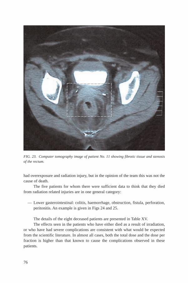
had overexposure and radiation injury, but in the opinion of the team this was not thecause of death.
The five patients for whom there were sufficient data to think that they diedfrom radiation related injuries are in one general category:
— Lower gasterointestinal: colitis, haemorrhage, obstruction, fistula, perforation,peritonitis. An example is given in Figs 24 and 25.
The details of the eight deceased patients are presented in Table XV.The effects seen in the patients who have either died as a result of irradiation,
or who have had severe complications are consistent with what would be expectedfrom the scientific literature. In almost all cases, both the total dose and the dose perfraction is higher than that known to cause the complications observed in thesepatients.
76
FIG. 23. Computer tomography image of patient No. 11 showing fibrotic tissue and stenosisof the rectum.
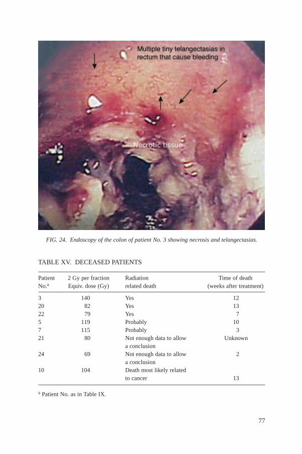
77
FIG. 24. Endoscopy of the colon of patient No. 3 showing necrosis and telangectasias.
TABLE XV. DECEASED PATIENTS
Patient 2 Gy per fraction Radiation Time of deathNo.a Equiv. dose (Gy) related death (weeks after treatment)
3 140 Yes 1220 82 Yes 1322 79 Yes 75 119 Probably 107 115 Probably 321 80 Not enough data to allow Unknown
a conclusion24 69 Not enough data to allow 2
a conclusion10 104 Death most likely related
to cancer 13
a Patient No. as in Table IX.

7.7.3. Sequence of events leading to death
It appears that there are two groups of patients who have had or will havedifferent sequences of events leading to death.
The first group died very rapidly, only a few weeks after irradiation. From thenecropsy findings and from the clinical charts, the most likely explanation seems tobe that :
— Their bowel mucosa were largely or totally destroyed.— As a consequence, the patients experienced severe (sometimes bloody)
diarrhoea, leading to dehydration, malnutrition, hypoalbuminaemia, anaemia,and severe impairment of their general condition with loss of weight.
— Because of the disappearance of the mucosal barrier, bowel bacteria migratedto the blood, resulting in septicaemia and septic shock (likely to be fatal inpatients in such an impaired clinical condition).
78
FIG. 25. Endoscopy of the colon of the same patient (No. 3) showing haemorrhage two daysbefore death.
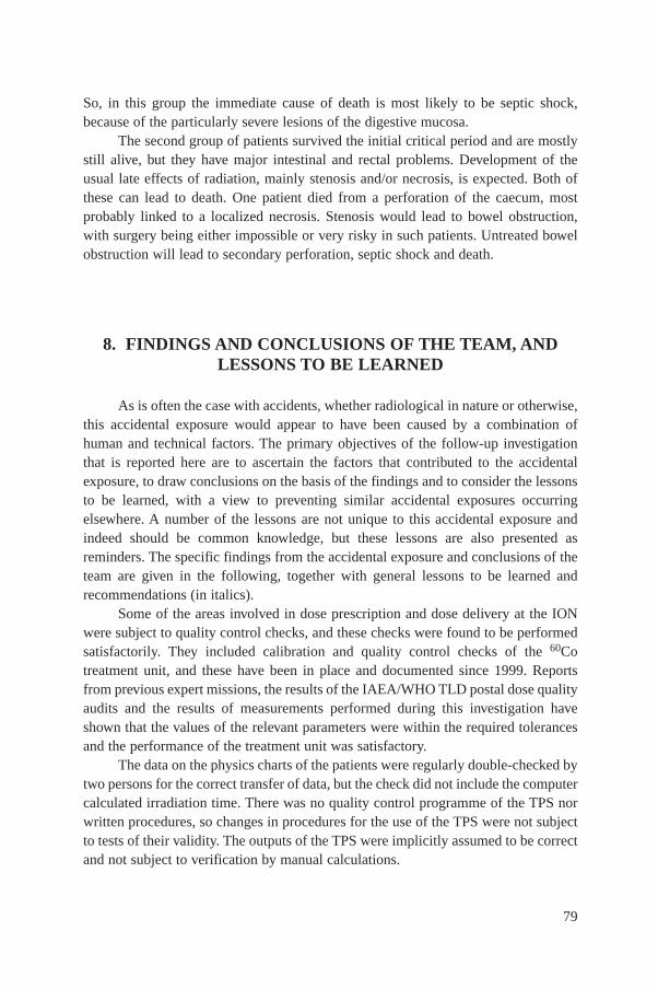
So, in this group the immediate cause of death is most likely to be septic shock,because of the particularly severe lesions of the digestive mucosa.
The second group of patients survived the initial critical period and are mostlystill alive, but they have major intestinal and rectal problems. Development of theusual late effects of radiation, mainly stenosis and/or necrosis, is expected. Both ofthese can lead to death. One patient died from a perforation of the caecum, mostprobably linked to a localized necrosis. Stenosis would lead to bowel obstruction,with surgery being either impossible or very risky in such patients. Untreated bowelobstruction will lead to secondary perforation, septic shock and death.
8. FINDINGS AND CONCLUSIONS OF THE TEAM, ANDLESSONS TO BE LEARNED
As is often the case with accidents, whether radiological in nature or otherwise,this accidental exposure would appear to have been caused by a combination ofhuman and technical factors. The primary objectives of the follow-up investigationthat is reported here are to ascertain the factors that contributed to the accidentalexposure, to draw conclusions on the basis of the findings and to consider the lessonsto be learned, with a view to preventing similar accidental exposures occurringelsewhere. A number of the lessons are not unique to this accidental exposure andindeed should be common knowledge, but these lessons are also presented asreminders. The specific findings from the accidental exposure and conclusions of theteam are given in the following, together with general lessons to be learned andrecommendations (in italics).
Some of the areas involved in dose prescription and dose delivery at the IONwere subject to quality control checks, and these checks were found to be performedsatisfactorily. They included calibration and quality control checks of the 60Cotreatment unit, and these have been in place and documented since 1999. Reportsfrom previous expert missions, the results of the IAEA/WHO TLD postal dose qualityaudits and the results of measurements performed during this investigation haveshown that the values of the relevant parameters were within the required tolerancesand the performance of the treatment unit was satisfactory.
The data on the physics charts of the patients were regularly double-checked bytwo persons for the correct transfer of data, but the check did not include the computercalculated irradiation time. There was no quality control programme of the TPS norwritten procedures, so changes in procedures for the use of the TPS were not subjectto tests of their validity. The outputs of the TPS were implicitly assumed to be correctand not subject to verification by manual calculations.
79
The initiating event of this accidental exposure was an attempt to obtain atreatment plan, with isodose calculations, for fields involving more shielding blocksthan the number for which the TPS had been designed. A method was found todigitize more than four blocks by entering the data into the TPS for multiple shieldingblocks together as if they were a single block. The output from the TPS using the newmethod of data entry was not verified and was not compared with the output producedusing the previous method. In addition, several characteristics of the TPS made iteasier for the error to occur:
— It is questionable whether the information in the instructions was sufficientlyclear to guide the user in detail on the manner in which the blocks should bedigitized.
— Several different methods of digitizing blocks were accepted by the computer.— There was no warning on the computer screen when blocks were digitized in a
manner different from the one described in the instructions.— When several blocks were digitized together as if they were a single block
describing two loops, the TPS produced a diagram which was the same as thatproduced when data were entered correctly, thereby giving the impression thatthe results were correct.
This method of digitizing the block data was used for a number of patientswithout any recognition of the longer than necessary times of exposure. Althoughsome symptoms of excessive exposure were noted, they were not effectivelyinvestigated, with the consequence that the accidental exposure went unnoticed for afurther number of months.
8.1. OPERATING ORGANIZATION: RADIOTHERAPY DEPARTMENTS
8.1.1. Quality assurance and radiotherapy
A single error in the method of entering data into the TPS led to the delivery ofwrong doses to patients and to severe, and in some cases fatal, consequences. Inradiotherapy, a single error can be fatal if it goes unnoticed. An efficient system fordetecting and correcting errors therefore needs to be in place: this implies a QAprogramme with sufficient double and independent checks.
A comprehensive QA programme needs to be in place in any radiotherapyfacility. In addition to the staff involved in the implementation of the programme, allhospital managers and administrators need to be made aware of this and of theconsequences of not having it, as part of their training.
80

A committee should be set up to exercise oversight of the QA programme. Thiscommittee should consider the completeness of the QA programme, the validity ofexisting and revised procedures and the results of quality control checks. The typicalcomposition of such a committee is: a radiation oncologist, a medical physicist, aradiotherapy technologist and an administrator representing the management of thehospital. The BSS place a strong emphasis on QA in radiotherapy.
8.1.2. Treatment planning systems as a safety issue
The accidental exposures involved the use of a computerized TPS. Externalaudits and the team mission confirmed that quality control protocols were properlyimplemented for the 60Co treatment units and that calibration was satisfactory, but nosuch protocols were in place for the computerized TPS.
Computerized TPSs for radiotherapy are to be regarded as safety relateddevices, for which a safety assessment and quality and safety measures are asimportant as they are for cobalt units and accelerators. The TPS needs to be includedin the QA programme.
8.1.3. Manual calculation check of the computer calculated plan
The staff performed double checks of the data transfer from the prescriptionand computer output into the patients’ treatment charts, but these checks did notinclude the treatment time calculated by the computer. It was implicitly assumed thatthe computer output was correct.
Results provided by the TPS need to be checked, and this should includeverification by manual calculation of the treatment time and dose to the selectedpoint. This verification should be part of the QA programme.
8.1.4. Changes in procedures
The method of entering the co-ordinates of shielding blocks into the TPS whencalculating dose distributions and irradiation times for particular treatments waschanged without subsequent testing. This led to incorrect results. Written proceduresrequiring approval, testing and documentation of any changes before they are adoptedwere not available at the ION.
Every step in the radiotherapy process should be reflected in the writtenprocedures. New procedures or changes in procedures should require formal testing,approval and documentation, as part of the QA programme.
81
8.1.5. Workload and team integration
The calculated treatment times for the affected patients were substantiallylonger than those for other patients with similar treatments. Although this wasabnormal, the professionals involved were not aware of it.
Individual and team awareness of the treatment arrangements for each patientare essential to ensure that abnormal situations are noticed quickly. The members ofthe team will normally be the radiation oncologist, the medical physicist and theradiotherapy technologist in charge of a particular patient. This team would beexpected to meet for a “new patient planning conference” [21], as well as during thesimulation of the treatment and when the patient is first set up for treatment. Theseoccasions provide opportunities to discuss the details of the treatment and to come toa common understanding of them, and they therefore present opportunities fordetecting any abnormal features or unusual values of parameters in the treatmentplan.
The level of individual and team awareness may have been inadequate at theION. The division of the team between two hospitals and the limited presence ofsome professionals may have contributed to some degree of fragmentation. This wasmade worse by the heavy workload involving some 70–80 patients being treated perday, many of them during the evenings with only a technologist present.
Pressure due to a heavy workload, if not properly managed, can result in areduction of quality and safety. It can cause the staff to perform in a more mechanicalmanner, reducing awareness and allowing indicators of abnormal situations to beoverlooked. Errors may ‘fall between the cracks’ instead of being picked up.
The workload should not result in a lowering of quality and safety standards.Staff should conform to the guidance provided by standards of good practice (usuallygiven by professional bodies) and their work should be kept under review andreexamined with regard to the workload (number of patients) and to any issue thatplaces an extra burden on them, such as the introduction of a new technique.
An integrated team approach to radiotherapy, combined with well definedindividual functions and responsibilities, should be part of the design andimplementation of a radiotherapy department.
8.1.6. Observation of unusual reactions of patients
Patients affected by the excessive dose showed unusual reactions, which weremore severe than those expected from the prescribed doses. The first review did notreveal the cause of the problem, and treatment of the patients continued or wasresumed.
Careful and frequent patient observation, followed up by comprehensiveinvestigation of the possible causes of unexpected symptoms, are indispensable for
82

the early discovery of errors and the mitigation of their consequences. Observationsof unexpected symptoms should be prompt and should include manual recalculationsand dose measurements on a phantom.
8.1.7. In vivo dosimetry
Errors in dose delivery can be detected by in vivo dosimetry, by the use of solidstate detectors placed on the patients. This provides evidence that the correct dose hasbeen delivered to a patient, and is therefore an additional level of defence in depth.
Implementation of in vivo dosimetry requires the allocation of resources interms of equipment, calibration of detectors, quality assurance and, most important,adequate training. These requirements are difficult to meet in some countries.Nevertheless, with appropriate planning and allocation of resources, in vivodosimetry can be implemented even in small radiotherapy departments [22], and thisis a desirable feature.
In vivo dosimetry should be promoted as far as practicable in radiotherapydepartments, but proper preparation for such a programme is necessary.
8.1.8. Local advice from the manufacturer/supplier
The users of the TPS were looking for a solution to an unusual request from thetreating oncologist in the use of the TPS. The co-operation and advice of themanufacturer/supplier in fulfilling this request was not sought.
For proper use of equipment, co-operation between the user and the supplier isessential, and this co-operation should be established from the time of purchase of theequipment. In addition, procedures not included in the manufacturer’s instructionsshould only be attemped in consultation with the manufacturer/supplier.
8.2. NATIONAL AUTHORITIES
8.2.1. Quality assurance
The regulations in Panama require a QA programme to be implemented for theclinical and physical parameters of radiotherapy. There was some quality control atthe hospital, but it was incomplete. It covered calibration and quality control of theirradiation unit, and was associated with adequate equipment calibration andperformance. The error that caused this accidental exposure was in an area which wasnot addressed by the quality control that was in place.
Inspections by the regulatory authority, and the external audit (in 1999), hadbeen carried out before the accidental exposure. The recommendations resulted in
83

better radiation protection and safety procedures within the department anddocumented calibration and quality control of the radiotherapy equipment.Unfortunately, procedures for the TPS were not developed.
A QA programme for radiotherapy should be a mandatory requirement in theregulations, and the requirement should be enforced (as was the case in Panama). Theprotocols used should be in accordance with well proven programmes developedeither at the national or regional level.
National authorities should promote external audits; recommendations arisingfrom the audits should be evaluated by a QA and radiation protection committee andimplementation should be closely followed up.
8.2.2. Communication between regulators and users of radiation
The records at the regulatory authority indicate that the ION was repeatedlyrequested to provide information on the QA procedures. Although the quality controlprotocols of IAEA-TECDOC-1151 [6] had been used for some time for calibrationand quality control of the 60Co unit, the ION does not seem to have responded to therequests of the regulatory authority for information.
Users of radiation should understand that they share a common objective withthe regulatory authority, which is safe operation, and that the purpose of monitoringcompliance with the regulatory requirements is oriented to that objective.
8.3. EQUIPMENT MANUFACTURERS AND SUPPLIERS
8.3.1. Software in treatment planning
In the TPS involved in this event, the manual of instructions does not describeprecisely how to digitize co-ordinates of shielding blocks and there are not enoughrelevant illustrations. In addition, it does not provide specific warning against dataentry approaches that are different from the one described. The indication on thismatter is vague, other than a general disclaimer at the beginning of the manual.
Instructions and explanations which do not make clear exactly what is and whatis not allowed leave open the possibility of users choosing an approach that was nottested by the manufacturer.
Software should be tested to ensure that it is as foolproof as possible. Theinstructions should guide the user explicitly and fully through the process, followingoptions that are allowed and have been tested, so as to prevent users from tryinganother method that may not have been tested by the manufacturer. Deviation fromthe steps given in the instructions should be prevented by a warning inserted both inthe instructions and on the computer screen display.
84
8.4. THE MEDICAL COMMUNITY
8.4.1. Findings
The accidental exposures at the ION in Panama were very serious. Many patientshave suffered severe radiation effects due to excessive dose. Both morbidity andmortality have increased significantly. This series of accidental exposures is unique.Previous radiation therapy accidental exposures that resulted in mortality had involvedexcessive doses of 30–50% more than prescribed. There are no reported previousaccidental expoures in which the doses delivered were 50–100% above prescribedradiotherapy doses, with all affected patients being treated in the pelvic region.
Not all of the radiation effects observed in these patients can be attributed to theaccidental dose. A few of the patients who received treatment at the time of theaccidental dose have radiation effects typical of those that would be expected fromthe prescribed doses. Some patients have pains or disorders that cannot be attributedto radiation. However, although some damage to normal tissue is expected as a resultof standard radiotherapy protocols, the incidence of serious and obvious radiationeffects caused by this accidental exposure was much higher than would be expectedfrom the prescribed doses.
Additional radiation effects will become apparent over the next months andyears, and given the radiation doses received, the morbidity and mortality can beexpected to increase. Most of the surviving patients already have serious medicalproblems related mainly to bowel and bladder overexposure. Most of the untowardbowel and bladder effects cannot be remedied.
8.4.2. Recommendation on patient care and follow-up
The following recommendations applicable to this case are also generallyapplicable to other accidental exposures of radiotherapy patients.
In association with the evaluation of the event leading to the accidental exposure:
— A clinical–pathological conference should be held between the medicalexaminer and the clinicians that continue to care for the surviving patients.
— Given the internal nature of the injuries, examinations that allow inspection ofinternal organs, such as endoscopy, should be carried out.
Patients should be made aware of the facts that:
— Appropriate nutrition is extremely important. They should be informed andhelped in obtaining a low residue, high protein, high calorie and iron rich diet.In some cases, hyperalimentation may be necessary.
85

— Psychological support may provide significant benefits.
The medical follow-up of the patients should take into consideration that:
— Medical care and surveillance should continue to be provided for the survivingpatients. The approach should be interdisciplinary.
— Home care (rather than hospital care) programmes should be favouredwhenever possible.
— Medical care should be supportive and conservative. — Surgery of highly radiation exposed tissue is very risky and should only be
performed on extremely strong indications. — An autopsy is strongly recommended when, unfortunately, a patient dies.
86

Annex I
TERMINATION REPORT TO THE CONTACT POINTS RADIOLOGICAL EMERGENCY IN PANAMA
Termination Report to the Contact Points Identified under the Convention on EarlyNotification of a Nuclear Accident and the Convention on Assistance in the Case of aNuclear Accident or Radiological Emergency
On 22 May 2001, the IAEA informed Contact Points identified under theConvention on Early Notification of a Nuclear Accident and the Convention onAssistance in the Case of a Nuclear Accident or Radiological Emergency (theAssistance Convention) of a radiological emergency at a radiotherapy facility in theNational Oncology Institute in Panama affecting 28 patients undergoing radiotherapy.On 2 June 2001, the Contact Points were provided with additional advisoryinformation, which informed them of the preliminary conclusions of an expert teamthat had been sent by the IAEA to assist the Panamanian Government under theauspices of the Assistance Convention.
The purpose of this advisory information is to inform Contact Points that theIAEA team has completed its mission and confirmed the preliminary conclusionsincluded in the advisory information provided on 2 June 2001, and to provide someadditional details of this emergency.
The experts found that the radiotherapy equipment had been working properlyand had been adequately calibrated. The experts confirmed that the cause of theemergency lay with the entering of data into the computerized treatment planningsystem which is used at the Institute in question. Shielding blocks are used to protecthealthy tissue of patients undergoing radiotherapy at the Institute, as is the normalpractice. Data on the shielding blocks are entered into the computer, which calculatesthe dose distributions in patients and the treatment times.
Until August 2000, the practice had been to enter data in one batch for eachshielding block. The treatment planning system has a limitation on the number ofshielding blocks for which data can be entered in this way. It was reported that thepractice at the facility was changed from August 2000 in order to overcome thislimitation for some treatments that require more shielding blocks. For the 28 patientswho were affected, data were entered in a batch for several shielding blocks at once.However, this approach apparently caused the treatment planning system to calculateincorrect radiation doses and, consequently, incorrect treatment times.
The team found that it was possible to enter data in one batch for severalshielding blocks in different ways; and that for some ways of entering the data, whichwere accepted by the treatment planning system, the output values were calculatedincorrectly. However, whichever way was used, the computer produced a printout
87

drawing that showed the treatment field and the shielding blocks as if the data hadbeen entered correctly. The isodose curves for a single treatment field are somewhatdifferent, but for multiple treatment fields the differences are not so obvious. (Itshould be noted that, for irradiation treatments in the pelvic region, which was theregion of treatment for all the patients concerned, multiple treatment fields are alwaysused in the Institute1.) These factors, together with an apparent omission of manualchecking of computer calculations, resulted in the patients concerned being exposedat radiation levels that were set too high.
The IAEA team was informed that, of the 28 patients concerned, eight havesince died; and the team confirmed that five of these deaths are probably attributableto the patients’ overexposure to radiation. Of the other three deaths, one is consideredto have been related to the patient’s cancer; while there was insufficient informationavailable to draw conclusions in respect of the other two deaths. Of the surviving 20patients, most injuries are related to the bowel, with a number of patients sufferingpersistent bloody diarrhoea, necrosis (tissue death), ulceration and anaemia. Aboutthree-quarters of the surviving 20 patients may be expected to develop seriouscomplications, which in some cases may ultimately prove fatal.
The IAEA team provided the Government of Panama with a briefing on thefindings and conclusions of the mission, which were consistent with those of the localgroup of investigators. The Government has agreed that the findings and conclusionsidentified be shared on an urgent basis with the international community in order tohelp prevent other overexposures where such an approach for treatment may be in use.
The Contact Points are requested to draw these findings and conclusionsurgently to the attention of the relevant national authorities, who are encouraged tourge users to check that any relevant systems are being operated in accordance withan appropriate quality assurance programme. It is reiterated that particular emphasisshould be given to the need to follow written quality assurance procedures, whichinclude:
• ensuring that the procedures require manual checks of the doses to theprescription points as calculated by computer, for each individual patient,before the first treatment; and
• Performing verification measurements using a phantom in exceptional cases ofcomplicated treatments, for which manual calculations may not be practicable.
The IAEA plans to publish a detailed report on the circumstances of thisemergency and the lessons to be learned as soon as feasible.
88
1 ‘Treatment field’ is the term used in radiotherapy to denote the direction of the beamand the size and shape of its cross-section. Treatments in the pelvic region often requiremultiple treatment fields (that is, irradiation from different directions).

Annex II
LITERATURE REVIEW OF RADIATION EFFECTS IN PERTINENT TISSUES IN THIS ACCIDENTAL EXPOSURE
Extensive discussions of radiation effects on specific tissues can be found inseveral textbooks [19, 23, 24].
SKIN
Changes of the skin that have been described in radiation therapy usuallyinvolve a course totalling 40–50 Gy from an orthovoltage source in 20–25 equalfractions over a four to five week time period. In such circumstances, patients maydemonstrate a faint erythema due to capillary dilatation during the first week oftreatment. Some epilation is noted at 10–14 days. The true erythema usually occursin the third week of treatment, with the skin becoming red, warm and edematous.Moist desquamation begins at the fourth week with oozing of serum.
In radiotherapeutic situations, desquamation is usually healed by the timetreatment is ended because there is compensatory regeneration in the basal layer ofthe skin. This regenerative capacity usually exceeds the destructive capacity duringconventional radiotherapy. Dry desquamation occurs if irradiation is halted during thethird week at the 30 Gy level. In these circumstances, the skin may itch, and scalingand increased pigmentation may occur. The erythematous changes and desquamationare almost always confined to the treatment field, although occasionally they mayextend beyond it. In addition, a generalized skin reaction may occur, perhaps due tothe indirect effect of a circulating product resulting from the breakdown of tissue asa result of radiation. If doses exceed those discussed, necrosis of the structuresunderlying the epidermis may occur.
Skin ulceration may occur very early with high absorbed doses. These ulcersmay heal but ultimately will recur. With more conventional doses such as those usedin radiotherapy, painful, slowly healing ulcers may occur and persist for years. Theprobable cause of these late ulcers is ischaemia due to the arteriolar and small arterychanges mentioned earlier.
With relatively large doses of radiation such as a single dose of 20–40 Gy ormore, a bullous type, moist desquamation may occur in four weeks. In this situation,small blisters tend to coalesce and rupture. If the dose is high enough, blisters may beformed from beneath the basal cell layer. At this stage, the clinical lesion may appearvery similar to a second or third degree thermal burn, but an important differentialdiagnostic point is that the patient will not remember having been burned. In such
89
circumstances, the bullae may become infected, and there also may be sloughing ofthe epidermis. A week or two after sloughing of the epidermis, the affected areas maybecome covered with epidermis, although ulcers tend to recur with later arteriolarobliterative changes. In patients who have developed late radiation ulcers followingfractionated radiotherapy with doses of 40–120 Gy, there is a reduction in circulationthat can be measured by radionuclide techniques. Venous and lymphatic vesselocclusion with swelling of an extremity have also been reported.
With radiotherapy, temporary loss of hair (epilation) occurs in about threeweeks with 3–5 Gy; hair begins to return during the second month and this continuesfor up to one year. Single doses of 7 Gy may cause permanent epilation, with thelatent period being less than three weeks. Not all body areas have the same radiationepilation sensitivity. The scalp and beard are most sensitive, with the chest wall,axillary, abdominal, eyebrow, eyelash, and pubic hair being less sensitive, in thatorder. The hair follicles of children are more sensitive than those of adults. Hair thathas regrown is always finer and slower growing than the original hair. It may also beof a different colour.
Skin tolerance to radiation depends significantly on the volume of tissueirradiated. As the volume of skin irradiated becomes smaller, the dose required toproduce necrosis increases. For example, the skin tolerance dose (TD) for a circularfield of 150 cm2 is approximately 15 Gy in a single dose, whereas for a circular fieldof 50 cm2 the TD is almost 20 Gy. For radiotherapy situations the skin tolerance doseis about 50 Gy for a skin area of 100 cm2, 58 Gy for 16 cm2, 84 Gy for 4 cm2 and392 Gy for 1 cm2. Occasionally, there are rare atypical skin reactions afterradiotherapy that resemble erythema multiforme, pemphigus and other entities. Suchreactions can begin in the irradiated area but then become more generalized.Chemotherapeutic agents may also play a role in the occurrence of such reactions.Radiation therapy also can exacerbate existing conditions such as psoriasis.
SMALL INTESTINE
The small bowel demonstrates mucosal reactions in radiotherapy schemes inwhich 30–40 Gy are given over four weeks. Higher doses may cause obstruction andother complications. Prior surgery, with formation of adhesions, reduces the toleranceof the bowel to radiotherapy. The small intestine is quite sensitive to radiation injurybecause of the rapidly proliferating cells of the mucosal epithelium in the crypts ofLieberkuhn. These columnar cells, which divide approximately once every 24 h, pushthe more mature cells up the villi to the intestinal lumen, where they mature to theirfinal state when they reach the tips of the villi. The epithelium of the crypt is replacedin fewer than seven days, making this the most rapidly proliferating tissue and one ofthe most radiosensitive tissues in the body. The relatively high sensitivity of the
90

mucosal lining compared with underlying vascular and stromal components meansthat acute changes are of the most clinical significance and that late changes due toarteriolar narrowing rarely occur. The radiosensitivity of the small intestine is clearlyaffected by the intestine’s ability to move. Because the terminal ileum, the duodenumand the most proximal portion of the jejunum are relatively fixed, these areas are themost frequently involved in complications of radiation injury.
Because the small intestine was relatively inaccessible until the advent ofendoscopy, there exists little in the way of human observations. A single case ofaccidental exposure has, however, been reported. Within seven days after single dosesin excess of 15 Gy, superficial erosion, pyknosis and sloughing of the epithelium intothe lumen occur. At somewhat lower doses, mucosal regeneration begins by sevendays. In therapeutic situations, within 12–24 h after daily doses of 1.5–3 Gy, cellnecrosis in the walls of the crypts can be identified. There is progressive loss of cells,as well as atrophy of the villi. During the period of mucosal sloughing, patientsexperience nausea, vomiting, cramping, pain, diarrhoea, fluid and electrolyteimbalance, and sepsis. Hypoproteinemia due to protein leakage through the damagedmucosal cells may occur.
The pathophysiology of radiation enteritis is poorly understood, and increasedprostaglandin levels have been implicated; however, Lifshitz et al. [25] haveexamined patients receiving radiotherapy and have found no increase in prostaglandinlevels. Other investigators have concluded that lactose malabsorption is a factor in thenausea, diarrhoea and vomiting experienced by patients undergoing pelvicradiotherapy. Henriksson et al. [26] have pointed out that sucralfate (an aluminiumhydroxide complex of sulfated sucrose) can protect against radiation induceddiarrhoea and bowel discomfort in patients receiving bowel radiotherapy. Agastrointestinal radiographic contrast examination of the patient during the acuteclinical period generally demonstrates a rapid transit time of barium from the stomachto the colon. Hypermotility is demonstrated in just under one half of the patientsreceiving radiation therapy to the small bowel. Lentz et al. [27] have reported thatafter whole-abdomen radiation therapy in the range of 29 to 51 Gy for gynaecologicalmalignancies, a transient chylous ascites developed in about 3% of patients.
During the chronic period, delayed effects are generally manifested asintermittent abdominal pain or obstruction. The diagnosis is difficult to make withoutthe history of radiation exposure. Additional symptoms include occasional bleeding,diarrhoea, cramping, abdominal bloating, nausea, vomiting and laboratory findings ofhypoproteinemia and malabsorption. Perkins et al. [28] have published aradiological–pathological correlative study of nine patients with radiation injury ofthe small bowel. In general, barium studies demonstrate a lack of distensibility of abowel segment without sharp margins and the persistence of edematous appearingmucosa with a ‘saw-toothed’ appearance. Donaldson et al. [29] have reviewed the latecomplications in children after whole-abdominal radiation therapy for Wilms’
91

tumour, teratoma or lymphoma. Of 14 long term survivors, five developed severeradiation injury, with small bowel obstruction, within two months of therapycompletion. The average age at treatment was six years, and the treatment was 31 Gyin 7–20 fractions over 11–39 days. Coia and Hanks [30] reviewed the complicationsin 1026 patients treated with large field infradiaphragmatic radiation therapy forHodgkin’s disease and seminoma. The most frequent complications weregastrointestinal injury such as peptic ulceration, haemorrhage, chronic diarrhoea andintestinal obstruction. The bowel complications occurred in 1% of patients at dosesof less than 35 Gy and 3% of patients for doses equal to or greater than 35 Gy.Histologically, during the subacute and chronic period, the villae of the mucosa areoften blunt and thickened, and the mucosal cells are often flattened. The laminapropria may be normal or may demonstrate severe fibrosis. Telangiectasia mayoccasionally occur as well. Overall, collagen deposition throughout the submucosa isdemonstrated most consistently. The arterioles, as in other tissues, show endothelialproliferation and intimal fibrosis.
COLON, SIGMOID AND RECTUM
The mucosal cells of the colon have a somewhat longer turnover time (4–8days) than those of the small intestine. There are also fewer epithelial cells at risk fora given surface area, and some of the cells remain in prolonged interphase. Thus, theepithelial portions of most of the colon have somewhat less radiosensitivity than thesmall intestine and about the same radiosensitivity as the oesophagus. The bloodvessels and underlying muscle have a radiosensitivity similar to that of the remainderof the gastrointestinal tract.
The pathological basis of radiation induced changes in the colon and rectum issimilar to that already discussed for the small intestine. Acute changes are easilydemonstrated during a course of radiation therapy in which the total dose exceeds30–40 Gy. These changes include hyperemic mucosa and abnormalities in mucusproduction. Pathologically, the peritoneal surfaces are roughened, with variableamounts of fibrin or fibrous plaques, and shallow mucosal ulcers may be present.When superficial ulcers appear, the changes are usually relatively well healed withina month. Treatment usually consists of low residue diets and symptomaticmanagement of diarrhoea.
Six to 12 months after radiation therapy, the patient may exhibit painless rectalbleeding. In mild stages, the rectal changes consist of mucosal thickening andexudate; however, there may ultimately be progression to ulceration, rectal stricturesor fistulas. The mucous membrane is usually granular, and the ulcers may be eithersolitary or multiple, usually 1–4 cm in diameter, and are located in a transverse
92

direction. The appearance on barium enema in these circumstances may be confusedwith that of a recurrent tumour.
The best method for determining radiation proctitis is by endoscopy. Gehriget al. [31] have indicated that after fractionated radiotherapy, the incidence of proctitisone to six years later is 0% at 40 Gy, 20% at 60 Gy and 50% at 90 Gy. The mostcharacteristic findings described by other authors are arteriolar narrowing,telangiectasia and diminished distensibility. Some relief of radiationproctosigmoiditis can be achieved through the use of rectal steroids or sucralfate. Theoccurrence of fistulas, perforation and small bowel injury can lead to mortalities inthe range of 25%.
Chronic changes consist of shortening and fibrosis of the colon, with occasionalareas of tapered stenosis. At this stage, the mucosa may be normal or atrophic.Occasionally, mucosal glands are present deep in the muscle, probably as a result ofhealing ulcers. The strictures generally result from extensive submucosal fibrosis. Theradiological diagnosis may be suggested by an hourglass type deformity on bariumenema or a ‘lead pipe’ appearance, such as is seen with long standing ulcerativecolitis. Treatment of these stenotic areas usually is symptomatic. Dilation issometimes utilized, although occasionally surgical intervention is necessary. Thepathology of the late changes is a result of progressive endarteritis and subsequentfibrosis. In general, the fixed position of the rectosigmoid portion of the colon causesit to be relatively susceptible to radiation injury when compared with the transversecolon.
The rectum is relatively resistant to radiation, although loss of the epitheliumoccurs on a transient basis with doses of 30–40 Gy. Chau [32] has reported theincidence of bowel complications in supervoltage pelvic irradiation to a total dose of60 Gy to be between 1 and 3%. As can be expected, the incidence of acute changesincreases dramatically with an increase in the volume of tissue irradiated. Daily dosesto the entire abdomen in excess of 1 Gy are usually poorly tolerated.
In terms of the importance of biological factors, increased age, race andprevious surgery are thought to be associated with a greater number of complications.The anatomic vascular distribution may be of some importance, although it has beendemonstrated that intestinal ulcerations do not always occur at the area of maximalirradiation or least vascularity, but appear to result more often from combined injury,mechanical trauma and bacterial action.
BLADDER
The bladder is relatively more radioresistant than the kidneys, with the mostresistant portion of the genito-urinary system being the ureters. Changes in the ureters
93

and bladder were identified as early as 1930 by Schmitz [33], who reported the firstcase of ureteral stricture. Other historical aspects have been well discussed anddescribed by Rubin and Casarett [19]. Bladder injury, as well as distal ureteralproblems, became apparent with the wide clinical experience gained throughtreatment of cervical carcinoma and other pelvic malignancies, particularly withradium therapy. The epithelium of the bladder and ureters is composed ofproliferating vegetative intermitotic cells in the deep layer of the epithelium. Thesegerminal cells are analogous to the same cells in the epidermis. Thus, the effectsdescribed for skin and mucosa are generally applicable to the mucosa of the urinarybladder.
Acute cystitis may occur four to six weeks after a course of radiotherapy.Symptoms include dysuria, nocturia and frequency. Hyperemia and oedema of themucosa may be seen. At high doses, partial desquamation occurs. In severe cystitiswith accompanying infection, ureteritis and transient hydro-ureter may be identified.The treatment of radiation cystitis is similar to that of cystitis due to other causes.
In a series of 527 patients studied after therapy for cervical cancer, Montanaet al. [34] indicated that the risk of cystitis was 3% for those with a bladder dose ofless than 50 Gy and 12% for those receiving fractionated doses in the range of 80 Gyor more to the bladder.
In the subacute clinical period (six months to two years after therapy), painlesshaematuria may be a sign of a trigonal ulcer. Cystoscopically, there is telangiectasiaof the vessels in the region of the trigone; if obliteration of the smaller arteriolesoccurs as well, ulceration and fistula formation may result. The bladder can usuallytolerate 55–60 Gy in 20 fractions over four weeks. Higher doses may be deliveredwith radium implants, and the changes observed include erythema, fibrosis,ulceration and fistula formation. If the distal ureters are involved, strictures andhydronephrosis may result. Goodman and Dalton [35] indicate that pelvicinflammatory disease, urinary tract infection and surgical manipulation increase thelikelihood of ureteral stenosis. In general doses exceeding 50–60 Gy over a five weektherapeutic regimen or 20–25 Gy in a single application carry a relatively high risk oflate radiation reactions. With radium application for cervical carcinoma, the dose tothe bladder is usually in the range of 50–60 Gy, and rarely above 70 Gy.
Parliament et al. [36] have reported that about 3% (10 of 328) of patients treatedwith curative intent for cervical carcinoma developed obstructive uropathy. Eight caseswere unilateral, and the median time to obstruction was 26 months. Of course,recurrent cervical carcinoma is a much more common cause of obstruction. Behr et al.[37] report a higher incidence (12%) of hydronephrosis and a very high incidence ofincontinence, probably due to fibrotic changes of the bladder and urethra.
In males treated for prostatic carcinoma, the incidence of incontinence issometimes reported to be as high as 18% after radiotherapy, while other authorssuggest that it is mostly related to surgery. A very large study by Lawton et al. [38]
94
reported on 1020 patients followed for at least seven years after external beamradiotherapy for prostate cancer. Only a total dose greater than 70 Gy was found tohave a significant impact on the incidence of urinary complications. The totalincidence of complications was: cystitis 2.6%, haematuria 3.1%, urethral stricture4.6%, and bladder contracture 0.7%.
One case report presents retroperitoneal fibrosis causing ureteral obstruction13 years after 30 Gy of external beam radiotherapy for stage I testicular carcinoma.Since there are quite a number of causes of retroperitoneal fibrosis and there are notmore reports following radiotherapy suggest that the fibrosis may not be radiationrelated.
In the chronic stages, the bladder may become contracted, thick and indurated.There may be multiple areas of ulceration, oedema, and telangiectasia. Collagen mayreplace muscular tissue, and breakdown of the bladder wall may occur. There also issubmucosal fibrosis, which is equally prominent in early and late stages. Patients withbladder cancer who have had a cystectomy and a urinary diversion to an ileal loophave a high risk of complications following radiotherapy doses of 55 Gy in20 fractions over four weeks.
UTERUS
The relative radioresistance of the uterus compared with other structures hasbeen demonstrated by its ability to withstand being utilized as a cavity for placementof radium during radiotherapy. Rubin and Casarett [19] have termed the radiationtolerance of the uterus “amazing”. It is not known what doses are required to causesterility on the basis of uterine irradiation because most of the therapeutic methodsemployed have also involved ovarian ablation. Doll and Smith [39] reported a studyof 1068 women who were treated for metropathia haemorrhagica. At dose levelsranging from 6.25 to 10.5 Gy, 97% had induced menopause. It has also been reportedin other studies that doses of 1.5–2.25 Gy in three doses over three weeks decreasedmenstrual periods in 75% of patients; however, over one half became pregnant later.Applications of radium in the region of the uterine cervix can result in doses to thecervix of 100–200 Gy. Hamberger et al. [40] have analysed the dose schedulescausing severe complications for treatment of carcinoma of the cervix. Standardradiotherapy doses for endometrial carcinoma used in a pre-operative fashion rangebetween 45 and 60 Gy over four to seven weeks.
Pathological study of the uterus is difficult after radiotherapy becauseco-existent tumours cause difficulties in interpretation. In the region of theendometrium, acute necrosis of the glands is identified with haemorrhage andfibrinous exudates. Approximately six weeks after radiotherapy, atrophy, which may
95

occasionally be severe, as well as atypia in the cells, occurs. Fajardo [23] indicatesthat lipid containing histiocytes have been described in the endometrium and thatulceration is common, probably as a result of endometrial ‘burns’ due to the contactof the endometrium with the radium sources. These often heal and are replaced byscar and fibrosis with telangiectasia of the blood vessels in the area. The myometriumdoes not appear to develop significant fibrosis or atrophy. Changes in arterioles occur,but such changes are often seen in the outer third of the uterine wall (without aradiation exposure history) as patients increase in age. Fajardo [23] suggests that thefinding of foamy histiocytes in the endothelial cells with myointimal proliferationshould raise the suspicion of radiation treatment.
The cervix has received special attention because of the frequent use of radiumtherapy for cervical carcinoma. Upon removal of a radium applicator, oedema andexudate often result, lasting for up to three weeks. Atrophy becomes apparent sixweeks later, with cervical ulceration being relatively common and healing asdescribed earlier. Actual necrosis of the cervix may occur within one year of therapy;however, in most instances it is due to recurrent tumour. Scarring of the cervix withatrophic epithelium and dysplasia of the epithelium often occur years after thetherapy. The dysplasia identified is quite common; whether it represents an in situcarcinoma or a benign situation is uncertain. The actual dose to the cervix is difficultto ascertain because such treatments are usually measured in terms of milligram hoursof radium (mgh Ra), and in recent years, 137Ca and 192Ir have been utilized. As a ruleof thumb, 1 mgh Ra is equal to 79 mGy.
VAGINA
The vaginal mucosa reacts in a fashion very similar to that of the mucousmembrane in other portions of the body, such as the pharynx, including initial stagesof moist desquamation, confluent mucositis and occasional ulceration.
The relative sensitivity of the vaginal lining compared with the endometriumis due in large part to the endometrium being composed of reverting post-mitoticcells that are relatively radioresistant, whereas the squamous epithelial lining in thevagina represents proliferating vegetative intermitotic cells that are relativelyradiosensitive.
Descriptions of the external genitalia in females after irradiation are difficult tofind; however, the tolerance to radiation is low compared with that of most skin,probably because of the effects of moisture and friction. Fractionated doses of30–50 Gy are capable of eliciting significant desquamative reaction. Vaginal necrosishas been reported in three patients after radiotherapy. All had pre-existingcardiovascular disease and received 70–90 Gy to large portions of the vagina.
96

PROSTATE AND SEMINAL VESICLES
The prostate and seminal vesicles are quite radioresistant. There is a vast literature derived from the radiotherapy of carcinoma of the rectum,prostate and bladder. On a historical basis, there are several reports of treating benign prostatic hypertrophy with doses in the range of 10–20 Gy. A 30%improvement in the symptomatology of these patients was reported. Currenttreatment schemes for pelvic carcinoma with external radiation therapy call forfractionated doses totalling 60–70 Gy; this regimen is usually followed withoutevidence of adverse effects. Very high doses are occasionally achieved throughradioactive implants. Ureteral stricture is a rare complication, but fistula formationhas been reported.
Fajardo [23] indicates that within weeks of a typical course of externalradiotherapy, acute inflammation of the prostate is very uncommon. Within monthsto years, the parenchyma is reduced in volume, with marked atrophy. Vascularchanges may be severe, with myointimal proliferation and foamy cells in theendothelium in a large number of cases. In the region of the seminal vesicles, theperivesicular tissue is replaced by dense collagen.
Chan et al. [41] have used magnetic resonance imaging to study 38 patientstreated for prostatic or pelvic tumours. The pattern of signal seen in the prostate wasvariable, but the most common pattern was a diffuse, low signal intensity on T2weighted images. The tumour usually has increased signal.
PENIS, URETHRA AND SCROTUM
In general, the penis, urethra and scrotum appear able to tolerate fractionatedradiotherapy of 60–65 Gy over six to eight weeks. Treatment for tumours has beenperformed not only by external radiotherapy but by application of isotope mouldsusing 60Co and 192Ir. Dose schedules of 20 Gy in 2 days or 50 Gy over 8 daysproduced severe reactions with late ulceration, probably due to myointimalobliteration. There have been exposures due to industrial radiography sources thathave necessitated significant surgical resection or amputation.
Radiotherapy doses in excess of 65 Gy are associated with penile complicationsincluding necrosis and urethral stenosis.
Radiation therapy of the prostate with high doses can cause fibrosis andsubsequent urinary incontinence. Potency and the ability to maintain a full erectioncan be reduced after radiotherapy for prostate cancer, but this is most pronounced inmen who were only borderline sexually active to begin with or had psychogenicproblems. Mittal [42] has studied penile blood flow both before and after prostatic
97

radiotherapy as well as in individuals who became impotent two to five months post-therapy. There was no measured change in blood flow.
98

Annex III
DATA ON INDIVIDUAL PATIENTS INVOLVED IN THIS ACCIDENTALEXPOSURE
Male, old
Prostate cancer, Gleason grade 6, PSA 22, past hormonal treatment.Radiation therapy to both breasts on 4 Mar. 2001.23 Nov. 2000–9 Feb. 2001; anterior and posterior 16 cm × 16 cm and8 cm × 16 cm lateral pelvic fields with 20 fractions each.Centre point: 119 Gy, 2 Gy fraction equivalent dose: 162 Gy.First course of radiotherapy stopped on 11 Dec. 2000 because ofrectal tenesmus. Rectal bleeding began on 19 Dec. 2000. Diarrhoeaoccurred constantly from March 2001 to the present. Rectal stenosis.Anaemia requiring transfusions. Urinary frequency three to fourhours with reduced volume. 16 kg weight loss. Intense skinpigmentation, pubic epilation.Marked effects, at high risk for future effects.(Category of LENT-SOMA: * *)
Patient No. 2
Tumour history:
Dates of therapy:
Estimated dose:Findings:
Conclusions:
a Patient No. corresponds to Table IX.b PSA: prostate specific antigen.
Male, middle aged
Prostate cancer, Gleason grade 1–2, PSAb 2.6, nodes negative.11 Dec. 2000–February 2001; anterior and posterior 16 cm × 16 cmand 8 cm × 16 cm lateral pelvic fields with a total of 15 fractions.Also received four oblique fields in eight fractions.Centre point: 111 Gy, 2 Gy fraction equivalent dose: 140 Gy.Treatment suspended on 26 Dec. 2000 after 12 fractions because ofrectal tenesmus and proctitis. Diarrhoea with blood loss continues,uniform skin pigmentation and hyperactive bowel sounds. Minimalurinary symptoms. Has lost about 20 kg.Marked effects, at high risk for future effects such as rectal stricture,ulceration and bowel necrosis.(Category of LENT-SOMA: * *)
Patient No. 1 a
Tumour history:Dates of therapy:
Estimated dose:Findings:
Conclusions:
99

Female, old
19 May 2001.Endometrial cancer, stage IV.24 Jan. 2001–2 Mar. 2001; anterior and posterior 14 cm × 16 cm andlateral 9 cm × 16 cm pelvic fields with 25 fractions each.Centre point: 94 Gy, 2 Gy fraction equivalent dose: 127 Gy.Severe diarrhoea, skin desquamation immediately after therapy.Abdominal pain during March 2001. In April 2001, diarrhoea andvaginal discharge began. Died at home.Exact cause of death is unknown but with timing and symptoms inthe preceding three months, death may well be radiation related.
Patient No. 5
Date of death:Tumour history:Dates of therapy:
Estimated dose:Findings:
Conclusions:
Male, old
Rectosigmoid cancer. Prior breast cancer radiotherapy 17 Mar. 2000.7 Dec. 2000–22 Jan. 2001; anterior and posterior 16 cm × 18 cm and10 cm × 18 cm pelvic fields with 28 fractions each.Centre point: 100 Gy, 2 Gy fraction equivalent dose: 131 Gy.No current symptoms, mild hyperpigmentation. Rectal bleeding butpatient has haemorrhoids.No obvious current effects, but high estimated dose suggests patientis at high risk for future effects.(Category of LENT-SOMA: * )
Patient No. 4
Tumour history:Dates of therapy:
Estimated dose:Findings:
Conclusions:
Male, old
20 May 2001, approx. four months after completion of therapy.Prostate cancer, no known metastases.29 Nov. 2000–27 Dec. 2000; anterior and posterior 16 cm × 16 cmand lateral 8 cm × 16 cm fields with 20 fractions each. Arc boost2 Jan. 2001–16 Jan. 2001.Centre point: 117 Gy, 2 Gy fraction equivalent dose: 171 Gy.After treatment there was malabsorption, 20 kg weight loss. A laparotomy was performed on 18 May 2001 with a caecostomyrevealing sigmoid stenosis. Decreased albumin. Haemoglobin:11.1. Died with 2 cm × 0.7 cm caecal perforation. Autopsy revealedgeneral fibrosis, 12 cm stenosis of the terminal ileum and rectalstenosis with necrosis.Death radiation related
Patient No. 3
Date of death:Tumour history:Dates of therapy:
Estimated dose:Findings:
Conclusions:
100
Female, old
6 Mar. 2001, about three weeks post-therapy.Cervical cancer, stage IIB.18 Jan. 2001–17 Feb. 2001; anterior and posterior 15 cm × 14 cmpelvic fields and 9 cm × 14 cm lateral fields in 25 fractions.Centre point: 91 Gy, 2 Gy fraction equivalent dose: 121 Gy.Patient had diarrhoea after 13 treatment fractions but continuedtreatment. At the end of treatment diarrhoea was uncontrollable. Died in another hospital. No autopsy carried out.Death is probably radiation related.
Patient No. 7
Date of death:Tumour history:Dates of therapy:
Estimated dose:Findings:
Conclusions:
Female, middle aged
Cervix, stage IB.14 Aug. 2000–25 Sep. 2000; anterior and posterior 15 cm × 15 cmpelvic fields and 9 cm × 15 cm lateral fields in 23 fractions.Brachytherapy 24 Oct. 2000.Centre point: 92 Gy, 2 Gy fraction equivalent dose: 122 Gy.After treatment, patient had a hysterectomy on 1 Jan. 2001. Currentproblem is that the hysterectomy incision has never completelyhealed and the patient has a 4 cm × 4 cm, 3 cm deep wound. Delayed wound healing in treatment field likely related tooverexposure. Pedicle graft may be needed. Marked effects, high riskfor future radiation effects(Category of LENT-SOMA: * *)
Patient No. 6
Tumour history:Dates of therapy:
Estimated dose:Findings:
Conclusions:
101

Female, old
Cervical cancer, stage IIIB.4 Sept. 2000–13 Oct. 2000; anterior and posterior 15 cm × 15 cmpelvic fields and 9 × 15 cm lateral fields. Additional four smalleroblique fields with 27 fractions.Centre point: 89 Gy, 2 Gy fraction equivalent dose: 110 Gy plusbrachytherapy 3410 mg h.Intermittent small bowel obstruction with abdominal bloating.Proctoscopy on 2 May 2001 showed enteritis with ulceration andnecrosis of the rectal mucosa 4–5 cm from the anus. Severe effects, at high risk for future effects of bowel necrosis,perforation and obstruction(Category of LENT-SOMA: * * *)
Patient No. 9
Tumour history:Dates of therapy:
Estimated dose:
Findings:
Conclusions:
Male, old
Prostate cancer, Gleason grade 6, bone scan negative.21 Nov. 2000–26 Dec. 2000; anterior and posterior 15.5 cm × 15.5 cm pelvic fields and 9 cm × 15.5 cm lateral fields with 23 frac-tions each. 28 Dec. 2000-12 Jan. 2001; arc boost in ten fractions.Centre point: 96 Gy, 2 Gy fraction equivalent dose: 115 Gy.Mild nausea and diarrhoea after 13 fractions. In May 2001,diarrhoea began again and has continued to be present but decreased.Skin changes are mild to moderate with pigmentation but nosignificant fibrosis. Mild to moderate symptoms now, but at high risk for late effects onbowel due to estimated dose(Category of LENT-SOMA: * )
Patient No. 8
Tumour history:Dates of therapy:
Estimated dose:Findings:
Conclusions:
102

Female, middle aged
Cervical cancer, stage IIB.28 Aug. 2000–6 Oct. 2000; anterior and posterior 14 cm × 15 cmpelvic fields and 8.5 cm × 15 cm (R) and 9.5 cm × 15 cm (L) lateralfields with 20 fractions; brachytherapy and boost.Centre point: 85 Gy, 2 Gy fraction equivalent dose: 103 Gy.Bowel stenosis requiring surgery with small bowel dilation.Severe radiation effects and at high risk for later effects.(Category of LENT-SOMA: * * *)
Patient No. 12
Tumour history:Dates of therapy:
Estimated dose:Findings:Conclusions:
Female, middle aged
Cervical cancer, stage IB.5 Sep. 2000–9 Oct. 2000; anterior and posterior 15 cm × 15 cmpelvic fields and 9 cm × 15 cm lateral fields as well as four obliquefields with 25 fractions each. Centre point: 81 Gy, 2 Gy fraction equivalent dose: 108 Gy.Perineal pain, constant diarrhoea with bleeding, anaemia. About 15 kg weight loss. No desire to eat. Possible rectal stenosis.Marked effects, high risk due to diarrhoea and high estimated dose.(Category of LENT-SOMA: * *)
Patient No. 11
Tumour history:Dates of therapy:
Estimated dose:Findings:
Conclusions:
Female, old
28 Dec. 2000, about ten weeks after completion of teletherapy.Cervical cancer, stage IIB; prior breast cancer in 1994 treated with5 fluorouracil, methotrexate and cyclophosphamide.4 Sep. 2000–10 Oct. 2000, brachytherapy on 30 Oct. 2000; anteriorand posterior 15 cm × 15 cm and 9 cm × 15 cm lateral fields andfour oblique fields with 17 fractions each + brachytherapy on 30 Oct. 2000.Centre point: 85 Gy, 2 Gy fraction equivalent dose: 110 Gy.Abdominal distension and diarrhoea began in Nov. 2000. On 1 Dec.2000 hospitalized with distension nausea, ascites, inhomogeneousliver on ultrasound and lung metastases on chest X ray. Discharged12 Dec. 2000; patient died at home.Death not radiation related but due to metastatic disease.
Patient No. 10
Date of death:Tumour history:
Dates of therapy:
Estimated dose:Findings:
Conclusions:
103
Female, old
Endometrial cancer, stage IC.23 Aug. 2000–10 Oct. 2000.Centre point: 80 Gy, 2 Gy fraction equivalent dose: 99 Gy. During treatment had dysuria and tenesmus but no bleeding. InMarch 2001 vaginal vault was 90% sealed with petechiae present.Currently with rectal bleeding, dysuria and occasional constipation.Moderate effects presently but at high risk for severe late boweleffects with the estimated dose.(Category of LENT-SOMA: * )
Patient No. 15
Tumour history:Dates of therapy:Estimated dose:Findings:
Conclusions:
Female, middle aged
Cervical cancer, stage IIA.4 Sep. 2000–10 Oct. 2000; anterior and posterior 15 cm × 15 cmpelvic fields and 9 cm × 15 cm lateral fields as well as four obliquefields with 17 fractions each; brachytherapy 29 Nov. 2000 (2056 mg h).Centre point: 80 Gy, 2 Gy fraction equivalent dose: 100 Gy plusbrachytherapy.Diarrhoea since January 2001. Colic pain. No urinary symptoms.Minimal skin changes.Mild effects now but at high risk for late bowel effects due toestimated dose level.(Category of LENT-SOMA: * )
Patient No. 14
Tumour history:Dates of therapy:
Estimated dose:
Findings:
Conclusions:
Female, middle aged
Endometrial cancer, stage IV.5 Nov. 2000–22 Dec. 2000; anterior and posterior 16 cm × 16 cmpelvic fields and 10 cm × 16 cm lateral fields with 25 fractions;brachytherapy with 35 Gy at point A (2800 mg h).Centre point: 82 Gy, 2 Gy fraction equivalent dose: 102 Gy plusbrachytherapy.Recently had one episode of bloody diarrhoea but none before. Nourinary symptoms. No significant skin changes. Minor radiation effects or symptoms at this time but at moderate riskfor future effects with the estimated dose.(Category of LENT-SOMA: 0)
Patient No. 13
Tumour history:Dates of therapy:
Estimated dose:
Findings:
Conclusions:
104
Female, old
Mullerian sarcoma.16 Nov. 2000–28 Dec. 2000; anterior and posterior 16 cm ×15 cmand 8 cm × 15 cm lateral pelvic fields with 25 fractions each.Brachytherapy in Feb. 2001.Centre point: 79 Gy, 2 Gy fraction equivalent dose: 98 Gy plusbrachytherapy.Mild weight loss, vaginal and bladder prolapse. Malaise andabdominal pain. Persistent diarrhoea with bleeding. Anterior fibrosis.Marked effects, at high risk for future severe bowel effects.(Category of LENT-SOMA: * *)
Patient No. 17
Tumour history:Dates of therapy:
Estimated dose:
Findings:
Conclusions:
Female, middle aged
Endometrial cancer, stage I.29 Aug. 2000–5 Oct. 2000; anterior and posterior 15 cm × 15 cm and9 cm × 15 cm lateral pelvic fields with 18 fractions each. Alsoreceived four smaller oblique fields with 7 fractions each;.brachytherapy 20 Nov. 2000 (2056 mg h). Centre point: 77 Gy, 2 Gy fraction equivalent dose: 94 Gy plusbrachytherapy.Lower abdominal colicky pain. Mucous per rectum. Urinarysymptoms of low volume and frequency. No diarrhoea currently.Mild bowel symptoms currently. At moderate risk for severe latebowel effects with estimated dose.(Category of LENT-SOMA: * )
Patient No. 16
Tumour history:Dates of therapy:
Estimated dose:
Findings:
Conclusions:
105

Female, middle aged
Vaginal cancer, squamous cell, stage II.5 Sep. 2000–10 Oct. 2000; anterior and posterior 14 cm × 14 cm and8 cm × 14 cm lateral fields with 23 fractions each. Treatment times1.0, 1.0, 1.0 and 0.74 min.Centre point: 74 Gy, 2 Gy fraction equivalent dose: 92 Gy. Also hadadditional caesium brachytherapy (2740 mg h).Hysterectomy in 1992 for carcinoma in situ. Bloody diarrhoea sinceSep. 2000 requiring transfusions. Urinary frequency with somehaematuria. Haemoglobin on 24 May was 9.5. Currently anorexic.Minimal skin changes.Marked effects now and at high risk for future effects.(Category of LENT-SOMA: * *)
Patient No. 19
Tumour history:Dates of therapy:
Estimated dose:
Findings:
Conclusions:
Male, old
Rectal adenocarcinoma with prior chemotherapy. 30 Aug. 2000–6 Oct. 2000; anterior and posterior 14 cm × 20 cm and10 cm × 20 cm lateral pelvic fields with 25 fractions each. Boostwith five fractions but no documentation. Centre point: 81 Gy, 2 Gy fraction equivalent dose: 93 Gy.In April 2001 diarrhoea began and patient must wear diapers.Occasional bleeding but not currently. Fibrosis with hyper- anddepigmentation anteriorly and posteriorly. Rectal cancer still presentby proctoscopy. Marked radiation effects causing symptoms. At high risk for futurebowel effects.(Category of LENT-SOMA: * *)
Patient No. 18
Tumour history:Dates of therapy:
Estimated dose:Findings:
Conclusions:
106

Female, old
28 March 2001.Cervical cancer, stage IIB.8 Jan. 2001–9 Feb. 2001. Anterior and posterior 15 cm × 15 cm and10 cm × 15 cm lateral pelvic fields with 16 fractions each.Centre point: 61 Gy, 2 Gy fraction equivalent dose: 84 Gy.Radiation treatment had to be suspended after 16 treatments becauseof dehydration and diarrhoea. Anaemia and continuous diarrhoeaduring February and March 2001. In March, the total albumin was0.9, BUN 35 and creatinine 2.1. Patient developed renal insufficiencyand hyperkalemia and died in hospital.Death assumed to be radiation related.
Patient No. 22
Date of death:Tumour history:Dates of therapy:
Estimated dose:Findings:
Conclusions:
Female, middle aged
15 Dec. 2000.Cervical cancer, stage IIIB.16 Aug. 2000–9 Sep. 2000 (partial treatment only), 21 fractions.Centre point: 68 Gy, 2 Gy fraction equivalent dose: 84 Gy. Expired. No data available.Died, cause unknown.
Patient No. 21
Date of death:Tumour history:Dates of therapy:Estimated dose:Findings:Conclusions:
Female, middle aged
7 May 2001.Cervical cancer, stage IB.10–29 Jan. 2001; anterior and posterior 15 cm × 16 cm and 9 cm × 16 cm lateral pelvic fields with 13 fractions each. Treatmenttimes 1.4, 1.4, 1.7, 1.7 min.Centre point: 58 Gy, 2 Gy fraction equivalent dose: 88 Gy.Hysterectomy 5 September 2000. Radiation treatment during Januarybut had to be stopped because of severe reactions. 30 Jan. 2001admitted to hospital with weakness, nausea, subacute bowelobstruction and diarrhoea. Readmitted on 29 March 2001 withenteritis. Readmitted 17 April 2001 with Klebsiella sepsis, bloodydiarrhoea and diffuse intravascular coagulopathy. Operated upon on24 April; died in hospital.Death radiation related.
Patient No. 20
Date of death:Tumour history:Dates of therapy:
Estimated dose:Findings:
Conclusions:
107

Female, old
19 Oct. 2000, approximately two weeks post-radiation therapy.Endometrial cancer, stage IIIB. Hysterectomy 21 July 2000.4 Sep. 2000–5 Oct. 2000; anterior and posterior 15 cm × 15 cm and 9 cm × 15 cm lateral pelvic fields in 11 fractions. Four oblique fieldsin seven fractions terminated early.Centre point: 58 Gy, 2 Gy fraction equivalent dose: 73 Gy. Anteriorand posterior 15 cm × 15 cm and 9 cm ×15 cm lateral pelvic fields in11 fractions. There were four additional oblique pelvic fields with 7fractions each. No brachytherapy.Treatment terminated due to nausea, vomiting and diarrhoea.Hospitalized on 17 October with hypotension, anasarca, creatinine of2.8, haemoglobin 10, platelets 39 000, white blood cells 3900.Expired in hospital two days later.No autopsy carried out. With limited data and high stage tumour, it isunknown if the death was radiation related.
Patient No. 24
Date of death:Tumour history:Dates of therapy:
Estimated dose:
Findings:
Conclusions:
Female, old
Endometrial cancer, stage T2.28 Aug. 2000–4 Oct. 2000; anterior and posterior 14 cm × 16 cm and8.5 cm × 16 cm lateral pelvic fields with 14 fractions each. Fouroblique fields as well, with 11 fractions. Caesium brachytherapyplanned but not given.Centre point: 69 Gy, 2 Gy fraction equivalent dose: 89 Gy.Diarrhoea and occasional bleeding during therapy. Insulin dependentdiabetic. Occasional vomiting since February. No diarrhoea orbladder symptoms presently.No current severe effects. Minimal skin changes. Diabetes maypredispose patient to higher than expected risk of future effects.(Category of LENT-SOMA: *)
Patient No. 23
Tumour history:Dates of therapy:
Estimated dose:Findings:
Conclusions:
108

Male, old
Prostate cancer diagnosed in 1995. Hormonal therapy, but biopsy in2000 was positive and there was hydronephrosis. PSA>700.2–20 Feb. 2001; anterior and posterior 15 cm ×15 cm and 9 cm × 15 cm lateral pelvic fields with 15 fractions each. Centre point: 55 Gy, 2 Gy fraction equivalent dose: 73 Gy.Patient did not complete the 25 treatments because he refused toreturn after 17 fractions due to colicky pain and rectal bleeding. The bleeding continued for three weeks post-therapy. Now with nodiarrhoea but mild lower abdominal pain and urinary frequency. The skin showed mild pigmentation anteriorly and somedepigmentation posteriorly. Mild pubic epilation.Low risk for effects from the accidental exposure. Still at risk forexpected incidence of late complications that might be expected fromstandard radiotherapy. (Category of LENT-SOMA: *)
Patient No. 26
Tumour history:
Dates of therapy:
Estimated dose:Findings:
Conclusions:
Female, middle-aged
Cervical cancer, stage IIB.30 Nov. 2000–28 Dec. 2000; anterior and posterior 16 cm × 16 cmand 8 cm × 16 cm lateral pelvic fields with 18 fractions each.Centre point: 47 Gy, 2 Gy fraction dose equivalent: 53 Gy.Treatment interrupted for personal reasons. Currently no symptoms.No excess risk likely as a result of the accidental exposure. May havenormal post-treatment incidence of complications.(Category of LENT-SOMA: 0)
Patient No. 25
Tumour history:Dates of therapy:
Estimated dose:Findings:Conclusions:
109
Male, old
Prostate cancer, Gleason grade 5, PSA 9.5. Prior hormone treatment.16–29 Nov. 2000. Patient had six treatments and went for personalreasons to another hospital to complete therapy.Centre point: 19 Gy, 2 Gy fraction equivalent dose: 25 Gy. Anteriorand posterior 16 cm × 17 cm and 9 cm × 17 cm lateral pelvic fieldswith six fractions each. Total dose is not known since therapy recordsfrom the second hospital were not available.After the 16th treatment patient had vomiting and diarrhoea for threeweeks. Now with occasional diarrhoea depending upon diet. Nourinary symptoms at present. Posterior pigmentation with small areasof depigmentation. No fibrosis.Low risk for effects from the accidental exposure. Still at slightlyhigher than the normal incidence of expected side effects since totaldose was probably slightly higher than planned.
Patient No. 28
Tumour history:Dates of therapy:
Estimated dose:
Findings:
Conclusions:
Male, old
Prostate cancer, Gleason grade 5, PSA 12.5.5 Feb. 2001–2 Mar. 2001; anterior and posterior 15 cm × 15 cm and10 cm × 15 cm lateral pelvic fields with 17 fractions each. Originally25 fractions planned. Centre point: 54 Gy, 2 Gy fraction equivalent dose: 67 Gy. Patient terminated treatment because of severe diarrhoea as well asbladder and rectal tenesmus. Now with occasional bloating andintermittent diarrhoea. No urinary complaints. CT scan on 11 May2001 showed thickened wall of the bladder and rectosigmoid. Recentendoscopy showed some necrosis of all of the right colon.Colonoscopy scheduled.Higher than normal risk because of early symptoms and bowelfindings on CT scan and endoscopy.(Category of LENT-SOMA: *)
Patient No. 27
Tumour history:Dates of therapy:
Estimated dose:Findings:
Conclusions:
110
REFERENCES
[1] FOOD AND AGRICULTURE ORGANIZATION OF THE UNITED NATIONS,INTERNATIONAL ATOMIC ENERGY AGENCY, INTERNATIONAL LABOURORGANISATION, OECD NUCLEAR ENERGY AGENCY, PAN AMERICANHEALTH ORGANIZATION, WORLD HEALTH ORGANIZATION, InternationalBasic Safety Standards for Protection against Ionizing Radiation and for the Safety ofRadiation Sources, Safety Series No. 115, IAEA, Vienna (1996).
[2] INTERNATIONAL ATOMIC ENERGY AGENCY, Accidental Overexposure ofRadiotherapy Patients in San José, Costa Rica, IAEA, Vienna (1998).
[3] INTERNATIONAL ATOMIC ENERGY AGENCY, Lessons Learned from AccidentalExposure in Radiotherapy, Safety Reports Series No. 17, IAEA, Vienna (1999).
[4] FLETCHER, G.H. (Ed.), Textbook of Radiotherapy, Lea & Febiger, Philadelphia(1980).
[5] PÉREZ, C.A., BRADY, L.W. (Eds), Principles and Practices of Radiation Oncology,J.B. Lippincott, Philadelphia (1992).
[6] ORGANISMO INTERNACIONAL DE ENERGÍA ATÓMICA, Aspectos Físicos de laGarantía de Calidad en Radioterapia: Protocolo de control de Calidad, IAEA-TECDOC-1151, IAEA, Vienna (2000).
[7] IZEWSKA, J., ANDREO, P., The IAEA/WHO TLD postal programme for radiotherapyhospitals, Radiother. Oncol. 54 (2000) 65–72.
[8] UNIDAD TÉCNICA DE PROTECCIÓN RADIOLÓGICA: DEPARTAMENTO DESALUD RADIOLÓGICA, MINISTERIO DE SALUD, Informe técnico: Accidenteradiológico, Instituto Oncológico Nacional, Panamá, Informe del 25 Mayo 2001.
[9] PAN AMERICAN HEALTH ORGANIZATION, Letter from PAHO regional office inWashington to PAHO representative in Panama, 24 March 2001.
[10] PAN AMERICAN HEALTH ORGANIZATION, Letter from PAHO regional office inWashington to PAHO representative in Panama, 16 April 2001.
[11] AGUIRRE, A., ALMOND, P., LINDBERG, R., Report of a consultation visit to theNational Oncology Center, Panama City, 17–18 April 2001.
[12] INTERNATIONAL ATOMIC ENERGY AGENCY, Standardized Quality AuditProcedures for On-Site Visits to Radiotherapy Hospitals, Internal Rep. IAEA-DMRP-199907-IU, IAEA, Vienna (1999).
[13] INTERNATIONAL ATOMIC ENERGY AGENCY, Design and Implementation of aRadiotherapy Programme: Clinical, Medical Physics and Radiation Protection andSafety Aspects, IAEA-TECDOC-1040, IAEA, Vienna (1998).
[14] INTERNATIONAL ATOMIC ENERGY AGENCY, Absorbed Dose Determination inPhoton and Electron Beams: An International Code of Practice, 2nd edn, TechnicalReports Series No. 277, IAEA, Vienna (1997).
[15] BRITISH INSTITUTE OF RADIOLOGY, Central Axis Depth Data for Use inRadiotherapy, Joint Working Party of the British Institute of Radiology and theInstitution of Physics and Engineering in Medicine and Biology, BJR Supplement 25(1996).
[16] MULTIDATA SYSTEMS INTERNATIONAL CORP., RTP/2 User and ReferenceGuide, Release 2.1 & Up, St. Louis, MO.
111

[17] STEEL, G.G., ARNOLD, E., (Eds.), Basic Clinical Radiobiology, Arnold, London(1993).
[18] DALRYMPLE, G.V., et al., Medical Radiation Biology, W.B. Saunders, Philadelphia,PA, (1973), Table 1-13 (p. 23).
[19] SCHERER, E., STREFFER C., TROTT, K., Radiopathology of Organs and Tissues,Springer-Verlag, Berlin (1991).
[20] RUBIN, P., CASARETT, G.W., Clinical Radiation Pathology, W.B. Saunders,Philadelphia, PA (1968).
[21] AMERICAN ASSOCIATION OF PHYSICISTS IN MEDICINE. Comprehensive QAfor Radiation Oncology. Rep. AAPMTG-40, Med. Phys. 21 (1994) 1–38.
[22] KESTELOOT, K., DUTREIX, A., VAN DER SCHUEREN, E., A model for calculatingthe costs of in vivo dosimetry and portal imaging in radiotherapy departments,.Radiother. Oncol. 28 (1993) 108–117.
[25] LIFSHITZ, S., SAVAGE, J.E., TAYLOR, K.A., Plasma prostaglandin levels in radiation-induced enteritis, Int. J. Radiat. Oncol. Biol. Phys. 8 (1982) 275–277.
[26] HENRIKSSON, R., FRANZEN, L., LITTBRAND, B., Effects of sucralfate on acuteand late bowel discomfort following radiotherapy of pelvic cancer, J. Clin. Oncol. 10,(1992) 969–975.
[27] LENTZ, S., SCHRAY, M., WILSON, T., Chylous ascites after whole abdomenirradiation for gynecologic malignancy, Int. J. Radiat. Oncol. Biol. Phys. 19 (1990)435–438.
[28] PERKINS, D.E., SPJUT, H.J., Intestinal stenosis following radiation therapy, Am. J.Radiol. 88 (1962) 953.
[29] DONALDSON, S., JUNDT, S., RICOUR, C., et al., Radiation enteritis in children.Cancer 35 (1975) 1167–1178.
[30] COIA, L., HANKS, G., Compications from large field intermediate doseinfradiaphramatic radiation: An analysis of the patterns of care outcome studies orHodgkin’s disease and seminoma, Int. J. Radiat. Oncol. Biol. Phys. 15 (1988) 29–35.
[31] GEHRIG, J., HACKI, W., SCHULTHESS, H., et al., Strahlenproktitis nachGynaekologischer Radiotherapie: Eine endoskopische Studie, Schweiz. Med.Wochenschr. 117 (1987) 1326–1332.
[32] CHAU, P.M., et al., Complications in high dose pelvis irradiation in female pelviccancer, Am. J. Radiol. 87 (1962) 22–40.
[33] SCHMITZ, H., Complications of the urinary tract due to carcinoma of the cervix orradiation treatment, Am. J. Raiol. 24 (1930) 47.
[34] MONTANA, G., FOWLER, W., Carcinoma of the cervix: Analysis of bladder and rectalradiation dose and complications, Int. J. Radiat. Oncol. Biol. Phys. 16 (1989) 95–100.
[35] GOODMAN, M., DALTON, J.R., Ureteral structures following radiotherapy: Incidence,etiology and treatment guidelines, J. Urol. 128 (1982) 21–24.
[36] PARLIAMENT, M., et al., Obstructive ureteropathy following radiation therapy forcarcinoma of the cervix, Gynecol. Oncol. 33 (1989) 237–240.
[37] BEHR, J., WINKLER, M., WILLGEROTH, F., Functional changes of the lower urinarytract after irradiation of cervical carcinoma, Strahlenther. Onkol. 166 (1990) 135–139.
112
[38] LAWTON, C., et al., Long term treatment sequel following external beam irradiation foradenocarcinoma of the prostate. Analysis of RTOG studies 7506 and 7706, Int. J. Radiat.Oncol. Biol. Phys. 21 (1991) 935–939.
[39] DOLL, R., SMITH, P.G., The long-term effects of X irradiation in patients treated formetropathia haemorrhagica, Br. J. Radiol. 41 (1968) 362–368.
[40] HAMBERGER, A.D., et al., Analysis of severe complications of irradiation ofcarcinoma of the cervix: Whole pelvic irradiation and intracavitary radium, Int. J.Radiat. Oncol. Biol. Phys. 9 (1983) 367–371.
[41] CHAN, T., KRESSEL, H., Prostrate and seminal vesicles after irradiation: MRappearance, J. Magn. Res. Imag. 1 (1991) 503–511.
[42] MITTAL, B., A study of penile circulation before and after radiation in patients withprostate cancer and its effect on impotence, Int. J. Radiat. Oncol. Biol. Phys. 11 (1985)1121–1125.
113

CONTRIBUTORS TO DRAFTING AND REVIEW
Akashi, M. Research Center for Radiation Emergency Medicine of the National Institute of Radiological Sciences, Japan
Borrás, C. Pan American Health Organization
Cosset, J.-M. Département de Radiothérapie, Institut Curie, France
Gibbs, E. Departmento de Salud Radiológica, Panama
Gourmelon, P. Centre d’Etudes Nucléaires, Institut de Protection et de SûretéNucléaire, France
Izewska, J. International Atomic Energy Agency
Konchalovsky, M.V. Hematologic Department of the Scientific (representing WHO) Research Center of Russia, Russian Federation
Mettler, F.A., Jr. Department of Radiology, The University of New Mexico, School ofMedicine, USA
Miles, J. National Radiological Protection Board, United Kingdom
Morales, E. Dirección General de Salud, Panama
Nogueira, C. International Atomic Energy Agency
Oresegun, M. International Atomic Energy Agency
Ortiz López, P. International Atomic Energy Agency
Rudder, D. Consultant to the Pan American Health Organization
Vatnitsky, S. International Atomic Energy Agency
Wrixon, A.D. International Atomic Energy Agency
115