Embed Size (px)
Citation preview
AACPDM 2015 Pre-Course 4 Hands-On Ultrasound Course: Muscle Localizationeview of Scanning Techniques and Hands On
Training Moderators Katharine Alter MD Florian Heinen MD Sebastian Schroeder MD Faculty, Lecture Session: •Katharine Alter MD •Steffen Berweck MD •Bradley Fullerton MD •Florian Heinen MD •Sebastian Schroeder MD
•Faculty Hands on Session: •Katharine Alter MD •Steffen Berweck MD •Bob Cooper MD •Florian Heinen MD •Heakung Kim, MD •Steve Nichols MD
• Ultrasound Machines for Hands On Session Provided by:
– Terason
Disclosures: BoNT Injections for Muscle Hypertonia
• In the USA – BoNT injections are off label for children
• In other countries BoNT is on label for children with CP
– BoNT is on label in adults for many indications including • Spasticity: upper limb, post stroke • Cervical dystonia • Over active bladder • Others ……
– All BoNTs carry a boxed warning related to potential distant spread/dysphagia/respiratory complications and or death
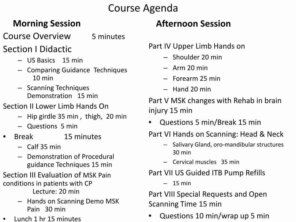
Course Agenda Morning Session
Course Overview 5 minutes
Section I Didactic – US Basics 15 min – Comparing Guidance Techniques
10 min – Scanning Techniques
Demonstration 15 min Section II Lower Limb Hands On
– Hip girdle 35 min , thigh, 20 min – Questions 5 min
• Break 15 minutes – Calf 35 min – Demonstration of Procedural
guidance Techniques 15 min Section III Evaluation of MSK Pain conditions in patients with CP Lecture: 20 min
– Hands on Scanning Demo MSK Pain 30 min
• Lunch 1 hr 15 minutes
Afternoon Session
Part IV Upper Limb Hands on – Shoulder 20 min – Arm 20 min – Forearm 25 min – Hand 20 min
Part V MSK changes with Rehab in brain injury 15 min • Questions 5 min/Break 15 min Part VI Hands on Scanning: Head & Neck
– Salivary Gland, oro-mandibular structures 30 min
– Cervical muscles 35 min
Part VII US Guided ITB Pump Refills – 15 min
Part VIII Special Requests and Open Scanning Time 15 min • Questions 10 min/wrap up 5 min

Why Should you Learn a New Technique for Chemodenervation?
US for Chemodenervation Procedures
• Evidence supports US guidance is – More accurate than other localization
techniques for invasive procedures – May improve efficacy of BoNT or
chemodenervation procedures
– Owing to • Direct visualization of
– Target location/depth – Structures to be avoided – Needle /injectate location
• Continuous needle visualization during the procedure
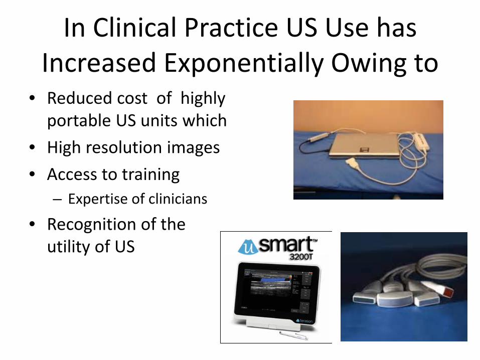
In Clinical Practice US Use has Increased Exponentially Owing to
• Reduced cost of highly portable US units which
• High resolution images • Access to training
– Expertise of clinicians
• Recognition of the utility of US
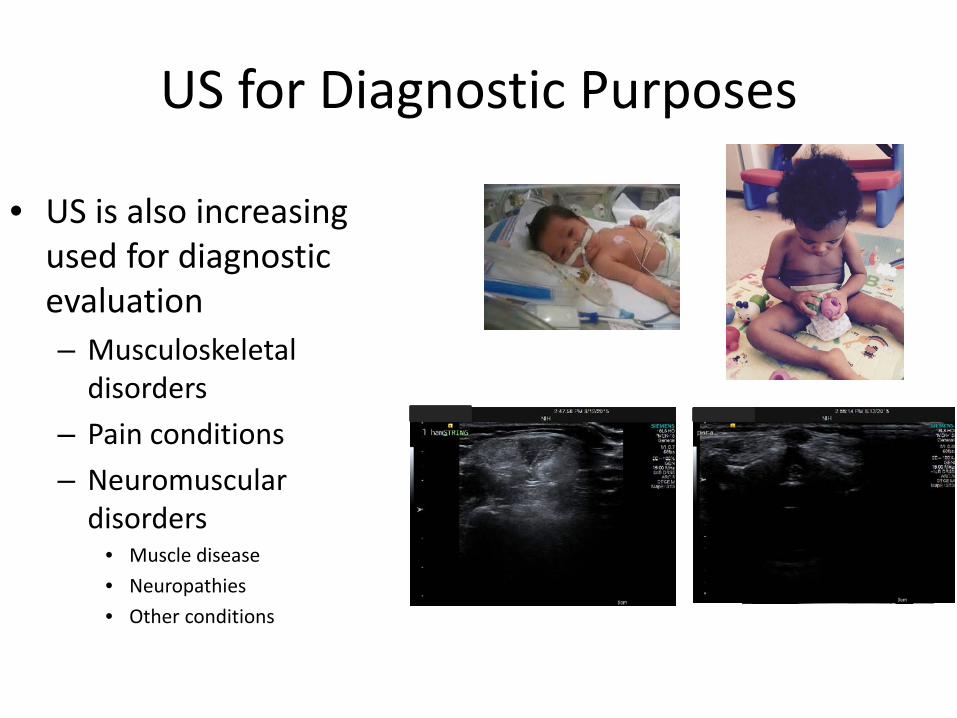
US for Diagnostic Purposes
• US is also increasing used for diagnostic evaluation – Musculoskeletal
disorders – Pain conditions – Neuromuscular
disorders • Muscle disease • Neuropathies • Other conditions
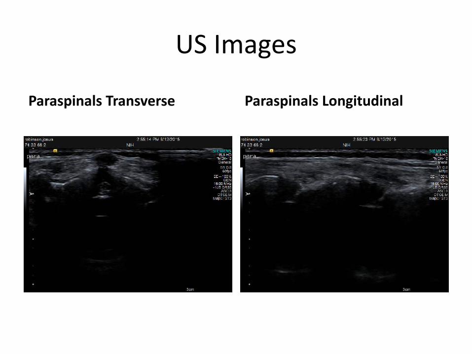
US Images
Paraspinals Transverse Paraspinals Longitudinal

21 m.o. referred to NIH for abnormal gait
• Unremarkable
– Pregnancy – Birth History – Neonatal – Infancy/medical
• Developmental history – Speech/cognitive: Nl
• Talking in 2-3 word sentences
• >100 words • Follows 1 and 2 step
directions • Understands cause/effect

US BASIC PHYSICS What you need to know to start scanning?
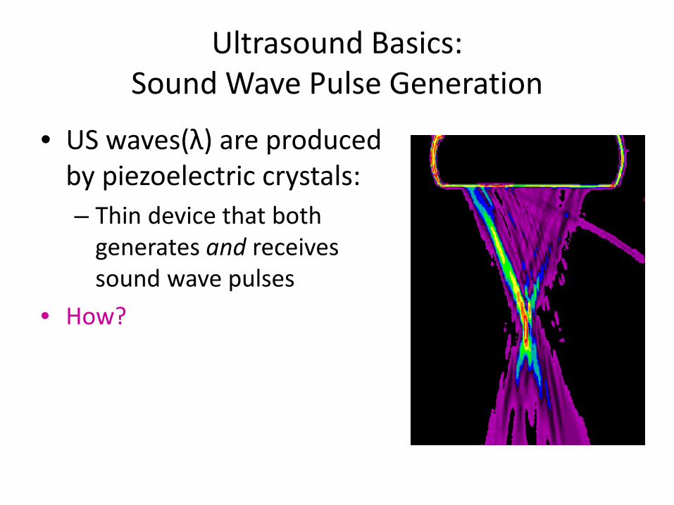
Ultrasound Basics: Sound Wave Pulse Generation
• US waves(λ) are produced by piezoelectric crystals: – Thin device that both
generates and receives sound wave pulses
• How?

Ultrasound Pulse Generation and Reception
Piezoelectric Crystals • Convert electrical pulses into
vibrations • Converts returning vibrations
back into electrical pulses • A linear crystal array is used to
create planar images • Returning echoes are
processed to create grey scale 2D/3D/4D images
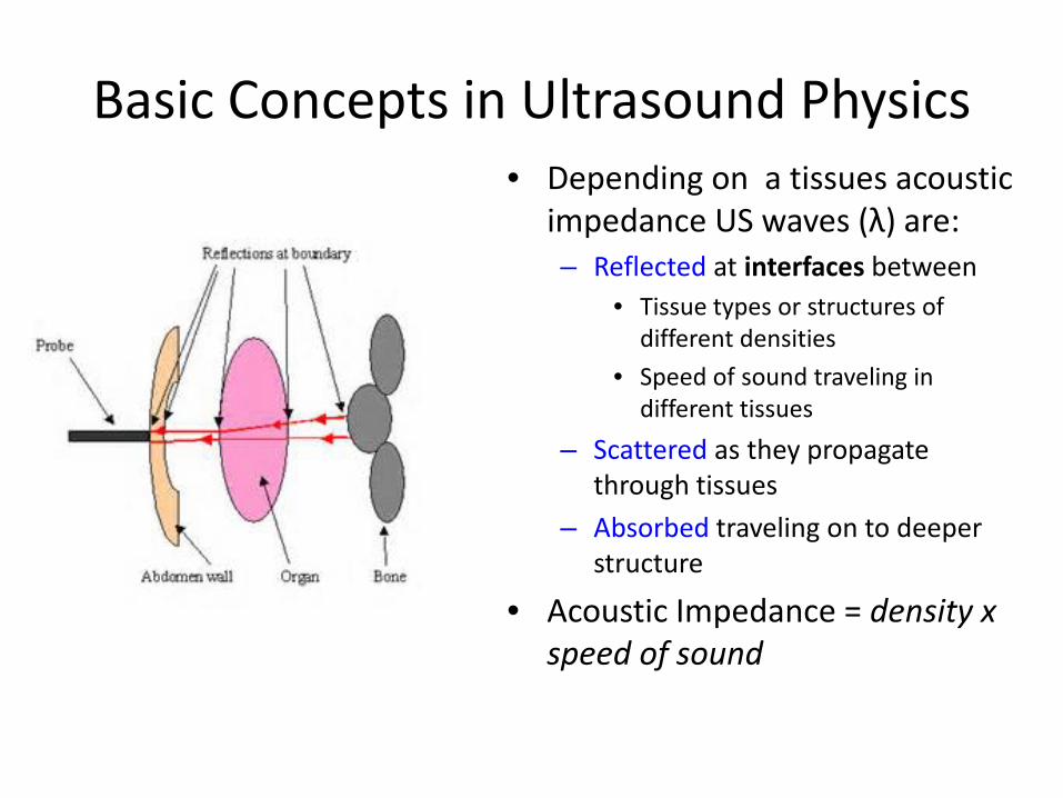
Basic Concepts in Ultrasound Physics • Depending on a tissues acoustic
impedance US waves (λ) are: – Reflected at interfaces between
• Tissue types or structures of different densities
• Speed of sound traveling in different tissues
– Scattered as they propagate through tissues
– Absorbed traveling on to deeper structure
• Acoustic Impedance = density x speed of sound
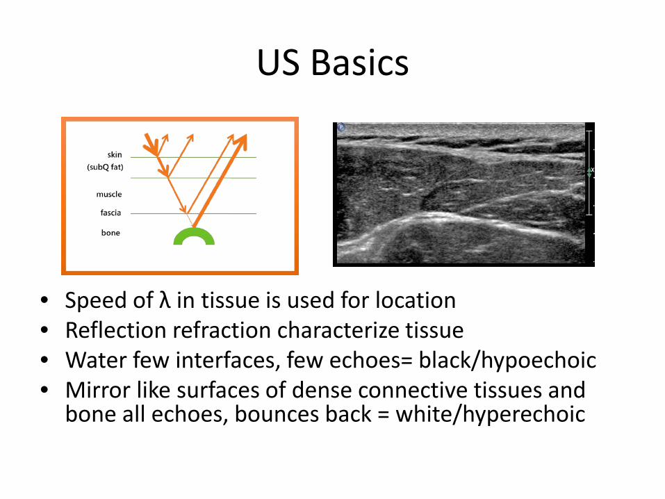
US Basics
• Speed of λ in tissue is used for location • Reflection refraction characterize tissue • Water few interfaces, few echoes= black/hypoechoic • Mirror like surfaces of dense connective tissues and
bone all echoes, bounces back = white/hyperechoic
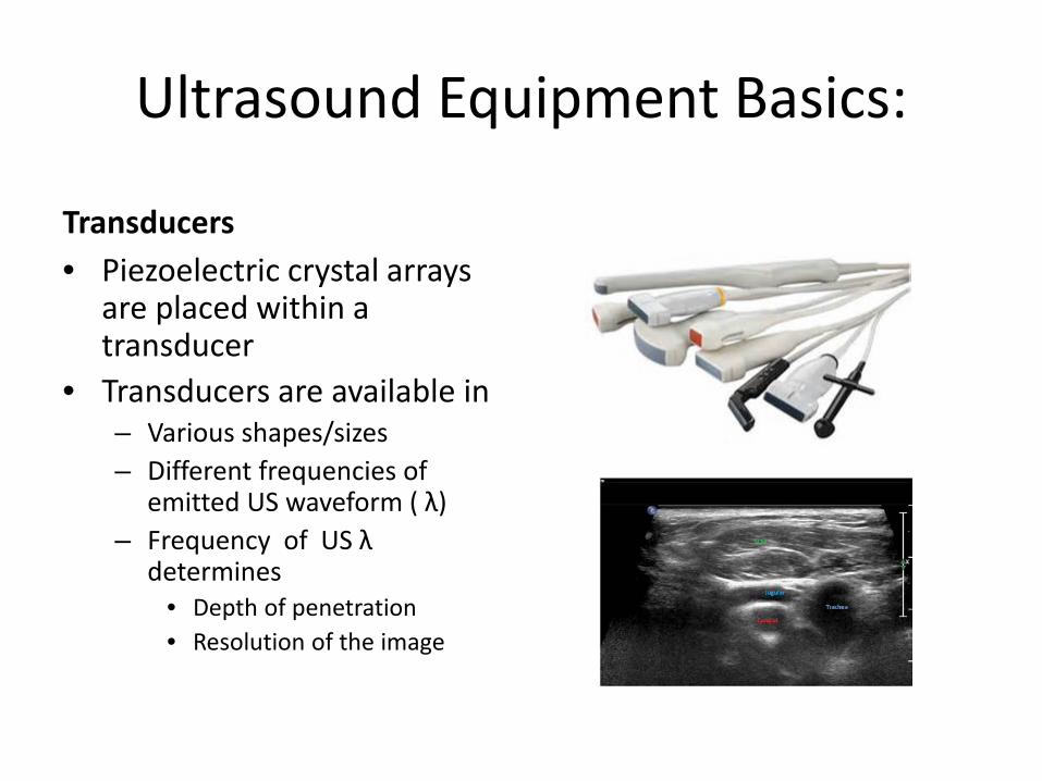
Ultrasound Equipment Basics:
Transducers • Piezoelectric crystal arrays
are placed within a transducer
• Transducers are available in – Various shapes/sizes – Different frequencies of
emitted US waveform ( λ) – Frequency of US λ
determines • Depth of penetration • Resolution of the image
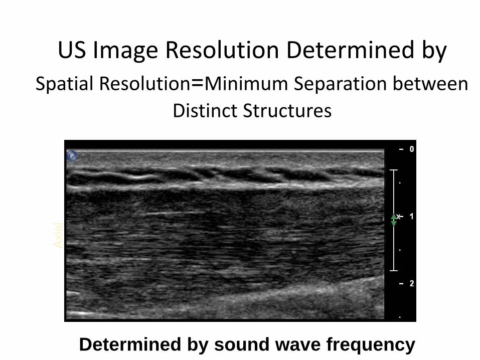
US Image Resolution Determined by Spatial Resolution=Minimum Separation between
Distinct Structures
Axi
al
Determined by sound wave frequency

Resolution Lateral: Ability to discriminate 2 side by side objects
Axial: Ability to discriminate 2 objects at different depths
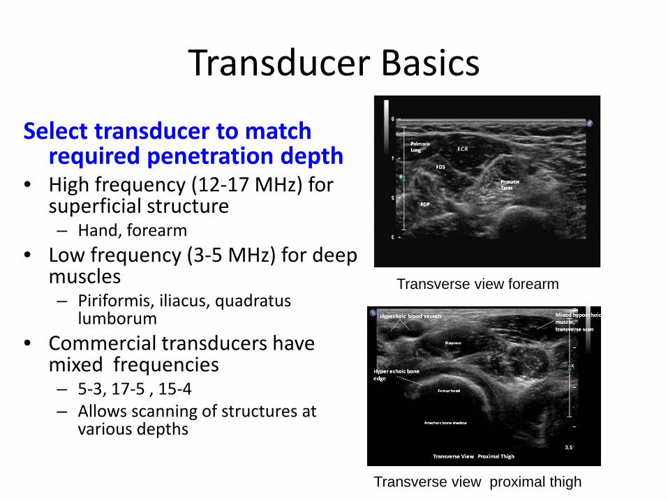
Transducer Basics Select transducer to match
required penetration depth • High frequency (12-17 MHz) for
superficial structure – Hand, forearm
• Low frequency (3-5 MHz) for deep muscles – Piriformis, iliacus, quadratus
lumborum • Commercial transducers have
mixed frequencies – 5-3, 17-5 , 15-4 – Allows scanning of structures at
various depths
Transverse view forearm
Transverse view proximal thigh
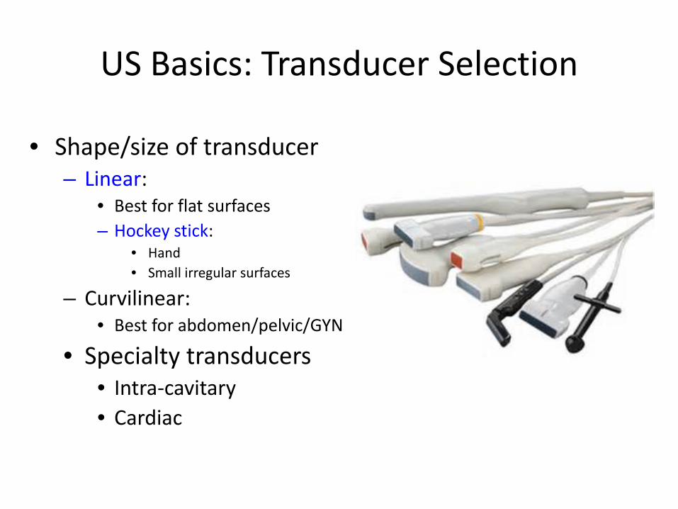
US Basics: Transducer Selection
• Shape/size of transducer – Linear:
• Best for flat surfaces – Hockey stick:
• Hand • Small irregular surfaces
– Curvilinear: • Best for abdomen/pelvic/GYN
• Specialty transducers • Intra-cavitary • Cardiac

US Basics: View convention
• Top of screen/image • Superficial
• Bottom of screen/image • Deeper structures
Superficial
Deep
left
Right
Transverse view, posterior calf

US Basics: View convention
• Transverse scans – How do you place the
transducer on the patient?
– Conventions vary • Standard cross sectional
imaging – Screen left = patient right
• Simplified cross sectional imaging
– Screen left = medial
Superficial
Deep
left
Right
Transverse view, posterior calf

US Basics: View convention
Longitudinal view Convention • Place the transducer on the
patient so that – Proximal = screen left – Distal = screen right
Qadriceps tendon and patella
Distal Proximal
Superficial
Deep
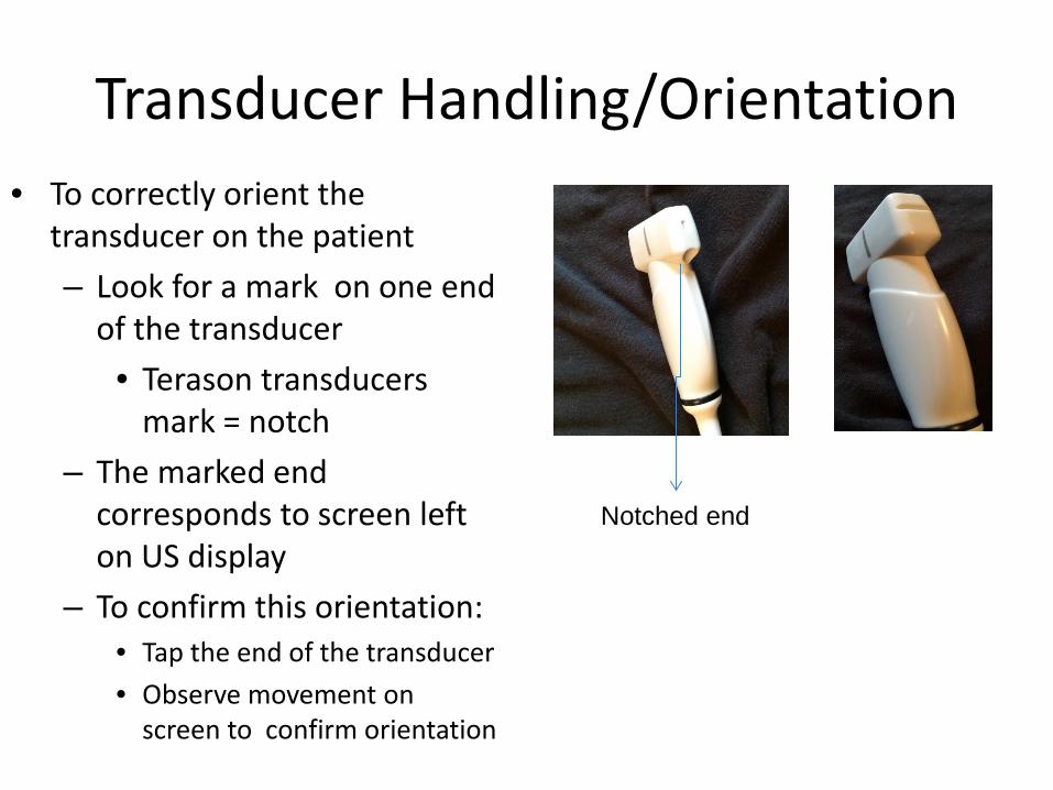
Transducer Handling/Orientation • To correctly orient the
transducer on the patient – Look for a mark on one end
of the transducer • Terason transducers
mark = notch – The marked end
corresponds to screen left on US display
– To confirm this orientation: • Tap the end of the transducer • Observe movement on
screen to confirm orientation
Notched end

ULTRASOUND PROPERTIES OF TISSUES
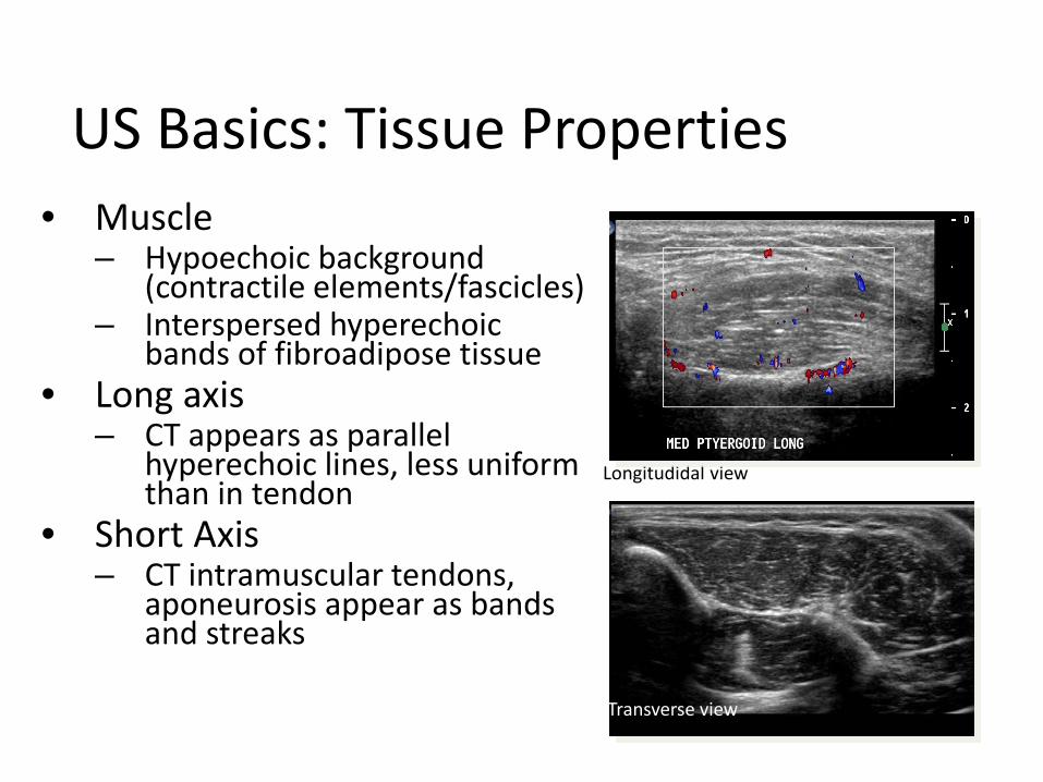
US Basics: Tissue Properties • Muscle
– Hypoechoic background (contractile elements/fascicles)
– Interspersed hyperechoic bands of fibroadipose tissue
• Long axis – CT appears as parallel
hyperechoic lines, less uniform than in tendon
• Short Axis – CT intramuscular tendons,
aponeurosis appear as bands and streaks
Transverse view
Longitudidal view
Transverse view
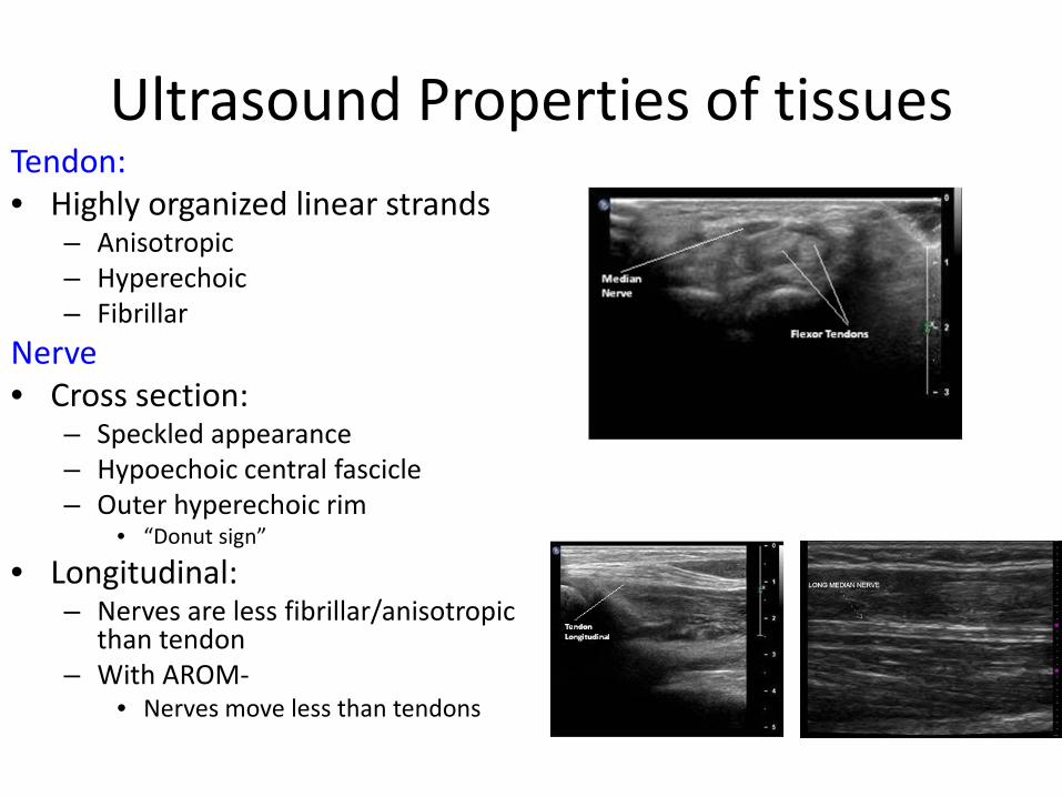
Ultrasound Properties of tissues Tendon: • Highly organized linear strands
– Anisotropic – Hyperechoic – Fibrillar
Nerve • Cross section:
– Speckled appearance – Hypoechoic central fascicle – Outer hyperechoic rim
• “Donut sign”
• Longitudinal: – Nerves are less fibrillar/anisotropic
than tendon – With AROM-
• Nerves move less than tendons
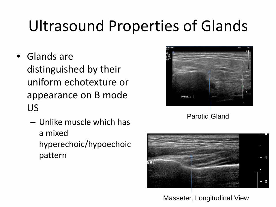
Ultrasound Properties of Glands
• Glands are distinguished by their uniform echotexture or appearance on B mode US – Unlike muscle which has
a mixed hyperechoic/hypoechoic pattern
Parotid Gland
Masseter, Longitudinal View

US Muscle identification
• Identification of muscles is based on pattern recognition of – Contour lines – Adjacent structures
• Bones • Vessels • Other muscles
– Real-time • Use AROM/PROM to
assist muscle identification
Transverse view, proximal calf
Transverse view, proximal anterior thigh
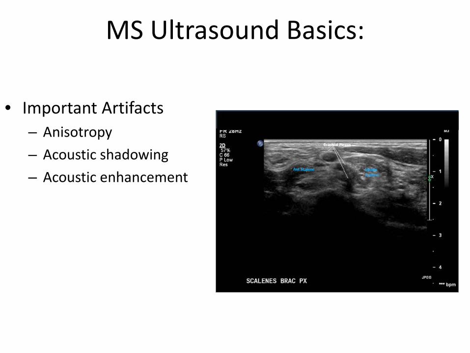
MS Ultrasound Basics:
• Important Artifacts – Anisotropy – Acoustic shadowing – Acoustic enhancement

Anisotropy: Incidence/angle of US beam
Illustration from Rutten M J C M et al. Radiographics 2006;26:589-604 ©2006 by Rad Society of North America
Property of tendon/muscle/nerve: Echogenicity determined by incidence/angle of US beam Artifactualy hypoechoic if US beam is not perpendicular to imaged structure ► May mimic pathology: Ex. partial tendon tear
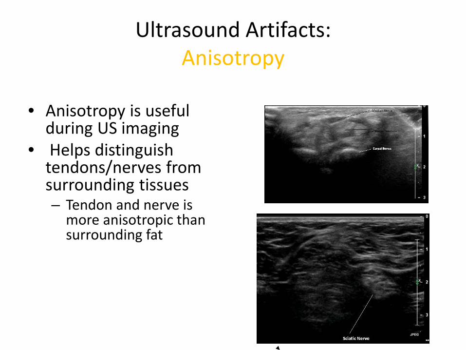
Ultrasound Artifacts: Anisotropy
• Anisotropy is useful
during US imaging • Helps distinguish
tendons/nerves from surrounding tissues – Tendon and nerve is
more anisotropic than surrounding fat
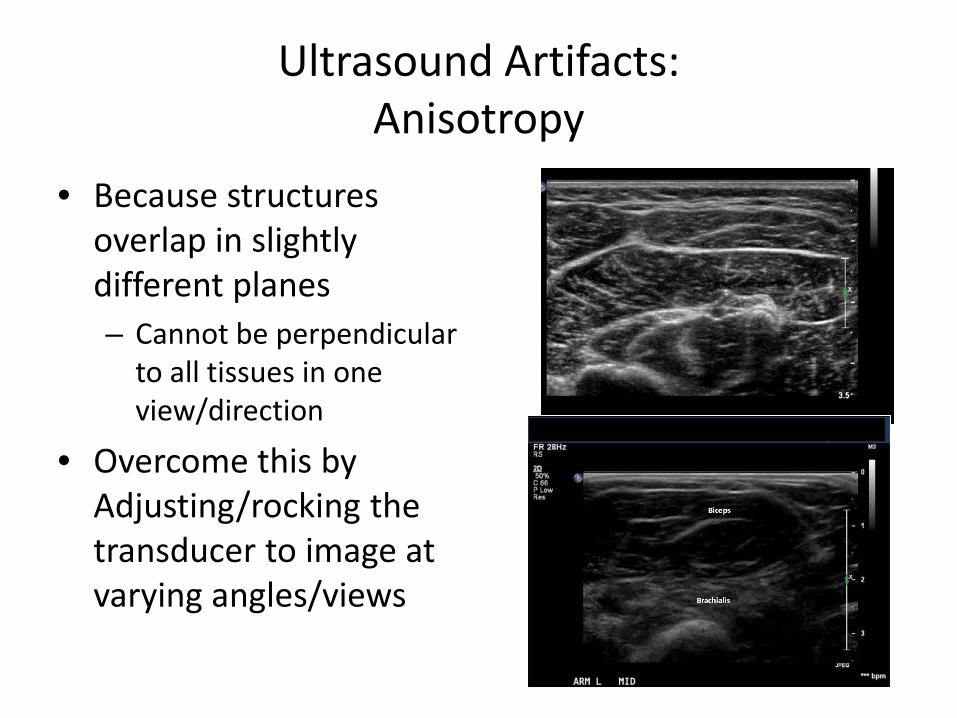
Ultrasound Artifacts: Anisotropy
• Because structures overlap in slightly different planes – Cannot be perpendicular
to all tissues in one view/direction
• Overcome this by Adjusting/rocking the transducer to image at varying angles/views
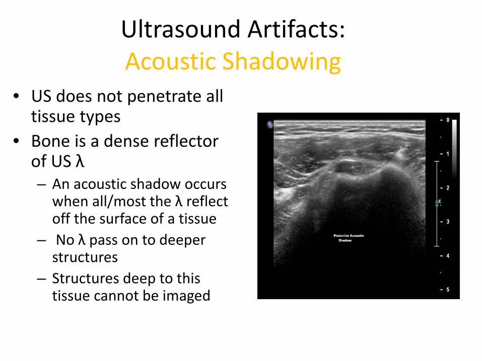
Ultrasound Artifacts: Acoustic Shadowing
• US does not penetrate all tissue types
• Bone is a dense reflector of US λ – An acoustic shadow occurs
when all/most the λ reflect off the surface of a tissue
– No λ pass on to deeper structures
– Structures deep to this tissue cannot be imaged
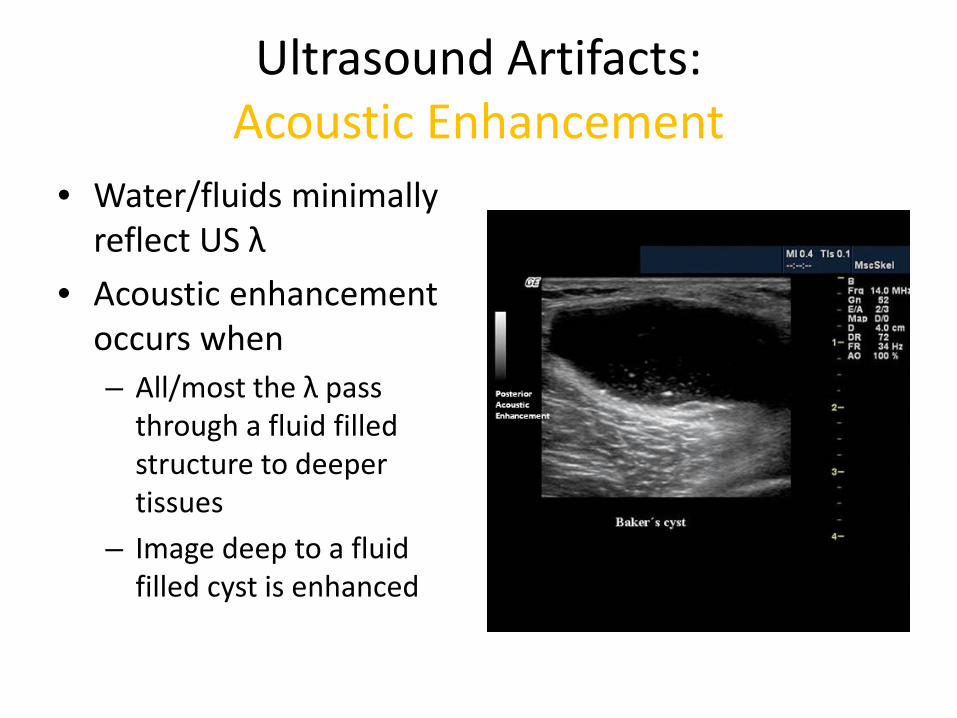
Ultrasound Artifacts: Acoustic Enhancement
• Water/fluids minimally reflect US λ
• Acoustic enhancement occurs when – All/most the λ pass
through a fluid filled structure to deeper tissues
– Image deep to a fluid filled cyst is enhanced

TECHNICAL SKILLS

How to Hold the Transducer
Correct Incorrect
Hold transducer with thumb Index +/- middle finger - Maintain contact with patient - -Use heel of hand or 4th& 5th fingers
“Free handing” the transducer - Hand is not in contact with patient - Transducer may slip out of place
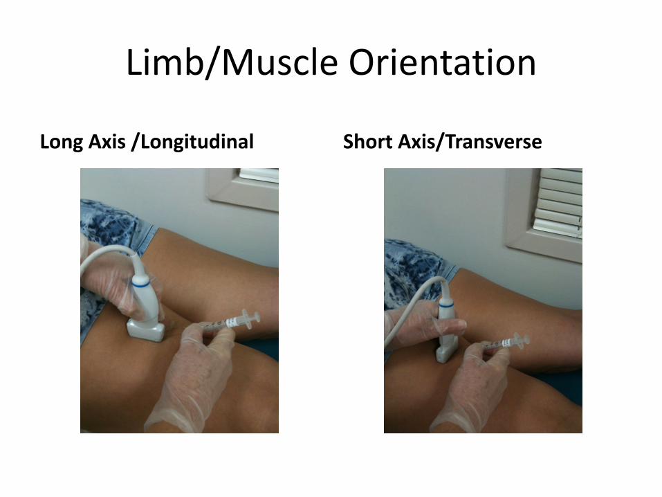
Limb/Muscle Orientation
Long Axis /Longitudinal Short Axis/Transverse

Interventional MS US: Clinical Pearls • In plane/long Axis needle view:
– Keep needle parallel to transducer – Insert needle at flat angle – Poor needle visualization
• Oblique position • Steep angle needle
• Out of plane/short axis needle view: – Keep needle tip under US beam
• If needle tip is outside of US beam, visualization is lost
• May be in untargeted structure or muscle
– Walk down technique • Follow movement of needle tip
passing through tissues planes to target

• Real time injection • US beam is narrow
– Only the width of a credit card – Not the width of the transducer
• Keep needle within the US beam – If travel out side of the narrow
beam needle visualization is lost
• May not be in target structure
Interventional MS Ultrasound: Clinical Pearls

Interventional MS Ultrasound: Pearls of Wisdom
• Larger needles are easier to see than small needles – Larger needles hurt more – 27g hypodermic needles are easily seen – Non-insulated needles are visualized better than insulated.
Etched Needles are also available • Small amount of air or injectate (.2-.3 ml) helps define
needle location • Billing: In the USA, to charge/bill for US, a picture or
cine-loop must be saved to document the procedure • Current CPT Code: 76942: Ultrasound for Needle guidance, aspiration

COMPARISON OF GUIDANCE TECHNIQUES
Chemodenervation Procedures

Traditional Localization Techniques for BoNT Injections: Palpation, EMG, E-Stim
Advantages: • Anatomic:
– No equipment needed (other than reference guides)
• EMG/E-Stim – Clinician familiarity
• Some muscles may be easily/quickly isolated – Many are not
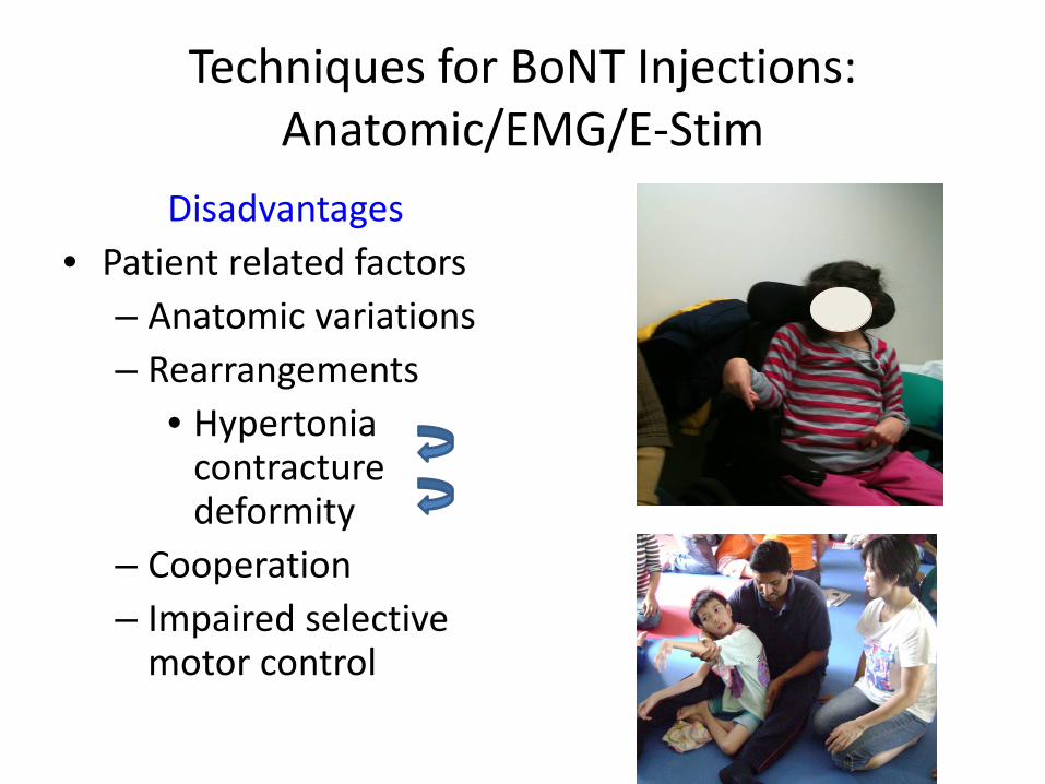
Techniques for BoNT Injections: Anatomic/EMG/E-Stim
Disadvantages • Patient related factors
– Anatomic variations – Rearrangements
• Hypertonia contracture deformity
– Cooperation – Impaired selective
motor control

Localization Techniques for BoNT: EMG/Anatomic
Disadvantages • Difficult to isolate deep/overlapping
muscles • Co contraction, mass synergy,
impaired selective motor control – EMG signal falsely attributed to target
when needle is in another muscle
• E-Stim – Over stimulation
• Volume conduction can lead to errors
– Pain from stimulation often requires sedation

5 year old female 11 year old female
12 year old male 29 year old male
Anatomic/EMG/E-stim Localization Disadvantages FCR Transverse view - Examples
Patient related factors: Muscle size, architecture and shape all vary with age
Heinen et al

Anatomic/EMG/Estim Localization Disadvantages
Sonography Diameter
Echogenicity
GMFCS I GMFCS III
Muscle Size: Inversely related to impairment level
Heinen et al
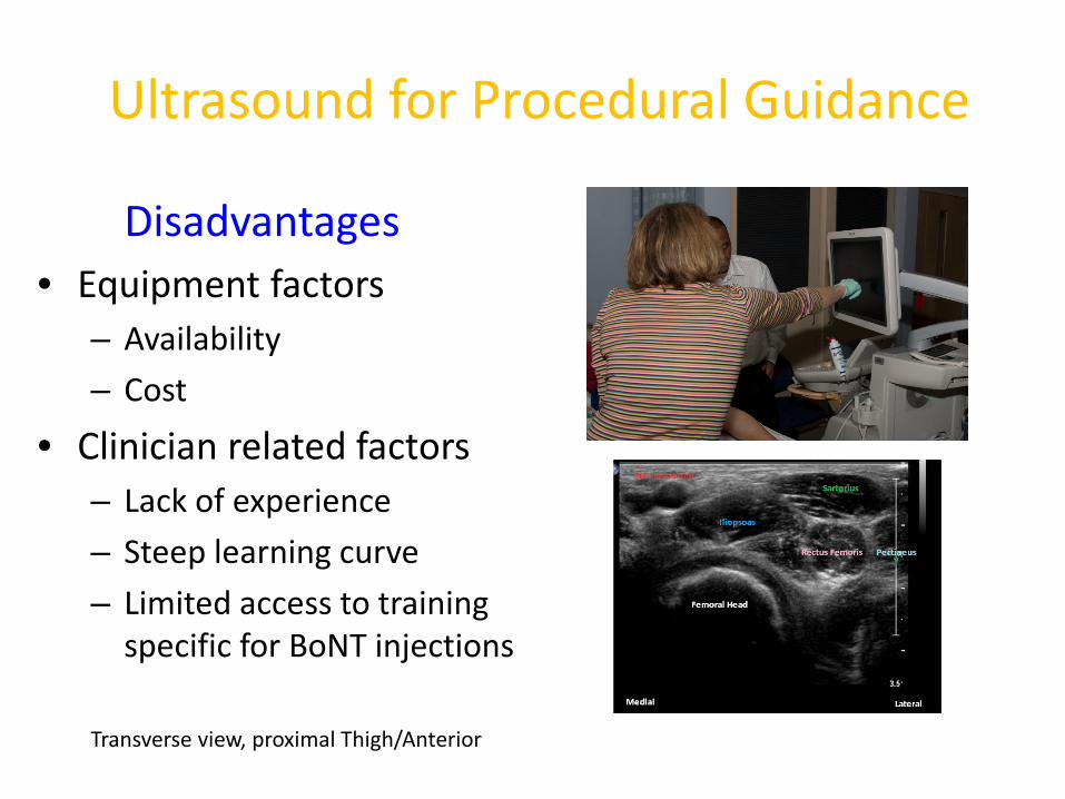
Ultrasound for Procedural Guidance
Disadvantages • Equipment factors
– Availability – Cost
• Clinician related factors – Lack of experience – Steep learning curve – Limited access to training
specific for BoNT injections
Transverse view, proximal Thigh/Anterior

US for BoNT Injections: Advantages
Improved accuracy – Complex/overlapping
anatomy obscures muscle identification
– Small/large patients • Provides direct
assessment of target – Depth – Location – Structures to be avoided
Transverse View, Forearm
Transverse View Proximal Thigh

US for BoNT Injections: Advantages • Visualize/isolate target
muscles – Quickly – Easily – Accurately
• Less painful – Smaller needles
• US often distracts patients during procedure – Reducing anxiety/stress
In plane injection lateral Gastroc
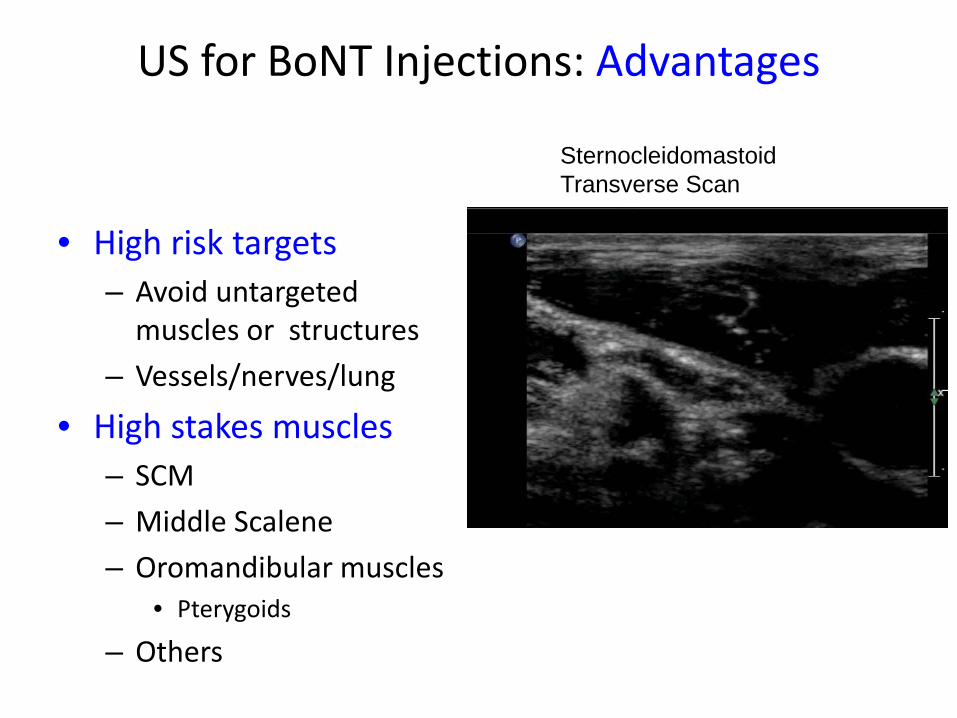
US for BoNT Injections: Advantages
• High risk targets – Avoid untargeted
muscles or structures – Vessels/nerves/lung
• High stakes muscles – SCM – Middle Scalene – Oromandibular muscles
• Pterygoids
– Others
Sternocleidomastoid Transverse Scan
Adductors, Transverse Doppler

US for BoNT Injections: Advantages
Improved accuracy • When localization limited by:
– Involuntary muscle activity – Co-contraction – Motor contro – Deformity – Post surgical changes – Patient cooperation
• US does not require AROM to isolate muscle
• Muscle identification is based on pattern recognition

BoNT Injections: Why Use US?
Focal dystonia • Goal: identify and target
individual muscle fascicles – Ex: FDS digit 3 vs. 4
• US increases accuracy and decreases time to isolate correct muscle fascicles
• Reduces pain
FDS longitudinal view, mid forearm Short axis view of needle
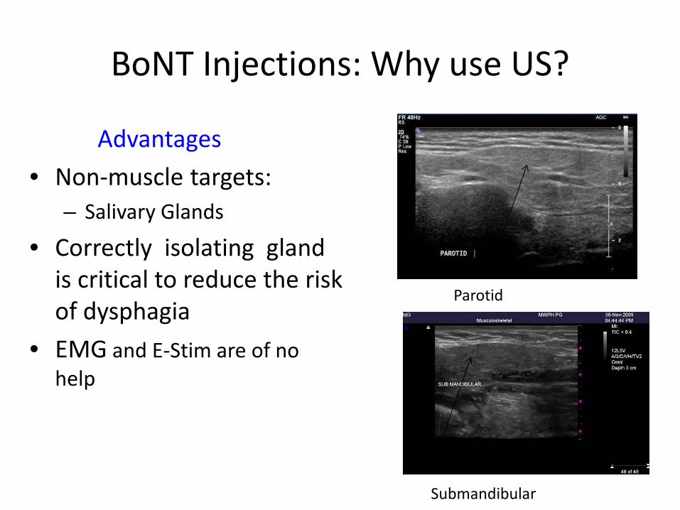
BoNT Injections: Why use US?
Advantages • Non-muscle targets:
– Salivary Glands
• Correctly isolating gland is critical to reduce the risk of dysphagia
• EMG and E-Stim are of no help
Parotid
Submandibular
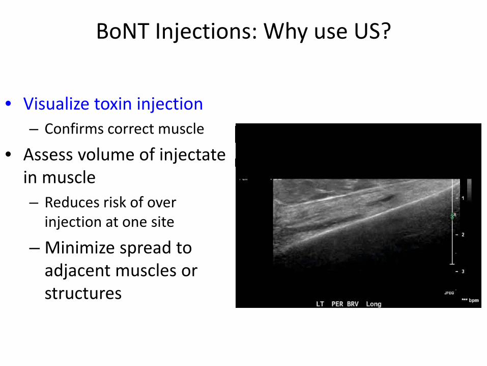
BoNT Injections: Why use US?
• Visualize toxin injection – Confirms correct muscle
• Assess volume of injectate in muscle – Reduces risk of over
injection at one site
– Minimize spread to adjacent muscles or structures
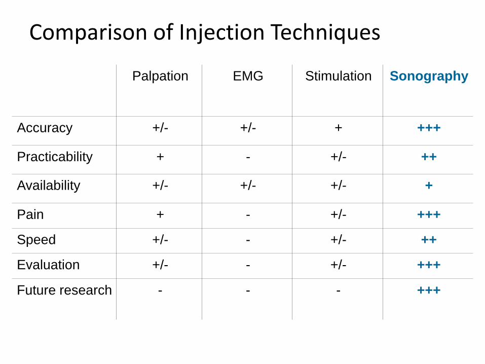
Comparison of Injection Techniques
Palpation EMG Stimulation Sonography
Accuracy +/- +/- + +++
Practicability + - +/- ++
Availability +/- +/- +/- +
Pain + - +/- +++
Speed +/- - +/- ++
Evaluation +/- - +/- +++
Future research - - - +++
Ultrasound for BoNT Injection: Summary
• Localization techniques – Palpation – EMG – Nerve stimulators – Ultrasound
• All have advantages & disadvantages
• Best Strategy: – Be skilled in multiple
techniques

Ultrasound for BoNT Injection: Summary
• US is a useful technique to add to your tool box for BoNT injections – Improved speed/accuracy of target localization – Decreased pain – Reduced risk of harm
• Initial learning curve is steep – Worth the time and effort

US SCANNING DEMONSTRATION

SECTION II Lower Limb Hands on Scanning,
SECTION III MSK PAIN CONDITIONS IN CEREBRAL PALSY
Lecture and Demonstration

SECTION IV Upper Limb Scanning, Hands on
SECTION V MSK changes with Rehab in brain injury

SECTION VI Head and Neck Hands on Scanning

SECTION VII Hands on Scanning: Open scan time, special requests

US Scanning Demonstration
• How to hold the transducer….and why • Scanning limbs/structures • Injection Techniques
– In plane – Out of plane

Hands on Demonstration and Scanning
Lower Limb Muscles • Iliopsoas, Adductors
– Obturator nerve
• Hamstrings/Quadriceps • Lower leg
– Antero-lateral calf • Fibularis longus, Extensor
hallicus longus – Posterior calf
• Gastrocnemius, Tib. Posterior, Soleus, FDL, FHL
Upper limb • Shoulder Girdle
– Pectoralis Major – Latissimus dorsi/subscapularis
• Arm – Biceps/Brachialis – Brachioradialis
• Flexor Forearm – FCR, FCU, FDS, FDP, FPL – Pronators
• Hand

Hands On Scanning Demonstration
Head Neck
• Sternocleidomastoid • Levator Scapulae • Masseter • Parotid/Submandibular