Embed Size (px)
Citation preview

Introduction to the Study of Cell Biology
วัตถุประสงค เพ่ือใหนิสิตสามารถ
1. สืบคนแหลงขอมูลที่เกี่ยวของกับการศึกษาโครงสรางและการทํางานของเซลล
2. อธิบายทฤษฎีเซลล
3. อธิบายโครงสรางและหนาที่ของเซลลสัตว พืช สาหราย เช้ือยีสต เช้ือรา เช้ือแบคทีเรีย
4. เลือกใชกลองจุลทรรศนใหเหมาะสมกับการศึกษาชีววิทยาของเซลล

Introduction to the Study of Cell Biology
Cell is the smallest basic unit of a plant and animal. This is the definition defined
according to Cambridge Dictionaries Online
(http://dictionary.cambridge.org/dictionary/british/cell_1?q=cell). However, this definition
does not include other life forms such as bacteria, yeast, fungi, protozoa and algae, although
these organisms consist of single or multiple unit of cell. Also the definition does not state
the function of a cell. In biology, cell can be defined as “small, membrane-enclosed units
filled with a concentrated aqueous solution of chemicals and endowed with the
extraordinary ability to create copies of themselves by growing and dividing in two” (Albert
et al., 2010). This gives more details about cell physiology and its ability to regenerate.
As you can imagine about complexity in structure and function of human body, similar
condition can be found in the structure and function of our cells. However, complexity in
structure and function of cells is also depending on whether the cell belongs to prokaryote
or eukaryote. Generally, eukaryotic cell structures are more complex than those of
prokaryote. Furthermore, there are some differences in plant cells structures compared with
that of animal cells. Bacterium, a prokaryotic microorganism, is probably the simplest type of
cells. Great diversity is also found in size and appearance of cells as shown in figure 1.
Figure 1 Size and Shape of different cells (Albert et al., 2010)

Question: Is there so much variation of cells and tissues in terms of size, shape, and
organization in the past billion years compared with the present day? And why?
Structure of animal, plant, algal, bacterial, yeast and fungal cells
Animal, plant, algae, bacteria, yeast and fungi compose of cells. Each cell type has some
specific structures and functions. For example, flagellum is commonly found in organisms
that are able to move. Plant cell wall composes of polysaccharide such as cellulose. There
is no nucleus and nucleus membrane in a bacterial cell. Chloroplast is a common energy-
producing unit in a plant cell. This structure is not observed in yeast, fungal and animal
cells. It is also possible to detect variation in cellular structure within the same species.
However, there are some common structures among the mentioned organisms.
Structure of bacterial cell
A good resource about details of bacterial cell structure can be found online at Todar’s
Online Textbook of Bacteriology (http://textbookofbacteriology.net/structure.html). Figure 2
shows drawing of a typical bacterial cell. Table 1 gives function of some bacterial structures.
Figure 2 drawing of bacterial cell structure
(http://textbookofbacteriology.net/schematic_bacterium.jpg)

Not all bacteria have all these cell structures. Some may not have flagellum while others
may have more than one flagella. The composition of cell wall of Gram’s positive differs
from Gram’s negative bacteria. Some bacteria have large inclusion.
Table 1 function of bacterial structures
Structure Function(s) Predominant chemical composition
Flagella Swimming movement Protein
Pili
Sex pilus Stabilizes mating bacteria during DNA
transfer by conjugation
Protein
Common pili or
fimbriae
Attachment to surfaces; protection against
phagotrophic engulfment
Protein
Capsules (includes
"slime layers" and
glycocalyx)
Attachment to surfaces; protection against
phagocytic engulfment, occasionally killing
or digestion; reserve of nutrients or
protection against desiccation
Usually polysaccharide; occasionally
polypeptide
Cell wall
Gram-positive
bacteria
Prevents osmotic lysis of cell protoplast
and confers rigidity and shape on cells
Peptidoglycan (murein) complexed with
teichoic acids
Gram-negative
bacteria
Peptidoglycan prevents osmotic lysis and
confers rigidity and shape; outer membrane
is permeability barrier; associated LPS and
proteins have various functions
Peptidoglycan (murein) surrounded by
phospholipid protein-lipopolysaccharide
"outer membrane"
Plasma membrane Permeability barrier; transport of solutes;
energy generation; location of numerous
enzyme systems
Phospholipid and protein
Ribosomes Sites of translation (protein synthesis) RNA and protein
Inclusions Often reserves of nutrients; additional
specialized functions
Highly variable; carbohydrate, lipid, protein
or inorganic
Chromosome Genetic material of cell DNA
Plasmid Extrachromosomal genetic material DNA

Structure of algal cells
Algae are classified as a eukaryotic organism. There are both unicellular and multi-cellular.
There is a great diversity within algae. Some are unicellular organism; others are plant-like
organism, like seaweed. However, algae derive energy from sun light through photosynthetic
process. Although algae are similar to plants, they lack structures such as root, xylem and
phloem. Some algae do not have chlorophyll. They use other types of pigment to produce
energy. The structure and function of organelle in an algal cell is presented in Figure 3.
Figure 3 a Unicellular alga (a) and Yeast cell structure (b) (Talaro, 2002)
Structure of a yeast cell
Yeast is a microorganism which belongs to eukaryote. The structure of a yeast cell is
represented in Figure 3. A number of organelles inside the yeast cell can be observed
including vacuole, nucleus, endoplasmic reticulum, mitochondria, centrioles, etc. This is

typical of the eukaryotic organism. As yeast reproduce asexually by budding, there is a bud
scar left on the surface of cell wall.
Cellular structure of fungi
Fungi are multicellular organisms in the form of filament. Septum is a cellular structure that
separates two adjacent fungal cells from each other. Some species of fungi lacks this
structure. Asexually, the growth of fungi is at the tip of hyphae. A special cellular structure
of fungi is spore. There are many types of spores depending on the specie of fungi.
Internally, the ultrastructures are similar to that of other eukaryotes and presented in Figure
4. The growth of fungi is active at their tips. On the contrary, the aged hyphae may break
due to autolysis.
Figure 4 Ultrastructure of fungal hyphae (Deacon, 2006)

Cellular structure of animal and plant cells
The structures of animal and plant cells are similar because both of them are eukaryotic
cell. Plant cells tend to be bigger. They consist of membrane-bound organelles such as
nucleus, mitochondria, endoplasmic reticulum, etc. The functions of organelles of the two
are similar. However, they do have some differences. This includes the lacks of cell wall,
vacuoles and chloroplast in animal cells compared with plant cells. Figure 5 shows
structural details of animal and plant cells.
Figure 5 The structure of bacteria, animal and plant cells (Albert, 2003)

Basic structure of prokaryotic and eukaryotic cells
The previous section gives examples of cell components of bacteria, fungi, yeast, algae,
animal and plant. Several analogous components can be observed, although there are some
variations. As we know now, cells can be classified into either prokaryotic or eukaryotic type.
The first one is simpler. It consists of a single closed compartment that is surrounded by the
plasma membrane, lacks a defined nucleus, and has a relatively simple internal
organization. Prokaryote can be either single (bacteria) or multi-cellular organisms (some
cyanobacteria). On the other hand, eukaryotic cells are commonly larger and more complex.
Structurally, they contain membrane-enveloped nucleus and the organelles. Both types of
organisms contain significant amount of water inside their cells, cytosol. Figure 6 shows basic
structures of prokaryotic and eukaryotic cells.
Figure 6 Basic structures of prokaryotic and eukaryotic cells
Question: Structurally, how are prokaryotes differ from eukaryotes?
Question: What is the function of each cell component?

Origin of life
A cell is the basic unit of life. One of the most skeptical questions is where the cell comes
from. In the cell theory of Schlieden and Schwan, a cell must comply with three principles
including
1. All living organisms are composed of one or more cells
2. The cell is the most basic unit of life.
3. All cells arise from pre-existing, living cells.
Based on this theory, the first and the second principles are true. However, the third
principle cannot be applied with the first cell on Earth. This raises the question “How is the
first cell occur on Earth?”. In 1950s, Stanley Miller and the colleges showed that they can
synthesize amino acids by charging atmosphere consisting of H2, CH4 and NH3 in the
presence of water. Although this is not the precise condition of primitive Earth, they clearly
show that simple molecules can be created spontaneously by nature. Similarly, the
occurrence of macromolecules such as proteins and DNA should be the same scenario.
Apart from these two components, proteins and DNA or RNA, primitive cells must consist of
at least cell membrane to protect and enclose pools of enzyme, DNA or RNA and those
necessary for cell replication inside. In order for cell to thrive and self-replicate, it must have
some mechanisms to generate energy. It is expected that there is no oxygen in the
atmosphere of Earth at the beginning. Therefore, the energy-generating reaction should
involve the breakdown of organic molecules in the absence of oxygen. This is similar to
glycolysis. The next evolution of origin of life is photosynthesis reaction. This reaction
produces oxygen and change Earth’s atmosphere. Later, oxidative metabolism (respiration)
evolves. Another critical in cell evolution is acquisition of membrane-enclosed sub-cellular
organelles, especially mitochondria and chloroplast, by endosymbiosis. This is supported by
the evidence that both mitochondria and chloroplast have their own DNA and systems for
self-replication. Multicellular organisms, then, evolve from aggregation of unicellular
organisms (eukaryote). Complexity of multicellular organisms increases as a result of cell
specialization. This is the case for animal cells, which has several types of cells. The first
cells seem to emerge on Earth around 3.8 billion year ago.

Question : What is endosymbiosis?, Please explain the evolution of mitochondria and
chloroplast?
Surprisingly, researchers at J. Craig Venter Institute in United State of America have
successfully created a chemically synthesized genome that can control cells of
Mycobacterium mycoides in their laboratory
(http://en.wikipedia.org/wiki/Mycoplasma_laboratorium). This proves that DNA can be
created from a chemical synthesis. Their work is very useful for the future applications. For
example, bacteria that have a specific function might be created such as biogas producing-
or carbon dioxide absorbing bacteria.
.
Figure 7 Spontaneous formation of organic molecules (Cooper and Hausman, 2007)
History and progress of cell studies
Initially, the history of cell studies closely associates with the invention of
microscope. The first compound microscope is invented in 1950 by Francis Janssen and
Zacharias Janssen. Their microscope can magnify only at 10x to 30x. During 1564 – 1642,
Galileo Galilei invented a simple microscope having only single lens. The use of microscope
to study biological samples such as tissues of kidney, brain, liver, spleen, etc. is done by

Marcello Malpighi (1628 – 1694). However, the first scientist which was the first to define the
word “cell” is Robert Hooke (1635 – 1703). Then, in 1675, Anton van Leeuwenhoek was the
first to examine free-living cell by using his microscope. During 19th century, cell theory has
been formulated through many discoveries. Mathias Jacob Schleiden and Theodor Schwan
postulated that a cell is basic unit of life during 1838 and 1839. Then, K. Nageli as well as
Rudolf Virchow suggested that cells arise from the division of the pre-exist cells. Since then,
numerous discoveries in cell biology have been made as shown in the table below (Verma
and Agarwal, 2005). These findings correlate with the advances in novel technologies such as
electron microscope, fluorescence microscopy, X-ray diffraction, immune-technology,
chromatography and electrophoresis. Some of microscopic instruments commonly used to
study cell are reviewed in the following section.
Microscope
Prior to go to the detail of each microscope, it is worth to look into the scale of
things, especially cells. The scale of things is presented in Figure 8. Individual cell of
organisms is generally small that hardly be seen by unaided eye because human eye can
see the smallest objects no less than 0.1 mm long
(http://learn.genetics.utah.edu/content/cells/scale/). The first invented compound
microscope belongs to light microscope classification. Its magnification is very low, ~ 9x
(http://www.history-of-the-microscope.org/history-of-the-microscope-who-invented-the-
microscope.php). To date, modern light microscopes can magnifiy object up to 2000x
(Talaro, 2002). In detail, the limitation is not about the magnification power. The limitation
depends on resolution. Resolution or resolving power is defined as the ability of an optical
system to separate two objects from one another (Talaro, 2002). Resoving power of any light
microscopic system can be calculated from an equation provided below.
푟푒푠표푙푣푖푛푔 푝표푤푒푟 = 휆
2 × 푁. 퐴.
This means that resolving power of a microscope system can be decreased by decreasing
wavelength or increasing numerical apperture of objective lens.

As we all know, microscopes are invented to help researchers to study detail of minuscule
objects. Light microscopes use light as a light source with broad spectrum of wavelength. To
use this microscope effectively, samples are needed to be prepared as thin as possible.
Staining helps researchers to see objects clearer. Nevertheless, some specific details inside a
cell, expecially for the very tiny one, can not be seen or differentiated from other
components. There are other types of light microscopes such as dark field-, phase contrast-,
and differential interference microscopes.
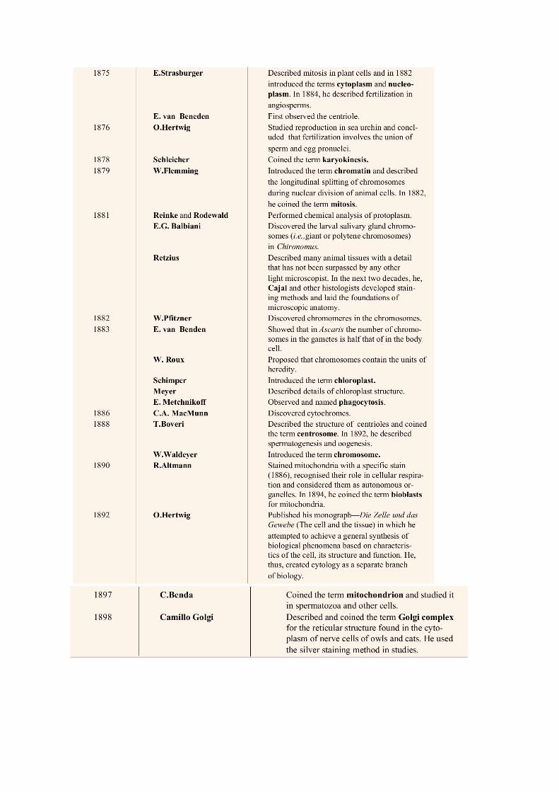

Figure 8 Size of object and tools commonly used to study (http://micro.magnet.fsu.edu/cells/)
Fluorescence microscope
Fluorescence microscope can be classified as one type of light microscope. Fluorescence
microscope use UV light as their light source. It is a sensitive method to study intracellular
structure of cells. Generally, fluoresence occur when high energy light (a wavelength) is
radiated on some substances that absorb light energy making a shift in electron to an exited
state and then release energy as emission light or fluorescence out (another wavelength).
The electron come back to their normal state. Fluorescence dyes coupling with antibodies
are used to stain or fix on the specific structure of a cell. This is very useful. The staining can
be used with living cells as well. With an advance in DNA recombinant and green
fluorescence protein (GFP), the DNA sequence of this protein can be fused into DNA
segement of the target protein which when expressed, it can be seen by fluorescence
microscope. Nevertheless, the images obtained by fluorescence microscope are not clear as
a result of out-of-fucus fluorescence. Confocal microscopy solves this problem by detecting
fluorescence signal from only a single point at a time. Laser is used as a light source. The
image from confocal microscope is very clear compared to that of fluorescence microscope.
Nevertheless, both of them still have limitation of resolution as a result of light diffraction
which both of them still use light source. Recently, there is a break through in fluorescence
microscopy. The new technique is collectively called super-resolution fluorescence
microscopy. This techninue solves the problem of light diffraction by various methods

including SNOM/NSOM, TIRFM, SIM, STED or PALM (Schermelleh et al., 2010). In principle, the
fluorescence signal from fluorephore dyes is reduced to that of single spot of single
molecule. Each method have their own principle and limitation. Nevertheless, the resolution
of these super-resolution fluorecence microscopy is much better than normal fluorescence
microscopy. For example, STED and PALM give images with the lowest resolution (xy) at 20
nm. Figure 9 shows pictures taken by super-resolution fluorescence microscopy compared
with other methods. It can be seen that the details of images taken by super-resolution
fluorescence microscopy is much better than the conventional methods. Apart from its
specificity, which is one of many advantages, it can be use to study structures and well as
interaction between structures of living cells.
Electron microscope
Electron microscope or EM is pobably one of the most powerful microscopes. Instead of
light, EM use electron which have very short wavelength, 0.004 nm. This value should give
vaery low resolution. Another factor affecting resolution is numerical apperture of lens. In
EM, electromegnetic lens is used instead of glass lens. The numerical apperture of
electromegnetic lens is 0.01. Theoretically, the limitation in resolution of images taken by
EM is at 0.2 nm. In reality, it is 1-2 nm. There are two types of EM; Scanning electron
microscope (SEM) and Transmission electron microscope (TEM). In TEM, the sample is
prepared as thin as possible using a microtome and stained with salt of heavy metals. The
beam of electron that pass through the stained sample is collected and the signal is used to
create an image (apear white). The stained components appear dark as a result of electron
scattering. The chemicals commonly that are used as stains are osmium tetroxide, uranyl
acetate, and lead citrate. Apart from these chemicals, Antibodies labelled with heavy metal
salts can also be used similar to that used in fluorescence microscopy. For SEM, it is used to
study 3 dimentional structure of a sample. The electron beam does not pass through the
sample as in TEM. The samples in SEM have to be coated with heavy metal, so the scatter
electron from the surface is then collected and use to create an image. The resolution of
SEM is about 10 nm, therefore it is suitable to study structure of whole cell instead of detail
structure inside. The drawnbacks of the EM are the incapability in studying live cells as well
as the tedious sample preaparation and costs compared with light microscopy.

Scanning probe microscopy
Scanning probe microscopy was invented in 1981. It uses several kinds of probes to examine
the surface of a specimen by using specially design probes. Its resolution is very high to an
atomic level. In addition, there is no need for sample preparation. Scanning tunnelling
microscopy and atomic force microscopy are two examples of scanning probe microscopy.
Question: What kind of instrument that is suitable for investigating the structure and
function of mitochondria, cell surface, flagella, microtubule? And Why?

Figure 9 Images talen by super-resolution fluorescence microscopy (Schermelleh et al.,
2010)