Embed Size (px)
Citation preview
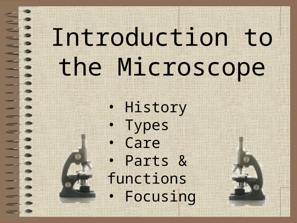
Introduction to the Microscope
• History• Types• Care• Parts & functions• Focusing
Compound microscopes are light illuminated. The image seen with this type of microscope is two dimensional. This microscope is the most commonly used. You can view individual cells, even living ones. It has high magnification. However, it has a low resolution.
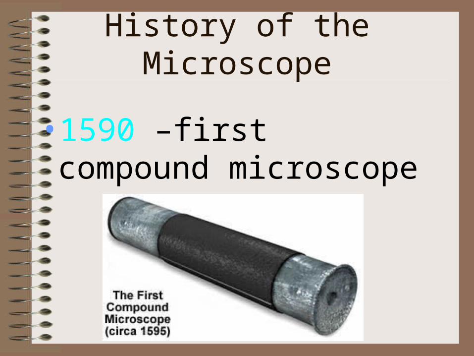
History of the Microscope
•1590 –first compound microscope
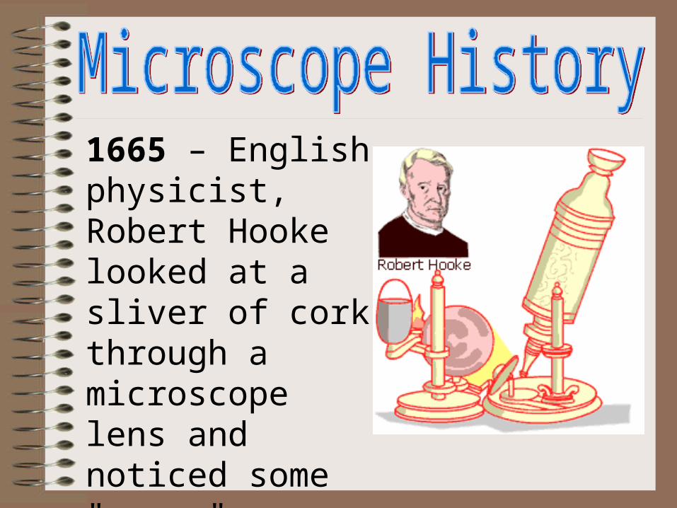
1665 – English physicist, Robert Hooke looked at a sliver of cork through a microscope lens and noticed some "pores" or "cells" in it.
1674 – Anton van Leeuwenhoek built a simple microscope with only one lens to examine blood, yeast, insects and many other tiny objects. Leeuwenhoek was the first person to describe bacteria, and he invented new methods for grinding and polishing microscope lenses that allowed for curvatures providing magnifications of up to 270 diameters, the best available lenses at that time.
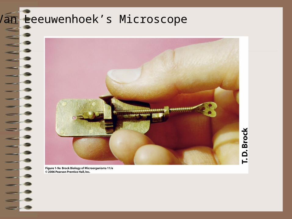
Van Leeuwenhoek’s Microscope
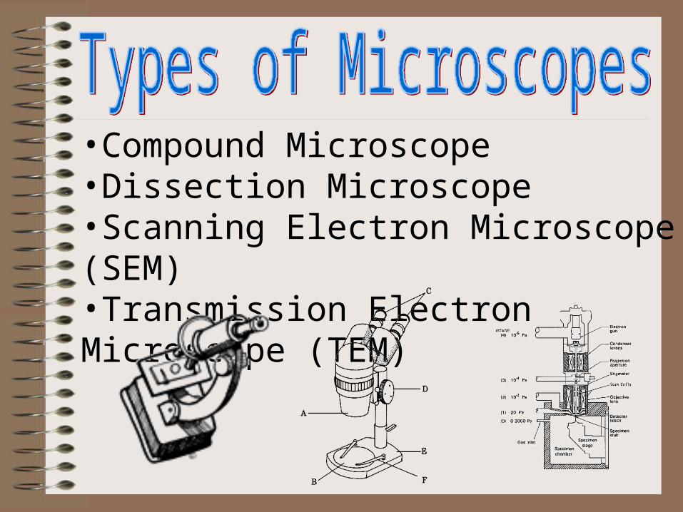
•Compound Microscope •Dissection Microscope •Scanning Electron Microscope (SEM)•Transmission Electron Microscope (TEM)
Microscope Vocabulary
•Magnification: increase of an object’s apparent size
•Resolution: power to show details clearly
•Both are needed to see a clear image
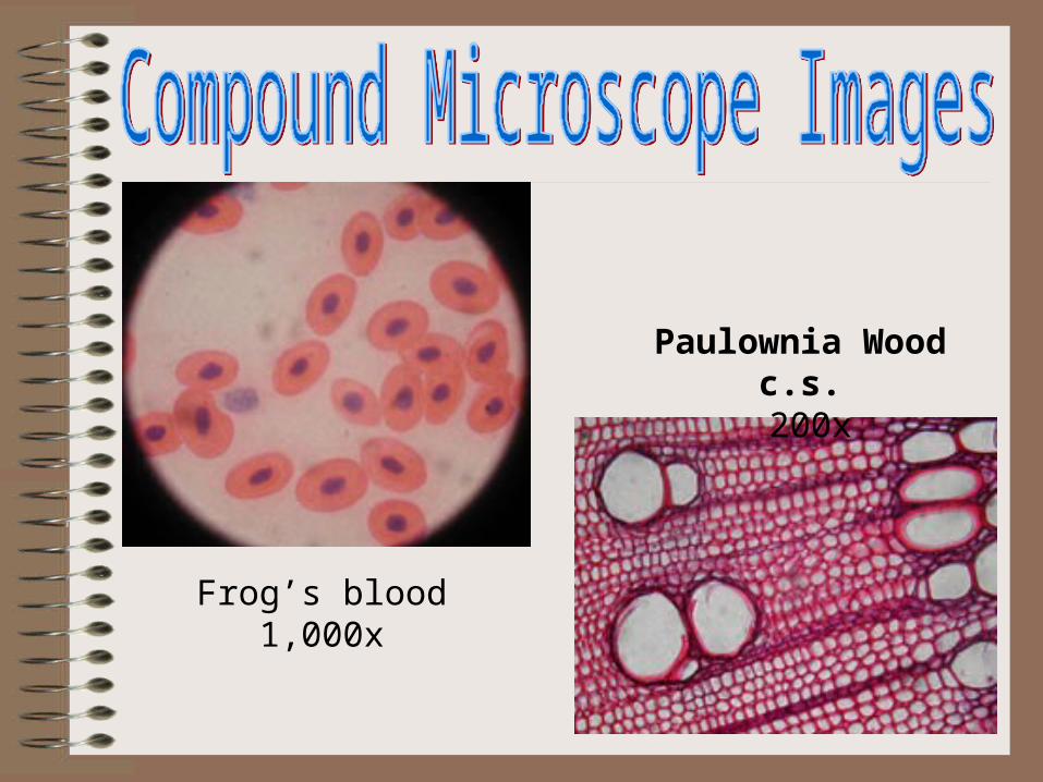
Frog’s blood1,000x
Paulownia Wood c.s. 200x
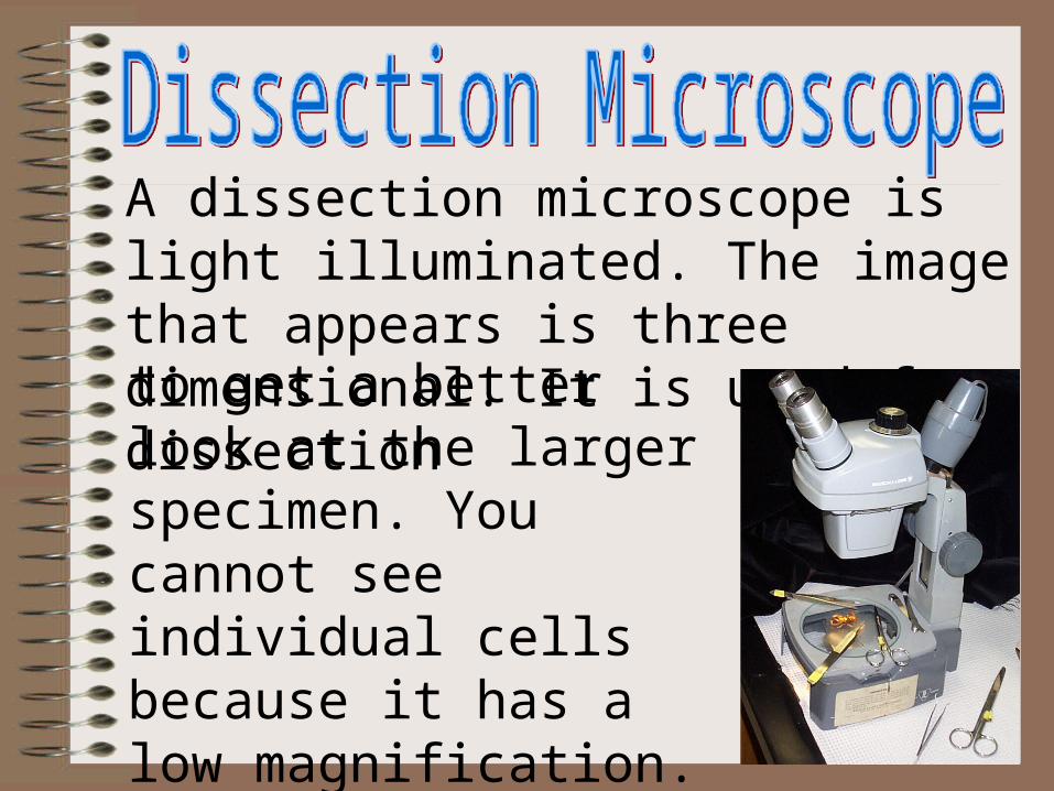
A dissection microscope is light illuminated. The image that appears is three dimensional. It is used for dissectionto get a better look at the larger specimen. You cannot see individual cells because it has a low magnification.(also called stereo microscope)
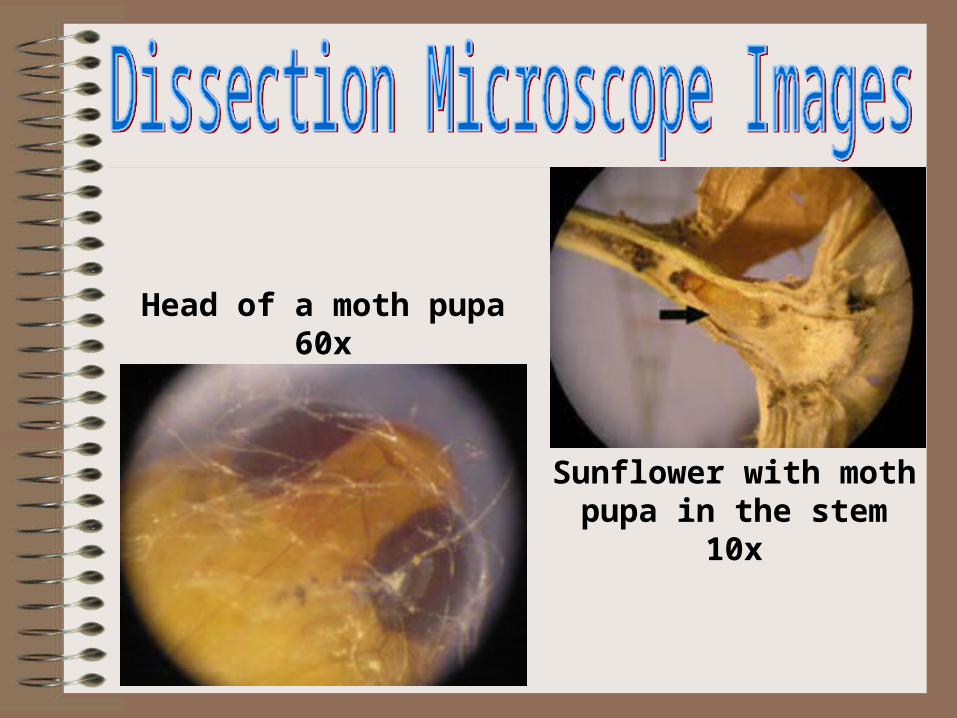
Sunflower with moth pupa in the stem
10x
Head of a moth pupa60x
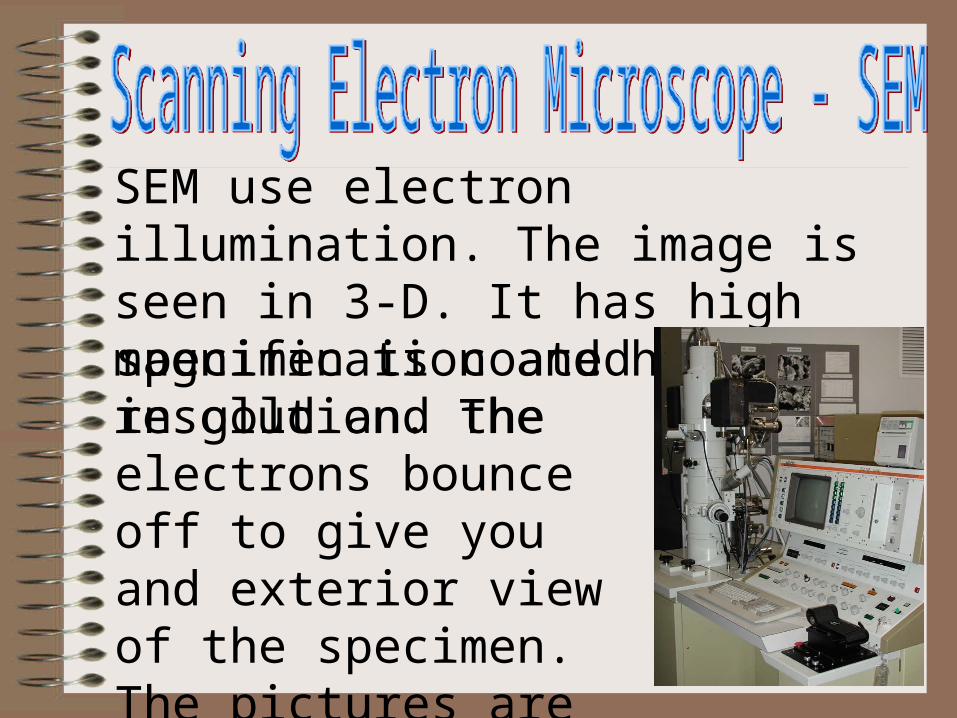
SEM use electron illumination. The image is seen in 3-D. It has high magnification and high resolution. Thespecimen is coated in gold and the electrons bounce off to give you and exterior view of the specimen. The pictures are in black and white.
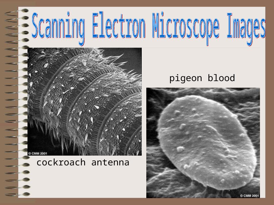
cockroach antenna
pigeon blood
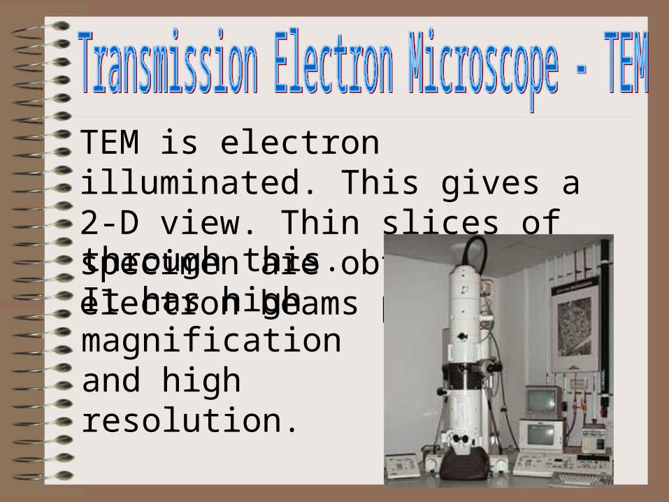
TEM is electron illuminated. This gives a 2-D view. Thin slices of specimen are obtained. The electron beams passthrough this. It has high magnification and high resolution.
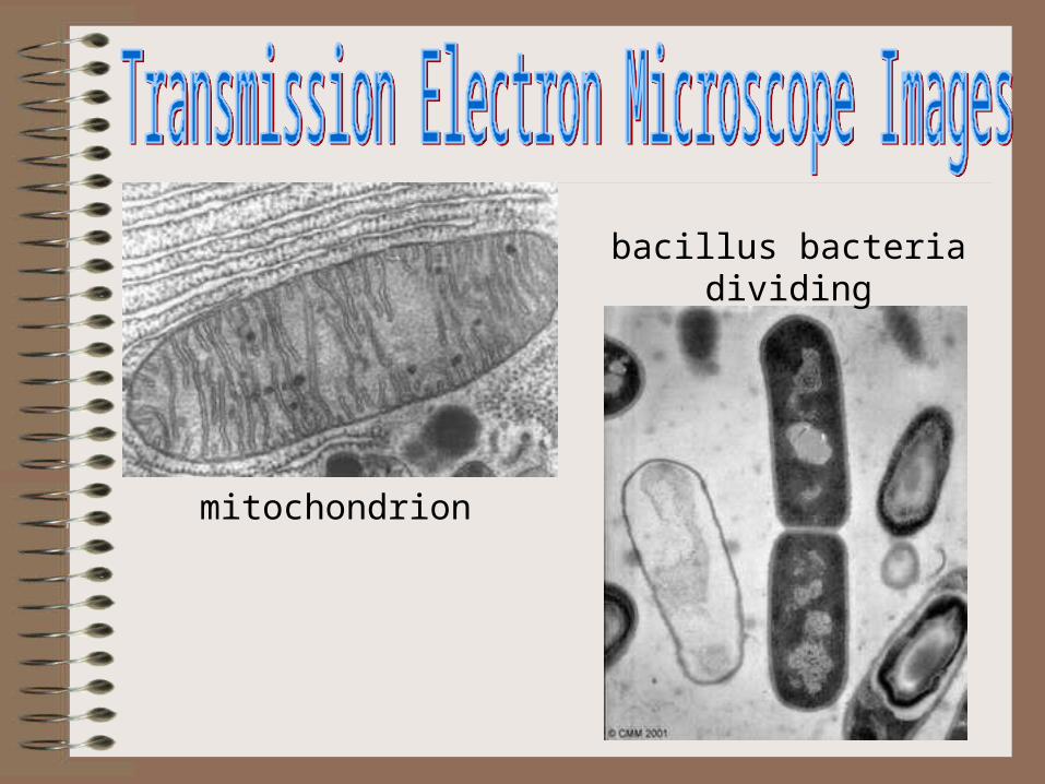
mitochondrion
bacillus bacteriadividing
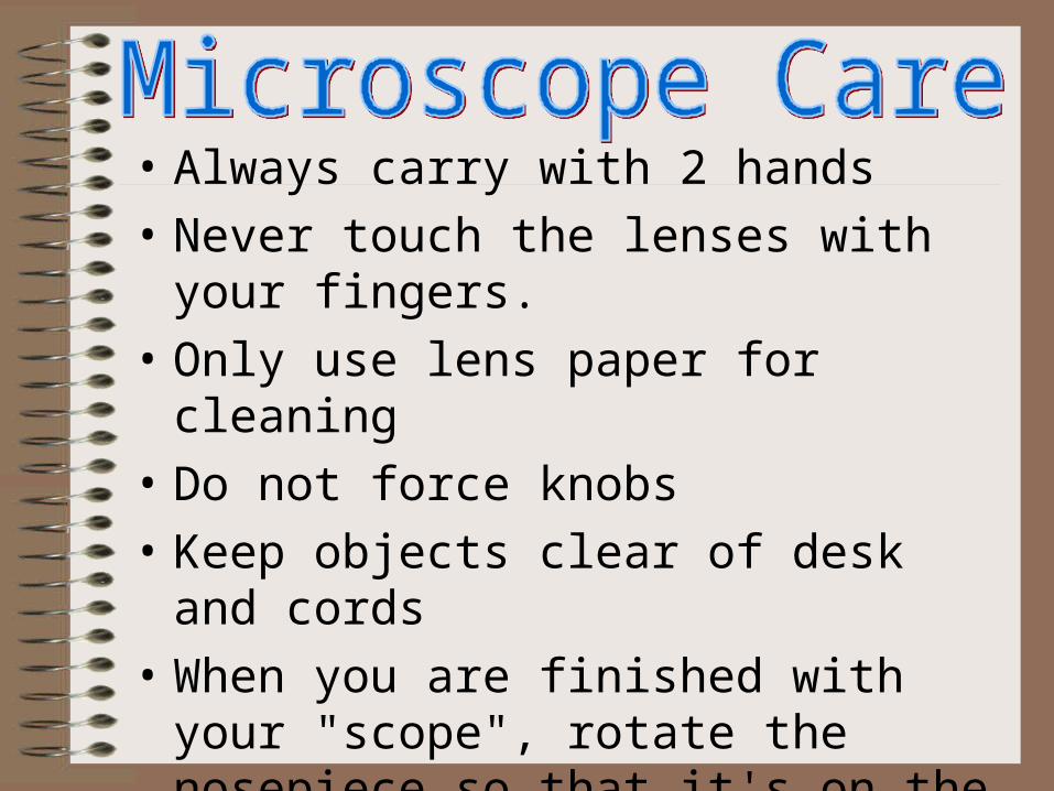
• Always carry with 2 hands
• Never touch the lenses with your fingers.
• Only use lens paper for cleaning
• Do not force knobs
• Keep objects clear of desk and cords
• When you are finished with your "scope", rotate the nosepiece so that it's on the low power objective, roll the stage down to lowest level, rubber band the cord, then replace the dust cover.
• .
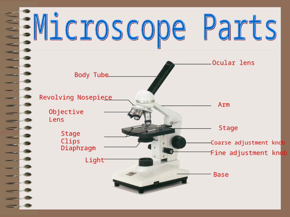
Ocular lens
Body Tube
Revolving NosepieceArm
Objective Lens
StageStage Clips
Coarse adjustment knob
Fine adjustment knob
Base
Diaphragm
Light
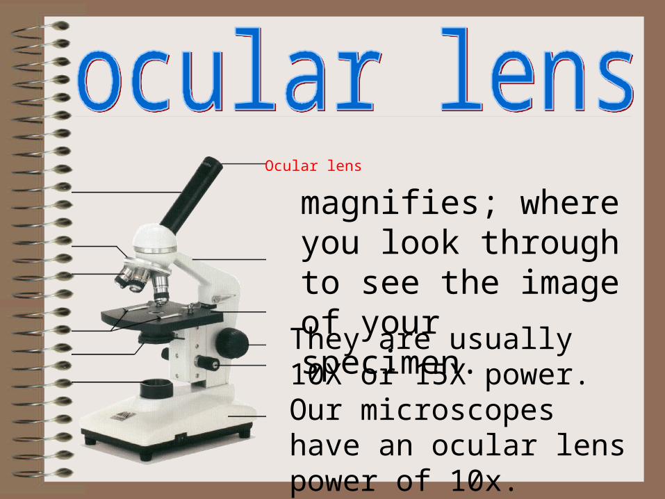
Ocular lens
magnifies; where you look through to see the image of your specimen.
They are usually 10X or 15X power. Our microscopes have an ocular lens power of 10x.
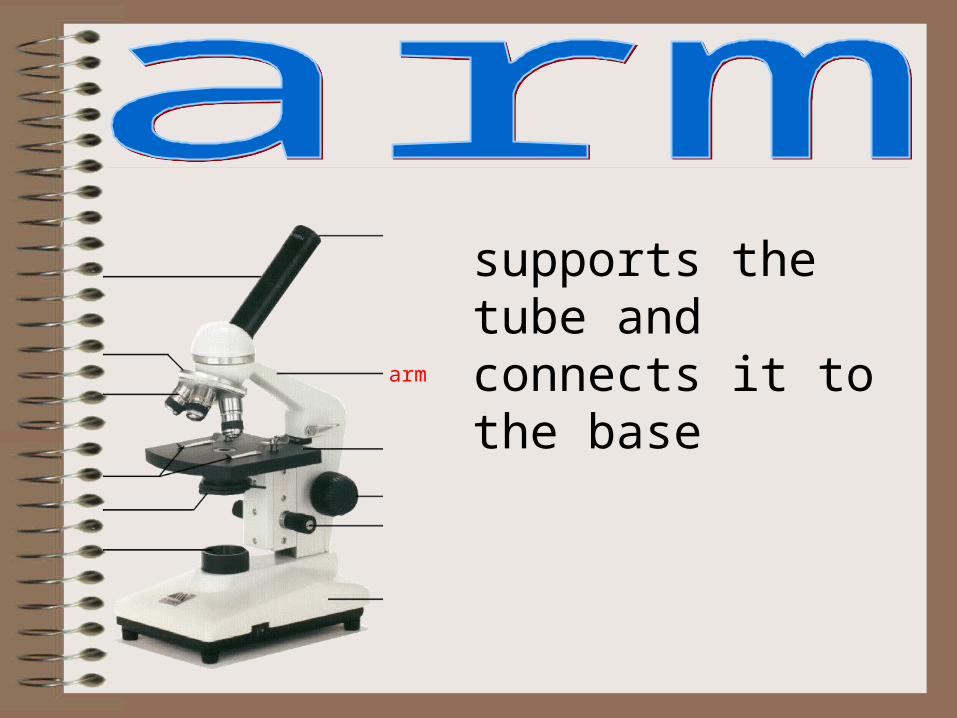
arm
supports the tube and connects it to the base

stage
the flat platform where you place your slides

coarse adjustment knob
moves stage (or body tube) up and down
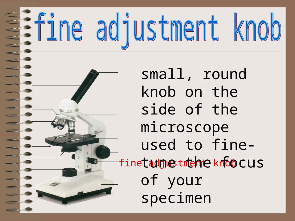
fine adjustment knob
small, round knob on the side of the microscope used to fine-tune the focus of your specimen
after using the coarse adjustment knob

base
the bottom of the microscope, used for support
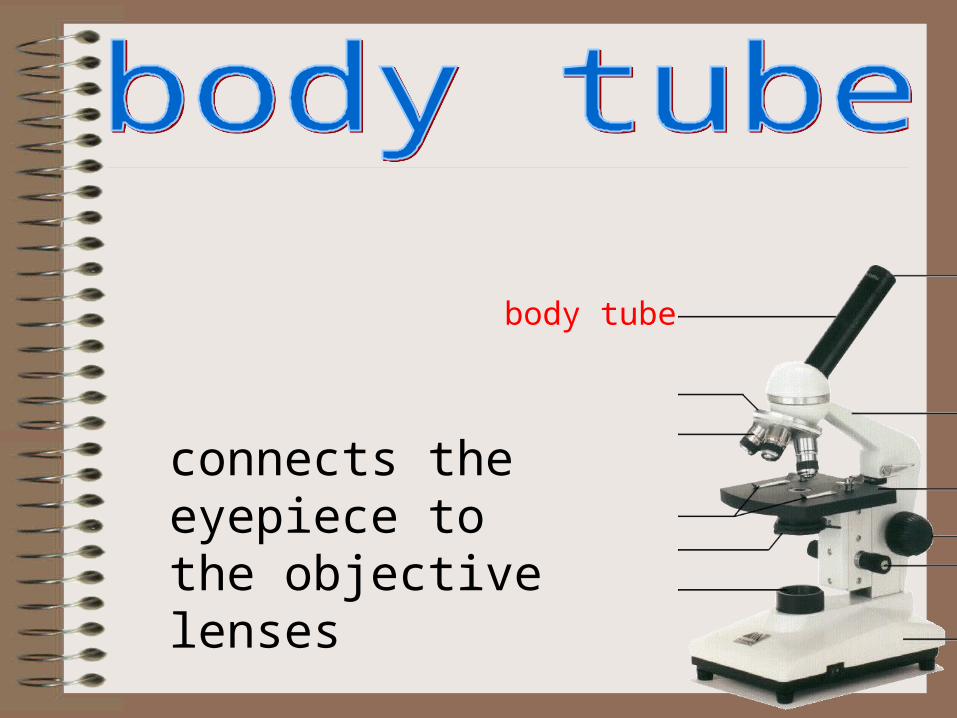
body tube
connects the eyepiece to the objective lenses

revolving nosepiece
the part that holds two or more objective lenses
and can be rotated toeasily change power
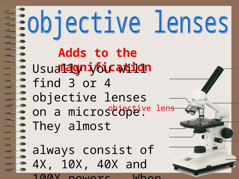
objective lens
Adds to the magnificationUsually you will find 3 or 4 objective lenses on a microscope. They almost
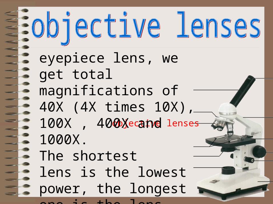
always consist of 4X, 10X, 40X and 100X powers. When coupled with a 10X (most common)
objective lenses
eyepiece lens, we get total magnifications of 40X (4X times 10X), 100X , 400X and 1000X.The shortestlens is the lowest power, the longest one is the lens with the greatest power. Lenses are color coded.
objective lenses
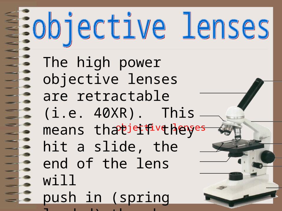
The high power objective lenses are retractable (i.e. 40XR). This means that if they hit a slide, the end of the lens will push in (spring loaded) thereby protecting the lens and the slide.
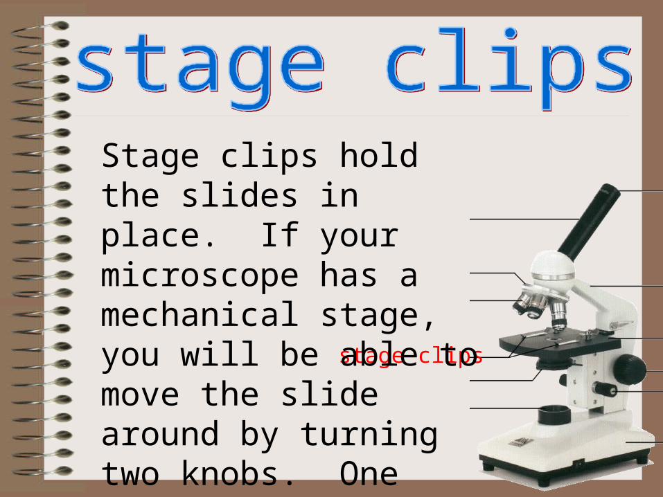
stage clips
Stage clips hold the slides in place. If your microscope has a mechanical stage, you will be able to move the slide around by turning two knobs. One moves it left and right, the other moves it up and down.
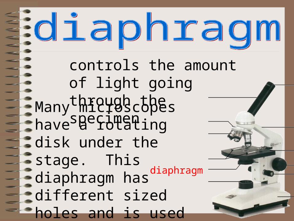
diaphragm
controls the amount of light going through the specimen
Many microscopes have a rotating disk under the stage. This diaphragm has different sized holes and is used to vary the intensity and size of the cone of light
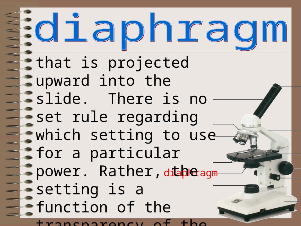
diaphragm
that is projected upward into the slide. There is no set rule regarding which setting to use for a particular power. Rather, the setting is a function of the transparency of the specimen, the degree of contrast you desire and the particular objective lens in use.
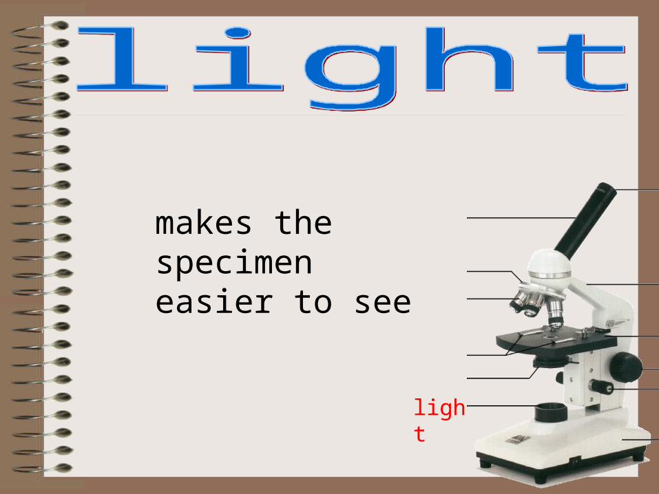
light
makes the specimen easier to see
The proper way to focus a microscope is to start with the lowest power objective lens first and while looking from the side, crank the lens down as close to the specimen as possible without touching it. Now, look through the eyepiece lens and focus upward only until the image is sharp. If you can't get it in focus, repeat the process again.
Once the image is sharp with the low power lens, you should be able to simply click in the next power lens and do minor adjustments with the focus knob. If your microscope has a fine focus adjustment, turning it a bit should be all that's necessary. Continue with subsequent objective lenses and fine focus each time.
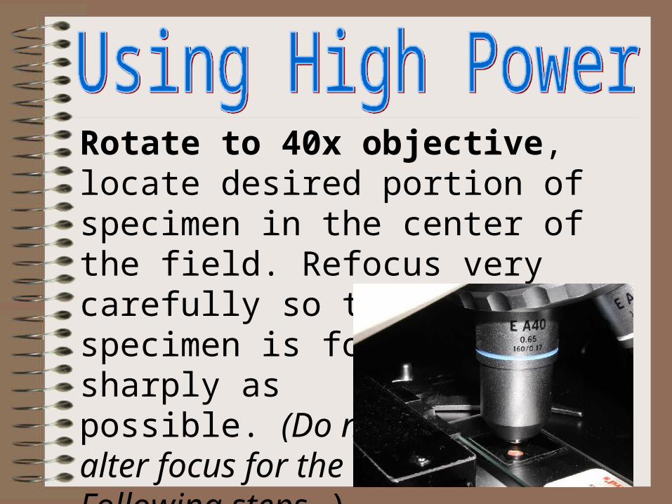
Rotate to 40x objective, locate desired portion of specimen in the center of the field. Refocus very carefully so that the specimen is focused as sharply as possible. (Do not alter focus for the Following steps )
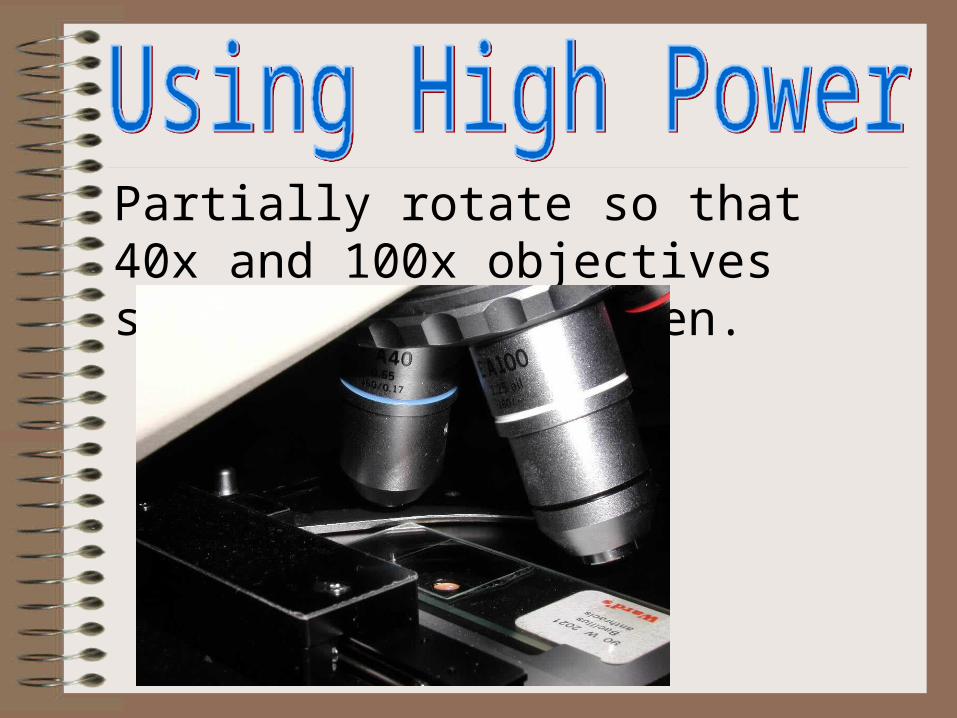
Partially rotate so that 40x and 100x objectives straddle the specimen.
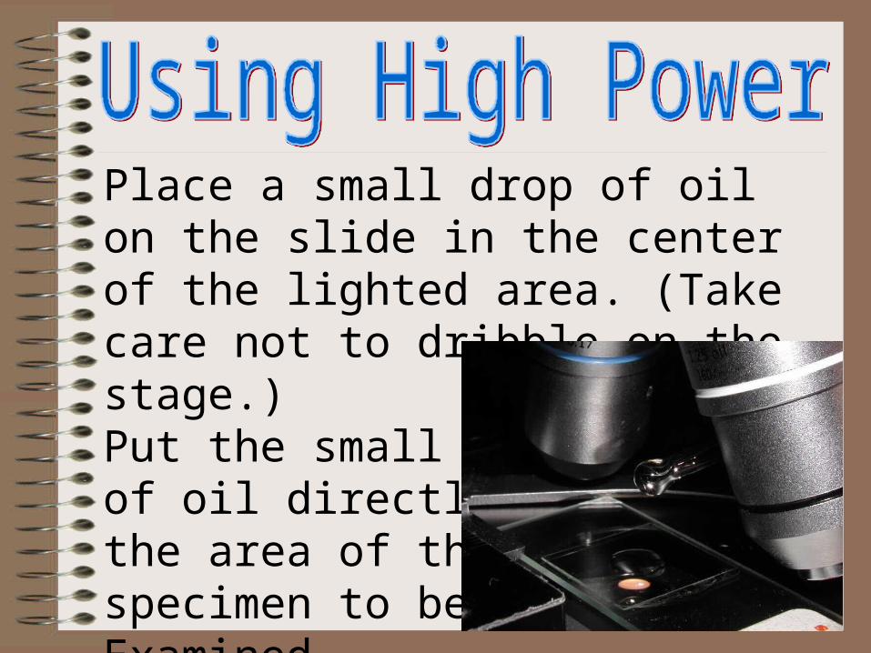
Place a small drop of oil on the slide in the center of the lighted area. (Take care not to dribble on the stage.)Put the small drop of oil directly over the area of the specimen to be Examined.
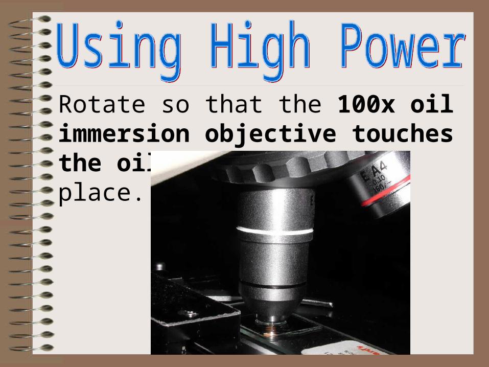
Rotate so that the 100x oil immersion objective touches the oil and clicks into place.
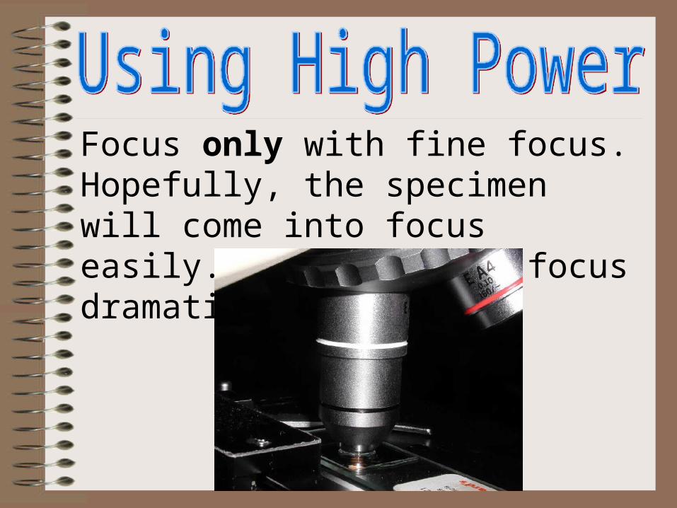
Focus only with fine focus. Hopefully, the specimen will come into focus easily. Do not change focus dramatically.

Clean up!: When you have finished for the day, wipe the 100x oil immersion objective carefully with lens paper to remove all oil. Wipe oil from the slide thoroughly with a Kimwipe. Cleanse stage should any oil have spilled on it. Recap the immersion oil container securely, replace in drawer.