Embed Size (px)
Citation preview

Introduction, Basic techniques: Restriction enzymes and restriction digestion, other enzymes used for DNA
manipulation: synthesis, joining and modification, , gel-electrophoresis: Principal, types, process
Mitesh Shrestha

Gene cloning
• DNA cloning is a technique for reproducing DNA fragments.
• It can be achieved by two different approaches:
– cell based
– Using polymerase chain reaction (PCR).
• a vector is required to carry the DNA fragment of interest into the host cell.

Gene cloning
• DNA cloning allows a copy of any specific part of a DNA (or RNA) sequence to be selected among many others and produced in an unlimited amount.
• This technique is the first stage of most of the genetic engineering experiments:
– Production of DNA libraries
– PCR
– DNA sequencing

Basic Steps of Gene Cloning
• Generation of DNA fragment/s – Restriction Digestion – PCR – Synthesis
• Insert into appropriate vector – Cloning Vector – Shuttle Vector – Expression Vector – Integrative Vector
• Insert the vector into appropriate host – Cloning Host/Intermediate Host – Expression Host
• Identify/Confirm the DNA fragment – Restriction Digestion – PCR – Transcriptome Analysis – Protein Expression Analysis – Sequencing

Enzymes used in molecular cloning
• Enzymes are tools of gene isolation, gene cloning, genetic engineering and gene transfer.
• Seven types of enzymes:
– Alkaline Phosphatase
– Terminal Transferase
– Polymerases
– Bacteriophage RNA Polymerases
– Nucleases: DNase and Rnase
– Polynucleotide Kinase and
– DNA Ligase.

Enzymes are main tool used for
• Cutting and joining- a DNA manipulative technique.
• Besides, DNA can be shortened, lengthened, copied into RNA or to new DNA and modified by addition and removal of specific group- all needs enzymes that are functioning in side living cell
• Nuclease Cleave DNA: endonuclease and exonuclease
• Modification of DNA: kinase, terminal transferase etc
• Synthesis of NA: DNA - and RNA polymerases, reverse transcriptase
• Joining two DNA fragments: ligase
• Industries produce these enzymes commercially

Alkaline Phosphatase
• Alkaline phosphatase removes 5′ phosphate groups from DNA and RNA.
• It will also remove phosphates from nucleotides and proteins.
• These enzymes are most active at alkaline pH therefore known as alkaline phosphatase.

Alkaline Phosphatase
• There are several sources of alkaline phosphatase that differ in how easily they can in-activated:
– Bacterial alkaline phosphatase (BAP) is the most active of the enzymes, but also the
most difficult to destroy at the end of the dephosphorylation reaction.
– Calf intestinal alkaline phosphatase (CIP) is purified from bovine intestine. This is
phosphatase most widely used in molecular biology labs because, although less active than BAP, it can be effectively destroyed by protease digestion or heat (75 °C for 10 minutes in the presence of 5 mM EDTA).
– Shrimp alkaline phosphatase is derived from a cold-water shrimp and is promoted for
being readily destroyed by heat (65°C for 15 minutes).
Alkaline Phosphatase
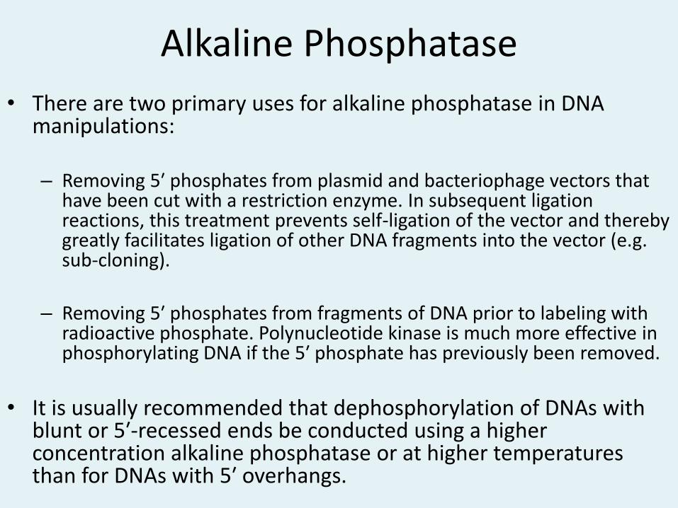
• There are two primary uses for alkaline phosphatase in DNA manipulations: – Removing 5′ phosphates from plasmid and bacteriophage vectors that
have been cut with a restriction enzyme. In subsequent ligation reactions, this treatment prevents self-ligation of the vector and thereby greatly facilitates ligation of other DNA fragments into the vector (e.g. sub-cloning).
– Removing 5′ phosphates from fragments of DNA prior to labeling with
radioactive phosphate. Polynucleotide kinase is much more effective in phosphorylating DNA if the 5′ phosphate has previously been removed.
• It is usually recommended that dephosphorylation of DNAs with
blunt or 5′-recessed ends be conducted using a higher concentration alkaline phosphatase or at higher temperatures than for DNAs with 5′ overhangs.

Terminal Transferase
• Terminal transferase catalyzes the addition of nucleotides to the 3′ terminus of DNA.
• Interestingly, it works on single-stranded DNA, including 3′ overhangs of double-stranded DNA, and is thus an example of a DNA polymerase that does not require a primer.
• It can also add homopolymers of nucleotides to the 3′ end of DNA.
• The much preferred substrate for this enzyme is protruding 3′ ends, but it will also, less efficiently, add nucleotides to blunt and 3′-recessed ends of DNA fragments.
• Cobalt is a necessary cofactor for activity of this enzyme. • Terminal transferase is a mammalian enzyme, expressed in
lymphocytes. • The enzyme purchased commercially is usually produced by
expression of the bovine gene in E. coli.

Terminal Transferase Uses
• Labeling the 3′ ends of DNA
– Most commonly, the substrate for this reaction is a fragment of DNA generated by digestion with a restriction enzyme that leaves a 3′ overhang, but oligodeoxynucleotides can also be used.
– When such DNA is incubated with tagged nucleotides and terminal transferase, a string of the tagged nucleotides will be added to the 3′ overhang or to the 3′ end of the oligonucleotide.
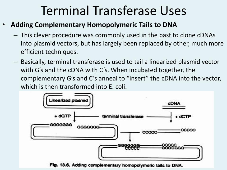
Terminal Transferase Uses • Adding Complementary Homopolymeric Tails to DNA
– This clever procedure was commonly used in the past to clone cDNAs into plasmid vectors, but has largely been replaced by other, much more efficient techniques.
– Basically, terminal transferase is used to tail a linearized plasmid vector with G’s and the cDNA with C’s. When incubated together, the complementary G’s and C’s anneal to “insert” the cDNA into the vector, which is then transformed into E. coli.

Polymerases
• DNA polymerases are enzymes that catalyse the synthesis of a new DNA strand from a pre-existing strand.
• The enzyme adds deoxyribonucleotides to the free 3’-OH of the chainundergoing elongation. The direction of synthesis is 5’-3’.
• It has three major requirements for its activity – a template strand for which the enzyme synthesizes a
complementary strand; – a primer with a free 3’-OH group that hybridizes with the
template to form a double stranded region that initiates the polymerization and
– A pool of all the four dNTPs that are used to synthesize the new DNA strand.
• In addition, some cofactors like Mg2+ ions may be required in a buffer solution with correct pH for optimum activity.

Different types of DNA polymerases
• E. coli DNA Polymerase I
• Klenow Fragment
• Thermostable DNA Polymerases
• Reverse Transcriptase
E.coli DNA polymerase • Single polypeptide 105,000 mol weight
• Add nucleotides to 3’ end of DNA primer hybridized to ssDNA template
• Short ss DNA or RNA work as primer
• Complementary strand (single strand) of synthesized DNA works as template
• Show 5’ to 3’ polymerase activity
• Show 5’ to 3’ exonuclease activity for proof reading
• Show 3’ to 5’ exonuclease activity in single strand.
• Proof reading activity: error rate 10-6 without exonuclease activity it will be 10-4
• Dual activity-DNA polymerization and DNA degradation
• Used for DNA labelling and nick translation
E. coli DNA Polymerase I
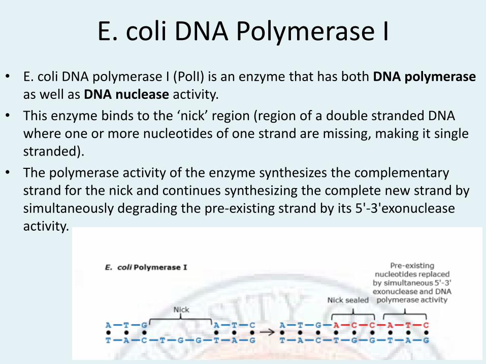
• E. coli DNA polymerase I (PolI) is an enzyme that has both DNA polymerase as well as DNA nuclease activity.
• This enzyme binds to the ‘nick’ region (region of a double stranded DNA where one or more nucleotides of one strand are missing, making it single stranded).
• The polymerase activity of the enzyme synthesizes the complementary strand for the nick and continues synthesizing the complete new strand by simultaneously degrading the pre-existing strand by its 5'-3'exonuclease activity.
E. coli DNA Polymerase I
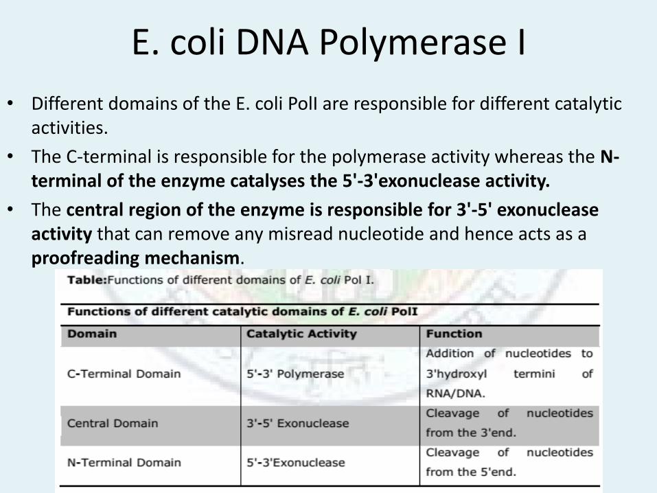
• Different domains of the E. coli PolI are responsible for different catalytic activities.
• The C-terminal is responsible for the polymerase activity whereas the N-terminal of the enzyme catalyses the 5'-3'exonuclease activity.
• The central region of the enzyme is responsible for 3'-5' exonuclease activity that can remove any misread nucleotide and hence acts as a proofreading mechanism.

Klenow fragment • The first 323 AA fragment of E.coli polymerase shows nuclease
activity so its removal retain polymerase function. • Thus produced larger fragment of mol. Wt. 68,000 is called Klenow
fragment. • It shows DNA polymerase activity with low 3’ to 5’ exonuclease
activity. • No 5’ To 3’ exonuclease activity • Used to synthesise DNA on single strand template, label 3’end with
radioactive nucleotides • To fill nick or gap-nick translation
5’P 3’OH 3’OH 5’P
↓Klenow Fragment 5’P 3’OH 3’OH 5’P
Fig. 3’-End labelling

Klenow fragment
• If the E. coli Pol I holoenzyme is treated with amild protease, it results in the formation of two fragments.
• A larger fragment retaining both 5'-3' polymerase and 3'-5‘ exonuclease activities; while the smaller one has only the 5'-3' exonuclease activity. The larger fragment is known as ‘Klenow fragment’.

Klenow fragment
• Klenow fragment can synthesize the new DNA strand complementary to the template but cannot degrade the existing strand.
• Klenow fragment is predominantly used in DNA sequencing.
• Other uses in recombinant DNA technology where Klenow fragment is used are
– Synthesis of double stranded DNA from single stranded template.
– Filling of 5’ overhangs created by restriction enzymesto create blunt ends.
– Digestion of protruding 3’ overhangs to produce blunt ends.

Bacteriophage T4 and T7 polymerase
• Produced by E.coli cells infected by bacteriophage T4 or T7 T4-polymerase • 3’to 5’ single strand exonuclease activity stronger than Klenow fragment • No 5’ to 3’ exonuclease activity • The enzyme creates blunt end from both 5'-protruding and 5'-recessed
DNA termini • Used to fill 5’ protruding end, to add radioactive nucleotide
5’P 3’OH 3’OH 5P’ 3’OH 5’P 5’P 3’OH Mg2+ ↓ 3’to5’ exonuclease activity Mg2+↓ polymerase activity 5’P 3’OH 3’OH 5’P 3’OH 5’P 5’P Fig. 3’to 5’ Exonuclease and polymerase activity

T7-polymerase
• High 3’ to 5’ exonuclease activity
• Modified enzymes with low 3’ to 5’ exonuclease activity used in DNA sequencing.
5’P (primer)
3’OH 5’P
Mg2+, dNTPs↓Polymerase activity
5’P 3’OH
3’OH 5’P
Fig. Polymerase activity
Taq polymerase • Active also at high temperature. Found in a hot spring bacteria Thermus acquaticus. The enzyme is resistant to thermal denaturation • Used in PCR as high polymerase activity is seen at higher temperature. • No 3’ to 5’ exonuclease activity so no proof reading

Thermo-stable DNA Polymerases
• The thermophilic DNA polymerases, like other DNA polymerases, catalyze template-directed synthesis of DNA from nucleotide triphosphates.
• In general, they have maximal catalytic activity at 75° to 80°C, and substantially reduced activates at lower temperatures.
• At 37°C, Taq polymerase has only about 10% of its maximal activity.
• In addition to Taq DNA polymerase, several other thermostable DNA polymerases have been isolated and expressed from cloned genes.
Reverse transcriptase
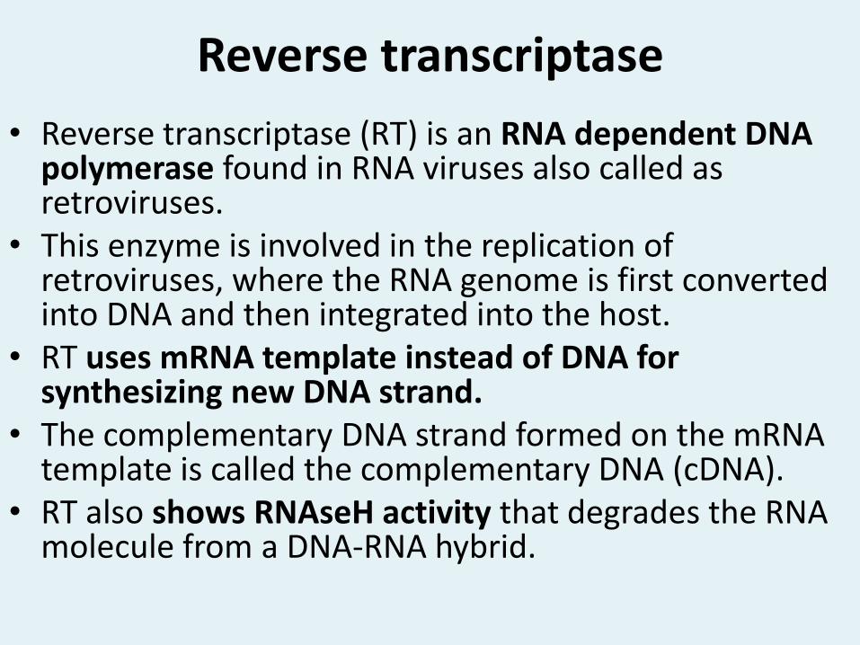
• Reverse transcriptase (RT) is an RNA dependent DNA polymerase found in RNA viruses also called as retroviruses.
• This enzyme is involved in the replication of retroviruses, where the RNA genome is first converted into DNA and then integrated into the host.
• RT uses mRNA template instead of DNA for synthesizing new DNA strand.
• The complementary DNA strand formed on the mRNA template is called the complementary DNA (cDNA).
• RT also shows RNAseH activity that degrades the RNA molecule from a DNA-RNA hybrid.

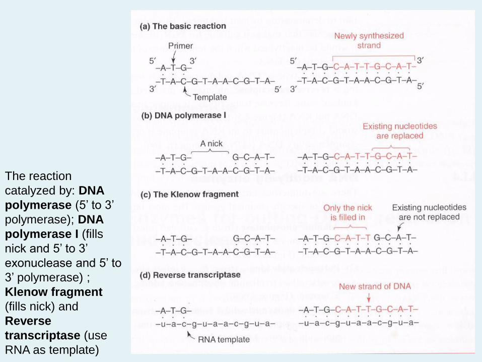
The reaction
catalyzed by: DNA
polymerase (5’ to 3’
polymerase); DNA
polymerase I (fills
nick and 5’ to 3’
exonuclease and 5’ to
3’ polymerase) ;
Klenow fragment
(fills nick) and
Reverse
transcriptase (use
RNA as template)

Bacteriophage RNA Polymerases
• Phage encoded DNA dependent RNA polymerases are used for in vitro transcription to generate defined RNAs.
• Most commonly, the reaction utilizes ribonucleotides that are labeled with radionuclides or some other tag, and the resulting labeled RNA is used as a probe for hybridization.
• Other applications of in vitro transcription including making RNAs for in vitro translation or to study RNA structure and function.
• Several bacteriophage RNA polymerases are commercially available. They are named after the phage that encodes them, and either purified from phage infected bacteria or produced as recombinant proteins.
• Many of the plasmids used for carrying cloned DNA incorporate promoters for bacteriophage RNA polymerases adjacent to the cloning site. This allows one to readily obtain either mRNA sense or antisense transcripts from the inserted DNA.
• The process is often called runoff transcription, because the plasmid is cut with a restriction enzyme downstream of the inserted DNA, Which causes the polymerase to fall off the template when it reaches that spot.


Nucleases
• Deoxyribonucleases (DNases) and ribonucleases (RNases) have certain indispensible roles in molecular biology laboratories.
• Numerous types of DNase and RNase have been isolated and characterized.
• They differ among other things in substrate specificity, cofactor requirements, and whether they cleave nucleic acids internally (endonucleases), chew in from the ends (exonucleases) or attack in both of these modes.
• In many cases, the substrate specificity of a nuclease depends upon the concentration of enzyme used in the reaction, with high concentrations promoting less specific cleavages.
Nucleases
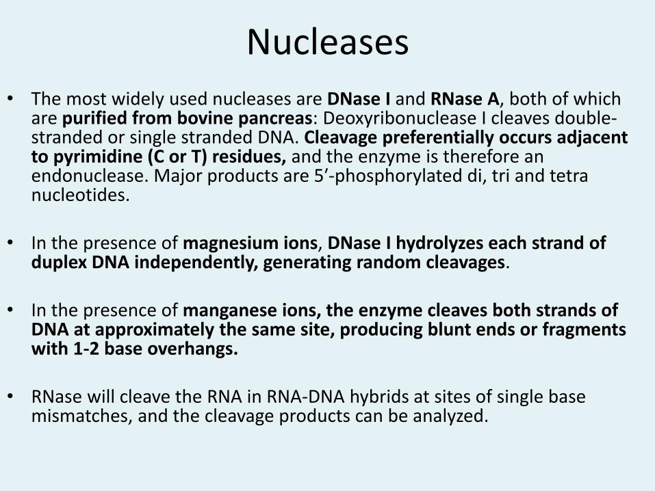
• The most widely used nucleases are DNase I and RNase A, both of which are purified from bovine pancreas: Deoxyribonuclease I cleaves double-stranded or single stranded DNA. Cleavage preferentially occurs adjacent to pyrimidine (C or T) residues, and the enzyme is therefore an endonuclease. Major products are 5′-phosphorylated di, tri and tetra nucleotides.
• In the presence of magnesium ions, DNase I hydrolyzes each strand of duplex DNA independently, generating random cleavages.
• In the presence of manganese ions, the enzyme cleaves both strands of DNA at approximately the same site, producing blunt ends or fragments with 1-2 base overhangs.
• RNase will cleave the RNA in RNA-DNA hybrids at sites of single base mismatches, and the cleavage products can be analyzed.

Dnase I Applications
• Eliminating DNA (e.g. plasmid) from preparations of RNA.
• Analyzing DNAprotein interactions via DNase foot printing.
• Nicking DNA prior to radiolabeling by nick translation.

Nucleases
• Nucleases are enzymes that degrade DNA molecules by breaking the phosphodiester bonds that link one nucleotide to the next in a DNA strand.
• Nucleases can be broadly categorized into
– Exonucleases
– Endonucleases.
• Exonuclease removes the terminal nucleotide of the DNA molecule by breaking the phosphodiester bond, whereas endonuclease breaks the internal phosphodiester bond.


Exonuclease
• Different types of exonucleases can be categorised on the basis of number of strands they degrade in a double stranded DNA molecule.
• An exonuclease named Bal31 is isolated from a marine bacterium Alteromonas espejiana. It is a Ca2+ dependent enzyme that degrades the nucleotides from both the strands of dsDNA molecule. The longer the DNA is incubated with Bal31, the shorter the DNA molecule becomes.
• On the contrary, an enzyme isolated from E. coli called exonuclease III digests only one strand of the dsDNA molecule. It removes the nucleotide from the 3' terminus of the strand, thus leaving protruding 5' overhangs. Exonuclease III is used for generating single stranded templates.


Endonucleases
• Similar to exonucleases, endonucleases can also be categorised based on whether they act on single or double stranded DNA.
• S1 nuclease is an endonuclease that is isolated from the fungus Aspergillus oryzae. It is a heat stable enzyme that functions at high ionic strength, low pH and in the presence of Zn2+ ions. It cleaves only single stranded DNA. Also, it is able to cleave the single stranded nicks in dsDNA molecules.
• Another type of endonuclease called as DNase I that is isolated from cow’s pancreas is a non– specific enzymes. It is able to cleave both single and double stranded DNAs. It can cleave any of the internal phosphodiester bonds, thus prolonged digestion of DNA with DNase I results in its complete chewing leaving only a mixture of mononucleotides.

Restriction Endonucleases
• Another class of endonucleases are called as restriction endonucleases.
• The cleavage of DNA by these enzymes is very specific at ‘particular sites’.
• Specific order of nucleotide sequences are recognized by restriction enzymes on the DNA that are then cleaved.
• Two kinds of ends (blunt or staggered) may be formed due to digestion of DNA by different kinds of restriction enzymes.


Molecular Scissors: Restriction Endonucleases
• The foundation of molecular cloning was laid with the discovery of restriction enzymes.
• For DNA cloning, the vehicle carrying the DNA i.e., the vector must be cleaved to open up the circle. It is absolutely essential that the cutting is very precise.
• If the vector is cut in a random fashion generating two or more fragments, the vector DNA becomes nonfunctional. Restriction endonucleases enable this precise cleavage and hence are also known as ‘molecular scissors’.
• Generally every cloning vector has a ‘polylinker region’ also called as ‘multiple cloning site’ that is comprised of many restriction sites.
• This enables the molecular biologists to choose different restriction enzymes for multiple cloning procedures. The discovery of restriction enzymes was a crucial step that has revolutionized the field of genetic engineering.


Types of Restriction Endonucleases
• Type I
• Type II
• Type III
• Type IV
• Type V

Type I
• Type I restriction enzymes have a complex structure with three different subunits (endonuclease, methyl transferase and recognition).
• Need ATP, Mg 2+ and S-adenosylmethionine as cofactors.
• Recognition site is 15 bp length and cleavage site is at least 1000 bp away from the recognition site.
• Cleaves DNA when DNA loops back. • Cleavage occurs only if the Adenine of recognition site is not methylated. • Recognition site is specific but cleavage site is unspecific.
• Can cut only once and is inactivated.
• Their restriction sites are also complex and discontinuous with spacers.
• They may have biological significance but are of very little practical value since the site where the DNA is going to be digested cannot be predicted.


Type III
• With two subunits one for site recognition and modification and other for nuclease activity.
• Require ATP and Mg 2+ as cofactor.
• Cleave at specific sequence (non-palindromic) near to recognition site (two separate non-palindromic sequences that are inversely oriented) usually ~24 to 28 bp down stream. Single strand end produce but the end differ always and can not be recommended for cloning
• Not used in gene cloning.


Type IV
• Recognition sites for these enzymes consist of modified DNA, usually methylated sequences.
• Examples are the McrBC and Mrr systems of E. coli.

Type V
• These enzymes use guide RNAs to target specific non-palindromic sequences found in invading organisms, and cut DNA to generate fragments of variable lengths.
• An example is the cas9-gRNA complex from CRISPRs, which can be utilised for genetic engineering applications.

Type II • Main molecular scissors of dsDNA used in cloning
• Cut both strands of DNA within cleavage site-at
middle of recognition site.
• Separate proteins operating independently show endonuclease and methylase activities.
• Specific recognition site and cleave also at this site. The recognisiotion site is tetra-, penta-, hexa- or octanucleotide (4, 5, 6 or 8).

Type II • How often cleaves a RE depends on the length of
Recog. site: 6 at every 4096, 4 at every 256 calculated by 4n, where n is the number of nucleotide in recognition site.
• Require Mg 2+ as cofactor.
• The two strands of recognition site are always palindromic sequence (Inverse repeating sequence). The sequences are always written 5’ to 3’ direction for sense strand and 3’ to 5’ direction for antisense strand when both strand is written.
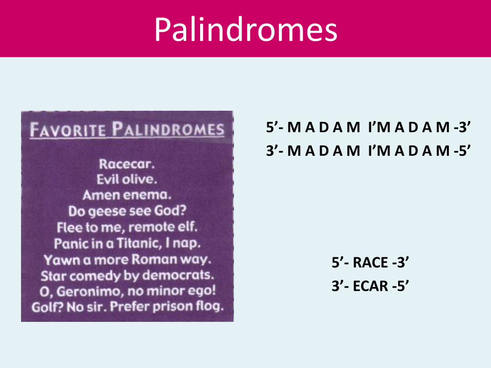
Palindromes
5’- M A D A M I’M A D A M -3’
3’- M A D A M I’M A D A M -5’
5’- RACE -3’
3’- ECAR -5’

• All DNA cleaved by same RE have identical termini
• Recognition site of EcoRI is
5’–GAATTC-3’
3’–CTTAAG-5’
Staggered cut: Cut two strands in a distance of few bases. After cleavage produce sticky ends (cohesive ends) in the fragments.
↓
5’-GAATTC-3’OH
3’-CTTAAG-5’
↑
P5’-G3’OH 5’P-AATTC-3’OH
3’-CTTAA-5’P 3’OHG-5’
Cleavage by EcoRI producing sticky ends

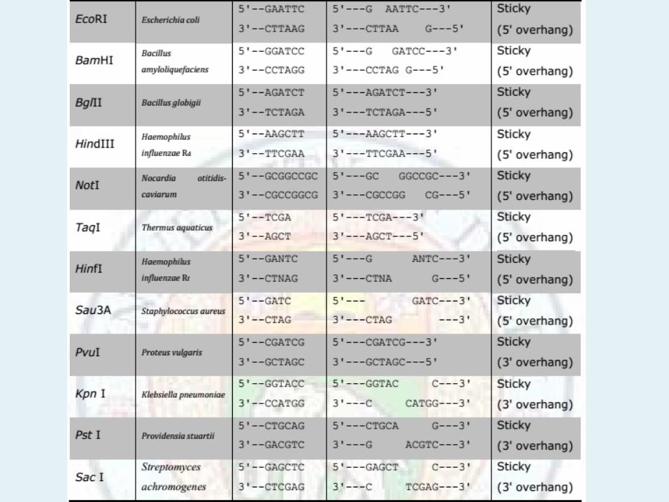
The overhanging ends may be with 5’P or 3’OH
EcoRI produce 5’P overhanging end where as PstI (from Providencia stuarti) produce 3’OH overhanging ends.
↓
5’-CTGCAG-3 3’-GACGTC-5’ ↑ 5’-CTGCA3’OH OH3’-G 3’-G5’P HO3’ACGTC-5’
Fig.Cleavage by RE PstI

Nomenclature of Restriction Endonucleases
• At present, more than 3000 restriction endonucleases have been identified.
• Each has been provided a definite name.
• Enzyme nomenclature is based on the bacterium from which it was isolated. The first three letters of the enzyme name are derived from the first letter of the genus name and the first two letters of the species name. Since each bacterium may contain several different restriction enzymes, a Roman numeral is also used to identify each enzyme.
• In the case of enzymes isolated from different strains of the same bacterium, first letter of the strain is used before the Roman numeral.
• For example, EcoRI is derived from Escherichia (genus),coli(species), RY1 (strain), I (first identified from bacterium).

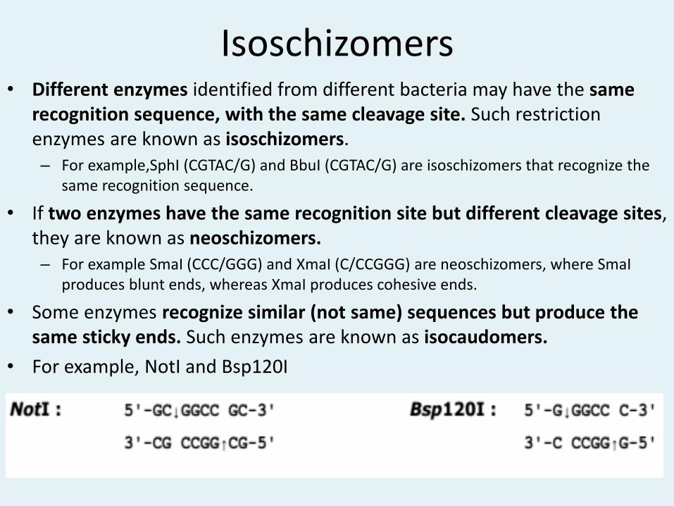
Isoschizomers • Different enzymes identified from different bacteria may have the same
recognition sequence, with the same cleavage site. Such restriction enzymes are known as isoschizomers. – For example,SphI (CGTAC/G) and BbuI (CGTAC/G) are isoschizomers that recognize the
same recognition sequence.
• If two enzymes have the same recognition site but different cleavage sites, they are known as neoschizomers. – For example SmaI (CCC/GGG) and XmaI (C/CCGGG) are neoschizomers, where SmaI
produces blunt ends, whereas XmaI produces cohesive ends.
• Some enzymes recognize similar (not same) sequences but produce the same sticky ends. Such enzymes are known as isocaudomers.
• For example, NotI and Bsp120I

• Isochizomeres: Two REs with same recognition and cleavage site. Example:
Hind II and HsuI both with same recognition and cleavage site are isochizomeres
↓ 5’–AAGCTT-3’ 3’-TTCGAA-5’ ↑ Two REs may have same recognition site but different cleavage
sites. ↓ ↓ 5’CCCGGG3’ 5’CCCGGG3 3’GGGCCC5’ 3’GGGCCC5’ ↑
↑ XmaI SmaI

Some REs recognize different recognition sequence but produce same sticky ends
↓ ↓
5’-GGATCC-3’ 5’-AGATCT-3
3’-CCTAGG-5’ 3’-TCTAGA-5’ ↑ ↑
BamHI BglII
5’-G-3’ 5’-GATCT-3’ 3’-CCTAG-5’ 3’-A-5’
Joining two fragment result fragment that is not cut by
both enzymes

BamH I and Bgl II
producing similar
sticky ends

Some RE are not quite specific
• Acc I is not quite specific. Its recognition and cleavage site is
↓AG
5’-GTCTAC-3’ 3’-CAGATG-5’ TC↑ If C replaced by A and/or G by T the changed
sequence will be also recognized by the enzyme.

Restriction of the λ
DNA molecule by
REs producing
different size
fragments.
RE activity is dependent upon
• pH, salt concentration, methylation of bases of recognition
sequence. Optimum buffer (in 10X supplied with enzyme).
• RE can work in wide range of pH and salt concentration but the
efficiency will decrease
• Methylation of bases may reduce the efficiency completely but
some enzyme recognize the sequence also after methylation

Star activity
• Change of relaxation of specificity.
• Some enzymes change their specificity to restriction site when reaction condition changes like change in pH (high), change in NaCl concentration (low), Mg 2+ replaced by Mn 2+ , use of organic solvents, methylation.
• Example: EcoRI recognizes GAATTC but if condition changes also recognize AATT.

DNA Methylase
• E.coli consists of common dam - and dcm methylase-methylate A and C respectively present at specific sequence. The enzymes transfer methyl group from S-adenosyl methionine at N6 of adenine and 5 of cytosine at specific sequence present in recognition sequence GATC and CCAGG or CCTGG respectively.
• EcoRI methylase methylate restriction site of EcoRI as -GAmATTC- the enzyme used in clonning cDNA fragment


Applications of restriction enzymes
• Traditional Cloning
• DNA Mapping
• Understanding epigenetic modifications

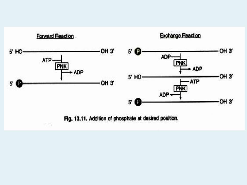
Polynucleotide Kinase
• Polynucleotide kinase (PNK) is an enzyme that catalyzes the transfer of a phosphate from ATP to the 5′ end of either DNA or RNA.
• It is a product of the T4 bacteriophage, and commercial preparations are usually products of the cloned phage gene expressed in E. coli.

Utilized in two types of reactions
• In the “forward reaction”, PNK transfers the gamma phosphate from ATP to the 5′ end of a polynucleotide (DNA or RNA). The target nucleotide is lacking a 5′ phosphate either because it has been dephosphorylated or has been synthesized chemically.
• In the “exchange reaction”, target DNA or RN A that has a 5′ phosphate is incubated with an excess of ADP, PNK will first transfer the phosphate from the nucleic acid onto an ADP, forming ATP and leaving a dephosphorylated target. PNK will then perform a forward reaction and transfer a phosphate from ATP onto the target nucleic acid.

Applications of Polynucleotide Kinase
• Phosphorylating linkers and adaptors (fragments of DNA ready for ligation) which require a 5′ phosphate. This includes products of polymerase chain reaction, which are typically generated using non- phosphorylated primers.
• Radiolabelling oligonucleotides, usually with 32P, for use as hybridization probes. PNK is inhibited by small amounts of ammonium ions, so ammonium acetate should not be used to precipitate nucleic acids prior to phosphorylation. Low concentrations of phosphate ions, or NaCl concentrations greater than about 50 mM, also inhibit this enzyme.

DNA Ligase
• The term recombinant DNA includes the concept of recombining fragments of DNA from different sources into a new and useful DNA molecule. Joining linear DNA fragments together with covalent bonds is called ligation.
• More specifically, DNA ligation involves creating a phosphodiester bond between the 3′ hydroxyl of one nucleotide and the 5′ phosphate of another.
• The enzyme used to ligate DNA fragments is T4 DNA ligase, which originates from the T4 bacteriophage. This enzyme will ligate DNA fragments having overhanging, cohesive ends that are annealed together.
• This is equivalent to repairing “nicks” in duplex DNA.
• T4 DNA ligase will also ligate fragments with blunt ends, although higher concentrations of the enzyme are usually recommended for this purpose.


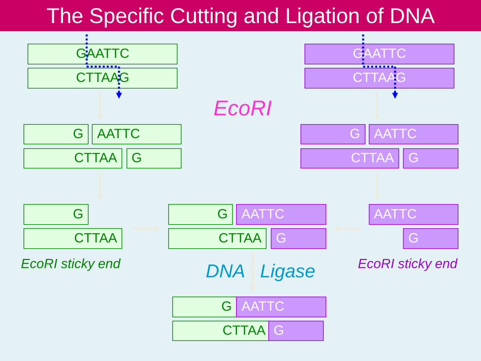
The Specific Cutting and Ligation of DNA
GAATTC
CTTAAG
GAATTC
CTTAAG
G
CTTAA
AATTC
G
AATTC
G
G
CTTAA
G
CTTAA
AATTC
G
G
CTTAA
AATTC
G
G
CTTAA
AATTC
G
EcoRI
DNA Ligase EcoRI sticky end EcoRI sticky end