Embed Size (px)
Citation preview

8/12/2019 Intro Histo Male Genital
http://slidepdf.com/reader/full/intro-histo-male-genital 1/51
HISTOLOGY DEPARTMENT
MEDICAL FACULTY
BANDUNG ISLAMIC UNIVERSITY
1
8/12/2019 Intro Histo Male Genital
http://slidepdf.com/reader/full/intro-histo-male-genital 2/51
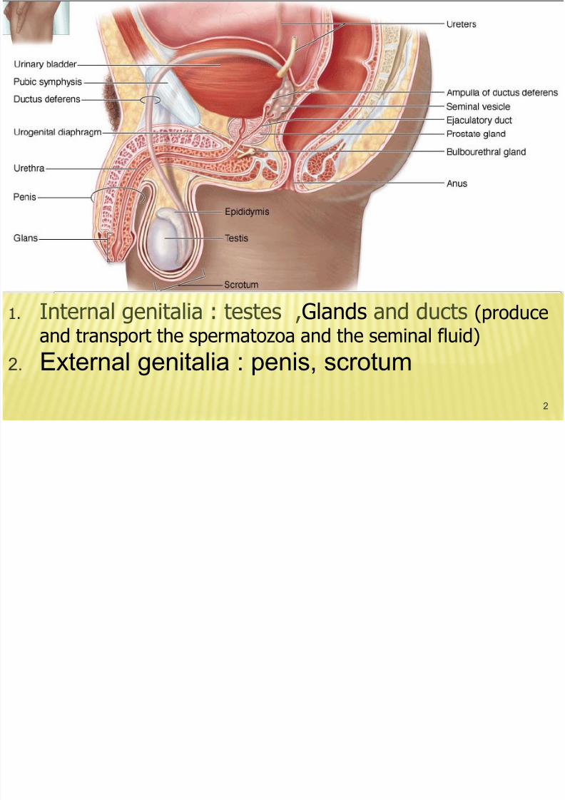
1. Internal genitalia : testes ,Glands and ducts (produce
and transport the spermatozoa and the seminal fluid)
2. External genitalia : penis, scrotum
2

8/12/2019 Intro Histo Male Genital
http://slidepdf.com/reader/full/intro-histo-male-genital 3/51
3
Urethra
Penis
Corpus cavernosumof the penis
Corpus cavernosumof the urethra
Prepuce
Glans penis
Testicular lobules
Tunica albuginea
Tunica vaginalis
Tubuli recti Rete testis
Bladder
Prostate
Ampulla
Seminal vesicle
Ejaculatoryduct
Ductus deferens
Ductusepididymidis
Ductuliefferentes
Epididymis
Mediastinumtestis
Bulbourethralgland
Diagram of the
male genital system
(shown in color).The testis and the
epididymis are
shown in different
scales than the
other parts of the
reproductive
system. Observe
the communication
between the
testicular lobules.

8/12/2019 Intro Histo Male Genital
http://slidepdf.com/reader/full/intro-histo-male-genital 4/51
8/12/2019 Intro Histo Male Genital
http://slidepdf.com/reader/full/intro-histo-male-genital 5/51
5
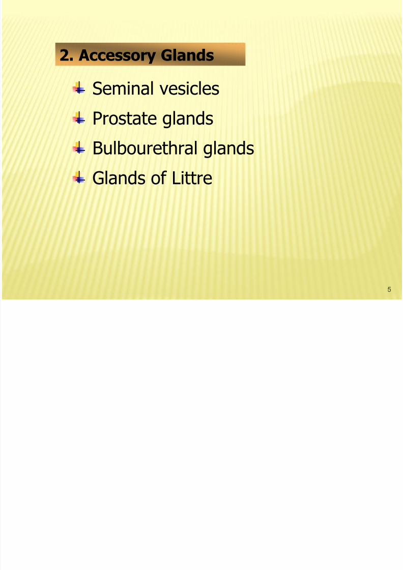
Seminal vesicles
Prostate glands
Bulbourethral glands
Glands of Littre
2. Accessory Glands
8/12/2019 Intro Histo Male Genital
http://slidepdf.com/reader/full/intro-histo-male-genital 6/51
6
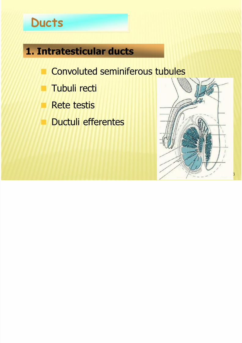
Convoluted seminiferous tubules
Tubuli recti
Rete testis
Ductuli efferentes
Ducts
1. Intratesticular ducts

8/12/2019 Intro Histo Male Genital
http://slidepdf.com/reader/full/intro-histo-male-genital 7/517
Penis and scrotum
External Genitalia
Ductus epididymidisDuctus deferens
Ejaculatory duct
Urethra
2. Excretory Genital Ducts

8/12/2019 Intro Histo Male Genital
http://slidepdf.com/reader/full/intro-histo-male-genital 8/518
a. Tunica vaginalis
Double layered mesothelial sac, covers the
anterior surface of each testis
b. Tunica albuginea
Dense fibrous connective tissue capsule
Thickens along posterior surfacemediastinum testis
Testes
1. External Covering

8/12/2019 Intro Histo Male Genital
http://slidepdf.com/reader/full/intro-histo-male-genital 9/51
TESTES
9

8/12/2019 Intro Histo Male Genital
http://slidepdf.com/reader/full/intro-histo-male-genital 10/51
10
a. Septa : Extension oftunica albuginea, 250
lobules
b. Lobules
2. Internal Structure

8/12/2019 Intro Histo Male Genital
http://slidepdf.com/reader/full/intro-histo-male-genital 11/51
11
Each long : 40 –70 cm, convoluted, packed into small space.
The walls composed of 3 layers :
a. Tunica propria
Fibrous connective tissue + fibroblasts
Innermost layers : contractile myoid cells
Seminiferous Tubule
1. General Structure
8/12/2019 Intro Histo Male Genital
http://slidepdf.com/reader/full/intro-histo-male-genital 12/51
12
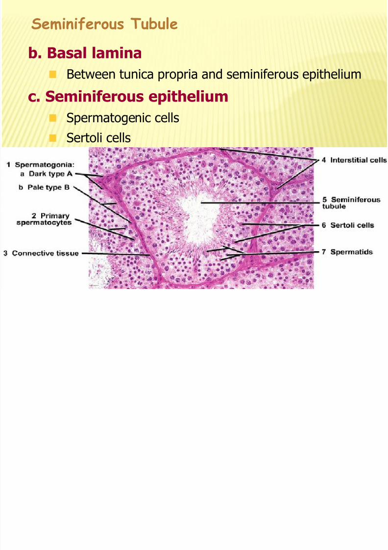
b. Basal lamina
Between tunica propria and seminiferous epithelium
c. Seminiferous epithelium
Spermatogenic cells
Sertoli cells
Seminiferous Tubule

8/12/2019 Intro Histo Male Genital
http://slidepdf.com/reader/full/intro-histo-male-genital 13/51
13

8/12/2019 Intro Histo Male Genital
http://slidepdf.com/reader/full/intro-histo-male-genital 14/51
14
Diagram of the structure of part of a seminiferous tubule and
interstitial tissue. This figure does not show the lymphaticvessels found in the connective tissue.
Latespermiogenesis
Initial spermiogenesis
Meiosis
Basal lamina
Fibroblast
Interstitial cells
Cytoplasmicbridges
Earlyspermatids
Secondaryspermatocytes
Primaryspermatocytes
Spermatogonium

8/12/2019 Intro Histo Male Genital
http://slidepdf.com/reader/full/intro-histo-male-genital 15/51
SPERMATOGENESIS
These changes involve flattening of the nucleus, formationof an acrosome which resembles a large lysosome,
growth of a flagellum (tail) from the basal body,
reorganization of the mitochondria in the midpiece region,
and shedding of unneeded cytoplasm as a residual body.
15

8/12/2019 Intro Histo Male Genital
http://slidepdf.com/reader/full/intro-histo-male-genital 16/51
16
a. Spermatogonia
Small round cells,
near the basal lamina
Round nucleus +
Heterochromatin
2. Spermatogenic Cells
b P i t t

8/12/2019 Intro Histo Male Genital
http://slidepdf.com/reader/full/intro-histo-male-genital 17/51
17
b. Primary spermatocytes
Closer to the lumen thanspermatogonium
Largest germ cells
Large round nucleus, heterochromatin

8/12/2019 Intro Histo Male Genital
http://slidepdf.com/reader/full/intro-histo-male-genital 18/51
18
c. Secondary spermatocytes
Closer to lumen than primary spermatocytes
Half size
Rare
d Spermatids

8/12/2019 Intro Histo Male Genital
http://slidepdf.com/reader/full/intro-histo-male-genital 19/51
19
d. Spermatids
Products of second meiotic division
located next to the lumen.
Small cells :
Dark heterochromatic nuclei
8/12/2019 Intro Histo Male Genital
http://slidepdf.com/reader/full/intro-histo-male-genital 20/51
20
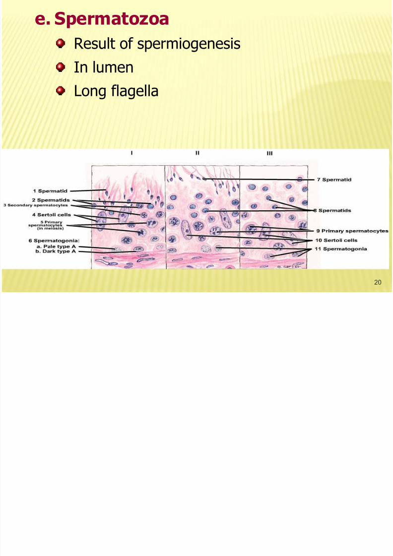
e. Spermatozoa
Result of spermiogenesis
In lumenLong flagella

8/12/2019 Intro Histo Male Genital
http://slidepdf.com/reader/full/intro-histo-male-genital 21/51
21
MesodermElongated, branched,pyramidal epithelialcells
Cytoplasmic enfolding
Bound tightly toneighboring
supporting cells
occluding junction.
3. Supporting Sertoli Cells
8/12/2019 Intro Histo Male Genital
http://slidepdf.com/reader/full/intro-histo-male-genital 22/51
22
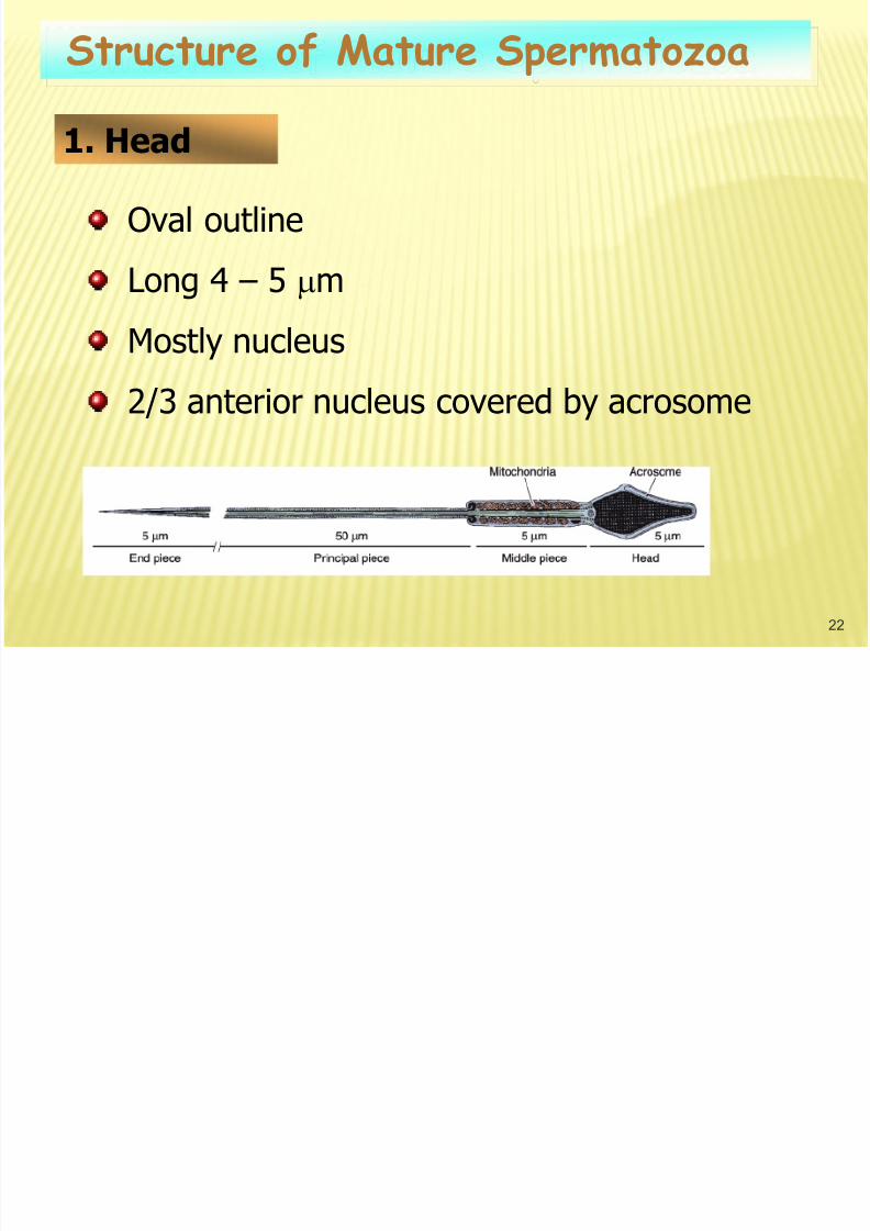
Oval outline
Long 4 – 5 m
Mostly nucleus
2/3 anterior nucleus covered by acrosome
Structure of Mature Spermatozoa
1. Head

8/12/2019 Intro Histo Male Genital
http://slidepdf.com/reader/full/intro-histo-male-genital 23/51
23
Enveloped by plasma membrane
Middle piece : 5 – 7 m
mitochondrial arranged end to end inhelical sheath around flagellum
• Principal piece
• Outer fibrous sheath with dorsal andventral longitudinal columns• Flagellum
• End piece
Lacks fibrous sheath
2. Tail
I t titi l C ll (L di )

8/12/2019 Intro Histo Male Genital
http://slidepdf.com/reader/full/intro-histo-male-genital 24/51
24
Mesoderm
Secrete :testosterone
Vascular nests ofpale acidophilic cellsin loose connectivetissue between the
seminiferoustubules
Interstitial Cells (Leydig)

8/12/2019 Intro Histo Male Genital
http://slidepdf.com/reader/full/intro-histo-male-genital 25/51
25
Protects the developing sperm fromdamage by an autoimmune response. Thebarriers consists of a continuous belt of
junctional complexes joining the sertolicells at their lateral surfaces.
G. Blood – Testis Barrier
8/12/2019 Intro Histo Male Genital
http://slidepdf.com/reader/full/intro-histo-male-genital 26/51
26
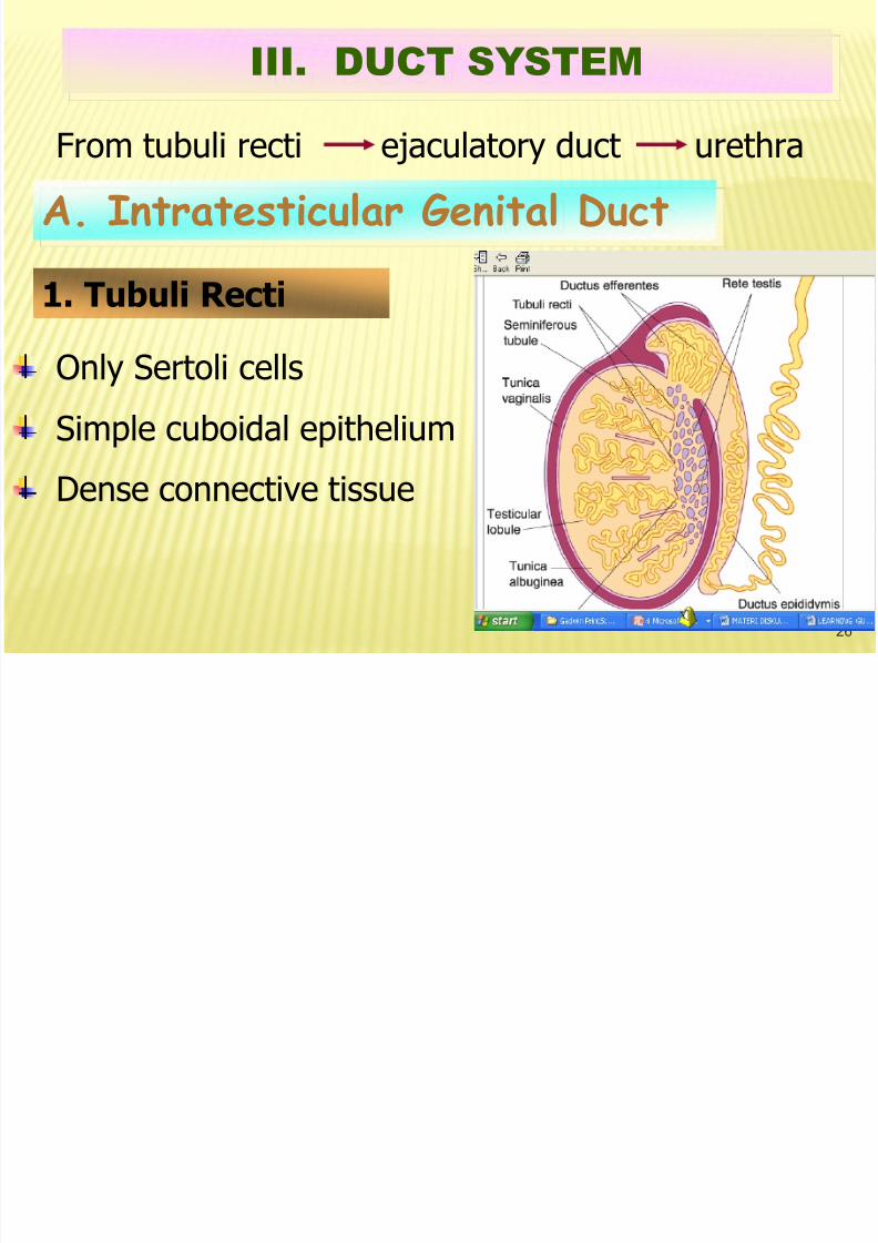
From tubuli recti ejaculatory duct urethra
III. DUCT SYSTEM
A. Intratesticular Genital Duct
1. Tubuli Recti
Only Sertoli cells
Simple cuboidal epithelium
Dense connective tissue

8/12/2019 Intro Histo Male Genital
http://slidepdf.com/reader/full/intro-histo-male-genital 27/51
27
2. Rete Testis
Anastomosing networkMediastinum testis
Low cuboidal epithelium

8/12/2019 Intro Histo Male Genital
http://slidepdf.com/reader/full/intro-histo-male-genital 28/51
28
3. Ductuli Efferentes
Walls :
Epithelium
Simple cuboidal : absorb fluid
Ciliated columnar cells : Cilia sweep
spermatozoa
Smooth muscle
The ductules form the head of the epididymis
10 – 20 ducts

8/12/2019 Intro Histo Male Genital
http://slidepdf.com/reader/full/intro-histo-male-genital 29/51
29
SEMINIFEROUS TUBULES, STRAIGHT TUBULES,RETE TESTIS AND DUCTULI EFFERENTES
1. Seminiferous
tubules
2. Straighttubules
3. Connectivetissue ofmediastinum
4. Rete testistubules
5. Ductuliefferentes(efferentductules
6. Rete testistubules
8/12/2019 Intro Histo Male Genital
http://slidepdf.com/reader/full/intro-histo-male-genital 30/51
30
Single, coiled 4 – 6 m long tube
Comprises the body and tail epididymis
Epithelium : pseudostratified columnar +stereocilia
Circular smooth muscle
Function : - Maturation of sperm
- Absorption
- Secretion (glycoprotein)
- Sperm storage
B. Excretory Genital Duct
1. Ductus Epididymidis

8/12/2019 Intro Histo Male Genital
http://slidepdf.com/reader/full/intro-histo-male-genital 31/51
31
1. Connectivetissue
2. Cross sectionsof the ductusepididymidis
3. Basementmembrane
4. Pseudostratifiedcolumnarepithelium withstereocilia
5. Sectionthrough of U-bend of theductusepididymidis
6. Epididymalwall cuttangentially
7. Smoothmuscle fibers
8. Stereocilia
9. Columnar cells
10. Basal cell
Ductus Epididymidis (Duct of the Epididymis)

8/12/2019 Intro Histo Male Genital
http://slidepdf.com/reader/full/intro-histo-male-genital 32/51
32

8/12/2019 Intro Histo Male Genital
http://slidepdf.com/reader/full/intro-histo-male-genital 33/51
33
Straight tube with thickmuscular walls
Longitudinal mucosal fold
Pseudostratifiedcolumnar epithelial +stereocilia
Three layer smooth
muscle
Termination : ampulla
2. Ductus Deferens

8/12/2019 Intro Histo Male Genital
http://slidepdf.com/reader/full/intro-histo-male-genital 34/51
34
1. Outerlongitudinalmuscle layer
2. Circularmuscle layer
3. Innerlongitudinalmuscle layer
4. Nerve andblood vesselsin theadventitious
5. Lamina propria
6. Longitudinal crestof lamina propria
7. Epithelium
8. Adipose tissue
Ductus Deferens (Transverse section)

8/12/2019 Intro Histo Male Genital
http://slidepdf.com/reader/full/intro-histo-male-genital 35/51
35
Short 2 cm/long
Epithelium,pseudostratifiedcolumnar
Penetrates prostate
empty to prostaticurethra
3. Ejaculatory Duct

8/12/2019 Intro Histo Male Genital
http://slidepdf.com/reader/full/intro-histo-male-genital 36/51
36
SEMINAL VESICLE
PROSTATE GLAND
BULBO URETHRAE GLANDS (COWPER’S
GLAND)
LITTRE’S GLAND
IV. ACCESSORY GENITAL DUCTS

8/12/2019 Intro Histo Male Genital
http://slidepdf.com/reader/full/intro-histo-male-genital 37/51
37
Paired
Each consist of two highly coiled 15 cm long
tubes
Mucosa : highly folded
Pseudostratified low columnar epithelium
A. Seminal Vesicle

8/12/2019 Intro Histo Male Genital
http://slidepdf.com/reader/full/intro-histo-male-genital 38/51
38
Secretory product : thick yellowish liquid rich
in fructose
Make up 70% human ejaculate
Smooth muscle : underlying the lamina
propria

8/12/2019 Intro Histo Male Genital
http://slidepdf.com/reader/full/intro-histo-male-genital 39/51
39
Seminal Vesicle
4. Glandularepithelium
5. Primary fold inthe mucosa
6. Secondary folds
7. Lamina propria
1. Crypts in themucosa
2. Muscular coat
3. Adventitia

8/12/2019 Intro Histo Male Genital
http://slidepdf.com/reader/full/intro-histo-male-genital 40/51
40
B. Prostate Gland
Largest male accessory sex glands30 – 50 compound tubuloalveolar glands
The glands of the prostate are collected into
three major groups
Mucosal glands
Submucosal glands
Main glands
Drained by a discreteductal systemprostatic urethra

8/12/2019 Intro Histo Male Genital
http://slidepdf.com/reader/full/intro-histo-male-genital 41/51
41

8/12/2019 Intro Histo Male Genital
http://slidepdf.com/reader/full/intro-histo-male-genital 42/51
42
Mucosa : folded, epithelium varies from tall
cuboidal to pseudostratified columnar.Produces : fluid, rich in citric acid and acid
phosphatase, amylase
Surrounded by fibroelastic capsule containingsmooth muscle capsule
Characteristic :
Corpora amylacea in lumen
Composed of glycoprotein calcified

8/12/2019 Intro Histo Male Genital
http://slidepdf.com/reader/full/intro-histo-male-genital 43/51
43
Submucosalglands
Diagram illustrating the position of the
prostatic glands
8/12/2019 Intro Histo Male Genital
http://slidepdf.com/reader/full/intro-histo-male-genital 44/51
44
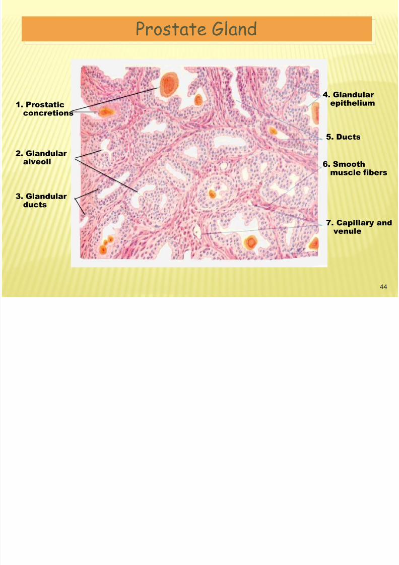
4. Glandularepithelium
5. Ducts
6. Smoothmuscle fibers
7. Capillary andvenule
1. Prostaticconcretions
2. Glandularalveoli
3. Glandularducts
Prostate Gland
C Bulbo Urethral Glands

8/12/2019 Intro Histo Male Genital
http://slidepdf.com/reader/full/intro-histo-male-genital 45/51
45
Located within the musculature of the pelvicdiaphragm
Lined by cuboidal to columnar epithelium
Their secretion consists of a clear, mucus-likematerial ducts empty into membranous urethra
C. Bulbo Urethral Glands(Cowper’s Glands)

8/12/2019 Intro Histo Male Genital
http://slidepdf.com/reader/full/intro-histo-male-genital 46/51
V PENIS
2 CORPORA CAVERNOSUS
CORVUS SPONGIOSUM46

8/12/2019 Intro Histo Male Genital
http://slidepdf.com/reader/full/intro-histo-male-genital 47/51
47
Two dorsal erectile cylinders
Deep arterySheathed by tunica albuginea (thickdense connective tissue)
Spaces of erectile tissue : largest inthe central region
V. PENIS
A. Consists of three cylindric bodies of spongyerectile tissue surrounded by a looseconnective tissue sheath and covered byhairless thin skin.
1. Corpora Cavernous

8/12/2019 Intro Histo Male Genital
http://slidepdf.com/reader/full/intro-histo-male-genital 48/51
48
Drawing of a transverse section of the penis
Corpus
cavernosumof the penis
Erectiletissue
Corpuscavernosum
of the urethra
Urethra
Dorsalarteries
Superficialdorsal vein
Deep dorsal
vein
Tunicaalbuginea
Deep artery

8/12/2019 Intro Histo Male Genital
http://slidepdf.com/reader/full/intro-histo-male-genital 49/51
49
Single, smaller
Ventral cylinder
Surrounded by a thinnerconnective tissue sheath
Expanded distal tip glanspenis
Spaces of erectile tissue :uniform size
2. Corpus Spongiosum(Corpus Cavernosum Urethra)

8/12/2019 Intro Histo Male Genital
http://slidepdf.com/reader/full/intro-histo-male-genital 50/51
50

8/12/2019 Intro Histo Male Genital
http://slidepdf.com/reader/full/intro-histo-male-genital 51/51