Embed Size (px)
Citation preview
Intrinsic bursting enhances the robustness of a neuralnetwork model of sequence generation by avianbrain area HVC
Dezhe Z. Jin & Fethi M. Ramazanoğlu &
H. Sebastian Seung
Received: 25 September 2006 /Revised: 9 February 2007 /Accepted: 13 March 2007# Springer Science + Business Media, LLC 2007
Abstract Avian brain area HVC is known to be importantfor the production of birdsong. In zebra finches, each RA-projecting neuron in HVC emits a single burst of spikesduring a song motif. The population of neurons is acti-vated in a precisely timed, stereotyped sequence. We pro-pose a model of these burst sequences that relies on twohypotheses. First, we hypothesize that the sequential orderof bursting is reflected in the excitatory synaptic connec-tions between neurons. Second, we propose that the neu-rons are intrinsically bursting, so that burst duration is setby cellular properties. Our model generates burst sequencessimilar to those observed in HVC. If intrinsic bursting isremoved from the model, burst sequences can also be pro-duced. However, they require more fine-tuning of synapticstrengths, and are therefore less robust. In our model,intrinsic bursting is caused by dendritic calcium spikes, and
strong spike frequency adaptation in the soma contributesto burst termination.
Keywords Songbird . Associative chaining model .
Dendritic spike . Sequence generation . Computationalmodel
1 Introduction
How does the brain generate behaviors that are composedof long sequences of actions, such as language and musicalperformance? According to one idea, sequential behaviorsare generated by the sequential activation of groups ofneurons. In the associative chaining model, the neurons ofone group directly excite the neurons of the next group, sothat sequential order is directly embedded in the structure ofexcitatory synaptic connectivity. This model was criticizedby Lashley on theoretical grounds as insufficient forexplaining the full complexity of sequential behaviors(Lashley 1951). However, the model has never actuallybeen tested through neurobiological experiments.
Recent progress in the neurobiology of birdsong has setthe stage for testing the associative chaining model in anon-human animal (Chi and Margoliash 2001; Doupe andKuhl 1999; Fee et al. 2004; Hahnloser et al. 2002; Konishi1965; Leonardo and Fee 2005; Nottebohm et al. 1976;Williams 2004; Yu and Margoliash 1996). The goal of thispaper is to give a biophysically realistic implementation ofthe associative chaining model applied to birdsong. As willbe seen later, our implementation of the model leads to anumber of predictions that can be tested experimentally.
The zebra finch sings a single, highly stereotyped songthat consists of repetitions of a motif, typically 0.5–1 s in
J Comput NeurosciDOI 10.1007/s10827-007-0032-z
Action Editor: Alain Destexhe
D. Z. Jin (*)Department of Physics,The Pennsylvania State University,104 Davey Lab, University Park, PA 16802, USAe-mail: [email protected]
F. M. RamazanoğluDepartment of Physics,Massachusetts Institute of Technology,Cambridge, MA 02139, USA
H. S. SeungHoward Hughes Medical Instituteand Department of Brain and Cognitive Sciences,Massachusetts Institute of Technology,Cambridge, MA 02139, USA
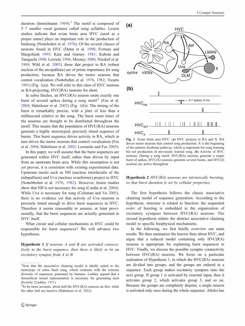
duration (Immelmann 1969).1 The motif is composed of3–7 smaller vocal gestures called song syllables. Lesionstudies indicate that avian brain area HVC (used as aproper name) plays an important role in the production ofbirdsong (Nottebohm et al. 1976). Of the several classes ofneurons found in HVC (Dutar et al. 1998; Fortune andMargoliash 1995; Katz and Gurney 1981; Kubota andTaniguchi 1998; Lewicki 1996; Mooney 2000; Nixdorf et al.1989; Wild et al. 2005), those that project to RA (robustnucleus of the arcopallium) are of prime importance for songproduction, because RA drives the motor neurons thatcontrol vocalization (Nottebohm et al. 1976, 1982; Vicario1991) (Fig. 1(a)). We will refer to this class of HVC neuronsas RA-projecting, HVC(RA) neurons for short.
In zebra finches, an HVC(RA) neuron emits exactly oneburst of several spikes during a song motif 2 (Fee et al.2004; Hahnloser et al. 2002) (Fig. 1(b)). The timing of theburst is remarkably precise, with a jitter of less than amillisecond relative to the song. The burst onset times ofthe neurons are thought to be distributed throughout themotif. This means that the population of HVC(RA) neuronsgenerate a highly stereotyped, precisely timed sequence ofbursts. This burst sequence drives activity in RA, which inturn drives the motor neurons that control vocalization (Feeet al. 2004; Hahnloser et al. 2002; Leonardo and Fee 2005).
In this paper, we will assume that the burst sequences aregenerated within HVC itself, rather than driven by inputfrom an upstream brain area. While this assumption is notyet proven, it is consistent with existing experimental data.Upstream nuclei such as NIf (nucleus interfacialis of thenidopallium) and Uva (nucleus uvaeformis) project to HVC(Nottehbohm et al. 1976, 1982). However, lesion studiesshow that NIf is not necessary for song (Cardin et al. 2004).While Uva is necessary for song (Coleman and Vu 2005),there is no evidence yet that activity of Uva neurons isprecisely timed enough to drive burst sequences in HVC.Therefore it seems reasonable to assume, at least provi-sionally, that the burst sequences are actually generated inHVC itself.
What circuit and cellular mechanisms in HVC could beresponsible for burst sequences? We will advance twohypotheses.
Hypothesis 1 If neurons A and B are activated consecu-tively in the burst sequence, then there is likely to be anexcitatory synapse from A to B.
Hypothesis 2 HVC(RA) neurons are intrinsically bursting,so that burst duration is set by cellular properties.
The first hypothesis follows the classic associativechaining model of sequence generation. According to thehypothesis, structure is related to function: the sequentialorder of bursting is embedded in the organization ofexcitatory synapses between HVC(RA) neurons. Thesecond hypothesis relates the abstract associative chainingmodel to specific biophysical mechanisms.
In the following, we first briefly overview our mainresults. We then summarize the known facts about HVC, andargue that a reduced model containing only HVC(RA)neurons is appropriate for explaining burst sequences inHVC. Finally, we discuss the possible synaptic connectivitybetween HVC(RA) neurons. We focus on a particularrealization of Hypothesis 1, in which the HVC(RA) neuronsare divided into groups, and the groups are ordered in asequence. Each group makes excitatory synapses onto thenext group. If group 1 is activated by external input, then itactivates group 2, which activates group 3, and so on.Because the groups are completely disjoint, a single neuronis activated only once during the whole sequence. Abeles has
LMAN
DLM
Uva
NIf
X
RA
HVC
syrinx nXIIts
HVCRA
HVCINT
HVCX
3-7 spikes, 6 ms
(a)
(b)
Fig. 1 Avian brain area HVC. (a) HVC projects to RA and X. RAdrives motor neurons that control song production. X is the beginningof the anterior forebrain pathway, which is important for song learningbut not production of previously learned song. (b) Activity of HVCneurons. During a song motif, HVC(RA) neurons generate a singleburst of spikes, HVC(X) neurons generate several bursts, and HVC(I)neurons are active throughout
1Note that the associative chaining model is ideally suited to thestereotypy of zebra finch song, which contrasts with the extremediversity of sequences generated by humans. Lashley argued that ahierarchical neural representation is necessary for generating suchdiversity (Lashley 1951).2To be more accurate, about half the HVC(RA) neurons do this, whilethe other half are inactive (Hahnloser et al. 2002).
J Comput Neurosci
utilized this synaptic connectivity in his synfire chain model,which stresses synchronous spiking of neurons in each group(Abeles 1982, 1991). Others have utilized this connectivityin neural network models based on firing rates3 (Amari1972; Kleinfeld 1986; Sompolinsky and Kanter 1986).
1.1 Overview
In the Results, we first study an associative chaining modelwithout intrinsic bursting in HVC(RA) neurons. We showthat the model can generate burst sequences like those seenin HVC. However, there is a problem: the model requiresfine-tuning of synaptic strengths. Since the response of aneuron depends on the amplitude and duration of synapticinput, the number of spikes in a burst depends strongly onsynaptic strengths. If synapses are too strong, then there isrunaway activity, in which successive neurons in thesequence produce longer and longer bursts of spikes. Ifsynapses are too weak, then the activity decays to zero.
To solve this robustness problem, we utilize Hypothesis2, which is that HVC(RA) neurons are equipped withintrinsic cellular mechanisms for generating bursts. When aburst of spikes is initiated, it is a stereotyped event, withonly a weak dependence on the amplitude or duration ofsynaptic input. As a result, little tuning of synaptic strengthsis required to produce burst sequences. This improvedrobustness is demonstrated with numerical simulations ofour model.
Our Hypothesis 2 is not the only way of solving therobustness problem, but it is arguably the simplest.Obviously it is easier to construct a neural circuit thatgenerates bursts, if the elements in the circuit producebursts intrinsically. Other ways of solving the robustnessproblem are mentioned in Section 4.
Whether HVC(RA) neurons indeed possess intrinsicbursting mechanisms is currently unknown. A number ofsuch mechanisms have been studied in other neurons(Brumberg et al. 2000; Franceschetti et al. 1995; Mattiaet al. 1997; Schwindt and Crill 1999; Traub et al. 1994;Wong and Stewart 1992), and any of them would besufficient for generating the bursts observed in HVC. Wecan only speculate as to the mechanisms that could beinvolved. In our model, the dendrites of HVC(RA) neuronsproduce a calcium spike, due to the presence of voltage-activated calcium channels. This calcium spike depolarizesthe soma, producing a burst of sodium spikes.
Calcium spikes can last tens of milliseconds in hippo-campal and cortical neurons (Golding et al. 1999; Schwindtand Crill 1999; Wei et al. 2001). If HVC(RA) neurons have
calcium spikes of similar duration, what could account forthe fact that bursts in HVC have an average duration of6 ms (Hahnloser et al. 2002)? To limit the duration ofsodium spiking, our model incorporates a biophysicalproperty that has been observed in HVC(RA) neurons invitro, strong spike frequency adaptation (Dutar et al. 1998;Mooney and Prather 2005; Wild et al. 2005). As discussedin Section 3, this causes sodium spiking to terminate beforethe end of the calcium spike. One could also imagine thatHVC neurons possess calcium spikes that last for just 6 ms,in which case strong spike frequency adaptation in thesoma would be less crucial.
The burst duration in our model is determined by intrinsiccellular properties, rather than circuit properties. Therefore itis natural to ask whether fine-tuning of intrinsic cellularproperties is required to generate bursting. We show that thisis not the case, as the number of spikes per burst is not verysensitive to parameters of our model neuron.
Before proceeding further, we should say a few wordsabout our methodology, which could be described as “top-down.” Table 1 lists four levels of description of HVC. Webegin near the top, with burst sequences. In order to explainthem, we make Hypothesis 1 about correlational connec-tivity and Hypothesis 2 about intrinsic bursting. Hypothesis2 is further elaborated by adding strong spike frequencyadaptation. Our model shows how these hypotheticalneuron and network properties could help generate burstsequences, and therefore demonstrates their potentialfunctional significance for birdsong. Moving further downin the table, specific channels are proposed as biophysicalmechanisms for the hypothetical single neuron properties.These proposals are useful, because they suggest specificways of testing the neuron-level hypotheses experimentally(see Section 4). However, the primary subject of this paperis the functional implications of intrinsic cellular propertiesfor sequence generation, rather than the detailed biophys-ical mechanisms of these intrinsic properties.
We have pursued a top-down modeling approach,because the bottom-up approach is not possible at thistime. The channel properties of HVC neurons cannot serveas a starting point for modeling, because so little is knownabout them. A top-down approach has the advantage that itguides research by focusing attention on neuron and
3In the more general correlation matrix model studied by theseauthors, a single neuron is allowed to belong to more than one group.
Table 1 HVC model and levels of description
Behavior SongNetwork Burst sequences
Correlational connectivityNeuron Intrinsic bursting
Strong spike frequency adaptationChannel Dendrite: ICa, ICaK
Soma: IKLT, IKHT
J Comput Neurosci
channel properties that are most significant for behavioralfunction.
1.2 Synaptic organization of HVC
In this section, we summarize the current knowledge aboutsynaptic connectivity of HVC, and introduce a reducedmodel for sequence generation that focuses on theexcitatory synapses between HVC(RA) neurons, and omitsthe other classes of neurons in HVC.
HVC contains two classes of projection neurons, those thatproject to RA (HVC(RA)), and those that project to area X(HVC(X)); it also contains interneurons (HVC(I)) (Dutar et al.1998; Fortune and Margoliash 1995; Katz and Gurney 1981;Kubota and Taniguchi 1998; Lewicki 1996; Mooney 2000;Nixdorf et al. 1989; Wild et al. 2005). The projection to RA isimportant for song production, as RA drives motor neuronsthat control vocalization (Nottebohm et al. 1976, 1982; Vicario1991). The projection to X enters the anterior forebrainpathway, which is important for song learning but notnecessary for song production (Brainard and Doupe 2000).
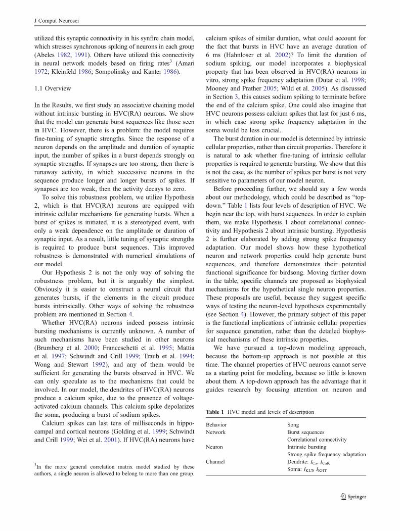
Figure 2(a) is our best guess about the synapticconnectivity of HVC, given the limited information thatexists in the literature (Mooney and Prather 2005).According to the figure, HVC(RA) neurons make excitato-ry synapses onto both HVC(RA) and HVC(X) neurons. Incontrast, HVC(X) neurons make no excitatory synapsesonto other projection neurons. Both types of projectionneurons make excitatory synapses on interneurons, andreceive inhibitory synapses from them.4 In the following,we assume that the diagram of Fig. 2(a) is correct, andargue that the simplified versions shown in Fig. 2(b and c)should be good approximations.
Interneurons are much less temporally selective thanprojection neurons in their song-related activity (Fig. 1(b)).During a song motif, HVC(RA) neurons fire a single burstof spikes (Hahnloser et al. 2002), and HVC(X) neurons firea few bursts (Kozhevnikov and Fee 2006). In contrast,HVC(I) neurons fire at many times during a song motif(Hahnloser et al. 2002).
If we make the approximation that interneurons haveconstant firing rates during song, their dynamic inhibitoryinput to the projection neurons can be replaced by a staticconductance. With this approximation, we can omit theinterneurons from Fig. 2(a), which yields the reducedmodel shown in Fig. 2(b).
Neglecting synaptic inhibition may seem like a drasticstep. However, it seems justified provided that the goal of
modeling is to explain sequence generation. Since inhibi-tion lacks temporal selectivity, its main role is likely to beregulation of the overall level of activity in projectionneurons, which might enhance the robustness of sequencegeneration.
In the reduced model of Fig. 2(b), HVC(RA) neuronssend feedforward drive to HVC(X) neurons, but receive nosignals from them, which implies that HVC(X) neurons areirrelevant to the dynamics of HVC(RA) neurons. Thereforewe omit the HVC(X) neurons, as the projection to area X isnot necessary for song production anyway (Brainard andDoupe 2000). This leads to a reduced model containingonly HVC(RA) neurons, as shown in Fig. 2(c). Note thatthis reduction depends on the assumption that there are nosynapses from HVC(X) neurons to HVC(RA) neurons, asin Fig. 2(a). This is consistent with the fact that targeteddestruction of HVC(X) neurons does not cause deteriora-tion of song in adult zebra finches (Scharff et al. 2000).
1.3 Correlational models of synaptic connectivity
In the preceding section, we argued that a reduced model ofHVC containing only HVC(RA) neurons is a good startingpoint for understanding sequence generation. But thesynaptic connectivity between HVC(RA) neurons is largelyunknown. In the Introduction, we proposed Hypothesis 1: ifneurons A and B are activated consecutively in the burstsequence, then there is likely to be an excitatory synapse
4This diagram is based primarily on the work of Mooney and Prather(2005). The evidence for recurrent inhibition is strong, but theexcitatory interactions between projection neurons are somewhatspeculative.
HVCRA HVCX
HVCRA
(a)
(c)
HVCRA
HVCIN
HVCX
(b)
Fig. 2 Synaptic organization of HVC and reduced models. (a) Hypothe-sized synaptic organization of HVC. HVC(RA) neurons excite eachother and HVC(X) neurons. Both HVC(RA) and HVC(X) neuronsexcite HVC(I) neurons, and receive inhibition from them. Theevidence for recurrent inhibition (solid lines) is strong (Mooney andPrather 2005). The excitatory interactions between projection neurons(dashed lines) are more speculative. The lack of synapses from HVC(X)to HVC(RA) neurons is based on Scharff et al. (2000). (b) Reducedmodel with projection neurons only. If the HVC(I) neurons have firingrates that are approximately constant in time, then they can be omitted,leaving a reduced model consisting of projection neurons only. (c)Further reduced model with HVC(RA) neurons only. Since there areessentially no backprojections from HVC(X) neurons to HVC(RA)neurons, one can consider a reduced model consisting of HVC(RA)neurons only
J Comput Neurosci
from A to B. In other words, if neuron A fires just beforeneuron B, we can infer that A probably helped cause B tofire by giving it direct excitatory synaptic input. Theinference is probabilistic, since some imprecision of wiringis expected in a neurobiological system. Since we areinferring causation from temporal correlation, Hypothesis 1can be called a correlational model of connectivity.
To make the implications of the correlational modelmore explicit, it is worth stating the conditions under whichan excitatory synapse is not expected between neurons Aand B. If neuron A fires long before neuron B, there shouldbe no synapse between them (temporal contiguity isimportant). If neuron A fires after neuron B, there shouldbe no synapse from A to B (temporal order is important).5
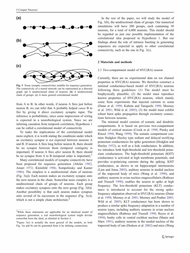
Many correlational models of synaptic connectivity havebeen proposed for sequence generation (Abeles 1982;Amari 1972; Kleinfeld 1986; Sompolinsky and Kanter1986). The simplest is a unidirectional chain of neurons(Fig. 3(a)). Each neuron makes an excitatory synapse ontothe next neuron in the chain. Somewhat more complex is aunidirectional chain of groups of neurons. Each groupmakes excitatory synapses onto the next group (Fig. 3(b)).Another possibility is that each neuron makes synapsesonto several of its successors in the sequence (Fig. 3(c)),which is not a simple chain architecture.6
In the rest of the paper, we will study the model ofFig. 3(b), the unidirectional chain of groups. Our numericalsimulations will have 200 groups, each containing 30neurons, for a total of 6,000 neurons. This model shouldbe regarded as just one possible implementation of thecorrelational idea proposed in Hypothesis 1. But ourfindings about the role of intrinsic bursting in generatingsequences are expected to apply to other correlationalconnectivity, such as the one in Fig. 3(c).
2 Materials and methods
2.1 Two-compartment model of HVC(RA) neuron
Currently, there are no experimental data on ion channelproperties in HVC(RA) neurons. We therefore construct aminimal conductance-based model of HVC(RA) neuronsfollowing three guidelines: (1) The model must bebiophysically plausible; (2) the model must reproduceknown properties of HVC(RA) neurons, which mainlycome from experiments that injected currents to soma(Dutar et al. 1998; Kubota and Taniguchi 1998; Mooneyet al. 2001; Wild et al. 2005); (3) the model must exhibitrobust burst spike propagation through excitatory connec-tions between neurons.
The minimal model consists of somatic and dendriticcompartments. It is based on previous two-compartmentmodels of cortical neurons (Crook et al. 1998; Pinsky andRinzel 1994; Wang 1999). The somatic compartment con-tains Hudgkin–Huxley type sodium and delayed rectifyingpotassium conductances for spike generation (Hodgkin andHuxley 1952), as well as a leak conductance. In addition,we introduce both high-threshold and low-threshold potas-sium conductances. The high-threshold potassium (KHT)conductance is activated at high membrane potentials, andprovides re-polarizing currents during the spiking. KHTconductance, as shown in rat hippocampal interneurons(Lien and Jonas 2003), auditory neurons in medial nucleusof the trapezoid body of mice (Wang et al. 1998), andauditory neurons in avian nucleus magnocellularis (Rathouzand Trussell 1998), enables the neuron to spike at highfrequency. The low-threshold potassium (KLT) conduc-tance is introduced to account for the strong spike-frequency adaptation observed in HVC(RA) neurons (Dutaret al. 1998; Mooney et al. 2001; Mooney and Prather 2005;Wild et al. 2005). KLT conductance has been shown toproduce a similar spike frequency adaptation in a number ofneuron types, including auditory neurons in avian nucleusmagnocellularis (Rathouz and Trussell 1998; Reyes et al.1994), bushy cells in ventral cochlear nucleus (Manis andMarx 1991), auditory neurons in the medial nucleus of thetrapezoid body of rats (Dodson et al. 2002) and mice (Wang
5While these statements are applicable for an idealized model ofsequence generation, a real neurobiological system might deviatesomewhat from the ideal, as detailed in Section 4.6Figure 3(c) is actually the most general of the models, as bothFig. 3(a and b) can be generated from it by deleting connections.
(a)
(b)
(c)
Fig. 3 Some synaptic connectivities suitable for sequence generation.The connectivity of a neural network can be represented as a directedgraph. (a) A unidirectional chain of neurons. (b) A unidirectionalchain of groups. (c) A more general correlational model
J Comput Neurosci
et al. 1998), and auditory neurons in the gerbil medialsuperior olive (Svirskis et al. 2002). KLT conductance isactivated at subthreshold membrane potentials.
The dendritic compartment contains a leak conductance,a high-threshold calcium conductance, and a calcium-activated potassium conductance. The calcium and potassi-um conductance enable a calcium spike in the dendrite ifdepolarized over the threshold, as observed in dendrites ofmany types of neurons, such as mammalian hippocampaland cortical neurons, as well as cerebellar Purkinje neurons(Golding et al. 1999; Hausser et al. 2000). Biophysicalplausibility of our model is ensured since the conductancesare taken from previous experimental data in other types ofneurons. In the man text, we have shown that this minimalmodel also satisfies the other two guidelines.
The membrane potentials Vs and Vd of soma and dendriteobey the following dynamical equations:
CmAsdVs
dt¼ AsðILs þ INa þ IK þ IKHT þ IKLT þ IFFsÞ
þ Iext þ Vd % Vsð Þ=Rc;
CmAddVd
dt¼ Ad ILd þ ICa þ ICaK þ Isyn þ IFFd
! "
þ Vs % Vdð Þ=Rc:
Here Cm=1 μF/cm2 is the membrane capacitance, As=100 μm2 and Ad=50,000 μm2 are the surface areas of thesoma and the dendrite, respectively. For the soma, ILs=gLs(Er−Vs) is the leak current, with conductance gLs=0.05 mS/cm2 and reversal potentialEr=−85 mV; INa=gNam
3&h(ENa−Vs) is the sodium current, with conductance gNa=100 mS/cm2, reversal potential ENa=55 mV, and gatingvariables m and h; IK=gKn
4(EK−Vs) is the potassiumcurrent, with conductance gK=2 mS/cm2, reversal potentialEK=−90 mV, and gating variable n; IKHT=gKHTw(EK−Vs) isthe high threshold potassium current, with conductance gKHT=300 mS/cm2 and gating variable w. IKLT=gKLTl(EK−Vs) isthe low threshold potassium current, with conductance gKLT=25 mS/cm2 and gating variable l; IFFs=−gFFsVs is thefeedforward excitatory input to the soma, with a constantconductance gFFs; Iext is the external current injection tosoma; Rc=250 MΩ is the resistance of the connectionbetween soma and dendrite. For the dendrite, ILd=gLd(Er−Vd) is the leak current with gLd=0.1 mS/cm2; ICa ¼gCam2
1 ECa % Vdð Þ is the high threshold calcium current withconductance gCa=200 mS/cm2, reversal potential ECa=120 mV, and voltage dependent factor m∞=1/(1+exp(−(Vd−20)/15)); ICaK=gCaKq(EK−Vd) is the calcium dependentpotassium current, with conductance gCaK=100 mS/cm2,and calcium dependent variable q; Isyn=−gsynVd is theexcitatory synaptic current. Calcium concentration followsa first order kinetics d[Ca2+]/dt=0.1ICa−[Ca2+]/τCa, with the
decay time constant τCa=100 ms. IFFd=−gFFdVd is thefeedforward excitatory to the dendrite, with a constant con-ductance gFFd; The synaptic conductance follows a “kick-and-decay” kinetics: gsyn→gsyn+G when a spike arrivesfrom another HVC(RA) neuron at a synapse with conduc-tance G, and dgsyn/dt=−gsyn/τsyn in between spikes withsynaptic time constant τsyn=5 ms.
The general equation for the gating variables m, h, n is
dxdt
¼ ax Vð Þ 1% xð Þ % bx Vð Þx;
where x=m, h, n. The voltage dependent coefficients of thegating variables are:
αm ¼ %0:5 V þ 22ð Þ= exp % V þ 22ð Þ=10ð Þ % 1ð Þ;
βm ¼ 20 exp % V þ 47ð Þ=18ð Þ;
αh ¼ 0:35 exp % V þ 34ð Þ=20ð Þ;βh ¼ 5= exp % V þ 4ð Þ=10ð Þ þ 1Þ;ð
αn ¼ %0:075 V þ 30ð Þ= exp % V þ 30ð Þ=10ð Þ % 1ð Þ;βn ¼ 0:1 exp % V þ 40ð Þ=80ð Þ:
The general equation for the gating variables x=w, l, q is
dxdt
¼ x1 Vð Þ % xð Þ=tx:
Here, w∞(V ) = 1/(exp(−V/5)+1), τw = 1 ms; l∞=1/(exp(−(V + 40) /5) + 1), τl = 10 ms; and q∞( [Ca
2+]) = (0.0005[Ca2+])2, τq([Ca
2+])=(0.0338)/(min(0.0001[Ca2+], 0.01)+0.001). The kinetics of the gating variable q of the calciumdependent potassium conductance is taken from Crooket al. (1998).
The properties of the model neuron under currentinjection to the soma are illustrated in Fig. 4, which showsthat the KLT conductance in the soma produces a strongspike frequency adaptation as observed in the experiments(Dutar et al. 1998; Mooney et al. 2001; Wild et al. 2005).The key properties of the KLT conductance are that it islarge when the membrane potential is large and that it doesnot inactivate. Figure 4(a) shows traces of subthresholdsomatic membrane potentials under step current injections.For the largest inject current in the figure, a briefsubthreshold overshoot of the membrane potential can beseen. This is because the activation of the KLT conductanceis slower than the rise of the membrane potential when themagnitude of the step current is large. Activation of theKLT conductance is also responsible for a sublineardependence of the equilibrium membrane potential on theapplied current, as shown in Fig. 4(b). The slow activationof the KLT conductance permits a time window forproducing a few transient spikes with suprathreshold stepcurrents, as shown in Fig. 4(c). In this case, as the KLTconductance becomes fully activated, the membrane poten-
J Comput Neurosci
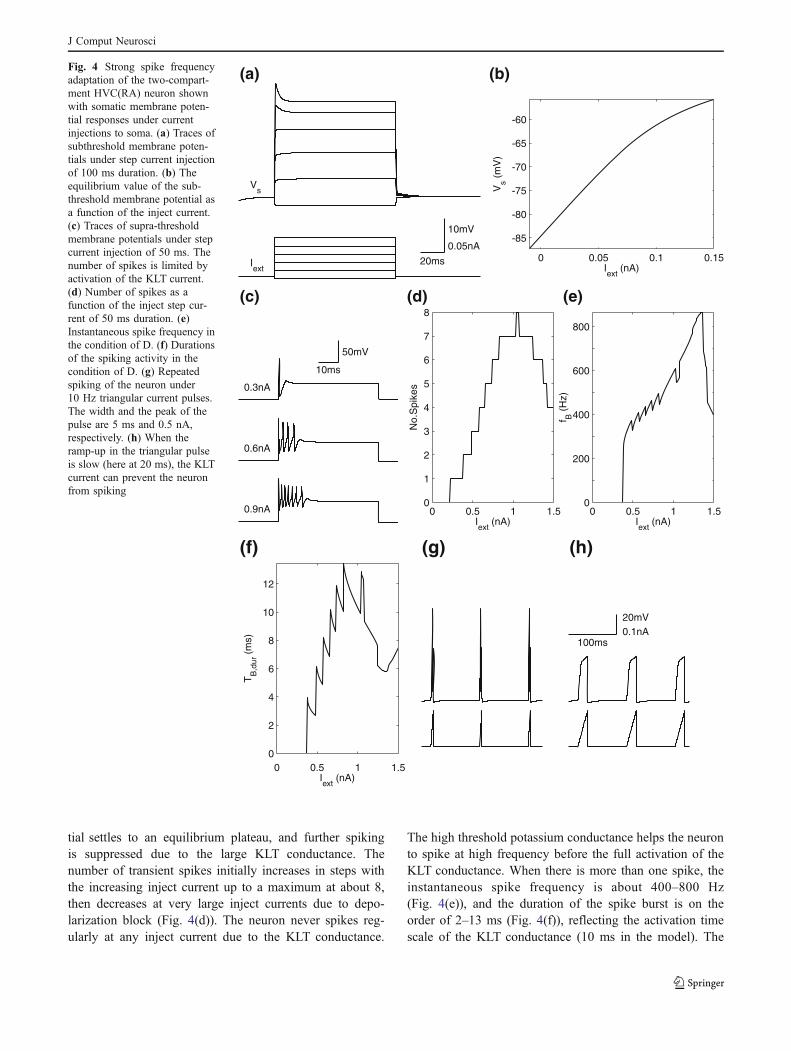
tial settles to an equilibrium plateau, and further spikingis suppressed due to the large KLT conductance. Thenumber of transient spikes initially increases in steps withthe increasing inject current up to a maximum at about 8,then decreases at very large inject currents due to depo-larization block (Fig. 4(d)). The neuron never spikes reg-ularly at any inject current due to the KLT conductance.
The high threshold potassium conductance helps the neuronto spike at high frequency before the full activation of theKLT conductance. When there is more than one spike, theinstantaneous spike frequency is about 400–800 Hz(Fig. 4(e)), and the duration of the spike burst is on theorder of 2–13 ms (Fig. 4(f)), reflecting the activation timescale of the KLT conductance (10 ms in the model). The
20ms
10mV
0.05nA
Vs
Iext
0 0.05 0.1 0.15
-85
-80
-75
-70
-65
-60
Vs (
mV
)
Iext
(nA)
0.3nA
0.6nA
0.9nA
10ms
50mV
0 0.5 1 1.50
1
2
3
4
5
6
7
8
Iext
(nA)
No.
Spi
kes
0 0.5 1 1.50
200
400
600
800
Iext
(nA)
f B (
Hz)
0 1 1.50
2
4
6
8
10
12
T B,d
ur (
ms) 100ms
20mV0.1nA
(a)
(c) (e)
(f) (g) (h)
(b)
(d)
0.5Iext
(nA)
Fig. 4 Strong spike frequencyadaptation of the two-compart-ment HVC(RA) neuron shownwith somatic membrane poten-tial responses under currentinjections to soma. (a) Traces ofsubthreshold membrane poten-tials under step current injectionof 100 ms duration. (b) Theequilibrium value of the sub-threshold membrane potential asa function of the inject current.(c) Traces of supra-thresholdmembrane potentials under stepcurrent injection of 50 ms. Thenumber of spikes is limited byactivation of the KLT current.(d) Number of spikes as afunction of the inject step cur-rent of 50 ms duration. (e)Instantaneous spike frequency inthe condition of D. (f) Durationsof the spiking activity in thecondition of D. (g) Repeatedspiking of the neuron under10 Hz triangular current pulses.The width and the peak of thepulse are 5 ms and 0.5 nA,respectively. (h) When theramp-up in the triangular pulseis slow (here at 20 ms), the KLTcurrent can prevent the neuronfrom spiking
J Comput Neurosci

20ms
80mV
gEEmax
=0.05mS/cm2
50 100 150 200 250Time (ms)
Neu
ron
ID
20ms
80mV
gEEmax
=0.05mS/cm2
20ms
80mV
gEEmax
=0.081mS/cm2
0 0.05 0.1 0.150
10
20
30
40
50
60
gEEmax
(ms/cm2)
Ave
N.S
pike
s (b
lack
) an
d M
axN
.Spi
kes
(gre
y)
0 0.05 0.1 0.150
10
20
30
40
50
gEEmax
(ms/cm2)
Ave
N.S
pike
s (b
lack
) an
d M
axN
.Spi
kes
(gre
y)
(b) (d)
(e)
unstable
unstable
gFFs
=1mS/cm2
gFFs
=5mS/cm2
gFFs
=8mS/cm2
10ms
50mV
0 2 4 6 8 100
5
10
15
20
25
gFFs
(mS/cm2)
N.S
pike
s
(a)
(c)
(f)
J Comput Neurosci
model neuron can fire repeatedly under short currentpulses: 10 Hz triangular current pulses with 5 ms rise-timecan induce spikes at each pulse (Fig. 4(g)). This is due to afaster rise of the membrane potential compared to the acti-vation of KLT conductance at each pulse, and the reductionof KLT conductance when the membrane potential returnsto the resting potential after each pulse. In contrast,triangular pulses with 20 ms rise-time cannot make theneuron spike, as shown in Fig. 4(h).
2.2 Single compartment model
To show how the strong spike-frequency adaptation and thedendritic compartment contribute to robust propagation ofburst spikes, we also study spike propagation in networksof the standard Hodgkin–Huxley model with a leakconductance, fast sodium and potassium conductances,and an additional KHT conductance. The properties of theconductances are the same as in the two-compartmentmodel.
2.3 Noise injection
Noisy fluctuation of the membrane potential of each neuronis induced by injection of Poisson spike trains to anexcitatory synapse (noise synapse) on the neuron. At eachnoise spike arrival, the conductance added to the noisesynapse is randomly chosen from a range. The parametersof the noise injection are chosen to make the membranepotential to fluctuate with a standard deviation of approx-imately 3 mV. Specifically, the frequencies and themaximum conductances of the noise spike trains are:
200 Hz and 0.016 mS/cm2 for the two-compartment model,injected to both the soma and the dendrite; 200 Hz and0.031 mS/cm2 for the single compartment models.
2.4 Numerical integration
The dynamical equations are integrated numerically with afourth-order Runge–Kutta method (Press et al. 1992) usinga fixed time step of 0.01 ms. The computer code related tothe model is available upon request.
3 Results
3.1 Non-robust burst sequence generation
A complete model of burst sequences in HVC requires notonly synaptic connectivity, but also dynamical models ofaction potential generation and synaptic transmission. In thepast, theorists have studied models of sequence generationin which the output of a single neuron is binary-valued(Amari 1972; Kleinfeld 1986), while others have used morerealistic spiking neuron models (Abeles 1982, 1991; Cateauand Fukai 2001; Diesmann et al. 1999; Hermann et al.1995). In our numerical simulations, we have used spiking,conductance-based model neurons. This is because our goalis to identify which intrinsic cellular properties of HVC(RA) neurons are important in the generation of burstsequences.
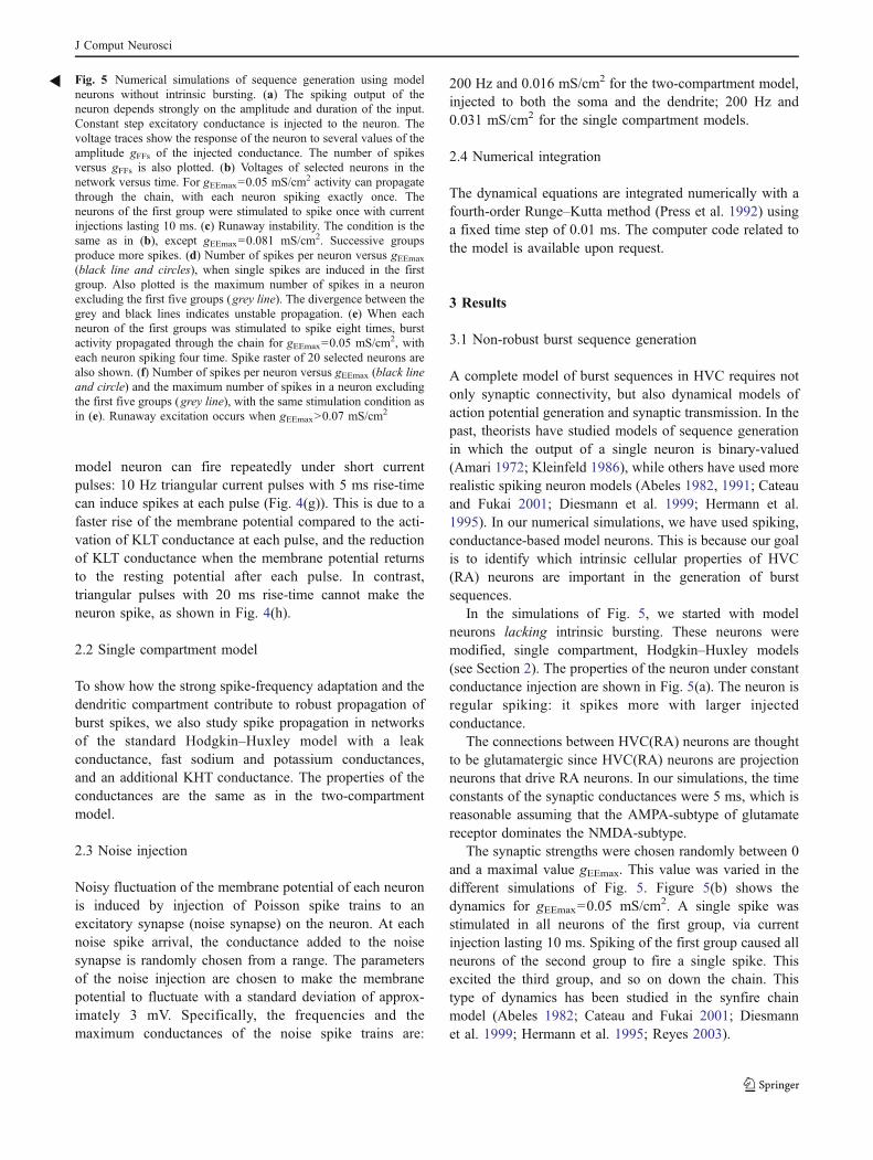
In the simulations of Fig. 5, we started with modelneurons lacking intrinsic bursting. These neurons weremodified, single compartment, Hodgkin–Huxley models(see Section 2). The properties of the neuron under constantconductance injection are shown in Fig. 5(a). The neuron isregular spiking: it spikes more with larger injectedconductance.
The connections between HVC(RA) neurons are thoughtto be glutamatergic since HVC(RA) neurons are projectionneurons that drive RA neurons. In our simulations, the timeconstants of the synaptic conductances were 5 ms, which isreasonable assuming that the AMPA-subtype of glutamatereceptor dominates the NMDA-subtype.
The synaptic strengths were chosen randomly between 0and a maximal value gEEmax. This value was varied in thedifferent simulations of Fig. 5. Figure 5(b) shows thedynamics for gEEmax=0.05 mS/cm2. A single spike wasstimulated in all neurons of the first group, via currentinjection lasting 10 ms. Spiking of the first group caused allneurons of the second group to fire a single spike. Thisexcited the third group, and so on down the chain. Thistype of dynamics has been studied in the synfire chainmodel (Abeles 1982; Cateau and Fukai 2001; Diesmannet al. 1999; Hermann et al. 1995; Reyes 2003).
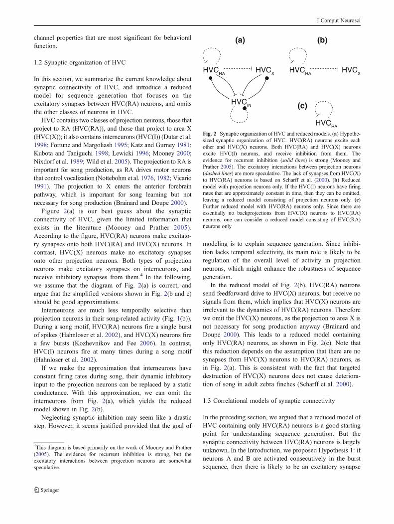
Fig. 5 Numerical simulations of sequence generation using modelneurons without intrinsic bursting. (a) The spiking output of theneuron depends strongly on the amplitude and duration of the input.Constant step excitatory conductance is injected to the neuron. Thevoltage traces show the response of the neuron to several values of theamplitude gFFs of the injected conductance. The number of spikesversus gFFs is also plotted. (b) Voltages of selected neurons in thenetwork versus time. For gEEmax=0.05 mS/cm2 activity can propagatethrough the chain, with each neuron spiking exactly once. Theneurons of the first group were stimulated to spike once with currentinjections lasting 10 ms. (c) Runaway instability. The condition is thesame as in (b), except gEEmax=0.081 mS/cm2. Successive groupsproduce more spikes. (d) Number of spikes per neuron versus gEEmax
(black line and circles), when single spikes are induced in the firstgroup. Also plotted is the maximum number of spikes in a neuronexcluding the first five groups (grey line). The divergence between thegrey and black lines indicates unstable propagation. (e) When eachneuron of the first groups was stimulated to spike eight times, burstactivity propagated through the chain for gEEmax=0.05 mS/cm2, witheach neuron spiking four time. Spike raster of 20 selected neurons arealso shown. (f) Number of spikes per neuron versus gEEmax (black lineand circle) and the maximum number of spikes in a neuron excludingthe first five groups (grey line), with the same stimulation condition asin (e). Runaway excitation occurs when gEEmax>0.07 mS/cm2
R
J Comput Neurosci
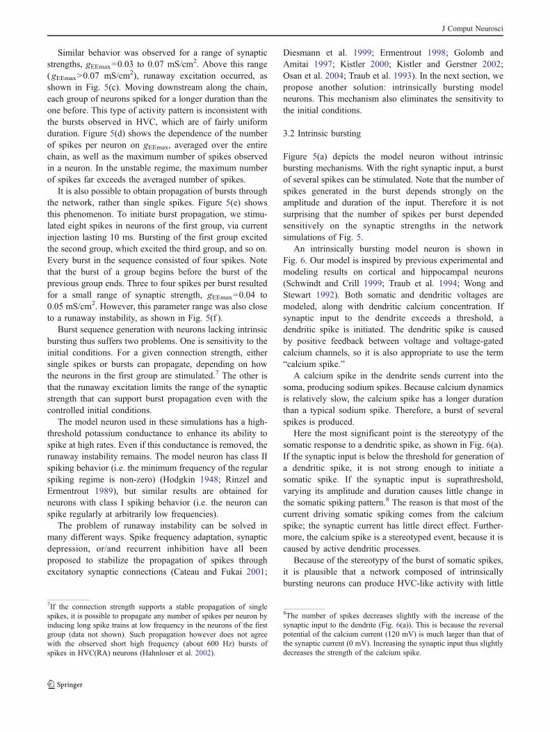
Similar behavior was observed for a range of synapticstrengths, gEEmax=0.03 to 0.07 mS/cm2. Above this range( gEEmax>0.07 mS/cm2), runaway excitation occurred, asshown in Fig. 5(c). Moving downstream along the chain,each group of neurons spiked for a longer duration than theone before. This type of activity pattern is inconsistent withthe bursts observed in HVC, which are of fairly uniformduration. Figure 5(d) shows the dependence of the numberof spikes per neuron on gEEmax, averaged over the entirechain, as well as the maximum number of spikes observedin a neuron. In the unstable regime, the maximum numberof spikes far exceeds the averaged number of spikes.
It is also possible to obtain propagation of bursts throughthe network, rather than single spikes. Figure 5(e) showsthis phenomenon. To initiate burst propagation, we stimu-lated eight spikes in neurons of the first group, via currentinjection lasting 10 ms. Bursting of the first group excitedthe second group, which excited the third group, and so on.Every burst in the sequence consisted of four spikes. Notethat the burst of a group begins before the burst of theprevious group ends. Three to four spikes per burst resultedfor a small range of synaptic strength, gEEmax=0.04 to0.05 mS/cm2. However, this parameter range was also closeto a runaway instability, as shown in Fig. 5(f ).
Burst sequence generation with neurons lacking intrinsicbursting thus suffers two problems. One is sensitivity to theinitial conditions. For a given connection strength, eithersingle spikes or bursts can propagate, depending on howthe neurons in the first group are stimulated.7 The other isthat the runaway excitation limits the range of the synapticstrength that can support burst propagation even with thecontrolled initial conditions.
The model neuron used in these simulations has a high-threshold potassium conductance to enhance its ability tospike at high rates. Even if this conductance is removed, therunaway instability remains. The model neuron has class IIspiking behavior (i.e. the minimum frequency of the regularspiking regime is non-zero) (Hodgkin 1948; Rinzel andErmentrout 1989), but similar results are obtained forneurons with class I spiking behavior (i.e. the neuron canspike regularly at arbitrarily low frequencies).
The problem of runaway instability can be solved inmany different ways. Spike frequency adaptation, synapticdepression, or/and recurrent inhibition have all beenproposed to stabilize the propagation of spikes throughexcitatory synaptic connections (Cateau and Fukai 2001;
Diesmann et al. 1999; Ermentrout 1998; Golomb andAmitai 1997; Kistler 2000; Kistler and Gerstner 2002;Osan et al. 2004; Traub et al. 1993). In the next section, wepropose another solution: intrinsically bursting modelneurons. This mechanism also eliminates the sensitivity tothe initial conditions.
3.2 Intrinsic bursting
Figure 5(a) depicts the model neuron without intrinsicbursting mechanisms. With the right synaptic input, a burstof several spikes can be stimulated. Note that the number ofspikes generated in the burst depends strongly on theamplitude and duration of the input. Therefore it is notsurprising that the number of spikes per burst dependedsensitively on the synaptic strengths in the networksimulations of Fig. 5.
An intrinsically bursting model neuron is shown inFig. 6. Our model is inspired by previous experimental andmodeling results on cortical and hippocampal neurons(Schwindt and Crill 1999; Traub et al. 1994; Wong andStewart 1992). Both somatic and dendritic voltages aremodeled, along with dendritic calcium concentration. Ifsynaptic input to the dendrite exceeds a threshold, adendritic spike is initiated. The dendritic spike is causedby positive feedback between voltage and voltage-gatedcalcium channels, so it is also appropriate to use the term“calcium spike.”
A calcium spike in the dendrite sends current into thesoma, producing sodium spikes. Because calcium dynamicsis relatively slow, the calcium spike has a longer durationthan a typical sodium spike. Therefore, a burst of severalspikes is produced.
Here the most significant point is the stereotypy of thesomatic response to a dendritic spike, as shown in Fig. 6(a).If the synaptic input is below the threshold for generation ofa dendritic spike, it is not strong enough to initiate asomatic spike. If the synaptic input is suprathreshold,varying its amplitude and duration causes little change inthe somatic spiking pattern.8 The reason is that most of thecurrent driving somatic spiking comes from the calciumspike; the synaptic current has little direct effect. Further-more, the calcium spike is a stereotyped event, because it iscaused by active dendritic processes.
Because of the stereotypy of the burst of somatic spikes,it is plausible that a network composed of intrinsicallybursting neurons can produce HVC-like activity with little
7If the connection strength supports a stable propagation of singlespikes, it is possible to propagate any number of spikes per neuron byinducing long spike trains at low frequency in the neurons of the firstgroup (data not shown). Such propagation however does not agreewith the observed short high frequency (about 600 Hz) bursts ofspikes in HVC(RA) neurons (Hahnloser et al. 2002).
8The number of spikes decreases slightly with the increase of thesynaptic input to the dendrite (Fig. 6(a)). This is because the reversalpotential of the calcium current (120 mV) is much larger than that ofthe synaptic current (0 mV). Increasing the synaptic input thus slightlydecreases the strength of the calcium spike.
J Comput Neurosci
need for fine tuning. This will be demonstrated in the nextsection.
Another important feature of the dendritic spike is itslong refractory period. In Fig. 6(b), the dendrite receivesthree identical synaptic inputs, spaced by 80 ms intervals.Although the first input causes a dendritic spike, the secondand third fail to do so. This is because calcium concentra-tion decays slowly after the dendritic spike, and activates acalcium-dependent potassium current, which reduces theexcitability of the dendrite. As will be seen in the nextsection, the long refractory period also contributes toprevention of runaway instability.
It should be noted that there is presently no publishedevidence for dendritic calcium spikes in HVC(RA) neurons.We predict that this phenomenon will be found (seeSection 4).
3.3 Robust burst sequence generation
We simulated a network of intrinsically bursting modelneurons, with the synaptic connectivity of Fig. 3(b). Somesimulated burst sequences are shown in Fig. 7(a). Eachburst of somatic spikes rides on top of a “plateau” involtage. The plateau is due to current generated by thedendritic spike. In effect, dendrites use somatic spikes to
communicate with each other. If we considered onlysomatic spike times, the activity of this model wouldlook very similar to the activity shown in Fig. 5. How-ever, the model is very different, because the fundamentalpropagating event is the dendritic spike, not the somaticspike.
Intrinsic bursting gives the model improved robustness.If gEEmax is varied, the number of spikes per burst variesfrom 4 to 6 (Fig. 7(c)). As explained before, this is becausea burst is a stereotyped event. Furthermore, there is norunaway instability. The stereotypy of the calcium spikemakes the neural response insensitive to the synaptic in-puts. This prevents amplification of the activity during thepropagation. The long refractory period of the dendriticspike also contributes to the stability by suppressing con-secutive activations of the dendrtic spikes.
3.4 Strong spike frequency adaptation
Our model neuron has another important feature, which isthat the burst of somatic spikes has a shorter duration thanthe calcium spike (see Fig. 6(a)). The somatic spikesterminate because the soma has a low threshold potassium(KLT) conductance, which strongly dampens excitabilitywhen the soma is depolarized.
Vs
Vd
[Ca2+]
10ms
50mV1mM
gFFd=1mS/cm2
0 2 4 6 8 100
1
2
3
4
5
6
7
gFFd (mS/cm2)
N.S
pike
s
Vs
Vd
20ms
50mV
(a)
(b)
Fig. 6 The dendritic mecha-nism of burst generation in thetwo-compartment model ofHVC(RA) neuron. (a) Thespiking output of the neurondepends only weakly on theamplitude gFFd and duration ofconstant excitatory conductanceinput on the dendrite. Somaticvoltage, dendritic voltage, anddendritic calcium concentrationare shown. Input conductance tothe dendrite causes a dendriticspike, which drives a burst ofsomatic spikes. (b) The dendriticspike has a long refractory peri-od. Only the first synaptic inputin a series of three is able toactivate a dendritic spike. Thevertical bars indicate the timesof excitatory synaptic inputs tothe dendrite
J Comput Neurosci

Our use of a KLT conductance is inspired by Dutar et al.(1998), who used sharp electrodes to record from HVC(RA) neurons in brain slices from adult zebra finches.They found that HVC(RA) neurons responded with justone or two spikes to sustained current injection. They alsonoted that the strong spike frequency adaptation might be
due to a rapidly activating potassium conductance. Indeed,the KLT conductance has been shown to lead to a similarspike response in a number of other neuron types, in-cluding auditory neurons in avian nucleus magnocellularis(Rathouz and Trussell 1998; Reyes et al. 1994), bushy cellsin ventral cochlear nucleus (Manis and Marx 1991), audi-tory neurons in medial nucleus of the trapezoid body ofrats (Dodson et al. 2002) and mice (Wang et al. 1998), andauditory neuron in gerbil medial superior olive (Svirskiset al. 2002).
Whether a KLT conductance is likewise the mechanismof spike frequency adaptation in HVC(RA) neurons ispresently unknown. Our model cannot resolve this issue;further experiments are needed. The point of our model isdifferent: it proposes a possible function for spikefrequency adaptation, regardless of its biophysical mecha-nism. Adaptation allows the duration of somatic spiking tobe shorter than the duration of the dendritic calcium spike.This is an important feature, since the typical burst ofsodium spikes in HVC(RA) neurons during singing is just6 ms (Hahnloser et al. 2002), whereas the dendritic calciumcan last tens of milliseconds if the hypothetical calciumspikes in dendrities of HVC(RA) neurons are similar tocalcium spikes in cortical and hippocampal neurons. In ourmodel, the short burst duration is set by the properties ofthe KLT conductance.
3.5 Robustness of intrinsic bursting
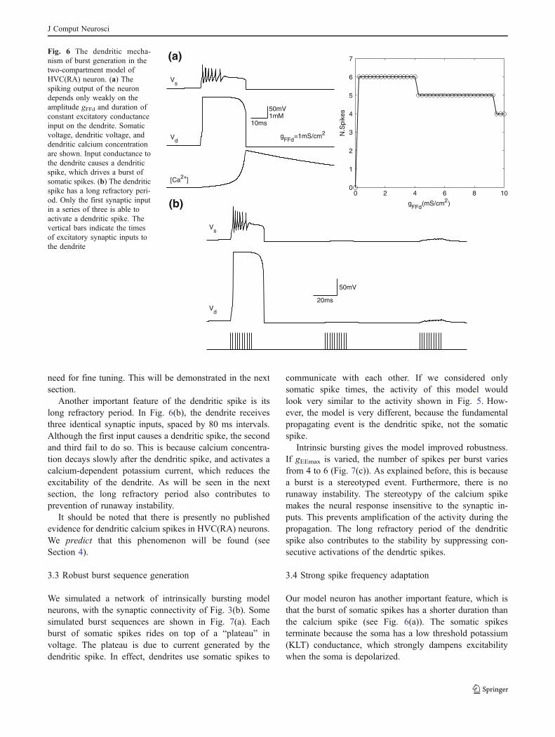
If the neurons are intrinsically bursting, the need forfine-tuning of synaptic strengths is removed from ourmodel. However, one might ask whether fine-tuning ofintrinsic cellular properties is necessary to achieve theproper burst duration and number of spikes per burst. Toinvestigate this issue, we experimented with varyingparameters of the model dendrite. Figure 8 shows theeffect of these manipulations on the number of spikes perburst.
Figure 8(a) shows how the spike number depends on thecoupling resistance Rc between the dendritic and somaticcompartments, and the total dendritic area Ad. All otherparameters are left at their default values. The spike numberis insensitive to Ad. The dependence on Rc is stronger, butthere is a reasonable range of values yielding more thanthree spikes.
Figure 8(b) plots the dependence of the number ofsomatic spikes on the conductances gCa and gCaK ofcalcium channels and calcium-activated potassium chan-nels, respectively. There is a large parameter regime inwhich the spike number is 5 or 6.
The problem of how excitable cells regulate theirconductances to achieve dynamical behaviors is a fascinat-ing one. The problem is not peculiar to our model of HVC,
50 100 150 200 250Time (ms)
Neu
ron
ID
20ms
80mV
0 0.1 0.2 0.3 0.4 0.50
1
2
3
4
5
6
gEEmax
(ms/cm2)
Ave
N.S
pike
s(a)
(b)
(c)
Fig. 7 Robust propagation of burst spikes in networks of the HVC(RA) neurons with active dendrites. (a) Traces of somatic membranepotentials of five selected neurons in a run of the dynamics. Theconnections between the neurons are made to the dendrites. (b) Spikeraster of 20 selected neurons. (c) Average number of spikes per neuronversus the connection strength gEEmax
J Comput Neurosci
but is relevant to just about any neural system. It seemslikely that such regulatory mechanisms exist (Davis 2006).Figure 8 suggests that such mechanisms would not have totune parameters very precisely to achieve the requiredbursting behavior.
4 Discussion
In conclusion, we have described a theory of burstsequences in HVC(RA) neurons that is based on two mainhypotheses. Hypothesis 1 is that the connectivity betweenHVC(RA) neurons is correlational, i.e., based on temporalcorrelations in their activity. Hypothesis 2 is that HVC(RA)neurons are intrinsic bursters.
4.1 Correlational connectivity
Hypothesis 1 makes a clear prediction: two HVC(RA)neurons that burst in succession should be connected by anexcitatory synapse, with high probability. While thishypothesis is easy to state, the only direct experimentaltests that one can imagine are very difficult. For example,one could perform dual intracellular recordings of pairs ofHVC(RA) neurons in singing birds. One could test whetherthe neurons are connected, and then compare their bursttimings. To a neurophysiologist, this experiment alreadysounds extremely difficult. To make things worse, con-nections between HVC(RA) neurons are likely to be verysparse. In our numerical simulations, we used 200 groupsof neurons. If a pair of neurons is chosen randomly fromour model, its probability of being connected is less than1%. Therefore, our model predicts that it would be difficultto find connected pairs by simply recording randomly fromneurons.9
If HVC were spatially organized, the experimental situa-tion would be more favorable. For example, suppose thatneurons that burst simultaneously were at the same locationin HVC. Then it would be much easier to find connectedpairs of neurons. However, no such spatial organization hasbeen detected so far (Fee, unpublished observations).
An alternative to the neurophysiological approachmentioned above is a purely anatomical approach. Recent-ly, new high-throughput anatomical techniques have beeninvented (Denk and Horstmann 2004; Tsai et al. 2003). Inparticular, automated serial-section electron microscopycould potentially allow the reconstruction of the entiresynaptic connectivity of HVC. If HVC were found to havea connectivity similar to one of those shown in Fig. 3, thiswould be strong evidence for Hypothesis 1.
To some, Hypothesis 1 may seem so intuitively obviousthat its truth must be a foregone conclusion. But this is notthe case, as competing models for sequence generationhave been proposed. For example, Estes argued thatsequential order is determined by inhibitory synapticconnectivity, not excitatory connectivity (Estes 1972).
4.2 Intrinsic bursting
We have shown that intrinsic bursting enhances the robustnessof burst sequence generation by a neural network. Accordingto our Hypothesis 2, HVC(RA) neurons are intrinsic bursters.How strong is this prediction? It is consistent with theobservation that the spontaneous bursts of HVC(RA) neuronduring sleep have the same stereotypy as during singing
9While Mooney and Prather reported synaptic interactions betweenpairs of HVC(RA) neurons in vitro (Mooney and Prather 2005), it isnot clear whether these connections were monosynaptic.
Ad(α
m)
100 300 500 70020000
280000
540000
800000
2
3
4
5
6
gCa(mS/cm2)
g CaK
(mS
/cm
2 )
5 105 205 3055
105
205
305
3
4
5
6
Rc(M )
(a)
(b)
6 5 4 3 253
6
5
Fig. 8 Robustness of intrinsic bursting to changes in parameters ofthe HVC(RA) neuron model. (a) Dependence of spike number onsoma-dendrite coupling and total dendritic area. (b) Dependence ofspike number on conductances of calcium channels and calcium-activated potassium channels
J Comput Neurosci

(Hahnloser et al. 2002, 2006). While intrinsic bursting is onesolution to the problem of robustness, it is not the only one.Other biophysical mechanisms could also improve robust-ness, such as spike frequency adaptation, synaptic depres-sion, and recurrent inhibition (Cateau and Fukai 2001;Diesmann et al. 1999; Ermentrout 1998; Golomb and Amitai1997; Kistler 2000; Kistler and Gerstner 2002; Osan et al.2004; Traub et al. 1993). While our theoretical arguments aresuggestive, they are not conclusive. Ultimately, this issue canonly be settled by experiment.
In previous experiments in vitro, it has been found thatsomatic current injection does not trigger intrinsic burstingin HVC(RA) neurons (Dutar et al. 1998; Kubota andTaniguchi 1998; Mooney 2000; Wild et al. 2005). This maysound inconsistent with our Hypothesis 2, but actually it isnot, for two reasons. First, somatic current injection is anindirect way of testing for a dendritic spike. In our model, itmay or may not trigger a calcium spike in the dendrite. Thisdepends on model parameters like the strength of couplingbetween the soma and dendrite, and the voltage thresholdfor a dendritic spike. Second, the intrinsic properties ofHVC(RA) neurons appear to be different in vitro and in thebrain of a singing bird. For example, the peak firing ratesobserved in vitro are never as high as those observed duringsong (Dutar et al. 1998; Kubota and Taniguchi 1998).Perhaps HVC(RA) neurons in vitro are missing someneuromodulator that is necessary for the intrinsic propertiesappropriate for song-related activity, and this neuromodu-lator could have effects on intrinsic bursting.
Given these facts, one can imagine a number of ways oftesting Hypothesis 2. Drugs that affect voltage-gatedcalcium channels or calcium-activated potassium channelscould lower the threshold for a dendritic spike, and permitthe initiation of a burst by somatic current injection. Further-more, such drugs could change burst duration or spikenumber during song in vivo. In addition, glutamate un-caging could be used to stimulate dendrites directly, ashas been done with cortical neurons (Schiller et al. 2000;Wei et al. 2001). Also in vivo imaging of calcium dynamicsin dendrites could be revealing (Euler et al. 2002;Trachtenberg et al. 2002).
4.3 Spike frequency adaptation
We have argued that strong spike frequency adaptation inHVC(RA) neurons could function to make burst durationshorter than the duration of the hypothetical dendriticcalcium spike. However, we should make the caveat thatthere is some disagreement about the strength of spikefrequency adaptation in HVC(RA) neurons. Kubota andTaniguchi (1998) found weaker spike frequency adaptationin HVC(RA) neurons, which in their experiments producedmany spikes in response to sustained current injection.
Perhaps their results were different from Dutar et al. (1998)because they used patch electrodes rather than sharpelectrodes and juvenile rather than adult birds.
Therefore we should also consider the alternative sce-nario in which spike frequency adaptation is only a weakeffect, and has little effect on burst duration. In this sce-nario, the dendritic calcium spike itself has short duration,unlike calcium spikes seen in cortical and hippocampalneurons (Golding et al. 1999; Schwindt and Crill 1999; Weiet al. 2001).
In short, two scenarios should be considered: burst dura-tion could be set by the time course of the dendritic spike,or by somatic spike frequency adaptation. These possibil-ities can be tested pharmacologically by applying drugs thataffect calcium dynamics or spike frequency adaptation andobserving the effects on burst duration during song.
4.4 Refractory period
In our model, the dendritic spike has a long refractoryperiod, due to the slow decay of calcium and the calcium-dependent potassium current (Fig. 5(b)). Previously weargued that the refractory period could function to preventrunaway instability.
Here we mention another possible effect: the refractoryperiod could enhance robustness to sloppiness in theconnectivity of the HVC(RA) neurons. In our idealizedmodel, if neuron A bursts after neuron B, there is nosynaptic connection from A to B. In Fig. 3, this dependenceon temporal order is reflected in the fact that there are only“forward” connections, and no “backward” connections.However, even if there were some backward connections,they would have no effect on propagation of bursts throughthe network, because they would deliver synaptic input toneurons only while their dendrites are refractory. Therefore,some sloppiness in the connectivity can be tolerated.
Note that burst propagation would be possible in themodels of Fig. 3, even if the connectivity were truly bidi-rectional, with equal numbers of forward and backwardconnections. Then propagation in either direction would bepossible, so that the bird could sing its song either forwardor backward.10 Since this is not observed, it seems morelikely that the connectivity is biased in the forward direction.
4.5 Inhibition
In our model, we neglected the influence of HVC(I)neurons on HVC(RA) neurons. We argued that this was a
10Bidirectional propagation is standard for most excitable media. Forexample, an axon can support either orthodromic or antidromicpropagation of an action potential, though only the orthodromic isseen in natural conditions.
J Comput Neurosci

good approximation because the spiking of HVC(I) neuronsis much less temporally selective, and therefore their inputto HVC(RA) neurons can be approximated as constant intime. We suspect that interneurons lack temporal selectivitybecause they sum the outputs of many projection neurons.Because the projection neurons are active at a wide varietyof times, the sum of their outputs is temporally unselective.
While inhibition is not expected to determine thetemporal order in which projection neurons are activated,it may have the function of regulating the overall level ofactivity of the projection neurons. In other words, inhibitioncould be a circuit mechanism that prevents runawayinstability, in addition to the cellular mechanisms that havebeen discussed previously.
4.6 Learning
Suppose that HVC(RA) neurons possess a correlationalconnectivity, like those of Fig. 3. How could thisconnectivity be created during learning or development?
The simplest possibility would be spatial organization.Suppose that the HVC(RA) neurons were arranged sothat their temporal selectivities progressed in an orderlyfashion across HVC. Then if synaptic connections werelocal in space and biased in one direction, they wouldconform to Fig. 3. However, as mentioned previously, nosuch spatial organization has been detected in HVC(RA)neurons thus far.
The obvious alternative is that Hebbian synaptic plastic-ity could create correlational structure. This idea has beenpopular in models of associative memory, in which synapticstrengths are given by correlation matrices. The idea wasproposed for temporal sequences in particular by Amari(1972).
4.7 Previous models of HVC sequence generation
In a previous model of sequence generation by HVC,Troyer and Doupe proposed that sequences are generatedby an associative chain of motor and sensory representa-tions (Troyer and Doupe 2000), where the motor represen-tation resides in the HVC(RA) neurons and the sensoryrepresentation is an efference copy in the HVC(X) neurons.The sequence is generated by reciprocal excitatory inter-actions between the HVC(RA) and HVC(X) neural pop-ulations. In contrast, we have argued that the associativechain is within the HVC(RA) population itself, and doesnot involve the HVC(X) neurons. Support for this ideacomes from the fact that targeted destruction of HVC(X)neurons does not cause deterioration of song in adult zebrafinches (Scharff et al. 2000). Accordingly, we believe thatthere are excitatory synapses from HVC(RA) to HVC(X)neurons, but not vice versa (see Fig. 2).
Drew and Abbott proposed a model of sequencegeneration in HVC (Drew and Abbott 2003). In the model,HVC neurons are driven with a common periodic input.The sequential firing of the neurons arise from chainedinhibitory connections between neurons, disinhibition, aswell as strong afterhyperpolarization currents that preventneurons to spike repeatedly in short time. Their work isclosely related to the general model of sequence generationproposed by Estes (1972). Although there is some evidenceof sporadic timed inputs from Uva (Vu et al. 1994;Williams and Vicario 1993), it has not been demonstratedthat there are timing signals with high enough resolution todrive sequential burst of spiking lasting about 6 ms. Ourmodel does not require a timing signal inputs. The timingarises from the excitatory connectivity between the neu-rons. However our model does require an external input tostart the spiking activity in the first group.
In their model of birdsong learning, Doya and Sejnowski(1999) took the HVC activation patterns as given. They didnot address the mechanism of sequence generation in HVC.
4.8 Synfire chains
Spiking network models utilizing the connectivity ofFig. 3(b) are often called synfire chains. Should this termalso be applied to the present model? The answer to thisquestion depends on the exact definition of the term.Typically synchronous spiking of one group of neurons isrequired to drive the next group over threshold in a synfirechain. Having a high threshold for spiking helps to makethe dynamics robust to noise. Also, each neuron in a groupgenerates a single spike, so that the precise timing of thisspike is clearly important. On the other hand, suppose thateach neuron generates a burst of more than one spike, andsynapses must temporally integrate successive spikes todrive the next group of neurons above threshold. In thiscase, the spiking of neurons in a group must still occurwithin a window that is set by the synaptic integration time.However, the width of this window is longer than theinterspike interval during a burst, so it is not clear whetherthis qualifies as precise synchrony.
In our model of burst sequences, the dendritic calciumspike is the fundamental propagating event, rather than thesomatic sodium spikes. Therefore, the term synburst chainmight be more appropriate.11
11Another ambiguity of definition arises when considering theconnectivity of Fig. 3(c). In this model, the connectivity is uni-directional but the neurons are not divided into groups. Here the spiketimes of the neurons will not cluster into groups, but are expected tobe fairly uniformly distributed in time. Nevertheless, synchronous(within a synaptic integration time) spiking may be required forpropagation of activity. It is not clear whether this should be called asynfire chain.
J Comput Neurosci
Acknowledgement Research was supported by The Huck Instituteof Life Sciences at the Pennsylvania State University and Alfred P.Sloan Fellowship (DZJ), and Howard Hughes Medical Institute (FR,HSS). DZJ thanks the Kavli Institute for Theoretical Physics atUniversity of California, Santa Barbara for partial support of thiswork. We thank Michael Long, Anthony Leonardo and Michale Feefor useful discussions.
References
Abeles, M. (1982). Local cortical circuits: An electrophysiologicalstudy (pp. 83–92). Berlin Heidelberg New York: Springer.
Abeles, M. (1991). Corticonics. Cambridge, UK: CambridgeUniversity Press.
Amari, S. (1972). Learning patterns and pattern sequences by self-organizing nets of threshold elements. IEEE Transactions onComputers, C-21, 1197–1206.
Brainard, M. S., & Doupe, A. J. (2000). Interruption of a basalganglia-forebrain circuit prevents plasticity of learned vocal-izations. Nature, 404, 762–766.
Brumberg, J. C., Nowak, L. G., & McCormick, D. A. (2000). Ionicmechanisms underlying repetitive high-frequency burst firing insupragranular cortical neurons. Journal of Neuroscience, 20,4829–4843.
Cardin, J. A., Raksin, J. N., & Schmidt, M. F. (2004). Thesensorimotor nucleus NIf is necessary for auditory processingbut not vocal motor output in the avian song system. Journal ofNeurophysiology.
Cateau, H., & Fukai, T. (2001). Fokker–Planck approach to thepulse packet propagation in synfire chain. Neural Networks, 14,675–685.
Chi, Z., & Margoliash, D. (2001). Temporal precision and temporaldrift in brain and behavior of zebra finch song. Neuron, 32,899–910.
Coleman, M. J., & Vu, E. T. (2005). Recovery of impaired songsfollowing unilateral but not bilateral lesions of nucleus uvaefor-mis of adult zebra finches. Journal of Neurobiology, 63, 70–89.
Crook, S. M., Ermentrout, G. B., & Bower, J. M. (1998). Spike fre-quency adaptation affects the synchronization properties of net-works of cortical oscillations. Neural Computation, 10, 837–854.
Davis, G. W. (2006). Homeostatic control of neural activity: Fromphenomenology to molecular design. Annual Review of Neuro-science, 29, 307–323.
Denk, W., & Horstmann, H. (2004). Serial block-face scanningelectron microscopy to reconstruct three-dimensional tissuenanostructure. PLoS Biol, 2, e329.
Diesmann, M., Gewaltig, M. O., & Aertsen, A. (1999). Stablepropagation of synchronous spiking in cortical neural networks.Nature, 402, 529–533.
Dodson, P. D., Barker, M. C., & Forsythe, I. D. (2002). Twoheteromeric Kv1 potassium channels differentially regulateaction potential firing. Journal of Neuroscience, 22, 6953–6961.
Doupe, A. J., & Kuhl, P. K. (1999). Birdsong and human speech:Common themes and mechanisms. Annual Review of Neurosci-ence, 22, 567–631.
Doya, K., & Sejnowski, T. J. (1999). A computational model of aviansong learning. In M. S. Gazzaniga (Ed.), The new cognitiveneurosciences. Cambridge, MA: MIT Press.
Drew, P. J., & Abbott, L. F. (2003). Model of song selectivity andsequence generation in area HVc of the songbird. Journal ofNeurophysiology, 89, 2697–2706.
Dutar, P., Vu, H. M., & Perkel, D. J. (1998). Multiple cell typesdistinguished by physiological, pharmacological, and anatomic
properties in nucleus HVc of the adult zebra finch. Journal ofNeurophysiology, 80, 1828–1838.
Ermentrout, B. (1998). The analysis of synaptically generated travel-ing waves. Journal of Computational Neuroscience, 5, 191–208.
Estes, W. K. (1972). An associative basis for coding and organisationin memory. In A. W. Melton, & E. Martin (Eds.), Codingprocesses in human memory. Washington, DC: Winston.
Euler, T., Detwiler, P. B., & Denk, W. (2002). Directionally selectivecalcium signals in dendrites of starburst amacrine cells. Nature,418, 845–852.
Fee, M. S., Kozhevnikov, A. A., & Hahnloser, R. H. (2004). Neuralmechanisms of vocal sequence generation in the songbird.Annals of the New York Academy of Sciences, 1016, 153–170.
Fortune, E. S., & Margoliash, D. (1995). Parallel pathways andconvergence onto HVc and adjacent neostriatum of adult zebrafinches (Taeniopygia guttata). Journal of Comparative Neurology,360, 413–441.
Franceschetti, S., Guatteo, E., Panzica, F., Sancini, G., Wanke, E., &Avanzini, G. (1995). Ionic mechanisms underlying burst firing inpyramidal neurons: Intracellular study in rat sensorimotor cortex.Brain Research, 696, 127–139.
Golding, N. L., Jung, H. Y., Mickus, T., & Spruston, N. (1999).Dendritic calcium spike initiation and repolarization are con-trolled by distinct potassium channel subtypes in CA1 pyramidalneurons. Journal of Neuroscience, 19, 8789–8798.
Golomb, D., & Amitai, Y. (1997). Propagating neuronal dischargesin neocortical slices: Computational and experimental study.Journal of Neurophysiology, 78, 1199–1211.
Hahnloser, R. H., Kozhevnikov, A. A., & Fee, M. S. (2002). An ultra-sparse code underlies the generation of neural sequences in asongbird. Nature, 419, 65–70.
Hahnloser, R. H., Kozhevnikov, A. A., & Fee, M. S. (2006). Sleep-related neural activity in a premotor and a basal-ganglia pathwayof the songbird. Journal of Neurophysiology, 96, 794–812.
Hausser, M., Spruston, N., & Stuart, G. J. (2000). Diversity anddynamics of dendritic signaling. Science, 290, 739–744.
Hermann, M., Hertz, J. A., & Prugel-Bennet, A. (1995). Analysisof synfire chains. Network: Computation in Neural Systems, 6,403–414.
Hodgkin, A. L. (1948). The local electric changes associated withrepetitive action in a non-medulated axon. Journal of Physiology,107, 165–181.
Hodgkin, A. L., & Huxley, A. F. (1952). A quantitative description ofmembrane current and its application to conduction and exci-tation in nerve. Journal of Physiology (London), 117, 500–544.
Immelmann, K. (1969). Song development in the zebra finch andother estrildid finches. In R. Hinde (Ed.), Bird vocalization(pp. 61–74). Cambridge, UK: Cambridge University Press.
Katz, L. C., & Gurney, M. E. (1981). Auditory responses in the zebrafinch’s motor system for song. Brain Research, 221, 192–197.
Kistler, W. M. (2000). Stability properties of solitary waves andperiodic wave trains in a two-dimensional network of spikingneurons. Physical Review. E, Statistical Physics, Plasmas, Fluids,and Related Interdisciplinary Topics, 62, 8834–8837.
Kistler, W. M., & Gerstner, W. (2002). Stable propagation of activitypulses in populations of spiking neurons. Neural Computation,14, 987–997.
Kleinfeld, D. (1986). Sequential state generation by model neuralnetworks. Proceedings of the National Academy of Sciences ofthe United States of America, 83, 9469–9473.
Konishi, M. (1965). The role of auditory feedback in the control ofvocalization in the white-crowned sparrow. Zeitschrift fürTierpsychologie, 22, 770–783.
Kozhevnikov, A., & Fee, M. S. (2006). Singing-related activity ofidentified HVC neurons in the zebra finch. Journal ofNeurophysiology.
J Comput Neurosci
Kubota, M., & Taniguchi, I. (1998). Electrophysiological character-istics of classes of neuron in the HVc of the zebra finch. Journalof Neurophysiology, 80, 914–923.
Lashley, K. S. (1951). The problem of serial order in behavior. InL. A. Jeffress (Ed.), Cerebral mechanisms in behavior (the HixonSymposium) (pp. 112–136). New York: Wiley.
Leonardo, A., & Fee, M. S. (2005). Ensemble coding of vocal controlin birdsong. Journal of Neuroscience, 25, 652–661.
Lewicki, M. S. (1996). Intracellular characterization of song-specificneurons in the zebra finch auditory forebrain. Journal of Neuro-science, 16, 5855–5863.
Lien, C. C., & Jonas, P. (2003). Kv3 potassium conductance isnecessary and kinetically optimized for high-frequency actionpotential generation in hippocampal interneurons. Journal ofNeuroscience, 23, 2058–2068.
Manis, P. B., & Marx, S. O. (1991). Outward currents in isolatedventral cochlear nucleus neurons. Journal of Neuroscience, 11,2865–2880.
Mattia, D., Kawasaki, H., & Avoli, M. (1997). In vitro electrophys-iology of rat subicular bursting neurons. Hippocampus, 7, 48–57.
Mooney, R. (2000). Different subthreshold mechanisms underlie songselectivity in identified HVc neurons of the zebra finch. Journalof Neuroscience, 20, 5420–5436.
Mooney, R., Hoese, W., & Nowicki, S. (2001). Auditory representa-tion of the vocal repertoire in a songbird with multiple songtypes. Proceedings of the National Academy of Sciences of theUnited States of America, 98, 12778–12783.
Mooney, R., & Prather, J. F. (2005). The HVC microcircuit: Thesynaptic basis for interactions between song motor and vocalplasticity pathways. Journal of Neuroscience, 25, 1952–1964.
Nixdorf, B. E., Davis, S. S., & DeVoogd, T. J. (1989). Morphology ofGolgi-impregnated neurons in hyperstriatum ventralis, parscaudalis in adult male and female canaries. Journal of Compar-ative Neurology, 284, 337–349.
Nottebohm, F., Kelley, D. B., & Paton, J. A. (1982). Connections ofvocal control nuclei in the canary telencephalon. Journal ofComparative Neurology, 207, 344–357.
Nottebohm, F., Stokes, T. M., & Leonard, C. M. (1976). Centralcontrol of song in the canary, Serinus canarius. Journal ofComparative Neurology, 165, 457–486.
Osan, R., Curtu, R., Rubin, J., & Ermentrout, B. (2004). Multiple-spike waves in a one-dimensional integrate-and-fire neuralnetwork. Journal of Mathematical Biology, 48, 243–274.
Pinsky, P. F., & Rinzel, J. (1994). Intrinsic and network rhythmo-genesis in a reduced Traub model for CA3 neurons. Journal ofComputational Neuroscience, 1, 39–60.
Press, W., Teukolsky, S., Vetterling, W., & Flannery, B. (1992). Numericalrecipes in C. Cambridge, UK: Cambridge University Press.
Rathouz, M., & Trussell, L. (1998). Characterization of outwardcurrents in neurons of the avian nucleus magnocellularis. Journalof Neurophysiology, 80, 2824–2835.
Reyes, A. D. (2003). Synchrony-dependent propagation of firing ratein iteratively constructed networks in vitro. Nature Neuroscience,6, 593–599.
Reyes, A. D., Rubel, E. W., & Spain, W. J. (1994). Membraneproperties underlying the firing of neurons in the avian cochlearnucleus. Journal of Neuroscience, 14, 5352–5364.
Rinzel, J., & Ermentrout, G. B. (1989). Analysis of neural excitabilityand oscillations. In C. Koch, & I. Segev (Eds.), Methods inneuronal modeling. Cambridge: MIT Press.
Scharff, C., Kirn, J. R., Grossman, M., Macklis, J. D., &Nottebohm, F. (2000). Targeted neuronal death affects neuronalreplacement and vocal behavior in adult songbirds. Neuron, 25,481–492.
Schiller, J., Major, G., Koester, H. J., & Schiller, Y. (2000). NMDAspikes in basal dendrites of cortical pyramidal neurons. Nature,404, 285–289.
Schwindt, P., & Crill, W. (1999). Mechanisms underlying burst andregular spiking evoked by dendritic depolarization in layer 5cortical pyramidal neurons. Journal of Neurophysiology, 81,1341–1354.
Sompolinsky, H., & Kanter, I. I. (1986). Temporal association inasymmetric neural networks. Physical Review Letters, 57,2861–2864.
Svirskis, G., Kotak, V., Sanes, D. H., & Rinzel, J. (2002).Enhancement of signal-to-noise ratio and phase locking for smallinputs by a low-threshold outward current in auditory neurons.Journal of Neuroscience, 22, 11019–11025.
Trachtenberg, J. T., Chen, B. E., Knott, G. W., Feng, G., Sanes, J. R.,Welker, E., et al. (2002). Long-term in vivo imaging ofexperience-dependent synaptic plasticity in adult cortex. Nature,420, 788–794.
Traub, R. D., Jefferys, J. G., & Miles, R. (1993). Analysis of thepropagation of disinhibition-induced after-discharges along theguinea-pig hippocampal slice in vitro. Journal of Physiology,472, 267–287.
Traub, R. D., Jefferys, J. G., Miles, R., Whittington, M. A., & Toth, K.(1994). A branching dendritic model of a rodent CA3 pyramidalneurone. Journal of Physiology, 481(Pt 1), 79–95.
Troyer, T. W., & Doupe, A. J. (2000). An associational model ofbirdsong sensorimotor learning II. Temporal hierarchies and thelearning of song sequence. Journal of Neurophysiology, 84,1224–1239.
Tsai, P. S., Friedman, B., Ifarraguerri, A. I., Thompson, B. D.,Lev-Ram, V., Schaffer, C. B., et al. (2003). All-opticalhistology using ultrashort laser pulses. Neuron, 39, 27–41.
Vicario, D. S. (1991). Organization of the zebra finch song controlsystem: II. Functional organization of outputs from nucleusRobustus archistriatalis. Journal of Comparative Neurology, 309,486–494.
Vu, E. T., Mazurek, M. E., & Kuo, Y. C. (1994). Identification of aforebrain motor programming network for the learned song ofzebra finches. Journal of Neuroscience, 14, 6924–6934.
Wang, L. Y., Gan, L., Forsythe, I. D., & Kaczmarek, L. K. (1998).Contribution of the Kv3.1 potassium channel to high-frequencyfiring in mouse auditory neurones. Journal of Physiology, 509(Pt 1), 183–194.
Wang, X. J. (1999). Fast burst firing and short-term synaptic plasticity:A model of neocortical chattering neurons. Neuroscience, 89,347–362.
Wei, D. S., Mei, Y. A., Bagal, A., Kao, J. P., Thompson, S. M., &Tang, C. M. (2001). Compartmentalized and binary behavior ofterminal dendrites in hippocampal pyramidal neurons. Science,293, 2272–2275.
Wild, J. M., Williams, M. N., Howie, G. J., & Mooney, R. (2005).Calcium-binding proteins define interneurons in HVC of thezebra finch (Taeniopygia guttata). Journal of ComparativeNeurology, 483, 76–90.
Williams, H. (2004). Birdsong and singing behavior. Annals of theNew York Academy of Sciences, 1016, 1–30.
Williams, H., & Vicario, D. S. (1993). Temporal patterning of songproduction: Participation of nucleus uvaeformis of the thalamus.Journal of Neurobiology, 24, 903–912.
Wong, R. K., & Stewart, M. (1992). Different firing patterns generatedin dendrites and somata of CA1 pyramidal neurones in guinea-pig hippocampus. Journal of Physiology, 457, 675–687.
Yu, A. C., & Margoliash, D. (1996). Temporal hierarchical control ofsinging in birds. Science, 273, 1871–1875.
J Comput Neurosci