Embed Size (px)
Citation preview

Intricate Hydrogen-Bonded Networks: Binary and Ternary Combinations of Uracil, PTCDI,and Melamine
Jules A. Gardener,*,† Olga Y. Shvarova, G. Andrew D. Briggs, and Martin R. Castell*Department of Materials, UniVersity of Oxford, Oxford, OX1 3PH, United Kingdom
ReceiVed: NoVember 29, 2009; ReVised Manuscript ReceiVed: January 31, 2010
We report the formation of two- and three-component porous supramolecular networks from combinations ofuracil, PTCDI, and melamine. The structures, which are formed on Au(111) in ultra-high vacuum (UHV)and studied by scanning tunneling microscopy (STM), are stabilized by hydrogen bonds. We show that twobimolecular networks comprising uracil and PTCDI can be formed, one of which contains two pore geometriesand is composed of 28 molecules per unit cell. In addition, we observe two different ordered structures frommixtures of melamine and uracil. By combining all three of these species, we demonstrate the formation ofa single ternary structure that contains 33 molecules per unit cell. Our results demonstrate the capacity ofhydrogen bonding to produce highly complex structures, and open up the possibility of forming a wide rangeof new structures from combinations of nucleic bases and other small organic molecules.
Introduction
A range of porous two-dimensional supramolecular networkscan be constructed on surfaces through careful choice of themolecular components and the substrate.1 This is primarilyachieved through hydrogen bonding, metal-organic co-ordina-tion, van der Waals interactions, or covalent bonding. Here, weconcentrate on combinations of organic molecules with thecapacity to form hydrogen bonds. Such an approach is advanta-geous since hydrogen bonds are highly directional, leading toordered structures, but sufficiently weak to allow moleculardiffusion on surfaces at room temperature. These properties canresult in the formation of ordered molecular structures over largeareas. Further, some of these networks may contain pores andcan act as templates for the ordering of guest molecules suchas fullerenes.2–9
Some nucleic bases and other small organic molecules canform monomer porous two-dimensional structures stabilized byhydrogen bonding.10–14 However, a greater variety of networkscan be generated through the combination of two complementarymolecular species. For example, four different supramolecularnetworks of 3,4,9,10-perylenetetracarboxylic diimide (PTCDI)and1,3,4-triazine-2,4,6-triamine(melamine)havebeenreported,2,6–8,15,16
of which three contain pores that act as absorption sites for guestmolecules. Bimolecular networks generally have more complexunit cells than their monomer counterparts, and offer a widerrange of pore sizes, geometries, and interpore separations.Recently, coronene-templated three-component networks havebeen reported, which are even larger and more intricate (wedefine intricacy in terms of the number of molecules per unitcell).17,18 However, these appear to show little capacity to hostmolecules other than coronene.
In this work, we have combined two different systems thatare well-known to form hydrogen-bonded supramolecularnetworks: a nucleic base with PTCDI-melamine. The nucleicbase that we have chosen is uracil, a molecule that forms planar
hydrogen-bonded structures in two and three dimensions.19–21
The chemical structures of the three molecules studied are shownin Figure 1. Uracil is prochiral; when constrained to lie parallelto a surface, it can adopt one of two possible orientations andthese have no mirror symmetry, giving rise to chirality.However, PTCDI and melamine are not chiral, even whenconstrained to two dimensions. Here, we deposit uracil, PTCDI,and melamine onto Au(111), by sublimation. Through thecareful selection and postdeposition processing of the componentmolecules, we demonstrate the formation of ordered binary andternary supramolecular networks, some of which have remark-ably intricate unit cells.
Experimental Methods
All experiments were performed in situ under UHV conditions(base pressure of ∼1 × 10⁻⁸ Pa). Au(111) films on mica(Agilent) were prepared by Ar+ sputtering and annealing to∼600 °C, a procedure that produced large flat terraces displayingthe characteristic 22 × �3 reconstruction. Melamine (Aldrich),PTCDI (TCI Europe), and uracil (Aldrich) were sublimed attypical rates of 8-15 ML/h (ML)monolayers), 0.6-0.8 ML/h, and 0.1-0.3 ML/h (unless otherwise stated), respectively,onto the Au substrates, which were held at room temperature.The samples were subjected to postdeposition annealing for10-14 h; shorter anneals (typically a few hours) and lowertemperatures resulted in less ordered structures. A JEOLJSTM4500S STM was used to obtain constant current images(bias applied to the sample) at room temperature, usingelectrochemically etched W tips. All images were processedwith WSxM software.22
* To whom correspondence should be addressed. [email protected] [email protected].
† Present address: Department of Physics, Harvard University, Cambridge,MA 02138.
Figure 1. The molecular structures of (a) PTCDI, (b) melamine, and(c) uracil. Balls represent carbon (gray), oxygen (red), nitrogen (blue),and hydrogen (white) atoms.
J. Phys. Chem. C 2010, 114, 5859–5866 5859
10.1021/jp9113249 2010 American Chemical SocietyPublished on Web 03/10/2010

Results and Discussion
First, we characterize the self-assembly of uracil afterdeposition onto a clean 22 × �3 reconstructed Au(111) filmon mica. We observe two different structures, as shown in Figure2. At moderately low coverages, chain-like features are formed,as shown in Figure 2a. These structures often form closed rings,but show no long-range ordering. High-resolution images, suchas Figure 2b, show that the chains comprise double rows ofuracil molecules, while pentagonal clusters are frequentlyobserved where multiple chains join (for example, as indicatedby an arrow in the figure). The uracil chains are similar to thoseformed by flat-lying cytosine or thymine on Au(111), stabilizedby in-plane hydrogen bonds;23–25 any differences in molecularpositioning within the chains are attributed to the nonequivalenthydrogen bonding sites of different nucleic bases. At highermolecular coverages we find close-packed regions (Figure 2c).In this arrangement, each uracil is generally surrounded by sixother uracil molecules, again forming a hydrogen-bondednetwork.19,20
New porous structures are observed after sequential depositionof 0.1-0.3 ML of uracil then 0.3-0.4 ML of PTCDI at room
temperature, followed by annealing to ∼85 °C for ∼10 h,examples of which are shown in Figures 3 and 4. On average,these structures cover a combined total of 34% of the Au(111)surface, while some residual close-packed PTCDI patchesremain (any excess uracil is likely to have desorbed from thesurface during annealing). The differences in size, shape, andelectronic structure of PTCDI and uracil allows them to bedistinguished in the STM images; PTCDI appear as brightrectangles, while uracil molecules are seen as smaller, roundfeatures. However, we cannot rule out the additional presenceof small adsorbate molecules (such as water) within thesestructures.
The network shown in panels a and b of Figure 3 comprisesdouble rings of uracil, linked by parallel pairs of PTCDImolecules arranged in a cross-like manner. We will refer to thisstructure as the “double-cross”. The uracil double-rings areslightly elliptical in shape and show distinct similarities to thechain structure of Figure 2 since, in both of these structures,neighboring uracil molecules from each ring are aligned radially.However, the outer ring of the double-cross structure appearsto be missing four uracil molecules (one in each quadrant), the
Figure 2. (a) Disordered chain-like structures of uracil (Vs ) +1.35 V, It ) 0.07 nA, 85 nm × 85 nm). (b) A higher resolution image of the chains,which are double rows of uracil molecules (Vs ) +1.44 V, It ) 0.07 nA, 16.0 nm × 12.0 nm). A black arrow highlights a pentagonal cluster ofuracil. (c) A region of close-packed uracil (Vs ) -1.40 V, It ) 0.10 nA, 16.0 nm × 12.0 nm). Uracil was deposited at a rate of 0.9-1.0 ML/h forall images shown in this figure.
5860 J. Phys. Chem. C, Vol. 114, No. 13, 2010 Gardener et al.
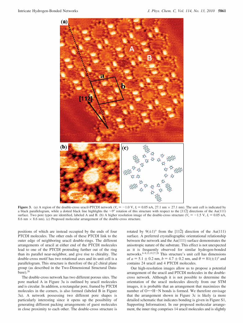
positions of which are instead occupied by the ends of fourPTCDI molecules. The other ends of these PTCDI link to theouter edge of neighboring uracil double-rings. The differentarrangements of uracil at either end of the PTCDI moleculeslead to one of the PTCDI protruding further out of the ringthan its parallel near-neighbor, and give rise to chirality. Thedouble-cross motif has two rotational axes and its unit cell is aparallelogram. This structure is therefore of the p2 chiral planegroup (as described in the Two-Dimensional Structural Data-base).26
The double-cross network has two different porous sites. Thepore marked A in Figure 3a is outlined by uracil moleculesand is circular. In addition, a rectangular pore, framed by PTCDImolecules in the corners, is also formed (labeled B in Figure3a). A network possessing two different pore shapes isparticularly interesting since it opens up the possibility ofgenerating different packing arrangements of guest moleculesin close proximity to each other. The double-cross structure is
rotated by 9((1)° from the [112j] direction of the Au(111)surface. A preferred crystallographic orientational relationshipbetween the network and the Au(111) surface demonstrates theanisotropic nature of the substrate. This effect is not unexpectedas it is frequently observed for similar hydrogen-bondednetworks.6–8,15,19,27,28 This structure’s unit cell has dimensionsof a ) 5.1 ( 0.2 nm, b ) 4.7 ( 0.2 nm, and θ ) 81((1)° andcontains 24 uracil and 4 PTCDI molecules.
Our high-resolution images allow us to propose a potentialarrangement of the uracil and PTCDI molecules in the double-cross network. Although it is not possible to determine theorientation of the uracil molecules directly from our STMimages, it is probable that an arrangement that maximizes thenumber of O · · ·H-N bonds is formed. We therefore envisagethat the arrangement shown in Figure 3c is likely (a moredetailed schematic that indicates bonding is given in Figure S1,Supporting Information). In our proposed molecular arrange-ment, the inner ring comprises 14 uracil molecules and is slightly
Figure 3. (a) A region of the double-cross uracil-PTCDI network (Vs ) -1.0 V, It ) 0.05 nA, 27.1 nm × 27.1 nm). The unit cell is indicated bya black parallelogram, while a dotted black line highlights the ∼9° rotation of this structure with respect to the [112j] directions of the Au(111)surface. Two pore types are identified, labeled A and B. (b) A higher resolution image of the double-cross structure (Vs ) -1.5 V, It ) 0.05 nA,8.6 nm × 8.6 nm). (c) Proposed molecular arrangement of the double-cross structure.
Intricate Hydrogen-Bonded Networks J. Phys. Chem. C, Vol. 114, No. 13, 2010 5861

elliptical in shape, consistent with our experimental observations.The inner uracil molecules are of one chirality, while the outeruracil molecules are of the opposite chirality. Although thislocally leads to a slight imbalance of the ratio of the two uracilchiralities, this is globally relieved by the formation of bothcomplementary chiral structures, as we have observed.
The double-cross structure coexists with a different porousnetwork, which is shown in Figure 4a,b. This comprises rowsof end-to-end PTCDI pairs sandwiching two overlappingpentagonal uracil clusters. We denote this structure the “stair-case” network. The unit cell contains 8 uracil and 2 PTCDImolecules, and has dimensions a ) 2.4 ( 0.2 nm, b ) 3.5 (0.2 nm, and θ ) 63((2)°. We propose a possible molecularstructure of the staircase network (Figure 4c, also Figure S2 inthe Supporting Information), again based on our experimentalobservations and the requirement to maximize the possiblenumber of hydrogen bonds. This arrangement contains equalnumbers of the two chiralities of uracil. Similarities within this
network are observed with the monomer structures, such as thepentagonal uracil features (Figure 2b) and the end-on bindingof PTCDI molecules.16 In general, domains of the staircasenetwork are larger than those of the double-cross structure, withaverage domain areas of 3800 nm2 (standard deviation of 2200nm2) and 440 nm2 (standard deviation of 400 nm2), respectively.Presumably this difference arises from the larger intricacy ofthe latter. The smaller double-cross fragments, however, aremore numerous than the staircase domains, although still onlycover 7-13% of the total surface area (we observe that thestaircase network covers 11-37% of the Au(111) surface).
We now turn to combinations of uracil and melamine, fromwhich we have successfully generated two different networkswhich cover most of the surface (Figure 5). Small fragments ofthese networks are formed after codeposition at room temper-ature, but after annealing to ∼90 °C for ∼12 h these structuresgrow in size and number, while single-component melamineand uracil regions are no longer observed. Both networks look
Figure 4. (a) An image showing two regions of the uracil-PTCDI staircase structure (Vs ) -1.5 V, It ) 0.05 nA, 60.6 nm × 60.6 nm). A dottedblack line runs parallel to the translation vector. This network is oriented 7((1)° from the [112j] directions of the Au(111) surface. (b) A moredetailed image of the staircase network, in which the unit cell is indicated (Vs ) -1.5 V, It ) 0.10 nA, 7.3 nm × 7.3 nm). (c) Our proposed modelof the staircase structure.
5862 J. Phys. Chem. C, Vol. 114, No. 13, 2010 Gardener et al.

very different from those of pure uracil (Figure 2) and puremelamine29 on Au(111). We therefore attribute these structuresto mixtures of melamine and uracil. The most commonlyobserved network, shown in Figure 5a, is rotated by 7((1)°with respect to the [112j] directions of the Au(111) surface. Theunit cell is indicated in the inset and has dimensions a ) b )2.3 ( 0.2 nm and θ ) 115((1)°. In contrast, the unit cell ofthe network shown in Figure 5b is more elongated, with a )2.9 ( 0.2 nm, b ) 1.2 ( 0.2 nm, and θ ) 98((1)°. It has notbeen possible to distinguish between the uracil and melaminemolecules in these images, owing to their similar sizes, and sowe refrain from proposing a structural model for either network.However, we suggest that both supramolecular networks are
predominantly stabilized by hydrogen bonds, in a similar mannerto three-dimensional melamine-uracil crystals30 and their mono-mer two-dimensional counterparts.19,20,29
We have established that a range of supramolecular structurescan be constructed from uracil-PTCDI and uracil-melamine,while melamine and PTCDI are well-known to form a range ofporous networks on Au(111).6–8,15,16 We now report on ternarymixtures of all three species. Combining three molecules withthe potential for hydrogen bonding is an attractive approach togenerating more intricate and even larger supramolecularnetworks. Figure 6 shows the highly ordered porous structureformed from 0.1-0.3 ML of melamine, 0.2-0.3 ML of uracil,and 0.3-0.4 ML of PTCDI after annealing to ∼120 °C for10-12 h. Patches of this structure cover on average 24-38%of the surface, with only close-packed PTCDI regions otherwiseobserved. When the three components are mixed and annealedat a lower temperature of ∼100 °C for ∼2 h, small fragmentsof the bimolecular networks are often found. Examples of suchare given in Figure 7. In Figure 7a, small regions of aparallelogram structure formed by PTCDI and melamine6 areseen, as highlighted by white arrows. Combinations of PTCDIand uracil are also observed, for example, as shown in Figure7b, where the double-cross structure is formed. However, aftersubsequent annealing to ∼130 °C, we do not observe any ofthe possible binary structures, nor any single phase uracil ormelamine regions (any excess of the latter two are likely tohave sublimed from the surface during annealing). The forma-tion of one ternary structure, as opposed to the several possiblebimolecular structures, demonstrates the capacity of hydrogenbonding to drive the formation of highly complex structures.
As can be seen from Figure 6a,b, the network formed fromPTCDI, melamine, and uracil has large hexagonal pores eachof which are surrounded by six smaller triangular voids. Wename this structure the “star” network due to its similarity tothe Star of David. Chevron-shaped clusters of three uracil and/or melamine protrude from the apex points of each hexagongiving rise to chirality. Networks of the two complementarychiralities are observed (one of each is shown in panels a andb of Figure 6). The intricate unit cell of the star structure, whichis marked in Figure 6a, has dimensions a ) b ) 6.6 ( 0.3 nmand θ )121((2)° and contains 33 molecules. The average areaof the star domains is 1200 nm2 (standard deviation of 1000nm2).
The orientation of the star network with respect to theAu(111) surface can be inferred from Figure 6c. It is likely thatthis image has been acquired with an adsorbate on the tip,causing the molecules to appear darker than the Au substratebut enhancing the resolution of the surface reconstruction. Forcomparison, an image of the same region obtained after applyinga brief electrical pulse to the tip to remove the absorbate isprovided in the inset. Two regions of opposite chirality areshown in Figure 6c; more detailed images (not shown) revealthat region A is the same as Figure 6a, while region B is of thesame chirality as the structure shown in Figure 6b. Althoughboth unit cells are offset by 9((1)° with respect to the Au(111)[112j] directions, the local chirality of the network appears todefine the orientation of each unit cell with respect to thesubstrate. We note that in all cases the hexagonal rings arealigned parallel to [112j] directions of the Au(111) surface, i.e.,they are rotated by 9((1)° from the translation vectors, asindicated by the dotted line in Figure 6a.
Although we are unable to confidently distinguish betweenuracil and melamine in the star structure, we look to similaritieswith other known binary structures to enable us to propose a
Figure 5. (a) A region of a supramolecular network of melamine anduracil (Vs ) +1.0 V, It ) 0.07 nA, 61.4 nm × 61.4 nm). A higherresolution image is shown in the inset, in which the unit cell is marked(Vs )+1.0 V, It ) 0.07 nA, 17.0 nm × 17.0 nm). (c) A second structureof melamine-uracil (Vs ) +1.0 V, It ) 0.07 nA, 66.0 nm × 66.0 nm).Dotted lines indicate the direction of the translation vectors for eachnetwork, while the [112j] direction of the Au(111) surface is alsoindicated in each image.
Intricate Hydrogen-Bonded Networks J. Phys. Chem. C, Vol. 114, No. 13, 2010 5863
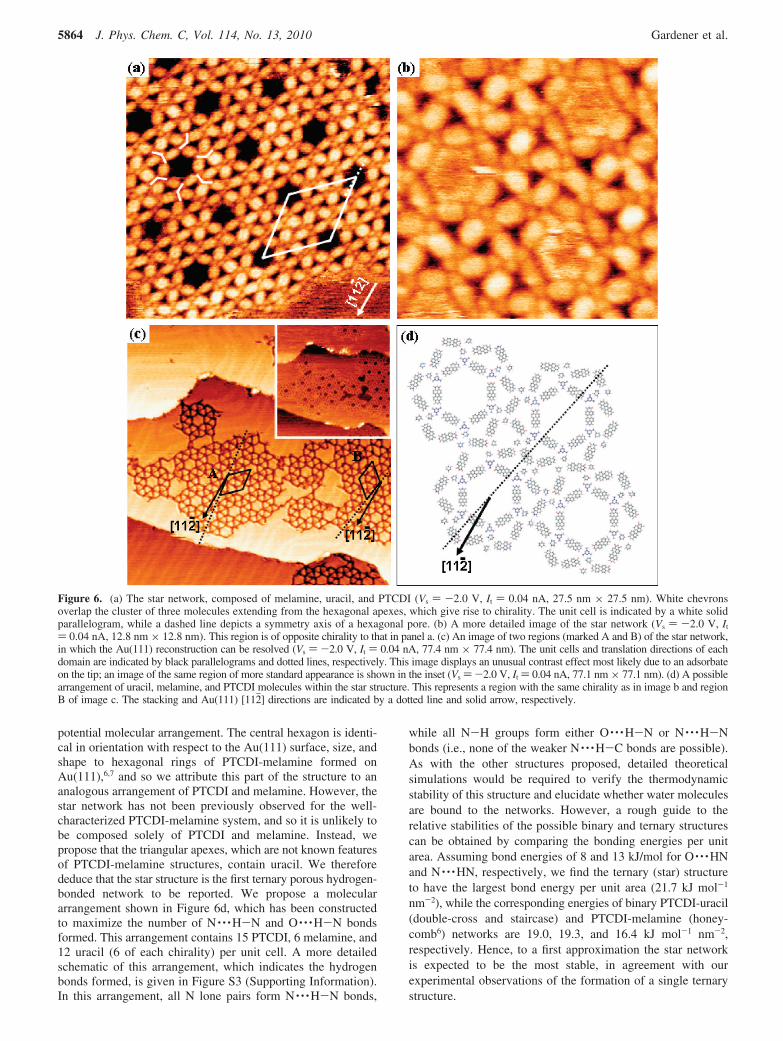
potential molecular arrangement. The central hexagon is identi-cal in orientation with respect to the Au(111) surface, size, andshape to hexagonal rings of PTCDI-melamine formed onAu(111),6,7 and so we attribute this part of the structure to ananalogous arrangement of PTCDI and melamine. However, thestar network has not been previously observed for the well-characterized PTCDI-melamine system, and so it is unlikely tobe composed solely of PTCDI and melamine. Instead, wepropose that the triangular apexes, which are not known featuresof PTCDI-melamine structures, contain uracil. We thereforededuce that the star structure is the first ternary porous hydrogen-bonded network to be reported. We propose a moleculararrangement shown in Figure 6d, which has been constructedto maximize the number of N · · ·H-N and O · · ·H-N bondsformed. This arrangement contains 15 PTCDI, 6 melamine, and12 uracil (6 of each chirality) per unit cell. A more detailedschematic of this arrangement, which indicates the hydrogenbonds formed, is given in Figure S3 (Supporting Information).In this arrangement, all N lone pairs form N · · ·H-N bonds,
while all N-H groups form either O · · ·H-N or N · · ·H-Nbonds (i.e., none of the weaker N · · ·H-C bonds are possible).As with the other structures proposed, detailed theoreticalsimulations would be required to verify the thermodynamicstability of this structure and elucidate whether water moleculesare bound to the networks. However, a rough guide to therelative stabilities of the possible binary and ternary structurescan be obtained by comparing the bonding energies per unitarea. Assuming bond energies of 8 and 13 kJ/mol for O · · ·HNand N · · ·HN, respectively, we find the ternary (star) structureto have the largest bond energy per unit area (21.7 kJ mol-1
nm-2), while the corresponding energies of binary PTCDI-uracil(double-cross and staircase) and PTCDI-melamine (honey-comb6) networks are 19.0, 19.3, and 16.4 kJ mol-1 nm-2,respectively. Hence, to a first approximation the star networkis expected to be the most stable, in agreement with ourexperimental observations of the formation of a single ternarystructure.
Figure 6. (a) The star network, composed of melamine, uracil, and PTCDI (Vs ) -2.0 V, It ) 0.04 nA, 27.5 nm × 27.5 nm). White chevronsoverlap the cluster of three molecules extending from the hexagonal apexes, which give rise to chirality. The unit cell is indicated by a white solidparallelogram, while a dashed line depicts a symmetry axis of a hexagonal pore. (b) A more detailed image of the star network (Vs ) -2.0 V, It
) 0.04 nA, 12.8 nm × 12.8 nm). This region is of opposite chirality to that in panel a. (c) An image of two regions (marked A and B) of the star network,in which the Au(111) reconstruction can be resolved (Vs ) -2.0 V, It ) 0.04 nA, 77.4 nm × 77.4 nm). The unit cells and translation directions of eachdomain are indicated by black parallelograms and dotted lines, respectively. This image displays an unusual contrast effect most likely due to an adsorbateon the tip; an image of the same region of more standard appearance is shown in the inset (Vs )-2.0 V, It ) 0.04 nA, 77.1 nm × 77.1 nm). (d) A possiblearrangement of uracil, melamine, and PTCDI molecules within the star structure. This represents a region with the same chirality as in image b and regionB of image c. The stacking and Au(111) [112j] directions are indicated by a dotted line and solid arrow, respectively.
5864 J. Phys. Chem. C, Vol. 114, No. 13, 2010 Gardener et al.

The star structure has one main pore and eight smaller voidsper unit cell which could be used to trap guest molecules, suchas fullerenes. We have sublimed C82 onto the structure and haveobserved that they occupy both classes of site, as shown inFigure 8. Between three and six C82 molecules are situatedwithin the hexagonal pores; the distribution of occupancynumber is consistent with that of C84 in the hexagonal PTCDI-melamine structure.9 In addition, single C82 can be trapped inthe triangular voids. This confirms that the intricacy of the star
structure gives rise to multiple absorption sites for hostmolecules and can be used to produce complex ordering offullerenes.
Conclusions
In conclusion, we have formed a variety of two-dimensionalporous networks from combinations of uracil, melamine, andPTCDI. Two different supramolecular structures have beenproduced from uracil and PTCDI, both of which contain featuresobserved in pure uracil double-chains. One of these structures,which we have termed the double-cross, has a unit cell of 28molecules, while the staircase structure contains 10 moleculesper unit cell. In addition, melamine and uracil have been shownto form two different structures. It therefore seems likely thatmany other bimolecular networks, with a range of pore shapesand sizes, could be generated by combinations of other nucleicbases with either PTCDI or melamine.
Through the combination of PTCDI, uracil, and melamine,we have produced a highly intricate network. The somewhatsurprising formation of a three-component network, rather thanphase segregated binary regions, opens up the possibility ofdesigning a range of new, complex structures. This could beachieved through the use of other nucleic bases, small cyclicmolecules (such as trimesic acid) or perylene derivatives, orthrough chemical functionalization of the molecular compo-nents.31 Such networks could then be used to generate a varietyof complicated arrangements of guest molecules.
Acknowledgment. The authors thank the EPSRC for funding(EP/D048761/1 and GR/S15808/01). We thank Dr. K. Porfyrakisfor provision of C82 and C. Spencer (JEOL UK) for technicalsupport.
Supporting Information Available: Schematics showingthe possible hydrogen bond formed within the PTCDI-uracil
Figure 7. Mixtures of uracil, melamine, and PTCDI formed afterannealing to ∼100 °C for 2 h (both images obtained at roomtemperature). (a) An image in which many disordered regions can beobserved, although small fragments of PTCDI-melamine in the paral-lelogram structure are highlighted by white arrows (Vs ) -1.5 V, It )0.04 nA, 79.9 nm × 79.9 nm). (b) A different region of the same samplethat is again largely disordered (Vs ) -1.5 V, It ) 0.04 nA, 49.8 nm× 49.8 nm). A small fragment of the double-cross PTCDI-uracilstructure is indicated by a white arrow.
Figure 8. The arrangement of C82 molecules after deposition onto thestar network (Vs ) -2.0 V, It ) 0.04 nA, 53.5 nm × 53.3 nm). Smallclusters of C82 are observed within the hexagonal pores (examples ofwhich are indicated by black arrows), while individual C82 occupy thesmaller triangular pores.
Intricate Hydrogen-Bonded Networks J. Phys. Chem. C, Vol. 114, No. 13, 2010 5865

and PTCDI-uracil-melamine networks. This material is availablefree of charge via the Internet at http://pubs.acs.org.
References and Notes
(1) Kudernac, T.; Lei, S.; Elemans, J. A. A. W.; De Feyter, S. Chem.Soc. ReV. 2009, 38, 402–421.
(2) Theobald, J. A.; Oxtoby, N. S.; Phillips, M. A.; Champness, N. R.;Beton, P. H. Nature 2003, 424, 1029–1031.
(3) Zhang, H. L.; Chen, W.; Huang, H.; Chen, L.; Wee, A. T. S. J. Am.Chem. Soc. 2008, 130, 2720–2721.
(4) Kiebele, A.; Bonifazi, D.; Cheng, F.; Stohr, M.; Diederich, F.; Jung,T.; Spillmann, H. Chem. Phys. Chem. 2006, 7, 1462–1470.
(5) Li, M.; Deng, K.; Lei, S.-B.; Yang, Y.-L.; Wang, T.-S.; Shen, Y.-T.; Wang, C.-R.; Zeng, Q.-D.; Wang, C. Angew. Chem. 2008, 120, 6819–6823.
(6) Silly, F.; Shaw, A. Q.; Porfyrakis, K.; Briggs, G. A. D.; Castell,M. R. Appl. Phys. Lett. 2007, 91, 253109.
(7) Perdigao, L. M. A.; Perkins, E. W.; Ma, J.; Staniec, P. A.; Rogers,B. L.; Champness, N. R.; Beton, P. H. J. Phys. Chem. B 2006, 110, 12539–12542.
(8) Staniec, P. A.; Perdigao, L. M. A.; Saywell, A.; Champness, N. R.;Beton, P. H. Chem. Phys. Chem. 2007, 8, 2177–2181.
(9) Theobald, J. A.; Oxtoby, N. S.; Champness, N. R.; Beton, P. H.;Dennis, T. J. S. Langmuir 2005, 21, 2038–2041.
(10) Otero, R.; Schock, M.; Molina, L. M.; Loegsgaard, E.; Stensgaard,I.; Hammer, B.; Besenbacher, F. Angew. Chem., Int. Ed. 2005, 44, 2270–2275.
(11) Sivakova, S.; Rowan, S. J. Chem. Soc. ReV. 2005, 34, 9–21.(12) Griessl, S.; Lackinger, M.; Edelwirth, M.; Hietschold, M.; Heckl,
W. M. Single Mol. 2002, 3, 25–31.(13) Pawin, G.; Wong, K. L.; Kwon, K.-Y.; Bartels, L. Science 2006,
313, 961–962.(14) Kong, X.-H.; Deng, K.; Yang, Y.-L.; Zeng, Q.-D.; Wang, C. J.
Phys. Chem. C 2007, 111, 17382–17387.(15) Silly, F.; Shaw, A. Q.; Briggs, G. A. D.; Castell, M. R. Appl. Phys.
Lett. 2008, 92, 023102.
(16) Silly, F.; Shaw, A. Q.; Castell, M. R.; Briggs, G. A. D. Chem.Commun. 2008, 1907–1909.
(17) Lei, S.; Surin, M.; Tahara, K.; Adisoejoso, J.; Lazzaroni, R.; Tobe,Y.; De Feyter, S. Nano Lett. 2008, 8, 2541–2546.
(18) Li, Y.; Ma, Z.; Deng, K.; Lei., S.; Zheng, Q.; Fan, X.; De Feyter,S.; Juang, W.; Wang, C. Chem.sEur. J. 2009, 15, 5418–5423.
(19) Dretschkow, Th.; Dakkouri, A. S.; Wandlowski, Th. Langmuir1997, 13, 2843–2856.
(20) Sowerby, S. J.; Stockwell, P. A.; Heckl, W. M.; Petersen, G. B.Origins Life EVol. Biospheres 2000, 30, 81–99.
(21) Parry, G. S. Acta Crystallogr. 1954, 7, 313–320.(22) Horcas, I.; Fernandez, R.; Gomez-Rodriquez, J. M.; Colchero, J.;
Gomez-Herrero, J.; Baro, A. M. ReV. Sci. Instrum. 2007, 78, 013705.(23) Kelly, R. E. A.; Lukas, M.; Kantorovich, L. N.; Otero, R.; Xu, W.;
Mura, M.; Laegsgaard, E.; Stensgaard, I.; Besenbacher, F. J. Chem. Phys.2008, 129, 184707.
(24) Otero, R.; Lukas, M.; Kelly, R. E. A.; Xu, W.; Laegsgaard, E.;Stensgaard, I.; Kantorovich, L. N.; Besenbacher, F. Science 2008, 319, 312–315.
(25) Xu, W.; Kelly, R. E. A.; Otero, R.; Schock, M.; Laegsgaard, E.;Stensgaard, I.; Kantorovich, L. N.; Besenbacher, F. Small 2007, 12, 2011–2014.
(26) Plass, K. E.; Grzesiak, A. L.; Matzger, A. J. Acc. Chem. Res. 2007,40, 287–293.
(27) Zhang, H.-M.; Xie, Z.-X.; Long, L.-S.; Zhong, H.-P.; Zhao, W.;Mao, B.-W.; Xu, X.; Zhen, L.-S. J. Phys. Chem. C 2008, 112, 4209–4218.
(28) Su, G.-J.; Zhang, H.-M.; Wan, L.-J.; Bai, C.-L.; Wandlowski, T.J. Phys. Chem. B 2004, 108, 1931–1937.
(29) Silly, F.; Shaw, A. Q.; Castell, M. R.; Briggs, G. A. D.; Mura, M.;Martsinovich, N.; Kantorovich, L. J. Phys. Chem. C 2008, 112, 11476–11480.
(30) Thomas, R.; Kulkarni, G. U. Beilstien J. Org. Chem. 2007, 3, 17.(31) Perdigao, L. M. A.; Saywell, A.; Fontes, G. N.; Staniec, P. A.;
Goretzki, G.; Phillips, A. G.; Champness, N. R.; Beton, P. H. Chem.sEur.J. 2008, 14, 7600–7607.
JP9113249
5866 J. Phys. Chem. C, Vol. 114, No. 13, 2010 Gardener et al.