Embed Size (px)
Citation preview

CMLS, Cell. Mol. Life Sci. 56 (1999) 683–7001420-682X/99/080683-18 $ 1.50+0.20/0© Birkhauser Verlag, Basel, 1999
Review
Intrathalamic sensory connections mediated by the thalamicreticular nucleus
J. W. Crabtree
Department of Anatomy, School of Medical Sciences, University of Bristol, Bristol BS8 1TD (UK),Fax +44 117 929 1687, e-mail: [email protected]
Received 11 May 1999; received after revision 15 July 1999; accepted 21 July 1999
Abstract. The thalamus and cerebral cortex are linked and auditory sectors of TRN share many of thesame organizational features. Each of these sectorstogether to form a vast network of interconnections.
Different modes of interactions among the cells in this contains maps, which are related to its inputs andoutputs, and organizational components called ‘slabs.’network underlie different states of consciousness,
such as wakefulness and sleep. Interposed between the It is proposed that, during wakefulness, TRN is cru-cially involved in resetting the activity levels in sen-dorsal thalamus and cortex are the GABAergic neu-
rons of the thalamic reticular nucleus (TRN), which sory nuclei of the dorsal thalamus, which allows thecortex to actively and periodically compare its on-play a pivotal role not only in switching between the
awake and sleep states but also in sensory processing going sensory processing with the available sensoryduring the awake state. The visual, somatosensory, information.
Key words. Sensory processing; GABAergic neurons; reticular sectors; cortical projections; intrathalamic projec-tions; organizational components; sensory maps; reticular discontinuities.
Introduction
In mammals, neurons in the thalamus and cerebralcortex form a vast network of interconnections [1–7].Different modes of interactions within this networkunderlie different states of consciousness, such as wake-fulness and sleep [1, 4, 8–11]. The neural mechanismsthat give rise to changes from one conscious state toanother operate in the thalamus [1, 3, 4, 6, 8–17].During wakefulness, sensory information gains accessto the cortex only after considerable processing in thethalamus and, when falling asleep, the thalamus func-tionally disengages the cortex from ascending sensoryinputs. A key player involved in these mechanisms is thethalamic reticular nucleus (TRN), which in carnivoresincludes the perigeniculate nucleus (PGN). The crucialrole that TRN plays in promoting sleep- and wake-
related thalamic activity is well established [1, 4, 8–11,14, 18; see also 3, 6, 13, 15–17, 19, 20]. However, thefunction of this nucleus in thalamic sensory processingduring wakefulness is still far from clear. Thus, thepurpose of this review is to inquire into the role(s) thatTRN might play in sensory processing in relation to ourcurrent understanding of the anatomical connectivity ofthis nucleus.
The thalamic reticular nucleus
A derivative of the ventral thalamus [21], TRN is asheet of cells that surrounds much of the dorsal thala-mus and lies between the dorsal thalamus and thecortex (fig. 1). The nucleus is bordered medially by theexternal medullary lamina (EML) and laterally by the

J. W. Crabtree Reticular-mediated intrathalamic connections684
internal capsule (IC). Because of its anatomical posi-tion, TRN is traversed across its thickness by virtuallyall axons passing between the dorsal thalamus andcortex, giving the nucleus its reticulated appearance andname. As these thalamocortical and corticothalamicaxons pass through TRN, many of them give off collat-erals [22–24], providing the nucleus with both dorsalthalamic [24] and cortical [24, 25] sources of innervation(fig. 1). These collaterals extend from their parent axonsat approximately right angles and thus are generallyoriented parallel to the reticular plane [26–32]. Further-more, the cortical afferents to TRN are specifically fromlayer VI [30, 31]. TRN also receives afferents from avariety of nonthalamic subcortical structures, such asthe brainstem reticular formation [33–40] and the basalforebrain [35, 36, 41–43].The main targets of the efferents of TRN are dorsalthalamic nuclei [22–24, 44]. Some of these nuclei can beclassified as ‘first order’ or ‘higher order’ [5, 6, 45] andrepresentations of each sensory modality are pre-sumably contained in at least one first-order nucleusand at least one higher-order nucleus. First-order thala-mic nuclei receive their main driving inputs throughascending sensory pathways and transmit informationabout stimuli in the sensory periphery to the cortex.Higher-order thalamic nuclei are thought to receive
Figure 2. A labelled thalamic reticular cell and labelled thalamo-cortical and corticothalamic axons after a small crystal of DiI wasplaced into the dorsal thalamus of a rabbit. Note the extensivedistribution of the reticular dendrites and their position relative tothe traversing axons.
their main driving inputs through descending pathwaysfrom cortical layer V and transmit information aboutsensory processing in one cortical area to another corti-cal area.The mammalian TRN appears to be made up entirelyof �-aminobutyric acid (GABA)ergic neurons [rodent:46–49; primate: 50; carnivore: 47, 51–54; lagomorphand marsupial: 55]. The reticular somata are usuallyelongated parallel to the plane of TRN and the reticulardendrites extend for relatively long distances, formingdisc-shaped fields that also lie parallel to the TRN plane[23, 53, 56–62]. Figure 2 shows a reticular cell in therabbit’s TRN, labelled with the fluorescent tracer DiI,and illustrates the orthogonal relationship between thedendrites of a reticular cell and traversing thalamocorti-cal and corticothalamic axons.
Thalamic reticular sectors
One striking organizational feature of TRN is its parti-tion into several well-defined sectors that are distributedalong the plane of the nucleus like a patchwork quilt. ATRN sector is defined as that region that (i) receivesprojections from a particular dorsal thalamic nucleusand the cortical area that is reciprocally connected tothat nucleus and (ii) sends projections back to the
Figure 1. Schematic representation in the coronal plane of theposition of the thalamic reticular nucleus (R) in relation to thedorsal thalamus (DT) and the cerebral cortex (CTX). Axons fromthe dorsal thalamus and cortex give off collaterals as they traversethe reticular nucleus.

CMLS, Cell. Mol. Life Sci. Vol. 56, 1999 685Review Article
dorsal thalamic nucleus from which it receives projec-tions [24, 25, 63–65]. The topographic organization ofthe reticular sectors was initially thought to reflect ageneral cortical organization, whereby adjacent areasalong the cortical sheet would be represented by adja-cent sectors along the reticular sheet. However, we nowknow that several functionally related cortical areas cansend projections to the same reticular sector and thatone reticular sector can receive projections from andsend projections to several functionally related dorsalthalamic nuclei. The projections of single cells from agiven reticular sector usually have restricted terminalfields within a dorsal thalamic nucleus [62] and, togetherwith the thalamoreticular projections, these reticulotha-lamic projections usually form open-loop rather thanclosed-loop circuits [66]. In regard to the role of TRN insensory processing during wakefulness, I will focus onwhat is known about the functional connectivity of thevisual, somatosensory, and auditory sectors of TRN.
The visual reticular sector
The visual sector of the mammalian TRN occupies adiscrete dorsocaudal region of the nucleus [rodent: 58,67, 68; lagomorph: 69–71; carnivore: 72; primate: 73–76], which specifically includes PGN in carnivores [72,77, 78]. This region lies adjacent to the dorsal lateralgeniculate nucleus (dLGN), a first-order visual nucleusof the dorsal thalamus. The visual sector receives excita-tory afferents from the visual cortex [52, 57, 58, 79, 80],which appear to provide predominantly ‘modulatory’inputs [17, 81]. The visual sector also receives excitatoryafferents from dLGN [67, 68, 70, 78, 82–85] and fromhigher-order visual nuclei of the dorsal thalamus, thelateral posterior nucleus (LP) and pulvinar (Pul) [84–87]. These thalamic afferents provide ‘driving’ inputs[17, 52, 57, 58, 80]. In return, the visual sector sendsefferents to dLGN, which make F-type synapses [76,88–90] and which have inhibitory effects on lateralgeniculate neurons [67, 91–99]. Thus, cells in dLGNand the visual sector of TRN form intrathalamic excita-tory-inhibitory networks [18, 100–104]. It would appearthat open-loop circuits are a more common feature ofthese networks than closed-loop circuits [97, 104]. Thevisual sector also sends efferents to LP and Pul [89,105].
Organizational components
Each first-order sensory nucleus in the dorsal thalamusis a three-dimensional structure that contains a map ofa peripheral receptor surface (e.g., retina, skin, or coch-lea). The map of the retina and the map of the skin eachoccupy two dimensions within a nucleus, whereas the
map of the cochlea occupies only one nuclear dimen-sion. The other dimension(s) of a nucleus containsorganizational components, which are continuously dis-tributed without borders and are regions of relativeisorepresentation of local areas of the receptor surface.In dLGN these components are ‘columns’ [106, 107; seealso 108] that extend along one entire dimension of thenucleus perpendicular to the dimensions that containthe retinotopic map. The columns are formed either byterminals in dLGN from focal areas of cortex or bylateral geniculate neurons that project to focal areas ofcortex. Because the visual sector of TRN is also athree-dimensional structure, it should also contain orga-nizational components and these would indicate notonly the presence of a map but also its orientation. Itwas initially thought that each sector of TRN wouldreflect a cortical organization [25, 63, 64] whereby theorganizational components would extend across thethickness of the nucleus. Contrary to this expectation,in the visual sector of the rabbit [71; see also 69], thebushbaby [75, 76; see also 73], and the rat [109], theorganizational components are ‘slabs’ that are orientedparallel to the plane of TRN. As demonstrated in therabbit’s visual sector [71], figure 3 shows horizontalsections through both a slab of terminal labelling (fig.3A), which resulted from an injection of horseradishperoxidase (HRP) into visual cortical area 1 (V1), and aslab of labelled TRN cells (fig. 3B), which resulted froman injection of HRP into dLGN. However, the organi-zational components in the carnivore PGN presumablyextend across the thickness of this nucleus perpendicu-lar to the retinotopic map (see below).
Topographic organization of inputs and outputs
The visual sector receives afferents from several, if notall, visual areas of the cortex [72, 74]. The terminaldistributions of these afferents provide a major clue asto the organization of the efferents of the visual sector(cf. figs. 3A and B). In this sector, inputs from V1 (area17) and from higher-order visual areas terminate, re-spectively, in an outer zone (closest to IC) and an innerzone (closest to EML) in the rabbit [69, 71], the bush-baby [75, 76], and the rat [109, 110]. Furthermore, inthe outer V1/area 17-related zone, there is a well-orga-nized retinotopic map of cortical inputs that is orientedperpendicular to the plane of TRN and to the organiza-tional components of the map (see fig. 3A) [69, 71, 75,109]. However, in the cat, inputs from area 17 terminatein the inner-lying PGN [32] and the map of corticalinputs in this nucleus is presumably oriented parallel toits plane [72].The visual sector sends efferents to both first-order andhigher-order dorsal thalamic nuclei and the thalamicprojections of single neurons from this sector have

J. W. Crabtree Reticular-mediated intrathalamic connections686
restricted terminal fields [62, 90, 111, 112]. Figure 4shows the axonal distribution of an HRP-labelled PGNcell within dLGN of the cat [111]. The outputs from thevisual sector to dLGN and to LP and/or Pul arise fromouter and inner zones, respectively, in the rabbit [71],the bushbaby [75, 76], and the rat [109, 110; see also 84,
Figure 4. Reconstruction of an HRP-labelled cell in the perigenic-ulate nucleus (PGN) showing a dense axonal arbor that is re-stricted to lamina A of the dorsal lateral geniculate nucleus.Dorsal is at the top and medial is to the left (scale bar=100 �m).Below is shown a lower-power outline of the dorsal lateral genic-ulate nucleus and the location of the labelled perigeniculate soma(asterisk) (scale bar=1.0 mm). Reprinted with permission fromUhlrich et al. [111] T, © 1999 The American Physiological Soci-ety, Bethseda, MD.
Figure 3. Horizontal sections through the thalamic reticular nu-cleus of the rabbit. Rostral is at the top and medial is to the right.Scale bar=200 �m. Reprinted with permission from Crabtree andKillackey [71], © 1999 Blackwell Science Ltd, Oxford. (A) La-belled terminals after an injection of horseradish peroxidase(HRP) into visual cortical area 1. The borders of the nucleus areindicated by the dashed line. (B) Labelled cells after an injectionof HRP into the dorsal lateral geniculate nucleus. The borders ofthe reticular nucleus are indicated by the dashed line.
85]. Furthermore, the reticular cells projecting to dLGNform a well-organized retinotopic map [71, 75]. Thismap is oriented perpendicular to the plane of TRN andto the organizational components of the map (see fig.3B) and is in register with the map formed by V1/area17 inputs. Presumably, the dLGN-related map in thecarnivore PGN is oriented parallel to the plane of thisnucleus. However, the reticular cells projecting to LPand/or Pul do not appear to form a map [75, 86, 87].

CMLS, Cell. Mol. Life Sci. Vol. 56, 1999 687Review Article
The visual sector also receives afferents from first-orderand higher-order nuclei of the dorsal thalamus [24, 26,68, 86, 87]. In the rabbit [71] and the bushbaby [75],there appears to be a well-organized retinotopic map oflateral geniculate inputs in the visual sector, which is inregister with the maps formed by V1/area 17 inputs andby reticular cells that project to dLGN. However, in thecat, inputs in the visual sector from cells in the LP-Pulcomplex do not appear to form a map [86].
Receptive fields
Compared to the receptive fields of neurons in dLGN,the receptive fields of neurons in the visual sector ofTRN, including PGN in carnivores, differ substantially[68, 77, 78, 81, 113]. They are noticeably larger, exhibitboth on- and off-center responses, and are binocular.Nevertheless, there is evidence for a well-organizedretinotopic map in the rat’s TRN [68] and the cat’sPGN [77]. The map in the cat’s PGN is unique in thatit reflects a direct spatial continuation of the organiza-tional components in a first-order dorsal thalamic nu-cleus (i.e., dLGN).
The somatosensory reticular sector
In the mammalian TRN, the somatosensory sector oc-cupies a discrete centroventral region of the nucleus[rodent: 58, 114–117; primate: 118; carnivore: 53, 118–120; lagomorph: 121]. This region lies adjacent to thefirst-order somatosensory nuclei of the dorsal thalamus,the ventrobasal complex (VB). The somatosensory sec-tor receives excitatory afferents from the somatosensorycortex [122, 123], which presumably provide modula-tory inputs. The somatosensory sector also receivesexcitatory afferents from VB [53, 114, 118, 123–126]and from the medial part of the posterior complex(POm) [127], which is a higher-order somatosensorynucleus of the dorsal thalamus. These thalamic afferentsprovide driving inputs. In return, the somatosensorysector sends efferents to VB, which make F-typesynapses [122, 128, 129] and which have inhibitoryeffects on ventrobasal neurons [93, 125, 126, 130–136].These intrathalamic connections allow cells in VB andthe somatosensory sector of TRN to form excitatory-in-hibitory networks [137], which appear to be largelymade up of open-loop circuits [125; see also 131]. Thesomatosensory sector also sends efferents to POm [53,127, 138].
Organizational components
Organizational components in VB are composed ofelongated clusters of neurons that form ‘barreloids’ in
the rodent [139, 140] or ‘rods’ in the primate [141, 142].These components in VB extend along one dimensionof the complex at right angles to the dimensions thatcontain the somatotopic map. In the cat, the nucleithemselves that make up VB provide a rather strikingexample of large-scale organizational components. Fig-ure 5A shows an oblique coronal section through thecat’s VB following staining with monoclonal antibodyCat-301 [143]. The staining pattern exactly correspondsnot only to the nuclei of VB, the ventroposterior medial
Figure 5. (A) Oblique coronal section through the ventrobasalcomplex of a cat showing immunoreactivity to monoclonal anti-body Cat-301. Dorsal is at the top and medial is to the right. TheCat-301-stained tissue is divided into three compartments corre-sponding medially to laterally with the ventroposterior medial, themedial part of the ventroposterior lateral, and the lateral part ofthe ventroposterior lateral nuclei. Scale bar=500 �m. Reprintedwith permission from Crabtree and Kind [143], © 1999 KluwerAcademic Publishers, Dordrecht. (B) Schematic representation ofan oblique coronal section through the cat’s ventrobasal complex.The somatotopic map as determined by evoked potentials toperipheral stimulation is shown. Adapted from Mountcastle andHenneman [144] and Rose and Mountcastle [145].

J. W. Crabtree Reticular-mediated intrathalamic connections688
Figure 6. Horizontal sections through the somatosensory thala-mus of the cat. Rostral is at the top and medial is to the right.Reprinted with permission from Crabtree [120], © 1999 JohnWiley & Sons, Inc., New York. (A) Labelled cells in the thalamicreticular nucleus after the injection of wheatgerm agglutinin(WGA)-HRP shown in B. The dashed lines indicate the rostrolat-eral border of the ventrobasal complex (lower right dashed line)and the inner and outer borders of the reticular nucleus (centralpair of dashed lines). Scale bar=200 �m. (B) Reaction productafter an injection of WGA-HRP in the medial part of the ventro-posterior lateral nucleus. The dashed lines indicate the borders ofthe nuclei of the ventrobasal complex (center) and the thalamicreticular nucleus (upper left-hand corner). Scale bar=500 �m.
strated in the cat’s somatosensory sector [120], figure 6shows a horizontal section through a slab of labelledcells in TRN (fig. 6A) and a horizontal section throughan injection site in VB (fig. 6B). The labelled cells inTRN (fig. 6A) resulted from a large injection of wheat-germ agglutinin (WGA)-HRP in the forelimb represen-tation of VB (fig. 6B).
Topographic organization of inputs and outputs
The somatosensory sector receives afferents from some,if not all, somatosensory areas of the cortex. Again, theterminal distributions of these afferents provide a majorclue to the organization of the efferents of the so-matosensory sector. In this sector in the mouse [117],the cat [119], and the rabbit [121], inputs from so-matosensory area 1 (S1) terminate across the entirethickness of TRN. In the cat [119], these S1-relatedinputs are overlapped by those from somatosensoryarea 2. Moreover, there is a well-organized somatotopicmap of S1 inputs that is oriented perpendicular to theplane of TRN and to the organizational components ofthe map [117, 119, 120].The somatosensory sector sends efferents to both first-order and higher-order dorsal thalamic nuclei. Theoverall impression is that the thalamic projections ofsingle neurons from this sector have relatively restrictedterminal fields [53, 138, 146]. For the rat, figure 7 showsthe axonal distribution of a biocytin-labelled TRN cellwithin VB [146] and figure 8 shows the axonal distribu-tion of a biocytin-labelled TRN cell within POm [138].Whereas in the rat the outputs from the somatosensorysector to VB and to POm tend to arise mainly fromouter and inner zones, respectively [127, 138], in the catthese outputs arise from overlapping zones across thethickness of the sector [120]. Furthermore, well overhalf of the cells in the cat’s somatosensory sectorprojects to both VB and POm. In addition, the reticularcells projecting to VB form a well-organized somato-topic map [120, 138]. This map is oriented perpendicu-lar to the plane of TRN and to the organizationalcomponents of the map (see fig. 5A) and is in registerwith the map formed by S1 inputs. However, the reticu-lar cells projecting to POm do not appear to form amap [120, 138].The somatosensory sector also receives afferents fromfirst-order and higher-order nuclei of the dorsal thala-mus [24, 27, 28]. In the cat [120], there appears to be awell-organized somatotopic map of ventrobasal inputsin the somatosensory sector, which is in register withthe maps formed by S1 inputs and by reticular cells thatproject to VB. However, inputs in the somatosensorysector from cells in POm do not appear to form a map[120].
nucleus and the medial and lateral divisions of theventroposterior lateral nucleus, but also to the somato-topic map in VB (fig. 5B) as determined electrophysio-logically [144, 145]. The somatosensory sector of TRNshould also contain organizational components, indicat-ing the presence and orientation of a map. Again, theorganizational components in this sector are ‘slabs,’formed either by terminals from focal areas of cortex orby reticular cells that project to the dorsal thalamus,and these slabs lie parallel to the plane of TRN [cat:119, 120; rabbit: 121; rat: 138; see also 117]. As demon-

CMLS, Cell. Mol. Life Sci. Vol. 56, 1999 689Review Article
Receptive fields
Neurons in the somatosensory sector of TRN haveresponse properties that differ substantially from thoseof VB neurons. In the cat [53, 118] and the macaquemonkey [118], neurons in the somatosensory sectorhave quite large receptive fields. Although neurons inthe somatosensory sector of the rat have small receptivefields [124, 126], such neurons lack direction selectivity,which is a property of VB neurons. There is evidencefor a well-organized somatotopic map in the rat’s TRN[115] and some evidence for such a map in TRN of thecat and the macaque monkey [118].
Figure 8. Reconstruction of a biocytin-labelled cell in the thala-mic reticular nucleus (R) showing a dense axonal arbor within themedial part of the posterior complex (POm). The reticular axontraverses the ventrobasal complex (VB) to reach the posteriorcomplex. Rostral is at the top and medial is to the left. The insetshows a lower-power outline of the section, indicating the location(stippling) of the labelled axonal arbor. Scale bar=200 �m.Reprinted with permission from Pinault et al. [138], © 1999Blackwell Science Ltd, Oxford.
Figure 7. Reconstruction of a biocytin-labelled cell in the thala-mic reticular nucleus (R) showing a dense axonal arbor within theventrobasal complex (VB). Rostral is at the top and medial is tothe right. The inset shows a lower-power outline of the section,indicating the location of the labelled thalamic reticular soma.Scale bar=100 �m. Reprinted with permission from Cox et al.[146], © 1999 John Wiley & Sons, Inc., New York.
The auditory reticular sector
The auditory sector of the mammalian TRN occupies adiscrete ventrocaudal region of the nucleus [rodent: 58,147; carnivore: 148–151; primate: 152]. This region liesadjacent to the medial geniculate complex (MG), whichcontains first-order and higher-order nuclei of the dor-sal thalamus. The auditory sector presumably receivesexcitatory modulatory afferents from the auditory cor-tex [29]. The auditory sector also receives excitatoryafferents from MG [147], which provide driving inputs.In return, the auditory sector sends efferents to MG,which make F-type synapses [153] and which have in-hibitory effects on medial geniculate neurons [93, 147,154]. Presumably, as in the visual and somatosensorythalamus, cells in MG and the auditory sector of TRNform intrathalamic excitatory-inhibitory networks.
Organizational components
In the ventral nucleus of MG (MGv), a first-orderdorsal thalamic nucleus, organizational components are‘lamellae’ of somata, dendrites, and afferent fibers [155;see also 156–158]. These components in MGv extend

J. W. Crabtree Reticular-mediated intrathalamic connections690
along two dimensions of the nucleus perpendicular tothe dimension that contains the one-dimensional coch-leotopic map. The auditory sector of TRN shouldalso contain organizational components and thesewould indicate not only the presence of a map butalso its orientation. Once again, the organizationalcomponents in this sector are ‘slabs,’ formed by reticu-lar cells that project to the dorsal thalamus, and theseslabs are also oriented parallel to the plane of TRN[151; see also 152]. As demonstrated in the cat’sauditory sector [151], figure 9 shows horizontal sec-tions through slabs of labelled TRN cells following aninjection of WGA-HRP in the middle- to high-fre-quency representation in MGv (fig. 9A) and in themiddle- to low-frequency representation in MGv (fig.9B).
Topographic organization of inputs and outputs
The auditory sector receives afferents from some, if notall, auditory areas of the cortex [152]. As in the visualand somatosensory sectors of TRN, the terminal distri-butions of these afferents may have an organizationsimilar to that of the efferents of the auditory sector. Inthe auditory sector of the bushbaby, inputs from audi-tory areas 1 and 2 terminate across most or all of theentire thickness of TRN [152]. Whether there are coch-leotopic maps of inputs from these cortical areas re-mains to be determined.The auditory sector sends efferents to both first-orderand higher-order dorsal thalamic nuclei. In the cat, theoutputs from the auditory sector to the medial part ofMGv (MGvm) and to the lateral part of MGv (MGvl)arise from outer and inner zones, respectively [151; seealso 152]. Thus, the reticular neurons projecting toMGv appear to form a well-organized cochleotopicmap. This map is oriented perpendicular to the plane ofTRN and to the organizational components of the map(see fig. 9). Furthermore, the outputs from the auditorysector to the medial nucleus of MG (MGm) and to thedorsal nucleus of MG (MGd) also arise from outer andinner zones, respectively [151; see also 152]. Thus, out-puts to MGvm and MGm arise from overlapping zones,as do outputs to MGvl and MGd. In addition, manycells in the cat’s auditory sector project either to bothMGvm and MGm or to both MGvl and MGd [151].MGm and MGd are presumably higher-order nuclei.The auditory sector also receives afferents from first-or-der and higher-order nuclei of the dorsal thalamus [24,151]. There appears to be a well-organized map ofventral medial geniculate inputs in the auditory sector[151; see also 152], which is in register with the mapformed by reticular cells that project to MGv.
Receptive fields
Compared to receptive fields of neurons in MG, thereceptive fields of neurons in the auditory sector ofTRN differ substantially [149, 150]. They have muchmore complex discharge properties and exhibit broaderfrequency tuning. In the cat there is some evidence for acochleotopic map in the TRN [150].
Overview of sensory thalamic reticular sectors
The visual, somatosensory, and auditory sectors ofTRN have many similar organizational features. Eachof these sectors occupies a discrete region of TRN andreceives excitatory afferents from a particular set offunctionally related areas of the cerebral cortex andnuclei of the dorsal thalamus. Afferents from the dorsalthalamus provide driving inputs and afferents while
Figure 9. Horizontal sections through the thalamic reticular nu-cleus (TRN) of a cat. Rostral is at the top and medial is to theright. The lower left dashed line indicates the borders of the optictract and the ventral lateral geniculate nucleus, the lower rightdashed line indicates the inner border of the dorsal part of thezona incerta, and the central pair of dashed lines indicates theinner and outer borders of TRN. Reprinted with permission fromCrabtree [151], © 1999 John Wiley & Sons, Inc., New York. (A)Labelled cells after an injection of WGA-HRP in the medial partof the ventral nucleus of the medial geniculate complex. (B)Labelled cells after an injection of WGA-HRP in the lateral partof the ventral nucleus of the medial geniculate complex. Scalebar=500 �m.

CMLS, Cell. Mol. Life Sci. Vol. 56, 1999 691Review Article
those from the cortex appear to provide mainly modu-latory inputs. Furthermore, each sector contains zones,or compartments, that are related to different corticalareas and to different dorsal thalamic nuclei. However,differences can be seen among the sensory sectors in thedegree to which these compartments overlap or, con-versely, in the degree to which they are segregated. Inaddition, each sector receives convergent inputs fromboth the cortex and dorsal thalamus, which is particu-larly evident from the receptive field properties of sen-sory TRN cells. Thus, the receptive fields of these cellswould have components that reflect the properties ofdifferent sources of inputs. Moreover, each sensorysector sends inhibitory efferents back to the functionallyrelated dorsal thalamic nuclei from which it receivesinputs. Overall, these outputs have relatively restrictedterminal fields and there is good evidence that sensoryTRN neurons can project to more than one dorsalthalamic nucleus. The outputs from the sensory sectorscomplete intrathalamic circuits, which appear to bepredominantly composed of open loops rather thanclosed loops. Thus, sensory cells in the dorsal thalamusand TRN form a network of excitatory-inhibitoryinterconnections.The sensory sectors of TRN each contain organiza-tional components. These have been identified as eitherslabs of cortical terminals or slabs of cells projecting tothe dorsal thalamus. Contrary to an early expectation,in which the components were thought to reflect acortical organization and to extend across the thicknessof TRN (fig. 10 top), the slabs of cortical terminals, forexample, are oriented parallel to the plane of TRN (fig.10 bottom). Consequently, these slabs are discretelyaligned with the dendrites of cells that occupy the samezone within the thickness of TRN (fig. 10 bottom)rather than with the dendrites of cells that occupy thesame region across the thickness of the nucleus (fig. 10top). Terminals from the dorsal thalamus are alsoaligned with the reticular dendrites. This remarkablespatial alignment of cortical and dorsal thalamic termi-nals and reticular dendrites accounts for the similarorientation between slabs of terminals and slabs of cellsthat project to the dorsal thalamus (cf. fig. 3A with figs3B, 6A, 9). Furthermore, the sensory sectors containmaps that are related to inputs from primary, or first-order, cortical areas, reticular cells projecting to a first-order dorsal thalamic nucleus, and inputs from afirst-order dorsal thalamic nucleus. These maps lie per-pendicular to the plane of TRN and to the organiza-tional components and, where information is available,all of the input and output maps are in register. How-ever, the orientation of the organizational componentsand map in the carnivore PGN appears to be exactlyorthogonal to the orientation of those in the carnivoreTRN and in TRN of other mammalian species. In
Figure 10. Schematic representations of possible orientations oforganizational components in the sensory sectors of the thalamicreticular nucleus. These components are shown as cylinders andthe inner and outer borders of the reticular nucleus are indicatedby the dashed lines. Note the very different relationship betweenthe components and the dendrites of the reticular neurons in eachscheme. The scheme at the top represents the traditional view ofthe organization reticular nucleus [25, 63, 64]. That at the bottomrepresents the current view of the organization in the sensoryreticular sectors. The exception to this current view is the organi-zation in the perigeniculate nucleus (part of the visual reticularsector in carnivores), which presumably reflects the organizationalscheme shown at the top. The reconstructed cells are reprintedwith permission from Lubke [60], © 1999 John Wiley & Sons,Inc., New York.
addition, sensory TRN cells that project to a higher-or-der dorsal thalamic nucleus or the thalamoreticularinputs from such a nucleus appear not to be mapped.Whether there are topographic projections from higher-order cortical areas to the sensory TRN sectors remainsto be determined.Each sensory sector of TRN has an organization thatmimics rather than mirrors the organization in thefunctionally related first-order nucleus of the dorsalthalamus. Such mimicry is seen not only in the orienta-tion of the reticular organizational components but alsoin the positioning of the axes (visual and somatosensorysectors) or axis (auditory sector) of the reticular maps.Because these maps, except the one in PGN, lie perpen-dicular to the plane of TRN, continuous mapping ofcortical inputs, dorsal thalamic inputs, or outputs to thedorsal thalamus is constrained by the inner and outerborders of the nucleus. Such constraints create signifi-cant discontinuities, or breaks, between sensory sectorsrelative to their cortical or thalamic connections. Forexample, when mapped inputs from one cortical area(or dorsal thalamic nucleus) reach an inner or outerborder of TRN, then inputs from an adjacent cortical
J. W. Crabtree Reticular-mediated intrathalamic connections692
area (or dorsal thalamic nucleus) must project onto adifferent sector or onto the same sector that receivesinputs from the first cortical area (or dorsal thalamicnucleus). Similarly, when mapped outputs to one dorsalthalamic nucleus reach an inner or outer border ofTRN, then outputs to an adjacent dorsal thalamic nu-cleus must arise from a different sector or from thesame sector that sends outputs to the first dorsal thala-mic nucleus. Developmentally, these discontinuities mayprovide a mechanism whereby each sensory sector ofTRN can group together its connections with a particu-lar set of functionally related cortical areas and dorsalthalamic nuclei.
Thalamic reticular-mediated connections between dorsal
thalamic nuclei
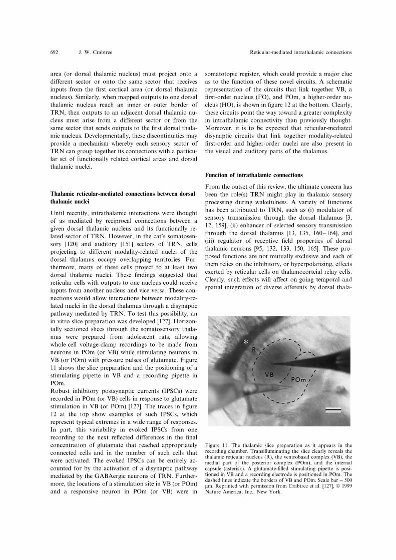
Until recently, intrathalamic interactions were thoughtof as mediated by reciprocal connections between agiven dorsal thalamic nucleus and its functionally re-lated sector of TRN. However, in the cat’s somatosen-sory [120] and auditory [151] sectors of TRN, cellsprojecting to different modality-related nuclei of thedorsal thalamus occupy overlapping territories. Fur-thermore, many of these cells project to at least twodorsal thalamic nuclei. These findings suggested thatreticular cells with outputs to one nucleus could receiveinputs from another nucleus and vice versa. These con-nections would allow interactions between modality-re-lated nuclei in the dorsal thalamus through a disynapticpathway mediated by TRN. To test this possibility, anin vitro slice preparation was developed [127]. Horizon-tally sectioned slices through the somatosensory thala-mus were prepared from adolescent rats, allowingwhole-cell voltage-clamp recordings to be made fromneurons in POm (or VB) while stimulating neurons inVB (or POm) with pressure pulses of glutamate. Figure11 shows the slice preparation and the positioning of astimulating pipette in VB and a recording pipette inPOm.Robust inhibitory postsynaptic currents (IPSCs) wererecorded in POm (or VB) cells in response to glutamatestimulation in VB (or POm) [127]. The traces in figure12 at the top show examples of such IPSCs, whichrepresent typical extremes in a wide range of responses.In part, this variability in evoked IPSCs from onerecording to the next reflected differences in the finalconcentration of glutamate that reached appropriatelyconnected cells and in the number of such cells thatwere activated. The evoked IPSCs can be entirely ac-counted for by the activation of a disynaptic pathwaymediated by the GABAergic neurons of TRN. Further-more, the locations of a stimulation site in VB (or POm)and a responsive neuron in POm (or VB) were in
somatotopic register, which could provide a major clueas to the function of these novel circuits. A schematicrepresentation of the circuits that link together VB, afirst-order nucleus (FO), and POm, a higher-order nu-cleus (HO), is shown in figure 12 at the bottom. Clearly,these circuits point the way toward a greater complexityin intrathalamic connectivity than previously thought.Moreover, it is to be expected that reticular-mediateddisynaptic circuits that link together modality-relatedfirst-order and higher-order nuclei are also present inthe visual and auditory parts of the thalamus.
Function of intrathalamic connections
From the outset of this review, the ultimate concern hasbeen the role(s) TRN might play in thalamic sensoryprocessing during wakefulness. A variety of functionshas been attributed to TRN, such as (i) modulator ofsensory transmission through the dorsal thalamus [3,12, 159], (ii) enhancer of selected sensory transmissionthrough the dorsal thalamus [13, 135, 160–164], and(iii) regulator of receptive field properties of dorsalthalamic neurons [95, 132, 133, 150, 165]. These pro-posed functions are not mutually exclusive and each ofthem relies on the inhibitory, or hyperpolarizing, effectsexerted by reticular cells on thalamocortcial relay cells.Clearly, such effects will affect on-going temporal andspatial integration of diverse afferents by dorsal thala-
Figure 11. The thalamic slice preparation as it appears in therecording chamber. Transilluminating the slice clearly reveals thethalamic reticular nucleus (R), the ventrobasal complex (VB), themedial part of the posterior complex (POm), and the internalcapsule (asterisk). A glutamate-filled stimulating pipette is posi-tioned in VB and a recording electrode is positioned in POm. Thedashed lines indicate the borders of VB and POm. Scale bar=500�m. Reprinted with permission from Crabtree et al. [127], © 1999Nature America, Inc., New York.

CMLS, Cell. Mol. Life Sci. Vol. 56, 1999 693Review Article
Figure 12. At the top, examples of inhibitory postsynaptic cur-rents (IPSCs) recorded from a POm cell (top trace) and a VB cell(bottom trace) in response to glutamate stimulation in VB orPOm, respectively. These IPSCs are not typical of the responsesrecorded from VB or POm cells but represent the extremes in arange of responses that is typical of both VB and POm cells. Atthe bottom, a schematic representation of a coronal sectionthrough the thalamus showing intrathalamic pathways linkingVB, a first-order nucleus (FO), and POm, a higher-order nucleus(HO). These are disynaptic pathways mediated by the thalamicreticular nucleus (R). The cells in the first-order and higher-ordernuclei project to the cortex (arrows).
order dorsal thalamic nuclei, in conjunction with topo-graphically organized inputs from a first-order corticalarea and a first-order dorsal thalamic nucleus. Thus,these reticular projections would exert local rather thanglobal effects on the cells in such nuclei. Before consid-ering what these local effects might achieve, it would beworthwhile to inquire as to whether dorsal thalamicinterneurons (i.e., local circuit neurons) could influencethese effects. Furthermore, the functional behavior ofthalamic neurons must also be taken into account.In addition to thalamocortical relay cells, dorsal thala-mic sensory nuclei also contain interneurons [2]. Theproportion of these interneurons varies widely acrossdifferent sensory nuclei and mammalian species [166].For example, interneurons account for about 25% ofthe neurons in the cat dLGN [51, 167, 168], whereasinterneurons are very scarce in the rat VB [48, 169].Interneurons in thalamic sensory nuclei receive F-typeterminals [129, 170–173], but whether the source ofthese terminals is TRN (or PGN), the interneuronsthemselves [172, 174], or pretectal cells [175–177] isunclear. What is known is that the projections of TRN/PGN cells preferentially target thalamocortical cells inthe sensory nuclei [89, 129, 172, 175; see also 100].Without a direct demonstration of a physiologicallyactive pathway from TRN/PGN to dorsal thalamicinterneurons, there is no compelling reason to believethat these interneurons play a significant role in intra-thalamic sensory processes mediated by TRN/PGN.Neurons in both the dorsal thalamus and TRN/PGNexhibit two main patterns of firing, tonic and burst[178–188]. Each of these firing modes is triggered bydepolarizing input and the cells switch between themodes in response to sustained changes in membranepotential. Figure 13 shows the tonic (upper trace) andthe burst (lower trace) firing modes for a typical thala-mic neuron that was recorded intracellularly and held atdifferent membrane potentials. With regard to TRN/PGN cells, modulatory glutamatergic inputs from thecortex are depolarizing [6, 9, 11] and promote the tonicfiring mode in these cells, whereas modulatory choliner-gic inputs from the brainstem are hyperpolarizing [189–196] and promote the burst firing mode in these cells.Either mode can be triggered by the driving and depo-larizing inputs from the dorsal thalamus [9, 11, 15].Figure 14 shows the powerful influence that burst firingin TRN/PGN (upper trace) has on a neuron in thedorsal thalamus (lower trace). Following the burst, abarrage of inhibitory, or hyperpolarizing, postsynapticpotentials was recorded in the dorsal thalamic cell,which would effectively silence the tonic firing of thecell.The organization and main functional connections ofTRN under consideration are summarized in figure 15,according to what we currently know about the sensory
mic cells [3, 6, 9, 12, 14–17, 20, 132, 133, 135], whichwill affect, in turn, the transmission of sensory informa-tion by these cells to the cerebral cortex. However, arigorous assessment of any proposed function for TRNalso relies on detailed knowledge of the connectivity ofthis nucleus.One striking attribute now recognized to be shared bythe different sensory sectors of TRN is the topographicorganization of the projections of reticular cells to first-

J. W. Crabtree Reticular-mediated intrathalamic connections694
sectors of this nucleus. In a given sensory sector ofTRN (e.g., visual, somatosensory, or auditory), organi-zational components A, B, and C are shown, which arespecifically connected to functionally related compo-nents A, B, and C in the first-order cortical area (CTX)and in the first-order dorsal thalamic nucleus (DT),respectively. The representation, or map, of the periph-eral receptor sheet (e.g., retina, skin, or cochlea) liesorthogonal to components A, B, and C in each struc-ture. It is assumed that the outputs of reticular neuronsin a given component (e.g., B) engage in within-compo-nent circuits within TRN [53, 58, 62, 97, 125, 146,197–201] and in predominantly open-loop circuitswithin the related component (i.e., B) in the dorsalthalamic nucleus [66, 97, 104, 125, 131]. Only inputs toand outputs from component B in TRN are shown,including inputs from the brainstem (BS). Although
Figure 14. Effect of burst firing recorded extracellularly in theperigeniculate nucleus (part of the carnivore thalamic reticularnucleus) on an intracellularly recorded neuron in the ferret’sdorsal lateral geniculate nucleus. The burst firing is shown in thetop trace. The resultant barrage of inhibitory postsynaptic poten-tials (arrow) in the dorsal lateral geniculate neuron is shown in thebottom trace. Reprinted with permission from Bal et al. [100],© 1999 The Journal of Physiology, Cambridge.
Figure 13. Different firing modes for an intracellularly recordedneuron in the cat’s dorsal lateral geniculate nucleus. The tonicmode is shown in the top trace and the burst mode is shown in thebottom trace. When the cell was held at a relatively depolarizedmembrane potential (−55 mV), a stream of conventional actionpotentials was triggered by a depolarizing 0.3 nA current pulse(bottom) injected into the cell. However, when the cell was held ata relatively hyperpolarized membrane potential (−70 mV), thesame depolarizing current pulse triggered a low-threshold Ca2+
spike with a burst of conventional action potentials riding on thecrest of the spike. Reprinted with permission from Sherman andGuillery [6], © 1999 The American Physiological Society,Bethseda, MD.
equally applicable to components A and C, respectively,in the cortex, dorsal thalamus, and TRN, we will nowconsider the following possibilities related only to com-ponent B in these structures.During periods of activity of cortical and brainsteminputs, the net effect of the tonic release of glutamateand acetylcholine, respectively, on thalamic reticularneurons would favor a cortical influence [178, 179,182–184]. This would result in a depolarizing shift inthe reticular cells, priming them for the tonic firingmode. Sensory transmission through a zone of dorsalthalamic component B, en route to cortical componentB, would then trigger the tonic firing mode in a zone ofreticular cells in component B. The tonic firing of thesecells would inhibit both the activity in an adjacent zoneof dorsal thalamic component B (small peaks) and in anadjacent zone of reticular cells in component B. Thislatter inhibition would disinhibit the activity of thedorsal thalamic zone in componet B (large peaks)through which sensory information is being transmit-ted. Thus, on-going sensory transmission through thedorsal thalamic nucleus would be enhanced. This pro-cess would be entirely consistent with models suggestingthat TRN enhances locally the transmission of salientsensory information through the dorsal thalamus [13,135, 160–164].Alternatively, during periods of activity of brainsteminputs and selective inactivity of cortical inputs fromcomponent B (�100 ms) [180], there would be a suffi-cient hyperpolarizing shift in component B reticularcells to prime them for the burst firing mode. Sensorytransmission through a zone of dorsal thalamic compo-nent B would then trigger the burst firing mode in a
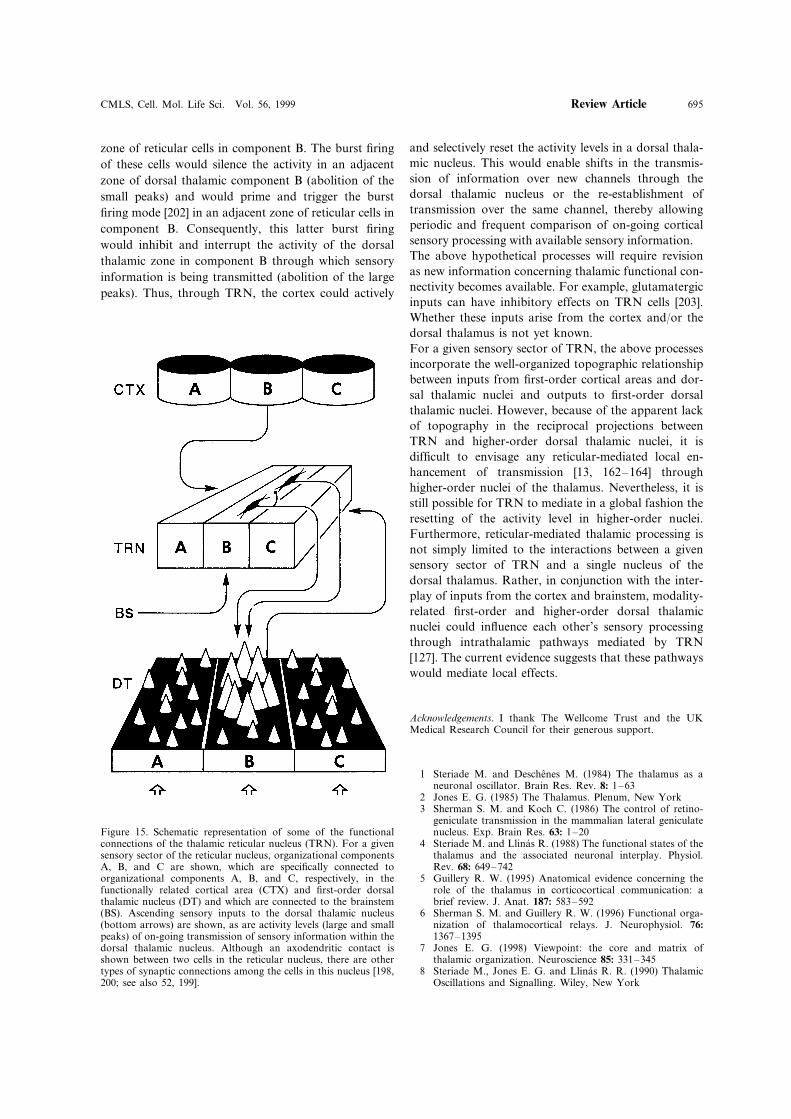
CMLS, Cell. Mol. Life Sci. Vol. 56, 1999 695Review Article
zone of reticular cells in component B. The burst firingof these cells would silence the activity in an adjacentzone of dorsal thalamic component B (abolition of thesmall peaks) and would prime and trigger the burstfiring mode [202] in an adjacent zone of reticular cells incomponent B. Consequently, this latter burst firingwould inhibit and interrupt the activity of the dorsalthalamic zone in component B through which sensoryinformation is being transmitted (abolition of the largepeaks). Thus, through TRN, the cortex could actively
and selectively reset the activity levels in a dorsal thala-mic nucleus. This would enable shifts in the transmis-sion of information over new channels through thedorsal thalamic nucleus or the re-establishment oftransmission over the same channel, thereby allowingperiodic and frequent comparison of on-going corticalsensory processing with available sensory information.The above hypothetical processes will require revisionas new information concerning thalamic functional con-nectivity becomes available. For example, glutamatergicinputs can have inhibitory effects on TRN cells [203].Whether these inputs arise from the cortex and/or thedorsal thalamus is not yet known.For a given sensory sector of TRN, the above processesincorporate the well-organized topographic relationshipbetween inputs from first-order cortical areas and dor-sal thalamic nuclei and outputs to first-order dorsalthalamic nuclei. However, because of the apparent lackof topography in the reciprocal projections betweenTRN and higher-order dorsal thalamic nuclei, it isdifficult to envisage any reticular-mediated local en-hancement of transmission [13, 162–164] throughhigher-order nuclei of the thalamus. Nevertheless, it isstill possible for TRN to mediate in a global fashion theresetting of the activity level in higher-order nuclei.Furthermore, reticular-mediated thalamic processing isnot simply limited to the interactions between a givensensory sector of TRN and a single nucleus of thedorsal thalamus. Rather, in conjunction with the inter-play of inputs from the cortex and brainstem, modality-related first-order and higher-order dorsal thalamicnuclei could influence each other’s sensory processingthrough intrathalamic pathways mediated by TRN[127]. The current evidence suggests that these pathwayswould mediate local effects.
Acknowledgements. I thank The Wellcome Trust and the UKMedical Research Council for their generous support.
1 Steriade M. and Deschenes M. (1984) The thalamus as aneuronal oscillator. Brain Res. Rev. 8: 1–63
2 Jones E. G. (1985) The Thalamus. Plenum, New York3 Sherman S. M. and Koch C. (1986) The control of retino-
geniculate transmission in the mammalian lateral geniculatenucleus. Exp. Brain Res. 63: 1–20
4 Steriade M. and Llinas R. (1988) The functional states of thethalamus and the associated neuronal interplay. Physiol.Rev. 68: 649–742
5 Guillery R. W. (1995) Anatomical evidence concerning therole of the thalamus in corticocortical communication: abrief review. J. Anat. 187: 583–592
6 Sherman S. M. and Guillery R. W. (1996) Functional orga-nization of thalamocortical relays. J. Neurophysiol. 76:
1367–13957 Jones E. G. (1998) Viewpoint: the core and matrix of
thalamic organization. Neuroscience 85: 331–3458 Steriade M., Jones E. G. and Llinas R. R. (1990) Thalamic
Oscillations and Signalling. Wiley, New York
Figure 15. Schematic representation of some of the functionalconnections of the thalamic reticular nucleus (TRN). For a givensensory sector of the reticular nucleus, organizational componentsA, B, and C are shown, which are specifically connected toorganizational components A, B, and C, respectively, in thefunctionally related cortical area (CTX) and first-order dorsalthalamic nucleus (DT) and which are connected to the brainstem(BS). Ascending sensory inputs to the dorsal thalamic nucleus(bottom arrows) are shown, as are activity levels (large and smallpeaks) of on-going transmission of sensory information within thedorsal thalamic nucleus. Although an axodendritic contact isshown between two cells in the reticular nucleus, there are othertypes of synaptic connections among the cells in this nucleus [198,200; see also 52, 199].

J. W. Crabtree Reticular-mediated intrathalamic connections696
9 McCormick D. A. (1992) Neurotransmitter actions in thethalamus and cerebral cortex and their role in neuromodula-tion of thalamocortical activity. Prog. Neurobiol. 39: 337–388
10 Steriade M., McCormick D. A. and Sejnowski T. J. (1993)Thalamocortical oscillations in the sleeping and arousedbrain. Science 262: 679–685
11 McCormick D. A. and Bal T. (1997) Sleep and arousal:thalamocortical mechanisms. Annu. Rev. Neurosci. 20: 185–215
12 Singer W. (1977) Control of thalamic transmission by corti-cofugal and ascending reticular pathways in the visual sys-tem. Physiol. Rev. 57: 386–420
13 Crick F. (1984) Function of the thalamic reticular complex:the searchlight hypothesis. Proc. Natl. Acad. Sci. USA 81:
4586–459014 McCormick D. A. and Bal T. (1994) Sensory gating mecha-
nisms of the thalamus. Curr. Opin. Neurobiol. 4: 550–55615 Salt T. E. and Eaton S. A. (1996) Functions of ionotropic
and metabotropic glutamate receptors in sensory transmis-sion in the mammalian thalamus. Prog. Neurobiol. 48: 55–72
16 Sherman S. M. (1996) Dual response mode in lateral genicu-late neurons: mechanisms and functions. Vis. Neurosci. 13:
205–21317 Sherman S. M. and Guillery R. W. (1998) On the actions
that one nerve cell can have on another: distinguishing‘drivers’ from ‘modulators’. Proc. Natl. Acad. Sci. USA 95:
7121–712618 Krosigk M. von, Bal T. and McCormick D. A. (1993)
Cellular mechanisms of a synchronized oscillation in thethalamus. Science 261: 361–364
19 Crunelli V. and Leresche N. (1991) A role for GABAB
receptors in excitation and inhibition of thalamocorticalcells. Trends Neurosci. 14: 16–21
20 Sillito A. M. (1992) GABA mediated inhibitory processes inthe function of the geniculo-striate system. Prog. Brain Res.90: 349–384
21 Rose J. E. (1942) The ontogenetic development of the rab-bit’s diencephalon. J. Comp. Neurol. 7: 61–129
22 Ramon y Cajal S. (1911) Histologie du Systeme Nerveux del’Homme et des Vertebres, vol. II, translated by L. Azoulay.Maloine, Paris
23 Scheibel M. E. and Scheibel A. B. (1966) The organizationof the nucleus reticularis thalami: a Golgi study. Brain Res.1: 43–62
24 Jones E. G. (1975) Some aspects of the organization of thethalamic reticular complex. J. Comp. Neurol. 162: 285–308
25 Carman J. B., Cowan W. M. and Powell T. P. S. (1964)Cortical connexions of the thalamic reticular nucleus. J.Anat. (Lond.) 98: 587–598
26 Friedlander M. J., Lin C.-S., Stanford L. R. and Sherman S.M. (1981) Morphology of functionally identified neurons inlateral geniculate nucleus of the cat. J. Neurophysiol. 46:
80–12927 Yen C.-T., Conley M. and Jones E. G. (1985) Morphological
and functional types of neurons in cat ventral posteriorthalamic nucleus. J. Neurosci. 5: 1316–1338
28 Harris R. M. (1987) Axon collaterals in the thalamic reticu-lar nucleus from thalamocortical neurons of the rat ven-trobasal thalamus. J. Comp. Neurol. 258: 397–406
29 Rouiller E. M. and Ribaupierre F. de (1990) Arborization ofcorticothalamic axons in the auditory thalamus of the cat: aPHA-L tracing study. Neurosci. Lett. 108: 29–35
30 Bourassa J. and Deschenes M. (1995) Corticothalamic pro-jections from the primary visual cortex in rats: a single fiberstudy using biocytin as an anterograde tracer. Neuroscience66: 253–263
31 Bourassa J., Pinault D. and Deschenes M. (1995) Cortico-thalamic projections from the cortical barrel field to thesomatosensory thalamus in rats: a single-fibre study usingbiocytin as an anterograde tracer. Eur. J. Neurosci. 7: 19–30
32 Murphy P. C. and Sillito A. M. (1996) Functional morphol-ogy of the feedback pathway from area 17 of the cat visualcortex to the lateral geniculate nucleus. J. Neurosci. 16:
1180–119233 Ahlsen G. and Lo F.-S. (1982) Projection of brain stem
neurons to the perigeniculate nucleus and the lateral genicu-late nucleus in the cat. Brain Res. 238: 433–438
34 Mackay-Sim A., Sefton A. J. and Martin P. R. (1983)Subcortical projections to lateral geniculate and thalamicreticular nuclei in the hooded rat. J. Comp. Neurol. 213:
24–3535 Hallanger A. E., Levey A. I., Lee H. J., Rye D. B. and
Wainer B. H. (1987) The origins of cholinergic and othersubcortical afferents to the thalamus in the rat. J. Comp.Neurol. 262: 105–124
36 Levey A. I., Hallanger A. E. and Wainer B. H. (1987)Cholinergic nucleus basalis neurons may influence the cortexvia the thalamus. Neurosci. Lett. 74: 7–13
37 Pare D., Smith Y., Parent A. and Steriade M. (1988) Projec-tions of brainstem core cholinergic and non-cholinergic neu-rons of cat to intralaminar and reticular thalamic nuclei.Neuroscience 25: 69–86
38 Smith Y., Pare D., Deschenes M., Parent A. and Steriade M.(1988) Cholinergic and non-cholinergic projections from theupper brainstem core to the visual thalamus in the cat. Exp.Brain Res. 70: 166–180
39 Uhlrich D. J., Cucchiaro J. B. and Sherman S. M. (1988)The projection of individual axons from the parabrachialregion of the brain stem to the dorsal lateral geniculatenucleus in the cat. J. Neurosci. 8: 4565–4575
40 Cornwall J., Cooper J. D. and Phillipson O. T. (1990)Afferent and efferent connections of the laterodorsal teg-mental nucleus in the rat. Brain Res. Bull. 25: 271–284
41 Steriade M., Parent A., Pare D. and Smith Y. (1987) Cholin-ergic and non-cholinergic neurons of cat basal forebrainproject to reticular and mediodorsal thalamic nuclei. BrainRes. 408: 372–376
42 Chen S. and Bentivoglio M. (1993) Nerve growth factorreceptor-containing cholinergic neurons of the basal fore-brain project to the thalamic reticular nucleus in the rat.Brain Res. 606: 207–212
43 Bickford M. E., Gunluk A. E., Van Horn S. C. and ShermanS. M. (1994) GABAergic projection from the basal forebrainto the visual sector of the thalamic reticular nucleus in thecat. J. Comp. Neurol. 348: 481–510
44 Minderhoud J. M. (1971) An anatomical study of the effer-ent connections of the thalamic reticular nucleus. Exp. BrainRes. 12: 435–446
45 Guillery R. W., Feig S. L. and Lozsadi D. A. (1998) Payingattention to the thalamic reticular nucleus. Trends Neurosci.21: 28–32
46 Houser C. R., Vaughn J. E., Barber R. P. and Roberts E.(1980) GABA neurons are the major cell type of the nucleusreticularis thalami. Brain Res. 200: 341–354
47 Oertel W. H., Graybiel A. M., Mugnaini E., Elde R. P.,Schmechel D. E. and Kopin I. J. (1983) Coexistence ofglutamic acid decarboxylase- and somatostatin-like im-munoreactivity in neurons of the feline nucleus reticularisthalami. J. Neurosci. 3: 1322–1332
48 Barbaresi P., Spreafico R., Frassoni C. and Rustioni A.(1986) GABAergic neurons are present in the dorsal columnnuclei but not in the ventroposterior complex of rats. BrainRes. 382: 305–326
49 De Biasi S., Frassoni C. and Spreafico R. (1986) GABAimmunoreactivity in the thalamic reticular nucleus of the rat:a light and electron microscopical study. Brain Res. 399:
143–14750 Hendrickson A. E., Ogren M. P., Vaughn J. E., Barber R. P.
and Wu J.-Y. (1983) Light and electron microscopic im-munocytochemical localization of glutamic acid decarboxy-lase in monkey geniculate complex: evidence for GABAergicneurons and synapses. J. Neurosci. 3: 1245–1262
CMLS, Cell. Mol. Life Sci. Vol. 56, 1999 697Review Article
51 Fitzpatrick D, Penny G. R. and Schmechel D. E. (1984)Glutamic acid decarboxylase immunoreactive neurons andterminals in the lateral geniculate nucleus of the cat. J.Neurosci. 4: 1809–1829
52 Montero V. M. and Singer W. (1984) Ultrastructure andsynaptic relations of neural elements containing glutamicacid decarboxylase (GAD) in the perigeniculate nucleus ofthe cat. Exp. Brain Res. 56: 115–125
53 Yen C.-T., Conley M., Hendry S. H. C. and Jones E. G.(1985) The morphology of physiologically identifiedGABAergic neurons in the somatic sensory part of thethalamic reticular nucleus in the cat. J. Neurosci. 5: 2254–2268
54 Clemence A. E. and Mitrofanis J. (1992) Cytoarchitectonicheterogeneities in the thalamic reticular nucleus of cats andferrets. J. Comp. Neurol. 322: 167–180
55 Penny G. R., Conley M., Schmechel D. E. and Diamond I.T. (1984) The distribution of glutamic acid decarboxylaseimmunoreactivity in the diencephalon of the opossum andrabbit. J. Comp. Neurol. 228: 38–56
56 Scheibel M. E. and Scheibel A. B. (1972) Specialized organi-zational patterns within the nucleus reticularis thalami of thecat. Exp. Neurol. 34: 316–322
57 Ide L. S. (1982) The fine structure of the perigeniculatenucleus in the cat. J. Comp. Neurol. 210: 317–334
58 Ohara P. T. and Lieberman A. R. (1985) The thalamicreticular nucleus of the adult rat: experimental anatomicalstudies. J. Neurocytol. 14: 365–411
59 Spreafico R., Battaglia G. and Frassoni C. (1991) Thereticular thalamic nucleus (RTN) of the rat: cytoarchitec-tural, Golgi, immunocytochemical, and horseradish peroxi-dase study. J. Comp. Neurol. 304: 478–490
60 Lubke J. (1993) Morphology of neurons in the thalamicreticular nucleus (TRN) of mammals as revealed by intracel-lular injections into fixed brain slices. J. Comp. Neurol. 329:
458–47161 Ohara P. T. and Havton L. A. (1996) Dendritic arbors of
neurons from different regions of the rat thalamic reticularnucleus share a similar orientation. Brain Res. 731: 236–240
62 Pinault D. and Deschenes M. (1998) Projection and innerva-tion patterns of individual thalamic reticular axons in thethalamus of the adult rat: a three-dimensional, graphic, andmorphometric analysis. J. Comp. Neurol. 391: 180–203
63 Chow K. L. (1952) Regional degeneration of the thalamicreticular nucleus following cortical ablations in monkeys. J.Comp. Neurol. 97: 37–60
64 Rose J. E. (1952) The cortical connections of the reticularcomplex of the thalamus. Res. Publ. Assoc. Res. Nerv.Ment. Dis. 30: 454–479
65 Steriade M., Parent A. and Hada J. (1984) Thalamic projec-tions of nucleus reticularis thalami of cat: a study usingretrograde transport of horseradish peroxidase and fluores-cent tracers. J. Comp. Neurol. 229: 531–547
66 Pinault D. and Deschenes M. (1998) Anatomical evidencefor a mechanism of lateral inhibition in the rat thalamus.Eur. J. Neurosci. 10: 3462–3469
67 Sumitomo I., Nakamura M. and Iwama K. (1976) Locationand function of the so-called interneurons of rat lateralgeniculate body. Exp. Neurol. 51: 110–123
68 Hale P. T., Sefton A. J., Baur L. A. and Cottee L. J. (1982)Interrelations of the rat’s thalamic reticular and dorsal lat-eral geniculate nuclei. Exp. Brain Res. 45: 217–229
69 Montero V. M., Guillery R. W. and Woolsey C. N. (1977)Retinotopic organization within the thalamic reticular nu-cleus demonstrated by a double label autoradiographic tech-nique. Brain Res. 138: 407–421
70 Lo F.-S. and Xie G.-Y. (1987) Location of interneurones inthe recurrent inhibitory circuit of the rabbit lateral geniculatenucleus. Exp. Brain Res. 66: 83–89
71 Crabtree J. W. and Killackey H. P. (1989) The topographicorganization and axis of projection within the visual sectorof the rabbit’s thalamic reticular nucleus. Eur. J. Neurosci. 1:
94–109
72 Updyke B. V. (1977) Topographic organization of the pro-jections from cortical areas 17, 18, and 19 onto the thalamus,pretectum and superior colliculus in the cat. J. Comp. Neu-rol. 173: 81–122
73 Symonds L. L. and Kaas J. H. (1978) Connections of striatecortex in the prosimian, Galago senegalensis. J. Comp. Neu-rol. 181: 477–512
74 Graham J., Lin C.-S. and Kaas J. H. (1979) Subcorticalprojections of six visual cortical areas in the owl monkey,Aotus tri�irgatus. J. Comp. Neurol. 187: 557–580
75 Conley M. and Diamond I. T. (1990) Organization of thevisual sector of the thalamic reticular nucleus in Galago :evidence that the dorsal lateral geniculate and pulvinar nu-clei occupy separate parallel tiers. Eur. J. Neurosci. 2: 211–226
76 Harting J. K., Van Lieshout D. P. and Feig S. (1991)Connectional studies of the primate lateral geniculate nu-cleus: distribution of axons arising from the thalamic reticu-lar nucleus of Galago crassicaudatus. J. Comp. Neurol. 310:
411–42777 Sanderson K. J. (1971) The projection of the visual field to
the lateral geniculate and medial interlaminar nuclei in thecat. J. Comp. Neurol. 143: 101–118
78 Dubin M. W. and Cleland B. G. (1977) Organization ofvisual inputs to interneurons of lateral geniculate nucleus ofthe cat. J. Neurophysiol. 40: 410–427
79 Ohara P. T. and Lieberman A. R. (1981) Thalamic reticularnucleus: anatomical evidence that cortico-reticular axonsestablish monosynaptic contact with reticulo-geniculate pro-jection cells. Brain Res. 207: 153–156
80 Montero V. M. (1989) Ultrastructural identification ofsynaptic terminals from cortical axons and from collateralaxons of geniculo-cortical relay cells in the perigeniculatenucleus of the cat. Exp. Brain Res. 75: 65–72
81 Xue J. T., Carney T., Ramoa A. S. and Freeman R. D.(1988) Binocular interaction in the perigeniculate nucleus ofthe cat. Exp. Brain Res. 69: 497–508
82 Ahlsen G. and Lindstrom S. (1982) Excitation of perigenicu-late neurones via axon collaterals of principal cells. BrainRes. 236: 477–481
83 Ahlsen G., Lindstrom S. and Lo F.-S. (1982) Functionaldistinction of perigeniculate and thalamic reticular neuronsin the cat. Exp. Brain Res. 46: 118–126
84 Sumitomo I., Hsiao C.-F. and Fukuda Y. (1988) Two typesof thalamic reticular cells in relation to the two visualthalamocortical systems in the rat. Brain Res. 446: 354–362
85 Zhu J. J. and Lo F.-S. (1997) Recurrent inhibitory interneu-rons of the rabbit’s lateral posterior pulvinar complex. J.Neurophysiol. 78: 3117–3124
86 FitzGibbon T. (1994) Rostral reticular nucleus of the thala-mus sends a patchy projection to the pulvinar lateralis-poste-rior complex of the cat. Exp. Neurol. 129: 266–278
87 FitzGibbon T., Tevah L. V. and Sefton A. J. (1995) Connec-tions between the reticular nucleus of the thalamus andpulvinar-lateralis posterior complex: A WGA-HRP study. J.Comp. Neurol. 363: 489–504
88 Ohara P. T., Sefton A. J. and Lieberman A. R. (1980) Modeof termination of afferents from the thalamic reticular nu-cleus in the dorsal lateral geniculate nucleus of the rat. BrainRes. 197: 503–506
89 Montero V. M. and Scott G. L. (1981) Synaptic terminals inthe dorsal lateral geniculate nucleus from neurons of thethalamic reticular nucleus: a light and electron microscopeautoradiographic study. Neuroscience 6: 2561–2577
90 Cucchiaro J. B., Uhlrich D. J. and Sherman S. M. (1991)Electron-microscopic analysis of synaptic input from theperigeniculate nucleus to the A-laminae of the lateral genicu-late nucleus in cats. J. Comp. Neurol. 310: 316–336
91 Burke W. and Sefton A. J. (1966) Recovery of responsive-ness of cells of lateral geniculate nucleus of rat. J. Physiol.187: 213–229
92 Burke W. and Sefton A. J. (1966) Inhibitory mechanisms inlateral geniculate nucleus of rat. J. Physiol. 187: 231–246

J. W. Crabtree Reticular-mediated intrathalamic connections698
93 Yingling C. D. and Skinner J. E. (1976) Selective regulationof thalamic sensory relay nuclei by nucleus reticularis tha-lami. Electroenceph. Clin. Neurophysiol. 41: 476–482
94 Lindstrom S. (1982) Synaptic organization of inhibitorypathways to principal cells in the lateral geniculate nucleusof the cat. Brain Res. 234: 447–453
95 Sillito A. M. and Kemp J. A. (1983) The influence ofGABAergic inhibitory processes on the receptive field struc-ture of X and Y cells in cat dorsal lateral geniculate nucleus(dLGN). Brain Res. 277: 63–77
96 French C. R., Sefton A. J. and Mackay-Sim A. (1985) Theinhibitory role of the visually responsive region of the thala-mic reticular nucleus in the rat. Exp. Brain Res. 57: 471–479
97 Lo F.-S. and Sherman S. M. (1994) Feedback inhibition inthe cat’s lateral geniculate nucleus. Exp. Brain Res. 100:
365–36898 Kim U., Sanchez-Vives M. V. and McCormick D. A. (1997)
Functional dynamics of GABAergic inhibition in the thala-mus. Science 278: 130–134
99 Sanchez-Vives M. V. and McCormick D. A. (1997) Func-tional properties of perigeniculate inhibition of dorsal lateralgeniculate nucleus thalamocortical neurons in vitro. J. Neu-rosci. 17: 8880–8893
100 Bal T., Krosigk M. von and McCormick D. A. (1995)Synaptic and membrane mechanisms underlying synchro-nized oscillations in the ferret lateral geniculate nucleus invitro. J. Physiol. 483: 641–663
101 Bal T., Krosigk M. von and McCormick D. A. (1995) Roleof the ferret perigeniculate nucleus in the generation ofsynchronized oscillations in vitro. J. Physiol. 483: 665–685
102 Kim U., Bal T. and McCormick D. A. (1995) Spindle wavesare propagating synchronized oscillations in the ferretLGNd in vitro. J. Neurophysiol. 74: 1301–1323
103 Kim U., Sanchez-Vives M. V. and McCormick D. A. (1997)Functional dynamics of GABAergic inhibition in the thala-mus. Science 278: 130–134
104 Kim U. and McCormick D. A. (1998) The functional influ-ence of burst and tonic firing mode on synaptic interactionsin the thalamus. J. Neurosci. 18: 9500–9516
105 Rodrigo-Angulo M. L. and Reinoso-Suarez F. (1988) Con-nections to the lateral posterior-pulvinar thalamic complexfrom the reticular and ventral lateral geniculate thalamicnuclei: a topographical study in the cat. Neuroscience 26:
449–459106 Rose J. E. and Malis L. I. (1965) Geniculo-striate connec-
tions in the rabbit. II. Cytoarchitectonic structure of thestriate region and of the dorsal lateral geniculate body:organization of the geniculo-striate projections. J. Comp.Neurol. 125: 121–140
107 Sanderson K. J. (1971) Visual field projection columns andmagnification factors in the lateral geniculate nucleus of thecat. Exp. Brain Res. 13: 159–177
108 Bishop P. O., Kozak W., Levick W. R. and Vakkur G. J.(1962) The determination of the projection of the visual fieldon to the lateral geniculate nucleus in the cat. J. Physiol 163:
503–539109 Lozsadi D. A., Gonzalez-Soriano J. and Guillery R. W.
(1996) The course and termination of corticothalamic fibresarising in the visual cortex of the rat. Eur. J. Neurosci. 8:
2416–2427110 Coleman K. A. and Mitrofanis J (1996) Organization of the
visual reticular thalamic nucleus of the rat. Eur. J. Neurosci.8: 388–404
111 Uhlrich D. J., Cucchiaro J. B., Humphrey A. L. and Sher-man S. M. (1991) Morphology and axonal projection pat-terns of individual neurons in the cat perigeniculate nucleus.J. Neurophysiol. 65: 1528–1541
112 Pinault D., Bourassa J. and Deschenes M. (1995) Thalamicreticular input to the rat visual thalamus: a single fiber studyusing biocytin as an anterograde tracer. Brain Res. 670:
147–152113 So Y. T. and Shapley R. (1981) Spatial tuning of cells in and
around lateral geniculate nucleus of the cat: X and Y relay
cells and perigeniculate interneurons. J. Neurophysiol. 45:107–120
114 Sugitani M. (1979) Electrophysiological and sensory proper-ties of the thalamic reticular neurones related to somaticsensation in rats. J. Physiol. 290: 79–95
115 Shosaku A., Kayama Y. and Sumitomo I. (1984) Somato-topic organization in the rat thalamic reticular nucleus.Brain Res. 311: 57–63
116 Bernardo K. L. and Woolsey T. A. (1987) Axonal trajecto-ries between mouse somatosensory thalamus and cortex. J.Comp. Neurol. 258: 542–564
117 Hoogland P. V., Welker E. and Van der Loos H. (1987)Organization of the projections from barrel cortex to thala-mus in mice studied with Phaseolus �ulgaris-leucoagglutininand HRP. Exp. Brain Res. 68: 73–87
118 Pollin B. and Rokyta R. (1982) Somatotopic organization ofnucleus reticularis thalami in chronic awake cats and mon-keys. Brain Res. 250: 211–221
119 Crabtree J. W. (1992) The somatotopic organization withinthe cat’s thalamic reticular nucleus. Eur. J. Neurosci. 4:1352–1361
120 Crabtree J. W. (1996) Organization in the somatosensorysector of the cat’s thalamic reticular nucleus. J. Comp.Neurol. 366: 207–222
121 Crabtree J. W. (1992) The somatotopic organization withinthe rabbit’s thalamic reticular nucleus. Eur. J. Neurosci. 4:1343–1351
122 De Biasi S., Frassoni C. and Spreafico R. (1988) The intrin-sic organization of the ventroposterolateral nucleus and re-lated reticular thalamic nucleus of the rat: a double-labelingultrastructural investigation with �-aminobutyric acid im-munogold staining and lectin-conjugated horseradish peroxi-dase. Somatosens. Res. 5: 187–203
123 Curtis M. de, Spreafico R. and Avanzini G. (1989) Excita-tory amino acids mediate responses elicited in vitro bystimulation of cortical afferents to reticularis thalami neu-rons of the rat. Neuroscience 33: 275–283
124 Shosaku A. (1985) A comparison of receptive field propertiesof vibrissa neurons between the rat thalamic reticular andventro-basal nuclei. Brain Res. 347: 36–40
125 Shosaku A. (1986) Cross-correlation analysis of a recurrentinhibitory circuit in the rat thalamus. J. Neurophysiol. 55:1030–1043
126 Sumitomo I. and Iwama K. (1987) Neuronal organization ofrat thalamus for processing information of vibrissal move-ments. Brain Res. 415: 389–392
127 Crabtree J. W., Collingridge G. L. and Isaac J. T. R. (1998)A new intrathalamic pathway linking modality-related nucleiin the dorsal thalamus. Nat. Neurosci. 1: 389–394
128 Peschanski M., Ralston H. J. and Roudier F. (1983) Reticu-laris thalami afferents to the ventrobasal complex of the ratthalamus: an electron microscope study. Brain Res. 270:325–329
129 Liu X.-B., Warren R. A. and Jones E. G. (1995) Synapticdistribution of afferents from reticular nucleus in ventropos-terior nucleus of cat thalamus. J. Comp. Neurol. 352: 187–202
130 Andersen P., Eccles J. C. and Sears T. A. (1964) Theventro-basal complex of the thalamus: types of cells, theirresponses and their functional organization. J. Physiol. 174:370–399
131 Salt T. E. (1989) Gamma-aminobutyric acid and afferentinhibition in the cat and rat ventrobasal thalamus. Neuro-science 28: 17–26
132 Lee S. M., Friedberg M. H. and Ebner F. F. (1994) The roleof GABA-mediated inhibition in the rat ventral posteriormedial thalamus. I. Assessment of receptive field changesfollowing thalamic reticular nucleus lesions. J. Neurophysiol.71: 1702–1715
133 Lee S. M., Friedberg M. H. and Ebner F. F. (1994) The roleof GABA-mediated inhibition in the rat ventral posteriormedial thalamus. II. Differential effects of GABAA andGABAB receptor antagonists on responses of VPM neurons.J. Neurophysiol. 71: 1716–1726

CMLS, Cell. Mol. Life Sci. Vol. 56, 1999 699Review Article
134 Huguenard J. R. and Prince D. A. (1994) Clonazepamsuppresses GABAB-mediated inhibition in thalamic relayneurons through effects in nucleus reticularis. J. Neurophys-iol. 71: 2576–2581
135 Warren R. A. and Jones E. G. (1994) Glutamate activationof cat thalamic reticular nucleus: effects on response proper-ties of ventroposterior neurons. Exp. Brain Res. 100: 215–226
136 Cox C. L., Huguenard J. R. and Prince D. A. (1997)Nucleus reticularis neurons mediate diverse inhibitory effectsin thalamus. Proc. Natl. Acad. Sci. USA 94: 8854–8859
137 Warren R. A., Agmon A. and Jones E. G. (1994) Oscillatorysynaptic interactions between ventroposterior and reticularneurons in mouse thalamus in vitro. J. Neurophysiol. 72:
1993–2003138 Pinault D., Bourassa J. and Deschenes M. (1995) The axonal
arborization of single thalamic reticular neurons in the so-matosensory thalamus of the rat. Eur. J. Neurosci. 7: 31–40
139 Van der Loos H. (1976) Barreloids in mouse somatosensorythalamus. Neurosci. Lett. 2: 1–6
140 Belford G. R. and Killackey H. P. (1979) The developmentof vibrissae representation in subcortical trigeminal centersof the neonatal rat. J. Comp. Neurol. 188: 63–74
141 Jones E. G. and Friedman D. P. (1982) Projection pattern offunctional components of thalamic ventrobasal complex onmonkey somatosensory cortex. J. Neurophysiol 48: 521–544
142 Jones E. G., Friedman D. P. and Hendry S. H. C. (1982)Thalamic basis of place- and modality-specific columns inmonkey somatosensory cortex: a correlative anatomical andphysiological study. J. Neurophysiol. 48: 545–568
143 Crabtree J. W. and Kind P. C. (1993) Monoclonal antibodyCat-301 selectively identifies a subset of nuclei in the cat’ssomatosensory thalamus. J. Neurocytol. 22: 903–912
144 Mountcastle V. and Henneman E. (1949) Pattern of tactilerepresentation in thalamus of cat. J. Neurophysiol. 12: 85–100
145 Rose J. E. and Mountcastle V. B. (1952) The thalamic tactileregion in rabbit and cat. J. Comp. Neurol. 97: 441–489
146 Cox C. L., Huguenard J. R. and Prince D. A. (1996)Heterogeneous axonal arborizations of rat thalamic reticularneurons in the ventrobasal nucleus. J. Comp. Neurol. 366:
416–430147 Shosaku A. and Sumitomo I. (1983) Auditory neurons in the
rat thalamic reticular nucleus. Exp. Brain Res. 49: 432–442148 Rouiller E. M., Colomb E., Capt M. and De Ribaupierre F.
(1985) Projections of the reticular complex of the thalamusonto physiologically characterized regions of the medialgeniculate body. Neurosci. Lett. 53: 227–232
149 Simm G. M., De Ribaupierre F., De Ribaupierre Y. andRouiller E. M. (1990) Discharge properties of single units inauditory part of reticular nucleus of thalamus in cat. J.Neurophysiol. 63: 1010–1021
150 Villa A. E. P. (1990) Physiological differentiation within theauditory part of the thalamic reticular nucleus of the cat.Brain Res. Rev. 15: 25–40
151 Crabtree J. W. (1998) Organization in the auditory sector ofthe cat’s thalamic reticular nucleus. J. Comp. Neurol. 390:
167–182152 Conley M., Kupersmith A. C. and Diamond I. T. (1991) The
organization of projections from subdivisions of the auditorycortex and thalamus to the auditory sector of the thalamicreticular nucleus in Galago. Eur. J. Neurosci. 3: 1089–1103
153 Montero V. M. (1983) Ultrastructural identification of axonterminals from the thalamic reticular nucleus in the medialgeniculate body in the rat: an EM autoradiographic study.Exp. Brain Res. 51: 338–342
154 Bartlett E. L. and Smith P. H. (1999) Anatomic, intrinsic,and synaptic properties of dorsal and ventral division neu-rons in rat medial geniculate body. J. Neurophysiol. 81:
1999–2016155 Morest D. K. (1965) The laminar structure of the medial
geniculate body of the cat. J. Anat. (Lond.) 99: 143–160
156 Aitkin L. M. and Webster W. R. (1972) Medial geniculatebody of the cat: organization and responses to tonal stimuliof neurons in ventral division. J. Neurophysiol. 35: 365–380
157 Calford M. B. and Webster W. R. (1981) Auditory represen-tation within principal division of cat medial geniculatebody: an electrophysiological study. J. Neurophysiol. 45:
1013–1028158 Imig T. J. and Morel A. (1985) Tonotopic organization in
ventral nucleus of medial geniculate body in the cat. J.Neurophysiol. 53: 309–340
159 Scheibel M. E. and Scheibel A. B. (1967) Structural organi-zation of nonspecific thalamic nuclei and their projectiontoward cortex. Brain Res. 6: 60–94
160 Skinner J. E. and Yingling C. D. (1977) Central gatingmechanisms that regulate event-related potentials and behav-ior: a neural model for attention. Prog. Clin. Neurophysiol.1: 30–69
161 Scheibel A. B. (1981) The problem of selective attention: apossible structural substrate. In: Brain Mechanisms of Per-ceptual Awareness, pp 319–326, Pompeiano O. and Ajmone-Marsan C. (eds), Raven, New York
162 LaBerge D., Carter M. and Brown V. (1992) A networksimulation of thalamic circuit operations in selective atten-tion. Neural Comp. 4: 318–331
163 Vidal de Carvalho L. A. (1994) Modeling the thalamocorti-cal loop. Int. J. Bio-Med. Comp. 35: 267–296
164 Taylor J. G. and Alavi F. N. (1995) A global competitiveneural network. Biol. Cybern. 72: 233–248
165 Villa A. E. P., Rouiller E. M., Simm G. M., Zurita P., DeRibaupierre Y. and De Ribaupierre F. (1991) Corticofugalmodulation of the information processing in the auditorythalamus of the cat. Exp. Brain Res. 86: 506–517
166 Spreafico R., Fressoni C., Regondi M. C., Arcelli P. and DeBiasi S. (1993) Interneurons in the mammalian thalamus. In:Thalamic Networks for Relay and Modulation, pp 17–28,Minciacchi D., Molinari M., Macchi G. and Jones E. G.(eds), Pergamon, Oxford
167 LeVay S. and Ferster D. (1979) Proportion of interneuronsin the cat’s lateral geniculate nucleus. Brain Res. 164: 304–308
168 Montero V. M. and Zempel J. (1985) Evidence for two typesof GABA-containing interneurons in the A-laminae of thecat lateral geniculate nucleus: a double-label HRP andGABA-immunocytochemical study. Exp. Brain Res. 60:
603–609169 Harris R. M. and Hendrickson A. E. (1987) Local circuit
neurons in the rat ventrobasal thalamus: a GABA immuno-cytochemical study. Neuroscience 21: 229–236
170 Montero V. M. and Singer W. (1985) Ultrastructural iden-tification of somata and neural processes immunoreactive toantibodies against glutamic acid decarboxylase (GAD) in thedorsal lateral geniculate nucleus of the cat. Exp. Brain Res.59: 151–165
171 Montero V. M. (1991) A quantitative study of synapticcontacts on interneurons and relay cells of the cat lateralgeniculate nucleus. Exp. Brain Res. 86: 257–270
172 Takacs J., Hamori J. and Silakov V. (1991) GABA-contain-ing neuronal processes in normal and cortically deafferenteddorsal lateral geniculate nucleus of the cat: an immunogoldand quantitative EM study. Exp. Brain Res. 83: 562–574
173 Erisir A., Van Horn S. C. and Sherman S. M. (1998)Distribution of synapses in the lateral geniculate nucleus ofthe cat: differences between laminae A and A1 and betweenrelay cells and interneurons. J. Comp. Neurol. 390: 247–255
174 Montero V. M. (1987) Ultrastructural identification ofsynaptic terminals from the axon of type 3 interneurons inthe cat lateral geniculate nucleus. J. Comp. Neurol. 264:
268–283175 Cucchiaro J. B., Bickford M. E. and Sherman S. M. (1991)
A GABAergic projection from the pretectum to the dorsallateral geniculate nucleus in the cat. Neuroscience 41: 213–226

J. W. Crabtree Reticular-mediated intrathalamic connections700
176 Cucchiaro J. B., Uhlrich D. J. and Sherman S. M. (1993)Ultrastructure of synapses from the pretectum in the A-lam-inae of the cat’s lateral geniculate nucleus. J. Comp. Neurol.334: 618–630
177 Wahle P., Stuphorn V., Schmidt M. and Hoffmann K.-P.(1994) LGN-projecting neurons of the cat’s pretectum ex-press glutamic acid decarboxylase mRNA. Eur. J. Neurosci.6: 454–460
178 Mukhametov L. M., Rizzolatti G. and Tradardi V. (1970)Spontaneous activity of neurones of nucleus reticularis tha-lami in freely moving cats. J. Physiol. 210: 651–667
179 Deschenes M., Paradis M., Roy J. P. and Steriade M. (1984)Electrophysiology of neurons of lateral thalamic nuclei incat: resting properties and burst discharges. J. Neurophysiol.51: 1196–1219
180 Jahnsen H. and Llinas R. (1984) Electrophysiological prop-erties of guinea-pig thalamic neurones: an in vitro study. J.Physiol. 349: 205–226
181 Jahnsen H. and Llinas R. (1984) Ionic basis for the electrore-sponsiveness and oscillatory properties of guinea-pig thala-mic neurones in vitro. J. Physiol. 349: 227–247
182 Domich L., Oakson G. and Steriade M. (1986) Thalamicburst patterns in the naturally sleeping cat: a comparisonbetween cortically projecting and reticularis neurones. J.Physiol. 379: 429–449
183 Mulle C., Madariaga A. and Deschenes M. (1986) Morphol-ogy and electrophysiological properties of reticularis thalamineurons in cat: in vivo study of a thalamic pacemaker. J.Neurosci. 6: 2134–2145
184 Steriade M., Domich L. and Oakson G. (1986) Reticularisthalami neurons revisited: activity changes during shifts instates of vigilance. J. Neurosci. 6: 68–81
185 Spreafico R., Curtis M. de, Frassoni C. and Avanzini G.(1988) Electrophysiological characteristics of morphologi-cally identified reticular thalamic neurons from rat slices.Neuroscience 27: 629–638
186 Avanzini G., Curtis M. de, Panzica F. and Spreafico R.(1989) Intrinsic properties of nucleus reticularis thalami neu-rones of the rat studied in vitro. J. Physiol. 416: 111–122
187 McCormick D. A. and Feeser H. R. (1990) Functionalimplications of burst firing and single spike activity in lateralgeniculate relay neurons. Neuroscience 39: 103–113
188 Lo F.-S., Lu S.-M. and Sherman S. M. (1991) Intracellularand extracellular in vivo recording of different responsemodes for relay cells of the cat’s lateral geniculate nucleus.Exp. Brain Res. 83: 317–328
189 Ben-Ari Y., Dingledine R., Kanazawa I. and Kelly J. S.(1976) Inhibitory effects of acetylcholine on neurons in thefeline nucleus reticularis thalami. J. Physiol. (Lond.) 261:647–671
190 Dingledine R. and Kelly J. S. (1977) Brain stem stimulationand the acetylcholine-evoked inhibition of neurones in thefeline nucleus reticularis thalami. J. Physiol. 271: 135–154
191 McCormick D. A. and Prince D. A. (1986) Acetylcholineinduces burst firing in thalamic reticular neurones by activat-ing a potassium conductance. Nature 319: 402–405
192 Francesconi W., Muller C. M. and Singer W. (1988) Cholin-ergic mechanisms in the reticular control of transmission inthe cat lateral geniculate nucleus. J. Neurophysiol. 59: 1690–1718
193 Hu B., Steriade M. and Deschenes M. (1989) The effects ofbrainstem peribrachial stimulation on perigeniculate neu-rons: the blockage of spindle waves. Neuroscience 31: 1–12
194 McCormick D. A. (1989) Cholinergic and noradrenergicmodulation of thalamocortical processing. Trends Neurosci.12: 215–221
195 Funke K. and Eysel U. T. (1993) Modulatory effects ofacetylcholine, serotonin, and noradrenaline on the activity ofcat perigeniculate neurons. Exp. Brain Res. 95: 409–420
196 Marks G. A. and Roffwarg H. P. (1991) Cholinergic modu-lation of responses to glutamate in the thalamic reticularnucleus of the anesthetized rat. Brain Res. 557: 48–56
197 Ahlsen G. and Lindstrom S. (1982) Mutual inhibition be-tween perigeniculate neurones. Brain Res. 236: 482–486
198 Deschenes M., Madariaga-Domich A. and Steriade M.(1985) Dendrodendritic synapses in the cat reticularis tha-lami nucleus: a structural basis for thalamic spindle synchro-nization. Brain Res. 334: 165–168
199 Williamson A. M., Ohara P. T., Ralston D. D., Milroy A.M. and Ralston H. J. (1994) Analysis of gamma-aminobu-tyric acidergic synaptic contacts in the thalamic reticularnucleus of the monkey. J. Comp. Neurol. 349: 182–192
200 Pinault D., Smith Y. and Deschenes M. (1997) Dendroden-dritic and axoaxonic synapses in the thalamic reticular nu-cleus of the adult rat. J. Neurosci. 17: 3215–3233
201 Sanchez-Vives M. V., Bal T. and McCormick D. A. (1997)Inhibitory interactions between perigeniculate GABAergicneurons. J. Neurosci. 17: 8894–8908
202 Bazhenov M., Timofeev I., Steriade M. and Sejnowski T. J.(1999) Self-sustained rhythmic activity in the thalamic reticu-lar nucleus mediated by depolarizing GABAA receptor po-tentials. Nat. Neurosci. 2: 168–174
203 Cox C. L. and Sherman S. M. (1999) Glutamate inhibitsthalamic reticular neurons. J. Neurosci. 19: 6694–6699
.